Note: Descriptions are shown in the official language in which they were submitted.
- 1 -
A DEVICE COMPRISING A SETON AND A TISSUE GROWTH
PROMOTER AND USES THEROF
Field of the invention
The present invention relates to devices and related methods for treating
fistulas
such as anal or recto-vaginal fistulas, in particular by the use of a seton to
secure a
tissue growth promoter such as a growth factor and/or fibrin. The various
devices
arc particularly suitable for positioning tissue growth promoters securely
within a
fistula. Thus, one device comprises a seton and a tissue growth promoter.
Further
related aspects of the invention include devices comprising an enclosure
provided
inbetween portions of a seton, devices comprising a seton and a plurality of
holes
for enabling the device to be sutured to tissue, devices comprising a probe
and a
seton that are releasably connectable end-to-end, devices comprising an
attachment
device to secure the ends of a seton, and devices comprising a fistula plug
adapted
to be secured to a seton.
Background of the invention
An anal fistula, otherwise known as an anorectal fistula, is an abnormal
passage formed
between the wall of the anal canal and the skin around the anus, typically the
perianal skin.
An anal fistula usually originates from an infection in an anal gland located
in the anal
canal. In the case of an anal gland becoming infected, an abscess may form
deep under the
skin around the anus which requires surgical drainage. After drainage, a tract
between the
drainage site and the wall of the anal canal may form resulting in an anal
fistula. Fistulas
cause intermittent symptoms of discharge and generally do not heal without
treatment or
surgical intervention. Anal fistulas are also a common feature of inflammatory
bowel
diseases, especially ulcerative colitis and Crohn's disease.
Tay open' fistulotomy is the conventional surgery for treating an anal fistula
and involves
dividing the tissue between the fistula and the skin so as to promote tissue
regeneration and
hence healing of the fistula. A disadvantage with this procedure is that it
causes discomfort
and scarring, and usually results in some level of incontinence.
CA 2831071 2019-01-22
CA 02831071 2013-09-23
WO 2011/151659 PCT/GB2011/051810
- 2 -
In an alternative procedure, a seton may be used which is passed through the
track of the
fistula by use of a fistula probe. The seton is a string preferably formed out
of silicon or
rubber that is typically threaded through an eye of the fistula probe. The
probe is then
.. passed through the track of the fistula pulling the seton along so that it
extends through the
entire length of the fistula. As the probe reaches the wall of the anal canal,
the probe is
passed through the anus and then removed from the scton such that the two
loose ends of
the seton can be tied together so as to form a loop. The seton is typically
either left in place
long-term and assists in draining any discharge from the fistula, or is tied
tight to produce a
slow form of fistulotomy, that is, division of tissues superficial to the
fistula.
Suitable devices for use in such a procedure are described in the prior art.
For example,
patent application WO 2005/020823 describes a device for the treatment of anal
fistulas
comprising a probe attached to a drainage thread. The probe is used to guide
the drainage
thread through the fistula, after which the probe may be removed and the two
ends of the
thread tied together. The thread acts to ensure that the fistula channel is
kept open,
allowing adequate drainage.
In some cases, particularly Crohn's disease, when the fistula becomes
infected, it may be
necessary for the patient to undergo a course of antibiotics and/or anti
inflammatory
medication prior to surgical treatment. Consequently, surgical treatment of
the fistula is
delayed causing discomfort. In an attempt to overcome this problem, Razvan
Arsenescu
has proposed the development of a biodegradable seton made from PLGA that
releases an
anti-inflammatory drug in a controlled fashion (see the proposal for an award
from the
Kentucky Science & Engineering Foundation entitled "Drug Eluting Biodegradable
Seton
for Treatment of Perianal Fistulas in Crohn's Disease" at
http://ksef.kstc.com/Dynamic/Awards).
A problem with the conventional technique is that the knot of the seton is
external to the
body and may cause discomfort as the patient sits down. In some cases the knot
becomes
undone causing the seton to fall out of the fistula and so the process of
inserting the seton
needs to be repeated.
CA 02831071 2013-09-23
WO 2011/151659 PCT/GB2011/051810
- 3 -
A further problem is that whilst movement of the seton within the fistula is
desirable to
assist in drainage, such movement may act to prevent tissue regeneration and
hence healing
of the fistula. Further surgical steps to close the fistula are therefore
often required after the
elimination of the infection.
As already indicated, an alternative to the above procedure is that the scton
can be used to
gradually divide the tissue superficial to the fistula. In this alternative
procedure, the
structures superficial to the fistula, such as the skin, subcutaneous fat and
sphincter muscle,
arc slowly divided by tying the seton tightly. The process of gradual
tightening causes the
seton to divide the tissue it surrounds thereby promoting tissue regeneration
and healing of
the fistula. Unfortunately, this often results in some impairment of
continence.
A device suitable for use in such a procedure is described in patent
application WO
2005/096957. The device describes a silicone thread with either a transversal
through hole
at its tip or a tip-piece that includes a transversal through hole. Also
provided is a
removable supporting jacket which serves to guide the thread through the
fistula. Once the
thread has been passed through the fistula, the supporting jacket is removed
and the distal
end of the thread is passed through the transversal through hole at the tip.
Fastening nodes
may be provided along the length of the thread so as to interfere with the
internal diameter
of the transversal through hole, thus tying the thread. When tied tightly, a
slow cutting
action is provided resulting in the opening-up, or "elastic traction" of the
fistula. In use, as
the loop slackens due to the cutting action, more of the thread along with
further fastening
nodes may be pulled through the transversal through hole so as to ensure that
the cutting
action is maintained.
The main disadvantage of dividing the tissue in this way is that it tends to
result in some
level of incontinence. Furthermore, the lose ends of the thread may again
cause discomfort
as a patient sits down, and rotation of the tightened thread through the
fistula may cause
additional discomfort and risk of infection.
As fistulas vary in configuration, complexity and length, surgeons have to
take great care
when inserting a probe and seton through the track of a fistula as surrounding
tissue can
easily be damaged as a result of the probe going off course. Furthermore, a
seton is
CA 02831071 2013-09-23
WO 2011/151659 PCT/GB2011/051810
- 4 -
typically attached to the probe by the seton being inserted through an eye of
the probe, or
by an end of the seton overlapping an end of the probe and the ends being
lashed together
using a thread. These types of connections are bulky and may cause the seton
to abrade or
damage surrounding tissue as the seton is pulled through the fistula.
Furthermore, it is also
apparent that these configurations arc often inadequate at maintaining the
seton attached to
the probe because the seton may become detached from the probe as it is pulled
through
the fistula. In such cases, the surgeon would need to repeat the procedure
which potentially
causes more trauma to the surrounding tissue and the patient.
In an attempt to avoid fistulotomy (the division of tissues superficial to the
fistula track), an
alternative recent procedure using a fibrin plug has been introduced. The
fibrin plug
comprises a scaffold of polymeric fibrin that is inserted into the fistula
tract so as to
promote tissue in-growth into the scaffold so as to restore natural tissue
healing and
formation.
A disadvantage with the fibrin plug and even its more recent modifications is
that it tends
to fall out of the fistula before the end of the 4 to 6 week healing process.
Although the
fibrin plug can be sutured to an inner wall of the anus to mitigate against
this problem, this
has proven to be insufficient to maintain the plug in position.
In an attempt to overcome the above disadvantages, a modified fibrin plug is
disclosed in
US patent application US 2007/0031508. The modified fibrin plug includes end
caps
and/or a "tail" which may be sutured to the patient after placement of the
plug so as to
secure the plug in position. Such a device again relies entirely on the
strength of the sutures
25 and surrounding tissue. Thus, whilst this represents an improvement on
the conventional
fibrin plug, it does not reliably overcome the problem of plug displacement.
There is also a need for devices that will enable other types of fistulas to
be treated.
30 Such fistulas include for instance recto-vaginal fistulas, which are
abnormal passages
between the rectum and the vagina. These are particularly difficult to treat
by conventional
surgery. Recto-vaginal fistulas frequently recur requiring multiple operations
resulting in
bowel incontinence, pain and deformity. Recto-vaginal fistulas have become a
bigger
CA 02831071 2013-09-23
WO 2011/151659 PCT/GB2011/051810
- 5 -
problem of late due to a rising incidence of inflammatory bowel disease
(especially Crohn's
disease), as well as operations to preserve continence for ulcerative colitis
sufferers, by
restorative proctocolectomy and ileal pouch anal anastomosis. Such procedures
are also
being performed on patients suffering from a variety of premalignant
conditions. There is
therefore a need for devices and procedures which will give a high chance of
achieving
primary healing in recto-vaginal fistulas without invasive surgical
intervention.
The present invention seeks to provide a device that overcomes or
substantially alleviates
the problems mentioned above.
Summary of the invention
According to a first aspect of the present invention, there is provided a
device
comprising a seton suitable for treating a fistula, wherein the device
comprises a
tissue growth promoter. Preferably the seton comprises a tissue growth
promoter.
The tissue growth promoter may comprise or be a tissue growth promoting agent,
i.e. a pharmaceutical substance which encourages tissue growth such as a
growth
factor. Suitable growth factors include but are not limited to basic
fibroblast growth
factor (FGF-2), transforming growth factor beta (TGF-beta), epidermal growth
factor (EGF), cartilage derived growth factor (CDGF), platelet derived growth
factor (PDGF), insulin-like growth factors I and II (IGF-I and IGF-II),
interferons
(e.g. interferon a, p, y) and the like.
Alternatively the tissue growth promoter may comprise or be a tissue growth
promoting matrix, i.e. a substance or structure which may act as a scaffold
onto
which and/or through which tissue may grow. Preferably tissue may grow both
onto
and through the scaffold.
In one embodiment of the first aspect of the present invention, the tissue
growth
promoting matrix comprises a microscopic scaffold, i.e. the scaffold structure
is not
visible to the naked eye. For instance, the scaffold may comprise a plurality
of
fibrils which may be non-interwoven or preferably interwoven and/or
CA 02831071 2013-09-23
WO 2011/151659 PCT/GB2011/051810
- 6 -
interconnected. Said fibrils may be interwoven and/or interconnected in an
ordered
or disordered structure.
Where a scaffold comprises a plurality of fibrils, preferably said fibrils
have a
diameter of from 0.1 nm to 1000 nm, more preferably from 1 nm to 500 nm, and
most preferably from 10 nm to 100 nm.
Typically, where a scaffold comprises a plurality of fibrils, at least 10% by
volume of
the scaffold is occupied by the fibrils. Preferably at least 25% by volume,
and more
preferably at least 50% by volume of the scaffold is occupied by the fibrils.
In another embodiment of the first aspect of the present invention the tissue
growth promoting matrix comprises a macroscopic scaffold, i.e. the scaffold
structure is visible to the naked eye. For instance, the scaffold may comprise
a
plurality of interwoven and/or interconnected strands. Said strands may be
interwoven and/or interconnected in an ordered or disordered structure.
In another embodiment, the scaffold may comprise a plurality of non-interwoven
strands, such as a plurality of strands affixed to a central core, for example
in a
brush-like array. Preferably said strands are afixed substantially
perpendicularly
from the central core. Preferably the central core is the seton.
Where a macroscopic scaffold comprises a plurality of strands, preferably said
strands have a diameter of from 1 [im to 1 mm, more preferably from 10 [1m to
100
,m.
Typically, where a scaffold comprises a plurality of strands, at least 10% by
volume
of the scaffold is occupied by the strands. Preferably at least 25% by volume,
and
more preferably at least 50% by volume of the scaffold is occupied by the
strands.
In one embodiment, the macroscopic scaffold comprises at least 10 strands per
cm3.
Preferably, the macroscopic scaffold comprises at least 100 strands per cm'.
More
preferably the macroscopic scaffold comprises at least 1000 strands per cm'.
CA 02831071 2013-09-23
WO 2011/151659 PCT/GB2011/051810
- 7 -
Alternatively, the scaffold may comprise a porous structure such as a sponge-
like
structure. Said porous structure may be microscopic or macroscopic.
Optionally, the tissue growth promoting matrix may comprise both a macroscopic
scaffold and a microscopic scaffold. In a preferred embodiment, the
macroscopic
scaffold is made from, coated with or embedded in a microscopic scaffold. Eor
instance, the strands and/or porous structure of the macroscopic scaffold may
comprise a microscopic scaffold.
In one embodiment of the first aspect of the present invention, at least 10%
by
volume of the tissue growth promoting matrix is void. Preferably at least 25%
by
volume, and more preferably at least 50 ,4 by volume of the tissue growth
promoting matrix is void.
The material from which the tissue growth promoting matrix is made may be
biological or non-biological, or a mixture thereof. Preferably the material
promotes
tissue remodelling and/or is remodelable. Preferably the material promotes
angiogenesis.
In a preferred embodiment of the first aspect of the present invention, the
tissue
growth promoting matrix is made from biocompatible materials.
As used herein, the term "biocompatible materials" refers to materials which
do not
have unacceptable adverse effects on the subject (e.g. human or other animal)
to be
treated. Preferably the biocompatible materials do not have unacceptable
adverse
effects on the subject to be treated when left in contact with the subject for
at least
two weeks, more preferably for at least 4 weeks, and most preferably for at
least 6
weeks.
In one embodiment of the first aspect of the present invention, the tissue
growth
promoting matrix comprises a biological material and/or a synthetic equivalent
thereof. Preferably the biological material and/or the synthetic equivalent
thereof is
CA 02831071 2013-09-23
WO 2011/151659 PCT/GB2011/051810
- 8 -
fibrous. Preferably, where the tissue growth promoting matrix comprises a
microscopic scaffold, the microscopic scaffold comprises a biological material
and/or a synthetic equivalent thereof.
The biological material may be xenogencic, allogencic (e.g. cadaveric),
autogcncic,
or a mixture thereof.
Typically, where the tissue growth promoting matrix comprises a biological
material,
the biological material is processed and/or purified, preferably so that the
biological
material is non-cellular.
Suitable substances which can provide a scaffold for tissue growth include
fibrin;
collagens such as Type I, Type II, Type III, Type IV or Type V collagen; other
extracted collagenous extracellular matrix (ECM) materials such as submucosa
tissue
(e.g. intestinal submucosa, urinary bladder submucosa or uterine submucosa),
fascial
tissue, renal capsule membrane tissue, dermal tissue (e.g. dermal collagen),
dura
mater, pericardium tissue, serosa, peritoneum, basement membrane layers and
the
like.
Other suitable substances which can provide a scaffold for tissue growth
include
fibrous biological materials or synthetic equivalents thereof which have been
cross-
linked, for example using cross-linking agents such as dialdehydes,
polyepoxides,
dichloroalkanes and the like to give material such as albumin crossed linked
with
glutaraldehyde. Cross-linking may also be achieved by the reaction of chemical
groups within the fibrous biological materials or synthetic equivalents
thereof, such
as by dehydration, the formation of disulphide bridges and the like.
In a particularly preferred embodiment, the tissue growth promoting matrix is
made
from fibrin and/or collagen. Preferably, where the tissue growth promoting
matrix
comprises a microscopic scaffold, the microscopic scaffold comprises fibrin
and/or
collagen. Most preferably, the tissue growth promoting matrix is made from
fibrin.
CA 02831071 2013-09-23
WO 2011/151659
PCT/GB2011/051810
- 9 -
As used herein, "fibrin" refers to a polymer formed from fibrin monomers,
which
themselves have been formed by the treatment of fibrinogen with thrombin.
In one embodiment of the first aspect of the present invention, the tissue
growth
promoting matrix comprises a biodegradable material. Preferably where the
tissue
growth promoting matrix comprises a macroscopic scaffold, the material from
which the macroscopic scaffold is made is biodegradable, such as a
biodegradable
polymer.
Alternatively or in addition, the tissue growth promoting matrix may comprise
a
non-biodegradable material such as a non-biodegradable polymer. Preferably,
where
the tissue growth promoting matrix comprises a non-biodegradable material, the
tissue growth promoting matrix comprises a macroscopic scaffold and the
macroscopic scaffold comprises the non-biodegradable material.
As used herein, a "biodegradable material" refers to a material that
decomposes on
contact with biological fluids or systems such as blood plasma, skin or
sphincter
muscle. Similarly a "biodegradable polymer" refers to a polymer that undergoes
hydrolysis on contact with biological fluids or systems such as blood plasma,
skin or
sphincter muscle. Conversely a "non-biodegradable" material or polymer refers
to a
material or polymer that does not substantially decompose or undergo
hydrolysis on
contact with biological fluids or systems. A polymer that is "entirely
biodegradable"
refers to a polymer wherein at least one covalent bond in every link between
constituent monomer units is able to undergo hydrolysis on contact with
biological
fluids or systems.
In one embodiment of any aspect of the present invention, a "biodegradable"
material or polymer decomposes or undergoes hydrolysis on contact with an
aqueous solution of pH between 5 and 9, preferably between 6 and 8, more
preferably about 7.
Preferably a "biodegradable" material or polymer undergoes decomposition or
hydrolysis on contact with biological fluids or systems at a rate such that it
takes on
CA 02831071 2013-09-23
WO 2011/151659
PCT/GB2011/051810
- 10 -
average at least 10 days for the material or polymer to degrade into its
constituent
non-biodegradable sections and/or constituent monomer units. More preferably
it
takes on average at least 20 days, at least 30 days or at least 40 days for
the material
or polymer to degrade into its constituent non-biodegradable sections and/or
constituent monomer units. Most preferably it takes on average at least 50
days for
the material or polymer to degrade into its constituent non-biodegradable
sections
and/or constituent monomer units.
Preferably a "biodegradable" material or polymer undergoes decomposition or
hydrolysis on contact with biological fluids or systems at a rate such that it
takes on
average less than 400 days for the material or polymer to degrade into its
constituent non-biodegradable sections and/or constituent monomer units. More
preferably it takes on average less than 200 days for the material or polymer
to
degrade into its constituent non-biodegradable sections and/or constituent
monomer units. Most preferably it takes on average less than 100 days for the
material or polymer to degrade into its constituent non-biodegradable sections
and/or constituent monomer units.
Biodegradable polymers suitable for use in the present invention include but
are not
limited to polyesters such as poly-lactic acids, poly-lactides, polyglycolic
acid,
polyglycolides, polycaprolactones, polycaprolactone diols, and
polycaprolactone
triols; polyanhydrides such as poly(sebacic acids), poly(adipic acids),
poly(fumaric
anhydrides), poly(stilbene dicarboxylic acid anhydrides) and poly[1,6-bisU)-
carboxy-
phenoxy)hexane]; polyphosphoesters such as poly[1,4-bis(hydroxyethyl)-
terephthalate-alt-ethyloxyphosphate]; polyphosphazenes such as poly(bis(1,4-
dioxapentyl)phosphazenes), poly(bis(4-carboxyphenoxy)phospha2enes) and poly-
[bis(1-(ethoxycarbony1)-2-phenylethylamino)phosphazene]; polyethers such as
polypropylene oxides and polyethylene glycols; other synthetic polymers such
as
polycarbonates, polycyanoacrylates, polydioxanones, poly(1,5-dioxepan-2-one),
polyaminoacids, polyamides, polyhydroxybutyrates, polyhydroxyvalerates,
polyesteramides, polyvinyl pyrrolidone, polyurethanes, polyalkylene
succinates,
poly(malic acid), polyalkylene oxalates, polyorthocarbonates, polyorthoesters,
polyamines, polyhydroxycelluloses, polyvinyl alcohol, polyacetals, polyketals
and
CA 02831071 2013-09-23
WO 2011/151659 PCT/GB2011/051810
- 11 -
cyclodextrins; and natural polymers such as albumin, chitin, chitosan,
collagen,
dextran, fibrin, fibrinogen, gelatine, polysaccharides, carrageenan,
tragacanth, acacia,
xanthan gum and poly(alginic acid).
Biodegradable polymers can also include copolymers of any of the above,
including
alternating copolymers, periodic copolymers, random copolymers and block
copolymers. Examples of such copolymers include poly(lactic acid-co-glycolic
acids), poly(lactide-co-glycolides), poly(lactide-co-caprolactoncs),
poly(lactidc-co-
caprolactone-co-glycolidc), poly[(lactide-co-ethylene glycol)-co-
ethyloxyphosphatc],
to poly[(1,6-bis-carboxyphenoxy)hexanc)-co-sebacic acid],
poly(hydroxybutyric acid-
co-hydroxyvaleric acid), poly[1,4-bis(hydroxyethyl)terephthalate-alt-ethyloxy-
phosphate]-co-1,4-bis(hydroxyethyl)terephthalate-co-terephthalate,
poly(ethylene
glycol)-poly(caprolactone) methyl ether block copolymers, poly(ethylene
glycol)-
polylactide methyl ether block copolymers, poly(ethylene glycol)methyl ether-
poly-
lactide polylactide block copolymers, poly(ethylene oxide)-polycaprolactone
block
copolymers, poly(ethylene oxide)-polylactide block copolymers,
polycaprolactone-
polytetrahydrofuran-polycaprolactone block copolymers, polylactide-
poly(ethylene
glycol)-polylactide block copolymers, and polyoxyethylene-polypropylene block
copolymers.
Suitable non-biodegradable polymers for use in the present invention include
celluloses such as cellulose ethers, ethyl celluloses, hydroxypropyl methyl
celluloses,
hydroxypropyl celluloses, hydroxyethyl celluloses, hydroxyethylmethyl
celluloses,
methyl celluloses, cellulose acetates and their derivatives and copolymers
thereof.
25 Other suitable non-biodegradable polymers include polyacrylates,
polymethacrylates, polypyrrolidones, polyoxyethylenes, polyoxyethylene-
polypropylene copolymers, polymethylmethacrylatyes, polybutylmethacrylates,
polysiloxanes, shellac, acrylic and methacrylic acid based polymers, and
copolymers
thereof.
In a further embodiment of the first aspect of the present invention, the
tissue
growth promoter may comprise one or more tissue growth promoting matrix
precursors, i.e. substances which are able to react to form a tissue growth
CA 02831071 2013-09-23
WO 2011/151659 PCT/GB2011/051810
- 12 -
promoting matrix. For instance, the tissue growth promoter may be in the form
of
two or more agents which together are able to form a tissue growth promoting
matrix, such as fibrinogen and thrombin.
The tissue growth promoter may also comprise a combination of one or more
tissue
growth promoting agents, one or more tissue growth promoting matrices, and/or
agents which together are able to form tissue growth promoting matrices. For
instance, the tissue growth promoter may comprise a combination of a tissue
growth promoting matrix and a tissue growth promoting agent; for example
fibrin
and a growth factor.
Optionally the tissue growth promoting matrix may be coated or impregnated
with
one or more tissue growth promoting agents, and/or one or more tissue growth
promoting matrix precursors, and/or other pharmaceutical agents.
In one embodiment of the first aspect of the present invention, a segment or
all of
the seton comprises or consists of a tissue growth promoting matrix.
Preferably the
segment to be positioned within the fistula comprises or consists of a tissue
growth
promoting matrix.
In another embodiment of the first aspect of the present invention, a tissue
growth
promoting matrix is attached to the seton. For instance, the seton may be
thread
through a tissue growth promoting matrix such as a fibrin plug. Optionally, in
such
an embodiment, the seton itself does not comprise a tissue growth promoter.
Where the seton is thread through a tissue growth promoting matrix such as a
fibrin
plug, the tissue growth promoting matrix may be securely attached such that
movement of the tissue growth promoting matrix along the length of the seton
is
prevented. Alternatively, the tissue growth promoting matrix may be free to
move
along the length of the seton. Alternatively still the tissue growth promoting
matrix
may be a friction fit on the seton, such that it may be moved along the length
of the
seton only by the application of a force greater than gravity, such as hand
force.
CA 02831071 2013-09-23
WO 2011/151659 PCT/GB2011/051810
- 13 -
Optionally, two or more tissue growth promoting matrices such as fibrin plugs
may
be attached to the seton.
In a further embodiment of the first aspect of the present invention, a tissue
growth
promoter is coated on the seton, preferably such that tissue growth promoter
is
released immediately on contact with the patient. Preferably in such an
embodiment
the tissue growth promoter is a tissue growth promoting agent and/or a tissue
growth promoting matrix precursor.
In yet another embodiment of the first aspect of the present invention, a
tissue
growth promoter is impregnated or encapsulated within part or all of the
seton,
preferably such that the tissue growth promoter is released in a delayed
and/or
sustained manner. For instance, the tissue growth promoter may be impregnated
or
encapsulated within the seton itself, or within a coating provided upon the
seton.
Again, preferably in such an embodiment the tissue growth promoter is a tissue
growth promoting agent and/or a tissue growth promoting matrix precursor.
Optionally, the device of the first aspect of the present invention may
further
comprise one or more additional pharmaceutical agents. The one or more
additional
pharmaceutical agents are preferably selected from anti-inflammatories, anti-
bacterial agents, immunomodulators or combinations thereof.
In one embodiment the anti-inflammatory is a COX inhibitor. As used herein, a
'COX inhibitor' refers to an inhibitor of cyclooxygenase. For instance, the
COX
inhibitor may be a non-steroidal anti-inflammatory drug (NSAID). Preferably
said
NSAID is selected from:
(a) an aminoarylcarboxylic acid derivative such as enfenamic acid,
etofenamate,
flufenamic acid, isonixin, meclofenamic acid, mefenamic acid, niflumic acid,
talniflumate, terofenamate or tolfenamic acid;
(b) an arylacetic acid derivative such as aceclofenac, acemetacin,
alclofenac,
amfenac, amtolmetin guacil, bromfenac, bufexamac, diclofenac, etodolac,
felbinac,
fenclozic acid, fentiazac, glucametacin, ibufenac, indomethacin, isoxepac,
lonazolac,
CA 02831071 2013-09-23
WO 2011/151659
PCT/GB2011/051810
- 14 -
metiazinic acid, mofezolac, nepafenac, oxametacine, proglumetacin, sulindac,
tiaramide, tolmetin, tropesin or zomepirac;
(c) an arylbutyric acid derivative such as bumadizon, butibufen,
butixirate or
fenbufen;
(d) an arylcarboxylic acid derivative such as kctorolac or tinoridinc;
(e) an arylpropionic acid derivative such as alminoprofcn, bermoprofen,
carprofcn, fenoprofcn, flunoxaprofen, flurbiprofcn, ibuprofen, ibuproxam,
ketoprofen, loxoprofen, naproxcn, oxaprozin, piketoprofen, pranoprofen,
suprofcn,
tiaprofenic acid, ximoprofen or zaltoprofen;
(I) a pyrazolc derivative such as difenamizole or epirizole;
(g) a pyrazolone derivative such as apazone, feprazone, mofebutazone,
morazone, oxyphenbutazone, phenylbutazone, pipebuzone, propyphenazone,
ramiphenaz one or suxibuzone;
(h) a salicylic acid derivative such as acetaminosalol, aspirin,
balsalazide,
benorylate, diflunisal, fendosal, gentisic acid, glycol salicylate, imidazole
salicylate,
lysine acetylsalicylate, mesalamine, morpholine salicylate, 1-napthyl
salicylate,
olsalazine, parsalmide, phenyl acetylsalicylate, phenyl salicylate,
salicylamide 0-
acetic acid, salicylsulphuric acid, salsalate, salicylic acid or
sulfasalazine;
(i) a thiazinecarboxamide derivative such as ampiroxicam, lornoxicam,
.. meloxicam, piroxicam or tenoxicam;
a selective COX-2 inhibitor such as celecoxib, etoricoxib, lumiracoxib,
parecoxib, rofecoxib or valdecoxib; or
(k) another NSAID such as e-acetamidocaproic acid, S-adenosylmethionine,
ajulemic acid, 3-amino-4-laydroxybutyric acid, bendazac, benzydamine, ct-
bisabolol,
bucolome, difenpiramide, ditazol, emorfazone, fepradinol, guaiazulene,
lexipafant,
licofelone, nabumetone, nimesulide, oxaceprol, perisoxal, proquazone,
superoxide
dismutase or tenidap.
In a preferred embodiment, said NSAID is an arylpropionic acid derivative such
as
ibuprofen or naproxen. In another preferred embodiment, said NSAID is a
selective
COX-2 inhibitor.
CA 02831071 2013-09-23
WO 2011/151659
PCT/GB2011/051810
- 15 -
In another embodiment, the anti-inflammatory is a steroid. Preferably the
steroid is
a corticosteroid such as 21-acetoxypregnenolone, fludrocortisone, fluticasone
furoate, fluticasone propionate, alclometasone, algestone, amcinonide,
bec1omethasonc, betamethasone, budesonide, chloroprednisone, ciclesonide,
clobcstasol, clobetasonc, clocortolonc, cloprednol, cortisone, cortivazol,
dcflazacort, dcsonidc, dcsoximetasone, dexamethasonc, diflorasonc,
diflucortolonc,
difluprednate, cnoxolonc, etiprednol dicloacctatc, fluazacort, flucloronidc,
flumethasonc, flunisolide, fluocinolonc acctonidc, fluocinonidc, fluocortin
butyl,
fluocortolonc, fluorometholone, fluperolone acetate, fluprednidene acetate,
fluprednisolone, flurandrenolide, fluticasonc propionate, formocortal,
halcinonide,
halobetasol propionate, halometasone, halopredone acetate, hydrocortamate,
hydrocortisone, loteprednol etabonate, mazipredone, medrysone, meprednisone,
methylprednisolone, mometasone furoate, paramethasone, prednicarbate,
prednisolone, prednisone, prednival, prednylidene, rimexolone, tixocortol,
triamcinolone, triamcinolone acetonide, triamcinolone benetonide or
triamcinolone
hexacetonide.
Most preferably, the steroid is selected from hydrocortisone, betamethasone,
cortisone, deflazacort, dexamethasone, methylprednisolone, prednisoione,
.. triamcinolone, fludrocortisone, beclomethasone, budesonide, ciclesonide,
fluticasone furoate, mometasone furoate, flunisolide, flumethasone,
fluorometholone or loteprednol etabonate.
In yet another embodiment, the anti-inflammatory is an anti-interleukin-6
agent
such as tocilizumab, elsilimomab, anti-IL-6 chimeric monoclonal antibody,
ALD518
or CNTO 136. Preferably the anti-interleukin-6 agent is tocilizumab.
In yet another embodiment, the anti-inflammatory is a TNF inhibitor. The TNF
inhibitor may be for instance a monoclonal antibody such as infliximab,
adalimumab, certolizumab pegol or golimumab, a circulating receptor fusion
protein
such as etanercept, or a xanthine derivative such as pentoxifylline or
bupropion.
CA 02831071 2013-09-23
WO 2011/151659
PCT/GB2011/051810
- 16 -
In one embodiment of the first aspect of the present invention, the
antibacterial
agent is an antibiotic. The antibiotic may be selected from:
(a) aminoglycosides such as amikacin, arbekacin, bambermycins, butirosin,
dibckacin, dihydrostrcptomycin, fortimicins, gcntamicin, iscpamicin,
kanamycin,
micronomicin, neomycin, nctilmicin, paromomycin, ribostamycin, sisomicin,
spcctinomycin, streptomycin or tobramycin;
(b) amphcnicols such as azidamfcnicol, chloramphcnicol or thiamphcnicol;
(c) ansamycins such as rifamide, rifampin, rifamycin SV, rifapcntinc or
rifaximin;
(d) p-lactams including carbacephems such as loracarbcf; carbapcnems such
as
biapenem, doripenem, ertapenem, imipenem, meropenem or panipenem;
cephalosporins such as cefaclor, cefadroxil, cefamandole, cefatrizine,
cefazedone,
cefazolin, cefcapene pivoxil, cefdinir, cefditoren, cefepime, cefetamet,
cefixime,
cefmenoxime, cefodizime, cefonicid, cefoperazone, ceforanide, cefoselis,
cefotaxime, cefotiam, cefozopran, cefpimizole, cefpiramide, cefpirome,
cefpodoxime proxetil, cefprozil, cefroxadine, cefsulodin, ceftazidime,
cefteram,
ceftezole, ceftibuten, ceftizoxime, ceftobiprole medocaril, ceftriaxone,
cefuroxime,
cefuzonam, cephacetrile sodium, cephalexin, cephaloglycin, cephaloridine,
cephalosporin C, cephalothin, cephapirin sodium, cephradine or pivcefalexin;
cephamycins such as cefbuperazone, cefmetazole, cefminox, cefotetan or
cefoxitin;
monobactams such as aztreonam or carumonam; oxacephems such as flomoxef or
moxalactam; penems such as faropenem or ritipenem; and penicillins such as
amdinocillin, amdinocillin pivoxil, amoxicillin, ampicillin, apalcillin, asp
oxicillin,
azidocillin, azlocillin, bacampicillin, carbenicillin, carindacillin,
clometocillin,
cloxacillin, cyclacillin, dicloxacillin, epicillin, fenbenicillin,
floxacillin, hetacillin,
lenampicillin, metampicillin, methicillin sodium, mezlocillin, nafcillin,
oxacillin,
penamecillin, penethamate hydriodide, penicillin G, penicillin G benzathine,
penicillin G procaine, penicillin N, penicillin 0, penicillin V,
phenethicillin
potassium, pip eracillin, pivampicillin, propicillin, quinacillin,
sulbenicillin,
sultamicillin, talampicillin, temocillin or ticarcillin;
(e) lincosamides such as clindamycin or lincomycin;
(f) macrolides such as azithromycin, cethromycin, clarithromycin,
dirithromycin,
erythromycin, erythromycin ethyl succinate, erythromycin acistrate,
erythromycin
CA 02831071 2013-09-23
WO 2011/151659
PCT/GB2011/051810
- 17 -
estolate, erythromycin glucoheptonate, erythromycin lactobionate, erythromycin
propionate, erythromycin stearate, josamycin, leucomycins, midecamycins,
miokamycin, oleandomycin, primycin, rokitamycin, rosaramicin, roxithromycin,
spiramycin, tclithromycin or tro1candomycin;
(g) polypcptidcs such as amphomycin, bacitracin, bacitracin zinc,
caprcomycin,
colistin, dalbavancin, daptomycin, cnduracidin, cnviomycin, fusafunginc,
gramicidin (s), gramicidin S, iscganan, oritavancin, polymyxin, quinupristin,
ramoplanin, ristocetin, tcicoplanin, tclavancin, thiostrepton,
tuberactinomycin,
tyrocidine, tyrothricin, vancomycin or viomycin;
(h) tetracyclines such as chlortetracycline, clomocycline, demeclocycline,
doxycycline, guamecycline, lymecycline, meclocycline, methacycline,
minocycline,
oxytetracycline, pipacycline, rolitetracycline, tetracycline or tigecycline;
or
(1) other antibiotics such as cycloserine, dalfopristin, fosfomycin,
fusidic acid,
mupirocin, pristinamycin or virginiamycin.
Preferably the antibiotic is selected from amikacin, gentamicin, neomycin,
tobramycin, cefaclor, cefadroxii, cephalexin, cefixime, cefotaxime,
cefpodoxime
proxetil, cephradine, ceftazidime, ceftriaxone, cefuroxime, azithromycin,
clarithromycin, erythromycin, erythromycin ethylsuccinate, erythromycin
stearate,
taithromycin, amoxicillin, ampicillin, floxacillin, penicillin G, penicillin
V,
piperacillin, ticarcillin, rifampin, demeclocycline, doxycycline, lymecycline,
minocycline, oxytetracycline, tetracycline, az treonam, chloramphenico1,
clindamycin,
colistin, daptomycin, doripenem, ertapenem, imipenem, meropenem, quinupristin,
dalfopristin, fusidic acid, teicoplanin, tigecycline or vancomycin.
In another embodiment, the antibacterial agent is a synthetic antibacterial
agent.
The synthetic antibacterial agent may be selected from:
(a) 2,4-diaminopyrimidines such as brodimoprim, tetroxoprim or
trimethoprim;
(b) nitrofurans such as furaltadone, furazolium chloride, nifurate1,
nifurfoline,
nifurpirinol, nifurtoinol or nitrofurantoin;
(c) oxazolidinones such as linezolid;
(d) quinolones and analogs thereof such as balofloxacin, cinoxacin,
ciprofloxacin, clinafloxacin, enoxacin, fleroxacin, flumequine, garenoxacin,
CA 02831071 2013-09-23
WO 2011/151659 PCT/GB2011/051810
- 18 -
gatifloxacin, gemifloxacin, grepafloxacin, levofloxacin, lomefloxacin,
miloxacin,
moxifloxacin, nadifloxacin, nalidixic acid, norfloxacin, ofloxacin, oxolinic
acid,
pazufloxacin, pefloxacin, pipemidic acid, piromidic acid, prulifloxacin,
rosoxacin,
rufloxacin, sitafloxacin, sparfloxacin, tosufloxacin or trovafloxacin;
(e) sulfonamides such as acetyl sulfamcthoxypyrazinc, chloraminc-B,
chloraminc-T, dichloraminc T, N2-formylsulfisomidine, mafcnidc,
noprylsulfamidc,
phthalylsulfacctamide, phthalylsulfathiazole, salazosulfadimidinc,
succinylsulfathiazolc, sulfabenzamidc, sulfacctamidc, sulfachloropyridazinc,
sulfachrysoidinc, sulfacytine, sulfadiazine, sulfadicramidc, sulfadoxinc,
sulfaethidolc,
.. sulfaguanidine, sulfaguanole, sulfalcnc, sulfaloxic acid, sulfamcrazine,
sulfametcr,
sulfamethazine, sulfamethizole, sulfamethomidine, sulfamethoxazole,
sulfamethoxypyridazine, sulfametrole, sulfamidochrysoidine, sulfamoxole,
sulfanilamide, N4-sulfanilylsulfanilamide, sulfanilylurea, N-sulfanily1-3,4-
xylamide,
sulfaperine, sulfaphenazole, sulfaproxyline, sulfapyrazine, sulfapyridine,
sulfathiazole, sulfathiourea, sulfisomidine or sulfisoxazole;
(f) sulfones such as acediasulfone, dap sone, glucosulfone sodium,
solasulfone,
succisulfone, sulfanilic acid, p-sulfanilylbenzylamine, sulfoxone sodium or
thiazolsulfone; or
(g) other synthetic antibacterial agents such as clofoctol, methenamine,
metronidazole, nitroxoline, noxythiolin, pexiganan, taurolidine, tinidazole or
xibornol.
Preferably the synthetic antibacterial agent is selected from metronidazole,
tinidazole, ciprofloxacin, levofloxacin, ofloxacin, moxifloxacin, nalidixic
acid,
trimethoprim, sulfamethoxazole, linezolid or noxythiolin.
In a further embodiment, the antibacterial agent is an anti-rickettsial agent
such as
p-aminobenzoic acid, chloramphenicol or tetracycline.
In yet another embodiment, the antibacterial agent is an antibacterial
adjunct.
Preferably the antibacterial adjunct is a p-lactamase inhibitor such as
clavulanic acid,
sulbactam, sultamicillin or tazobactam. More preferably, the antibacterial
adjunct is
selected from clavulanic acid or tazobactam.
CA 02831071 2013-09-23
WO 2011/151659 PCT/GB2011/051810
- 19 -
In one embodiment of the first aspect of the present invention, the
immunomodulator is selected from acemannan, actimid, aldesleukin, amiprilose,
ampligcn, bucillamine, ditiocarb sodium, glatiramcr, imiquimod, inosinc
pranobcx,
interfcron-, interferon-y, leflunomide, lcnalidomide, lcntinan, lcvamisole,
lisofyllinc, macrophage colony-stimulating factor, mitoxantronc, pidotimod,
platonin, polyoxidonium, procodazolc, propagcrmanium, rcsiquimod, romurtide,
tcriflunomide, thalidomide, thymalfasin, thymomodulin, thymopentin,
thymostimulin, ubenimcx or virulizin .
Alternately, the immunomodulator may be an immunosuppressant, such as
abatacept, abetimus sodium, alefacept, alemtuzumab, azathioprine, basiliximab,
belatacept, brequinar, cyclosporins, daclizumab, efalizumab, everolimus,
fingolimod,
gusperimus, 6-mercaptopurine, mizoribine, muromonab CD3, mycophenolic acid,
pimecrolimus, rapamycin or tacrolimus.
In one embodiment of the first aspect of the present invention, the one or
more
additional pharmaceutical agents are coated on the seton, preferably such that
the
one or more additional pharmaceutical agents are released immediately on
contact
with the patient.
In another embodiment, the one or more additional pharmaceutical agents are
impregnated or encapsulated within part or all of the seton, preferably such
that the
one or more additional pharmaceutical agents are released in a delayed and/or
sustained manner.
Where a tissue growth promoter or pharmaceutical agent is released in a
sustained
manner, preferably the tissue growth promoter or pharmaceutical agent is
released
over a period of from 1 to 400 days. More preferably the tissue growth
promoter or
pharmaceutical agent is released over a period of from 7 to 100 days. Most
preferably the tissue growth promoter or pharmaceutical agent is released over
a
period of from 25 to 50 days.
CA 02831071 2013-09-23
WO 2011/151659
PCT/GB2011/051810
-20 -
In certain embodiments of the first aspect of the present invention, the
tissue
growth promoter or pharmaceutical agent may be impregnated within a non-
biodegradable polymer matrix, preferably such that the tissue growth promoter
or
pharmaceutical agent is able to leach out of the matrix over a period of time.
In some embodiments of the first aspect of the present invention, the seton
comprises a biodegradable material such as a biodegradable polymer. Preferably
the
biodegradable polymer is selected from a polyester, a polyanhydride, a
polyphosphocster, a polyphosphazene or a polyether.
In one embodiment of the first aspect of the present invention, all of the
seton is
made from a biodegradable material.
In another embodiment of the first aspect of the present invention, part of
the
seton is made from a biodegradable material. Preferably the remainder of the
seton
is made from a pharmaceutical agent (such as a tissue growth promoter) and/or
a
non-biodegradable material.
For instance, a segment of the seton to be positioned within the fistula may
be
biodegradable. Thus, this segment may slowly biodegrade over time once the
seton
is inserted, eventually causing the seton loop to break and the seton to fall
out. The
need for surgical removal of the seton is thereby obviated.
Alternatively the device may comprise a tissue growth promoting matrix such as
a
fibrin plug located inbetween biodegradable portions of the seton. Thus, in
use
tissue growth into the matrix and biodegradation of the connecting portions of
the
seton simultaneously occur. Accordingly, the tissue growth promoting matrix is
held
in place by the remainder of the seton long enough for it to become attached
to the
surrounding tissue of the patient, by which time the connecting portions of
the
seton have degraded, causing the remainder of the seton to break free from the
tissue growth promoting matrix and fall out of the patient.
CA 02831071 2013-09-23
WO 2011/151659
PCT/GB2011/051810
- 21 -
Similarly, where the seton is thread through a tissue growth promoting matrix
such
as a fibrin plug, the portion of the seton passing through the tissue growth
promoting matrix may be biodegradable.
In other embodiments of the first aspect of the present invention, the tissue
growth
promoter or pharmaceutical agent may be impregnated or encapsulated within the
biodegradable material. Accordingly, the tissue growth promoter or
pharmaceutical
agent may then be able to leach out of the biodegradable material over a
period of
time and/or be released as the biodegradable material degrades.
A second aspect of the present invention provides a seton suitable for
treating a
fistula, wherein the device further comprises an enclosure provided inbetween
portions of the seton. Preferably said enclosure provides access means for
placing a
substance such as a pharmaceutical agent within the enclosure. For instance,
two
halves of the enclosure may unscrew to allow access thereto. Alternatively,
one or
more ends of the enclosure may unscrew from the seton at the point of
attachment,
so as to allow access to the inside of the enclosure.
Alternatively still, the enclosure may be sealed on manufacture with a
substance
such as a pharmaceutical agent provided therein.
In one embodiment, the enclosure is formed of a permeable wall such as a mesh
wall. Conveniently, the seton and/or any wall of the enclosure are made from a
biodegradable material, such as any outlined for use in relation to the first
aspect of
the present invention.
Alternatively, the seton and/or any wall of the enclosure may be made from a
non-
biodegradable material, such as any non-biodegradable polymer outlined for use
in
relation to the first aspect of the present invention.
In one embodiment of the second aspect of the present invention, one or more
walls of the enclosure are made from a biodegradable material and the
remainder of
the seton is made from a non-biodegradable material.
CA 02831071 2013-09-23
WO 2011/151659 PCT/GB2011/051810
- 22 -
Preferably, the enclosure is loaded with one or more pharmaceutical agents,
such as
a tissue growth promoter and/or any other pharmaceutical agent as outlined for
use
in relation to the first aspect of the present invention. For instance a
tissue growth
promoting matrix, such as a fibrin plug or fibrin pellets, may be inserted
into the
enclosure.
In another embodiment of the second aspect of the present invention, the seton
and/or any wall of the enclosure is coated or impregnated with one or more
tissue
growth promoting agents, and/or one or more tissue growth promoting matrix
precursors, and/or other pharmaceutical agents, such as any outlined for use
in
relation to the first aspect of the present invention.
According to a third aspect of the present invention, there is provided a
device
comprising a seton suitable for treating a fistula, wherein the device
comprises a
plurality of holes for enabling the device to be sutured to tissue. Such a
device is
particularly suitable for use in securing a tissue growth promoter within a
fistula
since it enables the seton to be fixed tightly in place, preventing
displacement of the
seton loop and hence preventing the tissue growth promoter from sliding out of
the
fistula as the seton loop becomes displaced.
In one embodiment of the third aspect of the present invention the seton
comprises
a plurality of holes for enabling the device to be sutured to tissue.
Alternatively or
in addition, another part of the device such as an attachment device as
discussed
below may be provided with a plurality of holes for enabling the device to be
sutured to tissue.
In a preferred embodiment of the third aspect of the present invention, the
device
is also a device according to the first and/or second aspect of the present
invention.
In one embodiment of any of the first to third aspects of the present
invention, the
device further comprises a probe, wherein the probe and the seton are
connectable
together end-to-end.
CA 02831071 2013-09-23
WO 2011/151659
PCT/GB2011/051810
-23 -
Conveniently, the probe and the seton have cooperating means to connect the
probe to the seton end-to-end.
The probe and the seton may each terminate in an end face and their respective
end
faces face each other when connected together.
In one embodiment, the seton is swaged to the probe such that the scton and
the
probe are fused together. Conveniently, the seton is swaged or fused to the
probe
by melting an end of the seton and an end of the probe and by locating the two
melted ends against one another. Thus, the ends blend together such that when
the
ends have cooled down, a bond is created between the seton and the probe.
The probe may be formed with a hollow end and an end portion of the seton
locates in the hollow end of the probe, the connectable ends of the seton and
the
probe being fused together.
Similarly, the seton may be formed with a hollow end and an end portion of the
probe locates in the hollow end of the seton, the connectable ends of the
seton and
.. the probe being fused together.
In another embodiment, the probe is formed with a hollow end and an end
portion
of the seton locates in the hollow end of the probe, the hollow end of the
probe
being configured to tightly fit around the end portion of the seton.
Similarly, the seton may be formed with a hollow end and an end portion of the
probe locates in the hollow end of the seton, the hollow end of the seton
being
configured to tightly fit around the end portion of the probe.
According to a fourth aspect of the present invention, there is provided a
device
comprising a probe and a seton suitable for treating a fistula, wherein, the
probe
and the seton have cooperating means to releasably connect the probe to the
seton
end-to-end.
CA 02831071 2013-09-23
WO 2011/151659 PCT/GB2011/051810
- 24 -
It follows from the fourth aspect of the present invention that further
aspects of the
present invention may provide a probe adapted for use in the fourth aspect of
the
present invention, and/or a seton adapted for use in the fourth aspect of the
present invention.
Typically, the probe and the scton each terminate in an end face and their
respective
end faces face each other when connected together
In a preferred embodiment of the fourth aspect of the present invention, the
device
is also a device according to any of the first to third aspects of the present
invention.
Preferably said cooperating means allow the probe and the seton to be
repeatedly
.. connected and disconnected, preferably by hand.
For instance, the probe and the seton may comprise a threaded screw and screw
attachment means so as to enable the probe and the seton to be connected to
each
other.
In one embodiment of the fourth aspect of the present invention, the probe
comprises a screw extending from an end and the seton comprises a hollow end
for
receiving the screw of the probe so that the seton can be connected to the
probe.
The hollow end of the seton may be threaded.
Alternatively, the probe comprises a hollow end and the seton comprises an end
having a screw extending therefrom, the hollow end of the probe being threaded
so
that it can be screwed onto the screw of the seton.
In another embodiment of the fourth aspect of the present invention, the
cooperating means click-fit.
CA 02831071 2013-09-23
WO 2011/151659 PCT/GB2011/051810
-25 -
For instance, an end of the seton may be formed with a protrusion having a
flange
and the probe may comprise a hollow end having a recess corresponding to the
shape of the flange. Alternatively an end of the probe may be formed with a
protrusion having a flange and the seton may comprise a hollow end having a
recess
corresponding to the shape of the flange.
Preferably, the flange and/or the hollow end is resilient, typically such that
as the
protrusion is pushed into the hollow end, temporary deformation occurs until
the
flange locates in the corresponding recess.
Preferably, the means of connection are configured such that the force
required to
remove the protrusion from the hollow end is greater than the force required
to pull
the probe and the seton through a fistula so as to avoid the seton from
detaching
from the probe as the seton is being fitted.
In any of the first to fourth aspects of the present invention, wherein the
device
comprises a probe, preferably, the probe and the seton each have an outer
diameter,
wherein the overall diameter of the connected probe and seton in the region of
the
connection does not exceed the sum of the outer diameters of said probe and
seton.
More preferably, the overall diameter does not exceed 2 times, or 1.5 times
the
outer diameter of said probe or seton. Most preferably, the overall diameter
does
not exceed the outer diameter of said probe or seton.
In one embodiment, the probe and the seton each have a longitudinal axis and
when
the probe and the seton are connected together end-to-end, the longitudinal
axes
extending through the end of the probe and the seton are approximately
coaxial.
The body of the probe is preferably made from a malleable material with
sufficient
rigidity to act as a guide when passed through the fistula. For instance,
suitably
malleable metals or plastics may be used.
The probe is typically elongate in shape, with a rounded blunt end distal from
the
end connectable to the seton. In one embodiment, the probe is between 3 and
30cm
CA 02831071 2013-09-23
WO 2011/151659
PCT/GB2011/051810
-26 -
in length. More preferably the probe is between 4 and 20cm in length. Most
preferably the probe is between 5 and 15cm in length. Optionally, the probe is
between 0.1 and 5mm in diameter. Preferably the probe is between 0.2 and 4mm
in
diameter. Most preferably the probe is between 0.5 and 2mm in diameter.
In a preferred embodiment of any of the first to fourth aspects of the present
invention, the probe and the seton arc connected together end-to-end.
According to a fifth aspect of the present invention, there is provided a
device suitable
to for treating a fistula comprising a seton and an attachment device, the
seton being
formed with a first end and a second end that are securable to each other by
the
attachment device.
In a preferred embodiment of the fifth aspect of the present invention, the
device is
also a device according to any of the first to fourth aspects of the present
invention.
Preferably, the first end of the seton is provided with the attachment device,
the
attachment device being configured to receive the second end such that the
seton is
formed into a loop.
In one embodiment of the fifth aspect of the present invention, the first end
of the seton
is provided with the attachment device and the second end is connectable to a
probe. Optionally the second end is connected to a probe. The probe and the
manner of connection of the seton to the probe are preferably as described in
relation to any of the first to fourth aspects of the present invention.
Conveniently, the attachment device comprises a housing formed with an
aperture,
preferably arranged such that the aperture and the second end of the seton are
formed with cooperating means so as to enable the second end of the seton to
be
inserted through the aperture but prevented from being withdrawn from the
aperture.
CA 02831071 2013-09-23
WO 2011/151659 PCT/GB2011/051810
- 27 -
In one embodiment, the cooperating means comprises ratchet teeth formed on the
second end of the seton and a pawl formed on the aperture of the housing for
engagement with said teeth.
Preferably, the ratchet teeth arc formed along a length of the second end of
the
seton so as to permit the scton to be pulled through the aperture of the
attachment
device to the required extent.
In one embodiment of the fifth aspect of the present invention, the device is
to formed with one or more cooperating elements adapted to align the first
end and
the second end of the seton, preferably in a secure manner. Typically, the one
or
more cooperating elements are adapted to align the redundant second end of the
seton to the first end of the seton after the second end of the seton has
passed
through the aperture of the attachment device.
For instance, said cooperating element may comprise a loop through which the
second end of the seton may be thread so as to retain the second end of the
seton
against the first end. Optionally, the loop may be affixed to the seton
adjacent to the
attachment device, such that in use the redundant end of the seton that has
passed through
the aperture of the attachment device may be thread through the loop so as to
align the
redundant end against the first end of the seton.
Preferably, the first end of the seton is formed with cooperating elements and
the
second end of the seton is formed with corresponding cooperating elements such
that the first end and the second end can be aligned, preferably in a secure
manner.
For instance, the first end and the second end of the seton may click
together.
The cooperating elements enable the redundant end of the seton that has been
passed
through the aperture of the attachment device to be securely aligned to a
portion of the
seton adjacent to the housing. As the first and second ends of the seton are
held in
alignment by the cooperating means, the likelihood of the redundant end of the
seton
interfering with surrounding tissue is reduced.
CA 02831071 2013-09-23
WO 2011/151659
PCT/GB2011/051810
- 28 -
In another embodiment of the fifth aspect of the present invention, where the
first
end of the seton is provided with the attachment device and the attachment
device
is configured to receive the second end such that the seton is formed into a
loop,
the attachment device comprises means for automatically tightening the loop.
Typically, the attachment device may automatically tighten the loop at a rate
of
between 0.01 and 2 cm per day. Preferably the attachment device automatically
tightens the loop at a rate of between 0.05 and 1 cm per day. More preferably,
the
attachment device automatically tightens the loop at a rate of between 0.1 and
0.5
cm per day. Preferably, said rate of tightening occurs when the loop is thread
through a fistula such as an anal fistula or a recto-vaginal fistula.
In one embodiment, the device may be spring-loaded.
Alternatively, the device may be fitted with a spring loaded cog which engages
with
corresponding teeth formed on the seton, so as to pull the seton through the
attachment device after the attachment device has received the second end of
the
seton.
Alternatively still, the device may comprise a motor such as an electric or
clockwork
motor which acts on the seton, so as to pull the seton through the attachment
device after the attachment device has received the second end of the seton.
In any of the above embodiments of the fifth aspect of the present invention,
the
attachment device may be provided with one or more holes for enabling it to be
sutured to tissue.
According to a sixth aspect of the present invention, there is provided a
device
comprising a plug suitable for treating a fistula, wherein the plug is adapted
to be
secured to a seton. Preferably the plug is secured to a seton. Optionally, two
or
more such plugs may be secured to a seton.
CA 02831071 2013-09-23
WO 2011/151659 PCT/GB2011/051810
-29 -
In one embodiment of the sixth aspect of the present invention, the plug is
approximately cylindrical or conical in shape. Typically, the plug may be from
1 to
20 cm long. Preferably, the plug is from 2 to 10 cm long. Most preferably, the
plug
is from 4 to 8 cm long.
Typically, the plug has an average diameter of from 2 to 10 mm. Preferably,
the plug
has an average diameter of from 5 to 7 mm.
Optionally, the plug may further comprise a flange located at one end of the
plug.
Typically, said flange extends beyond the diameter of the plug. Preferably,
the flange
is configured not to pass through the fistula, but to abut the tissue surface
adjacent
to the point of exit of the fistula, thereby aiding the secure positioning of
the plug
within the fistula.
In one embodiment the plug comprises a hole through which the seton may be
thread. Preferably the hole runs parallel to the longitudinal axis of the plug
or is
coaxial with the longitudinal axis of the plug. Typically, the hole is between
0.1 and
5mm in diameter. Preferably the hole is between 0.2 and 4mm in diameter. Most
preferably the hole is between 0.5 and 2mm in diameter.
In another embodiment the plug comprises one or more holes to enable the plug
to
be sutured to the seton. Preferably the plug comprises a plurality of such
holes
dispersed along the length of the plug.
25 In yet another embodiment, the plug is formed with cooperating elements
such that
in use, the plug may be secured to a seton with corresponding cooperating
elements.
In a preferred embodiment of the sixth aspect of the present invention, the
plug
comprises a tissue growth promoting matrix, such as any outlined for use in
relation
30 to the first aspect of the present invention. Most preferably the plug
is formed from
fibrin and/or collagen.
CA 02831071 2013-09-23
WO 2011/151659
PCT/GB2011/051810
- 30 -
In any embodiment of any aspect of the present invention, it is preferred that
the seton
and/or the device as a whole is made from biocompatible materials.
For instance, where the seton or a segment thereof is not biodegradable, it
may be formed
from flexible materials such as rubber, silicone, silk, flexible plastics such
as polypropylene
and the like.
Preferably the segment of the seton to be positioned within the fistula does
not
contain exposed metal. More preferably, the segment of the seton to be
positioned
within the fistula does not contain metal. Most preferably, the seton does not
contain
metal.
Typically, in any embodiment of any aspect of the present invention, the seton
is
between 5 and 100cm in length. More preferably the seton is between 10 and
50cm
in length. Most preferably the seton is between 15 and 30cm in length.
Typically,
the seton is between 0.1 and 5mm in diameter. Preferably the seton is between
0.2
and 4mm in diameter. Most preferably the seton is between 0.5 and 2mm in
diameter.
In one embodiment of any aspect of the present invention, the device may
further
comprise one or more washers. The seton may be thread through and optionally
attached to said washers. In use the washers are preferably positioned
adjacent to
the portion of the seton that passes through the fistula, such that they abut
the
tissue surface adjacent to the point of exit of the fistula.
According to a seventh aspect of the present invention, there is provided a
device
according to any of the first to sixth aspects of the present invention, for
use in
medicine. Preferably said device is for use in the treatment of a fistula such
as an
anal fistula or a recto-vaginal fistula.
As used herein, a "fistula" refers to any abnormal passage or communication
through the body between two epithelial surfaces, including those occurring
naturally, e.g. as a result of infection, those occurring as a result of
injury, e.g. as a
CA 02831071 2013-09-23
WO 2011/151659 PCT/GB2011/051810
- 31 -
result of impalement, and those man-made, for example as a result of surgery
or
body piercing.
In a preferred embodiment of any aspect of the present invention, the fistula
to be
treated is selected from:
(i) a body piercing or skin-to-skin fistula;
(ii) an anal or anorectal fistula, which may be classified anatomically as
intersphinctcric, transphinctcric, suprasphincteric or extrasphincteric;
(iii) a recto-vaginal fistula such as an anovulval, anovaginal,
rectovulval,
rectovaginal or rectovestibular fistula, wherein the recto-vaginal fistula
may be classified anatomically as infrasphincteric, transphincteric, or
suprasphincteric;
(iv) a recto-prostatic fistula;
(v) a gastrointestinal fistula such as a trachea-oesophageal, gastro-
cutaneous,
ileo-cutaneous, cob-cutaneous, cob-vaginal or gastrointestinal-vascular
fistula; or
(vi) a urinary fistula such as a urethrocutaneous, urethrovaginal,
urethrovesical, vesciovaginal, rectovesical or rectourethral fistula.
.. Typically said fistulas are complete (i.e. both ends open on a mucosal or
exterior
surface of the body). Complete fistulas may be external (i.e. between a hollow
organ
and an external surface of the body) or bimucosal (i.e. both ends open on a
mucosal
surface of the body).
Preferably said fistulas are simple (i.e. contain no blind tracts and contain
only one
opening at each end of the tract). Optionally however said fistulas include
blind
tracts and/or are complex (i.e. include more than two openings due to division
of
the tract). An example of a complex fistula is a horseshoe fistula (where two
ends of
the fistula tract open on an exterior surface of the body and a third end
opens into a
hollow organ such as the anal canal).
More preferably, the fistula to be treated is selected from an anal fistula or
a recto-
vaginal fistula. Most preferably the fistula is an anal fistula
CA 02831071 2013-09-23
WO 2011/151659 PCT/GB2011/051810
- 32 -
According to an eighth aspect of the present invention, there is provided a
method of
treating a fistula comprising the use of a device according to any of the
first to sixth
aspects of the present invention. Preferably the fistula is an anal fistula or
a recto-
vaginal fistula. Preferably said method comprises inserting the scton and/or
the
plug into the fistula. Most preferably said method comprises inserting a
tissue
growth promoter into the fistula and securing the tissue growth promoter using
a
seton.
According to a ninth aspect of the present invention, there is provided a
seton for use in
the treatment of a fistula, said treatment comprising the use of the seton to
secure a
tissue growth promoter.
According to a tenth aspect of the present invention, there is provided a
tissue growth
promoter for use in the treatment of a fistula, said treatment comprising the
use of
a seton to secure the tissue growth promoter.
According to an eleventh aspect of the present invention, there is provided a
method of
treating a fistula comprising the use of a seton to secure a tissue growth
promoter.
The tissue growth promoter and the seton of any of the ninth to eleventh
aspects of
the present invention are preferably as described in relation to any of the
first to
sixth aspects of the present invention.
Preferably the patient to be treated in any of the preceding aspects of the
invention
is a human. Optionally, the patient may also be suffering from an inflammatory
bowel disease such as Crohn's disease.
For the avoidance of doubt, insofar as is practicable any embodiment of a
given aspect of
the present invention may occur in combination with any other embodiment of
the same
aspect of the present invention. In addition, insofar as is practicable it is
to be understood
that any preferred or optional embodiment of any aspect of the present
invention should
CA 02831071 2013-09-23
WO 2011/151659 PCT/GB2011/051810
- 33 -
also be considered as a preferred or optional embodiment of any other aspect
of the
present invention.
Detailed description of the invention
Embodiments of the present invention will now be described by way of example
only, with
reference to the accompanying drawings, in which:
Figure 1 shows a planar view of a device comprising a probe and seton
according to the
present invention;
Figure 2 shows a perspective view of a locking device of the device in Figure
1;
Figure 3 shows a perspective view of an alternative embodiment of the
connection between
the seton and the probe shown in Figure 1;
Figure 4 shows a planar view of an embodiment of the device wherein a segment
of the
seton comprises a tissue growth promoting matrix;
Figure 5 shows a planar view of an embodiment of the device comprising a
fibrin plug
thread onto the seton;
Figure 6 shows a planar view of an embodiment of the device comprising an
enclosure
holding pharmaceutical agents;
Figure 7 shows a schematic diagram of a seton of the present invention
positioned within
an anal fistula; and
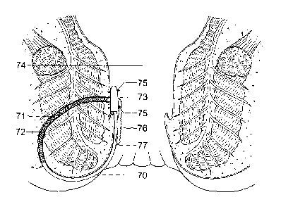
Figure 8 shows a schematic diagram of a seton of the present invention
positioned within a
recto-vaginal fistula.
The devices of the present invention are particularly useful for the treatment
of fistulas
such as anal or recto-vaginal fistulas. Depending on the fistula to be treated
however, it
CA 02831071 2013-09-23
WO 2011/151659 PCT/GB2011/051810
- 34 -
may be desirable and/or necessary to pre-treat the fistula prior to the use of
the devices of
the present invention. For instance, if the fistula is heavily infected, it
may desirable to
insert a drainage seton according to the prior art methods discussed above.
The drainage
seton may optionally incorporate antibiotics and/or anti-inflammatories in
order to help
reduce the extent of infection. After a period of time the drainage scton may
be removed
prior to the insertion of a device according to the present invention.
Optionally, prior to the insertion of a device according to the present
invention, the fistula
tract may be cleaned, for example using a jet of water and/or a suitable
brush.
Referring now to Figure 1, there is shown a device 1 according to an
embodiment of the
present invention comprising a probe 2 and a seton 3 for assisting in the
healing of fistulas.
The probe 2 comprises an elongate body having a blunt end 4 and an opposite
end 5 to
which the seton 3 is attached. The probe 2 is inserted into the fistula with
the blunt end
first such that the surgeon can use the probe to discover the track of the
fistula. The end 4
is blunt rather than sharp so as to reduce damage to surrounding tissue and to
minimise the
possibility of the probe going off track creating false passages.
The probe 2 is made out of a malleable metal or plastic that is easily bent so
that it can be
formed into a desired shape by the surgeon. This enables the surgeon to tailor
the shape of
the probe 2 to the track of the fistula so that the probe 2 can more easily be
fed through
the fistula with minimal damage to surrounding tissue. The probe 2 may vary in
size
depending on the type of fistula that is to be treated. It is envisaged that
the probe is
between 6 to 12 cm long and 0.5-2.0 mm in diameter.
The seton 3 is in the form of a flexible thread and is preferably made out of
a tough flexible
rubber or plastic such as polypropylene, in particular when treating fistulas
wherein the
seton has to be left in position for a longer period of time. Alternatively,
the seton 3 may
be formed out of a biodegradable material such as polyglycolic acid such that
the seton 3
biodegrades in situ with time, thereby obviating the need to remove the seton
3 from the
treatment site.
CA 02831071 2013-09-23
WO 2011/151659 PCT/GB2011/051810
- 35 -
The end 5 of the probe 2 opposite to the blunt end 4 is hollow and an end
portion of the
seton 3 locates therein and is secured to the probe 2 by the end 5 of the
probe 2 being
reduced in diameter such that a tight fit is created between the end 5 of the
probe and the
end of the seton 3. The diameter of the end 5 of the probe may be reduced by
passing the
end 5 of the probe through an aperture of a die. The aperture is configured to
be of a
smaller diameter than the initial diameter of the end 5 of the probe. The
diameter of the
end 5 of the probe can also be reduced by shaping the end 5 by hammering
Alternatively,
the end of the seton 3 may be swaged to the corresponding hollow end of the
probe 5 such
that the seton 3 and the probe 5 are fused together end-to-end.
In an alternative embodiment, the end 5 of the probe opposite to the blunt end
4 may be
solid such that an end face of the probe is directly swaged or fused to an end
face of the
seton 3. All of the aforementioned methods securely attach the seton 3 to the
probe 2 such
that as the surgeon feeds the probe 2 through the fistula the seton 3 follows
the probe
without detaching. In contrast, a seton that is threaded through an eye of a
probe as known
from the prior art may easily detach as it is pulled through the fistula such
that the surgeon
may have to repeat the process causing further discomfort to the patient.
As can be appreciated from Figure I, at the point of attachment of the seton 3
and the
probe 2, the longitudinal axis of the seton 3 is aligned with the longitudinal
axis of the
probe 2 and the overall diameter of the device in the region of the connection
does not
exceed the diameter of the probe 2. The region of the connection should be
understood to
mean the region where the seton engages with the probe, in particular where
the seton is
swaged to the probe or where a tight fit is created between the seton and the
probe.
Preferably, the region of the connection of the aforementioned embodiments is
less than 2
cm in a direction parallel to the longitudinal axis of the seton and the
probe.
The features of the longitudinal axis of the seton 3 being aligned with the
longitudinal axis
of the probe 2 and/or the overall diameter of the device in the region of the
connection
not exceeding the diameter of the probe 2, minimise damage to surrounding
tissue when
the probe 2 and the seton 3 are fed through the fistula in contrast to devices
known from
the prior art wherein the seton is thread through an eye of a probe. That is,
because as a
seton is thread through an eye of a probe it is transverse to the longitudinal
axis of the
CA 02831071 2013-09-23
WO 2011/151659 PCT/GB2011/051810
- 36 -
probe and as a result of resilient characteristics of the seton, the
transverse portion of the
seton thread through the eye of the probe may extend beyond the diameter of
the probe
thereby abrading the surrounding tissue as the probe and the seton are fed and
pulled
through the fistula.
Furthermore, as the scton is thread through the eye of the probe a double
layer of the
scton is formed which also may extend beyond the diameter of the probe. As
discussed in
the introduction, it is also known from the prior art to overlap an end of the
scton with an
end of the probe and thereafter to lash or suture the ends together. The
overall diameter of
the region in which the seton and the probe arc connected is therefore bulky
and extends
beyond the diameter of the probe. Again, this may abrade and damage
surrounding tissue
as the probe and the seton are fed and pulled through the fistula.
The seton 3 of the present invention is optionally secured to the probe 2 by
any of the
methods discussed herein during manufacturing so that the surgeon is not
required to
assemble the seton 3 and the probe 2 prior to use. For instance, the seton may
be glued
or welded onto the probe. Therefore, the device 1 is easier to use than those
known from
the prior art which require the surgeon to carefully thread or tie the seton
to the probe.
Furthermore, the fact that the device is pre-assembled reduces the required
surgical
preparation time.
As best shown in Figure 1, on one side of the seton 3 proximal to its
attachment to the
probe 2 ratchet teeth 7 are formed. In Figure 1 (and also Figures 2 and 4-6),
the ratchet
teeth 7 are shown to protrude from the seton 3 such that they extend beyond
the diameter
of the seton 3. In an alternative embodiment, it is envisaged that the ratchet
teeth do not
extend beyond the diameter of the seton. This may be achieved by making or
moulding
incisions into the seton transversely to the longitudinal axis of the seton so
as to form
ratchet teeth.
The ratchet teeth 7 engage with a locking device 8 also referred to as an
attachment device
as shown in Figures 1 and 2. The locking device 8 comprises a housing 9 formed
with an
aperture 10 that is transverse to the plane of the housing 9. A resiliently
deformable pawl
(not shown) extends into the aperture 10 for engagement with the ratchet teeth
7 when the
CA 02831071 2013-09-23
WO 2011/151659 PCT/GB2011/051810
- 37 -
seton 3 is inserted through the aperture 10, such that the seton 3 cannot be
withdrawn
from the aperture 10.
It is envisaged that once the surgeon has inserted the probe 2 and the scton 3
through the
fistula such that either ends of the scton 3 extend from either ends of the
fistula, the
surgeon feeds the blunt end 4 of the probe 2 followed by the scton 3 through
the aperture
of the locking device 8 so as to form a loop. Thereafter, the surgeon cuts the
scton 3
proximal to the probe 2. In an alternative method, the surgeon may cut the
scton 3
adjacent to thc probe and thereafter feed the free end of the seton 3,
provided with ratchet
10 teeth 7, through the aperture of the locking device 8. In either method,
cooperation
between the ratchet teeth 7 and the pawl permits the seton 3 to be pulled
through the
aperture 10 of the locking device 8 to the required extent.
By the locking device 8 securing the opposite end of the seton 3 so as to form
a loop, the
seton 3 remains in its position throughout the treatment period. Therefore,
the problem of
a knot securing the seton becoming undone as known from the prior art is
overcome and
so the probability of the surgeon having to re-insert a seton is reduced.
The ratchet teeth 7 and the locking device 8 enable the seton 3 to form a loop
of variable
size, as the seton can be pulled through and secured to the locking device at
any desired
length where the ratchet teeth 7 are formed. Therefore, the device 1 can be
specifically
adjusted to individual patients and their needs.
As can be appreciated from Figure 2, the housing 9 of the locking device 8 is
formed with
four holes 11. These holes enable a surgeon to suture the locking device 8 to
the wall of the
anal canal. Advantageously, as the seton 3 is held in place and the locking
device 8 is
retained internally, discomfort caused by a knot of two tied ends of a seton
as known from
the prior art is overcome.
A portion of the seton 3 adjacent to the locking device 8 is formed with a
coupling element
so as to be secured to a corresponding coupling element formed at the same end
as the
ratchet teeth 7. In the illustrated embodiment shown in Figure 1 and 2, the
coupling
elements of the seton 3 comprise protrusions 12 formed adjacent to the locking
device 8
CA 02831071 2013-09-23
WO 2011/151659
PCT/GB2011/051810
- 38 -
and corresponding recesses 13 formed on the opposite side of the ratchet teeth
7. The
protrusions 12 are a friction fit within corresponding recesses 13 to secure
the end of the
seton 3 that has been passed through the aperture 10 of the locking device 8,
to a portion
of the scton 3 adjacent to the ratchet teeth 7. The redundant end of the seton
3 is thereby
aligned with the remaining seton 3 and any excess cut off.
It will be appreciated that the coupling elements of the seton 3 arc optional.
Alternative
means for attaching the redundant end of the seton to the end of the seton
proximal to the
locking device may be used or omitted altogether.
It should be understood by those skilled in the art that the present invention
is not limited
to the locking device described above. Any locking device that securely holds
the two loose
ends of the seton together falls within the scope of the present invention.
An alternative embodiment will now be described with reference to Figure 3.
The
configuration of this embodiment only differs to the aforementioned embodiment
in how
the seton is attached to the probe and so a general description of the device
will be
omitted.
In Figure 3, an end 25 of a probe 22 is shown. The end 25 is formed with a
threaded screw
26 for receiving a seton 23. The screw 26 extends from the end 25 of the probe
22 and its
longitudinal axis is aligned with the longitudinal axis of the probe and its
diameter is
preferably smaller than that of the probe 22. Preferably, the probe 22 and the
screw 26 are
integrally formed. The seton 23 is formed with corresponding screw attachment
means in
that it has a hollow end 27. Although the hollow end 27 of the seton 23 is not
threaded it is
screwed onto the screw 26 by the flexible nature of the material used for the
seton 23. In
an alternative embodiment, the internal surface of the hollow end 27 of the
seton 23 is
threaded so that the seton 23 is screwed onto the threaded screw 26 of the
probe 22.
It shall be understood that the threaded screw may alternatively be formed on
an end of
the seton and the probe may be formed with a corresponding threaded screw
attachment
means such as a hollow end so that the probe can be screwed onto the seton. In
this
embodiment, the seton and the screw may be integrally formed.
CA 02831071 2013-09-23
WO 2011/151659 PCT/GB2011/051810
- 39 -
The aforementioned embodiments described with reference to Figure 3, have the
same
advantages as those described with reference to Figure 1. In particular, as
the longitudinal
axis of the probe and the longitudinal axis of the seton arc aligned at the
point of
attachment the probability of damaging surrounding tissue as the probe and
seton arc
pulled through is reduced. This is also achieved by the fact that the diameter
of the screw is
smaller than the diameter of the probe and the seton so that as the seton and
the probe arc
connected, the outer surfaces of the probe and the seton are flush or the
diameter of the
seton is smaller than that of the probe. As a result, the diameter of the
region of the
connection does not exceed the overall diameter of the device. The region of
connection
should be understood to mean the region where the screw is received in the
corresponding
screw attachment means. Preferably, the region of the connection of the seton
and the
probe of the present embodiments is less than 2 cm in a direction parallel to
the
longitudinal axis of the seton and the probe.
It should be appreciated that the seton of the device according to the present
invention can be secured to the probe by alternative means than those
described
hereinbefore. For example, the seton and the probe may be secured by 'click
fitting'.
In this un-illustrated embodiment, an end of the seton may be formed with a
protrusion having a resilient flange and the probe may comprise a hollow end
having a recess corresponding to the shape of the flange. By inserting the
protrusion of the seton into the hollow end of the probe, the flange
temporarily
deforms until it locates in the corresponding recess. In this embodiment, the
means
of connection are configured such that the force required to insert the
protrusion of
the seton into the hollow end of the probe is greater than the force required
to pull
the probe and the seton through a fistula so as to avoid the seton from
detaching
from the probe as the seton is being fitted.
An advantage of the probe and the seton being releasably connectable, such as
by
means of the screw attachment or click-fitting described above, is that in use
the
surgeon can rapidly interchange probes without having to undertake the fiddly
and
time-consuming task of threading the seton through an eve of the probe.
CA 02831071 2013-09-23
WO 2011/151659 PCT/GB2011/051810
-40 -
Furthermore, where it is desired to treat complex fistulas such as horseshoe
fistulas,
the use of two or more setons may be required. In such a scenario, a further
advantage of the probe and the seton being releasably connectable is that a
single
probe may be used for the insertion of both setons, thus saving on the
equipment
required and hence the cost of the procedure.
In another un-illustrated embodiment, the device is spring loaded so that the
seton will
retain a snug fit through the fistula for an extended period of time. Often,
for a seton to
promote optimal healing of a fistula, it is desirable for the seton to be
snugly fitted
throughout the treatment period. This is particularly important in cases where
the scton is
used to gradually cut through the sphincter muscle. However, setons as known
from the
prior art may become slack as they gradually cut through the sphincter muscle
resulting in
the seton becoming less effective in dividing the remaining muscle tissue. To
overcome this
problem, the device according to the present invention may be spring loaded so
as to
absorb any slacking so that the seton is continuously snugly fitted through
the fistula for a
desired period of time. Preferably, a spring is fitted between the locking
device and the
seton so as to absorb any slacking. Alternatively, the device may be fitted
with a spring
loaded cog which engages with corresponding teeth formed on the seton. The cog
could be
configured to comprise a coil spring and preferably the cog would engage with
the
.. redundant end of the seton that has been pulled through the locking device
such that the
cog pulls the seton through the locking device as the seton starts to slack.
A preferred embodiment is now described which may comprise any combination of
individual features as described above. In this embodiment the seton is
provided with a
.. tissue growth promoter such as fibrin so as to further promote tissue
repair of the fistula.
The seton may further be provided with one or more additional pharmaceutical
agents so
as to reduce inflammation or infection by locally delivering the specific
agents to the
treatment site. In this manner, many of the drawbacks associated with systemic
drug
delivery are avoided and relatively high concentrations of the required
pharmaceutical agent
can be targeted at the point of need with minimal side effects.
In one embodiment, the seton is provided with fibrinogenic materials which
include any
material based on fibrin. The seton may be coated directly in fibrinogenic
material or the
CA 02831071 2013-09-23
WO 2011/151659 PCT/GB2011/051810
-41 -
fibrinogenic material may be incorporated into a biodegradable polymer such as
alginate
which coats the seton.
Alternatively, as illustrated in Figure 4, a segment 29 of the scton may
itself be made from a
tissue growth promoting matrix such as fibrin, with the remainder of the
device 28 being as
described in relation to Figure 1.
Thus, in usc the portion of the scton being made from or provided with a
tissue growth
promoter is positioned within the fistula tract so as to encourage tissue
growth therein.
It is envisaged that fibrinogenic material can best be used in cases where
there has been no
active sepsis, that is to say when the tract of the fistula is a single tunnel
with no blind tracts
and no intervening abscess cavity in the path of the fistula.
In another embodiment, the seton may be provided with a fibrin plug, as
illustrated in
Figure 5. For example, the seton 50 may be threaded through the centre of a
fibrin plug 51
commercially sold as Biodesign0 and manufactured by Cook Medical. The seton
is thus
able to hold the fibrin plug firmly in place. When used in conjunction with
the previously
described locking device 52, the seton and hence the fibrin plug may be
further secured in
an exact position by the use of sutures through the holes 53.
Referring now to Figure 6, a cage 30 is shown which is attached to the seton
33. The cage
is made out of a biodegradable material and it comprises a wall 31 shaped into
a hollow
cylinder. The wall 31 is formed out of a mesh so as to allow tissue in-growth
and efficient
25 release of pharmaceutical agents. The cage 30 is also formed with means
of attachment 34,
for example screws, on either end. A portion of the seton 33 is attached to
either end of
the cage 30 via the means of attachment 34. One portion comprises the locking
device 38
and the cooperating elements and the other portion comprises the ratcheting 37
and the
corresponding cooperating elements as described in relation to the
aforementioned
30 embodiments. The cage 30 defines a space for holding pharmaceutical
agents such as a
fibrin plug (not shown), for example the fibrin plug manufactured by Cook
Medical, or
pellets of fibrinogenic material. The fibrin plug or any other pharmaceutical
material that is
to be used is inserted into the cage prior to attaching the portions of seton
33 to the means
CA 02831071 2013-09-23
WO 2011/151659
PCT/GB2011/051810
-42 -
of attachment 34. Thus, access to the inside of the cage is either at one or
other end at the
point of attachment 34 of the cage to the seton 33. Alternatively, it could be
in the middle
of the cage using a separate screw attachment device not shown or numbered in
Figure 6.
The seton 33 provided with the cage 30 is inserted through a fistula using a
probe 32 in a
similar way as described above. The scton is cut proximal to the probe and is
secured by
inserting the freed end of the seton 33 through the aperture 39 of the device.
Preferably,
the locking device 38 is formed with holes 40 so that the locking device 38
can be sutured
to the wall of the anal canal which thereby prevents the loop of seton 33 from
moving or
rotating. The advantage of the seton 33 being provided with a cage 30 is that
it retains the
fibrin plug in its position throughout the treatment period thereby promoting
primary
healing.
Preferably, the seton provided with fibrinogenic materials or a fibrin plug
with or without a
cage is formed out of biodegradable material. After a period of time,
typically 4 to 6 weeks,
this allows for the seton to be cut off at the entry and exit points of the
fistula such that a
segment of the seton and the fibrin with or without a cage remain in the
fistula as they are
gradually broken down by the body and fully or partially replaced by new
tissue formation.
Depending on the type of fistula to be treated the fibrinogenic material and
the fibrin plug
with or without the cage may extend for a length of 2 to 6 cm, and preferably
for a length
of 3 to 5 cm. The cage is preferably 2.5mm in diameter.
In some embodiments, the seton may further be provided with antibiotics, anti-
inflammatories such as steroids or tumour necrosis factor (TNF) inhibitors, or
immunomodulators and the like. A seton provided with antibiotics such as
gentamicin would be used for treating local septic areas of the fistula,
thereby
avoiding or reducing systemic side effects associated with oral medication. A
seton
provided with steroids or immunomodulators would be particularly useful in
patients suffering from Crohn's disease where local treatment of affected
tissue may
increase the rate of tissue regeneration and potentially reduce any systemic
side
effects resulting from oral medication. Similarly, a seton provided with a TNF
inhibitor would provide effective targeting of tumour cells in the area of the
fistula.
CA 02831071 2013-09-23
WO 2011/151659 PCT/GB2011/051810
-43 -
For the embodiments described above wherein the seton is provided with one or
more
tissue growth promoters and/or pharmaceutical agents, it is preferred to
suture the locking
device to the anal wall. This ensures that the portion of the seton provided
with active
agents is always located in the fistula and prevents the loop of the seton
from
rotating.
Referring to Figure 7, a schematic diagram of a seton 70 of the present
invention
positioned within an anal fistula 71 is illustrated. A segment 72 of the seton
70 provided
with a tissue growth promoter is secured within the anal fistula 71. The seton
loop is held
together by means of the attachment device 73 which is positioned within the
anal canal 74.
The attachment device is sutured to the wall of the anal canal by means of
sutures 75 so as
to prevent displacement of the tissue growth promoting segment out of the
fistula by
rotation of the seton loop. The redundant second end 76 of the seton that has
passed
through the attachment device and from which the probe has been removed is
securely
aligned against the first end 77 of the seton by means of the previously
described
cooperating elements. Discomfort to the patient caused by loose ends of the
seton is
thereby avoided.
By securely positioning the tissue growth promoting segment 72 within the
fistula 71 by
means of the seton 70, tissue in-growth is encouraged, thereby allowing the
fistula to heal.
Furthermore, the seton ensures that the tissue growth promoter is robustly
held in place
within the fistula, thus the problems encountered in the prior art due to
fistula plugs falling
out of the fistula are obviated.
Finally, referring to Figure 8 a schematic diagram of a seton 80 of the
present invention
positioned within a recto-vaginal fistula 81 is illustrated.
Thus, in use the seton 80 attached to the probe (not shown) may be thread
through the
recto-vaginal fistula 81 from the vaginal opening and then through the
attachment device
82 attached to the other end of the seton, which may be located inside the
anal canal 83 so
as to secure the seton loop. The probe may then be cut off and the redundant
end 84 of
the seton that has passed through the attachment device may be securely
aligned against the
CA 02831071 2013-09-23
WO 2011/151659 PCT/GB2011/051810
- 44 -
other end 85 of the seton by means of cooperating elements. The attachment
device may
be sutured to the wall of the anal canal by means of sutures 86.
Accordingly, it can be seen that a segment 87 of the scton 80 provided with a
tissue growth
promoter is secured within the recto-vaginal fistula 81. Thus, tissue in-
growth is
encouraged, thereby allowing the fistula to heal. Typically, after a 4 to 6
week healing
period, the seton is cut proximal to the entry and exit of the fistula, so as
to leave the
absorbed tissue growth promoter within the fistula tract whilst allowing the
attachment
device and the remainder of the seton to be removed.
Of course, it will be appreciated that the devices according to the present
invention may be
used by the person skilled in the art to treat simple anal and recto-vaginal
fistulas using
variations of the techniques exemplified above. For instance, when used to
treat a recto-
vaginal fistula, the probe may be thread through the recto-vaginal fistula
from the rectal
opening and/or the attachment device may be sutured to the wall of the vagina.
Similarly, it will be appreciated that whilst uses of the devices according to
the present
invention have been described above in relation to simple anal and recto-
vaginal fistulas,
the person skilled in the art will understand that the devices may also find
application in
many other types of fistula.
The devices of the present invention find particular application in fistulas
adjacent to which
there is a substantial body of tissue about which the seton loop may be
affixed. Preferably
the fistulas to be treated comprise or are located near at least one external
opening such
.. that the seton loop lies partially outside of the body. That said however,
the use of the
devices of the present invention entirely internally is also envisaged.
Furthermore, the devices and the uses described may be best adapted by the
surgeon in
view of the particular fistula to be treated. For instance, where a fistula is
complex, two or
more such devices may be used to ensure that a tissue growth promoter is
positioned
within each tract.
CA 02831071 2013-09-23
WO 2011/151659
PCT/GB2011/051810
-45 -
Although embodiments of the invention have been shown and described, it will
be
appreciated by those persons skilled in the art that the foregoing description
should
be regarded as a description of preferred embodiments only. The examples are
not
intended to limit the scope of the invention. Various modifications and
embodiments can be made without departing from the scope and spirit of the
invention, which is defined by the following claims only.