Note: Descriptions are shown in the official language in which they were submitted.
HEPATOCYTE GROWTH FACTOR MIMICS AS THERAPEUTIC AGENTS
DESCRIPTION SUMMARY
Field of the Invention
The invention generally relates to the development of hepatocyte growth factor
(HGF) mimics that
can act as mimetics (agonists) or antagonists. Mimetics act: to enhance
cognitive function; as
general neuroprotective/neuroregenerative agents; to facilitate wound repair;
to improve insulin
sensitivity and glucose transport; and to decrease tissue or organ fibrosis in
order to prevent or
reverse the symptoms of dementia, to protect from or reverse neurodegenerative
disease, to facilitate
repair of traumatic injury to the nervous system, to augment tissue and organ
vascularization, to
improve impaired wound healing, and to decrease or reverse fibrotic changes in
organs like heart,
lung, kidney, and liver. Antagonists act, for example, as anti-angiogenic and
anti-cancer agents; to
treat various malignancies and diseases like macular degeneration and diabetic
retinopathy, which
are associated with hypervascularization.
Mimetics:
Dementia: There are approximately 10 million diagnosed dementia patients in
the United States
alone and that number continues to grow every year as the population ages. The
costs
- 1 -
CA 2832113 2018-08-10
CA 02832113 2013-10-02
WO 2012/138599
PCT/US2012/031815
of treatment and care of these patients are in excess of $70 billion annually
and are increasing
rapidly. Unfortunately, the current treatment options for the management of
dementia are
severely limited and largely ineffective. The lack of treatment options for a
burgeoning
health problem of this magnitude necessitates that new and innovative
therapeutic approaches
be developed as quickly as possible.
At its core dementia results from a combination of diminished synaptic
connectivity
among neurons and neuronal death in the entorhinal cortex, hippocampus and
neocortex.
Therefore, an effective treatment would be expected to augment synaptic
connectivity, protect
neurons from underlying death inducers, and stimulate the replacement of lost
neurons from
preexisting pools of neural stem cells. These clinical endpoints advocate for
the therapeutic
use of neurotrophic factors, which mediate neural development, neurogenesis,
neuroprotection, and synaptogenesis. Not unexpectedly neurotrophic factors
have been
considered as treatment options for many neurodegenerative diseases including
Alzheimer's
disease (see reviews- Nagahara and Tuszynski, 2011; Calissano et al., 2010).
One particularly
attractive but mostly overlooked neurotrophic factor is HGF, which has a
proven ability to
both stimulate neurogenesis (Shang et al., 2011, Wang et al, 2011) and
synaptogenesis (see
preliminary studies below). The realization that HGF application might
represent a viable
treatment option for dementia should be no surprise. HGF is a potent
neurotrophic factor in
many brain regions (Kato et al., 2009; Ebens et al., 1996), while affecting a
variety of
neuronal cell types.
Neuroprotection/Neuroregeneration: HGF and c-Met are actively expressed in
both the
developing and adult brains and nerves. The Met system is essential for both
the central and
peripheral nervous systems to function properly. A large number of studies
have shown that
HGF and c-Met are expressed in multiple areas of the brain including the
frontal cortex,
subependyma, thalamus, cerebellar cortex, deep gray matter, and the
hippocampus, an
important area for cognition.
The biological activities described above also characterize Met functions in
the brain where
HGF/c-Met signaling is neurotrophic (Honda et al., 1995) and protective (Zhang
et al., 2000;
Takeo et al., 2007; Tyndall and Walikonis, 2007; Takeuchi et al.,
2008).Similar to its
activities in other tissues, Met in the brain is involved in development,
acting as a guidance
factor during differentiation, motogenesis and neuritogenesis (Ebens et al.,
1996; Sun et al.,
2002; Tyndall and Walikonis, 2007). HGF/ c-Met signaling has also been shown
to promote
-2-
CA 02832113 2013-10-02
WO 2012/138599
PCT/US2012/031815
healing of neuronal injury (Trapp et al., 2008), especially after ischemic
brain injury (Takeo
et al., 2007). HGF also displayed neuroprotective effects in animal models for
neurodegenerative diseases including amyotrophic lateral sclerosis (ALS). The
various
functions of HGF, plus its highly potent neurotrophic activities, promote HGF
as a potential
therapeutic agent for the treatment of various diseases of the nervous system.
Amyotrophic Lateral Sclerosis: ALS is a fatal rapid-onset neurodegenerative
disease that is
characterized by degeneration of motoneurons in the spinal cord and efferent
neurons in the
motor cortex and brainstem. The impact of this degeneration results in a
progressive loss of
muscle function culminating in total paralysis. Approximately 90% of the cases
of ALS are
classified as sporadic with no known etiology, while the remaining 10% appear
to be familial,
resulting in part from defects in copper/zinc superoxide dismutase 1 (SOD1),
which leads to
exaggerated oxidative stress and an unfolded protein response. The one thing
that both forms
of ALS have in common is that there is currently is no effective treatment
available.
Despite the paucity of effective treatment options, several studies have
highlighted the
potential benefits of using hepatocyte growth factor (HGF) as a therapeutic
agent. These
investigations have demonstrated that application of hepatocyte growth factor
(HGF) in a
murine or rat model of familial ALS significantly slows motoneuron
degeneration (Aoki et
al., 2009); reduces gliosis (Kadoyama et al. 2007), which contributes to the
degeneration
process; delays the onset of paralysis (Kadayama et al., 2009); and increases
lifespan (Sun et
al., 2002).
The realization that HGF application might represent a viable treatment option
for
ALS, however, should be unexpected. HGF along with its type I tyrosine kinase
receptor, c-
Met, have long been recognized for their role in the development of tubular
structures (Santos
et al., 1993) and their general proliferative, anti-apoptotic, motogenic, and
morphogenic
actions on hepatocytes and cells of epithelial origin . Most pertinent,
however, is the more
recent realization that HGF is a potent neurotrophic factor (Maina and Klein,
1993; Kato et al.,
2009) in many brain regions and that it is particularly effective as a pro-
survival/regenerative
factor for motoneurons (Ebens et al., 1996; Yamamoto et al., 1997; Hayashi et
al., 2006; Elsen
.. et al., 2009).
Parkinson's Disease: A treatment option long considered for many
neurodegenerative diseases
-3-
CA 02832113 2013-10-02
WO 2012/138599
PCT/US2012/031815
including Parkinson's disease (PD) has been the application of growth factors
with the
intention of halting disease progression, restoring lost function, or
hopefully both (review,
Rangasamy et al., 2010). However, this dream has gone largely unfulfilled at
the level of
clinical medicine because of limitations related to brain delivery and costs.
Growth factors are
universally large proteins that are both metabolically labile and too large to
pass the blood-
brain barrier (BBB). As such, most approaches to delivery have utilized gene
therapy methods
with the hope that the growth factor will be expressed in the correct location
at a high enough
concentration and for a long enough period to provide clinical relief.
Although a number of
creative and successful approaches in animal models have been employed to
deliver growth
factors like GDNF (Wang et al., 2011) to the brain, these methodologies are
technically
complex and prohibitively difficult to bring to practice with large numbers of
patients.
While many growth factor systems have been examined as potential therapeutic
targets
for PD one that has been largely, and we think mistakenly, overlooked is the
hepatocyte
growth factor (HGF)/c-Met (its type I tyrosine kinase- receptor) system.
Nevertheless, the
potential utility of HGF as a PD treatment has been highlighted in a study by
Koike et al.
(2006) in which an HGF plasmid injected directly into the substantia nigra
(SN) resulted in
localized over-expression of HGF, and acted dramatically to prevent neuronal
cell death and
preserve normal motor function in the 6-hydroxydopamine (6-0HDA) PD rat model.
This
observed neuroprotective effect of HGF on dopaminergic (DA) neurons meshes
with its ability
to augment the proliferation and migration of dopaminergic progenitor cells
(Lan et al., 2008)
The neuroprotective effect of the HGF on the nigrostriatal pathway, however,
should
be no surprise given its recognized role in stem cell regulation, the
development of tubular
structures (Santos et al., 1993) and its general proliferative, anti-
apoptotic, motogenic, and
morphogenic actions on many cell types including hepatocytes and cells of
epithelial origin
(Gherardi et al., 1993). Maina et al., Particularly pertinent is the
demonstration that HGF is a
potent neurotrophic factor for many neuronal cell types (Kato et al, 2009)
including
motoneurons ( Elsen et al., 2009; Hayashi et al, 2006), hippocampal neurons
Lim et al., 2008),
cerebellar granular cells (Ieraci et al., 2002), and sympathetic neurons
(1999). Moreover, HGF
appears to be a critical regulator of neural stern cell expansion and
differentiation (Nicoleau et
al., 2009) suggesting that neural as well as many types of peripheral stem
cells are under the
control of the HGF/c-Met system.
- 4 -
CA 02832113 2013-10-02
WO 2012/138599
PCT/US2012/031815
Traumatic Brain Injury/Spinal Cord Injury: TBI often negatively impacts
cognitive function
and can elicit effects that range from mild, with temporary decrements in
mental abilities, to
severe, with prolonged and debilitating cognitive dysfunction (Kane et al.,
2011). Cognitive
difficulties along with other neurological deficits including: anxiety,
aggressiveness, and
depression result in a significantly reduced quality of life (Masel and
DeWitt, 2010). With
military operations concluded in Iraq and continuing in Afghanistan TBI has
become the
major combat injury representing 28% of all combat casualties (Okie, 2005;
U.S. Medicine,
May 2006, Vol 42). Total estimates of military service members suffering TBIs
between
2001 and 2010 range from 180,000 to 320,000 (U.S. Defense and Veterans Brain
Injury
Center).
Underlying TBI is physical injury to the brain resulting in decreased synaptic
connectivity among neurons, loss and death of neurons, damage to cerebral
blood vessels
resulting in ischemic/hypoxic-induced damage, and secondary glial scaring.
This loss of
neurons and diminished synaptic connectivity is particularly apparent in the
hippocampus
(Gao et al., 2011; Zhang etal., 2011 a; Zhang et al., 2011b) resulting in
defective long-term
potentiation (Schwarzbach et al., 2006) and cognitive deficits (e.g. Dikrnen
et al., 2009; Patel
et al., 2010). The prevalence of TBI associated injuries that result in
neuronal loss and
decreased synaptic connectivity denote the need for therapies which support
neuronal repair
and/or replacement. These clinical endpoints advocate for the therapeutic use
of neurotrophic
factors which mediate neural development, neurogenesis, neuroprotection, and
synaptogenesis, for treating TBI. Not unexpectedly neurotrophic factors have
been considered
as treatment options for TBI (Kaplan et al., 2010; Richardson et al., 2010; Qi
et al., 2011).
One particularly attractive but mostly overlooked neurotrophic factor is HGF,
which has a
proven ability to both stimulate neurogenesis (Shang et al., 2011; Wang et
al., 2011) and
synaptogenesis (see preliminary studies below). The fact that HGF application
might
represent a viable treatment option for TBI stems from the recent realization
that HGF is a
potent neurotrophic factor in many brain regions (Kato et al., 2009; Ebens et
al, 1997), while
affecting a variety of neuronal cell types (Yamamoto et al., 1997; Hayashi et
al., 2006; Elsen
et al., 2009).
HGF and wound healing: Excessive scarring is typified by unnecessary
accumulation of
ECM components in the wound, due to an inappropriate balance between synthesis
and
-5-
CA 02832113 2013-10-02
WO 2012/138599
PCT/US2012/031815
degradation. Therapy for pathologic scarring may be directed at inhibiting the
synthesis and
promoting the degradation of the ECM. HGF in the skin promotes wound healing
effectively
in several ways: motivating the proliferation and motility of dermal vascular
endothelial cells;
stimulating the motility of epidermal keratinocytes; enhancing local blood
supply; and
accelerating the re-epithelialization of the wound (Nakanishi et al., 2002).
Re-
epithelialization inhibits the formation of scars. Studies have shown that HGF
gene transfer
accelerates dermal wound healing by stimulating angiogenesis and
reepithelialization
(Nakanishi et al., 2002). Therapeutic approaches that augment HGF/SF would be
expected to
promote wound healing and prevent scar formation.
HGF as a treatment option for metabolic syndrome and diabetes: Several recent
studies have
implicated the critical role of the HGF/c-Met system in the regulation of
glucose handling,
insulin secretion, and tissue insulin sensitivity. Together these
investigations have highlighted
the therapeutic potential of augmenting the HGF/c-Met system for the treatment
of type 2
diabetes and metabolic syndrome (Fafalios et al., 2011; Flaquer et al.,
2012)). These
investigators have shown that: 1) c-Met, the HGF receptor complexes with the
insulin
receptor; 2) c-Met is critically involved with hepatic glucose homoestasis; 3)
HGF restores
insulin responsiveness in a murine diabetic mouse model; 4) that HGF gene
therapy can
prevent the renal damage that typically accompanies diabetes, and 5) HGF
ameliorates the
vascular complication of diabetes (Peng et al., 2011).
The HGF/c-Met signaling pathway potentiating Angiogenesis: Angiogenesis is
defined as the
formation of new blood vessels from existing vascular bed, It is a prime
requirement in
physiological processes such as wound healing and the menstrual cycle, on the
other hand, it
is an essential step for multiple pathological conditions, like cancer,
macular degeneration,
atherosclerosis, diabetic retinopathy, neovascular glaucoma, psoriasis and
rheumatoid
arthritis. Consequently, the modulation of angiogenesis, whether it was
through encouraging
therapeutic angiogenesis or by stopping pathologic angiogenesis, is an
exhilarating prospect
for modern medicine. The equilibrium between physiological and pathological
angiogenesis
is mediated by the communication of numerous endogenous angiogenic and anti-
angiogenic
modulators.
Numerous studies have shown HGF to be a powerful inducer of neovasculature
-6--
CA 02832113 2013-10-02
WO 2012/138599
PCT/US2012/031815
formation. Moreover HGF/c-Met inhibitors are clinically relevant anti-
angiogenic agents.
(Gherardi et al, 2012).This is probably attained through multiple pathways,
achieved either by
direct or indirect action on endothelial cells.
HGF as anti-fibrotic agent: Fibrotic disease takes many forms and is a major
contributor to
degraded function in the heart, kidney, and liver secondary to many
pathological states
including myocardial infarction, diabetes, and alcoholism. Hepatocyte growth
factor (HGF) is
showing a strong anti-fibrotic effect with remarkable effectiveness in
ameliorating tissue
fibrosis in a wide range of animal models HGF exhibits a remarkably powerful
anti-fibrotic
effect that ameliorates tissue fibrosis in a wide range of animal models and
tissues (Liu and
Yang, 2006). Evidence has documented the therapeutic effect of exogenous HGF
in chronic
allograft nephropathic rats, a model of chronic inflammation and progressive
tissue scarring.
The intramuscular administration of the human HGF gene reduced the rate of
mortality,
restrained inflammation and infiltration, and reduced renal fibrosis (Liu and
Yang, 2006).
Coronary artery disease (CAD) ischemic events and myocardial infarction are
the
major causes of cardiac failure in the Western world. The only option for
severe coronary
blockage and atherosclerosis is bypass surgery. Two pathological events in CAD
play major
roles in the loss of cardiac function observed in CAD: 1) blockage of the
coronary arteries
resulting in decreased blood perfusion to the heart; and 2) the formation of
fibrotic tissue after
cardiac insult resulting in ventricle remodeling and decreased compliance.
Increased levels of
HGF in the circulation have been reported after acute myocardial Infarction
(Zhu et al., 2000;
Jin et al., 2003). This increase in circulating HGF can be used as biological
marker for heart
injury and gives a clue regarding its protective role (Ueda et al., 2001).
Pharmaceuticals that
enhance the HGF/Met signaling could potentially be used in the treatment of
myocardial
infarction, providing protection against oxidative stress and cell death due
to apoptosis as
well as reducing the formation of fibrotic tissue (Ahmet et al., 2002; Kondo
et al., 2004;
Pietronave et al., 2010). Moreover, another beneficial effect of HGF following
myocardial
infarction could lie in its ability to induce neovascularization, which could
support formation
of new cardiac vasculature that would improve reperfusion of the myocardium.
Although HGF is known to protect the liver against external insults, HGF
generation
has also been associated with several liver and extra-hepatic diseases.
Experimental and
clinical evidence indicates that HGF plays a crucial role in liver
regeneration. Liver cirrhosis
is the irreversible end result of fibrous scarring and hepatocellular
regeneration and is a major
-7-
CA 02832113 2013-10-02
WO 2012/138599
PCT/US2012/031815
cause of morbidity and mortality worldwide with no effective therapy. Although
there is no
specific etiology for this disease, cirrhosis has been defined as a chronic
disease of the liver in
which dispersed damage and regeneration of hepatic parenchymal cells have
taken place and
in which dissemination of connective tissue has resulted in inadequate
organization of the
lobular and vascular structures (Fujimoto and Kaneda, 1999; Kaibori et al.,
2002). Ideally,
approaches for the treatment of liver cirrhosis should include attenuation of
fibrogenesis,
encouragement of hepatocyte mitosis, and reformation of tissue architecture.
Studies have shown that exogenous administration of recombinant HGF increases
the
potential for liver regeneration after hepatoctomy especially in the cases of
cirrhotic liver
(Boros and Miller, 1995; Kaibori et al., 2002; Borowiak etal., 2004).
Conversely, studies
have shown that the clofibrate-related compounds, which increase HGF/SF
levels, can induce
hepatomegaly, proliferation of hepatic peroxisomes, and hepatic carcinoma (Xu
and Wu,
1999). The linkage of HGF/SF both positively and negatively to hepatic
diseases has made
HGF-related therapeutics a hot area for pharmaceutical development.
Limitations to the direct use of HGF: The direct use of HGF or any other
protein
neurotrophic factor as a therapeutic agent has two serious limitations: 1)
large size and
hydrophilic character precluding blood-brain barrier permeability (BBB); and
2) the need to
be manufactured by recombinant methods at high cost, thus limiting its
widespread use.
These impediments can be overcome using one or more of an extensive library of
small
molecule HGF mimetics which are described herein, some of which are orally
active,
display profound pro-cognitive/anti-dementia/ neuroprotective activity, and
are
inexpensive to synthesize.
Antagonists: Improper activation of the c-Met receptor can be encouraged by
genetic
activating mutations, transcriptional upregulation or by ligand-dependent
autocrine or
paracrine mechanisms.
c-Met activation in cancer: Cancer is a heterogeneous group of diseases that
result from the
accumulation of genetic mutations. These mutations cause altered function in
proto-
oncogenes leading to dysregulation of DNA repair, proliferation, and apoptotic
signaling
(Tarmock, 2005). The dysregulation in the signals within a group of cells
leads to the
uncontrolled growth, and invasion that either directly intrudes upon and
destroys adjacent
-8-
tissue or metastasizes and spread to other location in the body through the
lymphatic system or the
blood stream.
A dysfunctioning Met and Ha' system appears to be a critical trait of numerous
human
malignancies. Ectopical overexpression of HGF and/or c-Met in mouse and human
cell lines leads
them to develop tumorigenic and metastatic phenotypes in athymic nude mice
(Rong et al., 1994). A
large number of studies have shown that the HGF/c-Met pathway is one of the
most dysregulated
pathways in human malignancies, which include, but arc not limited to:
bladder, breast, cervical,
colorectal, endometrial, esophageal, gastric, head and neck, kidney, liver,
lung, nasopharyngeal,
ovarian, pancreatic, prostate, and thyroid cancers. Lastly, an activating
mutation of c-Met has been
discovered in sporadic and inherited forms of human renal papillary carcinomas
(Danilkovitch-
Miagkova and Zbar, 2002). These mutations which alter sequences within the
kinase domain have
also been found in other types of solid tumors and metastatic lesions. At this
point it's worth
mentioning that HGF over- or miss-expression often correlates with poor
prognosis and that the
down-regulation of c-Met or HGF expression in human tumor cells reduced their
tumorigenicity
(Abounader et al., 2002).
Activation of Met in cancer occurs most often through ligand autocrine or
paracrine
activation. Osteosarcomas and globlastoma mutliforme, which express both c-Met
and HGF are
examples of dysfunctional autocrine control. In other instances where
paracrine control is
paramount, c-Met over-expression has been reported in human primary tumors
while HGF is
provided by stromal cells and not the tumor itself (Houldsworth et al., 1990;
Kuniyasu et al., 1992;
Hara et al., 1998; Tong et al., 2004; Miller et al., 2006; Bean et al., 2007).
The list of neoplasms in which c-Met overexpression has been detected is
growing
relentlessly. In the case of carcinomas, excessive levels of c-Met expression
have been found in
virtually every malignancy (Danilkovitch-Miagkova and Zbar, 2002). Receptor
over-expression can
lead to local receptor oligomerization generating cells reactive to
subthreshold ligand concentrations.
HGF itself is able to trigger the transcription of c-Met (Boccaccio et al.,
1994), and it is thus HGF,
which is universally expressed by stromal cells throughout the body that
typically drives tumor over
expression of c-Met (Aguirre Ghiso et al., 1999; Parr et al., 2004). This
uniqueness of HGF permits
it to play a critical role, which engages paracrine positive feedback loops
that prop up the growth
and metastasis of cancer cells. Interestingly, this notion is in agreement
with the observation that c-
Met activating
- 9 -
CA 2832113 2018-08-10
CA 02832113 2013-10-02
WO 2012/138599
PCT/US2012/031815
mutations require HGF to enhance their catalytic effectiveness (Michieli et
al., 1999).
HGF can also abnon-nally stimulate c-Met in an autocrine manner, as depicted
in
gliobastomas (Weidner et al., 1990), breast carcinomas (Potempa and Ridley,
1998),
rhabdomyosarcomas (Hartmann et al., 1994) and osteosarcomas (Ridley et al.,
1995). With
multiple mechanisms of activation, it is clear that both Met and HGF are major
contributors
to the progression of most human cancers. Additionally, the demonstrated
activities of c-Met
and HGF in proliferation, invasion, angiogenesis and anti-apoptosis (Weidner
et al., 1990;
Rong et al., 1994; Kitamura et al., 2000; Xiao et al., 2001; Wang et al.,
2002; Derksen et al.,
2003) demarcate the different stages at which these molecules can participate
in tumor
development.
Although, c-Met is used as a general marker for cancer, is also an indicator
of
biological significance with respect to malignancy and patient prognosis, with
high levels
correlated with a poor prognosis. Molecules that inhibit c-Met and HGF can
therefore be
expected to interfere with the molecular causes of many cancers, and should
significantly help
in attenuating Recent studies from the Harding lab have confirmed the
potential use of HGF
antagonists as effective anti-cancer/anti-angiogenic agents (Yamamoto et al.,
2010, Kawas et
al., 2011; Kawas et al., 2012).
Macular degeneration/diabetic retinopolliy: Age-related macular degeneration
(ARMD) is
the most common cause of irreversible vision loss in Americans over the age of
60. It is
predicted that 10 million Americans will suffer from some level of this age-
related visual
damage during their retirement years. In normal healthy eyes, retinal pigment
epithelial (RPE)
cells form a polarized monolayer adjacent to the photoreceptors and are
involved in various
activities that are essential to retinal homeostasis and visual function. In
the case of macular
degeneration, unfortunately, adhesions and communication between RPE cells are
lost
because of inflammation. When inflammation occurs, RPE cells secrete many
growth factors
including HGF/SF, which stimulates the division and migration of RPE and the
foimation of
new vasculature from existing blood vessels (angiogenesis). HGF also
stimulates the
production of other growth factors (e.g. VEGF), which further promote the
formation of new
blood vessels that invade neighboring matrix (Jun et al., 2007). Hence the use
of HGF
blockers could be used either prophylactically, or as a treatment to slow down
the progression
of the disease and subsequent loss of vision.
Proliferative diabetic retinopathy (PDR), which entails a distinctive
-10-
neovascularization of the retina that is characterized by the invasion of
vessels into the vitreous
cavity, is coupled with bleeding and scarring around the proliferative channel
(Katsura et al., 1998).
There is substantial evidence that multiple growth factors are involved in the
onset and progression
of the neovascularization process in general and in the PDR in specifically.
These include basic
fibroblast growth factor (bFGF), Insulin-like growth factors (IGF-I), vascular
endothelial growth
factor (VEGF), and HGF. Of these, HGF has the most pronounced effects on
endothelial growth and
mitogenic activity (Boulton, 1999). Studies have found that levels of HGF in
the vitreous fluid of
PDR patients are considerably higher than in non-diabetic patients, and that
the levels of HGF are
especially high in the active stage of PDR (Katsura et al., 1998). This
suggests that HGF stimulates
or perpetuates neovascularization in PDR. Therefore, it is plausible to think
that an HGF antagonist
would be a promising option as a prophylactic treatment, or to ameliorate the
progression of PDR.
BRIEF DESCRIPTION OF THE DRAWINGS
Figure 1 A, B, and C. Effect of Dihexa on spatial learning in the water maze.
A: 30 minutes before
beginning testing rats were given scopolamine directly into the brain
intracerebroventricularly (ICV)
and 10 minutes later Dihexa was given ICV at 10 pmoles (low dose) or 100
pmoles (high dose). This
was done daily before the first training trial. There were 5 trials per day
for 8 days. The latency to
find the pedestal was considered a measure of learning and memory. Rats
receiving high Dihexa
were able to completely overcome the scopolamine deficits and were no
different than controls. B.
minutes before beginning testing rats were given scopolamine directly into the
brain
intracerebroventricularly (ICV) and 10 minutes later Dihexa was given orally
1.25 mg/kg/day (low
dose) and 2 mg/kg/day (high dose). This was done daily before the first
training trial. There were 5
trials per day for 8 days. The latency to find the pedestal was considered a
measure of learning and
25 memory. Rats receiving high dose Dihexa were able to completely overcome
the scopolamine
deficits and were no different than controls. C: Aged rats of mixed sex and
age (22-26 months) were
randomly assigned to a control/untreated group or a Dihexa treated group (2
mg/kg/day). Rats were
not prescreened. Note that normally-50% of aged rats show deficits, thus the
large group errors. The
Dihexa group performed significantly better than untreated controls.
- 11 -
CA 2832113 2018-08-10
Figure 2A and B. Dihexa and NIel-AngIV dose-dependently stimulate
spinogenesis. A)
Dihexa and B) Nlel-AngIV increase spine density in mR1-713-0-actin transfected
hippocampal neurons
in a dose-dependent manner. Neurons were stimulated with Dihexa or Nlei-AngIV
over a 5 day
period at a wide range of concentrations. Data obtained from separate
cultures; cultures were 12
days old at time of fixing. The number of dendritic spines on representative
50 [tm dendrite
segments were hand counted. ** = p <0.05 and *** = p <0.001 ; n = 50; mean
S.E.M.; 4 ¨
significantly different from control.
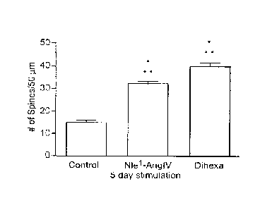
Figure 3A-E. Time dependent effects of Nlel-AngIV and Dihexa treated neurons
on
spinogenesis. Hippocampal neurons transfected with mRFP-13-actin were treated
with 10-12 M
Dihexa or Nlel-Ang IV for 5 days in culture or for 30 minutes prior to
fixation on day in vitro 12
(DIV12), promote spinogenesis. A) Representative image of the dendritic arbor
of a 5 day vehicle
treated hippocampal neuron. B) Representative image of a dendritic arbor from
a neuron stimulated
for 5 days with 10-12M Dihexa. C) Representative image of the dendritic arbor
of a neuron
stimulated with 10-12 M Nlel-Ang IV for 5 days. D) Bar graph representing the
number of spines per
50 tim dendrite length per treatment condition following a 5 day in vitro
treatment. *** P < 0.001 ; n
= 200. E) Bar graph representing the number of spines per 50 pm dendrite
length per treatment
condition following an acute 30 minute treatment. *** P < 0.001 ; n = 60.
*Data obtained from
separate cultures; cultures were 12 days old at time of fixing. Mean S.E.M.
by one-way ANOVA
and Tukey post hoc test.
Figure 4. Nlel-AngIV and Dihexa increase spine head width. The width of the
spine head was
measured as an indication of synaptic strength. Spine heads with a greater
surface area can
accommodate more neurotransmitter receptors and are more likely to form
functional synapses. The
AngIV analogue treatment-induced increase in spine head width suggests
facilitated
neurotransmission. *** = p < 0.001 ; mean S.E.M.; n = 100.
Figure 5A-G. Neurotransmitter patterns for Nlel-Ang1V and Dihexa stimulated
neurons.
Dihexa and Nle'-AngIV treated neurons were immunostained for the universal
presynaptic marker
synapsin and the glutamatergic presynaptic marker VGLUT1. The percent
correlation between the
postsynaptic spines (red) and presynaptic puncta (green) were measured as an
indication of
functional synapses. A) represents photographs of Dihexa and Nlel-AngIV
treated neurons
immunostained for the universal synaptic marker synapsin and the glutamatergic
presynaptic marker
VGLUT1. B) Bar graph representing the percent correlation of treatment-induced
postsynaptic
spines to the
- 12 -
CA 2832113 2018-08-10
glutamatergic presynaptic marker VGLUT1. A high percent correlation between
the presynaptic
marker and the postsynaptic spines suggests that functional connections are
formed (P> 0.05; mean
S.E.M.; n = 25). C) Bar graph representing an increase in the number of spines
following
treatment with vehicle, Nle 1 -AngIV or Dihexa, ensuring health of the neurons
(***=p < 0.001 ;
mean S.E.M.; n = 25). D) Bar graph representing the percent correlation of
treatment-induced
postsynaptic spines to the general presynaptic marker Synapsin. No significant
differences between
the stimulated neurons and vehicle control treated neurons were observed (P >
0.05; mean S.E.M.;
n = 25) suggesting a majority of the presynaptic input is glutamatergic. E)
Bar graph representing an
increase in the number of spines following treatment with vehicle, Nle 1 -
AngIV or Dihexa, ensuring
an active phenotype (***=P < 0.001 ; mean S.E.M.; n = 25). F) Bar graph
representing the percent
correlation of treatment-induced postsynaptic spines to the postsynaptic
marker PSD-95. G) Bar
graph shows no significant differences (P> 0.05; mean S.E.M.; n = 25)
between the postsynaptic
marker PSD-95 and the postsynaptic spines suggest that the newly formed spines
have a functional
postsynaptic element.
Figure 6A and B. Mini-excitatory postsynaptic currents (mEPSCs) in dissociated
hippocampal
neurons. NIel-AngIV and Dihexa treatment increase the frequency of mini-
excitatory postsynaptic
currents (mEPSCs). Recordings were done on dissociated hippocampal neurons
treated with vehicle,
10-12 M Nle 1 -AngIV or Dihexa for 5 days prior to recording. The currents
recorded were
spontaneous bursts of AMPA-mediated synaptic transmission in the absence of
action potentials
carried in the presence of strychnine, picrotoxin and tetrodotoxin. A) Bar
graph representing the
increase in AMPA-mediated frequencies from Nlel -AngIV or Dihexa treated
hippocampal neurons.
The increased frequencies indicate that spines induced by Nlel -Ang1V or
Dihexa support functional
synapses. *** = p < 0.001 ; S.E.M.; n = 25. B) Representative traces of
mEPSC recordings from
Niel-AngIV or Dihexa treated hippocampal neurons.
Figure 7A and B. Evaluation of Nlel-AngIV- and Dihexa-dependent spinogenesis
in CA1
hippocampal neurons from rat organotypic hippocampal slice cultures. Nlel-
AngIV- and
Dihexa were found to support spinogenesis in CAI hippocampal neurons.
Organotypic hippocampal
slice cultures (400 um thicknesses), representing a more intact environment,
were biolistically
transfected with the soluble red fluorescent protein Tomato. CA1 hippocampal
neurons were
selected for evaluation because of their known plastic
- 13 -
CA 2832113 2018-08-10
response during learning. Slices were obtained from postnatal day 5 rats. A)
Representative images
of CAI neuronal dendrites from Tomato transfected hippocampal slices. Images
represent a 2 day
treatment with 10-12 M Nlel-AngIV or Dihexa. B) Treatment- induced
spinogenesis is observed in
CA1 pyramidal hippocampal neurons. Spine numbers measured for control slices
were 7 per 50 gm
dendrite length vs. 11 spines per 50gm dendrite length for both NIel-AngIV and
Dihexa treated
neurons; Mean S.E.M., n = 17; ** = P < 0.01 Statistical significance by one-
way ANOVA
followed by Tukey Multiple Comparisons Test; Experiments were repeated at
least three times.
Figure 8. HGF dose-dependently enhances spinogenesis. Effect of HGF on
spinogenesis in
dissociated hippocampal neurons. Dissociated hippocampal neurons from 1 or 2
day old rats were
transfected with mRFP-P-actin and stimulated with HGF for 5 days. Treatment
with 2.5 ng/ml HGF
did not affect basal spine numbers and was considered subthreshold. Doses of
5, 10 and 20 ng/ml
significantly increased the number of spines per 50 gm dendrite lengths
compared to vehicle control
treated neurons. *** P <0.001; mean S.E.M.; n = 50 per treatment group.
Figure 9A and B. Effects of Dihexa and HGF on spinogenesis in organotypic
hippocampal slice
cultures. Hippocampal slice cultures were biolistically transfected with the
red soluble protein
Tomato on DIV3 and stimulated with Dihexa or HGF on DIVS. Organotypic
hippocampal slice
cultures maintain a more intact perforant path and therefore represent a more
intact environment. A)
Representative images of CA1 neurons, the neuronal type in the hippocampus
that exhibits learning
associated synaptic plasticity. Hippocampal slices were stimulated with
vehicle, 10-12 M Dihexa, or
10 ng/ml HGF for 2 days. B) Bar graph representing the number of spines per 50
gm dendrite length
for each treatment group. Dihexa and HGF significantly increase the number of
spines on CAI
hippocampal neurons compared to control treated neurons. *** = P <0.001 ; mean
S.E.M.; n = 20
for control, 26 for Dihexa and 38 for HGF stimulated neurons.
Figure I0A-D. Effect of HGF treatment on synaptogenesis in dissociated
hippocampal
neurons. HGF treatment supports the formation of functional synapses as
indicated by a high
correlation between postsynaptic spines (red) and markers of presynaptic
active zones (green). A)
Percent correlation of actin-enriched postsynaptic spines (red) juxtaposed to
the universal
presynaptic marker Synapsin (green). A high percent correlation suggests
functional synapses are
formed. B) Percent correlation of actin-enriched spines (red) juxtaposed to
the glutainatergic
presynaptic marker VGLUT1 (green). A greater than 95% correlation suggests
many of these inputs
are glutamatergic.
- 14 -
CA 2832113 2018-08-10
C) Bar graph representing an active phenotype as indicated by a significant
increase in the number
of spines per 50 tm dendrite length following stimulation with HGF (10 ng/ml).
Mean number of
spines = 33 vs. control = 23; *** = P < 0.001 by one-way ANOVA and Tukey
Multiple
Comparisons Test; mean S.E.M.; n = 25). D) Representative images of
hippocampal neurons
transfected with mRFP-p-actin on DIV6 and treated with 10 ng/ml of HGF or
vehicle for 5 days in
vitro. The neurons were stained for the general presynaptic marker Synapsin
and glutamatergic
presynaptic marker VGLUT I .
Figure 11. Effect of Dihexa and HGF treatment on the frequency of mEPSCs in
dissociated
hippocampal neurons. Dissociated hippocampal neurons transfected with mRFP-f3-
actin were
stimulated with 10-12 M Dihexa or 10 ng/ml for 5 days prior to recording
mEPSCs. Neurons were
treated with tetrodotoxin, picrotoxin, and strychnine to suppress action
potential, GABA-dependent
inhibition, and glycine-dependent inhibition. Treatment with both agonists
significantly enhanced
AMPA-mediated currents compared to vehicle treated neurons (** P <0.002;
S.E.M. by one-way
ANOVA followed by Newman-Keuls post hoc test; n = 9, 9 and 11 respectively).
Figure 12A and B. Effect of maximal and sub-threshold doses of Angiotensin IV
analogues and
HGF on spinogenesis. A) Sub-threshold levels of HGF, Dihexa or Nlel -AngIV do
not affect basal
spine numbers. Combined sub-threshold levels of Dihexa (10-1s M) and HGF (2.5
ng/ml) phenocopy
the effects of Dihexa at its biologically effective dose alone; # = 10-13 M
and $ = 2.5 ng/ml. B) A
sub-threshold dose of the parent compound Niel-Ang IV (l0-13 M) also does not
affect basal spine
levels. Combined sub-threshold levels of Dihexa (I M) and HGF (2.5 ng/ml)
phenocopy the
effects of NIel-AngIV at its biologically effective dose alone; # = 10-13 M
and $ = 2.5 ng/ml. The
ability of combined agonists at sub-threshold doses to generate maximal
responses suggests a
commonality of receptor pathways. *** P < 0.001 ; mean S.E.M.; n =50.
Figure 13A-D. The effect of the novel HGF antagonist Hinge on angiotensin IV
ligand-and
HGF-mediated spinogenesis. A) The effects of the HGF antagonist Hinge (10-12
M) on
spinogenesis were evaluated. Hinge does not affect spinogenesis in neurons
over a wide range of
doses; Dihexa was included to ensure the neurons were responsive to treatment.
B) Hinge inhibits
HGF- induced spinogenesis C) Hinge inhibits NIel-AngIV-induced
- 15 -
CA 2832113 2018-08-10
spinogenesis D) Hinge inhibits Dihexa-induced spinogenesis. # = 10-'2 M and $
= 10 ng/ml. The
above data further indicate that the actions of Niel -AngIV and Dihexa are
mediated by the HGF/c-
Met system. *** P < 0.001; mean S.E.M.; n = 50.
Figure 14A-D. Effect of the HGF antagonist Hinge on HGF- and Dihexa-mediated
enhancement of mEPSCs in dissociated hippocampal neurons. Dissociated
hippocampal neurons
were treated with Hinge (10-12 M), HGF, Dihexa (10-12 M) or HGF (lOng/m1) for
5 days after at
which time mEPSCs were recorded in the absence of action potentials. A) HGF
significantly
augments AMPA-mediated frequencies compared to control treated neurons. This
effect is
attenuated by Hinge while alone Hinge has no effect. B) Spontaneous AMPA-
mediated frequencies
are significantly increased following treatment with Dihexa and significantly
reduced following pre-
treatment with Hinge, which alone has no effect on base-line frequencies. * P
< 0.001 ; mean
S.E.M. by one way ANOVA followed by Newman-Keuls post hoc test. C)
Representative traces of a
Hinge treated neuron. D) Representative trace of a vehicle treated neuron.
Figure 15A-B. Distribution of c-Met protein in the adult rat brain. Gross
brain regions were
obtained from adult Sprague-Dawley rats and acutely frozen in liquid nitrogen.
The samples were
homogenized, separated by electrophoresis and immunoblotted for c-Met protein
and actin. A) A
representative Western blot of the samples probed against c-Met protein (bands
are at 145 kDa) and
actin serving as a loading control. Equal amounts of protein were loaded in
each lane based on BCA
protein determinations. B) The bar graph represents the amount of c-Met
(unspecified units) in
distinct brain regions of importance to cognition. The brain samples were
compared to liver where
HGF is produced.
Figure 16. Stimulation of c-Met phosphorylation by HGF and Dihexa in rat
hippocampal
slices. To test whether Dihexa could activate the c-Met receptor in the adult
rat brain, hippocampal
slices were acutely stimulated for 30 minutes with HGF, Dihexa or vehicle
(aCSF). Receptor
activation was measured by phosphorylation of the c-Met receptor by Western
blot. Saturating doses
of HGF (100 ng/ml) and Dihexa (10-10 M) effectively augment c-Met
phosphorylation in acutely
stimulated adult hippocampal slices compared to vehicle treated slices. Sub-
threshold doses of HGF
(50 ng/ml) and Dihexa (10-12 M) did not significantly increase c-Met receptor
phosphorylation
compared to control. However, combined sub-threshold doses of HGF and Dihexa
phenocopied the
saturating doses of HGF and Dihexa.
- 16 -
CA 2832113 2018-08-10
CA 02832113 2013-10-02
WO 2012/138599
PCT/US2012/031815
Figure 17. Effect of the HGF mimetic, Dihexa, on c-Met activation. HEK 293
cells were
treated with HGF +/- Dihexa at various doses, incubated at 37 C for 30
minutes, and then
analyzed for phosphorylated (activated) c-Met by immunoblotting. The results
clearly
demonstrate the ability of HGF and Dihexa to work synergistically to activate
c-Met.
Figure 18. Effect of the HGF mimetic, Dihexa, HGF-dependent cell scattering.
Cell
scattering was assessed in MDCK cells. Cells were grown to confluence on
coverslips,
which were then transferred to a clean plate. After treatment for four days,
the number of
cells that had scattered off the coverslip was quantitated. HEX=Dihexa at 10-1
M.
Figure 19. Verification of c-Met receptor knockdown. Receptor knockdown was
confirmed by transfecting HEK cells with mRFP-0-actin (untransfected), a 6Myc-
tagged
cMet gene product that served to verify presence of protein, shRNA (c-Met)
sequences (only
shl was employed for the knock-down experiment) and both shRNA's combined. The
transfected cells were cultured for a further 24 hours then lysed with RIPA
buffer and
prepared for gel electrophoresis. The samples were probed against Myc by
Western blot.
Untransfected cells serving as the negative control showed no signal, the 6-
Myc-tagged cMet
gene product was the positive control and had a strong signal. Both the shMet1
and shMet2
sequences considerably attenuated the signal and combined did not have a
signal indicating
effective knock down of the receptor.
Figure 20. Effect of c-Met knock-down on spinogenesis using a shRNA.The
picture
shows a Western blot probed for Myc. Hippocampal neurons transfected with mRFP-
J3-actin
alone or with shMet to knock down the c-Met receptor were stimulated with HGF
(10 ng/ml),
Dihexa (10-12 M) or Nlel-AngIV (10-12 M) for 48 hours. Neurons transfected
with mRFP-
13-actin and stimulated with HGF, Dihexa or Nlel-AngIV significantly increased
spinogenesis
(* P <0.05; mean S.E.M.; n = 100). Those neurons transfected with mRF'P-13-
actin and
shMet did not respond to stimulation with HGF, Dihexa or Nlel-AngIV treatment,
confirming HGF and c-Met are the target (P > 0.05; mean S.E.M.; n = 100).
Figure 21. HGF and c-Met have a function in spatial learning and memory. The
latency
to locate a submerged pedestal in the Morris water maze task of spatial
learning and memory
was tested on rats to ascertain the effects of HGF/c-Met on learning and
memory. Rats
received i.c.v. injections of amnestic drugs or HGF/c-Met receptor agonists.
Rats treated with
the scopolamine scopolamine are unable to learn the task as measured by
latency to
escape. The group latencies for rats treated with aCSF aCSF were
significantly shorter
-17-
CA 02832113 2013-10-02
WO 2012/138599
PCT/US2012/031815
than the scopolamine treated group on day one of training. Scopolamine Dihexa
treated
rats and rats treated with Hinge Hinge, while not significantly different
from the
scopolamine treated group on day one of training show rapid facilitation of
the task. The
group that received scopolamine + Hinge Dihexa was not significantly
different from the
scopolamine treated animals and has long latencies to escape. Group latencies
to locate a
submerged pedestal in the Morris water maze task of spatial learning and
memory. Hinge
alone has no effect on learning; however Hinge in addition to scopolamine
prevents
facilitation of the task.
Figure 22. Stability of Norleual in rat blood as compared to D-Nle-Tyr-Ile-NH-
(CH2)5-
CONH2.
Norleual and ¨al -D-Nle-Tyr-Ile-NH-(CH2)5-CONH2 were incubated in heparinized
rat
blood at 37 C; the figure shows percent recovery over time (mean SD). The
calculated
stability tu, based on single phase exponential decay for Norleual was 4.6 mm
and for D-Nle-
Tyr-Ile-NH-(CH2)5-CONH7 stability t112 was 79.97 min.
Figure 23. Binding of D-Nle-X-Ile-NH-(C112)5-CONH2 analogs to HGF.
Representative
curves illustrating the competition of D-Nle-X-Ile-NH-(CH2)5-CONH2 analogs for
3H-Hinge
binding to HGF. The D-Nle-X-Ile-NH-(CH/)5-CONH2 analogs and 3H-Hinge (13.3x10-
12M)
were incubated with 1.25ng of HGF for 40 min at 37 C in 0.25 ml of buffer. HGF-
bound
Hinge was eluted from Bio-Gel P6 columns after the addition of different
concentrations of
the D-Nle-X-Ile-NH-(CH2)5-CONH2 analogs (10-13-10-7M). The radioactivity of
the eluted
solutions was quantitated using scintillation counting. These data demonstrate
that the D-Nle-
X-Ile-NH-(CH2)5-CONH2 analogs exhibit a range of affinities for HGF. The Ks
for the Met,
Trp, Cys , and Tyr analogs were respectively determined to be: 1.375x10-07M ,
3.372x10- 9M,
1.330x10⁻¹⁰M, and 2.426x10⁻¹ M; N-9. D-Nle-Cys-Ile-NH-(CH2)5-CONH2, ====="'
D-
Nle-Met-Ile-NH-(CH2)5-CONH2, D-Nle-Trp-Ile-NH-(CH2)5- CONH2, D-Nle-
Tyr-Ile-NH-(CH2)5- CONH2.
Figure 24. Inhibition of HGF dimerization by D-Nle-X-Ile-NH-(CH2)5-CONH2
analogs.
HGF spontaneously dimerizes when incubated in PBS in the presence of heparin.
HGF was
incubated without (control) or with various drug candidates at 10-10M. These
include the
derivatives of D-Nle-X-Ile- (6) amino-hexanoic amide, an Ang1V-based analog
family, where
X= Tyr, Cys, Trp, and Met. After 30 minute incubation, samples were cross-
linked with BS3,
-18--
CA 02832113 2013-10-02
WO 2012/138599
PCT/US2012/031815
separated by gel electrophoresis, and silver stained. Band density was
quantified and used to
determine the level of HGF dimerization in each group. Treatment groups (Tyr,
Cys, Tip)
were statistically different than the HGF treated group (P<0.05; N=8) (A)
Representative gel.
(B) Pooled and quantified data.
Figure 25. Inhibition of Met phosphorylation by D-N1e-X-Ile-NH-(CH2)5-CONH2
analogs. HEK293 cells were treated for 10 min with HGF+/- Nle-X-Ile-(6) amino-
hexanoic
amide analogs at the indicated concentrations. HEK293 cell lysates were
immunoblotted with
anti-phospho-Met and anti-Met antibodies. The differences in the mean values
for Met
phosphorylation among the indicated treatment groups (Nle-X-Ile-(6) amino-
hexanoic amide
analogs) compared to the HGF treated group were greater than would be expected
by chance
(P <0.05; N=6). The Met group was not different than the HGF group (P>0.05;
N=6).
Figure 26. Effects of D-Nle-X-Ile-NH-(CH2)5-CONH2 analogs on MDCK cell
proliferation. MDCK cells were treated with a PBS vehicle (negative control),
HGF, or HGF
in combination with Nle-X-Ile-(6)-amino-hexanoic amide analogs (X= L-amino
acid) at 10-
10M concentration. The Hinge peptide (KDYIRN), which represents the
dimerization domain
of HGF, was included as a positive control. The cells were allowed to grow for
4 days. Cell
numbers were estimated on the fourth day with an MTT assay by measuring
absorbance at
590. HGF-dependent proliferation: control values were subtracted from all
values to
deteimine HGF-induced increase in cell proliferation. N=6. *** p<0.001. **
p<0.001, *
p<0.05, ns: not significant.
Figure 27. Effect of D-Nle-X-Ile-NH-(CH2)5-CONH2 analogs on HGF-dependent
scattering in MOCK cells. Cell scattering in which cells lose the cell-to-cell
contacts and
then migrate rapidly is the classic response to HGF. MDCK cells, the gold
standard cellular
model for studying the HGF/Met system, were grown to 100% confluence on cover
slips and
then placed in a clean plate. The cells were stimulated to scatter off of the
cover slip by
adding 20 ng/ml of HGF to the media alone or in combination with Nle-X-Ile-(6)
amino-
hexanoic amide analogs (X= L-amino acid). After 48 h of scattering, the cells
were fixed with
methanol and stained with Diff-Quik. The coverslips were removed to reveal the
ring of cells
that had scattered off of the cover slip and onto the plate. (A) The effect of
HGF on scattering
was quantitated by determining by densitometry of the digital images from
scattered cells.
ANOVA analysis indicates that the Tyr + HGF, Cys + HGF, and Trp + HGF treated
groups
-19-
were different from the HGF alone group but not different from the control
group. The HUE and HUE +
Met groups were not different. N=8, p<0.05 (B) Representative pictures of MDCK
cells scattering off
the coverslips.
Figure 28. Correlation between inhibition of MDCK cell scattering and
interference with dimerization
and the affinity to bind HGF. Three derivatives of the D-N1c-X-11e-(6)amino-
hexanoic amide, where X
is: Cys, Trp, or Met were examined to determine whether the percent of
inhibition of dimerization and
the binding affinity for each compound for HGF could be correlated to in vitro
cellular activity, namely
inhibition of MDCK cell scattering. The figure shows a strong correlation
between percent inhibition of
HGF dimerization (+; R2=0.9809) and for binding affinity to HGF (+ ; Ki
Values; R2=0.9903) and
percent inhibition of HGF-dependent cell scattering.
Figure 29. Inhibition of B16-F10 melanoma lung colonization by D-Nle-Cys-Ile-
NH-(CH2)5-CONF12.
400,000 B16-F10 murine melanoma cells were injected into the tail vein of
C57BL/6 mice. Mice
received daily IP injections of D-Nle-Cys-Ile-(6)-amino-hexanoic amide
(10m/kg/day or 100pg/kg/day)
or PBS vehicle.(A) After 14 days, the lungs from D-Nle-Cys-Ile-(6)-amino-
hexanoic amide treated mice
exhibited an obvious reduction in melanoma colonies when compared to untreated
controls. (B) After
removal, lungs were homogenized and total melanin content was determined
spectrophotometrically and
used to quantify total pulmonary melanoma colonization in vehicle treated and
D-Nle-Cys-Ile-(6)-
amino-hexanoic amide treated. Ungrafted age-matched control lungs exhibited a
background absorbance
at 410nm. N=15, Mean SEM; * P<0.05, *** P<0.001.
DETAILED DESCRIPTION
Peptide analogs or mimics of HGF (also referred to as "growth factor mimics"
or "analogs")
having a variety of therapeutic utilities have the following general
structural formula:
0
___________________ R2 ___ R3 __ NH¨ (C H2 )n ¨ C¨N H 2
1 2 3
¨ 20 -
CA 2832113 2017-06-08
CA 02832113 2013-10-02
WO 2012/138599
PCT/US2012/031815
where
R1 is an N-acyl group such as, for example, hexanoyl, heptanoyl, pentanoyl,
butanoyl, propanoyl, acetanoyl, or benzoyl,
a substituted or unsubsituted phenyl,
a D or L norleucine,
an amino acid (D or L) such as, for example, lysine, arginine, norvaline,
omithine, or S-benzyl cysteine amino acid residues;
R2 is an amino acid (D or L), such as, for example, tyrosine, cysteine,
phenyalanine,
aspartic acid, glutamic acid, glycine, tryptophan, lysine, homocysteine,
homoserine,
homophenylalanine;
R3 is a D or L isoleucine, leucine or valine amino acid residue; and
n ranges from 3-6;
and wherein covalent bonds 1, 2 and 3 are either peptide bonds (e.g. -CO-NH-
or reduced
peptide bonds (C1-2-N117).
An exemplary peptide bond and reduced peptide bond are depicted below:
Peptide bond Reduced peptide bond
o OH
I
Compounds within the general structural foimula have been synthesized and
analyzed
according to the following procedures.
Standard synthesis method:
All compounds were synthesized by solid phase methods using an AAPPTEC
Endeavor 90 peptide synthesizer using Fmoc protected amino acids. All peptide
amides were
synthesized on a Rink resin. The resin was pre-swollen in dimethylformamide
(DMF) and
deprotected with 20% piperidine/ DMF for 30 minutes. The piperidine/DMF was
then
removed by filtration. After deprotection, the N-a Fmoc protected amino acid
was added to
-21-
CA 02832113 2013-10-02
WO 2012/138599
PCT/US2012/031815
reaction vessel as a dry powder (3 equivalents). The vessel was then filled
with 2/3 full with
DMF and dry diisopropylethylamine (DIPEA; 3.5-4 equivalents) was added. Next N-
[(1H-
benzotriazol-1-y1)(dimethylamino)methylene]-N-methyl-methanaminium
hexafluorophosphate N-oxide (HBTU; 2.9 equivalents) was added and the
suspension mixed
for 30 minutes. The solution was then removed by filtration. The resin was
then washed twice
with DMF, twice with methanol, twice with dichloromethane, and finally twice
more with
DMF. Solutions were removed by filtration after each wash. Coupling efficiency
was
monitored using a Kaiser test for free amines. If the test was positive the
amino acid was re-
coupled to the resin or growing peptide chain. If the test indicated a good
linkage, the resin
was washed once more with DMF, deprotected with 20% piperidine/ DMF for 30
minutes as
indicated above, and again washed with DMF. The coupling then proceeded as
indicated
above.
Acylation of the N-terminal of the peptide:
After final deprotection, the peptide resin is incubated with 20% of the
appropriate
acyl anhydride in DMF and DIPEA (1.5 equivalents) for 30 minutes at room
temperature. The
resin was now washed twice with DMF, twice with methanol, twice with
dichloromethane,
and finally twice more with DMF. The solution was removed by filtration and a
Kaiser test
was performed to verify the completeness of the capping. If free amine was
detected the
capping procedure was repeated.
.. Insertion of an N-terminal reduced peptide bond:
After deprotection, hexanal (3 equivalents) DMF was added to the resin and
allowed
to mix for 5 minutes. Next, 3 equivalents of sodium cyanoborohydride were
added and the
suspension was mixed for an additional 2 hours. After the standard washing
procedure was
performed (see above), the Kaiser test was again used to verify the
completeness of the
reaction. If coupling was deemed incomplete, the procedure was repeated.
Cleavage of peptide from Rink Resin:
After the last amino acid was deprotected and washed the resin was transferred
to a
sintered glass funnel (4 porosity) and the DMF removed by vacuum. The semi-dry
resin was
then suspended in 20% trifluoroacetic acid (TFA) with 2.5% triisopropyl-silane
as a
.. scavenger, incubated at room temperature for 15 minutes, and filtered. The
resin was washed
three times with additional DMF and filtered. Ten volumes of ice-cold diethyl
ether were
added to the combined filtrates and the mixture allowed to set at 4 C
overnight. Precipitated
-22-
peptide was recovered by filtration and washed three times with ice-cold
ether. For very hydrophobic
peptides the combined ether washes were re-extracted with DMF, allowed to
precipitate peptide, and
filtered to recover additional peptide.
Peptide purification and analysis:
Crude peptides were first purified by reverse phase HPLC using a C18 column
using gradient
elution. The typical gradient was 10% to 40% component B over 30 minutes at a
flow rate of 1 ml/min
at 37 C where component A was 80 mM triethyamine phosphate, pH 3.0 and
component B was
acetonitrile (ACN). In all instances only a single peak with 215nm absorption
was detected and
collected. The collected compound was lyophilized and redissolved in 20%
methanol and injected onto a
second C18 column. The HPLC/MS system used was from Shimadzu (Kyoto, Japan),
consisting of a
CBM-20ATm communications bus module, LC20ADTM pumps, SIL20ACTM auto sampler,
SPD-
M2OATm diode array detector and LCMS-2010EVTm mass spectrometer. Data
collection and integration
were achieved using Shimadzu LCMS1m solution software. The analytical column
used was an
EconosphereTM C18 (100mm x 2.1mm) from Grace Davison Discovery Science
(Deerfield, IL, USA).
The mobile phase consisted of HPLC grade methanol and water with 0.1%
trifluoroacetic acid.
Separation was carried out using a non-isocratic method (20% - 50% methanol
over 30 min) at 37 C and
a flow rate of 0.3 mL/min. For MS analysis, a positive ion mode (Scan) was
used and peaks analyzed at
the anticipated m/z. Typical peak purity analysis revealed a peak purity index
of >0.95. Wavelength
rationing with the diode array detector further confirmed peak purity.
Table 1 below presents a listing of compounds in Family 1, drawn to mimetics,
and Famillies 2-
5, drawn to antagonists, all of which have been synthesized and analyzed
according to the procedures
described above.
TABLE 1
General Structure of Family 1 (Mimetics) and Families 2-5 (Antagonists)
0
____________________________ R2 __ R3 __ NH __ (CH2), __ C NH2
2 3
- 23 -
CA 2832113 2018-08-10
CA 02832113 2013-10-02
WO 2012/138599
PCT/US2012/031815
Arrows 1-3 denote pb = peptide bond; NI = reduced peptide bond (CH2-NH2)
n=5
Family # Ri(N-acyl group) R2 R3 1
1 hexanoyl Tyr Ile pb
heptanoyl Tyr Ile pb
pentanoyl Tyr Ile pb
butanoyl Tyr Ile pb
propanoyl Tyr Ile pb
acetanoyl Tyr Ile pb
benzoyl Tyr Ile pb
hexanoyl Tyr Ile kv
Family # RI R2 R3
2 D-Nle Tyr Ile
D-Nle Phe Ile
D-Nle Asp Ile
D-Nle Arg Ile
D-Nle Ile Ile
D-Nle Ser Ile
D-Nle His Ile
D-Nle Gly Ile
D-Nle Cys Ile
D-Nle Met Ile
D-Nle Trp Ile
-24-
CA 02832113 2013-10-02
WO 2012/138599
PCT/US2012/031815
D-Nle Lys Ile
D-Nle Val Ile
D-Nle Gly D-Ile
R1 R2 R3
3 D-Nle D-Tyr Ile
D-Nle D-Phe Ile
D-Nle D-Asp Ile
D-Nle D-Arg Ile
D-Nle D-Ile Ile
D-Nle D-Ser Ile
D-Nle D-His Ile
D-Nle D-Gly Ile
D-Nle D-Cys Ile
D-Nle D-Met Ile
D-Nle D-Trp Ile
D-Nle D-Lys Ile
R1 R2 R3
4 Tyr Tyr Ile
Phe Tyr Ile
Asp Tyr Ile
Arg Tyr Ile
Ile Tyr Ile
Ser Tyr Ile
His Tyr Ile
Gly Tyr Ile
Cys Tyr Ile
Met Tyr Ile
Typ Tyr Ile
-25-
CA 02832113 2013-10-02
WO 2012/138599
PCT/US2012/031815
Lys Tyr Ile
R1 R2 R3
5 D-Tyr Tyr Ile
D-Phe Tyr Ile
D-Asp Tyr Ile
D-Arg Tyr He
D-Ile Tyr Ile
D-Ser Tyr Ile
D-His Tyr Ile
D-Cys Tyr Ile
D-Met Tyr Ile
D-Typ Tyr Ile
D-Lys Tyr Ile
With reference to Table 1, while a number of compounds which have been
synthesized include tyrosine and isoleucine at R2 and R3, respectively, a wide
range of amino
acid and other residues might be used for the mimetics or agonists (Family 1
and Families 2-
5, respectively) in the practice of embodiments of the invention at these
other positions
including, without limitation, tyrosine, cysteine, methionine, phenylalaine,
aspartic acid,
glutamic acid, histidine, tryptophan, lysine, leucine, valine, homocysteine,
homoserine, and
homophenyalanine. Further, while the mimetics include certain N-acyl groups as
specified in
Table 1 (Family 1), in the practice of various embodiments of the invention
other N-acyl
groups or substituted or unsubstituted phenyl groups may be used at RI. In
addition, while a
number of the agonists in Table 1 (Families 2-5) have norleucine at RI, or an
amino acid
residue, in the practice of various embodiments of this invention a number of
an amino acid
residues (D or L) may be used at residue RI, including without limitation,
tyrosine,
phenylalanine, aspartic acid, arginine, isoleucine, serine, histidine,
glycine, cysteine,
methionine, tryptophan, norvaline, ornithine, S-benzyl cysteine amino acid
residues. Finally,
while all the compounds synthesized and tested in Table 1 included 5 methyl
repeats, the
methyl repeats (n) could range from 3-6 within the practice of the some of the
embodiments
-26-
CA 02832113 2013-10-02
WO 2012/138599
PCT/US2012/031815
of the present invention.
Compounds within Table 1 have also been assessed as follows:
Assessment of HGF mimetic activity:
HGF mimetic activity was typically assessed by one or both of two methods:
augmentation of HGF-dependent c-Met phosphorylation in HEK293 cells, or 2)
augmentation
of HGF-dependent cell scattering in MDCK cells. All the compounds in Family
one were
tested using the c-Met phosphorylation assay. N- hexanoyl-Tyr-Ile-(6)
aminohexamide was
further evaluated and found to have spectacularly augment HGF-dependent MDCK
cell
scattering. Table 2 presents a summary of the results.
TABLE 2
Compound ( 10-12M) HGF Mimetic Activity
N- heptanoyl-Tyr-Ile-(6) aminohexamide ++++
N- hexanoyl-Tyr-Ile-(6) aminohexamide
N- pentaanoyl-Tyr-I1e-(6) aminohexamide -H¨F+
N- butanoyl-Tyr-Ile-(6) aminohexamide +
N- propananoyl-Tyr-Ile-(6) aminohexamide
N- acetanoyl-Tyr-I1e-(6) aminohexamide
N- benzoyl-Tyr-Ile-(6) aminohexamide
N- hexanoyl-xv (CH2-NH2)-Tyr-Ile-(6) aminohexamide +++
Cell culture. Human embryonic kidney cells 293 (HEK293), Madin Darby canine
kidney
cells (MDCK), and B16F10 murine melanoma cells were grown in DMEM, 10% fetal
bovine
serum (FBS). Cells were grown to 90-100% confluency before use. For most but
not all
studies HEK and MDCK cells were serum starved for 24 hours prior to the
initiation of drug
treatment.
Western blotting. HEK293 cells were seeded in 6 well tissue culture plates and
grown to
95% confluency in DMEM containing 10% FBS. The cells were serum deprived for
24 hours
prior to the treatment to reduce the basal levels of phospho-Met. Following
serum starvation,
cocktails comprised of vehicle and HGF (2.5 ng/ml) with/without the test
compound were
prepared and pre-incubated for 30 minutes at room temperature. The cocktail
was then added
to the cells for 10 minutes to stimulate the Met receptor and downstream
proteins. Cells were
-27-
harvested using RIPA lysis buffer (Upstate) fortified with phosphatase
inhibitor cocktails 1 and 2
(Sigma-Aldrich; St. Louis, MO). The lysate was clarified by centrifugation at
15,000 g for 15
minutes, protein concentrations were determined using the BCA total protein
assay, and then
appropriate volumes of the lysates were diluted with 2x reducing Lacmmli
buffer and heated for ten
minutes at 95 C. Samples containing identical amounts of protein were resolved
using SDS-PAGE
(Criterion, BioRad Laboratories), transferred to nitrocellulose, and blocked
in Tris-buffered saline
(TBS) containing 5% milk for one hour at room temperature. The phospho-Met
antibody was added
to the blocking buffer at a final concentration of 1: 1000 and incubated at 4
C overnight with gentle
agitation. The membranes were then washed several times with water and TBS
(PBS, 0.05%
Tweenlm-20), a 1 :5000 dilution of horseradish-peroxidase conjugated goat anti-
rabbit antiserum was
added, and the membranes further incubated for one hour at room temperature.
Proteins were
visualized using the Supersignal West Picolm Chemiluminescent Substrate system
(Pierce, Fenton,
MO) and molecular weights determined by comparison to protein ladders
(BenchMarkrm, Invitrogen;
and KaleidoscopeTM, BioRad). Images were digitized and analyzed using a UVP
phosphoimager.
Scattering assay. MDCK cells were grown to 100% confluency on the coverslips
in six-well plates
and washed twice with PBS. The confluent coverslips were then aseptically
transferred to new six
well plates containing 900 tl serum free DMEM. Norleual, Hinge peptide, and/or
HGF (2.5 ng/ml)
were added to appropriate wells. Control wells received PBS vehicle. Plates
were incubated at 37 C
with 5% CO2 for 48 hours. Media was removed and cells were fixed with
methanol. Cells were
stained with Diff-Quik Wright-GiemsaTM (Dade-Behring, Newark, DE) and digital
images were
taken. Coverslips were removed with forceps and more digital images were
captured. Pixel
quantification of images was achieved using Image JTM and statistics were
performed using PrismTM
5 and InStatTM v.3.05.
For the general structural formula presented above, and reproduced below for
ease of
reference, there are several different compounds which can be prepared
according to the synthesis
procedures described above and used for therapies described below. Table 3
identifies various
exemplary families with various listed compounds in those families (identified
by substitution of
moieties within the general formula).
¨ 28 -
CA 2832113 2018-08-10
3
TABLE 3
General Structure:
0
R1 __ R2 __ R3 ___ NH __ (C __ H2),1 C N H 2
1 2 3
Arrows 1-3 may be pb = peptide bond; Iv = reduced peptide bond (0-12-N1-12)
Family # RI_ R2 R3 n 1 2 3
1 hexanoyl Y I 5 pb pb pb
heptanoyl Y I 5 pb pb pb
pentanoyl Y 1 5 pb pb pb
butanoyl Y I 5 pb ph pb
propanoyl Y I 5 pb pb pb
acetanoyl Y I 5 pb pb pb
isopropanoyl Y I 5 pb pb pb
tert-butanoyl Y I 5 pb pb pb
isobutanoyl Y I 5 pb pb pb
benzoyl Y I 5 pb pb pb
2 hexanoyl Y I 5 tv pb pb
heptanoyl Y I 5 ir pb pb
pentanoyl Y I 5 ii pb pb
butanoyl Y I 5 i pb pb
propanoyl Y I 5 tv pb pb
acetanoyl Y I 5 xi; pb pb
isopropanoyl Y I 5 tv pb pb
tert-butanoyl Y I 5 Ni pb pb
isobutanoyl Y I 5 kv pb pb
- 2 9 -
CA 2832113 2017-06-08
CA 02832113 2013-10-02
WO 2012/138599
PCT/US2012/031815
benzoyl Y I 5 w pb pb
3 hexanoyl Y I 5 w pb iv
heptanoyl Y I 5 w pb w
pentanoyl Y I 5 ky pb w
butanoyl Y I 5 w pb w
propanoyl Y I 5 iv pb w
acetanoyl Y I 5 w pb w
isopropanoyl Y I 5 iv pb w
tert-butanoyl Y I 5 iv pb w
isobutanoyl Y I 5 iv pb y
benzoyl Y I 5 iv pb iv
4 hexanoyl Y I 5 pb pb w
heptanoyl Y I 5 pb pb w
pentanoyl Y I 5 pb pb y
butanoyl Y I 5 pb pb w
propanoyl Y I 5 pb pb w
acetanoyl Y I 5 pb pb kv
isopropanoyl Y I 5 pb pb w
tert-butanoyl Y I 5 pb pb w
isobutanoyl Y I 5 pb pb w
benzoyl Y I 5 pb pb w
5 hexanoyl F I 5 pb pb pb
heptanoyl F I 5 pb pb pb
pentanoyl F I 5 pb pb pb
butanoyl F I 5 pb pb pb
propanoyl F I 5 pb pb pb
acetanoyl F I 5 pb pb pb
isopropanoyl F I 5 pb pb pb
-30-
CA 02832113 2013-10-02
WO 2012/138599
PCT/US2012/031815
tert-butanoyl F I 5 pb pb pb
isobutanoyl F I 5 pb pb pb
benzoyl F I 5 pb pb pb
6 hexanoyl F I 5 tv pb pb
heptanoyl F I 5 kv pb pb
pentanoyl F I 5 kv pb pb
butanoyl F I 5 w pb pb
propanoyl F I 5 w pb pb
acetanoyl F I 5 IV pb pb
isopropanoyl F I 5 w pb pb
tert-butanoyl F I 5 y pb pb
isobutanoyl F I 5 Ni pb pb
benzoyl F I 5 w pb pb
7 hexanoyl F I 5 kv pb kv
heptanoyl F I 5 NJ pb w
pentanoyl F I 5 w pb w
butanoyl F I 5 w pb w
propanoyl F I 5 kv pb w
acetanoyl F I 5 lif pb w
isopropanoyl F I 5 y pb w
tert-butanoyl F I 5 Ni pb w
isobutanoyl F I 5 w pb NJ
benzoyl F I 5 w pb kv
8 hexanoyl F I 5 pb pb w
heptanoyl F I 5 pb pb w
pentanoyl F I 5 pb pb xi;
butanoyl F I 5 pb pb w
propanoyl F I 5 pb pb Ni
-31-
CA 02832113 2013-10-02
WO 2012/138599
PCT/US2012/031815
acetanoyl F I 5 pb pb w
isopropanoyl F I 5 pb pb w
tert-butanoyl F I 5 pb pb w
isobutanoyl F I 5 pb pb w
benzoyl F I 5 pb pb w
9 hexanoyl C I 5 pb pb pb
heptanoyl C I 5 pb pb pb
pentanoyl C I 5 pb pb pb
butanoyl C I 5 pb pb pi)
propanoyl C I 5 pb pb pb
acetanoyl C I 5 pb pb pb
isopropanoyl C I 5 pb pb pb
tert-butanoyl C I 5 pb pb pb
isobutanoyl C I 5 pb pb pb
benzoyl C I 5 pb pb pb
10 hexanoyl C I 5 w pb pb
heptanoyl C I 5 Ni pb pb
pentanoyl C I 5 Iv pb pb
butanoyl C I 5 w pb pb
propanoyl C I 5 w pb pb
acetanoyl C I 5 w pb pb
isopropanoyl C I 5 w pb pb
tert-butanoyl C I 5 Ni pb pb
isobutanoyl C I 5 Ni ph pb
benzoyl C I 5 w pb pb
11 hexanoyl C I 5 w pb w
heptanoyl C I 5 w pb w
pentanoyl C I 5 NJ pb w
-32-
CA 02832113 2013-10-02
WO 2012/138599
PCT/US2012/031815
butanoyl C I 5 pb w
propanoyl C I 5 lif pb
acetanoyl C I 5 lif pb w
isopropanoyl C I 5 w pb w
tert-butanoyl C I 5 w pb
isobutanoyl C I 5 lif pb w
benzoyl C I 5 w pb w
12 hexanoyl C I 5 pb pb Ni
heptanoyl C I 5 pb pb xi
pentanoyl C I 5 pb pb w
butanoyl C I 5 pb pb w
propanoyl C I 5 pb pb y
acetanoyl C I 5 pb pb
isopropanoyl C I 5 pb pb
tert-butanoyl C I 5 pb pb xi/
is obutano yl C I 5 pb pb w
benzoyl C I 5 pb pb w
13-16 Same pattern as families 1-4 with R2=S
17-20 Same pattern as families 1-4 with R2=T
21-24 Same pattern as families 1-4 with R2=D
25-28 Same pattern as families 1-4 with R2=E
29-32 Same pattern as families 1-4 with R2=Y, R3=V
33-36 Same pattern as families 1-4 with R2=F, R3=V
37-40 Same pattern as families 1-4 with R2=C, R3=V
41-44 Same pattern as families 1-4 with R2=S, R3=V
45-48 Same pattern as families 1-4 with R2=T, R3=V
49-52 Same pattern as families 1-4 with R2=D, R3=V
53-56 Same pattern as families 1-4 with R2=E, R3=V
-33-
CA 02832113 2013-10-02
WO 2012/138599 PCT/US2012/031815
57-85 Same pattern as families 29-56 with R3=L
86-170 Same pattern as families 1-85 with n=3
171-256 Same pattern as families 1-85 with n=4
257-341 Same pattern as families 1-85 with n=6
RI R2 R3 11 1 2 3
342 D-norleucine Y I 5 pb pb pb
D-norleucine F I 5 pb pb pb
D-norleucine C I 5 pb pb pb
D-norleucine S I 5 ph pb pb
D-norleucine T 1 5 pb pb pb
D-norleucine D I 5 pb pb pb
D-norleucine E I 5 pb pb pb
D-norleucine G I 5 pb pb pb
343 D-norleucine Y I 5 pb pb w
D-norleucine F I 5 pb pb w
D-norleucine C I 5 pb pb w
D-norleucine S I 5 pb pb Ni
D-norleucine T I 5 pb pb w
D-norleucine D I 5 pb pb NI
D-norleucine E I 5 pb pb w
D-norleucine G I 5 pb pb Ni
344 D-norleucine Y I 5 w pb pb
D-norleucine F I 5 y pb pb
D-norleucine C I 5 w pb pb
D-norleucine S I 5 w pb pb
D-norleucine T I 5 w pb pb
D-norleucine D I 5 w pb pb
-34-
CA 02832113 2013-10-02
WO 2012/138599 PCT/US2012/031815
D-norleucine E I 5 NI pb pb
D-norleucine G I 5 w pb pb
345 D-norleucine Y I 5 iv pb w
D-norleucine F I 5 lif pb w
D-norleucine C I 5 iv pb w
D-norleucine S I 5 iv pb w
D-norleucine T I 5 iv pb w
D-norleucine D I 5 w pb w
D-norleucine E I 5 w pb tv
D-norleucine G I 5 w pb w
346-349 Same pattern as families 342-345 with R3=V
350-353 Same pattern as families 342-345 with R3=L
354-365 Same pattern as families 342-353 with R1=D norvaline
366-377 Same pattern as families 342-345 with R3=D-lysine
378-389 Same pattern as families 342-345 with R3=D-arginine
390-401 Same pattern as families 342-345 with R3=D S-methyl cysteine
402-457 Same pattern as families 342-401 with n=3
458-513 Same pattern as families 342-401 with n=4
514-569 Same pattern as families 342-401 with n=6
Alternatively, the analogs or growth factor mimics of the present invention
may also
be represented as comprised of four elements joined by covalent peptide or
reduced peptide
bonds, as follows:
I - II - III -IV
where
I = an acid such as heptanoic, hexanoic, pentanoic, butyric, proprionic,
acetic, benzoic, or
substituted benzoic acid, and isoforms thereof; or D or L norleucineõ lysine,
arginine,
-35-
CA 02832113 2013-10-02
WO 2012/138599
PCT/US2012/031815
norvaline, ornithine, or S-benzyl cysteine
= a D or L cysteine, phenyalanine, aspartic acid, glutamic acid, serine,
tyrosine, glycine,
homocysteine, homoserine or homophenylalanine amino acid residue;
III = a D or L isoleucine, leucine, or valine amino acid residue; and
IV = amino-hexanoic, amino-pentanoic or amino butyric acid; wherein elements
I, II, III and
IV are joined by peptide or reduced peptide bonds.
In one embodiment, the analog is: hexanoic-tyrosine-isoleucine-(6)-amino-
hexanoic
amide. Using Formula I as a generic formula, for this particular analog, R1=
hexanoyl; R2 is
Tyr; R3 is Ile; and n = 5. Alternatively, using the I - II - III - IV
nomenclature, in this
embodiment, I = hexanoic acid, II = Tyr; III = Ile; and IV = hexanoic amide.
Embodiments of the invention involve providing one or more HGF mimics to a
subject in need thereof. Exemplary subjects or patients which might benefit
from receiving
therapy such as administration of the one or more HGF mimics described herein
are generally
mammals, and usually humans, although this need not always be the case, since
veterinary
and research related applications of the technology are also contemplated.
Generally a
suitable subject or patient in need of therapy are identified by, for example,
a health care
professional or professionals using known tests, measurements or criteria. For
example, in
the treatment for dementia, a subjects already having symptoms of dementia, or
being at risk
of developing symptoms of dementia will be identified. Similar identification
processes will
be followed for other diseases and/or disorders (e.g., cancer therapy, other
cognitive
dysfunction therapies, etc.). A suitable treatment protocol is then developed
based on the
patient, the disease and/or disorder and its stage of development, and the HGF
mimic and its
dosage and delivery fortnat, as well as other relevant factors. The subject
then receives
treatment with HGF mimic. Embodiments of the invention also comprise one or
more steps
related to monitoring the effects or outcome of administration in order to
evaluate the
treatment protocol and/or to adjust the protocol as required or in a manner
that is likely to
provide more benefit, e.g. by increasing or decreasing doses of medication, or
by changing the
particular type of mimic that is administered, or by changing the frequency of
dosing or the
route of administration, etc. With particular reference to the embodiment of
providing
cognitive enhancement for example, while in some cases the improvement in
cognition (or
the prevention of loss of cognition) that occurs may be complete, e.g. the
functioning of the
patient returns to or remains normal (as assessed in comparison to suitable
control subjects or
-36-
CA 02832113 2013-10-02
WO 2012/138599 PCT/U
S2012/031815
standardized values obtained therefrom), this need not always be the case.
Those of skill in
the art will recognize that even a lower level of improvement in cognition may
be highly
beneficial to the patient, as may be the slowing of the progression of a
disease, as opposed to
a complete cure.
The methods of the invention involve administering compositions comprising the
HGF mimics disclosed herein to a patient in need thereof. The present
invention thus also
provides compositions which comprise the HGF analogs/mimics as described
herein, usually
together with a pharmacologically suitable carrier or diluent. In some
embodiments, one
substantially purified HGF mimic is present in a composition; in other
embodiments more
than one HGF mimic is present, each HGF mimic being substantially purified
prior to being
mixed in the composition. The preparation of pharmacologically suitable
compositions for
use as medicaments is well known to those of skill in the art. Typically, such
compositions
are prepared either as liquid solutions or suspensions, however solid forms
such as tablets,
pills, powders and the like are also contemplated. Solid forms suitable for
solution in, or
suspension in, liquids prior to administration may also be prepared. The
preparation may also
be emulsified. The active ingredients may be mixed with excipients which are
pharmaceutically acceptable and compatible with the active ingredients.
Suitable excipients
are, for example, water, saline, dextrose, glycerol, ethanol and the like, or
combinations
thereof In addition, the composition may contain minor amounts of auxiliary
substances such
as wetting or emulsifying agents, pH buffering agents, and the like. If it is
desired to
administer an oral form of the composition, various thickeners, flavorings,
diluents,
emulsifiers, dispersing aids or binders and the like may be added. The
composition of the
present invention may contain any such additional ingredients so as to provide
the
composition in a form suitable for administration. The final amount of HGF
mimic in the
formulations may vary. However, in general, the amount in the formulations
will be from
about 1% to about 99%.
The HGF mimic compositions (preparations) of the present invention may be
administered by any of the many suitable means which are well known to those
of skill in the
art, including but not limited to: by injection, inhalation, orally,
intravaginally, intranasally,
by ingestion of a food or product containing the mimic, topically, as eye
drops, via sprays,
etc. In preferred embodiments, the mode of administration is orally or by
injection. In
addition, the compositions may be administered in conjunction with other
treatment
-37-
CA 02832113 2013-10-02
WO 2012/138599
PCT/US2012/031815
modalities such as other agents which are used to treat, for example, dementia
or the
conditions which cause dementia in the patient, examples of which include but
are not limited
to the administration of anti-depressants and psychoactive drugs,
administration of dopamine
and similar agents. Similarly, in cancer treatment modalities, the HGF mimics
may be
administered together with analgesics and other suitable drugs. Thus, in
embodiments of the
invention, one or more HGF mimics may be used in combination with one or more
different
bioactive drugs.
The amount of HGF inhibitor that is administered may be in the range of from
about
0.1 to about 1,000 mg/kg, an preferably in the range of from about 1 to about
100mg/kg,
although as one of skill in the art will recognize, the precise amount may
vary depending on
one or more attributes of the drug recipient, including but not limited to:
weight, overall
health, gender, age, nationality, genetic history, other conditions being
treated, etc., and larger
or smaller doses are within the practice of this invention. Dosing may also
take place
periodically over a period of time, and the dosage may change (increase or
decrease) with
time.
The HGF mimics of the invention may be used to treat a variety of cognitive
function
disorders (cognitive dysfunction) as well as other disorders that are related
to HGF activity or
lack thereof. "Cognitive function" or "cognition" as used herein refers to a
range of high-level
brain functions, including but not limited to: the ability to learn and
remember infoimation;
the ability to organize, plan, and problem-solve; the ability to focus,
maintain, and shift
attention as necessary; and to understand and use language; the ability to
accurately perceive
the environment; the ability to perform calculations. Such functions include
but are not
limited to memory (e.g. acquiring, retaining, and retrieving new information);
attention and
concentration (particularly divided attention); information processing (e.g.
dealing with
information gathered by the five senses); executive functions (e.g. planning
and prioritizing);
visuospatial functions (e.g. visual perception and constructional abilities);
verbal fluency and
speech (e.g. word-finding); general intellect (e.g. "intelligence"); long-term
(remote) memory;
conversational skills; reading comprehension; etc. Conversely, by "cognitive
dysfunction"
we mean the loss of such abilities. Losses may be measured, detected and/or
diagnosed in any
of the many ways known to those of ordinary skill in the art. Such methods
include but are
not limited to: the use of standardized testing administered by a professional
(puzzles, word
games or problems, etc.); by self-reporting and/or the reports of caretakers,
friends and family
-38-
CA 02832113 2013-10-02
WO 2012/138599 PCT/U
S2012/031815
members of an afflicted individual; by observation of the activities, life
skills, habits and
coping mechanisms of the individual by professional or lay persons; by the
results of
questionnaires administered to an afflicted individual; etc.
Such disorders may be caused, for example, by a decrease in synaptic
connectivity
and/or neuron density due to a variety of factors. In some embodiments, the
loss is caused by
a brain injury, e.g. traumatic brain injury. Traumatic brain injury, which is
occurring at record
levels as a result of wars and sporting activities, is characterized by
reduced neuronal
connectivity. Hence, the use of HGF mimetics represents a viable treatment
option. Such
brain injuries may be the result of an external trauma to the brain, e.g.
caused by a high
impact accident (e.g. a car accident, a fall, etc.), a shooting incident, a
sports injury (e.g.
caused by impact to the head such a boxers and football players experience);
injuries received
in combat, etc. Alternatively, such injuries may be the result of internal
brain trauma, e.g. as
the result of stroke, aneurism, surgical procedure, tumor, etc. or other types
of conditions
which result in lack of oxygen to the brain or to sections of the brain;
injuries due to
inhalation of toxic gases; due to aging of the brain; to diseases and
disorders which exert a
deleterious effect on the nervous system and/or brain, such as multiple
sclerosis, Parkinson's
disease, Huntington's disease, brain disorders such as schizophrenia, etc.
As a specific example of a therapy contemplated by embodiments of the
invention, the
HGF mimics may be used for the treatment of dementia. By "dementia" we mean a
serious
loss of cognitive ability in a previously unimpaired person, beyond what might
be expected
from normal aging. It may be static, the result of a unique global brain
injury, or progressive,
resulting in long-term decline due to damage or disease in the body. Although
dementia is far
more common in the geriatric population, it may occur in any stage of
adulthood. For the
purposes of embodiments of this invention, the term "dementia" may include
and/or be
caused by e.g. Alzheimer's disease, vascular dementia, dementia with Lewy
bodies, etc. or
combinations of these. In other embodiments of the invention, Alzheimer's
disease may be
excluded from this definition. Other causes of dementia which may be treated
as described
herein include but are not limited to hypothyroidism and nomial pressure
hydrocephalus.
Inherited forms of the diseases which cause or are associated with dementia
that may treated
as described herein include but are not limited to: frontotemporal lobar
degeneration,
Huntington's disease, vascular dementia, dementia pugilistica, etc. In younger
populations,
progressive cognitive disturbance may be caused by psychiatric illness,
alcohol or other drug
-39-
CA 02832113 2013-10-02
WO 2012/138599
PCT/US2012/031815
abuse, or metabolic disturbances. Certain genetic disorders can cause true
neurodegenerative
dementia in younger populations (e.g. 45 and under). These include familial
Alzheimer's
disease, SCA17 (dominant inheritance); adrenoleukodystrophy (X-linked);
Gaucher's disease
type 3, metachromatic leukodystrophy, Niemann-Pick disease type C,
pantothenate kinase-
associated neurodegeneration, Tay-Sachs disease and Wilson's disease. Vitamin
deficiencies
and chronic infections may also occasionally mimic degenerative dementia.
These include
deficiencies of vitamin B12, folate or niacin, and infective causes including
cryptococcal
meningitis, HIV, Lyme disease, progressive multifocal leukoencephalopathy,
subacute
sclerosing panencephalitis, syphilis and Whipple's disease. With respect to
rapidly
progressive dementia, Creutzfeldt-Jakob disease typically causes a dementia
which worsens
over weeks to months, being caused by prions. The common causes of slowly
progressive
dementia also sometimes present with rapid progression, e.g. Alzheimer's
disease, dementia
with Lewy bodies, and frontotemporal lobar degeneration (including
corticobasal
degeneration and progressive supranuclear palsy).
In addition, encephalopathy or delirium may develop relatively slowly and
result in
dementia. Possible causes include brain infection (viral encephalitis,
subacute sclerosing
panencephalitis, Whipple's disease) or inflammation (limbic encephalitis,
Hashimoto's
encephalopathy, cerebral vasculitis); tumors such as lymphoma or glioma; drug
toxicity (e.g.
anticonvulsant drugs); metabolic causes such as liver failure or kidney
failure; and chronic
subdural hematoma. The dementia that is treated according to methods of the
present
invention may also be the result of other conditions or illnesses. For
example, there are many
medical and neurological conditions in which dementia only occurs late in the
illness, or as a
minor feature. For example, a proportion of patients with Parkinson's disease
develop
dementia, Cognitive impairment also occurs in the Parkinson-plus syndromes of
progressive
supranuclear palsy and corticobasal degeneration (and the same underlying
pathology may
cause the clinical syndromes of frontotemporal lobar degeneration). Chronic
inflammatory
conditions of the brain may affect cognition in the long term, including
Behyet's disease,
multiple sclerosis, sarcoidosis, Sjogren's syndrome and systemic lupus
erythematosus.
In addition, inherited conditions may also cause dementia alongside other
features include:
Alexander disease, Canavan disease, cerebrotendinous xanthomatosis, fragile X-
associated
tremor/ataxia syndrome, glutaric aciduria type 1, Krabbe's disease, maple
syrup urine disease,
Niemann Pick disease type C, Kufs' disease, neuroacanthocytosis, organic
acidemias,
-40-
CA 02832113 2013-10-02
WO 2012/138599
PCT/US2012/031815
Pelizaeus-Merzbacher disease, urea cycle disorders, Sanfilippo syndrome type
B, and
spinocerebellar ataxia type 2.
In addition to treating dementia, the HGF mimics of the invention may be used
for
neuroprotection and/or to treat neurodegenerative diseases, some of which also
involve
dementia as described above. For neuroprotection, the HGF mimics may be
administered
propylactically, i.e. prior to a subject's encounter with or exposure to a
potential neurohazard.
For example, the mimics may be administered prior to exposure to a drug,
chemical or
medical procedure that is known or likely to cause neuronal damage. With
respect to the
treatment of neurodegenerative diseases, the general pro-survival anti-
apoptotic activity of
HGF supports the use of HGF mimetics for treating neurodegenerative diseases
including but
not limited to Parkinson's disease, Huntington's disease, and amyotrophic
lateral sclerosis
(ALS), etc.
In addition, the mimics may be used for the treatment of "depression", by
which we
mean major depressive disorder (MDD) (also known as recurrent depressive
disorder, clinical
depression, major depression, unipolar depression, or unipolar disorder) and
also depression
that is characteristic of bipolar disorder, etc. Depression is ultimately a
disease in which
neurons and synaptic contacts are lost in the hippocampus. The capacity of HGF
to induce
new synaptic connections and stimulate neurogenesis in the hippocampus
supports the use of
HGF mimetics for the treatment of depression.
In addition, the cognitive abilities of persons afflicted with certain genetic
predispositions to cognitive dysfunction may also be increased, e.g. persons
with genetic
disorders such as Down's syndrome, lack of proper brain development e.g. due
to lack of
oxygen before or during birth, various congenital disorders which interfere
with brain
development, etc.
As demonstrated in the Examples below, the HGF mimics can inhibit the HGF/Met
system, and therefore can be used as anti-cancer agents. The HGF mimics may be
used to
attenuate malignant and metastatic transformations.
The HGF mimics have application in the therapy of Fibrotic Disease. Hepatic,
renal,
cardiac, and pulmonary fibrosis is a growing problem in our aging population.
Unfortunately,
the degradation of function that accompanies fibrotic changes is difficult to
treat. The
dramatic ability of HGF to inhibit or reverse tissue fibrosis suggests that
orally-active HGF
mimics provides a therapeutic option.
-41-
The HGF mimics have application in the therapy of Peripheral Vascular Disease:
Lower
Extremity Arterial Disease. Vascular disease resulting in poor perfusion is a
common sequel of
diabetes, obesity, and atherosclerosis. One treatment option is the induction
of new collateral vessels
in the effected organs and tissues. The potent angiogenic activity of HGF and
HGF mimics can
provide a clinical utility for the treatment of vascular insufficiency
including deep vein thrombosis
and coronary artery occlusion.
HGF mimics may also be used for Wound Healing. Defective wound healing is a
hallmark of
diabetics and burn victims. The ability of HGF to promote wound healing
because of its angiogenic
and mitogenic activities supports the use of HGF mimics to enhance the wound
healing process. Data
indicates that several HGF mimics are effective wound repair enhancers in both
normal and diabetic
individuals.
Without being bound by theory, it is believed that the likely mechanism
underlying this
marked pro-cognitive activity is augmented synaptic connectivity. This is
likely due to an increase in
miniature synaptic activity brought about by increasing dendritic spine
densities and altering the
morphological phenotype of postsynaptic spines.
The foregoing Examples are provided in order to illustrate various embodiments
of the
invention, but should not be interpreted as limiting the invention in any way.
EXAMPLES
EXAMPLE 1. Regulation of Synaptogenesis by Dihexa and NIel-AngIV.
The tetrapeptide (Nlel-YIH) and tripeptide (Nlel-YI) fragments of the Nlel-
AngIV analog
of AngIV were previously found to be the smallest active fragments capable of
overcoming
scopolamine-induced cognitive dysfunction in a spatial learning task. Using
the tripeptide as a new
template, additional active analogues were synthesized with improved metabolic
stability, blood
brain barrier permeability, and oral activity. In this Example, we show the
characterization of the
novel, orally active, angiotensin IV analogue Dihexa.
MATERIALS AND METHODS
Animals arid Surgery. Male Sprague-Dawley rats (Taconic derived) weighing 390-
450 g were
maintained with free access to water and food (Harland Tekland F6 rodent diet,
Madison, WI) except
the night prior to surgery when food was removed. Each animal was anesthetized
with Ketamine
hydrochloride plus Xylazine (100 and 2 mg/kg im. respectively; Phoenix
Scientific; St. Joseph, MO,
and Moby; Shawnee, KS). An intracerebroventricular
- 42 -
CA 2832113 2018-08-10
(icy) guide cannula (PE-60, Clay Adams; Parsippany, NY) was stereotaxically
positioned (Model
9001m, David Kopf Instruments; Tujunga, CA) in the right hemisphere using flat
skull coordinates
1.0 mm posterior and 1.5 mm lateral to bregma (refer to Wright et al. 1985).
The guide cannula
measured 2.5 cm in overall length and was prepared with a heat bulge placed
2.5 mm from its
beveled tip, thus acting as a stop to control the depth of penetration. Once
in position, the cannula
was secured to the skull with two stainless-steel screws and dental cement.
Post-operatively the
animals were housed individually in an American Accreditation for Laboratory
Animal Care-
approved vivarium maintained at 22+1 C on a 12-h alternating light/dark cycle
initiated at 06:00 h.
All animals were hand gentled for 5 min per day during the 5-6 days of post-
surgical recovery.
Histological verification of cannula placement was accomplished by the
injection of 5 1.1 fast-green
dye via the guide cannula following the completion of behavioral testing.
Correct cannula placement
was evident in all rats utilized in this study.
Behavioral testing. The water maze consisted of a circular tank painted black
(diameter: 1.6 m;
height: 0.6 m), filled to a depth of 26 cm with 26-28 C water. A black
circular platform (diameter: 12
cm; height: 24 cm) was placed 30 cm from the wall and submerged 2 cm below the
water surface.
The maze was operationally sectioned into four equal quadrants designated NW,
NE, SW, and SE.
For each rat the location of the platform was randomly assigned to one of the
quadrants and remained
fixed throughout the duration of training. Entry points were at the quadrant
corners (i.e. N, S, E, and
W) and were pseudo-randomly assigned such that each trial began at a different
entry point than the
preceding trial. Three of the four testing room walls were covered with extra-
maze spatial cues
consisting of different shapes (circles, squares, triangles) and colors. The
swimming path of the
animals was recorded using a computerized video tracking system
(ChromotrackTM; San Diego
Instruments, CA). The computer displayed total swim latency and swim distance.
Swim speed was
determined from these values.
Each member of the treatment groups in the scopolamine studies received an icy
injection of
scopolamine hydrobromide (70 nmol in 2 I aCSF over a duration of 20 s) 30 min
prior to testing
followed by Dihexa 10 mm prior to testing. Control groups received scopolamine
or aCSF 20 min
prior to testing followed by aCSF 10 min prior testing. The behavioral testing
protocol has been
described previously in detail (Wright et al. 1999). The rats in the aged rat
¨ 43 -
CA 2832113 2018-08-10
CA 02832113 2013-10-02
WO 2012/138599
PCT/US2012/031815
study on received Dihexa of aCSF (control group).Briefly, acquisition trials
were conducted
on 8 consecutive days with 5 trials/day. On the first day of training the
animal was placed on
the platform for 30 s prior to the first trial. Trials commenced with the
placement of the rat
facing the wall of the maze at one of the assigned entry points. The rat was
allowed a
maximum of 120 s to locate the platform. Once the animal located the platform
it was
permitted a 30 s rest period on the platform. If the rat did not find the
platform, the
experimenter placed the animal on the platform for the 30 s rest period. The
next trial
commenced immediately following the rest period.
Following day 8 of acquisition training, one additional trial was conducted
during which
the platform was removed (probe trial). The animal was required to swim the
entire 120 s to
determine the persistence of the learned response. Total time spent within the
target quadrant
where the platform had been located during acquisition and the number of
crossings of that
quadrant was recorded. Upon completion of each daily set of trials the animal
was towel-
dried and placed under a 100 watt lamp for 10-15 min and then returned to its
home cage.
Hippocampal cell culture preparation. Hippocampal neurons (2x10⁵ cells per
square cm)
were cultured from P1 Sprague Dawley rats on plates coated with poly-L-lysine
from Sigma
(St..Louis, MO; molecular weight 300,000). Hippocampal neurons were maintained
in
Neurobasal A media from Invitrogen (Carlsbad, CA) supplemented with B27 from
Invitrogen, 0.5 mM L-glutamine, and 5mM cytosine-D-arabinofuranoside from
Sigma added
at 2 days in vitro. Hippocampal neurons were then cultured a further 3-7 days,
at which time
they were either transfected or treated with various pharmacological reagents
as described in
(Wayman, Davare et al. 2008).
Transfection. Neurons were transfected with mRFP-{3-actin on day in vitro 6
(DIV6) using
LipofectAMINE TM 2000 (Invitrogen) according to the manufacturer's protocol.
This protocol
yielded the desired 3-5% transfection efficiency thus enabling the
visualization of individual
neurons. Higher efficiencies obscured the dendritic arbor of individual
neurons. Expression
of fluorescently tagged actin allowed clear visualization of dendritic spines,
as dendritic
spines are enriched in actin. On DIV7 the cells were treated with vehicle
(H20) or peptides
(as described in the text) added to media. On DIV12 the neurons were fixed (4%
paraformaldehyde, 3% sucrose, 60 mM PIPES, 25 mM HEPES, 5 mM EGTA, 1 mM MgCl2,
pH 7.4) for 20 min at room temperature and mounted. Slides were dried for at
least 20 hours
-44-
at 4 C and fluorescent images were obtained with Slidebook 42TM Digital
Microscopy Software
driving an Olympus IX81 TM inverted confocal microscope with a 60X oil
immersion lens, NA 1.4
and resolution 0.280 um Dendritic spine density was measured on primary and
secondary dendrites
at a distance of at least 150 [tm from the soma. Five 50 um long segments of
dendrite from at least 10
neurons per data point were analyzed for each data point reported. Each
experiment was repeated at
least three times using independent culture preparations. Dendrite length was
determined using the
National Institutes of Health's Image J 1.410 program (NIH, Bethesda, MD) and
the neurite tracing
program Neuron JTM (Meijering, Jacob et al. 2004) Spines were manually
counted.
Organotypic Hippocampal Slice Culture Preparation and Transfection. Hippocampi
from P4
Sprague Dawley rats were cultured as previously described (Wayman, Impey et
al. 2006). Briefly,
400 um slices were cultured on (Milipore, Billerica, MA) for 3 days after
which they were
biolistically transfected with tomato fluorescent protein (TFP) using a Helios
Gen GunTM (BioRad,
Hercules, CA), according to the manufacturer's protocol, to visualize
dendritic arbors. Following a 24
hour recovery period slices were stimulated with vehicle (H20), 1pM NIel-AngIV
or Dihexa for 2
days. Slices were fixed and mounted. Hippocampal CAI neuronal processes were
imaged and
measured as described above.
Immunocytochemistry. Transfected neurons were treated, fixed and stained.
Briefly, cells were
permeablized with 0.1% Triton X-100Tm detergent (Bio-Rad; Hercules, CA) for 10
minutes. An 8%
bovine serum albumin (Intergen Company; Burlington, MA) in PBS was used to
prevent non-
specific binding for one hour at R.T.; Primary antibody incubations were at a
1 :2500 dilution (see
below) in 1% BSA in PBS at 4 C overnight. Secondary antibody, 1 :3000
Alexafluor 488TM goat-
anti-mouse (Invitrogen: Carlsbad, CA) was applied for two hours at room
temperature. Coverslips
were mounted with ProLong GoldTM anti-fade reagent (Invitrogen; Carlsbad, CA)
and all washes
were done with PBS. Imaging and analysis were performed as described above.
For presynaptic
excitatory transmission the VGLUT1 (Synaptic Systems, Goettingen, Germany)
marker (Balsehun,
Moechars et al.) was employed and for general presynaptic transmission
synapsinl (Synaptic
Systems, Goettingen, Germany) (Ferreira and Rapoport 2002) was applied. A
postsynaptic function
was established by PSD95TM (Milipore, Billerica, MA) (El-Husseini, Schnell et
al. 2000). In each
instance the total number of spines was counted for the treatment groups,
control, Nlel -AngIV and
Dihexa, to ensure an active phenotype. The total number of actin enriched
spines adjacent to
VGLUT1
¨ 45 -
CA 2832113 2018-08-10
or SynapsinTM were counted and converted to a percentage as the percent
correlation of treatment-
induced spines to presynaptic markers is a strong indicator of ability to
transmit excitatory signals. In
our application the number of correlations consisted of red fluorescent-tagged
actin spines against
green PSD-95 immunopositive puncta which, when merged, resulted in an orange
spine.
Whole-cell recordings. Patch-clamp experiments were performed on mRFP-B-actin
transfected
cultured hippocampal neurons (vehicle control) and on transfected hippocampal
neurons with 1pM
Nlel-AngIV or Dihexa 5 day pretreatment. Recordings were taken from neurons
that were
pyramidal-like in shape (-20 um cell bodies and asymmetric dendrite
distribution). Thc time after
transfection was 6 days. The culture medium was exchanged by an extracellular
solution containing
(in mM) 140 NaCl, 2.5 KC1, 1 MgCl2, 3 CaCl2, 25 glucose, and 5 HEPES; pH was
adjusted to 7.3
with KOH; osmolality was adjusted to 310 mOsm. Cultures were allowed to
equilibrate in a
recording chamber mounted on inverted microscope (IX-71 ; Olympus optical,
Tokyo) for 30 min
before recording. Transfected cells were visualized with fluorescence (Olympus
optical). Recording
pipettes were pulled (P-97 Flaming/BrownTM micropipette puller; Sutter
Instrument, Novato, CA)
from standard-wall borosilicate glass without filament (OD = 1.5 mm; Sutter
Instrument). The
pipette-to-bath DC resistance of patch electrodes ranged from 4.0 to 5.2MS-2,
and were filled with an
internal solution of the following composition (in mM): 25 CsCl, 100 CsCH303S,
10
phosphocreatine, 0.4 EGTA, 10 HEPES, 2 MgCl2, 0.4 Mg-ATP, and 0.04 Na-GTP; pH
was adjusted
to 7.2 with Cs0H; osmolality was adjusted to 296 - 300 mOsm. Miniature EPSCs
(mEPSCs) were
isolated pharmacologically by blocking GABA receptor chloride channels with
picrotoxin (100 M;
Sigma), blocking glycine receptors with strychnine (1 uM; Sigma), and blocking
action potential
generation with tetrodotoxin (TTX, 500 nM; Tocris). Recordings were obtained
using a Multiclamp
700BTM amplifier (Molecular Devices, Sunnyvale, CA). Analog signals were low-
pass Bessel filtered
at 2 kHz, digitized at 10 kHz through a Digidata !44OATM interface (Molecular
Devices), and stored
in a computer using Clampex 10.2Tm software (Molecular Devices). The membrane
potential was
held at -70 mV at room temperature (25 C) during a period of 0.5 - 2 h after
removal of the culture
from the incubator. Liquid junction potentials were not corrected. Data
analysis was performed using
Clampfit 10.2 software (Molecular Devices), and Mini-Analysis 6=0TM software
(Synaptosoft Inc.;
Fort Lee, NJ). The criteria for successful recording included the electrical
resistance of
¨ 46 -
CA 2832113 2018-08-10
CA 02832113 2013-10-02
WO 2012/138599
PCT/US2012/031815
the seal between the outside surface of the recording pipette and the attached
cell >2 G11,
neuron input resistance >240 M. The mEPSCs had a 5-mM recording time.
RESULTS
Nlel-AngIV has long been known to be a potent cognitive enhancing agent
(Wright
and Harding, 2008 ) but is limited in terms of clinical utility by its
metabolic instability
(t112=1.40 minutes in rat serum). In order to exploit the pro-cognitve
properties of AngIV like
molecules more metabolically stable analogs needed to be developed. As part of
this
development process Dihexa (N-hexanoic-Tyr-ile-(6)-aminohexanoic amide) was
synthesized
and characterized (t1o=330minutes in rat serum). To determine if the
stabilized analog,
Dihexa still possessed pro-cognitive/ anti-dementia activity it was tested in
two dementia
models- the scolpolamine amnesia and the aged rat models. These studies
demonstrated that
Dihexa was able to reverse the cognitive deficits observed in both models.
Dihexa delivered
either intracerebroventricularly or orally by gavage improved water maze
performance
reaching performance levels seen in young healthy rats. In Figure lA Dihexa
delivered at 100
pmoles (n=8, p<.01)but not 10 pmoles reversed scopolamine-dependent learning
deficits as
evidenced by an escape latency equivalent to non-scopolamine treated controls.
Similar
results were seen when Dihexa was delivered orally (Figure 1B) at both low
(1.25mg/kg/day)
and high (2mg/kg/day). The high dose group's performance was no different than
controls
(n=8, p<.01). Randomly grouped aged rats ( 20-24 weeks) included both sexes
were similarly
treated with oral Dihexa over the 8 day test period (n=8) and compared to
untreated controls
(Figure 1C). The results indicate that the treated rats preformed
significantly better in the
water maze than untreated rats. (p<.05).
One hypothesis that was put forward to explain the pro-cognitive effects of
Nie1-
Ang1V and Dihexa was that they were acting as hepatocyte growth factor
mimetics and as
such may be supporting he expansion of neuronal connectivity by inducing the
growth of
dendritic spines and the establishment of numerous new synapses. To determine
the influence
of Dihexa on spinogenesis and synaptogenesis in high density mRFP-p-actin
transfected
hippocampal neuronal cultures was assayed. Actin-enriched spines increased in
response to
Dihexa and Nlel-AngIV treatment in a dose-dependent manner (Figure 2A and B).
An
apparent ceiling effect was produced by 10-12 M Dihexa application (mean
S.E.M.; 30
spines per 50 jam dendrite length vs. 19 for control; *** = P < 0.001; n = 50
and 100
respectively) while the results of a 10-13 M dose were not significantly
different from control
-47-
CA 02832113 2013-10-02
WO 2012/138599
PCT/US2012/031815
treated neurons (mean S.E.M.; 21 spines per 50 gin dendrite for both groups
vs. 19 for
control; * = P < 0.05; n = 95 and 100 respectively). They were however
statistically different
from the 10-12 M Dihexa dose. Neurons receiving a 10-10 M dose of Dihexa had
fewer spines
than vehicle treated neurons (Mean S.E.M.; 11 spines per 50 gm dendrite
length vs. 19 for
control; # = P <0.01; n = 50 and 100 respectively). Nlel-AngIV similarly
induced a dose-
dependent increase is spine density with a marked difference in the 10-10 M
dose which
promoted spinogenesis (mean S.E.M.; 22 spines per 50 gm dendrite length vs.
17 for
control; ** = P <0.01; n = 50). Maximal increases in spine density were again
observed
following treatment with a 10-12 M dose (mean S.E.M.; 25 and 26 spines per
50 gm
dendrite length respectively vs. 17 for control; ** = P <0.01; n = 50). The 10-
13 M dose of
Nlel-AngIV also had no effect on basal spine numbers (mean S.E.M.; 17 spines
per 50 gm
dendrite length vs. 17 for control; ** = P <0.01; n = 50).
The effects of a long-term application (5 days) of the AT4 agonists Dihexa and
Nlel-
AngIV were compared to an acute application of the agonists (30 minutes) at
the biologically
effective dose of 10-12 M (Figure 3A-E). The results revealed a near 3-fold
increase in the
number of spines stimulated by Dihexa and greater than 2-fold increase for
Nlel-AngIV
stimulated spines following a 5 day treatment (Figure 3 D). Both treatment
groups differed
significantly from the vehicle control group for which the average number of
spines per 50
gm dendrite length was 15. The average number of spines for the Dihexa and
Nlel-AngIV
treated groups was 41 and 32 spines per 50 gm dendrite lengths, respectively
(mean S.E.M.,
n = 200; *** = P <0.001 by one-way ANOVA and Tukey post hoc test). The
behavioral data
(data not shown) suggest a quick mechanism of action is taking place during
acquisition of
the spatial memory task. Therefore the ability of both Dihexa and Nlel-AngIV
to promote
spinogenesis was measured by an acute 30 minute application on the final day
of culturing
(Figure 3 E). The acute 30 minute application of Dihexa and Nlel-AngIV, on the
12th day
in vitro (DIV12) reveals a significant increase in spines compared to 30
minute vehicle
treated neurons (Dihexa mean spine numbers per 50 gm dendrite length = 23.9
S.E.M.;
Nlel-AngIV mean spine numbers = 2.6 S.E.M.; mean spine numbers for vehicle
control
treated neurons = 17.4 S.E.M.; n = 60; *** = p <0.0001 by one-way ANOVA
followed by
Tukey post-hoc test).
Strong correlations exist between spine size, persistence of spines, number of
AMPA-
receptors and synaptic efficacy. A correlation between the existence of long-
term memories
-48-
CA 02832113 2013-10-02
WO 2012/138599
PCT/US2012/031815
to spine volume has also been suggested (Kasai, Fukuda et al., 2001;
Yasumatsu, Matsuzaki
et al. 2008). With these considerations in mind spine head size measurements
were taken.
Results indicate that 1012 M doses of Dihexa and NIel-AngIV increased spine
head width
(Figure 4). Average spine head width for Nlel-AngIV = 0.87 gm (*** = P <0.001;
mean
S.E.M.) and Dihexa = 0.80 gm (** = P <0.01; mean S.E.M.) respectively
compared to
control head size (0.67 gm).
Dihexa and NIel-AngIV Mediate Synaptogenesis
To quantify synaptic transmission, mRFP-f3-actin transfected neurons were
immuno-
stained against synaptic markers. Hippocampal neurons were stimulated for 5
days in vitro
with 10'12 M Dihexa or Nlel-AngIV (Figure 5A-F). Nlel-AngIV and Dihexa's
neurotransmitter patterns were probed for excitatory synaptic transmission by
staining against
the glutamatergic presynaptic marker Vesicular Glutamate Transporter 1
(VGLUT1)
(Balschun, Moechars et al. 2010). The universal presynaptic marker Synapsin
was employed
to measure juxtaposition of the newly formed spines with presynaptic boutons
(Ferreira and
Rapoport 2002). PSD-95 served as a marker for the postsynaptic density (El
Husseini,
Schnell et al. 2000).
Dihexa and Nlel-AngIV treated neurons significantly augmented spinogenesis;
mean
spine numbers per 50 gm dendrite length for Niel -AngIV = 39.4; mean spine
numbers per 50
gm dendrite length for Dihexa = 44.2; mean spine numbers per 50 gm dendrite
length for
vehicle treated neurons = 23.1 (mean S.E.M., *** = P <0.001) (Figure 3B, D
and F and
Table 4). The percent correlation for the newly formed spines to the synaptic
markers was
calculated as a measure for the formation of functional synapses. Dihexa and
Nlel-AngIV
treatment-induced spines did not differ from control treated neurons in
percent correlation to
VGLUT1, Synapsin or PSD-95 (P > 0.05) (Figure 5A, C and E and Table 4).
Table 4. Summary of the percent correlation to markers of synaptic components
and the
number of spines induced by Dihexa and Niel-AngIV treatment.
Treatment Control NIel-AngIV Dihexa
Number of spines/50gm 22 39 44
% Correlation VGLUT1 95.2 95.1 94.4
Number of spines/50gm 19 31 37
% Correlation Synapsin 93.4 94.2 96.3
-49-
CA 02832113 2013-10-02
WO 2012/138599 PCT/U
S2012/031815
Number of spines/50gm 18 36 43
% Correlation PSD-95 98.03 97.38 98.71
The total number of spines for each treatment group is indicated as the number
of spines per
50 gm dendrite length. The percent correlation of the presynaptic marker
Synapsin, the
glutamatergic presynaptic marker VGLUT1 or the postsynaptic component PSD-95
is
reported directly below. N = 25 for each treatment group.
The above results suggest that the newly formed dendritic spines produced by
Dihexa
and Nlel-AngIV treatment are creating functional synapses. To further support
this
conclusion, mini postsynaptic excitatory currents (mEPSCs), the frequency of
which
corresponds to the number of functional synapses were recorded from mRFP-13-
actin
transfected hippocampal neurons. A near two-fold increase in the AMPA-mediated
currents
was measured following treatment with 10-12 M Nlel-AngIV and Dihexa (Figure 6A
and
B). The mean frequency of AMPA-mediated mEPSCs recorded from vehicle treated
neurons
was 3.06 0.23 Hz from 33 cells. Nlel-AngIV induced a 1.7 fold increase over
percent
control frequency (5.27 0.43 Hz from 25 cells; Mean S.E.M.; *** = P <0.001
vs. control
group and Dihexa produced a 1.6 fold increase (4.82 0.34 Hz from 29 cells;
*** = P <
0.001 vs. control group confirming an amplification of functional synapses. No
differences in
amplitude, rise- or decay-times were observed (data not shown) which suggests
that the
individual properties of the synapse were not altered.
To further assess the physiological significance of the spine induction
witnessed in
dissociated neonatal hippocampal neurons the effects of Dihexa and NIel-AngIV
on spine
formation in organotypic hippocampal slice cultures was evaluated. These
preparations, while
still neonatal in origin, represent a more intact and three dimensional
environment than
dissociated neurons. Hippocampal CA1 neurons, which have been functionally
linked to
hippocampal plasticity and learning/memory, could be easily identified based
on morphology
and were singled out for analysis. Dihexa and Nlel-AngIV significantly
augmented
spinogenesis in organotypic hippocampal slice cultures when compared to
vehicle treated
neurons. There were no differences in spine numbers between the Dihexa and
Nlel-AngIV
treatment groups (Figure 7A and B). Spine numbers measured for control slices
were 7 per
50 gm dendrite length vs. 11 spines per 50 gm dendrite length for both Nlel-
AngIV and
Dihexa treated neurons; mean S.E.M., n = 13-20; ** = P < 0.01.
-50-
CA 02832113 2013-10-02
WO 2012/138599
PCT/ES2012/031815
DISCUSSION
In this study, Dihexa like NIel-AngIV was a potent cognitive enhancer when
given
either ICV or orally. As predicted, Dihexa and NIel-AngIV both promoted
spinogenesis and
enhance synaptogenesis in cultured rat hippocampal neurons. As expected of an
angiotensin
IV analogue, Dihexa exerted spine induction effects at sub-nano-molar
concentrations
(Harding, Cook et al. 1992; Krebs, Hanesworth et al. 2000) with some spine
formation by
Dihexa and Nlel-AngIV occurring as early as 30 minutes after stimulation
(Figure 3D). The
maximal effect, however, requires a significantly longer treatment period
(Figure 3C).
Spine head size measurements were taken as an indicator of synaptic
potentiation.
Larger spines with a greater surface area tend to have larger synapses, a
larger PSD to recruit
scaffolding proteins, and a greater number of glutamatergic receptive
neurotransmitter
receptors (Kennedy 1997). Although not different from one another (P > 0.05),
both Dihexa
and Nlel-AngIV treatment groups exhibited large expansions in spine head size.
Changes in
spine morphology and numbers are proposed to be mechanisms for converting
short-term
synaptic changes into highly stable and long-lasting changes (Hering and Sheng
2001).
To evaluate the functional significance of these spine changes NIel-AngIV and
Dihexa stimulated hippocampal neurons were immunostained against the
glutamatergic
presynaptic marker VGLUT1 (Balschun, Moechars et al. 2010), the general
presynaptic
marker Synapsin (Ferreira and Rapoport 2002) and the postsynaptic marker PSD-
95
(Kennedy 1997; Han and Kim 2008) to decipher neurotransmitter phenotypes. The
high and
unaltered correlation between VGLUT1, Synapsin, and PSD-95 in both treated and
control
dendrites suggests that the newly minted spines support functional synapses
(Figure 5 and
Table 4) (Han and Kim 2008; Yasumatsu, Matsuzaki et al. 2008). Further, a near
perfect
con-elation between mRFP-p-actin labeled spines and the general presynaptic
marker
Synapsin and VLGUT1 staining, which identifies excitatory glutamatergic
synapses suggests
that most AngIV-dependent effects on hippocampal spines were restricted to
excitatory
synapses. These findings correspond nicely with the findings of De Bundel et
al. in which no
effect on the inhibitory neurotransmitter GABA by native angiotensin IV was
observed (De
Bundel, Demaegdt et al. 2010).
The increase in mEPSC frequency observed by Dihexa and Nlel-AngIV treated
preparations further supports that new spines form functional synapses
(Malgaroli and Tsien
1992; Hering and Sheng 2001; Tyler and Pozzo-Miller 2003). The consistent
strengthening
-51-
CA 02832113 2013-10-02
WO 2012/138599
PCT/US2012/031815
of neurotransmission initiated by Dihexa and Nle 1 -AngIV could not be
attributed to intrinsic
fluctuations of neurotransmitter release or metabolic and mechanical
influences (Yasumatsu,
Matsuzaki et al. 2008). The data presented here suggest that Niel -AngTV and
Dihexa
increase miniature synaptic activity by increasing dendritic spine densities
and altering the
morphological phenotype of postsynaptic spines in-vitro and may represent the
mechanism
that underlies facilitated learning observed AngIV analogues (Wright, Stubley
et al. 1999;
Lee, Albiston et al. 2004).
To bridge the adult behavioral data to the in vitro mechanistic theory,
organotypic
hippocampal slice cultures that maintain an environment representative of an
intact
hippocampus were employed and evaluated for treatment-induced spinogenesis.
Application
of 1012 M Nel-AngIV and Dihexa in ballistically transfected hippocampal slices
significantly
increase spine densities (Figure 7) implying that such changes may in fact be
occurring in the
intact hippocampus.
Thus, Dihexa fits the criteria necessary for an effective anti-dementia drug:
1) it is
orally active, as it survives passage through the gut and enters the brain; 2)
it augments
neuronal connectivity, a necessary property when faced with loss of neuronal
connectivity;
and 3) it is inexpensive to synthesize thus making it accessible to patients.
EXAMPLE 2. The Target of AngIV Analogs is Hepatocyte Growth Factor
This Example shows that the novel angiotensin IV ligand Dihexa and its parent
molecule Nlel-AngIV act through the HGF/c-Met receptor system.
MATERIALS AND METHODS
Animals and Surgery
Male Sprague-Dawley rats (Taconic derived) weighing 390-450 g were maintained
with free access to water and food (Harland Tekland F6 rodent diet, Madison,
WI) except the
night prior to surgery when food was removed. Each animal was anesthetized
with Ketamine
hydrochloride plus Xylazine (100 and 2 mg/kg im. respectively; Phoenix
Scientific; St.
Joseph, MO, and Moby; Shawnee, KS). An intracerebroventricular (icy) guide
cannula (PE-
60, Clay Adams; Parsippany, NY) was stereotaxically positioned (Model 900,
David Kopf
Instruments; Tujunga, CA) in the right hemisphere using flat skull coordinates
1.0 mm
posterior and 1.5 mm lateral to bregma (Wright et al., 1985). The guide
cannula measured
-52-
CA 02832113 2013-10-02
WO 2012/138599
PCT/US2012/031815
2.5 cm in overall length and was prepared with a heat bulge placed 2.5 mm from
its beveled
tip, thus acting as a stop to control the depth of penetration. Once in
position, the cannula
was secured to the skull with two stainless-steel screws and dental cement.
Post-operatively
the animals were housed individually in an American Accreditation for
Laboratory Animal
Care-approved vivarium maintained at 2211 C on a 12-h alternating light/dark
cycle initiated
at 06:00 h. All animals were hand gentled for 5 min per day during the 5-6
days of post-
surgical recovery.
Behavioral Testing
The water maze consisted of a circular tank painted black (diameter: 1.6 m;
height:
0.6 m), filled to a depth of 26 cm with 26-28 C water. A black circular
platform (diameter:
12 cm; height: 24 cm) was placed 30 cm from the wall and submerged 2 cm below
the water
surface. The maze was operationally sectioned into four equal quadrants
designated NW, NE,
SW, and SE. For each rat the location of the platform was randomly assigned to
one of the
quadrants and remained fixed throughout the duration of training. Entry points
were at the
quadrant comers (i.e. N, S, E, W) and were pseudo-randomly assigned such that
each trial
began at a different entry point than the preceding trial. Three of the four
testing room walls
were covered with extra-maze spatial cues consisting of different shapes
(circles, squares,
triangles) and colors. The swimming path of the animals was recorded using a
computerized
video tracking system (Chromotrack; San Diego Instruments, CA). The computer
displayed
total swim latency and swim distance. Swim speed was determined from these
values.
Each member of the treatment groups received an icy injection of scopolamine
hydrobromide (70 nmol in 2 ul aCSF over a duration of 20 s) 20 min prior to
testing followed
by Dihexa (300 pmol in 2 ul aCSF), Hinge (300 pmol in 2 jil aCSF), or Hinge +
Dihexa (300
pmol in 4 i1 aCSF) 5 min prior to testing. This scopolamine preparation is a
generally
accepted animal model of the spatial memory dysfunction that accompanies
dementia (Fisher
et al., 2003). Control groups received scopolamine or aCSF 20 min prior to
testing followed
by aCSF 5 min prior testing. The behavioral testing protocol has been
described previously in
detail (Wright et al., 1999). Briefly, acquisition trials were conducted on 8
consecutive days,
5 trials/day. On the first day of training the animal was placed on the
pedestal for 30 s prior to
the first trial. Trials commenced with the placement of the rat facing the
wall of the maze at
one of the assigned entry points. The rat was allowed a maximum of 120 s to
locate the
platform. Once the animal located the platform it was permitted a 30 s rest
period on the
-53-
CA 02832113 2013-10-02
WO 2012/138599
PCT/US2012/031815
platform.
If the rat did not find the platform, the experimenter placed the animal on
the platform
for the 30 s rest period. The next trial commenced immediately following the
rest period.
Upon completion of each daily set of trials the animal was towel-dried and
placed under a
100 watt lamp for 10-15 min and then returned to its home cage.
Statistical Analyses
One-way ANOVA was used to analyze the dendritic spine results and significant
effects were analyzed by Tukey post-hoc test. Morris water maze data set mean
latencies to
find the platform during each daily block of five trials were calculated for
each animal for
each day of acquisition. One-way ANOVAs were used to compare group latencies
on Days
1, 4, and 8 of training. Significant effects were analyzed by Newman-Keuls
post-hoc test
with a level of significance set at P < 0.05.
Scattering assay. MDCK cells were grown to 100% confluency on the coverslips
in six-well
plates and washed twice with PBS. The confluent coverslips were then
aseptically transferred
to new six well plates containing 900 p.1 serum free DMEM. Norleual, Hinge
peptide, and/or
HGF (20 ng/ml) were added to appropriate wells. Control wells received PBS
vehicle. Plates
were incubated at 37 C with 5% CO2 for 48 hours. Media was removed and cells
were fixed
with methanol. Cells were stained with Diff-Quik Wright-Giemsa (Dade-Behring,
Newark,
DE) and digital images were taken. Coverslips were removed with forceps and
more digital
images were captured. Pixel quantification of images was achieved using Image
J and
statistics were perfoimed using Prism 5 and InStat v.3.05.
Dissociated Hippocampal Neuronal cell culture preparation
Hippocampal neurons (2x10⁵ cells per square centimeter) were cultured from P1-
2
Sprague Dawley rats on plates coated with poly-L-lysine from Sigma (St.
.Louis, MO;
molecular weight 300,000). Hippocampal neurons were maintained in Neurobasal A
media
from Invitrogen (Carlsbad, CA) supplemented with B27 from Invitrogen, 0.5 mM L-
glutamine, and 5mM cytosine-D-arabinofuranoside from Sigma added at 2 days in
vitro.
Hippocampal neurons were then cultured a further 3-7 days, at which time they
were either
transfected or treated with various pharmacological reagents as described in
the text or figure
legends.
Transfection of Dissociated Hippocampal Neuronal Cell Cultures
-54-
CA 02832113 2013-10-02
WO 2012/138599
PCT/US2012/031815
Neurons were transfected with mREP-f3-actin on day in vitro 6 (DIV6) using
LipofectAMINE TM 2000 (Invitrogen) according to the manufacturer's protocol.
This
protocol yielded the desired 3-5% transfection efficiency thus enabling the
visualization of
individual neurons. Higher efficiencies obscured the dendritic arbor of
individual neurons.
Expression of fluorescently tagged actin allowed clear visualization of
dendritic spines, as
dendritic spines are enriched in actin. On DIV7 the cells were treated with
vehicle (H20) or
peptides (as described in the text) added to media. On DIV12 the neurons were
fixed (4%
paraformaldehyde, 3% sucrose, 60 mM PIPES, 25 mM HEPES, 5 mM EGTA, 1 mM MgCl2,
pH 7.4) for 20 min at room temperature and mounted. Slides were dried for at
least 20 hours
at 4 C and fluorescent images were obtained with Slidebook 4.2 Digital
Microscopy Software
driving an Olympus IX81 inverted confocal microscope with a 60X oil immersion
lens, NA
1.4 and resolution 0.280 um Dendritic spine density was measured on primary
and secondary
dendrites at a distance of at least 150 um from the soma. Five 50 um long
segments of
dendrite from at least 10 neurons per data point were analyzed for each data
point reported.
Each experiment was repeated at least three times using independent culture
preparations.
Dendrite length was determined using the National Institutes of Health's Image
J 1.410
program (NIH, Bethesda, MD) and the neurite tracing program Neuron J
(Meijering, Jacob et
al. 2004) Spines were manually counted.
Organotypic Hippocampal Slice Culture Preparation and Transfection
Hippocampi from P4 Sprague Dawley rats were cultured as previously described
(Wayman, Impey et al. 2006). Briefly, 400 um slices were cultured on
(Milipore, Billerica,
MA) for 3 days after which they were biolistically transfected with tomato
fluorescent protein
(TFP) using a Helios Gene Gun (BioRad, Hercules, CA), according to the
manufacturer's
protocol, to visualize dendritic arbors. Following a 24 hour recovery period
slices were
stimulated with 1pM NIel-AngIV or Dihexa for 2 days. Slices were fixed and
mounted.
Hippocampal CA1 neuronal processes were imaged and measured as described
above.
Acute Hippocampal Slices
Adult Sprague-Dawley rats (250g +) obtained from Harlan Laboratories (Ca, USA)
were anesthetized with isofluorane (Vet OneTM, MWI, Meridian, ID, USA) and
decapitated.
The brain was rapidly removed and placed into ice-chilled artificial
cerebrospinal fluid
(aCSF) for approximately 30 s. Both hemispheres were separated by a mid-
saggital cut and
both hippocampi removed. Slices were sectioned cross- and length-wise (400 um)
to ensure
-55-
CA 02832113 2013-10-02
WO 2012/138599
PCT/US2012/031815
penetrability of the drug, using a McIlwain tissue chopper (Brinkmann,
Gomshall, UK) and
transferred to a gassed (95% 07/5% C07) incubation chamber containing aCSF for
90
minutes at room temperature. Slices were transferred to fresh tubes, aCSF was
removed by
careful suctioning and replaced with aCSF containing vehicle (aCSF + aCSF),
100 ng/ml with
carrier free adult recombinant Hepatocyte Growth Factor (HGF) (R and D
Systems, MN,
USA) in aCSF, 10-10 M Hinge (Harding lab), 50 ng/ml in aCSF, 1010 M Dihexa
(Harding
lab) in aCSF, 10-12 M Dihexa in aCSF or 50 ng/ml HGF + 10-12 M Dihexa in aCSF
for 30
minutes at 37 C with gentle rocking. aCSF was removed and the slices were
lysed using
RIPA buffer (Upstate/Milipore, Billerica, MA) and inhibitor Cocktails I and II
(Sigma,
St.Louis, MO), sonicated on ice and clarified by centrifugation for 30
minutes, 13,000 rpm at
4 C. The supernatant was removed from the pellet and stored at -80 C or
processed
immediately for gel electrophoresis.
shRNA
A target sequence for c-Met was designed using RNAi central design program
(see the
website located at canean.cshl.edu/). The target sequence
GTGTCAGGAGGTGTTTGGAAAG (SEQ ID NO: 2) was inserted into pSUPER vector
(Oligoengine, Seattle WA) which drives endogenous production of shRNA under
the H1
promoter. The shRNA was transfected into cells using the lipofectamine method
described
above. Verification of receptor knockdown was done by creating a c-Met-6-Myc
tagged gene
product using the Gateway cloning system (Invitrogen). The Met protein coding
sequence
was cloned from rat whole brain cDNA using primers obtained from Integrated
DNA
Technologies, Inc. The amplified product was gel purified and a band
corresponding to 190
kDa band excised and cloned into a PCAGGS-6-Myc destination vector (Gateway).
Gel Electrophoresis and Western Blotting
Protein concentration of the samples was quantified using the BCA method
(Pierce,
Rockford, IL) following the manufacturers protocol. Samples were added to SDS-
PAGE
buffer and boiled for 10 min. before loading onto a 4-12% Bis-Tris pre-cast
gel (Invitrogen,
Carlsbad, CA) for electrophoresis. Proteins were transferred onto PVDF
membranes (Bio
Rad, Hercules, CA) and blocked with AquaBlockTM (New England Biolabs, Ipswich,
MA) for
1 hour at room temperature (RT). Primary antibody incubation was done in
AquaBlockTM
with rabbit anti-Met and anti-rabbit phospho-Met (Tyr1234/1235) (1:1000, Cell
Signaling
Technology, Danvers, MA) overnight at 4 C. Alternating washes were done with
PBS and
-56-
CA 02832113 2013-10-02
WO 2012/138599
PCT/US2012/031815
PBST. Secondary antibody (IRDye) (Rockland, Gilbertsville, PA) incubations
were done in
AquaBlockTM for one hour at RT. Blots were imaged using LI-COR Odyssey
Infrared
Imaging System (LI-COR Biosciences, Lincoln, NE).
Ininninoeytochemistry
Transfected neurons were treated, fixed and stained as previously described in
Chapter two. Briefly, cells were permeablized with 0.1% Triton X-100 detergent
(Bio-Rad;
Hercules, CA) for 10 minutes. An 8% bovine serum albumin (Intergen Company;
Burlington, MA) in PBS was used to prevent non-specific binding for one hour
at R.T.;
Primary antibody incubations were at a 1:2500 dilution (see below) in 1% BSA
in PBS at 4 C
overnight. Secondary antibody, 1:3000 Alexafluor 488 goat-anti-mouse
(Invitrogen:
Carlsbad, CA) was applied for two hours at room temperature. Coverslips were
mounted
with ProLong Gold anti-fade reagent (Invitrogen; Carlsbad, CA) and all washes
were done
with PBS. Imaging and analysis were performed as described above. For
presynaptic
excitatory transmission the VGLUT1 (Synaptic Systems, Goettingen, Germany)
marker
(Balschun, Moechars et al.) was employed and for general presynaptic
transmission synapsinl
(Synaptic Systems, Goettingen, Germany) (Ferreira and Rapoport 2002) was
applied. A
postsynaptic function was established by PSD-95 (Milipore, Billerica, MA) (El-
Husseini,
Schnell et al. 2000). In each instance the total number of spines was counted
for the
treatment groups, control, Nlel-AngIV and Dihexa, to ensure an active
phenotype.
The total number of actin enriched spines (red) adjacent to VGLUT1 or Synapsin
were
counted and converted to a percentage as the percent correlation of treatment-
induced spines
to presynaptic markers is a strong indicator of ability to transmit excitatory
signals. In our
application the number of correlations consisted of red fluorescent-tagged
actin spines against
green PSD-95 immuno-positive puncta which, when merged, resulted in an orange
spine.
Whole-cell recordings
Patch-clamp experiments were performed on mRFP-13-actin transfected cultured
hippocampal neurons (vehicle control) and on transfected hippocampal neurons
with 1pM
Hinge or Dihexa, or 10 ng/ml HGF (R&D Systems) 5 day pretreatment. Recordings
were
taken from neurons that were pyramidal-like in shape (-20 Jim cell bodies and
asymmetric
dendrite distribution). The time after transfection was 6 days. The culture
medium was
exchanged by an extracellular solution containing (in mM) 140 NaC1, 2.5 KC1, 1
MgCl2, 3
CaCl2, 25 glucose, and 5 HEPES; pH was adjusted to 7.3 with KOH; osmolality
was adjusted
-57-
CA 02832113 2013-10-02
WO 2012/138599
PCT/US2012/031815
to 310 mOsm. Cultures were allowed to equilibrate in a recording chamber
mounted on
inverted microscope (IX-71; Olympus optical, Tokyo) for 30 mm before
recording.
Transfected cells were visualized with fluorescence (Olympus optical).
Recording pipettes
were pulled (P-97 Flaming/Brown micropipette puller; Sutter Instrument,
Novato, CA) from
standard-wall borosilicate glass without filament (OD = 1.5 mm; Sutter
Instrument). The
pipette-to-bath DC resistance of patch electrodes ranged from 4.0 to 5.2M1-2,
and were filled
with a internal solution of the following composition (in mM): 25 CsCl, 100
CsCH303S, 10
phosphocreatine, 0.4 EGTA, 10 HEPES, 2 MgCl2, 0.4 Mg-ATP, and 0.04 Na-GTP; pH
was
adjusted to 7.2 with Cs0H; osmolality was adjusted to 296 - 300 mOsm.
Miniature EPSCs
(mEPSCs) were isolated pharmacologically by blocking GABA receptor chloride
channels
with picrotoxin (100 M; Sigma), blocking glycine receptors with strychnine (1
uM; Sigma),
and blocking action potential generation with tetrodotoxin (TTX, 500 nM;
Tocris).
Recordings were obtained using a Multiclamp 700B amplifier (Molecular Devices,
Sunnyvale, CA). Analog signals were low-pass Bessel filtered at 2 kHz,
digitized at 10 kHz
through a Digidata 1440A interface (Molecular Devices), and stored in a
computer using
Clampex 10.2 software (Molecular Devices). The membrane potential was held at -
70 mV at
room temperature (25 C) during a period of 0.5 ¨2 h after removal of the
culture from the
incubator. Liquid junction potentials were not corrected. Data analysis was
performed using
Clampfit 10.2 software (Molecular Devices), and Mini-Analysis 6.0 software
(Synaptosoft
Inc.; Fort Lee, NJ). The criteria for successful recording included the
electrical resistance of
the seal between the outside surface of the recording pipette and the attached
cell >2 GO,
neuron input resistance >240 Ma The mEPSCs had a 5-min recording time.
RESULTS
Hepatocyte Growth Factor Augments the dendritic architecture and Supports
Synaptogenesis
Dihexa and Nle 1 -AngIV have previously been shown to induce spinogenesis in
mRFP-f3-actin transfected hippocampal neurons (see Example 1); however the
mechanism
underlying this action was unknown. Because of the ability of Norleual,
another AngIV
analogue to block the action of HGF on c-Met (Yamamoto et al., 2010) we
hypothesized that
increases in spine density initiated by Dihexa and Nlel-AngIV are mediated by
the HGF/c-
.. Met system. As such, the effects of HGF on spinogenesis in dissociated
hippocampal
cultures were evaluated. Hippocampal neurons were transfected with mRFP-13-
actin on day
in vitro (DIV) 6 and stimulated with HGF for 5 days.
-58-
CA 02832113 2013-10-02
WO 2012/138599
PCT/US2012/031815
A dose-dependent increase in spine numbers following HGF stimulation was
observed
with the lowest effective dose being 5 ng/ml dose (mean spine numbers = 24.7;
**= p <0.01
vs. control; ns vs HGF 10 and 20 ng/ml). The most significant effects were
produced by 10
and 20ng/m1 doses (mean spine numbers = 27.5 and 27.0 respectively; n = 50 per
treatment
group; *** = p <0.001; df = 4/245; F = 13.5). A 2.5 ng/ml dose of HGF,
however, had no
effect on basal spine numbers (mean spine numbers = 18.6 vs. control = 18.0)
(Figure 8) and
was therefore considered to be sub-threshold.
To evaluate the ability of HGF to augment spinogenesis in a more
physiologically
relevant environment, organotypic hippocampal slices were employed.
Hippocampal slices,
which were biolistically transfected with the soluble red fluorescent protein
Tomato were
stimulated with 10 ng/ml HGF, 10-12 M Dihexa or vehicle for 48 hours. CA1
hippocampal
neurons, which are known to undergo plastic changes in response to learning
were easily
singled out for analysis based on morphology. Dihexa and HGF significantly
increased the
number of spines per 50 gm dendrite length in the CA1 hippocampal neurons
(mean spine
numbers = 15.0 and 18.5 respectively compared to mean control spine numbers =
6.1; *** =
P <0.001 and ** = P <0.01 between treatment groups; df = 2/81; F = 41.5)
(Figure 9A and
B).
Previous studies in which neurons were treated with Dihexa and Nlel-AngIV
indicated that most of dendritic spines that were induced co-localized with
both pre- and
postsynaptic markers indicated that these new spines supported functional
synapses. In
addition, the majority of synaptic input appeared to be glutamatergic. Because
Dihexa, Nlel -
AngIV, and HGF are proposed to all act through a common mechanism, the
functional
properties of HGF-induced spines was evaluated. mRFP-P-actin transfected
hippocampal
neurons were immunostained for a general marker of presynaptic active zones,
synapsin
(Ferreira and Rapoport; 2002) as well as a marker specific to glutamatergic
synapses,
Vesicular Glutamate Transporter 1 (VGLUT1) (Balschun, Moechars et al. 2010).
HGF
stimulation significantly augmented the number of postsynaptic spines (mean
number of
spines per 50 gm dendrite length for HGF = 33 vs. 23 for control; *** = P
<0.001; S.E.M.
by one-way ANOVA) thus ensuring an active phenotype by HGF-treatment (Figure
10A and
B). The number of postsynaptic spines adjacent to VGLUT1, or synapsin-positive
puncta
were counted and converted to a percentage of the total spines counted. For
HGF-treated
neurons (10 ng/ml) immunostained against Synapsinl a 98% correlation between
the
-59-
CA 02832113 2013-10-02
WO 2012/138599
PCT/US2012/031815
presynaptic marker and postsynaptic actin-enriched spine was observed (Figure
9C). A 95%
correlation for VGLUT1 and postsynaptic spines indicated that spines induced
by HGF were
almost exclusively glutamatergic (Figure 10D). The correlation between green
puncta and
red spines for vehicle treated neurons was similarly 94% for Synapsin and
VGLUT1 (Figure
10C and D).
The above data suggest that spines produced in response to HGF-treatment form
functional synapses. Furthermore, the high correlation with VGLUT1 suggests
that many of
these inputs are excitatory in nature. To further evaluate this conclusion, we
measured the
frequency of spontaneous AMPA-mediated mini-excitatory postsynaptic currents
(mEPSCs)
from neurons following HGF treatment and compared these data to those obtained
for
Dihexa, which had previously established to increase mEPSC frequency.
Recordings were
done on dissociated hippocampal neurons transfected with mRFP-0-actin and
treated with 10-
12
M Dihexa, 10 ng/ml HGF or an equivalent volume of vehicle for 5 days.
Both HGF (mean frequency = 7.09 0.53; n = 11) and Dihexa treatment (mean
frequency =
6.75 0.99; n = 9) increased excitatory synaptic transmission nearly two-fold
over control
(mean frequency = 3.55 0.60; n = 9; ** = P <0.002; mean S.E.M. by one-way
ANOVA
followed by Newman-Keuls post hoc test) treated neurons (Figure 11),
confirming the
supposition that HGF treatment supports increased synaptogenesis.
In order to ascertain whether angiotensin IV ligand actions are mediated by
HGF/c-
Met a synergy experiment was performed. Sub-threshold doses of HGF augmented
with sub-
threshold doses of Dihexa or Niel -AngIV were previously shown to promote
spinogenesis,
suggesting a common mechanism of action. Dissociated hippocampal neurons
transfected
with mRFP-P-actin were stimulated for 5 days with sub-threshold concentrations
of HGF and
Dihexa (2.5 ng/ml + 10-13 M, respectively), biologically active doses of HGF
(10 ng/ml),
Dihexa or Nle 1 -AngIV (102 M) or a combination of sub-threshold doses of 2.5
ng/ml HGF
+ 10-12 M Dihexa or 2.5 ng/ml HGF + 10-12 M Nlel-AngW. The results are
presented in
Figures 12 A and B. Sub-threshold concentrations of HGF (2.5 ng/ml), Dihexa
and Niel-
AngIV (10-is M) had no effect on basal spinogenesis and did not differ from
control treated
neurons (mean S.E.M. spine numbers for control = 17.4, HGF = 16.5, Dihexa =
17.1 and
Nlel-AngIV = 16.5 per 50 gm dendrite length; p> 0.05). Biologically active
doses of HGF
(10 ng/ml), Dihexa and Nlel-AngIV (10-12 M) produced a significant effect over
control
treated spines (mean S.E.M. spine numbers for HGF = 29.3, Dihexa = 26.4 and
Nlel-
- 6 0 -
CA 02832113 2013-10-02
WO 2012/138599
PCT/US2012/031815
AngIV = 29.8 per 50 gm dendrite). Combined sub-threshold doses of 2.5 ng/ml +
10-13 M
Dihexa and 2.5 ng/ml + 10-13 M Nlel-AngIV phenocopied the effects of each
agonist at its
biologically active dose alone (mean S.E.M. spine numbers for HGF + Dihexa
are 28.8 and
HGF + Nlel-AngIV are 26.2 per 50 gm dendrite length compared to control
treated neurons =
17.4; *** = P <0.001; mean + S.E.M.; by one-way ANOVA followed by Tukey post
hoc
test).
Seeking further substantiation for angiotensin IV ligand and HGF/c-Met
mediated
interactions, the novel HGF antagonist Hinge (DYIRNC, SEQ ID NO: 3) was
utilized (Kawas
et al., 20113 Hinge was confirmed as an HGF/c-Met receptor antagonist by its
ability to
inhibit scattering of Madin-Darby canine kidney (MDCK) cells, the gold
standard for
assessment of c-Met mediated activity. Cell scattering involves a loss of cell
adhesion
properties, cell migration and differentiation, the hallmarks of HGF and c-Met
actions
(Yamamoto, Elias et al., 2010; Birchmeier, Sonnenberg et al. 1993). Hinge was
tested for its
effects on dissociated hippocampal neurons and was found to have no effect on
spinogenesis
over a wide range of doses, thus indicating that Hinge and the HGF/c-Met
system do not have
a significant role in the basal spinogenesis seen in the cultured neurons
(Figure 13A).
However, Hinge did effectively inhibit spine formation in neurons stimulated
with 10 ng/ml
HGF (Figure 13B), 10.12 M Nlel-AngIV (Figure 13C) or 10.12 M Dihexa (Figure
12D)
further supporting the contention that these actions are mediated by the HGF/c-
Met system.
To assess the effects of Hinge on excitatory synaptic transmission mEPSCs were
recorded form mRFP-I3-actin transfected hippocampal neurons treated for 5 days
with Hinge
(10-12 M), HGF (10 ng/ml), Dihexa (10-12 M), Hinge + HGF (10-12 iv/
+ 10 ng/ml,
respectively) or Hinge + Dihexa (10-12 M each). Hinge alone does not affect
synaptic
transmission (mean frequency = 4.51 0.47) compared to vehicle treated
neurons (mean
frequency = 5.31 0.35; Figure 14A and B). HGF and Dihexa frequencies were
significantly increased compared to both Hinge and vehicle treated neurons
(mean frequency
for HGF = 9.66 0.20 and for Dihexa = 8.25 0.56). However these effects are
significantly
attenuated by stimulation in the presence of Hinge (mean frequencies for HGF +
Hinge = 5.25
+ 0.27 and Dihexa + Hinge = 5.57 + 0.65; Figure 14A and B). These results
suggest that the
newly generated spines are forming functional synapses and while Hinge has no
effect on
synaptic transmission, it is its ability to inhibit spinogenesis that
attenuates the AMPA-
mediated frequencies.
-61-
CA 02832113 2013-10-02
WO 2012/138599
PCT/US2012/031815
The proposed angiotensin IV receptor HGF is the ligand for the tyrosine kinase
receptor c-Met. Although the localization of c-Met and HGF mRNA in the brain
has been
well documented (Jung, Castren et al. 1994; Honda, Kagoshima et al. 1995;
Thewke and
Seeds 1996; Achim, Katyal et al. 1997) the presence and distribution of c-Met
protein has not
been examined. Therefore we probed several brain regions for the presence of c-
Met but
were unable to do so for HGF due to a lack of effective antibodies. High
levels of c-Met
protein were observed throughout most of the brain regions. Specifically, the
highest signal
of c-Met protein was seen in the hippocampus and appears to be greater than in
the liver
which is a major site of HGF production. A strong signal was also observed in
the prefrontal
cortex and midbrain, regions of importance to cognition, while neocortex had a
somewhat
attenuated signal the cerebellum produced the lowest signal (Figures 15 A and
B).
The apparent dependency of the actions of Dihexa on the HGF/c-Met system
predicted that Dihexa in the presence of sub-threshold levels of HGF should be
able to
stimulate c-Met phosphorylation and activation. Therefore acute adult rat
hippocampal slices
were stimulated with HGF, Dihexa at saturating and non-saturating
concentrations alone and
in combination and probed for phospho-Met. Phosphorylation of the c-Met
receptor indicates
receptor activation. Figure 16 shows phosphorylation of the c-Met receptor
following a 30
minute treatment with vehicle and various concentrations HGF or Dihexa.
Saturating doses
of HGF (100 ng/ml) and Dihexa (10-10 M) Dihexa both increased c-Met
phosphorylation
compared to control (aCSF) treated slices; (p < 0.007). Non-saturating doses
of HGF (50
ng/ml) and Dihexa (102 M) were not statistically different from control
treated slices (p>
0.05) and therefore considered to be sub-threshold. The sub-threshold doses of
HGF and
Dihexa combined, however, appeared to produce an effect similar to the
saturating doses of
HGF and Dihexa (p < 0.007). Thus dependent on the dose it appears that Dihexa
is
independently capable of activating the HGF/c-Met system in the adult rat
brain alone as well
as in conjunction with HGF. In concert with these findings Dihexa able to
dramatically
augment the ability of HGF to activate c-Met by phosphorylation in HEK293
cells (Figure
17) and stimulate MDCK cell scattering (Figure 18).
To irrefutably confirm that the AngIV analogues act via the HGF/e-met system
an
shRNA for c-Met was employed to knock-down the receptor. Dissociated
hippocampal
neurons were transfected with mRFP-13-actin and shMet RNA and receptor knock-
down was
allowed to take place for 48 hours prior to stimulating with 0.5 ktg (per
well) EIGF (10 ng/ml),
-62-
CA 02832113 2013-10-02
WO 2012/138599
PCT/US2012/031815
Dihexa or Nlel-AngIV (both at 10-12 M). Longer exposure appeared to be
detrimental or
toxic to the neurons. Effective c-Met receptor knock-down was verified by
transfecting
human embryonic kidney (HEK) cells with (0.1 p.g) 6-Myc- tagged c-Met, (0.1
ug)shMet or
mRFP-13-actin alone. Successful knockdown was confirmed by immunoblotting for
Myc
tagged c-met using an anti-Myc antibody (Figure 19).
Neurons transfected with mRFP-I3-actin alone, serving as the control, were
treated
with 10 ng/ml HGF, 10-12 M Dihexa or Nlel-AngIV. A significant increase in the
number of
spines compared to control treated neurons was observed (mean spine numbers
per 50 1.I.M
dendrite length = 13.2 vs HGF = 20.6; Dihexa = 21.8 and Nle 1 -AngIV = 20.0; p
< 0.05 by
one-way ANOVA followed by Tukey post hoc test). Neurons transfected with mRFP-
I3-actin
and shMet that were stimulated with 10 ng/ml HGF, 10-12 M Dihexa or NIel-
AngIV, did not
differ from control in terms of spine numbers (mean spine numbers per 50 p.m
dendrite length
= 13.5 vs HGF = 12.4; Dihexa = 12.0 and Nlel-AngIV = 12.1; p > 0.05 by one-way
ANOVA
followed by Tukey post hoc test) as shown in Figure 20. A scrambled RNA
sequence was
employed as the negative control and had no effect on basal or stimulated
spinogenesis (data
not shown). These results confirm that the effects of AngIV analogs are
mediated by the
HGF/c-Met system.
The Morris water maze, a hippocampal-dependent spatial learning task requiring
rats
to locate a pedestal hidden beneath the surface of the water by orienting
themselves to extra-
maze cues was employed to evaluate the impact of the HGF antagonist, Hinge, on
the pro-
cognitive effects of Dihexa. The groups tested included aCSF followed by aCSF,
scopolamine (70 nM) followed by aCSF, scopolamine followed by Dihexa (300 pM),
aCSF
followed by Hinge (300 pM) and scopolamine + Hinge followed by Dihexa. Figure
21
represents the mean latencies to find the hidden pedestal for days 1-8 of
training in the water
maze. None of the groups differed significantly in latency to find the
pedestal on day one of
training. Mean latencies for the vehicle control (aCSF --> aCSF) group = 89.3
s; the
scopolamine treated group = 114.7 s; the scopolamine + Hinge --> Dihexa
treated group
latency = 107.9 s; the Hinge group mean latency = 111.1 s; and the scopolamine
Dihexa
group = 115.2 s. By the fourth day of training, considered to be a crucial day
on which the
most improvement in training and neural plasticity occurs (Meighan et al.,
2006), the
scopolamine group (mean latency to fmd the pedestal = 102.4 s) and the
scopolamine + Hinge
Dihexa group (mean latency = 105.2 s) showed no signs of improvement compared
to the
-63-
CA 02832113 2013-10-02
WO 2012/138599
PCT/US2012/031815
vehicle control group (mean latency = 43.0 s), the Hinge group (mean latency =
78.3 s) and
the scopolamine ---dDihexa group (mean latency = 63.0 s). On the final day of
training when
maximal learning has occurred (Meighan, Meighan et al. 2006) the mean
latencies for the
scopolamine group (mean latency to find the pedestal = 84.8 s) and the
scopolamine + Hinge
Dihexa group (mean latency = 93.6 s) indicated little improvement in learning
compared
to the vehicle control group (mean latency = 43.0 s), the Hinge group (mean
latency = 46.1 s)
and the scopolamine Dihexa group (mean latency 62.3 s). These results
suggest that HGF
and c-Met play an important role in hippocampal-dependent cognitive processes.
Discussion
The pro-cognitive effects of angiotensin IV analogues suggest that anti-
dementia
drugs based on this system can be developed (Braszko, Kupryszewski et al.
1988; Stubley-
Weatherly, Harding et al. 1996; Pederson, Harding et al. 1998; Wright, Stubley
et al. 1999).
However, due to poor metabolic stability of angiotensin IV and many AngIV
analogues, the
inability of early analogues to penetrate the blood brain barrier, and the
failure to identify the
AT4 receptor, no pharmaceutical company has moved forward with their
development.
Dihexa, a novel angiotensin IV analogue synthesized by our laboratory, is
stable and orally
active and has thus overcome the major pharmacokinetic impediments preventing
development. Dihexa has been proven to be stable in the blood for over 5 hours
(not shown),
survived passage through the gut to penetrate the blood brain barrier, and
overcomes
cognitive deficits in acute and chronic models of dementia (not shown). A
general
mechanism, established for facilitation of the water maze task, involves
expansion of the
dendritic arbor in the form of newly developed postsynaptic spines and
accompanying
synaptogenesis. The last remaining hurdle to development was the lack of a
molecular
mechanism.
Here we demonstrate that the actions of AngIV analogues are dependent on the
HGF/c-Met system. Both systems appear to mediate similar physiological
effects. The
Angiotensin IV/AT4 system has cerebroprotective effects (Wright, Clemens et
al. 1996; Date,
Takagi et al. 2004), augments long term potentiation (Kramar, Armstrong et al.
2001;
Wayner, Armstrong et al. 2001; Akimoto, Baba et al. 2004; Davis, Kramar et al.
2006), has
well established pro-cognitive effects (Wright and Harding 2008), and is
suspected to regulate
neural stem cell development. The HGF/c-Met system also has pro-cognitive
effects
(Akimoto, Baba et al. 2004; Tyndall and Walikonis 2006; Tyndall and Walikonis
2007) and
-64-
CA 02832113 2013-10-02
WO 2012/138599 PCT/U
S2012/031815
is known to be involved in stem cell regulation (Urbanek, Rota et al. 2005;
Nicoleau,
Benzakour et al. 2009). In addition to functional similarities there is
sequence homology
between angiotensin IV and the "hinge" linker region of HGF (Wright, Yamamoto
et al.
2008). This notion was further solidified by the observation that the well
known AT4
antagonist, Norleual, is capable of blocking many HGF/c-Met regulated
functions such as
MDCK cell scattering (Yamamoto, Elias et al.2010).
Facilitation of the water maze task is effected by Dihexa and the parent
angiotensin IV
ligand, Nlel-AngIV, by augmentation of neurotransmission occurring through
elaboration of
the dendritic arbor. The hypothesized linkage between the action of AngIV
analogues and the
.. HGF/c-Met system predicted that like Dihexa and Nle 1 -AngIV HGF should be
able to
stimulate dendritic spine growth in dissociated hippocampal neurons.
As predicted, HGF promoted a dose-dependent increase in spinogenesis (Figure
7) in
dissociated hippocampal neurons. The most effective concentration of HGF (10
ng/ml) was
subsequently found to stimulate hippocampal neurons in organotypic hippocampal
slice
cultures which are more intact preparations similar to Dihexa (Figure 8A and
B) further
establishing a mechanistic link between Dihexa and HGF/c-Met. To evaluate the
physiological relevance of these new spines and to determine the
neurotransmitter signature
of resident synapses, HGF treatment-induced spines labeled with mRFP-p-actin
were
immunostained for the universal presynaptic marker Synapsin that is located in
the
.. presynaptic active zones (Ferreira and Rapoport 2002) and the excitatory
presynaptic marker
VGLUT1 that is found at glutamatergic presynaptic synapses (Balschun, Moechars
et al.).
The ratio of postsynaptic mRFP-P-actin labeled spines juxtaposed to Synapsin
or VGLUT1
spines was not different from control treated neurons suggesting treatment-
induced spines are
forming functional synapses (Figure 9A-D). Further validation of
synaptogenesis was
obtained by recording mEPSCs, spontaneous presynaptic bursts independent of
action
potentials, on HGF and Dihexa treated neurons. AMPA-mediated transmission was
amplified in response to HGF and Dihexa treatment as shown by increased
frequencies
(Figure 10).
Sub-threshold concentrations of Dihexa and HGF or Nlel-Ang1V and HGF were used
to stimulate hippocampal neurons in vitro to determine whether the angiotensin
IV ligands
Dihexa and Nlel-AngIV, and HGF affect the same signaling cascade or act on one
receptor
(c-Met). To determine whether Dihexa and Nlel-AngIV engage the same signaling
cascade
-65-
CA 02832113 2013-10-02
WO 2012/138599
PCT/US2012/031815
sub-threshold concentrations of AngTV ligands were combined with sub-threshold
doses of
HGF. While sub-threshold concentrations of each ligand alone did not alter
basal
spinogenesis, combined sub-threshold concentrations of 10-13 M Dihexa and 2.5
ng/ml HGF
or 10-1 3 M Nlel-AngTV and 2.5 ng/ml of HGF produced a near ceiling effect,
similar to
biological responsive doses of each ligand alone (Figure 11A and B). The
similarities in the
dendritic responses to the AngTV analogues and HGF are consistent with a
common
mechanism of action.
To further strengthen this perceived commonality of mechanism, the novel HGF
antagonist Hinge was employed and evaluated for its effects on hippocampal
neurons
stimulated with Ang1V analogues and HGF. Hinge, like the angiotensin IV
antagonist
Norleual, was established as a c-Met antagonist by its ability to block HGF-
dependent c-Met
phosphorylation and prevent HGF-dependent scattering in the MDCK epithelial
cell line.
Cell scattering, which is the hallmark of an HGF/c-Met interaction, leads to a
loss of cell
adhesion properties that allow cells to migrate (Yamamoto, Elias et al.;
Birchmeier,
Sonnenberg et al. 1993). Hinge was found to have no adverse effects on
cultured
hippocampal neurons and did not promote or hinder spinogenesis (Figure 12A).
At pico
molar concentrations, however, Hinge prevented HGF, Nle 1 -AngTV and Dihexa
induced
spinogenesis (Figure 12B-D) further suggesting that the effects observed for
our angiotensin
IV ligands are HGF/c-Met mediated. The effects of Hinge on synaptogenesis were
evaluated
by recording mEPSC frequencies on cultured hippocampal neurons. While Hinge
alone did
alter base-line synaptic transmission it attenuated HGF and Dihexa increases
in AMPA-
frequencies (Figure 13 A and B). This effect was likely due to attenuation of
spinogenesis
promoted by HGF and Dihexa treatments since, without the antagonizing effect
of Hinge,
each agonist increased mini-AMPA frequencies (Figure 13 A-B and Figure 10)
thus forming
functional synaptic connections. Taken together, these data suggest that
inhibiting HGF does
not alter the number of functional synapses in vehicle treated neurons but
attenuates the
effects of HGF and Dihexa on synaptogenesis by decreasing the number of
postsynaptic
spines.
To additionally support the contention that the agonists Dihexa and Nle1-AngIV
are
acting through HGF and its receptor c-Met, hippocampal neurons were
transfected with
shRNA to knockdown the c-Met receptor. Knockdown of the receptor was verified
by
immunoblotting against a Myc-tagged c-Met gene product (Figure 16). As
expected,
-66-
CA 02832113 2013-10-02
WO 2012/138599
PCT/US2012/031815
stimulation of hippocampal neurons transfected with mRFP-13-actin with HGF,
Dihexa and
Nle 1 -AngIV had significantly enhanced dendritic arbors while those
additionally transfected
with shc-Met RNA were no different from control treated neurons (Figure 17).
These data
provide conclusive support for our belief that angiotensin IV ligands Dihexa
and Nle 1 -AngIV
act through the HGF/e-Met system.
The newly developed angiotensin IV agonist ligand Dihexa has been shown to
facilitate acquisition of a spatial learning and memory task in scopolamine
treated rats (data
not shown). Because it is prohibitively expensive to test HGF in the water
maze, we instead
evaluated its involvement in cognition by employing the HGF antagonist Hinge
to block the
actions of Dihexa. Treatment with the muscarinic cholinergic receptor
antagonist
scopolamine renders rats acutely amnesic and therefore unable to learn the
task. A rescue
effect is observed in rats that are given Dihexa following scopolamine
pretreatment. These
rats exhibit rapid facilitation of the task and did not perform differently
from vehicle treated
rats. The group of rats that was pretreated with a scopolamine and Hinge did
not display the
rescue effect observed by Dihexa in the scopolamine preparation (Figures 14A
and B). These
data demonstrate a function for HGF and c-Met system in learning and memory,
and that
agents which mimic the action of HGF can be used to enhance learning and
memory in
subjects in need thereof
EXAMPLE 3: Development of Antiotensiii IV Analogs as Hepatocyte Growth
Factor/Met Modifiers
The 6-AH family [D-Nle-X-Ile-NH-(CI-12)5-CONH2; where X= various amino acids]
of Angiotensin IV analogs, bind directly to Hepatocyte Growth Factor (HGF) and
inhibit
HGF's ability to form functional dimers. The metabolically stabilized 6-AH
family member,
D-Nle-Tyr-Ile-NR-(CH2)5-CONI-12, had a t112 in blood of 80 min compared to the
parent
compound Norleual (Nle-Tyr-Leu-T-(CH2-NH2)3-4-His-Pro-Phe, SEQ ID NO: 1),
which had
a t112 in blood of < 5 mm. 6-AH family members were found to act as mimics of
the
dimerization domain of HGF (hinge region), and inhibited the interaction of an
HGF
molecule with a 3H-hinge region peptide resulting in an attenuated capacity of
HGF to
activate its receptor Met. This interference translated into inhibition of HGF-
dependent
signaling, proliferation, and scattering in multiple cell types at
concentrations down into the
low picomolar range. We also noted a significant correlation between the
ability of the 6-AR
family members to block HGF dimerization and inhibition of the cellular
activity. Further, a
-67-
CA 02832113 2013-10-02
WO 2012/138599
PCT/US2012/031815
member of the 6-AH family with cysteine at position 2, was a particularly
effective antagonist
of HGF-dependent cellular activities. This compound suppressed pulmonary
colonization by
B16-F10 murine melanoma cells, which are characterized by an overactive
HGF/Met system.
Together these data indicate that the 6-AH family of AngIV analogs exert their
biological
activity by modifying the activity of the HGF/Met system and offer the
potential as
therapeutic agents in disorders that are dependent on or possess an over-
activation of the
HGF/Met system.
INTRODUCTION
The multifunctional growth factor hepatocyte growth factor (HGF) and its
receptor
Met are important mediators for mitogenesis, motogenesis, and morphogenesis in
a wide
range of cell types (Birchmeier et al., 2003) including epithelial (Kakazu et
al., 2004),
endothelial (Kanda et al., 2006), and hematopoietic cells (Ratajczak et al.,
1997), neurons
(Thompson et al., 2004), melanocytes (Halaban et al., 1992), and hepatocytes
(Borowiak et
al., 2004). Furthermore, dysregulation of the HGF/Met system often leads to
neoplastic
changes and to cancer (in both human and animal) where it contributes to tumor
formation,
tumor metastasis, and tumor angiogenesis (Christensen et al., 2005; Liu et
al., 2008). Over-
activation of this signaling system is routinely linked to poor patient
prognosis (Liu et al.,
2010). Therefore molecules that inhibit the HGF/Met system can be expected to
exhibit anti-
cancer activity and attenuate malignant and metastatic transformations.
HGF is a vertebrate heteromeric polypeptide growth factor with a domain
structure
that closely resembles the proteinases of the plasminogen family (Donate et
al., 1994). HGF
consists of seven domains: an amino terminal domain, a dimerization-linker
domain, four
kringle domains (K1-K4), and a serine proteinase homology (SPH) domain (Lokker
et al.,
1992; Chirgadze et al., 1999). The single chain pro-polypeptide is
proteolytically processed
by convertases to yield a mature a (heavy chain 55 KDa), and 13 (light chain
34 KDa)
heterodimer, which are bound together by a disulfide link (Stella and
Comoglio, 1999;
Birchmeier et al., 2003; Gherardi et al., 2006). In addition to proteolytic
processing, HGF
requires dimerization to be fully activated (Lokker et al., 1992; Chirgadze et
al., 1999; Youles
et al., 2008). Several reports have shown that HGF forms dimers and/or
multimers, which are
arranged in a head-to-tail orientation, prior to its interaction with Met
(Gherardi et al., 2006).
The dimer interface, which encompasses the inter-domain linker amino acids
(K122, D123,
-68-
CA 02832113 2013-10-02
WO 2012/138599
PCT/US2012/031815
Y124, 1125, R126, and N127) is referred to as the hinge region (Gherardi et
al., 2006; Youles
et al., 2008). Although both pre-pro-HGF and the active disulfide-linked
heterodimer bind
Met with high affinity, it is only the heterodimer that is capable of
activating Met (Lokker et
al., 1992; Sheth et al., 2008).
Recent studies from our laboratory (Yamamoto et al., 2010) have shown that
picomolar concentrations of the AngIV analog, Norleual (Nle-Tyr-Leu-ti.i-(CH2-
NH2)3-4-His-
Pro-Phe), are capable of potently inhibiting the HGF/Met system and bind
directly to the
hinge region of HGF blocking its dimerization (Kawas et al., 2011). Moreover,
a hexapeptide
representing the actual hinge region possessed biochemical and pharmacological
properties
identical to Norleual's (Kawas et al., 2011). The major implication of those
studies was that
molecules, which target the dimerization domain of HGF, could represent novel
and viable
anti-cancer therapeutics. Additionally, these data support the development of
such molecules
using Norleual and/or the Hinge peptide as synthetic templates.
Despite its marked anti-cancer profile Norleual is highly unstable making its
transition
to clinical use problematic. Thus a family of metabolically stabile Ang IV-
related analogs has
been developed in our laboratory, which are referred to here as the 6-AH
family because of 6-
amnio hexanoic amide substituted at the C-terminal position. This substitution
along with D-
norleucine at the N-terminal enhances the metabolic resistance of family
members.
In this Example 3, it is demonstrated that 6-AH family members (i.e., HGF
Mimics)
have superior metabolic stability when compared to Norleual, bind to HGF with
high affinity,
and act as hinge region mimics; thus preventing HGF dimerization and
activation. This
interference translates into inhibition of HGF-dependent signaling,
proliferation, and
scattering in multiple cell types at concentration in the picomolar range. A
positive
correlation was evident between the ability to block dimerization and the
inhibition of the
cellular outcomes of HGF activation. Finally D-Nle-Cys-Ile-NH-(CH2)5-CONH2, a
member
of the 6-AH family suppressed pulmonary colonization by B16-F10 murine
melanoma cells,
which are characterized by an overactive HGF/Met system. This Example
highlights the
ability of AngIV-like molecules to bind to HGF, block HGF dimerization, and
inhibit the
HGF/Met system. Moreover, these HGF mimics have utility as AngIV-related
pharmaceuticals and can function as therapeutic agents in disorders where
inhibition of the
HGF/Met system would be clinically advantageous.
MATERIAL AND METHODS
-69-
CA 02832113 2013-10-02
WO 2012/138599
PCT/US2012/031815
Animals. C57BL/6 mice from Taconic farms were used in the lung colonization
studies. Male Sprague-Dawley rats (250+ g) were obtained from Harlan
Laboratories (CA,
USA) for use in pharmacokinetic studies. Animals were housed and cared for in
accordance
with NIH guidelines as described in the "Guide for the Care and Use of
Laboratory Animals".
Compounds. D-Nle-X-Ile-NH-(CH2)5-COOH; where X= various amino acids and
Norleual (Nle-Tyr-Leu-ip-(C1-12-NH2)3-4-His-Pro-Phe, SEQ ID NO: 1) were
synthesized using
Fmoc based solid phase methods in the Harding laboratory and purified by
reverse phase
HPLC. Purity and structure were verified by LC-MS. Hepatocyte growth factor
(HGF) was
purchased from R&D Systems (Minneapolis, MN).
Antibodies. Anti-Met was purchased from Cell Signaling Technology (Beverly,
MA)
and the phospho-Met antibody was purchased from AbCam, Inc (Cambridge,MA).
Cell culture. Human embryonic kidney cells 293 (HEK293) and Madin Darby canine
kidney cells (MDCK) were grown in DMEM, 10% fetal bovine serum (FBS). Cells
were
grown to 100% confluency before use. HEK and MDCK cells were serum starved for
2-24 h
prior to the initiation of drug treatment.
Blood Stability Studies. To compare the blood stability of Norleual and D-Nle-
Tyr-
Ile-NH-(CH2)5-CONH2, a representative member of the 6-AH family, 20 pL of
compound-
containing vehicle (water [Norleual] or 30% ethanol [D-Nle-Tyr-Ile-NH-(CH2)5-
CONH2])
was added to 180 pL of heparinized blood and incubated at 37 C for various
times. For
Norleual, 37 C incubations were stopped at 0, 20, 40, and 60 min, and for D-
Nle-Tyr-Ile-NH-
(CH2)5-CONH2, incubations were stopped at 0, 1, 3 and 5 h.
At the end of each incubation, 20 pL of Niel- AngIV (100 pg/ mL) was added to
each
sample as an internal standard. D-Nle-Tyr-Ile-NH-(C111)5-CONH2 samples were
centrifuged
at 4 C for 5 min at 2300x g to pellet erythrocytes, and the plasma was
transferred to clean
tubes. The Norleual and D-Nle-Tyr-I1e-NH-(CH2)5-CONH2 samples were
precipitated by
adding 3 vol of ice-cold acetonitrile (ACN) and the samples were vortexed
vigorously. All
samples were centrifuged at 4 C, 2300x g for 5 min and the supernatants were
transferred to
clean tubes. Samples were then evaporated to dryness in a Savant SpeedVac0
concentrator
(Thermo Fisher Scientific, Waltham, MA) , the residue was reconstituted in 225
pl 35%
methanol, vortexed briefly, transferred to HPLC autosampler vials, and 100 pl
injected into
the HPLC system.
Samples were then separated by HPLC on an Econosphere C18 (100mm x 2.1mm)
-70-
CA 02832113 2013-10-02
WO 2012/138599
PCT/US2012/031815
from Grace Davison Discovery Science (Deerfield, IL). Peaks were detected and
analyzed by
mass spectrographic methods using a LCMS-2010EV mass spectrometer (Shimadzu,
Kyoto
Japan). The mobile phase consisted of HPLC water (Sigma St. Louis, MO) with
0.1%
trifluoroacetic or 0.1% heptafluorobutyric acid (Sigma St. Louis, MO) and
varying
concentrations of ACN or methanol. Separation was carried out using a gradient
method, at
ambient temperature and a flow rate of 0.3 mL/min (see below for more
information).
Stability half-lives were determined assuming a normal single phase
exponential decay using
Prism 5 graphical/statistical program (GraphPad, San Diego, CA).
IV Pharmacokinetics.
Surgerical Procedures. Male Sprague-Dawley rats (250+ g) were allowed food
(Harlan
Teklad rodent diet) and water ad libitum in our AAALAC certified animal
facility. Rats were
housed in temperature-controlled rooms with a 12 h light/dark cycle. The right
jugular veins
of the rats were catheterized with sterile polyurethane HydrocoatTM catheters
(Access
Technologies, Skokie, IL, USA) under ketamine (Fort Dodge Animal Health, Fort
Dodge, IA,
USA) and isoflurane (Vet OneTM, MWI, Meridian, ID, USA) anesthesia. The
catheters were
exteriorized through the dorsal skin. The catheters were flushed with
heparinized saline
before and after blood sample collection and filled with heparin-glycerol
locking solution (6
mL glycerol, 3 mL saline, 0.5 mL gentamycin (100mg/mL), 0.5 mL heparin (10,000
u/mL))
when not used for more than 8 h. The animals were allowed to recover from
surgery for
several days before use in any experiment, and were fasted overnight prior to
the
phannacokinetic experiment.
Pharinacokinetic Study. Catheterized rats were placed in metabolic cages prior
to the
start of the study and time zero blood samples were collected. Animals were
then dosed
intravenously via the jugular vein catheters, with D-Nle-Tyr-Ile-NH-(CH2)5-
CONH2
(24mg/kg) in 30% ethanol. After dosing, blood samples were collected as
follows (times and
blood volumes collected are listed in chronological order):
Compound Time (min) Blood Volume Collected (p1)
D-Nle-Tyr-Ile- 0, 12, 30, 60, 90, 120, 180, 200, 200, 200, 200, 200, 300,
400, 500,
NH-(CH2)5- 240, 300 500
CONH,
-71-
CA 02832113 2013-10-02
WO 2012/138599
PCT/US2012/031815
After each blood sample was taken, the catheter was flushed with saline
solution and a
volume of saline equal to the volume of blood taken was injected (to maintain
total blood
volume).
Blood Sample Preparation. Upon collection into polypropylene microfuge tubes
without heparin, blood samples were immediately centrifuged at 4 C, 2300x g
for 5 min to
remove any cells and clots and the serum transferred into clean
microcentrifuge tubes. A
volume of internal standard (Niel -AngIV, 100 pg/mL) equal to 0.1 times the
sample serum
volume was added. A volume of ice-cold acetonitrile equal to four times the
sample serum
volume was then added and the sample vortexed vigorously for 30 s. The
supernatants were
transferred to clean tubes, then held on ice until the end of the experiment,
and stored at 4 C
afterward until further processing.
Serial dilutions of D-Nle-Tyr-Ile-NH-(CH2)5-CONfL in 30% ethanol were prepared
from the stock used to dose the animals for standard curves. 20 pL of each
serial dilution was
added to 180 pL of blood on ice for final concentrations of 0.01p g/mL,
0.1pg/mL, 1pg/mL
and 10pg/mL. The samples were centrifuged at 4 C, 2300x g for 5 min and the
serum
transferred into polypropylene microcentrifuge tubes. A volume of internal
standard (Niel -
AngIV, 100pg/mL) equal to 0.1 times the sample serum volume was added. A
volume of ice-
cold acetonitrile equal to four times the sample serum volume was then added
and the sample
vortexed vigorously for 30 s. The supernatants were transferred to clean tubes
and samples
stored at 4 C and processed alongside the pharmacokinetic study samples. All
samples were
evaporated to dryness in a Savant SpeedVac0 concentrator. The residue was
reconstituted in
225 pl 35% methanol and vortexed briefly. The samples were then transferred to
HPLC
autosampler vials and 100 pl was injected into the HPLC system a total of 2
times (2
HPLC/MS analyses) for each sample.
Chromatographic System and Conditions. The HPLC/MS system used was from
Shimadzu (Kyoto, Japan), consisting of a CBM-20A communications bus module, LC-
20AD
pumps, SIL-20AC auto sampler, SPD-M20A diode array detector and LCMS-2010EV
mass
spectrometer. Data collection and integration were achieved using Shimadzu
LCMS solution
software. The analytical column used was an Econosphere C18 (100mm x 2.1mm)
from
Grace Davison Discovery Science (Deerfield, IL, USA). The mobile phase
consisted of
HPLC grade methanol and water with 0.1% trifluoroacetic acid. Separation was
carried out
using a non-isocratic method ( 40% - 50% methanol over 10 min) at ambient
temperature and
-72 -
CA 02832113 2013-10-02
WO 2012/138599
PCT/US2012/031815
a flow rate of 0.3 mL/min. For MS analysis, a positive ion mode (Scan) was
used to monitor
the m/z of D-Nle-Tyr-Ile-NH-(CR))5-CONH2 at 542 and the m/z of Nlel-AngIV
(used for
internal standard) at 395. Good separation of D-N1e-Tyr-Ile-NH-(CH2)5-CONH2
and the
internal standard in blood was successfully achieved. No interfering peaks co-
eluted with the
analyte or internal standard. Peak purity analysis revealed a peak purity
index for D-Nle-Tyr-
Ile-NH-(CH2)5-CONH, of 0.95 and the internal standard of 0.94. D-Nle-Tyr-1le-
NH-(CH7)5-
CONH2 eluted at 5.06 min and the internal standard at 4.31 mm. Data were
normalized based
on the recovery of the internal standard.
Pharmacokinetic Analysis. Pharmacokinetic analysis was performed using data
from
individual rats. The mean and standard deviation (SD) were calculated for the
group. Non-
compartmental pharmacokinetic parameters were calculated from serum drug
concentration-
time profiles by use of WinNonlin software (Pharsight, Mountain View, CA,
USA). The
following relevant parameters were determined where possible: area under the
concentration-
time curve from time zero to the last time point (AUCo-last) or extrapolated
to infinity (AUC0-
-), Cmax concentration in plasma extrapolated to time zero (Co), terminal
elimination half-life
(t112), volume of distribution (Vd), and clearance (CL).
Microsomal Metabolism. Male rat liver microsomes were obtained from Celsis
(Baltimore, MD, USA). The protocol from Celsis for assessing microsomal-
dependent drug
metabolism was followed with minor adaptations. An NADPH regenerating system
(NRS)
was prepared as follows: 1.7 mg/mL NADP, 7.8 mg/mL glucose-6-phosphate and 6
units/mL
glucose-6-phosphate dehydrogenase were added to 10 mL 2% sodium bicarbonate
and used
immediately. 500 pM solutions of Norleual, D-Nle-Tyr-Ile-NH-(CH2)5-CONH2,
piroxicam,
verapamil and 7-ethoxycoumarin (low, moderate and highly metabolized controls,
respectively) were prepared in acetonitrile. Microsomes were suspended in 0.1M
Tris buffer
(pH 7.38) at 0.5 mg/mL and 100 pL of the microsomal suspension was added to
pre-chilled
microcentrifuge tubes on ice. To each sample, 640 pL 0.1M Tris buffer, 10 pL
500 pM test
compound, and 250 pL of NRS was added. Samples were incubated in a rotisserie
hybridization oven at 37 C for the appropriate incubation times (10, 20, 30 40
or 60 min).
500 pL from each sample was transferred to tubes containing 500 pL ice-cold
acetonitrile
with internal standard per incubation sample. Standard curve samples were
prepared in
incubation buffer and 500 pL added to 500 pL ice-cold acetonitrile with
internal standard. All
samples were then analyzed by high performance liquid chromatography/mass
spectrometry.
-73-
Drug concentrations were determined and loss of parent relative to negative
control samples
containing no microsomes was calculated. Clearance was determined by nonlinear
regression
analysis for ke and t112 and the equation Clint = ke Vd. For in vitro-in vivo
correlation, Clint per kg
body weight was calculated using the following measurements for Sprague-Dawley
rats: 44.8 mg of
protein per g of liver, 40 g of liver per kg of body weight.
HGF Binding. The binding of 6-AH analogs to HGF was assessed by competition
using a
soluble binding assay. 250 1 of PBS containing human HGF (1.25ng) were
incubated with 31-1-
Hinge, the central dimerization domain of HGF, in the presence of varying
concentrations of 6-AH
analogs between 10-13M to 10-7M (half-log dilutions) for 40 min at 37 C. The
incubates were then
spun through Bio-Gel p6TM spin columns (400 I packed volume) for I min to
separate free and
bound 3H-Hinge and the eluent was collected. Five milliliters of scintillation
fluid was added to the
eluent, which contained the HGF bound 3H-Hinge, and was then counted using
scintillation counter.
Total disintegrations per minute of bound 3H-Hinge were calculated based on
machine counting
efficiency. The Ki values for the binding of the peptides were determined
using the Prism 5.
Competition binding curves were performed in triplicate. Preliminary kinetic
studies indicated that
equilibrium binding was reached by 40 min of incubation at 37 C. 3H- Hinge has
recently been
shown to bind to HGF with high affinity (Kawas et al., 201 1).
HGF Dimerization. HGF dimerization was assessed using PAGE followed by silver
staining (Kawas et al., 201 1). Human HGF at a concentration of 0.08ng/p1 with
or without 6-AH
analogs was incubated with heparin at a final concentration of 5pg/ml. Loading
buffer was then
added to each sample and the mixture separated by native PAGE using gradient
Criterion XTI'm
precast gels (4-12% Bis-Tris; Biorad Laboratories, Hercules, CA). Next the gel
was silver stained for
the detection of the HGF monomers and dimers. Bands were quantitated from
digital images using a
UVP phosphoimager (Upland, CA).
Western blotting. HEK293 cells were seeded in 6 well tissue culture plates and
grown to
95% confluency in DMEM containing 10% FBS. The cells were serum deprived for
24 h prior to the
treatment to reduce the basal levels of phospho-Met. Following serum
starvation, cocktails
comprised of vehicle and HGF with/without 6-AH analogs were prepared and pre-
incubated for 30
min at room temperature. The cocktail was then added to the cells for 10 min
to stimulate the Met
receptor and downstream proteins. Cells were harvested using R1PA lysis buffer
(Millipore;
Billerica, MA) fortified with phosphatase
- 74 -
CA 2832113 2018-08-10
inhibitor cocktails 1 and 2 (Sigma-Aldrich; St. Louis, MO). The ly-sate was
clarified by centrifugation
at 15,000 nx g for 15 min, protein concentrations were determined using the
BCA total protein assay
(Pierce), and then appropriate volumes of the lysates were diluted with 2x
reducing Laemmli buffer
and heated for ten min at 95 C. Samples containing identical amounts of
protein were resolved using
SDS-PAGE (Criterion, BioRad Laboratories), transferred to nitrocellulose, and
blocked in Tris-
buffered saline (TBS) containing 5% milk for 1 h at room temperature. The
phospho-Met antibody
were added to the blocking buffer at a final concentration of 1 : 1000 and
incubated at 4 C overnight
with gentle agitation. The membranes were then washed several times with water
and TBS (PBS,
0.05% Tween-20), a 1 :5000 dilution of horseradish-peroxidase conjugated goat
anti-rabbit antiserum
was added, and the membranes further incubated for 1 h at room temperature.
Proteins were
visualized using the Supersignal West Pico Chemiluminescent Substrate system
(Pierce, Fenton,
MO) and molecular weights determined by comparison to protein ladders
(BencliMark, Invitrogen,
and Kaleidoscope, BioRad). Film images were digitized and analyzed using a
UVPTM
phosphoimager.
Cell proliferation. 5000 MDCK cells were seeded into the wells of a 96 well
plates in 10%
FBS DMEM. To induce cellular quiescence, the cells were serum deprived for 24
h prior to initiating
the treatments. Following serum starvation, 10 ng/ml HGF alone and with
various concentrations of
6-AH analogs or PBS vehicle were added to the media. The cells were allowed to
grow under these
conditions for 4 days with a daily addition of 6-AH analogs. On the fourth
day, 1 mg/ml of 1-(4, 5-
Dimethylthiazol-2-y1) 3, 5-diphenylformazan reagent (MTT, Sigma-Aldrich)
prepared in PBS was
added to the cells and incubated for 4 h. Dimethyl sulfoxide diluted in a .01
M glycine buffer was
added to solubilize the cell membranes and the absorbance of reduced MTT in
the buffer was
quantitated at 590 nm using a plate reader (Biotek Synergy 2, Winooski, VT).
HGF-dependent
proliferation was determined by subtracting the basal proliferation (in the
absence of HGF) from total
proliferation rates in groups containing HGF.
Scattering assay. MDCK cells were grown to 100% continency on the coverslips
in six-well
plates and washed twice with PBS. The confluent coverslips were then
aseptically transferred to new
six well plates containing 900 pl serum free DMEM. Norleual, Hinge peptide,
and/or HGF (20
ng/ml) were added to appropriate wells. Control wells received PBS vehicle.
Plates were incubated at
37 C with 5% CO2 for 48 h. Media was removed and cells
- 75 -
CA 2832113 2018-08-10
CA 02832113 2013-10-02
WO 2012/138599
PCT/US2012/031815
were fixed with methanol. Cells were stained with Diff-Quik Wright-Giemsa
(Dade-Behring,
Newark, DE) and digital images were taken. Coverslips were removed with
forceps and
more digital images were captured. Pixel quantification of images was achieved
using Image
J and statistics were perfoimed using Prism 5 and InStat v.3.05 (GraphPad; San
Diego, CA).
Lung colony formation. Six to eight month old C57BL/6 mice were injected with
400,000 B16-F10 cells in 200 pl PBS by tail vein injection and subsequently
received daily
intraperitoneal injections of either D-N1e-X-Cys-NH-(CH2)5-CONH2 (10 pg/kg and
100pg/kg) or a PBS vehicle control. Two weeks later, mice were anesthetized
and lungs
were perfused with PBS and removed. Photos were taken and lungs were
solubilized in 1%
Triton x-100, 20 mM Iris, 0.15 M NaCl, 2 mM EDTA, and 0.02% sodium azide.
Samples
were disrupted by sonication (Mixonix, Farmingdale, NY) and spun. The
supernatant was
transferred to a 96 well plate and melanin absorbance at 410nm was measured
using a plate
reader.
Statistics. Independent one-way analysis of variance (ANOVA) (InStat v.3.05
and
Prism 5) was used to determine differences among groups. Tukey-Kramar or
Bonferroni's
multiple comparison post-hoc tests were performed where necessary. Statistical
comparisons
of two groups were determined using the two-tailed Student's t-test (InStat
v.3.05 and Prism
5).
RESULTS
The AngIV analog D-Nle-Tyr-Ile-NH-(CH2)5-CONH2 is more metabolically stable
than Norleual (Nle-Tyr-Leu-11)-(CH2-NH2)3-4-His-Pro-Phe (SEQ ID NO: /):The
AngIV-
related peptidomimetic Norleual was previously shown to possess, anti-HGF/Met,
anti-
angiogenic, and anti-cancer activities (Yamamoto et al., 2010). The presence
of unprotected
peptide bonds at both the N- and C-terminal linkages predicts that Norleual
should have poor
metabolic stability and rapid clearance for the circulation, properties that
may limit its clinical
utility. In an attempt to overcome this limitation, a family of compounds, the
6-AH family
was designed and synthesized to offer defense against exopeptidases. Figure 22
demonstrates that as expected Norleual is unstable in heparinized blood while
D-Nle-Tyr-Ile-
NH-(CH2)5-CONH2 exhibited improved stability.
The AngIV analog D-Nle-Tyr-Ile-NH-(CH2)5-CONH2 has a much longer
circulating half-life than Norleual (1Vle-Tyr-Leu-11)-(CH2-NH2)3-4-His-Pro-Phe
(SEQ ID
NO: 1)):
-76-
CA 02832113 2013-10-02
WO 2012/138599
PCT/US2012/031815
As anticipated from the in-vitro blood stability data, D-Nle-Tyr-Ile-NH-(CH2)5-
CONH2
exhibited an extended in vivo elimination half-life of 1012 min after IV
injection in rats.
Other relevant pharmacokinetic parameters of D-Nle-Tyr-Ile-NH-(CH2)5-CONFI2
after a
single IV bolus dose are summarized in Table 5. Serum data were modeled using
WinNonlin software to perform non-compartmental analysis. D-N1e-Tyr-Ile-NH-
(CH2)5-
CONH2 appeared to be extensively distributed outside the central blood
compartment and/or
bound within the tissues as evidenced by its large volume of distribution
(Vd). D-Nle-Tyr-Ile-
NH-(CH2)5-CONH, is not expected to be highly bound to plasma proteins
according to
quantitative structure-activity relationship (QSAR) modeling (discussed below)
and since
total recovery from serum was greater than 35 %. These results, which suggest
that D-Nle-
Tyr-Ile-NH-(CFE)5-CONH2 is likely to be relatively hydrophobic, are in
agreement with the
outcome of QSAR modeling estimates generated by ADMET Predictor that
calculated an
octanol:water partition coefficient of 28.18 for D-Nle-Tyr-Ile-NH-(CR2)5-CONH2
(Table 6).
Not surprisingly because of its stability, hydrophobic character, and small
size, D-Nle-
Tyr-Ile-NH-(CH2)5-CONH7 was predicted to be orally bioavailable. The Paf value
represents
the predicted effective human jejunal permeability of the molecule. The
predicted Peff value
for D-Nle-Tyr-Ile-NH-(CH2)5-CONH2 (1.53) is intermediate between the predicted
Peff values
for enalapril (1.25) and piroxicam (2.14), two orally bioavailable drugs. D-
Nle-Tyr-Ile-NH-
(CH2)5-CONH2 was also predicted to be 42.68 percent unbound to plasma proteins
in
circulation, thus making it available for distribution into the tissues.
Also contributing to its slow removal from the blood was a lack of Phase I
metabolism for D-Nle-Tyr-Ile-NH-(C112)5-CONH2. D-Nle-Tyr-Ile-NH-(CH1)5-CONH2
exhibited no detectable metabolism over 90 mm in an in-vitro metabolism assay
using rat
liver microsomes (data not shown). Together these data indicate that D-Nle-Tyr-
Ile-NH-
(CH2)5-CONH2 is more metabolically stable than Norleual, possesses an
elongated half-life in
the circulation and penetrates tissue effectively. Overall these favorable
pharmacokinetic
properties justify the mechanistic and therapeutic evaluation of D-Nle-Tyr-Ile-
NH-(CH2)5-
CONH2 and related molecules.
D-Nle-X-Ile-NH-(CH2)5-CONH2 analogs bind HGF and compete with the 3H-
Hinge peptide for HGF binding:
Several members of the D-Nle-X-Ile-NH-(CH2)5-CONH2, 6-All family, were
analyzed for the
capacity to compete for 3H-Hinge binding to HGF. As will be evident below,
members of the
-77-
CA 02832113 2013-10-02
WO 2012/138599
PCT/US2012/031815
6-AH family display a varied ability to block the biological action of HGF. As
such, the HGF
binding properties of a selection of analogs with varying biological activity
was assessed to
determine if there was a relationship between inhibitory activity and affinity
for HGF. The
hypothesis that was put forth was that analogs are binding directly to HGF and
affecting the
sequestration of HGF in an inactive form. To begin the evaluation of this
idea, we used a 3H-
Hinge peptide as a probe to assess direct HGF binding of the peptides. The use
of 3H-Hinge
to probe the interaction was based on the ability of 3H-Hinge to bind
specifically and with
high affinity to HGF (Kawas et al., 2011). A competition study was initiated
with several
derivatives of the D-Nle-X-Ile-NH-(CH2)5-CONH, family. This study demonstrated
that
different analogs have variable abilities to bind HGF, and that the analogs
showing
antagonism to HGF are acting as a Hinge mimics. D-Nle-X-Ile-NH-(CH2)5-CONH,
derivatives were found to compete with Hinge for HGF binding and exhibited a
range of
affinities for HGF, with Ks ranging from 1.37x10⁻⁷- 1.33x10⁻¹⁰M (Figure 23).
As expected
it appears to be relationship between a compound's ability to bind HGF and its
capacity to
block dimerization and inhibit HGF-dependent activities (see Figures 25, 26,
27).
D-Nle-X-Ile-NH-(CH2)5-CONH2 analogs block HGF Dimerization: Several reports
have shown that HGF needs to form homodimers and/or multimers, prior to its
activation of
Met (Chirgadze et al., 1999; Gherardi et al., 2006). This dimer is arranged in
a head to tail
orientation; the dimer interface comprises a central region, the hinge region
that is important
for the proper dimer formation and orientation. A homologous sequence-
conservation screen
against all possible transcripts that were independent of and not derived from
angiotensinogen
looking for similarities to AngIV identified partial homology with the hinge
region
(Yamamoto et al., 2010) of the plasminogen family of proteins, which include
plasminogen
itself, its anti-angiogenic degradation product, angiostatin, and the protein
hormones
heptocyte growth factor (HGF) and macrophage stimulating protein (MSP).
Moreover, the
AngIV analog Norleual, which is a potent inhibitor of the HGF/Met system, was
shown to
bind to HGF and block its dimerization (Kawas et al., 2011). This knowledge
coupled with
the demonstration that some members of the 6-AR family bound with high
affinity to the
hinge region of HGF led to the expectation that other active AngIV analogs,
like 6-AH family
members, could be expected to inhibit HGF dimerization and that the ability of
an individual
analog to bind HGF and inhibit HGF-dependent processes should be reflected in
its capacity
to attenuate dimerization. The data in Figure 24 confirm this expectation by
demonstrating
-78-
CA 02832113 2013-10-02
WO 2012/138599
PCT/US2012/031815
that D-Nle-Cys-Ile-NH-(CH2)5-CONH2 and D-Nle-Tyr-Ile-NH-(CH2)5-CONH2 , which
bind
HGF with high affinity (Figure 23) and effectively attenuate HGF-dependent
processes
(Figures 25, 26, 27 ) completely block HGF dimer formation. Conversely D-Nle-
Met-Ile-
NH-(CH2)5-CONH2, which has low affinity for HGF (Figure 23) and exhibits
little anti-
HGF/Met activity, is unable to block dimerization at the concentration tested.
The D-Nle-Trp-
Ile-NH-(CH2)5-CONH2 analog, which exhibits intermediate inhibition of
dimerization,
predictably has a moderate affinity for HGF and a moderate ability to inhibit
HGF-dependent
processes (Figures 25, 26, 27). Together these data confirm the expectation
that active 6-AH
analogs can block dimerization and further that dimerization inhibitory
potential of an analog
translates, at least qualitatively, to its capacity to block HGF-dependent
processes.
D-Nle-X-Ile-NH-(CH2)5-CONH2 analogs attenuates HGF-dependent Met
signaling:
After establishing that the 6-AH family members exhibit a range of HGF binding
and
dimerization inhibitory profiles, we next determined whether these properties
would parallel a
compound's ability to inhibit Met signaling. Characteristic of tyrosine kinase-
linked growth
factor receptors like Met is a requisite tyrosine residue auto-phosphorylation
step, which is
essential for the eventual recruitment of various SH2 domain signaling
proteins. Thus we
evaluated the ability of several 6-AH analogs to induce Met tyrosine
phosphorylation. As
anticipated, the data in Figure 25 demonstrate that both D-Nle-Cys-Ile-NH-
(CH2)5-CONI12
and D-Nle-Tyr-Ile-NH-(CH2)5-CONH2 , which bind HGF with high affinity (Figure
23) and
effectively block its dimerization
(Figure 24) were able to block Met auto-phosphorylation. The D-Nle-Trp-Ile-NH-
(CH2)5-
CONH2 analog had intermediate inhibitory activity, and the D-Nle-Met-Ile-NH-
(CH2)5-
CONH2 analog showed no ability to effect on Met activation. Together, these
data indicate
that the capacity of 6-AH analogs to inhibit HGF-dependent Met activation
paralleled their
HGF binding affinity and their capacity to block dimerization.
D-Nle-X-Ile-NH-(CH2,)5-CONH2 analogs affect HGF/Met stimulated MDCK cell
proliferation:
Met activation initiates multiple cellular responses including increased
proliferation and
motility, enhanced survival, and differentiation (Zhang and Vande Woude,
2003). As an
initial test of the ability of 6-AH family members to alter HGF-dependent
cellular activity we
evaluated the capacity of several members of the family to modify the
proliferative activity of
-79-
CA 02832113 2013-10-02
WO 2012/138599
PCT/US2012/031815
Madin-Darby canine kidney (MDCK) cells, a standard cellular model for
investigating the
HGF/Met system (Stella and Comoglio, 1999). As seen in Figure 26 there is a
wide range of
inhibitory activity against HGF dependent cellular proliferation. Similar to
the results from
the binding and dimerization experiments the Cys2 and Tyr2 analogs exhibited
marked
inhibitory activity. The Asp2 analog, which had not been evaluated in the
earlier studies, also
exhibited pronounced inhibitory activity. The Trp2, Phe2, and Ser2 analogs all
showed
inhibitory activity, albeit less than that observed with the most potent
analogs. The decrease
in HGF-dependent MDCK proliferation below control levels for some compounds is
not
surprising since the experiment was carried in 2% serum, which likely contains
some level of
HGF. The Hinge peptide (KDYIRN), which represents the dimerization domain of
HGF, was
included as a positive control. A recent study has demonstrated that Hinge
binds to HGF with
high affinity blocking its dimerization and acting as a potent inhibitor of
HGF-dependent
cellular activities including MDCK proliferation (Kawas et al., 2011).
D-Nle-X-Ile-NH-(CH2)5-CONH2 analogs modify HGF/Met mediated cell scattering
in MDCK cells:
Cell scattering is the hallmark effect of HGF/Met signaling; a process
characterized by
decreased cell adhesion, increased motility, and increased proliferation. The
treatment of
MDCK cells with HGF initiates a scattering response that occurs in two stages.
First, the cells
lose their cell-to-cell adhesion and become polarized. Second, they separate
completely and
migrate away from each other. It is expected that if the 6-AH family members
are capable of
inhibiting the HGF/Met system then they should be able to modify HGF dependent
MDCK
cell scattering.
Figures 27 A & B indicate that those analogs that were previously found to
block
HGF dimerization were effective inhibitor of HGF/Met mediated cell scattering
in MDCK
cells, while those analogs with poor affinity for HGF were ineffective. Figure
28 shows a
correlation between the blockade of HGF dimerization and HGF binding affinity
and the
ability to prevent MDCK cell scattering.
D-Nle-Cys-Ile-NH-(CH2)5-CONH2 inhibits B16-F10 murine melanoma cell
migration and lung colony formation:
To evaluate the prospective utility of the 6-AH family members' as potential
therapeutics, we
examined the capacity of [D-Nle-Cys-I1e-NH-(CH2)5-CONH7], an analog that
exhibits a
strong inhibitory profile against HGF-dependent Met activation, to suppress
the migratory
-80-
CA 02832113 2013-10-02
WO 2012/138599
PCT/US2012/031815
and lung colony-forming capacity of B16-F10 murine melanoma cells. B16
melanoma cells
over-express Met (Ferraro et al., 2006), and were chosen for these studies
because Met
signaling is critical for their migration, invasion, and metastasis. As a
final test for the
physiological significance of the 6-AH family blockade of Met-dependent
cellular outcomes,
we evaluated the ability of D-Nle-Cys-Ile-NH-(CH2)5-CONH2 to inhibit the
formation of
pulmonary colonies by B16-F10 cells after tail vein injection in mice. Figure
29a illustrates
the inhibitory response that was observed with daily intraperitoneal
injections at two doses
(10 g/kg/day and 100 g/kg/day) of [D-Nle-Cys-Ile-NH-(CH,)5-CONH2]. Figure 29b
provides a quantitative assessment of pulmonary colonization by measuring
melanin content,
which reflects the level of melanoma colonization. Together these data
demonstrate that
treatment of melanoma cells with D-Nle-Cys-Ile-NH-(CW)5-CONH2 radically
prevented lung
colonization and highlight the utility of the 6-AH analogs as anti-cancer
agents.
DISCUSSION:
Recently interest has grown in developing therapeutics targeting the HGF/Met
system.
At present this interest has been primarily driven by the realization that
over-activation of the
HGF/c-Met system is a common characteristic of many human cancers (Comoglio et
al.,
2008; Eder et al., 2009). The potential utility of anti-HGF/Met drugs,
however, goes well
beyond their use as anti-cancer agents. For example, the recognized
involvement of the
HGF/c-Met system in the regulation of angiogenesis (see review- supports the
potential utility
of HGF/Met antagonists for the treatment of disorders in which control of
tissue
vascularization would be clinically beneficial. These could include hyper-
vascular diseases
of the eye like diabetic retinopathy and the wet type of macular degeneration.
In both cases
anti-angiogenic therapies are currently in use (see review- Jeganathan, 2011).
Anti-
angiogenics are also being examined as treatment options in a variety of other
disorders
ranging from obesity where adipose tissue vascularization is targeted
(Daquinag et al., 2011),
to chronic liver disease (Coulon et al., 2011), to psoriasis where topical
application of anti-
angiogenic drugs is being considered (Canavese et al., 2010).
Currently the pharmaceutical industry is employing two general approaches to
block
Met-dependent cellular activities (Eder et al., 2009; Liu X et al 2010). The
first involves the
development of single-arm humanized antibodies to HGF (Burgess et al., 2006;
Stabile et al.,
2008) or Met (Martens et al., 2006). The second approach utilizes "kinase
inhibitors", which
block the intracellular consequences of Met activation. These `kinase
inhibitors" are small
-81-
CA 02832113 2013-10-02
WO 2012/138599
PCT/US2012/031815
hydrophobic molecules that work intracellularly to compete for the binding of
ATP to the
kinase domain of Met thus inhibiting receptor autophosphorylation., 2002;
Christensen et al.,
2003; Sattler et al., 2003). Despite the promise of the biologic and kinase-
inhibitor
approaches, which are currently represented in clinical trials, both have
limitations arising
from toxicity or specificity considerations and/or cost (Hansel et al., 2010;
Maya, 2010).
A third approach, which our laboratory has been pursuing exploits a step in
the
activation process of the HGF-Met system; namely the need for HGF to pre-
dimerize before it
is able to activate Met. Thus we have targeted the dimerization process by
developing
molecules that mimic the dimerization domain, the hinge region, with idea that
they can act
as dominant negative replacements. Recent studies have validated this general
approach
demonstrating that molecules designed around angiotensin IV (Yamamoto et al,
2010) or the
hinge sequence itself (Kawas et al., 2011) can bind HGF, block its
dimerization, and
attenuate HGF-dependent cellular actions. The studies described herein
represent a first step
toward producing useful therapeutics targeted at HGF dimerization. The primary
focus of this
study was to improve the pharmaeokinetic characteristics of a parent compound,
Norleual
(Yamamoto et al., 2010) while maintaining biological activity. To this end we
successfully
synthesized and evaluated a family of new molecules, the 6-AH family [D-Nle-X-
Ile-NH-
(CH2)5-COOH]. A subset of these molecules not only had improved metabolic
stability and
cireulating tip but exhibited excellent in vitro and in vivo activity.
In addition to characterizing a new family of HGF/Met antagonists, this
Example
demonstrates a qualitative relationship between the ability of a compound to
bind HGF and
block HGF dimerization and its observed in vitro biological activity. Moreover
these studies
provide initial structure-activity data and pave the way for more extensive
evaluation. The
chemical modifications that were made at the N- and C-terminals of the AngIV
molecule and
the resultant improvement in metabolic stability highlight the critical role
played by
exopeptidases in the metabolism of AngIV-derived molecules. The demonstrated
importance
of protecting the terminals to pharmacokinetic characteristics suggests
numerous additional
synthetic approaches that may be applicable including the insertion of non-
peptide linkages
(see Sardinia et al., 1994) between the first and second amino acids, the
replacement of the N-
terminal amino acid with a non-a amino acid, and N-terminal acylation.
In sum these studies further validate the notion that targeting the
dimerization domain
of HGF is an effective means of inhibiting the HGF/Met system. Further they
demonstrate
-82-
CA 02832113 2013-10-02
WO 2012/138599
PCT/US2012/031815
that molecules with favorable pharmacokinetic characteristics can be produced
thus
highlighting their clinical utility.
Table 5. WinNonlin estimated pharmacokinetic parameters for D-Nle-Tyr-Ile-
N11-(C112)5-CONH2 after intravenous administration in adult male Sprague-
Dawley
rats Mean+/- SEM; n = 5. AUC0¨ = area under the curve. Vd= volume of
distribution. Cp =
initial concentration of drug in serum. t1n= biological half-life. KE= rate of
elimination. CL=
clearance rate.
Pharmacokinetic Parameter D-Nle-
Tyr-Ile-NH-(CH2)5-CONH2
(Mean SEM)
AUCO-co (min.mi/mL) 692.5 + 293.2
Vd (1__ 'kg) 104186.8 65034.3
Cp() (ng/m1.) 68.2 32.2
11/2 (min) 1012.0 391.4
KE (min-1) 0.001 0.0002
CL (L/min/kg) 58.3 15.6
Table 6. Predicted physiochemical properties of D-Nle-Tyr-Ile-NIT-(CH2)5-
CONH2. The
physiochemical properties of D-Nle-Tyr-Ile-NH-(CH2)5-CONH2 were estimated
following
modeling with ADMET Predictor software. LogP is the octanol:water
partitioning
coefficient. Peff is the predicted effective human jejunal permeability. Pavg
is the approximate
average intestinal permeability along the entire human intestinal tract.
PrUnbnd is the percent
unbound to plasma proteins.
Physicochemical Property Predicted Value
logP 1.45
Pell 1.53
Pa'.. 0.39
Prunbrid 42.68
While the invention has been described in terms of its preferred embodiments,
those
skilled in the art will recognize that the invention can be practiced with
modification within
the spirit and scope of the appended claims. Accordingly, the present
invention should not be
-83-
CA 02832113 2013-10-02
WO 2012/138599
PCT/US2012/031815
limited to the embodiments as described above, but should further include all
modifications
and equivalents thereof within the spirit and scope of the description
provided herein.
REFERENCES
Achim, C. L., S. Katyal, et al. (1997). "Expression of HGF and cMet in the
developing and
adult brain." Brain Res Dev Brain Res 102(2): 299-303.
Abounader R, Lal B, Luddy C, Koe G, Davidson B, Rosen EM and Laterra J (2002)
In vivo
targeting of SF/HGF and c-met expression via UlsnRNA/ribozymes inhibits glioma
growth and angiogenesis and promotes apoptosis. FASEB JOURNAL 16:108-110.
Aguirre Ghiso JA, Alonso DF, FarAfA-as EF, Gomez DE and de Kier JoffAfk- EB
(1999)
Deregulation of the signaling pathways controlling urokinase production. Its
relationship with the invasive phenotype. European journal of biochemistry /
FEBS
263:295-304.
Akimoto, M., A. Baba, et al. (2004). ''Hepatocyte growth factor as an enhancer
of iunda
currents and synaptic plasticity in the hippocampus." Neuroscience 128(1): 155-
62.
Aoki M, Warita H, Suzuki N, Itoyama Y. (2009) Development of motor neuron
restorative
therapy in amyotrophic lateral sclerosis using hepatocyte growth factor.
Rinsho
Shinkeigaku 49: 814-817.
Ahmet I, Sawa Y, Iwata K and Matsuda H (2002) Gene transfection of hepatocyte
growth
factor attenuates cardiac remodeling in the canine heart: A novel gene therapy
for
cardiomyopathy. Journal of Thoracic and Cardiovascular Surgery 124:957-963.
Balschun, D., D. Moechars, et al. "Vesicular glutamate transporter VGLUT1 has
a role in
hippocampal long-term potentiation and spatial reversal learning." Cereb
Cortex
20(3): 684-93.
Bean J, Brennan C, Shih J-Y, Riely G, Viale A, Wang L, Chitale D, Motoi N,
Szoke J,
Broderick S, Balak M, Chang W-C, Yu C-J, Gazdar A, Pass H, Rusch V, Gerald W,
Huang S-F, Yang P-C, Miller V, Ladanyi M, Yang C-H and Pao W (2007) MET
amplification occurs with or without T790M mutations in EGFR mutant lung
tumors
with acquired resistance to gefitinib or erlotinib. Proceedings of the
National
Academy of Sciences of the United States of America. 104:20932.
-84-
CA 02832113 2013-10-02
WO 2012/138599
PCT/US2012/031815
Birchmeier C, Birchmeier W, Gherardi E and Vande Woude GF (2003) MET,
METASTASIS, MOTILITY AND MORE, in Nature Reviews Molecular Cell Biology
pp 915-925, Nature Publishing Group.
Birchmeier, C., E. Sonnenberg, et al. (1993). "Tyrosine kinase receptors in
the control of
epithelial growth and morphogenesis during development." Bioessays 15(3): 185-
90.
Boccaccio C, Gaudino G, Gambarotta G, Galimi F and Comoglio PM (1994)
Hepatocyte
growth factor (HGF) receptor expression is inducible and is part of the
delayed-early
response to HGF. The Journal of biological chemistry 269:12846-12851.
Boros P and Miller CM (1995) Hepatocyte growth factor: A multifunctional
cytokine. Lancet
345:293.
Borowiak M, Garratt AN, WAfA14stefeld T, Strehle M, Trautwein C, Birchmeier C
and
Wigler MH (2004) Met Provides Essential Signals for Liver Regeneration.
Proceedings of the National Academy of Sciences of the United States of
America
101:10608-10613.
Boulton M (1999) A role for hepatocyte growth factor in diabetic retinopathy?
British journal
of ophthalmology. 83:763.
Braszko, J. J., G. Kupryszewski, et al. (1988). "Angiotensin II-(3-8)-
hexapeptide affects
motor activity, performance of passive avoidance and a conditioned avoidance
response in rats." Neuroscience 27(3): 777-83
Burgess T, Coxon A, Meyer S, Sun J, Rex K, Tsuruda T, Chen Q, Ho SY, Li L,
Kaufman S,
McDorman K, Cattley RC, Elliott G, Zhang K, Feng X, Jia XC, Green L, Radinsky
R
and Kendall R (2006) Fully human monoclonal antibodies to hepatocyte growth
factor
with therapeutic potential against hepatocyte growth factor/c-Met-dependent
human
tumors. Cancer research 66:1721-1729.
Calissano P, Matrone C, Amadoro G (2010) Nerve growth factor as a paradigm of
neurotrophins to Alzheimer's disease. Dev Neurobiol 70:372-383.
Canavese M, Altruda F, Ruzicka T, and Schauber J (2010 Barkmeier A. J &
Carvounis P E
(2011) Retinal pigment epithelial tears and the management of exudative age-
related
macular degeneration. Seminars in Ophthalmology 26: 94-103.
Christensen JG Burrows J and Salgia R (2005) c-Met as a target for human
cancer and
characterization of inhibitors for therapeutic intervention. Cancer Letters
225:1-26.
-85-
CA 02832113 2013-10-02
WO 2012/138599
PCT/US2012/031815
Christensen JG, Schreck R, Burrows J, Kuruganti P, Chan E, Le P, Chen J, Wang
X, Ruslim
L, Blake R, Lipson KE, Ramphal J, Do S, Cui JJ, Cherrington JM and Mendel DB
(2003) A selective small molecule inhibitor of c-Met kinase inhibits c-Met-
dependent
phenotypes in vitro and exhibits cytoreductive antitumor activity in vivo.
Cancer
Research 63:7345-7355.
Chirgadze DY, Hepple JP, Zhou H, Byrd RA, Blundell TL and Gherardi E (1999)
Crystal
structure of the NK1 fragment of HGF/SF suggests a novel mode for growth
factor
dimerization and receptor binding. Nature structural biology 6:72-79.
Comoglio PM, Giordano S and Trusolino L (2008) Drug development of MET
inhibitors:
targeting oncogene addiction and expedience. Nature reviews. Drug discovery
7:504-
516.
Coulon S, Heindryckx F, Geerts A, Van Steenkiste, C Colle I, and Van
Vlierberghe H (2011)
Angiogenesis in chronic liver disease and its complications. Liver
International 31:
146-162.
Danilkovitch-Miagkova A and Zbar B (2002) Dysregulation of Met receptor
tyrosine kinase
activity in invasive tumors. The Journal of clinical investigation 109:863-
867.
Daquinag A C, Zhang Y, and Kolonin MG (2011) Vascular targeting of adipose
tissue as an
anti-obesity approach. Trends in Pharmacological Sciences 32:300-307.
Date, I., N. Takagi, et al. (2004). "Hepatocyte growth factor improved
learning and memory
dysfunction of microspherc-embolized rats." J Neurosci Res 78(3): 442-53.
Davis, C. J., E. A. Kramar, et al. (2006). "AT4 receptor activation increases
intracellular
calcium influx and induces a non-N-methyl-D-aspartate dependent form of long-
term
potentiation." Neuroscience 137(4): 1369-79.
De Bundel, D., H. Demaegdt, et al. (2010) "Involvement of the AT1 receptor
subtype in the
effects of angiotensin IV and LVV-haemorphin 7 on hippocampal neurotransmitter
levels and spatial working memory.'' J Neurochem 112(5): 1223-34
Derksen PW, de Gorter DJ, Meijer HP, Bende RJ, van Dijk M, Lokhorst HM, Bloem
AC,
Spaargaren M and Pals ST (2003) The hepatocyte growth factor/Met pathway
controls
proliferation and apoptosis in multiple myeloma. Leukemia : official journal
of the
Leukemia Society of America, Leukemia Research Fund, UK 17:764-774.
-86-
CA 02832113 2013-10-02
WO 2012/138599
PCT/US2012/031815
Dikmen SS, Corrigan JD, Levin HS, Machamer J, Stiers W, Weisskopf MG (2009)
Cognitive
outcome following traumatic brain injury. J Head Trauma Rehabil. 24:430-438.
Donate LE, Gherardi E, Srinivasan N, Sowdhamini R, Aparicio S and Blundell TL
(1994)
Molecular evolution and domain structure of plasminogen-related growth factors
(HGF/SF and HGF1/MSP). Protein Science 3:2378-2394.
Ebens A, Brose K, Leonardo ED, Hanson MG, Jr., Bladt F, Birchmeier C, Barres
BA and
Tessier-Lavigne M (1996) Hepatocyte growth factor/scatter factor is an axonal
chemoattractant and a neurotrophic factor for spinal motor neurons. Neuron
17:1157-
1172.
Eder JP, Vande Woude GF, Boemer SA and LoRusso PM (2009) Novel therapeutic
inhibitors of the c-Met signaling pathway in cancer. Clinical cancer research
: an
official journal of the American Association for Cancer Research 15:2207-2214.
El-Husseini, A. E., E. Schnell, et al. (2000). "PSD-95 involvement in
maturation of excitatory
synapses." Science 290(5495): 1364-8.
Elsen GE, Choi LY, Prince VE, Ho RK. (2009) The autism susceptibility gene met
regulates
zebrafish cerebellar development and facial motor neuron migration. Dev Biol
335:
78-92.
Fafalios A, Ma J, Tan X, Stoops J, Luo J, Defrances MC, Zamegar R.(2011) A
hepatocyte
growth factor receptor (Met)-insulin receptor hybrid governs hepatic glucose
metabolism. Nat Med.17:1577-84.
Ferrari D, Corso S, Fasano E, Panieri E, Santangelo R, Borrello S, Giordano
S, Pani G and
Galeotti T (2006) Pro-metastatic signaling by Met through RAC-1 and reactive
oxygen species (ROS). Oncogene 25:3689-3698
Ferreira, A. and M. Rapoport (2002). "The synapsins: beyond the regulation of
neurotransmitter release." Cell Mol Life Sci 59(4): 589-95.
Fisher, A., Z. Pittel, et al. (2003). "Ml muscarinic agonists can modulate
some of the
hallmarks in Alzheimer's disease: implications in future therapy." J Mol
Neurosci
20(3): 349-56.
Flaquer M, Franquesa M, Vidal A, Bolafios N, Torras J, Lloberas N, Herrero-
Fresneda I,
Grinyo JM, Cruzado JM.(2010) Hepatocyte growth factor gene therapy enhances
infiltration of macrophages and may induce kidney repair in db/db mice as a
model of
diabetes. Diabetologia. Mar 30. [Epub ahead of print]
-87-
CA 02832113 2013-10-02
WO 2012/138599
PCT/US2012/031815
Fujimoto J and Kaneda Y (1999) Reversing liver cirrhosis: impact of gene
therapy for liver
cirrhosis. GENE THERAPY -BASINGSTOKE- 6:305-306.
Gao X, Deng P, Xu ZC, Chen J (2011) Moderate traumatic brain injury causes
acute
dendritic and synaptic degeneration in the hippocampal dentate gyrus..PLoS
One.6:e24566
Gherardi E, Sandin S, Petoukhov MV, Finch J, Youles ME, A"Fverstedt L-Gr,
Miguel RN,
Biundell TL, Vande Woude GF, Skoglund U and Svergun DI (2006) Structural basis
of hepatocyte growth factor/scatter factor and MET signalling, in Proceedings
of the
National Academy of Sciences of the United States of America pp 4046-4051.
Gherardi E, Birchmeier W, Birchmeier C, Vande Woude G (2012)Targeting MET in
cancer:
rationale and progress. Nat Rev Cancer. 12:89-103. Halaban R, Rubin JS,
Funasaka
Y, Cobb M, Boulton T, Faletto D, Rosen E, Chan A, Yoko K, White W and et al.
(1992) Met
and hepatocyte growth factor/scatter factor signal transduction in normal
melanocytes and
melanoma cells. Oncogene 7:2195-2206.
Han, K. and E. Kim (2008). "Synaptic adhesion molecules and PSD-95." Prog
Neurobiol
84(3): 263-83.
Hansel TT, Kropshofer H, Singer T, Mitchell JA and George AJ (2010) The safety
and side
effects of monoclonal antibodies. Nature Reviews. Drug Discovery 9:325-338.
Hara T, Ooi A, Kobayashi M, Mai M, Yanagihara K and Nakanishi 1(1998)
Amplification of
c-myc, K-sam, and c-met in Gastric Cancers: Detection by Fluorescence In Situ
Hybridization. Laboratory investigation. 78:1143.
Harding, J. W., V. I. Cook, et al. (1992). "Identification of an AII(3-8)
[ATV] binding site in
guinea pig hippocampus." Brain Res 583(1-2): 340-3.
Hai __ tmann G, Weidner KM, Schwarz H and Birchmeier W (1994) The motility
signal of
scatter factor/hepatocyte growth factor mediated through the receptor tyrosine
kinase
met requires intracellular action of Ras. The Journal of biological chemistry
269:21936-21939.
-88-
CA 02832113 2013-10-02
WO 2012/138599 PCT/US2012/031815
Hayashi Y, Kawazoe Y, Sakamoto T, Ojima M, Wang W, Takazawa T, Miyazawa D,
Ohya W,
Funakoshi H, Nakamura T, Watabe K. (2006) Adenoviral gene transfer of
hepatocyte growth
factor prevents death of injured adult motoneurons after peripheral nerve
avulsion. Brain Res
21: 187-195.
Hering, H. and M. Sheng (2001). ''Dendritic spines: structure, dynamics and
regulation." Nat
Rev Neurosci 2(12): 880-8.
Honda S, Kagoshima M, Wanaka A, Tohyama M, Matsumoto K and Nakamura T (1995)
Localization and functional coupling of HGF and c-Met/HGF receptor in rat
brain:
implication as neurotrophic factor. Brain research. Molecular brain research
32:197-
210.
Houldsworth J, Cordon-Cardo C, Ladanyi M, Kelsen DP and Chaganti RS (1990)
Gene
amplification in gastric and esophageal adenocarcinomas. Cancer research
50:6417-
6422.
Ieraci A, Forth PE, Ponzetto C. Viable hypomorphic signaling mutant of the Met
receptor reveals a
role for hepatocyte growth factor in postnatal cerebellar development. (2002)
Proc Nail Acad
Sci USA. 99:15200-5.
Jeganathan, V. S. E. (2011) Anti-angiogenesis drugs in diabetic retinopathy.
Current
Pharmaceutical Biotechnology 12:369-372.
Jin H, Yang R, Li W, Ogasawara AK, Schwall R, Eberhard DA, Zheng Z, Kahn D and
Paoni
NF (2003) Early Treatment with Hepatocyte Growth Factor Improves Cardiac
Function in Experimental Heart Failure Induced by Myocardial Infarction.
JOURNAL
OF PHARMACOLOGY AND EXPERIMENTAL THERAPEUTICS 304:654-705.
Jun EJ, Kim HS and Kim YH (2007) Role of HGF/c-Met in serum-starved ARPE-19
cells.
Korean journal of ophthalmology: KJO 21:244-250.
Jung, W., E. Castren, et al. (1994). "Expression and functional interaction of
hepatocyte
growth factor-scatter factor and its receptor c-met in mammalian brain." J
Cell Biol
126(2): 485-94.
Kadoyama K, Funakoshi H, Ohya-Shimada W, Nakamura T, Matsumoto K, Matsuyama S,
Nakamura T.(2009) Disease-dependent reciprocal phosphorylation of serine and
tyrosine
-89-
CA 02832113 2013-10-02
WO 2012/138599
PCT/US2012/031815
residues of c-Met/HGF receptor contributes disease retardation of a transgenic
mouse model
of ALS. Neurosci Res. 65:194-200.
Kadoyama K, Funakoshi H, Ohya W, Nakamura T. (2007) Hepatocyte growth factor
(HGF)
attenuates gliosis and motoneuronal degeneration in the brainstem motor nuclei
of a
transgenic mouse model of ALS. Neurosci Res 59: 446-456.
Kaibori M, Inoue T, Oda M, Naka D, Kawaguchi T, Kitamura N, Miyazawa K, Kwon
AH,
Kamiyama Y and Okumura T (2002) Exogenously Administered HGF Activator
Augments Liver Regeneration through the Production of Biologically Active HGF.
Biochemical and Biophysical Research Communications 290:475-481.
Kane MJ, Angoa-Perez M, Briggs DI, Viano DC, Kreipke CW, Kuhn DM (2011).A
mouse
model of human repetitive mild traumatic brain injury. J Neurosci Methods. Sep
12.
[Epub ahead of print]
Kaplan GB, Vasterling JJ, Vedak PC (2010) Brain-derived neurotrophic factor in
traumatic
brain injury, post-traumatic stress disorder, and their comorbid conditions:
role in
pathogenesis and treatment. Behav Pharmacol. 21:427-437.
Kasai, H., M. Fukuda, et al. (2001)"Structural dynamics of dendritic spines in
memory and
cognition." Trends Neurosci 33(3): 121-9.
Kakazu A, Chandrasekher G, and Bazan HE (2004) HGF protects corneal epithelial
cells
from apoptosis by the PI-3K/Akt-1/Bad- but not the ERK1/2-mediated signaling
pathway. Investigative Ophthalmology and Visual Science 45:3485-3492.
Kanda S, Kanetake H and Miyata Y (2006) HGF-induced capillary moiphogenesis of
endothelial cells is regulated by Src. Biochemical & Biophysical Research
Communications 344:617-622.
Kato N, Nakanishi K, Nemoto K. (2009) Efficacy of HGF gene transfer for
various nervous
injuries and disorders. Cent Nery Syst Agents Med Chem 9: 300-306.
Katsura Y, Okano T, Noritake M, Kosano H, Nishigori H, Kado S and Matsuoka T
(1998)
Hepatocyte growth factor in vitreous fluid of patients with proliferative
diabetic
retinopathy and other retinal disorders. Diabetes care 21:1759-1763.
-90-
CA 02832113 2013-10-02
WO 2012/138599
PCT/US2012/031815
Kawas LH, Yamamoto BJ, Wright JW, Harding JW. (2011) Mimics of the
dimerization
domain of hepatocyte growth factor exhibit anti-met and anti-cancer activity.
Journal
of Pharmacological and Experimental therapeutics, 339: 509-518.
Kawas LH, McCoy AT, Yamamoto BJ, Wright JW, Harding JW. (2012) Development of
Angiotensin IV Analogs as Hepatocyte Growth Factor/Met Modifiers. The Journal
of
Pharmacology and Experimental Therapeutics. (Nov 30 Epub ahaed of print).
Kennedy, M. B. (1997). "The postsynaptic density at glutamatergic synapses.''
Trends
Neurosci 20(6): 264-8.
Kitamura S, Kondo S, Shinomura Y, Kanayama S, Miyazaki Y, Kiyohara T, Hiraoka
S and
Matsuzawa Y (2000) Met/HGF receptor modulates bcl-w expression and inhibits
apoptosis in human colorectal cancers. British Journal of Cancer 83:668-673.
Koike H, Ishida A, Shimamura M, Mizuno S, Nakamura T, Ogiliara T, Kaneda Y,
Morishita
R. prevention of onset of Parkinson's disease by in vivo gene transfer of
human
hepatocyte growth factor in rodent model: a model of gene therapy for
Parkinson's
disease. (2006) Gen Ther 13:1639-1644.
Kondo I, Ohmori K, Oshita A, Takeuchi H, Fuke S, Shinomiya K, Noma T, Namba T
and
Kohno M (2004) Treatment of acute myocardial infarction by hepatocyte growth
factor gene transfer: the first demonstration of myocardial transfer of a
"functional"
gene using ultrasonic microbubble destruction. Journal of the American College
of
Cardiology 44:644-653.
Kramar, E. A., D. L. Armstrong, et al. (2001). "The effects of angiotensin IV
analogs on long-
term potentiation within the CAI region of the hippocampus in vitro." Brain
Res
897(1-2): 114-21.
Krebs, L. T., J. M. Hanesworth, et al. (2000). "A novel angiotensin analog
with subnanomolar
affinity for angiotensin-converting enzyme." J Pharmacol Exp Ther 293(1): 260-
7.
Kuniyasu H, Yasui W, Kitadai Y, Yokozaki H, Ito H and Tahara E (1992) Frequent
amplification of the c-met gene in scirrhous type stomach cancer. Biochemical
and
Biophysical Research Communications 189:227-232.
Lan F, Xu J, Zhang X, Wong VW, Li X, Lu A, Lu W, Shen L, Li L. Hepatocyte
growth factor
promotes proliferation and migration in immortalized progenitor cells. (2008)
Neuroreport. 19:765-9.
-91-
CA 02832113 2013-10-02
WO 2012/138599 PCT/US2012/031815
Lee, J., A. L. Albiston, et al. (2004). "Effect of I.C.V. injection of AT4
receptor ligands,
NLE1-angiotensin IV and LVV-hemorphin 7, on spatial learning in rats."
Neuroscience 124(2): 341-9.
Lim CS and Walikonis RS. Hepatocyte growth factor and c-Met promote dendritic
maturation
during hippocampal neuron differentiation via the Akt pathway. (2008) Cell
Signal. 20: 825-
35.
Liu X, Newton RC and Scherle PA (2009) Developing c-MET pathway inhibitors for
cancer
therapy: progress and challenges. Trends in molecular medicine 16:37-45.
Liu X, Yao W, Newton RC and Scherle PA (2008) Targeting the c-MET signaling
pathway
for cancer therapy. Expert opinion on investigational drugs 17:997-1011.
Liu X, Newton RC and Scherle PA (2010) Developing Met pathway inhibitors for
cancer
therapy: progress and challenges. Trends in Molecular Medicine 16:37-45.
Liu Y and Yang J (2006) Hepatocyte growth factor: New arsenal in the fights
against renal
fibrosis? Kidney International 70:238-240.
Lokker NA, Mark MR, Luis EA, Bennett GL, Robbins KA, Baker JB and Godowski PJ
(1992) Structure-function analysis of hepatocyte growth factor: identification
of
variants that lack mitogenic activity yet retain high affinity receptor
binding. The
EMBO journal 11:2503-2510.
Maina F, Klein R.(1999) Hepatocyte growth factor, a versatile signal for
developing
neurons .Nat Neurosci. 2:213-7.
Malgaroli, A. and R. W. Tsien (1992). "Glutamate-induced long-term
potentiation of the
frequency of miniature synaptic currents in cultured hippocampal neurons."
Nature
357(6374): 134-9.
Martens T, Schmidt N-0, Eckerich C, Fillbrandt R, Merchant M, Schwall R,
Westphal M and
Lamszus K (2006) A Novel One-Armed Anti-c-Met Antibody Inhibits Glioblastoma
Growth In vivo. Clinical cancer research : an official journal of the American
Association for Cancer Research. 12:6144.
Masel BE, DeWitt DS (2010) Traumatic brain injury: a disease process, not an
event. J
Neurotrauma. 27:1529-1540
-92-
CA 02832113 2013-10-02
WO 2012/138599 PCT/US2012/031815
Maya BL (2010) Endocrine side effects of broad-acting kinase inhibitors.
Endocrine-Related
Cancer 17:233-244.
Meighan, S. E., P. C. Meighan, et al. (2006). "Effects of extracellular matrix-
degrading
proteases matrix metalloproteinases 3 and 9 on spatial learning and synaptic
plasticity." J Neurochem 96(5): 1227-41.
Meijering, E., M. Jacob, et al. (2004). "Design and validation of a tool for
neurite tracing and
analysis in fluorescence microscopy images." Cytometry A 58(2): 167-76.
Michieli P, Basilico C, Pennacchietti S, Maffe A, Tamagnone L, Giordano S,
Bardelli A and
Comoglio PM (1999) Mutant Met-mediated transformation is ligand-dependent and
can be inhibited by HGF antagonists. Oncogene 18.
Miller CT, Lin L, Casper AM, Lim J, Thomas DG, Orringer MB, Chang AC, Chambers
AF,
Giordano TJ and Glover TW (2006) Genomic amplification of MET with boundaries
within fragile site FRA7G and upregulation of MET pathways in esophageal
adenocarcinoma. ONCOGENE -BASINGSTOKE- 25:409-418.
Morotti A, Mila S, Accomero P, Tagliabue E, and Ponzetto C (2002) K252a
inhibits the
oncogenic properties of Met, the HGF receptor. Oncogene 21:4885-4893
Nagahara and Tuszynski (2011) Potential therapeutic uses of BDNF in
neurological and
psychiatric disorders. Nat Rev Drug Discov 10:209-19.
Nakanishi K, Uenoyama M, Tomita N, Morishita R, Kaneda Y, Ogihara T, Matsumoto
K,
Nakamura T, Maruta A, Matsuyama S, Kawai T, Aurues T, Hayashi T and Ikeda T
(2002) Gene Transfer of Human Hepatocyte Growth Factor into Rat Skin Wounds
Mediated by Liposomes Coated witthe Sendai Virus (Hemagglutinating Virus of
Japan). The American journal of pathology. 161:1761.
Nicoleau C, Benzakour 0, Agasse F, Thiriet N, Petit J, Prestoz L, Roger M,
Jaber M, Coronas V.
Endogenous hepatocyte growth factor is a niche signal for subventricular zone
neural stem
cell amplification and self-renewal. (2009) Stem Cells. 27:408-19.
Parr C, Watkins G, Mansel RE and Jiang WG (2004) The hepatocyte growth factor
regulatory
factors in human breast cancer. Clinical cancer research : an official journal
of the
American Association for Cancer Research 10:202-211.
-93-
CA 02832113 2013-10-02
WO 2012/138599
PCT/US2012/031815
Patel AD, Gerzanich V, Geng Z, Simard JM. (2010) Glibenclamide reduces
hippocampal
injury and preserves rapid spatial learning in a model of traumatic brain
injury. J
Neuropathol Exp Neurol. 69; 1177-1190.
Pederson, E. S., J. W. Harding, et al. (1998). Attenuation of scopolamine-
induced spatial
learning impairments by an angiotensin IV analog. Regul Pept 74: 97-103.
Peng KY, Horng LY, Sung HC, Huang HC, Wu RT.(2011) Hepatocyte growth factor
has a
role in the amelioration of diabetic vascular complications via autophagic
clearance of
advanced glycation end products: Dispo85E, an HGF inducer, as a potential
botanical
drug. Metabolism. 60:888-92.
Pietronave S, Forte G, Locamo D, Merlin S, Zamperonc A, Nicotra G, Isidoro C,
Di Nardo P
and Prat M (2010) Agonist monoclonal antibodies against HGF receptor protect
cardiac muscle cells from apoptosis. American journal of physiology.
298:H1155.
Potempa S and Ridley AJ (1998) Activation of Both MAP Kinase and
Phosphatidylinositide
3-Kinase by Ras Is Required for Hepatocyte Growth Factor/Scatter Factor-
induced
Adherens Junction Disassembly. Molecular biology of the cell / 9:2185.
Qi L, Cui X, Dong W, Barrera R, Nicastro J, Coppa GF, Wang P, Wu R. (2011)
Ghrelin
Attenuates Brain Injury after Traumatic Brain Injury and Uncontrolled
Hemorrhagic
Shock in Rats. Mol Med. (Epub ahead of print).
Rangasamy SB, Soderstrom K, Bakay RAEand,Kordower JH. Neurotrophic factor
therapy for
Parkinson's disease. (2010) Progress in Brain Research 184: 237-264.
Ratajczak MZ, Marlicz W, Ratajczak J, Wasik M, Machalinski B, Carter A and
Gewirtz AM
(1997) Effect of hepatocyte growth factor on early human haemopoietic cell
development. British Journal of Haematology 99:228-236.
Richardson RM, Singh A, Sun D, Fillmore HL, Dietrich DW 3rd, Bullock MR (2010)
Stem
cell biology in traumatic brain injury: effects of injury and strategies for
repair. J
Neurosurg. 112:1125-1138.
Ridley AJ, Comoglio PM and Hall A (1995) Regulation of scatter
factor/hepatocyte growth
factor responses by Ras, Rae, and Rho in MDCK cells. Molecular and cellular
biology 15:1110-1122.
Rang S, Segal S, Anver M, Resau JH and Woude GFV (1994) Invasiveness and
Metastasis of
NIH 3T3 Cells Induced by Met-Hepatocyte Growth Factor/Scatter Factor Autocrine
- 94 -
CA 02832113 2013-10-02
WO 2012/138599
PCT/US2012/031815
Stimulation. Proceedings of the National Academy of Sciences of the United
States of
America 91:4731-4735.
Santos OF, Moura LA, Rosen EM, Nigam SK. (1993) Modulation of HGF-induced
tubulogenesis and branching by multiple phosphorylation mechanisms. Dev Biol
159:
535-548.
Sattler M, Pride YB, Ma P, Gramlich JL, Chu SC, Quinnan LA, Shirazian S, Liang
C, Podar
K, Christensen JG, and Salgia R (2003) A novel small molecule met inhibitor
induces
apoptosis in cells transformed by the oncogenic TPR-MET tyrosine kinase.
Cancer
Research 63:5462-5469.
Schwarzbach E, Bonislawski DP, Xiong G, Cohen AS (2006) Mechanisms underlying
the
inability to induce area CAI LTP in the mouse after traumatic brain injury.
Hippocampus. 16:541-550.
Shang A, Liu K, Wang H, Wang J, Hang X, Yang Y, Wang Z, Zhang C, Zhou D (2011)
Neuroprotective effects of neuroglobin after mechanical injury. Neurol Sci.
Sep 14.
[Epub ahead of print].
Sheth PR, Hays JL, Elfeiink LA and Watowich SJ (2008) Biochemical Basis for
the
Functional Switch That Regulates Hepatocyte Growth Factor Receptor Tyrosine
Kinase Activation, in Biochemistry pp 4028-4038.
Stabile LP, Rothstein ME, Keohavong P, Jin J, Yin J, Land SR, Dacic S, Luong
TM, Kim KJ,
Dulak AM and Siegfried JM (2008) Therapeutic targeting of human hepatocyte
growth
factor with a single neutralizing monoclonal antibody reduces lung
tumorigenesis.
Molecular Cancer Therapeutics 7:1913-1922.
Stella MC and Comoglio PM (1999) HGF: a multifunctional growth factor
controlling cell
scattering. The international journal of biochemistry & cell biology. 31:1357-
1362.
Stubley-Weatherly, L., J. W. Harding, et al. (1996). "Effects of discrete
kainic acid-induced
hippocampal lesions on spatial and contextual learning and memory in rats."
Brain
Res 716(1-2): 29-38
Sun W, Funakoshi H and Nakamura T (2002) Localization and functional role of
hepatocyte
growth factor (HGF) and its receptor c-met in the rat developing cerebral
cortex.
Molecular Brain Research 103.
-95-
CA 02832113 2013-10-02
WO 2012/138599
PCT/US2012/031815
Takeo S, Takagi N and Takagi K (2007) [Ischemic brain injury and hepatocyte
growth
factor]. Yakugaku zasshi Journal of the Pharmaceutical Society of Japan
127:1813-
1823.
Takeuchi D, Sato N, Shimamura M, Kurinami H, Takeda S, Shinohara M, Suzuki S,
Kojima
M, Ogihara T and Morishita R (2008) Alleviation of Abeta-induced cognitive
impairment by ultrasound-mediated gene transfer of HGF in a mouse model. Gene
therapy 15:561-571.
Tannock 1(2005) The basic science of oncology. McGraw-Hill, Medical Pub.
Division, New
York.
Thewke, D. P. and N. W. Seeds (1996). "Expression of hepatocyte growth
factor/scatter
factor, its receptor, c-met, and tissue-type plasminogen activator during
development
of the murine olfactory system." J Neurosci 16(21): 6933-44.
Thompson J, Dolcet X, Hilton M, Tolcos M and Davies AM (2004) HGF promotes
survival
and growth of maturing sympathetic neurons by PI-3 kinase- and MAP kinase-
dependent mechanisms. Molecular and Cellular Neurosciences 27:441-452.
Tong CY, Hui AB, Yin XL, Pang JC, Zhu XL, Poon WS and Ng HK (2004) Detection
of
oncogene amplifications in medulloblastomas by comparative genomic
hybridization
and array-based comparative genomic hybridization. Journal of neurosurgery
100:187-193.
Trapp T, Kogler G, El-Khattouti A, Sorg RV, Besselmann M, Ricking M, Billu-le
CP,
Trompeter I, Fischer JC and Wernet P (2008) Hepatocyte Growth Factor/c-MET
Axis-mediated Tropism of Cord Blood-derived Unrestricted Somatic Stem Cells
for
Neuronal Injury. Journal of Biological Chemistry 283.
Tyler, W. J. and L. Pozzo-Miller (2003). "Miniature synaptic transmission and
BDNF
modulate dendritic spine growth and form in rat CA1 neurones." J Physiol
553(Pt 2):
497-509.
Tyndall, S. J. and R. S. Walikonis (2006). "The receptor tyrosine kinase Met
and its ligand
hepatocyte growth factor are clustered at excitatory synapses and can enhance
clustering of synaptic proteins." Cell Cycle 5(14): 1560-8.
Tyndall SJ and Walikonis RS (2007) Signaling by hepatocyte growth factor in
neurons is
induced by pharmacological stimulation of synaptic activity. Synapse (New
York,
-96-
CA 02832113 2013-10-02
WO 2012/138599
PCT/US2012/031815
NY.) 61:199-204.
Ueda H, Nakamura T, Matsumoto K, Sawa Y and Matsuda H (2001) A potential
cardioprotective role of hepatocyte growth factor in myocardial infarction in
rats.
Cardiovascular research 51:41-50.
Urbanek, K., M. Rota, et al. (2005). "Cardiac stem cells possess growth factor-
receptor
systems that after activation regenerate the infarcted myocardium, improving
ventricular function and long-term survival." Circ Res 97(7): 663-73.
Wang TW, Zhang H, Gyetko MR, Parent JM. (2011) Hepatocyte growth factor acts
as a
mitogen and chemoattractant for postnatal subventricular zone-olfactory bulb
neurogenesis. Mol Cell Neurosci. 48: 38-50.
Wang X, DeFrances MC, Dai Y, Pediaditakis P, Johnson C, Bell A, Michalopoulos
GK and
Zarnegar R (2002) A mechanism of cell survival: sequestration of Fas by the
HGF
receptor Met. Molecular Cell 9:411-421.
Wayman, G. A., M. Davare, et al. (2008). "An activity-regulated microRNA
controls
dendritic plasticity by down-regulating p250GAP." Proc Nat! Acad Sci U S A
105(26): 9093-8
Wayman, G. A., S. Impey, et al. (2006). "Activity-dependent dendritic
arborization mediated
by CaM-kinase I activation and enhanced CREB-dependent transcription of Wnt-
2."
Neuron 50(6): 897-909
Wayner, M. J., D. L. Armstrong, et al. (2001). "Angiotensin IV enhances LTP in
rat dentate
gyrus in vivo." Peptides 22(9): 1403-14.
Weidner KM, Behrens Jr, Vandekerckhove J and Birchmeier W (1990) Scatter
Factor:
Molecular Characteristics and Effect on the Invasiveness of Epithelial Cells.
The
Journal of Cell Biology 111:2097-2108.
Wright, J. W., J. A. Clemens, et al. (1996). "Effects of LY231617 and
angiotensin IV on
ischemia-induced deficits in circular water maze and passive avoidance
performance
in rats." Brain Res 717(1-2): 1-11.
Wright, J. W. and J. W. Harding (1985) "The brain RAS and Alzheimer's
disease." Exp
Neurol 223(2): 326-33.
Wright, J. W. and J. W. Harding (2008). "The angiotensin AT4 receptor subtype
as a target
for the treatment of memory dysfunction associated with Alzheimer's disease."
J
-97-
CA 02832113 2013-10-02
WO 2012/138599 PCT/US2012/031815
Renin Angiotensin Aldosterone Syst 9(4): 226-37.
Wright, J. W., L. Stubley, et al. (1999). ''Contributions of the brain
angiotensin IV-AT4
receptor subtype system to spatial learning." J Neurosci 19(10): 3952-61.
Wright, J. W., B. J. Yamamoto, et al. (2008). "Angiotensin receptor subtype
mediated
physiologies and behaviors: new discoveries and clinical targets.' Prog
Neurobiol
84(2): 157-81.
Xiao G-H, Jeffers M, Bellacosa A, Mitsuuchi Y, Woude GFV and Testa JR (2001)
Anti-
Apoptotic Signaling by Hepatocyte Growth Factor/Met via the
Phosphatidylinositol 3-
Kinase/Akt and Mitogen-Activated Protein Kinase Pathways. Proceedings of the
National Academy of Sciences of the United States of America 98:247-252.
Xu Wand Wu SG (1999) The possible relationship between hepatomegaly and
release of
HGF into plasma induced by clofibrate in rats, in.
Yamamoto BJ, Elias PD, Masino JA, Hudson BD, McCoy AT, Anderson ZJ, Vamum MD,
Sardinia MF, Wright JW and Harding JW (2010) The Angiotensin IV Analog Nle-
Tyr-Leu-ψ-(CH2-NH2)3-4-His-Pro-Phe (Norleual) Can Act as a Hepatocyte
Growth Factor/c-Met Inhibitor. The Journal of pharmacology and experimental
therapeutics. 333:161.
Yamamoto Y, Livet J, Pollock RA, Garces A, Arce V, deLapeyriere 0, Henderson
CE. (1997)
Hepatocyte growth factor (HGF/SF) is a muscle-derived survival factor for a
subpopulation of
embryonic motoneurons. Development 124: 2903-2913.
Yasumatsu, N., M. Matsuzaki, et al. (2008). "Principles of long-term dynamics
of dendritic
spines." J Neurosci 28(50): 13592-608.
You WK, McDonald DM (2008) The hepatocyte growth factor/c-Met signaling
pathway as a
therapeutic target to inhibit angiogenesis. BMB Rep. 41:833-839.
Youles M, Holmes 0, Petoukhov MV, Nessen MA, Stivala S, Svergun DI and
Gherardi E
(2008) Engineering the NK1 Fragment of Hepatocyte Growth Factor/Scatter Factor
as
a MET Receptor Antagonist, in Journal of Molecular Biology pp 616-622.
Zhang B, Chen X, Lin Y, Tan T, Yang Z, Dayao C, Liu L, Jiang R, Zhang J (2011)
Impairment of synaptic plasticity in hippo campus is exacerbated by
methylprednisolone in a rat model of traumatic brain injury. Brain
Res.;1382:165-172.
Zhang BL, Chen X, Tan T, Yang Z, Carlos D, Jiang RC, Zhang JN (2011) Traumatic
brain
-98-
CA 02832113 2013-10-02
WO 2012/138599
PCT/US2012/031815
injury impairs synaptic plasticity in hippocampus in rats. Chin Med J (Engl).
124:740-
745.
Zhang L, Himi T, Morita I and Murota S (2000) Hepatocyte growth factor
protects cultured
rat cerebellar granule neurons from apoptosis via the phosphatidylinosito1-3
kinase/Akt pathway. Journal of neuroscience research 59:489-496.
Zhang YW and Vande Woude GF (2003) HGF/SF-met signaling in the control of
branching
morphogenesis and invasion. Journal of Cellular Biochemistry 88:408-417.
Zhu Y, Hojo Y, Ikeda U and Shimada K (2000) Production of hepatocyte growth
factor
during acute myocardial infarction. Heart (British Cardiac Society) 83:450-
455.
-99-