Note: Descriptions are shown in the official language in which they were submitted.
CA 02832678 2013-10-08
WO 2012/140224 PCT/EP2012/056823
- 1 -
Method and product for localised or spatial detection of nucleic acid in a
tissue sample
The present invention relates generally to the localised or spatial detection
of nucleic acid in a tissue sample. The nucleic acid may be RNA or DNA. Thus,
the
present invention provides methods for detecting and/or analysing RNA, e.g.
RNA
transcripts or genomic DNA, so as to obtain spatial information about the
localisation, distribution or expression of genes, or indeed about the
localisation or
distribution of any genomic variation (not necessarily in a gene) in a tissue
sample,
for example in an individual cell. The present invention thus enables spatial
genomics and spatial transcriptomics.
More particularly, the present invention relates to a method for determining
and/or analysing a transcriptome or genome and especially the global
transcriptome or genome, of a tissue sample. In particular the method relates
to a
quantitative and/or qualitative method for analysing the distribution,
location or
expression of genomic sequences in a tissue sample wherein the spatial
expression or distribution or location pattern within the tissue sample is
retained.
Thus, the new method provides a process for performing "spatial
transcriptomics"
or "spatial genomics", which enables the user to determine simultaneously the
expression pattern, or the location/distribution pattern of the genes
expressed or
genes or genomic loci present in a tissue sample.
The invention is particularly based on array technology coupled with high
throughput DNA sequencing technologies, which allows the nucleic acid molecule
(e.g. RNA or DNA molecules) in the tissue sample, particularly m RNA or DNA,
to
be captured and labelled with a positional tag. This step is followed by
synthesis of
DNA molecules which are sequenced and analysed to determine which genes are
expressed in any and all parts of the tissue sample. Advantageously, the
individual,
separate and specific transcriptome of each cell in the tissue sample may be
obtained at the same time. Hence, the methods of the invention may be said to
provide highly parallel comprehensive transcriptome signatures from individual
cells
within a tissue sample without losing spatial information within said
investigated
tissue sample. The invention also provides an array for performing the method
of
the invention and methods for making the arrays of the invention.
The human body comprises over 100 trillion cells and is organized into more
than 250 different organs and tissues. The development and organization of
CA 02832678 2013-10-08
WO 2012/140224 PCT/EP2012/056823
- 2 -
cornplex organs, such as the brain, are far from understood and there is a
need to
dissect the expression of genes expressed in such tissues using quantitative
methods to investigate and determine the genes that control the development
and
function of such tissues. The organs are in themselves a mixture of
differentiated
cells that enable all bodily functions, such as nutrient transport, defence
etc. to be
coordinated and maintained. Consequently, cell function is dependent on the
position of the cell within a particular tissue structure and the interactions
it shares
with other cells within that tissue, both directly and indirectly. Hence,
there is a need
to disentangle how these interactions influence each cell within a tissue at
the
transcriptional level.
Recent findings by deep RNA sequencing have demonstrated that a
majority of the transcripts can be detected in a human cell line and that a
large
fraction (75%) of the human protein-coding genes are expressed in most
tissues.
Similarly, a detailed study of 1% of the human genome showed that chromosomes
are ubiquitously transcribed and that the majority of all bases are included
in
primary transcripts. The transcription machinery can therefore be described as
promiscuous at a global level.
It is well-known that transcripts are merely a proxy for protein abundance,
because the rates of RNA translation, degradation etc will influence the
amount of
protein produced from any one transcript. In this respect, a recent antibody-
based
analysis of human organs and tissues suggests that tissue specificity is
achieved by
precise regulation of protein levels in space and time, and that different
tissues in
the body acquire their unique characteristics by controlling not which
proteins are
expressed but how much of each is produced.
However, in subsequent global studies transcriptome and proteome
correlations have been compared demonstrating that the majority of all genes
were
shown to be expressed. Interestingly, there was shown to be a high correlation
between changes in RNA and protein levels for individual gene products which
is
indicative of the biological usefulness of studying the transcriptome in
individual
cells in the context of the functional role of proteins.
Indeed, analysis of the histology and expression pattern in tissues is a
cornerstone in biomedical research and diagnostics. Histology, utilizing
different
staining techniques, first established the basic structural organization of
healthy
organs and the changes that take place in common pathologies more than a
CA 02832678 2013-10-08
WO 2012/140224 PCT/EP2012/056823
- 3 -
century ago. Developments in this field resulted in the possibility of
studying protein
distribution by immunohistochemistry and gene expression by in situ
hybridization.
However, the parallel development of increasingly advanced histological
and gene expression techniques has resulted in the separation of imaging and
transcriptome analysis and, until the methods of the present invention, there
has
not been any feasible method available for global transcriptome analysis with
spatial resolution.
As an alternative, or in addition, to in situ techniques, methods have
developed for the in vitro analysis of proteins and nucleic acids, i.e. by
extracting
molecules from whole tissue samples, single cell types, or even single cells,
and
quantifying specific molecules in said extracts, e.g. by ELISA, qPCR etc.
Recent developments in the analysis of gene expression have resulted in
the possibility of assessing the complete transcriptome of tissues using
microarrays
or RNA sequencing, and such developments have been instrumental in our
understanding of biological processes and for diagnostics. However,
transcriptome
analysis typically is performed on mRNA extracted from whole tissues (or even
whole organisms), and methods for collecting smaller tissue areas or
individual
cells for transcriptome analysis are typically labour intensive, costly and
have low
precision.
Hence, the majority of gene expression studies based on microarrays or
next generation sequencing of RNA use a representative sample containing many
cells. Thus the results represent the average expression levels of the
investigated
genes. The separation of cells that are phenotypically different has been used
in
some cases together with the global gene expression platforms (Tang F et a!,
Nat
Protoc. 2010; 5: 516-35; Wang D & Bodovitz S, Trends Biotechnol. 2010; 28:281-
90) and resulted in very precise information about cell-to-cell variations.
However,
high throughput methods to study transcriptional activity with high resolution
in
intact tissues have not, until now, been available.
Thus, existing techniques for the analysis of gene expression patterns
provide spatial transcriptional information only for one or a handful of genes
at a
time or offer transcriptional information for all of the genes in a sample at
the cost of
losing positional information. Hence, it is evident that methods to determine
simultaneously, separately and specifically the transcriptome of each cell in
a
sample are required, i.e. to enable global gene expression analysis in tissue
CA 02832678 2013-10-08
WO 2012/140224
PCT/EP2012/056823
- 4 -
samples that yields transcriptomic information with spatial resolution, and
the
present invention addresses this need.
The novel approach of the methods and products of the present invention
utilizes now well established array and sequencing technology to yield
transcriptional information for all of the genes in a sample, whilst retaining
the
positional information for each transcript. It will be evident to the person
of skill in
the art that this represents a milestone in the life sciences. The new
technology
opens a new field of so-called "spatial transcriptomics", which is likely to
have
profound consequences for our understanding of tissue development and tissue
and cellular function in all multicellular organisms. It will be apparent that
such
techniques will be particularly useful in our understanding of the cause and
progress of disease states and in developing effective treatments for such
diseases, e.g. cancer. The methods of the invention will also find uses in the
diagnosis of numerous medical conditions.
Whilst initially conceived with the aim of transcriptome analysis in mind, as
described in detail below, the principles and methods of the present invention
may
be applied also to the analysis of DNA and hence for genomic analyses also
("spatial genomics"). Accordingly, at its broadest the invention pertains to
the
detection and/or analysis of nucleic acid in general.
Array technology, particularly microarrays, arose from research at Stanford
University where small amounts of DNA oligonucleotides were successfully
attached to a glass surface in an ordered arrangement, a so-called "array",
and
used it to monitor the transcription of 45 genes (Schena M et al, Science.
1995;
270: 368-9, 371).
Since then, researchers around the world have published more than 30,000
papers using microarray technology. Multiple types of microarray have been
developed for various applications, e.g. to detect single nucleotide
polymorphisms
(SNPs) or to genotype or re-sequence mutant genomes, and an important use of
microarray technology has been for the investigation of gene expression.
Indeed,
the gene expression microarray was created as a means to analyze the level of
expressed genetic material in a particular sample, with the real gain being
the
possibility to compare expression levels of many genes simultaneously. Several
commercial microarray platforms are available for these types of experiments
but it
has also been possible to create custom made gene expression arrays.
CA 02832678 2013-10-08
WO 2012/140224
PCT/EP2012/056823
- 5 -
Whilst the use of microarrays in gene expression studies is now
commonplace, it is evident that new and more comprehensive so-called "next-
generation DNA sequencing" (NGS) technologies are starting to replace DNA
microarrays for many applications, e.g. in-depth transcriptome analysis.
The development of NGS technologies for ultra-fast genome sequencing
represents a milestone in the life sciences (Petterson E et al, Genomics.
2009; 93:
105-11). These new technologies have dramatically decreased the cost of DNA
sequencing and enabled the determination of the genome of higher organisms at
an unprecedented rate, including those of specific individuals (Wade CM et al
Science. 2009; 326: 865-7; Rubin J eta!, Nature 2010; 464: 587-91). The new
advances in high-throughput genomics have reshaped the biological research
landscape and in addition to complete characterization of genomes it is
possible
also to study the full transcriptome in a digital and quantitative fashion.
The
bioinformatics tools to visualize and integrate these comprehensive sets of
data
have also been significantly improved during recent years.
However, it has surprisingly been found that a unique combination of
histological, microarray and NGS techniques can yield comprehensive
transcriptional or genomic information from multiple cells in a tissue sample
which
information is characterised by a two-dimensional spatial resolution. Thus, at
one
extreme the methods of the present invention can be used to analyse the
expression of a single gene in a single cell in a sample, whilst retaining the
cell
within its context in the tissue sample. At the other extreme, and in a
preferred
aspect of the invention, the methods can be used to determine the expression
of
every gene in each and every cell, or substantially all cells, in a sample
simultaneously, i.e. the global spatial expression pattern of a tissue sample.
It will
be apparent that the methods of the invention also enable intermediate
analyses to
be performed.
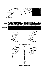
In its simplest form, the invention may be illustrated by the following
summary. The invention requires reverse transcription (RT) primers, which
comprise also unique positional tags (domains), to be arrayed on an object
substrate, e.g. a glass slide, to generate an "array". The unique positional
tags
correspond to the location of the RT primers on the array (the features of the
array).
Thin tissue sections are placed onto the array and a reverse transcription
reaction
is performed in the tissue section on the object slide. The RT primers, to
which the
RNA in the tissue sample binds (or hybridizes), are extended using the bound
RNA
CA 02832678 2013-10-08
WO 2012/140224 PCT/EP2012/056823
- 6 -
as a template to obtain cDNA, which is therefore bound to the surface of the
array.
As consequence of the unique positional tags in the RT primers, each cDNA
strand
carries information about the position of the template RNA in the tissue
section. The
tissue section may be visualised or imaged, e.g. stained and photographed,
before
or after the cDNA synthesis step to enable the positional tag in the cDNA
molecule
to be correlated with a position within the tissue sample. The cDNA is
sequenced,
which results in a transcriptome with exact positional information. A
schematic of
the process is shown in Figure 1. The sequence data can then be matched to a
position in the tissue sample, which enables the visualization, e.g. using a
computer, of the sequence data together with the tissue section, for instance
to
display the expression pattern of any gene of interest across the tissue
(Figure 2).
Similarly, it would be possible to mark different areas of the tissue section
on the
computer screen and obtain information on differentially expressed genes
between
any selected areas of interest. It will be evident that the methods of the
invention
result in data that is in stark contrast to the data obtained using current
methods to
study mRNA populations. For example, methods based on in situ hybridization
provide only relative information of single mRNA transcripts. Thus, the
methods of
the present invention have clear advantages over current in situ technologies.
The
global gene expression information obtainable from the methods of the
invention
also allows co-expression information and quantitative estimates of transcript
abundance. It will be evident that this is a generally applicable strategy
available for
the analysis of any tissue in any species, e.g. animal, plant, fungus.
As noted above, and described in more detail below, it will be evident that
this basic methodology could readily be extended to the analysis of genomic
DNA,
e.g. to identify cells within a tissue sample that comprise one or more
specific
mutations. For instance, the genomic DNA may be fragmented and allowed to
hybridise to primers (equivalent to the RT primers described above), which are
capable of capturing the fragmented DNA (e.g. an adapter with a sequence that
is
complementary to the primer may be ligated to the fragmented DNA or the
fragmented DNA may be extended e.g. using an enzyme to incorporate additional
nucleotides at the end of the sequence, e.g. a poly-A tail, to generate a
sequence
that is complementary to the primer) and priming the synthesis of
complementary
strands to the capture molecules. The remaining steps of the analysis may be
as
described above. Hence, the specific embodiments of the invention described
CA 02832678 2013-10-08
WO 2012/140224 PCT/EP2012/056823
- 7 -
below in the context of transcriptome analysis may also be employed in methods
of
analysing genomic DNA, where appropriate.
It will be seen from the above explanation that there is an immense value in
coupling positional information to transcriptome or genome information. For
instance, it enables global gene expression mapping at high resolution, which
will
find utility in numerous applications, including e.g. cancer research and
diagnostics.
Furthermore, it is evident that the methods described herein differ
significantly from the previously described methods for analysis of the global
transcriptome of a tissue sample and these differences result in numerous
advantages. The present invention is predicated on the surprising discovery
that the
use of tissue sections does not interfere with synthesis of DNA (e.g. cDNA)
primed
by primers (e.g. reverse transcription primers) that are coupled to the
surface of an
array.
Thus, in its first and broadest aspect, the present invention provides a
method for localised detection of nucleic acid in a tissue sample comprising:
(a) providing an array comprising a substrate on which multiple species of
capture probes are directly or indirectly immobilized such that each species
occupies a distinct position on the array and is oriented to have a free 3 end
to
enable said probe to function as a primer for a primer extension or ligation
reaction,
wherein each species of said capture probe comprises a nucleic acid molecule
with
5' to 3':
(i) a positional domain that corresponds to the position of the capture probe
on the array, and
(ii) a capture domain;
(b) contacting said array with a tissue sample such that the position of a
capture probe on the array may be correlated with a position in the tissue
sample
and allowing nucleic acid of the tissue sample to hybridise to the capture
domain in
said capture probes;
(c) generating DNA molecules from the captured nucleic acid molecules
using said capture probes as extension or ligation primers, wherein said
extended
or ligated DNA molecules are tagged by virtue of the positional domain;
(d) optionally generating a complementary strand of said tagged DNA and/or
optionally amplifying said tagged DNA;
CA 02832678 2013-10-08
WO 2012/140224
PCT/EP2012/056823
- 8 -
(e) releasing at least part of the tagged DNA molecules and/or their
complements or amplicons from the surface of the array, wherein said part
includes
the positional domain or a complement thereof;
(f) directly or indirectly analysing the sequence of the released DNA
molecules.
The methods of the invention represent a significant advance over other
methods for spatial transcriptomics known in the art. For example the methods
described herein result in a global and spatial profile of all transcripts in
the tissue
sample. Moreover, the expression of every gene can be quantified for each
position
or feature on the array, which enables a multiplicity of analyses to be
performed
based on data from a single assay. Thus, the methods of the present invention
make it possible to detect and/or quantify the spatial expression of all genes
in
single tissue sample. Moreover, as the abundance of the transcripts is not
visualised directly, e.g. by fluorescence, akin to a standard microarray, it
is possible
to measure the expression of genes in a single sample simultaneously even
wherein said transcripts are present at vastly different concentrations in the
same
sample.
Accordingly, in a second and more particular aspect, the present invention
can be seen to provide a method for determining and/or analysing a
transcriptome
of a tissue sample comprising:
(a) providing an array comprising a substrate on which multiple species of
capture probes are directly or indirectly immobilized such that each species
occupies a distinct position on the array and is oriented to have a free 3'
end to
enable said probe to function as a reverse transcriptase (RT) primer, wherein
each
species of said capture probe comprises a nucleic acid molecule with 5' to 3':
(i) a positional domain that corresponds to the position of the capture probe
on the array, and
(ii) a capture domain;
(b) contacting said array with a tissue sample such that the position of a
capture probe on the array may be correlated with a position in the tissue
sample
and allowing RNA of the tissue sample to hybridise to the capture domain in
said
capture probes;
(c) generating cDNA molecules from the captured RNA molecules using
said capture probes as RT primers, and optionally amplifying said cDNA
molecules;
CA 02832678 2013-10-08
WO 2012/140224
PCT/EP2012/056823
- 9 -
(d) releasing at least part of the cDNA molecules and/or optionally their
amplicons from the surface of the array, wherein said released molecule may be
a
first strand and/or second strand cDNA molecule or an amplicon thereof and
wherein said part includes the positional domain or a complement thereof;
(e) directly or indirectly analysing the sequence of the released molecules.
As described in more detail below, any method of nucleic acid analysis may
be used in the analysis step. Typically this may involve sequencing, but it is
not
necessary to perform an actual sequence determination. For example sequence-
specific methods of analysis may be used. For example a sequence-specific
amplification reaction may be performed, for example using primers which are
specific for the positional domain and/or for a specific target sequence, e.g.
a
particular target DNA to be detected (i.e. corresponding to a particular
cDNA/RNA
or gene etc.). An exemplary analysis method is a sequence-specific PCR
reaction.
The sequence analysis information obtained in step (e) may be used to
obtain spatial information as to the RNA in the sample. In other words the
sequence
analysis information may provide information as to the location of the RNA in
the
sample. This spatial information may be derived from the nature of the
sequence
analysis information determined, for example it may reveal the presence of a
particular RNA which may itself be spatially informative in the context of the
tissue
sample used, and/or the spatial information (e.g. spatial localisation) may be
derived from the position of the tissue sample on the array, coupled with the
sequencing information. Thus, the method may involve simply correlating the
sequence analysis information to a position in the tissue sample e.g. by
virtue of the
positional tag and its correlation to a position in the tissue sample.
However, as
described above, spatial information may conveniently be obtained by
correlating
the sequence analysis data to an image of the tissue sample and this
represents
one preferred embodiment of the invention. Accordingly, in a preferred
embodiment
the method also includes a step of:
(f) correlating said sequence analysis information with an image of said
tissue sample, wherein the tissue sample is imaged before or after step (c).
In its broadest sense, the method of the invention may be used for localised
detection of a nucleic acid in a tissue sample. Thus, in one embodiment, the
method of the invention may be used for determining and/or analysing all of
the
transcriptome or genome of a tissue sample e.g. the global transcriptome of a
tissue sample. However, the method is not limited to this and encompasses
CA 02832678 2013-10-08
WO 2012/140224 PCT/EP2012/056823
- 10 -
determining and/or analysing all or part of the transcriptome or genome. Thus,
the
method may involve determining and/or analysing a part or subset of the
transcriptome or genome, e.g. a transcriptome corresponding to a subset of
genes,
e.g. a set of particular genes, for example related to a particular disease or
condition, tissue type etc.
Viewed from another aspect, the method steps set out above can be seen
as providing a method of obtaining a spatially defined transcriptome or
genome,
and in particular the spatially defined global transcriptome or genome, of a
tissue
sample.
Alternatively viewed, the method of the invention may be seen as a method
for localised or spatial detection of nucleic acid, whether DNA or RNA in a
tissue
sample, or for localised or spatial determination and/or analysis of nucleic
acid
(DNA or RNA) in a tissue sample. In particular, the method may be used for the
localised or spatial detection or determination and/or analysis of gene
expression or
genomic variation in a tissue sample. The localised/spatial
detection/determination/analysis means that the RNA or DNA may be localised to
its native position or location within a cell or tissue in the tissue sample.
Thus for
example, the RNA or DNA may be localised to a cell or group of cells, or type
of
cells in the sample, or to particular regions of areas within a tissue sample.
The
native location or position of the RNA or DNA (or in other words, the location
or
position of the RNA or DNA in the tissue sample), e.g. an expressed gene or
genomic locus, may be determined.
The invention can also be seen to provide an array for use in the methods of
the invention comprising a substrate on which multiple species of capture
probes
are directly or indirectly immobilized such that each species occupies a
distinct
position on the array and is oriented to have a free 3' end to enable said
probe to
function as a reverse transcriptase (RT) primer, wherein each species of said
capture probe comprises a nucleic acid molecule with 5' to 3':
(i) a positional domain that corresponds to the position of the capture probe
on the array, and
(ii) a capture domain to capture RNA of a tissue sample that is contacted
with said array.
In a related aspect, the present invention also provides use of an array,
comprising a substrate on which multiple species of capture probe are directly
or
indirectly immobilized such that each species occupies a distinct position on
the
CA 02832678 2013-10-08
WO 2012/140224 PCT/EP2012/056823
- 11 -
array and is oriented to have a free 3' end to enable said probe to function
as a
reverse transcriptase (RT) primer, wherein each species of said capture probe
comprises a nucleic acid molecule with 5' to 3':
(i) a positional domain that corresponds to the position of the capture probe
on the array; and
(ii) a capture domain;
to capture RNA of a tissue sample that is contacted with said array.
Preferably, said use is for determining and/or analysing a transcriptome and
in particular the global transcriptome, of a tissue sample and further
comprises
steps of:
(a) generating cDNA molecules from the captured RNA molecules using
said capture probes as RT primers and optionally amplifying said cDNA
molecules;
(b) releasing at least part of the cDNA molecules and/or optionally their
amplicons from the surface of the array, wherein said released molecule may be
a
first strand and/or second strand cDNA molecule or an amplicon thereof and
wherein said part includes the positional domain or a complement thereof;
(c) directly or indirectly analysing the sequence of the released molecules;
and optionally
(d) correlating said sequence analysis information with an image of said
tissue sample, wherein the tissue sample is imaged before or after step (a).
It will be seen therefore that the array of the present invention may be used
to capture RNA, e.g. mRNA of a tissue sample that is contacted with said
array.
The array may also be used for determining and/or analysing a partial or
global
transcriptome of a tissue sample or for obtaining a spatially defined partial
or global
transcriptome of a tissue sample. The methods of the invention may thus be
considered as methods of quantifying the spatial expression of one or more
genes
in a tissue sample. Expressed another way, the methods of the present
invention
may be used to detect the spatial expression of one or more genes in a tissue
sample. In yet another way, the methods of the present invention may be used
to
determine simultaneously the expression of one or more genes at one or more
positions within a tissue sample. Still further, the methods may be seen as
methods
for partial or global transcriptome analysis of a tissue sample with two-
dimensional
spatial resolution.
The RNA may be any RNA molecule which may occur in a cell. Thus it may
be mRNA, tRNA, rRNA, viral RNA, small nuclear RNA (snRNA), small nucleolar
CA 02832678 2013-10-08
WO 2012/140224
PCT/EP2012/056823
- 12 -
RNA (snoRNA), microRNA (miRNA), small interfering RNA (siRNA), piwi-
interacting
RNA (piRNA), ribozymal RNA, antisense RNA or non-coding RNA. Preferably
however it is mRNA.
Step (c) in the method above (corresponding to step (a) in the preferred
statement of use set out above) of generating cDNA from the captured RNA will
be
seen as relating to the synthesis of the cDNA. This will involve a step of
reverse
transcription of the captured RNA, extending the capture probe, which
functions as
the RT primer, using the captured RNA as template. Such a step generates so-
called first strand cDNA. As will be described in more detail below, second
strand
cDNA synthesis may optionally take place on the array, or it may take place in
a
separate step, after release of first strand cDNA from the array. As also
described
in more detail below, in certain embodiments second strand synthesis may occur
in
the first step of amplification of a released first strand cDNA molecule.
Arrays for use in the context of nucleic acid analysis in general, and DNA
analysis in particular, are discussed and described below. Specific details
and
embodiments described herein in relation to arrays and capture probes for use
in
the context of RNA, apply equally (where appropriate) to all such arrays,
including
those for use with DNA.
As used herein the term "multiple" means two or more, or at least two, e.g.
3, 5, 10, 15, 20, 30, 40, 50, 60, 70, 80, 90, 100, 150, 200, 400, 500, 1000,
2000,
5000, 10,000, or more etc. Thus for example, the number of capture probes may
be
any integer in any range between any two of the aforementioned numbers. It
will be
appreciated however that it is envisaged that conventional-type arrays with
many
hundreds, thousands, tens of thousands, hundreds of thousands or even millions
of
capture probes may be used.
Thus, the methods outlined herein utilise high density nucleic acid arrays
comprising "capture probes" for capturing and labelling transcripts from all
of the
single cells within a tissue sample e.g. a thin tissue sample slice, or
"section". The
tissue samples or sections for analysis are produced in a highly parallelized
fashion, such that the spatial information in the section is retained. The
captured
RNA (preferably mRNA) molecules for each cell, or "transcriptomes", are
transcribed into cDNA and the resultant cDNA molecules are analyzed, for
example
by high throughput sequencing. The resultant data may be correlated to images
of
the original tissue samples e.g. sections through so-called barcode sequences
(or
CA 02832678 2013-10-08
WO 2012/140224
PCT/EP2012/056823
- 13 -
ID tags, defined herein as positional domains) incorporated into the arrayed
nucleic
acid probes.
High density nucleic acid arrays or microarrays are a core component of the
spatial transcriptome labelling method described herein. A microarray is a
multiplex
technology used in molecular biology. A typical microarray consists of an
arrayed
series of microscopic spots of oligonucleotides (hundreds of thousands of
spots,
generally tens of thousands, can be incorporated on a single array). The
distinct
position of each nucleic acid (oligonucleotide) spot (each species of
oligonucleotide/nucleic acid molecule) is known as a "feature" (and hence in
the
methods set out above each species of capture probe may be viewed as a
specific
feature of the array; each feature occupies a distinct position on the array),
and
typically each separate feature contains in the region of picomoles (10-12
moles) of
a specific DNA sequence (a "species"), which are known as "probes" (or
"reporters"). Typically, these can be a short section of a gene or other
nucleic acid
element to which a cDNA or cRNA sample (or "target") can hybridize under high-
stringency hybridization conditions. However, as described below, the probes
of the
present invention differ from the probes of standard microarrays.
In gene expression microarrays, probe-target hybridization is usually
detected and quantified by detection of visual signal, e.g. a fluorophore,
silver ion,
or chemiluminescence-label, which has been incorporated into all of the
targets.
The intensity of the visual signal correlates to the relative abundance of
each target
nucleic acid in the sample. Since an array can contain tens of thousands of
probes,
a microarray experiment can accomplish many genetic tests in parallel.
In standard microarrays, the probes are attached to a solid surface or
substrate by a covalent bond to a chemical matrix, e.g. epoxy-silane, amino-
silane,
lysine, polyacrylamide etc. The substrate typically is a glass, plastic or
silicon chip
or slide, although other microarray platforms are known, e.g. microscopic
beads.
The probes may be attached to the array of the invention by any suitable
means. In a preferred embodiment the probes are immobilized to the substrate
of
the array by chemical immobilization. This may be an interaction between the
substrate (support material) and the probe based on a chemical reaction. Such
a
chemical reaction typically does not rely on the input of energy via heat or
light, but
can be enhanced by either applying heat, e.g. a certain optimal temperature
for a
chemical reaction, or light of certain wavelength. For example, a chemical
immobilization may take place between functional groups on the substrate and
CA 02832678 2013-10-08
WO 2012/140224
PCT/EP2012/056823
- 14 -
corresponding functional elements on the probes. Such corresponding functional
elements in the probes may either be an inherent chemical group of the probe,
e.g.
a hydroxyl group or be additionally introduced. An example of such a
functional
group is an amine group. Typically, the probe to be immobilized comprises a
functional amine group or is chemically modified in order to comprise a
functional
amine group. Means and methods for such a chemical modification are well
known.
The localization of said functional group within the probe to be immobilized
may be used in order to control and shape the binding behaviour and/or
orientation
of the probe, e.g. the functional group may be placed at the 5' or 3' end of
the probe
or within sequence of the probe. A typical substrate for a probe to be
immobilized
comprises moieties which are capable of binding to such probes, e.g. to amine-
functionalized nucleic acids. Examples of such substrates are carboxy,
aldehyde or
epoxy substrates. Such materials are known to the person skilled in the art.
Functional groups, which impart a connecting reaction between probes which are
chemically reactive by the introduction of an amine group, and array
substrates are
known to the person skilled in the art.
Alternative substrates on which probes may be immobilized may have to be
chemically activated, e.g. by the activation of functional groups, available
on the
array substrate. The term "activated substrate" relates to a material in which
interacting or reactive chemical functional groups were established or enabled
by
chemical modification procedures as known to the person skilled in the art.
For
example, a substrate comprising carboxyl groups has to be activated before
use.
Furthermore, there are substrates available that contain functional groups
that can
react with specific moieties already present in the nucleic acid probes.
Alternatively, the probes may be synthesized directly on the substrate.
Suitable methods for such an approach are known to the person skilled in the
art.
Examples are manufacture techniques developed by Agilent Inc., Affymetrix
Inc.,
Roche Nimblegen Inc. or Flexgen By. Typically, lasers and a set of mirrors
that
specifically activate the spots where nucleotide additions are to take place
are
used. Such an approach may provide, for example, spot sizes (i.e. features) of
around 30 pm or larger.
The substrate therefore may be any suitable substrate known to the person
skilled in the art. The substrate may have any suitable form or format, e.g.
it may be
flat, curved, e.g. convexly or concavely curved towards the area where the
CA 02832678 2013-10-08
WO 2012/140224 PCT/EP2012/056823
- 15 -
interaction between the tissue sample and the substrate takes place.
Particularly
preferred is the where the substrate is a flat, i.e. planar, chip or slide.
Typically, the substrate is a solid support and thereby allows for an accurate
and traceable positioning of the probes on the substrate. An example of a
substrate
is a solid material or a substrate comprising functional chemical groups, e.g.
amine
groups or amine-functionalized groups. A substrate envisaged by the present
invention is a non-porous substrate. Preferred non-porous substrates are
glass,
silicon, poly-L-lysine coated material, nitrocellulose, polystyrene, cyclic
olefin
copolymers (COCs), cyclic olefin polymers (COPs), polypropylene, polyethylene
and polycarbonate.
Any suitable material known to the person skilled in the art may be used.
Typically, glass or polystyrene is used. Polystyrene is a hydrophobic material
suitable for binding negatively charged macromolecules because it normally
contains few hydrophilic groups. For nucleic acids immobilized on glass
slides, it is
furthermore known that by increasing the hydrophobicity of the glass surface
the
nucleic acid immobilization may be increased. Such an enhancement may permit a
relatively more densely packed formation. In addition to a coating or surface
treatment with poly-L-lysine, the substrate, in particular glass, may be
treated by
silanation, e.g. with epoxy-silane or amino-silane or by silynation or by a
treatment
with polyacrylamide.
A number of standard arrays are commercially available and both the
number and size of the features may be varied. In the present invention, the
arrangement of the features may be altered to correspond to the size and/or
density
of the cells present in different tissues or organisms. For instance, animal
cells
typically have a cross-section in the region of 1-100pm, whereas the cross-
section
of plant cells typically may range from 1-10000pm. Hence, NimblegenO arrays,
which are available with up to 2.1 million features, or 4.2 million features,
and
feature sizes of 13 micrometers, may be preferred for tissue samples from an
animal or fungus, whereas other formats, e.g. with 8x130k features, may be
sufficient for plant tissue samples. Commercial arrays are also available or
known
for use in the context of sequence analysis and in particular in the context
of NGS
technologies. Such arrays may also be used as the array surface in the context
of
the present invention e.g. an IIlumina bead array. In addition to commercially
available arrays, which can themselves be customized, it is possible to make
custom or non-standard "in-house" arrays and methods for generating arrays are
CA 02832678 2013-10-08
WO 2012/140224 PCT/EP2012/056823
- 16 -
well-established. The methods of the invention may utilise both standard and
non-
standard arrays that comprise probes as defined below.
The probes on a microarray may be immobilized, i.e. attached or bound, to
the array preferably via the 5' or 3' end, depending on the chemical matrix of
the
array. Typically, for commercially available arrays, the probes are attached
via a 3'
linkage, thereby leaving a free 5' end. However, arrays comprising probes
attached
to the substrate via a 5' linkage, thereby leaving a free 3' end, are
available and
may be synthesized using standard techniques that are well known in the art
and
are described elsewhere herein.
The covalent linkage used to couple a nucleic acid probe to an array
substrate may be viewed as both a direct and indirect linkage, in that the
although
the probe is attached by a "direct" covalent bond, there may be a chemical
moiety
or linker separating the "first" nucleotide of the nucleic acid probe from
the, e.g.
glass or silicon, substrate i.e. an indirect linkage. For the purposes of the
present
invention probes that are immobilized to the substrate by a covalent bond
and/or
chemical linker are generally seen to be immobilized or attached directly to
the
substrate.
As will be described in more detail below, the capture probes of the
invention may be immobilized on, or interact with, the array directly or
indirectly.
Thus the capture probes need not bind directly to the array, but may interact
indirectly, for example by binding to a molecule which itself binds directly
or
indirectly to the array (e.g. the capture probe may interact with (e.g. bind
or
hybridize to) a binding partner for the capture probe, i.e. a surface probe,
which is
itself bound to the array directly or indirectly). Generally speaking,
however, the
capture probe will be, directly or indirectly (by one or more intermediaries),
bound
to, or immobilized on, the array.
The use, method and array of the invention may comprise probes that are
immobilized via their 5' or 3' end. However, when the capture probe is
immobilized
directly to the array substrate, it may be immobilized only such that the 3'
end of the
capture probe is free to be extended, e.g. it is immobilized by its 5' end.
The
capture probe may be immobilized indirectly, such that it has a free, i.e.
extendible,
3' end.
By extended or extendible 3' end, it is meant that further nucleotides may be
added to the most 3' nucleotide of the nucleic acid molecule, e.g. capture
probe, to
extend the length of the nucleic acid molecule, i.e. the standard
polymerization
CA 02832678 2013-10-08
WO 2012/140224 PCT/EP2012/056823
- 17 -
reaction utilized to extend nucleic acid molecules, e.g. templated
polymerization
catalyzed by a polymerase.
Thus, in one embodiment, the array comprises probes that are immobilized
directly via their 3' end, so-called surface probes, which are defined below.
Each
species of surface probe comprises a region of complementarity to each species
of
capture probe, such that the capture probe may hybridize to the surface probe,
resulting in the capture probe comprising a free extendible 3' end. In a
preferred
aspect of the invention, when the array comprises surface probes, the capture
probes are synthesized in situ on the array.
The array probes may be made up of ribonucleotides and/or
deoxyribonucleotides as well as synthetic nucleotide residues that are capable
of
participating in Watson-Crick type or analogous base pair interactions. Thus,
the
nucleic acid domain may be DNA or RNA or any modification thereof e.g. PNA or
other derivatives containing non-nucleotide backbones. However, in the context
of
transcriptome analysis the capture domain of the capture probe must capable of
priming a reverse transcription reaction to generate cDNA that is
complementary to
the captured RNA molecules. As described below in more detail, in the context
of
genome analysis, the capture domain of the capture probe must be capable of
binding to the DNA fragments, which may comprise binding to a binding domain
that has been added to the fragmented DNA. In some embodiments, the capture
domain of the capture probe may prime a DNA extension (polymerase) reaction to
generate DNA that is complementary to the captured DNA molecules. In other
embodiments, the capture domain may template a ligation reaction between the
captured DNA molecules and a surface probe that is directly or indirectly
immobilised on the substrate. In yet other embodiments, the capture domain may
be ligated to one strand of the captured DNA molecules.
In a preferred embodiment of the invention at least the capture domain of
the capture probe comprises or consists of deoxyribonucleotides (dNTPs). In a
particularly preferred embodiment the whole of the capture probe comprises or
consists of deoxyribonucleotides.
In a preferred embodiment of the invention the capture probes are
immobilized on the substrate of the array directly, i.e. by their 5' end,
resulting in a
free extendible 3' end.
The capture probes of the invention comprise at least two domains, a
capture domain and a positional domain (or a feature identification tag or
domain;
CA 02832678 2013-10-08
WO 2012/140224 PCT/EP2012/056823
- 18 -
the positional domain may alternatively be defined as an identification (ID)
domain
or tag, or as a positional tag). The capture probe may further comprise a
universal
domain as defined further below. Where the capture probe is indirectly
attached to
the array surface via hybridization to a surface probe, the capture probe
requires a
sequence (e.g. a portion or domain) which is complementary to the surface
probe.
Such a complementary sequence may be complementary to a
positional/identification domain and/or a universal domain on the surface
probe. In
other words the positional domain and/or universal domain may constitute the
region or portion of the probe which is complementary to the surface probe.
However, the capture probe may also comprise an additional domain (or region,
portion or sequence) which is complementary to the surface probe. For ease of
synthesis, as described in more detail below, such a surface probe-
complementary
region may be provided as part, or as an extension of the capture domain (such
a
part or extension not itself being used for, or capable of, binding to the
target
nucleic acid, e.g. RNA).
The capture domain is typically located at the 3' end of the capture probe
and comprises a free 3' end that can be extended, e.g. by template dependent
polymerization. The capture domain comprises a nucleotide sequence that is
capable of hybridizing to nucleic acid, e.g. RNA (preferably mRNA) present in
the
cells of the tissue sample contact with the array.
Advantageously, the capture domain may be selected or designed to bind
(or put more generally may be capable of binding) selectively or specifically
to the
particular nucleic acid, e.g. RNA it is desired to detect or analyse. For
example the
capture domain may be selected or designed for the selective capture of mRNA.
As
is well known in the art, this may be on the basis of hybridisation to the
poly-A tail of
mRNA. Thus, in a preferred embodiment the capture domain comprises a poly-T
DNA oligonucleotide, i.e. a series of consecutive deoxythymidine residues
linked by
phosphodiester bonds, which is capable of hybridizing to the poly-A tail of
mRNA.
Alternatively, the capture domain may comprise nucleotides which are
functionally
or structurally analogous to poly-T i.e., are capable of binding selectively
to poly-A,
for example a poly-U oligonucleotide or an oligonucleotide comprised of
deoxythymidine analogues, wherein said oligonucleotide retains the functional
property of binding to poly-A. In a particularly preferred embodiment the
capture
domain, or more particularly the poly-T element of the capture domain,
comprises
at least 10 nucleotides, preferably at least 11, 12, 13, 14, 15, 16, 17, 18,
19 or 20
CA 02832678 2013-10-08
WO 2012/140224 PCT/EP2012/056823
- 19 -
nucleotides. In a further embodiment, the capture domain, or more particularly
the
poly-T element of the capture domain comprises at least 25, 30 or 35
nucleotides.
Random sequences may also be used in the capture of nucleic acid, as is
known in the art, e.g. random hexamers or similar sequences, and hence such
random sequences may be used to form all or a part of the capture domain. For
example, random sequences may be used in conjunction with poly-T (or poly-T
analogue etc.) sequences. Thus where a capture domain comprises a poly-T(or a
"poly-T-like") oligonucleotide, it may also comprise a random oligonucleotide
sequence. This may for example be located 5' or 3' of the poly-T sequence,
e.g. at
the 3' end of the capture probe, but the positioning of such a random sequence
is
not critical. Such a construct may facilitate the capturing of the initial
part of the
poly-A of mRNA. Alternatively, the capture domain may be an entirely random
sequence. Degenerate capture domains may also be used, according to principles
known in the art.
The capture domain may be capable of binding selectively to a desired sub-
type or subset of nucleic acid, e.g. RNA, for example a particular type of RNA
such
mRNA or rRNA etc. as listed above, or to a particular subset of a given type
of
RNA, for example, a particular mRNA species e.g. corresponding to a particular
gene or group of genes. Such a capture probe may be selected or designed based
on sequence of the RNA it is desired to capture. Thus it may be a sequence-
specific capture probe, specific for a particular RNA target or group of
targets
(target group etc). Thus, it may be based on a particular gene sequence or
particular motif sequence or common/conserved sequence etc., according to
principles well known in the art.
In embodiments where the capture probe is immobilized on the substrate of
the array indirectly, e.g. via hybridization to a surface probe, the capture
domain
may further comprise an upstream sequence (5' to the sequence that hybridizes
to
the nucleic acid, e.g. RNA of the tissue sample) that is capable of
hybridizing to 5'
end of the surface probe. Alone, the capture domain of the capture probe may
be
seen as a capture domain oligonucleotide, which may be used in the synthesis
of
the capture probe in embodiments where the capture probe is immobilized on the
array indirectly.
The positional domain (feature identification domain or tag) of the capture
probe is located directly or indirectly upstream, i.e. closer to the 5' end of
the
capture probe nucleic acid molecule, of the capture domain. Preferably the
CA 02832678 2013-10-08
WO 2012/140224 PCT/EP2012/056823
- 20 -
positional domain is directly adjacent to the capture domain, i.e. there is no
intermediate sequence between the capture domain and the positional domain. In
some embodiments the positional domain forms the 5' end of the capture probe,
which may be immobilized directly or indirectly on the substrate of the array.
As discussed above, each feature (distinct position) of the array comprises a
spot of a species of nucleic acid probe, wherein the positional domain at each
feature is unique. Thus, a "species" of capture probe is defined with
reference to its
positional domain; a single species of capture probe will have the same
positional
domain. However, it is not required that each member of a species of capture
probe
has the same sequence in its entirety. In particular, since the capture domain
may
be or may comprise a random or degenerate sequence, the capture domains of
individual probes within a species may vary. Accordingly, in some embodiments
where the capture domains of the capture probes are the same, each feature
comprises a single probe sequence. However in other embodiments where the
capture probe varies, members of a species of probe will not have the exact
same
sequence, although the sequence of the positional domain of each member in the
species will be the same. What is required is that each feature or position of
the
array carries a capture probe of a single species (specifically each feature
or
position carries a capture probe which has an identical positional tag, i.e.
there is a
single positional domain at each feature or position). Each species has a
different
positional domain which identifies the species. However, each member of a
species, may in some cases, as described in more detail herein, have a
different
capture domain, as the capture domain may be random or degenerate or may have
a random or degenerate component. This means that within a given feature, or
position, the capture domain of the probes may differ.
Thus in some, but not necessarily in all embodiments, the nucleotide
sequence of any one probe molecule immobilized at a particular feature is the
same
as the other probe molecules immobilized at the same feature, but the
nucleotide
sequence of the probes at each feature is different, distinct or
distinguishable from
the probes immobilized at every other feature. Preferably each feature
comprises a
different species of probe. However, in some embodiments it may be
advantageous
for a group of features to comprise the same species of probe, i.e.
effectively to
produce a feature covering an area of the array that is greater than a single
feature,
e.g. to lower the resolution of the array. In other embodiments of the array,
the
nucleotide sequence of the positional domain of any one probe molecule
CA 02832678 2013-10-08
WO 2012/140224 PCT/EP2012/056823
- 21 -
immobilized at a particular feature may be the same as the other probe
molecules
immobilized at the same feature but the capture domain may vary. The capture
domain may nonetheless be designed to capture the same type of molecule, e.g.
mRNA in general.
The positional domain (or tag) of the capture probe comprises the sequence
which is unique to each feature and acts as a positional or spatial marker
(the
identification tag). In this way each region or domain of the tissue sample,
e.g. each
cell in the tissue, will be identifiable by spatial resolution across the
array linking the
nucleic acid, e.g. RNA (e.g. the transcripts) from a certain cell to a unique
positional
domain sequence in the capture probe. By virtue of the positional domain a
capture
probe in the array may be correlated to a position in the tissue sample, for
example
it may be correlated to a cell in the sample. Thus, the positional domain of
the
capture domain may be seen as a nucleic acid tag (identification tag).
Any suitable sequence may be used as the positional domain in the capture
probes of the invention. By a suitable sequence, it is meant that the
positional
domain should not interfere with (i.e. inhibit or distort) the interaction
between the
RNA of the tissue sample and the capture domain of the capture probe. For
example, the positional domain should be designed such that nucleic acid
molecules in the tissue sample do not hybridize specifically to the positional
domain. Preferably, the nucleic acid sequence of the positional domain of the
capture probes has less than 80% sequence identity to the nucleic acid
sequences
in the tissue sample. Preferably, the positional domain of the capture probe
has
less than 70%, 60%, 50% or less than 40% sequence identity across a
substantial
part of the nucleic acids molecules in the tissue sample. Sequence identity
may be
determined by any appropriate method known in the art, e.g. the using BLAST
alignment algorithm.
In a preferred embodiment the positional domain of each species of capture
probe contains a unique barcode sequence. The barcode sequences may be
generated using random sequence generation. The randomly generated sequences
may be followed by stringent filtering by mapping to the genomes of all common
reference species and with pre-set Tm intervals, GC content and a defined
distance
of difference to the other barcode sequences to ensure that the barcode
sequences
will not interfere with the capture of the nucleic acid, e.g. RNA from the
tissue
sample and will be distinguishable from each other without difficulty.
CA 02832678 2013-10-08
WO 2012/140224
PCT/EP2012/056823
- 22 -
As mentioned above, and in a preferred embodiment, the capture probe
comprises also a universal domain (or linker domain or tag). The universal
domain
of the capture probe is located directly or indirectly upstream, i.e. closer
to the 5'
end of the capture probe nucleic acid molecule, of the positional domain.
Preferably
the universal domain is directly adjacent to the positional domain, i.e. there
is no
intermediate sequence between the positional domain and the universal domain.
In
embodiments where the capture probe comprises a universal domain, the domain
will form the 5 end of the capture probe, which may be immobilized directly or
indirectly on the substrate of the array.
The universal domain may be utilized in a number of ways in the methods
and uses of the invention. For example, the methods of the invention comprise
a
step of releasing (e.g. removing) at least part of the synthesised (i.e.
extended or
ligated) nucleic acid, e.g. cDNA molecules from the surface of the array. As
described elsewhere herein, this may be achieved in a number of ways, of which
one comprises cleaving the nucleic acid, e.g. cDNA molecule from the surface
of
the array. Thus, the universal domain may itself comprise a cleavage domain,
i.e. a
sequence that can be cleaved specifically, either chemically or preferably
enzymatically.
Thus, the cleavage domain may comprise a sequence that is recognised by
one or more enzymes capable of cleaving a nucleic acid molecule, i.e. capable
of
breaking the phosphodiester linkage between two or more nucleotides. For
instance, the cleavage domain may comprise a restriction endonuclease
(restriction
enzyme) recognition sequence. Restriction enzymes cut double-stranded or
single
stranded DNA at specific recognition nucleotide sequences known as restriction
sites and suitable enzymes are well known in the art. For example, it is
particularly
advantageous to use rare-cutting restriction enzymes, i.e. enzymes with a long
recognition site (at least 8 base pairs in length), to reduce the possibility
of cleaving
elsewhere in the nucleic acid, e.g. cDNA molecule. In this respect, it will be
seen
that removing or releasing at least part of the nucleic acid, e.g. cDNA
molecule
requires releasing a part comprising the positional domain of the nucleic
acid, e.g.
cDNA and all of the sequence downstream of the domain, i.e. all of the
sequence
that is 3' to the positional domain. Hence, cleavage of the nucleic acid, e.g.
cDNA
molecule should take place 5 to the positional domain.
CA 02832678 2013-10-08
WO 2012/140224 PCT/EP2012/056823
- 23 -
By way of example, the cleavage domain may comprise a poly-U sequence
which may be cleaved by a mixture of Uracil DNA glycosylase (UDG) and the DNA
glycosylase-Iyase Endonuclease VIII, commercially known as the USERTM enzyme.
A further example of a cleavage domain can be utilised in embodiments
where the capture probe is immobilized to the array substrate indirectly, i.e.
via a
surface probe. The cleavage domain may comprise one or more mismatch
nucleotides, i.e. when the complementary parts of the surface probe and the
capture probe are not 100% complementary. Such a mismatch is recognised, e.g.
by the MutY and T7 endonuclease I enzymes, which results in cleavage of the
nucleic acid molecule at the position of the mismatch.
In some embodiments of the invention, the positional domain of the capture
probe comprises a cleavage domain, wherein the said cleavage domain is located
at the 5' end of the positional domain.
The universal domain may comprise also an amplification domain. This may
be in addition to, or instead of, a cleavage domain. In some embodiments of
the
invention, as described elsewhere herein, it may be advantageous to amplify
the
nucleic acid, e.g. cDNA molecules, for example after they have been released
(e.g.
removed or cleaved) from the array substrate. It will be appreciated however,
that
the initial cycle of amplification, or indeed any or all further cycles of
amplification
may also take place in situ on the array. The amplification domain comprises a
distinct sequence to which an amplification primer may hybridize. The
amplification
domain of the universal domain of the capture probe is preferably identical
for each
species of capture probe. Hence a single amplification reaction will be
sufficient to
amplify all of the nucleic acid, e.g. cDNA molecules (which may or may not be
released from the array substrate prior to amplification).
Any suitable sequence may be used as the amplification domain in the
capture probes of the invention. By a suitable sequence, it is meant that the
amplification domain should not interfere with (i.e. inhibit or distort) the
interaction
between the nucleic acid, e.g. RNA of the tissue sample and the capture domain
of
the capture probe. Furthermore, the amplification domain should comprise a
sequence that is not the same or substantially the same as any sequence in the
nucleic acid, e.g. RNA of the tissue sample, such that the primer used in the
amplification reaction can hybridized only to the amplification domain under
the
amplification conditions of the reaction.
CA 02832678 2013-10-08
WO 2012/140224 PCT/EP2012/056823
- 24 -
For example, the amplification domain should be designed such that nucleic
acid molecules in the tissue sample do not hybridize specifically to the
amplification
domain or the complementary sequence of the amplification domain. Preferably,
the
nucleic acid sequence of the amplification domain of the capture probes and
the
complement thereof has less than 80% sequence identity to the nucleic acid
sequences in the tissue sample. Preferably, the positional domain of the
capture
probe has less than 70%, 60%, 50% or less than 40% sequence identity across a
substantial part of the nucleic acid molecules in the tissue sample. Sequence
identity may be determined by any appropriate method known in the art, e.g.
the
using BLAST alignment algorithm.
Thus, alone, the universal domain of the capture probe may be seen as a
universal domain oligonucleotide, which may be used in the synthesis of the
capture probe in embodiments where the capture probe is immobilized on the
array
indirectly.
In one representative embodiment of the invention only the positional
domain of each species of capture probe is unique. Hence, the capture domains
and universal domains (if present) are in one embodiment the same for every
species of capture probe for any particular array to ensure that the capture
of the
nucleic acid, e.g. RNA from the tissue sample is uniform across the array.
However,
as discussed above, in some embodiments the capture domains may differ by
virtue of including random or degenerate sequences.
In embodiments where the capture probe is immobilized on the substrate of
the array indirectly, e.g. via hybridisation to a surface probe, the capture
probe may
be synthesised on the array as described below.
The surface probes are immobilized on the substrate of the array directly by
or at, e.g. their 3' end. Each species of surface probe is unique to each
feature
(distinct position) of the array and is partly complementary to the capture
probe,
defined above.
Hence the surface probe comprises at its 5' end a domain (complementary
capture domain) that is complementary to a part of the capture domain that
does
not bind to the nucleic acid, e.g. RNA of the tissue sample. In other words,
it
comprises a domain that can hybridize to at least part of a capture domain
oligonucleotide. The surface probe further comprises a domain (complementary
positional domain or complementary feature identification domain) that is
complementary to the positional domain of the capture probe. The complementary
CA 02832678 2013-10-08
WO 2012/140224 PCT/EP2012/056823
- 25 -
positional domain is located directly or indirectly downstream (i.e. at the 3'
end) of
the complementary capture domain, i.e. there may be an intermediary or linker
sequence separating the complementary positional domain and the complementary
capture domain. In embodiments where the capture probe is synthesized on the
array surface, the surface probes of the array always comprise a domain
(complementary universal domain) at the 3' end of the surface probe, i.e.
directly or
indirectly downstream of the positional domain, which is complementary to the
universal domain of the capture probe. In other words, it comprises a domain
that
can hybridize to at least part of the universal domain oligonucleotide.
In some embodiments of the invention the sequence of the surface probe
shows 100% complementarity or sequence identity to the positional and
universal
domains and to the part of the capture domain that does not bind to the
nucleic
acid, e.g. RNA of the tissue sample. In other embodiments the sequence of the
surface probe may show less than 100% sequence identity to the domains of the
capture probe, e.g. less than 99%, 98%, 97%, 96%, 95%, 94%, 93%, 92%, 91% or
90%. In a particularly preferred embodiment of the invention, the
complementary
universal domain shares less than 100% sequence identity to the universal
domain
of the capture probe.
In one embodiment of the invention, the capture probe is synthesized or
generated on the substrate of the array. In a representative embodiment (see
figure
3), the array comprises surface probes as defined above. Oligonucleotides that
correspond to the capture domain and universal domain of the capture probe are
contacted with the array and allowed to hybridize to the complementary domains
of
the surface probes. Excess oligonucleotides may be removed by washing the
array
under standard hybridization conditions. The resultant array comprises
partially
single stranded probes, wherein both the 5' and 3' ends of the surface probe
are
double stranded and the complementary positional domain is single stranded.
The
array may be treated with a polymerase enzyme to extend the 3' end of the
universal domain oligonucleotide, in a template dependent manner, so as to
synthesize the positional domain of the capture probe. The 3' end of the
synthesized positional domain is then ligated, e.g. using a ligase enzyme, to
the 5'
end of the capture domain oligonucleotide to generate the capture probe. It
will be
understood in this regard that the 5' end of the capture domain
oligonucleotide is
phosphorylated to enable ligation to take place. As each species of surface
probe
CA 02832678 2013-10-08
WO 2012/140224 PCT/EP2012/056823
- 26 -
comprises a unique complementary positional domain, each species of capture
probe will comprise a unique positional domain.
The term "hybridisation" or "hybridises" as used herein refers to the
formation of a duplex between nucleotide sequences which are sufficiently
complementary to form duplexes via Watson-Crick base pairing. Two nucleotide
sequences are "complementary" to one another when those molecules share base
pair organization homology. "Complementary" nucleotide sequences will combine
with specificity to form a stable duplex under appropriate hybridization
conditions.
For instance, two sequences are complementary when a section of a first
sequence
can bind to a section of a second sequence in an anti-parallel sense wherein
the 3'-
end of each sequence binds to the 5'-end of the other sequence and each A,
T(U),
G and C of one sequence is then aligned with a T(U), A, C and G, respectively,
of
the other sequence. RNA sequences can also include complementary G=U or U=G
base pairs. Thus, two sequences need not have perfect homology to be
"complementary" under the invention. Usually two sequences are sufficiently
complementary when at least about 90% (preferably at least about 95%) of the
nucleotides share base pair organization over a defined length of the
molecule.
The domains of the capture and surface probes thus contain a region of
complementarity. Furthermore the capture domain of the capture probe contains
a
region of complementarity for the nucleic acid, e.g. RNA (preferably mRNA) of
the
tissue sample.
The capture probe may also be synthesised on the array substrate using
polymerase extension (similarly to as described above) and a terminal
transferase
enzyme to add a "tail" which may constitute the capture domain. This is
described
further in Example 7 below. The use of terminal transferases to add nucleotide
sequences to the end of an oligonucleotide is known in the art, e.g. to
introduce a
homopolymeric tail e.g. a poly-T tail. Accordingly, in such a synthesis an
oligonucleotide that corresponds to the universal domain of the capture probe
may
be contacted with the array and allowed to hybridize to the complementary
domain
of the surface probes. Excess oligonucleotides may be removed by washing the
array under standard hybridization conditions. The resultant array comprises
partially single stranded probes, wherein the 5' ends of the surface probes
are
double stranded and the complementary positional domain is single stranded.
The
array may be treated with a polymerase enzyme to extend the 3' end of the
universal domain oligonucleotide, in a template dependent manner, so as to
CA 02832678 2013-10-08
WO 2012/140224 PCT/EP2012/056823
- 27 -
synthesize the positional domain of the capture probe. The capture domain,
e.g.
comprising a poly-T sequence may then be introduced using a terminal
transferase
to add a poly-T tail to generate the capture probe.
The typical array of, and for use in the methods of, the invention may
contain multiple spots, or "features". A feature may be defined as an area or
distinct
position on the array substrate at which a single species of capture probe is
immobilized. Hence each feature will comprise a multiplicity of probe
molecules, of
the same species. It will be understood in this context that whilst it is
encompassed
that each capture probe of the same species may have the same sequence, this
need not necessarily be the case. Each species of capture probe will have the
same positional domain (i.e. each member of a species and hence each probe in
a
feature will be identically "tagged"), but the sequence of each member of the
feature (species) may differ, because the sequence of a capture domain may
differ.
As described above, random or degenerate capture domains may be used. Thus
the capture probes within a feature may comprise different random or
degenerate
sequences. The number and density of the features on the array will determine
the
resolution of the array, i.e. the level of detail at which the transcriptome
or genome
of the tissue sample can be analysed. Hence, a higher density of features will
typically increase the resolution of the array.
As discussed above, the size and number of the features on the array of the
invention will depend on the nature of the tissue sample and required
resolution.
Thus, if it is desirable to determine a transcriptome or genome only for
regions of
cells within a tissue sample (or the sample contains large cells) then the
number
and/or density of features on the array may be reduced (i.e. lower than the
possible
maximum number of features) and/or the size of the features may be increased
(i.e.
the area of each feature may be greater than the smallest possible feature),
e.g. an
array comprising few large features. Alternatively, if it is desirable to
determine a
transcriptome or genome of individual cells within a sample, it may be
necessary to
use the maximum number of features possible, which would necessitate using the
smallest possible feature size, e.g. an array comprising many small features.
Whilst single cell resolution may be a preferred and advantageous feature of
the present invention, it is not essential to achieve this, and resolution at
the cell
group level is also of interest, for example to detect or distinguish a
particular cell
type or tissue region, e.g. normal vs tumour cells.
CA 02832678 2013-10-08
WO 2012/140224 PCT/EP2012/056823
- 28 -
In representative embodiments of the invention, an array may contain at
least 2,5, 10, 50, 100, 500, 750, 1000, 1500, 3000, 5000, 10000, 20000, 40000,
50000, 75000, 100000, 150000, 200000, 300000, 400000, 500000, 750000,
800000, 1000000, 1200000, 1500000, 1750000, 2000000, 2100000. 3000000,
3500000, 4000000 or 4200000 features. Whilst 4200000 represents the maximum
number of features presently available on a commercial array, it is envisaged
that
arrays with features in excess of this may be prepared and such arrays are of
interest in the present invention. As noted above, feature size may be
decreased
and this may allow greater numbers of features to be accommodated within the
same or a similar area. By way of example. these features may be comprised in
an
area of less than about 20cm2, 10=2, 5cm2, M- -2,
1mm2, or 100pm2.
Thus, in some embodiments of the invention the area of each feature may
be from about 1 pm2, 2 pm2, 3 pm2, 4 pm2, 5 pm2, 10 pm2, 12 pm2, 15 pm2, 20
pm2,
50 pm2, 75 pm2, 100 pm2, 150 pm2, 200 pm2, 250 pm2, 300 pm2, 400 pm2, or 500
pm2.
It will be evident that a tissue sample from any organism could be used in
the methods of the invention, e.g. plant, animal or fungal. The array of the
invention
allows the capture of any nucleic acid, e.g. mRNA molecules, which are present
in
cells that are capable of transcription and/or translation. The arrays and
methods of
the invention are particularly suitable for isolating and analysing the
transcriptome
or genome of cells within a sample, wherein spatial resolution of the
transcriptomes
or genomes is desirable, e.g. where the cells are interconnected or in contact
directly with adjacent cells. However, it will be apparent to a person of
skill in the art
that the methods of the invention may also be useful for the analysis of the
transcriptome or genome of different cells or cell types within a sample even
if said
cells do not interact directly, e.g. a blood sample. In other words, the cells
do not
need to present in the context of a tissue and can be applied to the array as
single
cells (e.g. cells isolated from a non-fixed tissue). Such single cells, whilst
not
necessarily fixed to a certain position in a tissue, are nonetheless applied
to a
certain position on the array and can be individually identified. Thus, in the
context
of analysing cells that do not interact directly, or are not present in a
tissue context,
the spatial properties of the described methods may be applied to obtaining or
retrieving unique or independent transcriptome or genome information from
individual cells.
CA 02832678 2013-10-08
WO 2012/140224 PCT/EP2012/056823
- 29 -
The sample may thus be a harvested or biopsied tissue sample, or possibly
a cultured sample. Representative samples include clinical samples e.g. whole
blood or blood-derived products, blood cells, tissues, biopsies, or cultured
tissues or
cells etc. including cell suspensions. Artificial tissues may for example be
prepared
from cell suspension (including for example blood cells). Cells may be
captured in a
matrix (for example a gel matrix e.g. agar, agarose, etc) and may then be
sectioned
in a conventional way. Such procedures are known in the art in the context of
immunohistochemistry (see e.g. Andersson eta! 2006, J. Histochem. Cytochem.
54(12): 1413-23. Epub 2006 Sep 6).
The mode of tissue preparation and how the resulting sample is handled
may effect the transcriptomic or genomic analysis of the methods of the
invention.
Moreover, various tissue samples will have different physical characteristics
and it
is well within the skill of a person in the art to perform the necessary
manipulations
to yield a tissue sample for use with the methods of the invention. However,
it is
evident from the disclosures herein that any method of sample preparation may
be
used to obtain a tissue sample that is suitable for use in the methods of the
invention. For instance any layer of cells with a thickness of approximately 1
cell or
less may be used in the methods of the invention. In one embodiment, the
thickness of the tissue sample may be less than 0.9, 0.8, 0.7, 0.6, 0.5, 0.4,
0.3, 0.2
or 0.1 of the cross-section of a cell. However, since as noted above, the
present
invention is not limited to single cell resolution and hence it is not a
requirement that
the tissue sample has a thickness of one cell diameter or less; thicker tissue
samples may if desired be used. For example cryostat sections may be used,
which
may be e.g. 10-20 pm thick.
The tissue sample may be prepared in any convenient or desired way and
the invention is not restricted to any particular type of tissue preparation.
Fresh,
frozen, fixed or unfixed tissues may be used. Any desired convenient procedure
may be used for fixing or embedding the tissue sample, as described and known
in
the art. Thus any known fixatives or embedding materials may be used.
As a first representative example of a tissue sample for use in the invention,
the tissue may prepared by deep freezing at temperature suitable to maintain
or
preserve the integrity (i.e. the physical characteristics) of the tissue
structure, e.g.
less than -20 C and preferably less than -25, -30, -40, -50, -60, -70 or -80
C. The
frozen tissue sample may be sectioned, i.e. thinly sliced, onto the array
surface by
any suitable means. For example, the tissue sample may be prepared using a
CA 02832678 2013-10-08
WO 2012/140224 PCT/EP2012/056823
- 30 -
chilled microtome, a cryostat, set at a temperature suitable to maintain both
the
structural integrity of the tissue sample and the chemical properties of the
nucleic
acids in the sample, e.g. to less than -15 C and preferably less than -20 or -
25 C.
Thus, the sample should be treated so as to minimize the degeneration or
degradation of the nucleic acid, e.g. RNA in the tissue. Such conditions are
well-
established in the art and the extent of any degradation may be monitored
through
nucleic acid extraction, e.g. total RNA extraction and subsequent quality
analysis at
various stages of the preparation of the tissue sample.
In a second representative example, the tissue may be prepared using
standard methods of formalin-fixation and paraffin-embedding (FFPE), which are
well-established in the art. Following fixation of the tissue sample and
embedding in
a paraffin or resin block, the tissue samples may sectioned, i.e. thinly
sliced, onto
the array. As noted above, other fixatives and/or embedding materials can be
used.
It will be apparent that the tissue sample section will need to be treated to
remove the embedding material e.g. to deparaffinize, i.e. to remove the
paraffin or
resin, from the sample prior to carrying out the methods of the invention.
This may
be achieved by any suitable method and the removal of paraffin or resin or
other
material from tissue samples is well established in the art, e.g. by
incubating the
sample (on the surface of the array) in an appropriate solvent e.g. xylene,
e.g. twice
for 10 minutes, followed by an ethanol rinse, e.g. 99.5% ethanol for 2
minutes, 96%
ethanol for 2 minutes, and 70% ethanol for 2 minutes.
It will be evident to the skilled person that the RNA in tissue sections
prepared using methods of FFPE or other methods of fixing and embedding is
more
likely to be partially degraded than in the case of frozen tissue. However,
without
wishing to be bound by any particular theory, it is believed that this may be
advantageous in the methods of the invention. For instance, if the RNA in the
sample is partially degraded the average length of the RNA polynucleotides
will be
less and more randomized than a non-degraded sample. It is postulated
therefore
that partially degraded RNA would result in less bias in the various
processing
steps, described elsewhere herein, e.g. ligation of adaptors (amplification
domains),
amplification of the cDNA molecules and sequencing thereof.
Hence, in one embodiment of the invention the tissue sample, i.e. the
section of the tissue sample contacted with the array, is prepared using FFPE
or
other methods of fixing and embedding. In other words the sample may be fixed,
e.g. fixed and embedded. In an alternative embodiment of the invention the
tissue
CA 02832678 2013-10-08
WO 2012/140224 PCT/EP2012/056823
- 31 -
sample is prepared by deep-freezing. In another embodiment a touch imprint of
a
tissue may be used, according to procedures known in the art. In other
embodiments an unfixed sample may be used.
The thickness of the tissue sample section for use in the methods of the
invention may be dependent on the method used to prepare the sample and the
physical characteristics of the tissue. Thus, any suitable section thickness
may be
used in the methods of the invention. In representative embodiments of the
invention the thickness of the tissue sample section will be at least 0.1pm,
further
preferably at least 0.2, 0.3, 0.4, 0.5, 0.7, 1.0, 1.5, 2, 3, 4, 5, 6, 7, 8, 9
or 10pm. In
other embodiments the thickness of the tissue sample section is at least 10,
12, 13,
14, 15, 20, 30, 40 or 50pm. However, the thickness is not critical and these
are
representative values only. Thicker samples may be used if desired or
convenient
e.g. 70 or 100 pm or more. Typically, the thickness of the tissue sample
section is
between 1-100 pm, 1-50 pm, 1-30 pm, 1-25 pm, 1-20 pm, 1-15 pm, 1-10 pm, 2-
8pm, 3-7pm or 4-6pm, but as mentioned above thicker samples may be used.
On contact of the tissue sample section with the array, e.g. following
removal of the embedding material e.g. deparrafinization, the nucleic acid,
e.g. RNA
molecules in the tissue sample will bind to the immobilized capture probes on
the
array. In some embodiments it may be advantageous to facilitate the
hybridization
of the nucleic acid, e.g. RNA molecules to the capture probes. Typically,
facilitating
the hybridization comprises modifying the conditions under which hybridization
occurs. The primary conditions that can be modified are the time and
temperature
of the incubation of the tissue section on the array prior to the reverse
transcription
step, which is described elsewhere herein.
For instance, on contacting the tissue sample section with the array, the
array may be incubated for at least 1 hour to allow the nucleic acid, e.g. RNA
to
hybridize to the capture probes. Preferably the array may be incubated for at
least
2, 3, 5, 10, 12, 15, 20, 22 or 24 hours or until the tissue sample section has
dried.
The array incubation time is not critical and any convenient or desired time
may be
used. Typical array incubations may be up to 72 hours. Thus, the incubation
may
occur at any suitable temperature, for instance at room temperature, although
in a
preferred embodiment the tissue sample section is incubated on the array at a
temperature of at least 25, 26, 27, 28, 29, 30, 31, 32, 33, 34, 35, 36 or 37
C.
Incubation temperatures of up to 55 C are commonplace in the art. In a
particularly
preferred embodiment the tissue sample section is allowed to dry on the array
at
CA 02832678 2013-10-08
WO 2012/140224 PCT/EP2012/056823
- 32 -
37 C for 24 hours. Once the tissue sample section has dried the array may be
stored at room temperature before performing the reverse transcription step.
It will
be understood that the if the tissue sample section is allowed to dry on the
surface
of the array, it will need to be rehydrated before further manipulation of the
captured
nucleic acid can be achieved, e.g. the step of reverse transcribing the
captured
RNA.
Hence, the method of the invention may comprise a further step of
rehydrating the tissue sample after contacting the sample with the array.
In some embodiments it may be advantageous to block (e.g. mask or
modify) the capture probes prior to contacting the tissue sample with the
array,
particularly when the nucleic acid in the tissue sample is subject to a
process of
modification prior to its capture on the array. Specifically, it may be
advantageous to
block or modify the free 3' end of the capture probe. In a particular
embodiment, the
nucleic acid in the tissue sample, e.g. fragmented genomic DNA, may be
modified
such that it can be captured by the capture probe. For instance, and as
described in
more detail below, an adaptor sequence (comprising a binding domain capable of
binding to the capture domain of the capture probe) may be added to the end of
the
nucleic acid, e.g. fragmented genomic DNA. This may be achieved by, e.g.
ligation
of an adaptor or extension of the nucleic acid, e.g. using an enzyme to
incorporate
additional nucleotides at the end of the sequence, e.g. a poly-A tail. It is
necessary
to block or modify the capture probes, particularly the free 3' end of the
capture
probe, prior to contacting the tissue sample with the array to avoid
modification of
the capture probes, e.g. to avoid the addition of a poly-A tail to the free 3'
end of the
capture probes. Preferably the incorporation of a blocking domain may be
incorporated into the capture probe when it is synthesised. However, the
blocking
domain may be incorporated to the capture probe after its synthesis.
In some embodiments the capture probes may be blocked by any suitable
and reversible means that would prevent modification of the capture domains
during the process of modifying the nucleic acid of the tissue sample, which
occurs
after the tissue sample has been contacted with the array. In other words, the
capture probes may be reversibly masked or modified such that the capture
domain
of the capture probe does not comprise a free 3' end, i.e. such that the 3'
end is
removed or modified, or made inaccessible so that the capture probe is not
susceptible to the process which is used to modify the nucleic acid of the
tissue
CA 02832678 2013-10-08
WO 2012/140224
PCT/EP2012/056823
- 33 -
sample, e.g. ligation or extension, or the additional nucleotides may be
removed to
reveal and/or restore the 3' end of the capture domain of the capture probe.
For example, blocking probes may be hybridised to the capture probes to
mask the free 3' end of the capture domain, e.g. hairpin probes or partially
double
stranded probes, suitable examples of which are known in the art. The free 3'
end
of the capture domain may be blocked by chemical modification, e.g. addition
of an
azidomethyl group as a chemically reversible capping moiety such that the
capture
probes do not comprise a free 3' end. Suitable alternative capping moieties
are well
known in the art, e.g. the terminal nucleotide of the capture domain could be
a
reversible terminator nucleotide, which could be included in the capture probe
during or after probe synthesis.
Alternatively or additionally, the capture domain of the capture probe could
be modified so as to allow the removal of any modifications of the capture
probe,
e.g. additional nucleotides, that occur when the nucleic acid molecules of the
tissue
sample are modified. For instance, the capture probes may comprise an
additional
sequence downstream of the capture domain, i.e. 3' to capture domain, namely a
blocking domain. This could be in the form of, e.g. a restriction endonuclease
recognition sequence or a sequence of nucleotides cleavable by specific enzyme
activities, e.g. uracil. Following the modification of the nucleic acid of the
tissue
sample, the capture probes could be subjected to an enzymatic cleavage, which
would allow the removal of the blocking domain and any of the additional
nucleotides that are added to the 3' end of the capture probe during the
modification process. The removal of the blocking domain would reveal and/or
restore the free 3' end of the capture domain of the capture probe. The
blocking
domain could be synthesised as part of the capture probe or could be added to
the
capture probe in situ (i.e. as a modification of an existing array), e.g. by
ligation of
the blocking domain.
The capture probes may be blocked using any combination of the blocking
mechanisms described above.
Once the nucleic acid of the tissue sample, e.g. fragmented genomic DNA,
has been modified to enable it to hybridise to the capture domain of the
capture
probe, the capture probe must be unblocked, e.g. by dissociation of the
blocking
oligonucleotide, removal of the capping moiety and/or blocking domain.
In order to correlate the sequence analysis or transcriptome or genome
information obtained from each feature of the array with the region (i.e. an
area or
CA 02832678 2013-10-08
WO 2012/140224 PCT/EP2012/056823
- 34 -
cell) of the tissue sample the tissue sample is oriented in relation to the
features on
the array. In other words, the tissue sample is placed on the array such that
the
position of a capture probe on the array may be correlated with a position in
the
tissue sample. Thus it may be identified where in the tissue sample the
position of
each species of capture probe (or each feature of the array) corresponds. In
other
words, it may be identified to which location in the tissue sample the
position of
each species of capture probe corresponds. This may be done by virtue of
positional markers present on the array, as described below. Conveniently, but
not
necessarily, the tissue sample may be imaged following its contact with the
array.
This may be performed before or after the nucleic acid of the tissue sample is
processed, e.g. before or after the cDNA generation step of the method, in
particular the step of generating the first strand cDNA by reverse
transcription. In a
preferred embodiment the tissue sample is imaged prior to the release of the
captured and synthesised (i.e. extended or ligated) DNA, e.g. cDNA, from the
array.
In a particularly preferred embodiment the tissue is imaged after the nucleic
acid of
the tissue sample has been processed, e.g. after the reverse transcription
step, and
any residual tissue is removed (e.g. washed) from the array prior to the
release of
molecules, e.g. of the cDNA from the array. In some embodiments, the step of
processing the captured nucleic acid, e.g. the reverse transcription step, may
act to
remove residual tissue from the array surface, e.g. when using tissue
preparing by
deep-freezing. In such a case, imaging of the tissue sample may take place
prior to
the processing step, e.g. the cDNA synthesis step. Generally speaking, imaging
may take place at any time after contacting the tissue sample with the area,
but
before any step which degrades or removes the tissue sample. As noted above,
this may depend on the tissue sample.
Advantageously, the array may comprise markers to facilitate the orientation
of the tissue sample or the image thereof in relation to the features of the
array. Any
suitable means for marking the array may be used such that they are detectable
when the tissue sample is imaged. For instance, a molecule, e.g. a fluorescent
molecule, that generates a signal, preferably a visible signal, may be
immobilized
directly or indirectly on the surface of the array. Preferably, the array
comprises at
least two markers in distinct positions on the surface of the array, further
preferably
at least 3, 4, 5, 6, 7, 8, 9, 10, 12, 15, 20, 30, 40, 50, 60, 70, 80, 90 or
100 markers.
Conveniently several hundred or even several thousand markers may be used. The
markers may be provided in a pattern, for example make up an outer edge of the
CA 02832678 2013-10-08
WO 2012/140224 PCT/EP2012/056823
- 35 -
array, e.g. an entire outer row of the features of an array. Other informative
patterns
may be used, e.g. lines sectioning the array. This may facilitate aligning an
image
of the tissue sample to an array, or indeed generally in correlating the
features of
the array to the tissue sample. Thus, the marker may be an immobilized
molecule
to which a signal giving molecule may interact to generate a signal. In a
representative example, the array may comprise a marker feature, e.g. a
nucleic
acid probe immobilized on the substrate of array, to which a labelled nucleic
acid
may hybridize. For instance, the labelled nucleic acid molecule, or marker
nucleic
acid, may be linked or coupled to a chemical moiety capable of fluorescing
when
subjected to light of a specific wavelength (or range of wavelengths), i.e.
excited.
Such a marker nucleic acid molecule may be contacted with the array before,
contemporaneously with or after the tissue sample is stained in order to
visualize or
image the tissue sample. However, the marker must be detectable when the
tissue
sample is imaged. Thus, in a preferred embodiment the marker may be detected
using the same imaging conditions used to visualize the tissue sample.
In a particularly preferred embodiment of the invention, the array comprises
marker features to which a labelled, preferably fluorescently labelled, marker
nucleic acid molecule, e.g. oligonucleotide, is hybridized.
The step of imaging the tissue may use any convenient histological means
known in the art, e.g. light, bright field, dark field, phase contrast,
fluorescence,
reflection, interference, confocal microscopy or a combination thereof.
Typically the
tissue sample is stained prior to visualization to provide contrast between
the
different regions, e.g. cells, of the tissue sample. The type of stain used
will be
dependent on the type of tissue and the region of the cells to be stained.
Such
staining protocols are known in the art. In some embodiments more than one
stain
may be used to visualize (image) different aspects of the tissue sample, e.g.
different regions of the tissue sample, specific cell structures (e.g.
organelles) or
different cell types. In other embodiments, the tissue sample may be
visualized or
imaged without staining the sample, e.g. if the tissue sample contains already
pigments that provide sufficient contrast or if particular forms of microscopy
are
used.
In a preferred embodiment, the tissue sample is visualized or imaged using
fluorescence microscopy.
The tissue sample, i.e. any residual tissue that remains in contact with the
array substrate following the reverse transcription step and optionally
imaging, if
CA 02832678 2013-10-08
WO 2012/140224 PCT/EP2012/056823
- 36 -
imaging is desired and was not carried out before reverse transcription,
preferably
is removed prior to the step of releasing the cDNA molecules from the array.
Thus,
the methods of the invention may comprise a step of washing the array. Removal
of
the residual tissue sample may be performed using any suitable means and will
be
dependent on the tissue sample. In the simplest embodiment, the array may be
washed with water. The water may contain various additives, e.g. surfactants
(e.g.
detergents), enzymes etc to facilitate to removal of the tissue. In some
embodiments, the array is washed with a solution comprising a proteinase
enzyme
(and suitable buffer) e.g. proteinase K. In other embodiments, the solution
may
comprise also or alternatively cellulase, hemicelluase or chitinase enzymes,
e.g. if
the tissue sample is from a plant or fungal source. In further embodiments,
the
temperature of the solution used to wash the array may be, e.g. at least 30 C,
preferably at least 35, 40, 45, 50 or 55 C. It will be evident that the wash
solution
should minimize the disruption of the immobilized nucleic acid molecules. For
instance, in some embodiments the nucleic acid molecules may be immobilized on
the substrate of the array indirectly, e.g. via hybridization of the capture
probe and
the RNA and/or the capture probe and the surface probe, thus the wash step
should not interfere with the interaction between the molecules immobilized on
the
array, i.e. should not cause the nucleic acid molecules to be denatured.
Following the step of contacting the array with a tissue sample, under
conditions sufficient to allow hybridization to occur between the nucleic
acid, e.g.
RNA (preferably mRNA), of the tissue sample to the capture probes, the step of
securing (acquiring) the hybridized nucleic acid takes place. Securing or
acquiring
the captured nucleic acid involves a covalent attachment of a complementary
strand of the hybridized nucleic acid to the capture probe (i.e. via a
nucleotide bond,
a phosphodiester bond between juxtaposed 3'-hydroxyl and 5'-phosphate termini
of
two immediately adjacent nucleotides), thereby tagging or marking the captured
nucleic acid with the positional domain specific to the feature on which the
nucleic
acid is captured.
In some embodiments, securing the hybridized nucleic acid, e.g. a single
stranded nucleic acid, may involve extending the capture probe to produce a
copy
of the captured nucleic acid, e.g. generating cDNA from the captured
(hybridized)
RNA. It will be understood that this refers to the synthesis of a
complementary
strand of the hybridized nucleic acid, e.g. generating cDNA based on the
captured
RNA template (the RNA hybridized to the capture domain of the capture probe).
CA 02832678 2013-10-08
WO 2012/140224
PCT/EP2012/056823
- 37 -
Thus, in an initial step of extending the capture probe, e.g. the cDNA
generation,
the captured (hybridized) nucleic acid, e.g. RNA acts as a template for the
extension, e.g. reverse transcription, step. In other embodiments, as
described
below, securing the hybridized nucleic acid, e.g. partially double stranded
DNA,
may involve covalently coupling the hybridized nucleic acid, e.g. fragmented
DNA,
to the capture probe, e.g. ligating to the capture probe the complementary
strand of
the nucleic acid hybridized to the capture probe, in a ligation reaction.
Reverse transcription concerns the step of synthesizing cDNA
(complementary or copy DNA) from RNA, preferably mRNA (messenger RNA), by
reverse transcriptase. Thus cDNA can be considered to be a copy of the RNA
present in a cell at the time at which the tissue sample was taken, i.e. it
represents
all or some of the genes that were expressed in said cell at the time of
isolation.
The capture probe, specifically the capture domain of the capture probe,
acts as a primer for producing the complementary strand of the nucleic acid
hybridized to the capture probe, e.g. a primer for reverse transcription.
Hence, the
nucleic acid, e.g. cDNA, molecules generated by the extension reaction, e.g.
reverse transcription reaction, incorporate the sequence of the capture probe,
i.e.
the extension reaction, e.g. reverse transcription reaction, may be seen as a
way of
labelling indirectly the nucleic acid, e.g. transcripts, of the tissue sample
that are in
contact with each feature of the array. As mentioned above, each species of
capture probe comprises a positional domain (feature identification tag) that
represents a unique sequence for each feature of the array. Thus, all of the
nucleic
acid, e.g. cDNA, molecules synthesized at a specific feature will comprise the
same
nucleic acid "tag".
The nucleic acid, e.g. cDNA, molecules synthesized at each feature of the
array may represent the genome of, or genes expressed from, the region or area
of
the tissue sample in contact with that feature, e.g. a tissue or cell type or
group or
sub-group thereof, and may further represent genes expressed under specific
conditions, e.g. at a particular time, in a specific environment, at a stage
of
development or in response to stimulus etc. Hence, the cDNA at any single
feature
may represent the genes expressed in a single cell, or if the feature is in
contact
with the sample at a cell junction, the cDNA may represent the genes expressed
in
more than one cell. Similarly, if a single cell is in contact with multiple
features, then
each feature may represent a proportion of the genes expressed in said cell.
Similarly, in embodiments in which the captured nucleic acid is DNA, any
single
CA 02832678 2013-10-08
WO 2012/140224 PCT/EP2012/056823
- 38 -
feature may be representative of the genome of a single cell or more than one
cell.
Alternatively, the genome of a single cell may be represented by multiple
features.
The step of extending the capture probe, e.g. reverse transcription, may be
performed using any suitable enzymes and protocol of which many exist in the
art,
as described in detail below. However, it will be evident that it is not
necessary to
provide a primer for the synthesis of the first nucleic acid, e.g. cDNA,
strand
because the capture domain of the capture probe acts as the primer, e.g.
reverse
transcription primer.
Preferably, in the context of the present invention the secured nucleic acid
(i.e. the nucleic acid covalently attached to the capture probe), e.g. cDNA is
treated
to comprise double stranded DNA. However, in some embodiments, the captured
DNA may already comprise double stranded DNA, e.g. where partially double
stranded fragmented DNA is ligated to the capture probe. Treatment of the
captured nucleic acid to produce double stranded DNA may be achieved in a
single
reaction to generate only a second DNA, e.g. cDNA, strand, i.e. to produce
double
stranded DNA molecules without increasing the number of double stranded DNA
molecules, or in an amplification reaction to generate multiple copies of the
second
strand, which may be in the form of single stranded DNA (e.g. linear
amplification)
or double stranded DNA, e.g. cDNA (e.g. exponential amplification).
The step of second strand DNA, e.g. cDNA, synthesis may take place in situ
on the array, either as a discrete step of second strand synthesis, for
example using
random primers as described in more detail below, or in the initial step of an
amplification reaction. Alternatively, the first strand DNA, e.g. cDNA (the
strand
comprising, i.e. incorporating, the capture probe) may be released from the
array
and second strand synthesis, whether as a discrete step or in an amplification
reaction may occur subsequently, e.g. in a reaction carried out in solution.
Where second strand synthesis takes place on the array (i.e. in situ) the
method may include an optional step of removing the captured nucleic acid,
e.g.
RNA before the second strand synthesis, for example using an RNA digesting
enzyme (RNase) e.g. RNase H. Procedures for this are well known and described
in the art. However, this is generally not necessary, and in most cases the
RNA
degrades naturally. Removal of the tissue sample from the array will generally
remove the RNA from the array. RNase H can be used if desired to increase the
robustness of RNA removal.
CA 02832678 2013-10-08
WO 2012/140224 PCT/EP2012/056823
- 39 -
For instance, in tissue samples that comprise large amounts of RNA, the
step of generating the double stranded cDNA may yield a sufficient amount of
cDNA that it may be sequenced directly (following release from the array). In
this
case, second strand cDNA synthesis may be achieved by any means known in the
art and as described below. The second strand synthesis reaction may be
performed on the array directly, i.e. whilst the cDNA is immobilized on the
array, or
preferably after the cDNA has been released from the array substrate, as
described
below.
In other embodiments it will be necessary to enhance, i.e. amplify, the
amount of secured nucleic acid, e.g. synthesized cDNA to yield quantities that
are
sufficient for DNA sequencing. In this embodiment, the first strand of the
secured
nucleic acid, e.g. cDNA molecules, which comprise also the capture probe of
the
features of the array, acts as a template for the amplification reaction, e.g.
a
polymerase chain reaction. The first reaction product of the amplification
will be a
second strand of DNA, e.g. cDNA, which itself will act as a template for
further
cycles of the amplification reaction.
In either of the above described embodiments, the second strand of DNA,
e.g. cDNA, will comprise a complement of the capture probe. If the capture
probe
comprises a universal domain, and particularly an amplification domain within
the
universal domain, then this may be used for the subsequent amplification of
the
DNA, e.g. cDNA, e.g. the amplification reaction may comprise a primer with the
same sequence as the amplification domain, i.e. a primer that is complementary
(i.e. hybridizes) to the complement of the amplification domain. In view of
the fact
that the amplification domain is upstream of the positional domain of the
capture
probe (in the secured nucleic acid, e.g. the first cDNA strand), the
complement of
the positional domain will be incorporated in the second strand of the DNA,
e.g.
cDNA molecules.
In embodiments where the second strand of DNA, e.g. cDNA is generated in
a single reaction, the second strand synthesis may be achieved by any suitable
means. For instance, the first strand cDNA, preferably, but not necessarily,
released from the array substrate, may be incubated with random primers, e.g.
hexamer primers, and a DNA polymerase, preferably a strand displacement
polymerase, e.g. klenow (exo), under conditions sufficient for templated DNA
synthesis to occur. This process will yield double stranded cDNA molecules of
varying lengths and is unlikely to yield full-length cDNA molecules, i.e. cDNA
CA 02832678 2013-10-08
WO 2012/140224 PCT/EP2012/056823
- 40 -
molecules that correspond to entire mRNA from which they were synthesized. The
random primers will hybridise to the first strand cDNA molecules at a random
position, i.e. within the sequence rather than at the end of the sequence.
If it is desirable to generate full-length DNA, e.g. cDNA, molecules, i.e.
molecules that correspond to the whole of the captured nucleic acid, e.g. RNA
molecule (if the nucleic acid, e.g. RNA, was partially degraded in the tissue
sample
then the captured nucleic acid, e.g. RNA, molecules will not be "full-length"
transcripts or the same length as the initial fragments of genomic DNA), then
the 3'
end of the secured nucleic acid, e.g. first stand cDNA, molecules may be
modified.
For example, a linker or adaptor may be ligated to the 3' end of the cDNA
molecules. This may be achieved using single stranded ligation enzymes such as
T4 RNA ligase or CircligaseTM (Epicentre Biotechnologies).
Alternatively, a helper probe (a partially double stranded DNA molecule
capable of hybridising to the 3' end of the first strand cDNA molecule), may
be
ligated to the 3' end of the secured nucleic acid, e.g. first strand cDNA,
molecule
using a double stranded ligation enzyme such as T4 DNA ligase. Other enzymes
appropriate for the ligation step are known in the art and include, e.g. Tth
DNA
ligase, Taq DNA ligase, Thermococcus sp. (strain 9 N) DNA ligase (9ONTM DNA
ligase, New England Biolabs), and AmpligaseTM (Epicentre Biotechnologies). The
helper probe comprises also a specific sequence from which the second strand
DNA, e.g. cDNA, synthesis may be primed using a primer that is complementary
to
the part of the helper probe that is ligated to the secured nucleic acid, e.g.
first
cDNA strand. A further alternative comprises the use of a terminal transferase
active enzyme to incorporate a polynucleotide tail, e.g. a poly-A tail, at the
3' end of
the secured nucleic acid, e.g. first strand of cDNA, molecules. The second
strand
synthesis may be primed using a poly-T primer, which may also comprise a
specific
amplification domain for further amplification. Other methods for generating
"full-
length" double stranded DNA, e.g. cDNA, molecules (or maximal length second
strand synthesis) are well-established in the art.
In some embodiments, second strand synthesis may use a method of
template switching, e.g. using the SMARTTm technology from ClontechO. SMART
(Switching Mechanism at 5' End of RNA Template) technology is well established
in
the art and is based that the discovery that reverse transcriptase enzymes,
e.g.
Superscript II (lnvitrogen), are capable of adding a few nucleotides at the
3' end of
an extended cDNA molecule, i.e. to produce a DNA/RNA hybrid with a single
CA 02832678 2013-10-08
WO 2012/140224
PCT/EP2012/056823
- 41 -
stranded DNA overhang at the 3' end. The DNA overhang may provide a target
sequence to which an oligonucleotide probe can hybridise to provide an
additional
template for further extension of the cDNA molecule. Advantageously, the
oligonucleotide probe that hybridises to the cDNA overhang contains an
amplification domain sequence, the complement of which is incorporated into
the
synthesised first strand cDNA product. Primers containing the amplification
domain
sequence, which will hybridise to the complementary amplification domain
sequence incorporated into the cDNA first strand, can be added to the reaction
mix
to prime second strand synthesis using a suitable polymerase enzyme and the
cDNA first strand as a template. This method avoids the need to ligate
adaptors to
the 3' end of the cDNA first strand. Whilst template switching was originally
developed for full-length mRNAs, which have a 5' cap structure, it has since
been
demonstrated to work equally well with truncated mRNAs without the cap
structure.
Thus, template switching may be used in the methods of the invention to
generate
full length and/or partial or truncated cDNA molecules. Thus, in a preferred
embodiment of the invention, the second strand synthesis may utilise, or be
achieved by, template switching. In a particularly preferred embodiment, the
template switching reaction, i.e. the further extension of the cDNA first
strand to
incorporate the complementary amplification domain, is performed in situ
(whilst the
capture probe is still attached, directly or indirectly, to the array).
Preferably, the
second strand synthesis reaction is also performed in situ.
In embodiments where it may be necessary or advantageous to enhance,
enrich or amplify the DNA, e.g. cDNA molecules, amplification domains may be
incorporated in the DNA, e.g. cDNA molecules. As discussed above, a first
amplification domain may be incorporated into the secured nucleic acid
molecules,
e.g. the first strand of the cDNA molecules, when the capture probe comprises
a
universal domain comprising an amplification domain. In these embodiments, the
second strand synthesis may incorporate a second amplification domain. For
example, the primers used to generate the second strand cDNA, e.g. random
hexamer primers, poly-T primer, the primer that is complementary to the helper
probe, may comprise at their 5' end an amplification domain, i.e. a nucleotide
sequence to which an amplification primer may hybridize. Thus, the resultant
double stranded DNA may comprise an amplification domain at or towards each 5'
end of the double stranded DNA, e.g. cDNA molecules. These amplification
domains may be used as targets for primers used in an amplification reaction,
e.g.
CA 02832678 2013-10-08
WO 2012/140224 PCT/EP2012/056823
- 42 -
PCR. Alternatively, the linker or adaptor which is ligated to the 3' end of
the secured
nucleic acid molecules, e.g. first strand cDNA molecules, may comprise a
second
universal domain comprising a second amplification domain. Similarly, a second
amplification domain may be incorporated into the first strand cDNA molecules
by
template switching.
In embodiments where the capture probe does not comprise a universal
domain, particularly comprising an amplification domain, the second strand of
the
cDNA molecules may be synthesised in accordance with the above description.
The resultant double stranded DNA molecules may be modified to incorporate an
amplification domain at the 5' end of the first DNA, e.g. cDNA strand (a first
amplification domain) and, if not incorporated in the second strand DNA, e.g.
cDNA
synthesis step, at the 5' end of the second DNA, e.g. cDNA strand (a second
amplification domain). Such amplification domains may be incorporated, e.g. by
ligating double stranded adaptors to the ends of the DNA, e.g. cDNA molecules.
Enzymes appropriate for the ligation step are known in the art and include,
e.g. Tth
DNA ligase, Taq DNA ligase, Thermococcus sp. (strain 9 N) DNA ligase (9ONTM
DNA ligase, New England Biolabs), AmpligaseTM (Epicentre Biotechnologies) and
T4 DNA ligase. In a preferred embodiment the first and second amplification
domains comprise different sequences.
From the above, it is therefore apparent that universal domains, which may
comprise an amplification domain, may be added to the secured (i.e. extended
or
ligated) DNA molecules, for example to the cDNA molecules, or their
complements
(e.g. second strand) by various methods and techniques and combinations of
such
techniques known in the art e.g. by use of primers which include such a
domain,
ligation of adaptors, use of terminal transferase enzymes and/or by template
switching methods. As is clear from the discussion herein, such domains may be
added before or after release of the DNA molecules from the array.
It will be apparent from the above description that all of the DNA, e.g. cDNA
molecules from a single array that have been synthesized by the methods of the
invention may all comprise the same first and second amplification domains.
Consequently, a single amplification reaction, e.g. PCR, may be sufficient to
amplify
all of the DNA, e.g. cDNA molecules. Thus in a preferred embodiment, the
method
of the invention may comprise a step of amplifying the DNA, e.g. cDNA
molecules.
In one embodiment the amplification step is performed after the release of the
DNA,
e.g. cDNA molecules from the substrate of the array. In other embodiments
CA 02832678 2013-10-08
WO 2012/140224 PCT/EP2012/056823
- 43 -
amplification may be performed on the array (i.e. in situ on the array). It is
known in
the art that amplification reactions may be carried out on arrays and on-chip
thermocyclers exist for carrying out such reactions. Thus, in one embodiment
arrays which are known in the art as sequencing platforms or for use in any
form of
sequence analysis (e.g. in or by next generation sequencing technologies) may
be
used as the basis of the arrays of the present invention (e.g. IIlumina bead
arrays
etc.)
For the synthesis of the second strand of DNA, e.g. cDNA it is preferable to
use a strand displacement polymerase (e.g. 1)29 DNA polymerase, Bst (exo-) DNA
polymerase, klenow (exo-) DNA polymerase) if the cDNA released from the
substrate of the array comprises a partially double stranded nucleic acid
molecule.
For instance, the released nucleic acids will be at least partially double
stranded
(e.g. DNA:DNA, DNA:RNA or DNA:DNA/RNA hybrid) in embodiments where the
capture probe is immobilized indirectly on the substrate of the array via a
surface
probe and the step of releasing the DNA, e.g. cDNA molecules comprises a
cleavage step. The strand displacement polymerase is necessary to ensure that
the
second cDNA strand synthesis incorporates the complement of the positional
domain (feature identification domain) into the second DNA, e.g. cDNA strand.
It will be evident that the step of releasing at least part of the DNA, e.g.
cDNA molecules or their amplicons from the surface or substrate of the array
may
be achieved using a number of methods. The primary aim of the release step is
to
yield molecules into which the positional domain of the capture probe (or its
complement) is incorporated (or included), such that the DNA, e.g. cDNA
molecules
or their amplicons are "tagged" according to their feature (or position) on
the array.
The release step thus removes DNA, e.g. cDNA molecules or amplicons thereof
from the array, which DNA, e.g. cDNA molecules or amplicons include the
positional domain or its complement (by virtue of it having been incorporated
into
the secured nucleic acid, e.g. the first strand cDNA by, e.g. extension of the
capture
probe, and optionally copied in the second strand DNA if second strand
synthesis
takes place on the array, or copied into amplicons if amplification takes
place on the
array). Hence, in order to yield sequence analysis data that can be correlated
with
the various regions in the tissue sample it is essential that the released
molecules
comprise the positional domain of the capture probe (or its complement).
Since the released molecule may be a first and/or second strand DNA, e.g.
cDNA molecule or amplicon, and since the capture probe may be immobilised
CA 02832678 2013-10-08
WO 2012/140224 PCT/EP2012/056823
- 44 -
indirectly on the array, it will be understood that whilst the release step
may
comprise a step of cleaving a DNA, e.g. cDNA molecule from the array, the
release
step does not require a step of nucleic acid cleavage; a DNA, e.g. cDNA
molecule
or an amplicon may simply be released by denaturing a double-stranded
molecule,
for example releasing the second cDNA strand from the first cDNA strand, or
releasing an amplicon from its template or releasing the first strand cDNA
molecule
(i.e. the extended capture probe) from a surface probe. Accordingly, a DNA,
e.g.
cDNA molecule may be released from the array by nucleic acid cleavage and/or
by
denaturation (e.g. by heating to denature a double-stranded molecule). Where
amplification is carried out in situ on the array, this will of course
encompass
releasing amplicons by denaturation in the cycling reaction.
In some embodiments, the DNA, e.g. cDNA molecules are released by
enzymatic cleavage of a cleavage domain, which may be located in the universal
domain or positional domain of the capture probe. As mentioned above, the
cleavage domain must be located upstream (at the 5 end) of the positional
domain,
such that the released DNA, e.g. cDNA molecules comprise the positional
(identification) domain. Suitable enzymes for nucleic acid cleavage include
restriction endonucleases, e.g. Rsal. Other enzymes, e.g. a mixture of Uracil
DNA
glycosylase (UDG) and the DNA glycosylase-lyase Endonuclease VIII (USERTM
enzyme) or a combination of the MutY and T7 endonuclease I enzymes, are
preferred embodiments of the methods of the invention.
In an alternative embodiment, the DNA, e.g. cDNA molecules may be
released from the surface or substrate of the array by physical means. For
instance,
in embodiments where the capture probe is indirectly immobilized on the
substrate
of the array, e.g. via hybridization to the surface probe, it may be
sufficient to
disrupt the interaction between the nucleic acid molecules. Methods for
disrupting
the interaction between nucleic acid molecules, e.g. denaturing double
stranded
nucleic acid molecules, are well known in the art. A straightforward method
for
releasing the DNA, e.g. cDNA molecules (i.e. of stripping the array of the
synthesized DNA, e.g. cDNA molecules) is to use a solution that interferes
with the
hydrogen bonds of the double stranded molecules. In a preferred embodiment of
the invention, the DNA, e.g. cDNA molecules may be released by applying heated
water, e.g. water or buffer of at least 85 C, preferably at least 90, 91, 92,
93, 94, 95,
96, 97, 98, 99 C. As an alternative or addition to the use of a temperature
sufficient
to disrupt the hydrogen bonding, the solution may comprise salts, surfactants
etc.
CA 02832678 2013-10-08
WO 2012/140224 PCT/EP2012/056823
- 45 -
that may further destabilize the interaction between the nucleic acid
molecules,
resulting in the release of the DNA, e.g. cDNA molecules.
It will be understood that the application of a high temperature solution,
e.g.
90-99 C water may be sufficient to disrupt a covalent bond used to immobilize
the
capture probe or surface probe to the array substrate. Hence, in a preferred
embodiment, the DNA, e.g. cDNA molecules may be released by applying hot
water to the array to disrupt covalently immobilized capture or surface
probes.
It is implicit that the released DNA, e.g. cDNA molecules (the solution
comprising the released DNA, e.g. cDNA molecules) are collected for further
manipulation, e.g. second strand synthesis and/or amplification. Nevertheless,
the
method of the invention may be seen to comprise a step of collecting or
recovering
the released DNA, e.g. cDNA molecules. As noted above, in the context of in
situ
amplification the released molecules may include amplicons of the secured
nucleic
acid, e.g. cDNA.
In embodiments of methods of the invention, it may be desirable to remove
any unextended or unligated capture probes. This may be, for example, after
the
step of releasing DNA molecules from the array. Any desired or convenient
method
may be used for such removal including, for example, use of an enzyme to
degrade
the unextended or unligated probes, e.g. exonuclease.
The DNA, e.g. cDNA molecules, or amplicons, that have been released from
the array, which may have been modified as discussed above, are analysed to
investigate (e.g. determine their sequence, although as noted above actual
sequence determination is not required - any method of analysing the sequence
may be used). Thus, any method of nucleic acid analysis may be used. The step
of
sequence analysis may identify the positional domain and hence allow the
analysed
molecule to be localised to a position in the tissue sample. Similarly, the
nature or
identity of the analysed molecule may be determined. In this way the nucleic
acid,
e.g. RNA at given position in the array, and hence in the tissue sample may be
determined. Hence the analysis step may include or use any method which
identifies the analysed molecule (and hence the "target" molecule) and its
positional
domain. Generally such a method will be a sequence-specific method. For
example, the method may use sequence-specific primers or probes, particularly
primers or probes specific for the positional domain and/or for a specific
nucleic
acid molecule to be detected or analysed e.g. a DNA molecule corresponding to
a
CA 02832678 2013-10-08
WO 2012/140224
PCT/EP2012/056823
- 46 -
nucleic acid, e.g. RNA or cDNA molecule to be detected. Typically in such a
method sequence-specific amplification primers e.g. PCR primers may be used.
In some embodiments it may be desirable to analyse a subset or family of
target related molecules, e.g. all of the sequences that encode a particular
group of
proteins which share sequence similarity and/or conserved domains, e.g. a
family of
receptors. Hence, the amplification and/or analysis methods described herein
may
use degenerate or gene family specific primers or probes that hybridise to a
subset
of the captured nucleic acids or nucleic acids derived therefrom, e.g.
amplicons. In
a particularly preferred embodiment, the amplification and/or analysis methods
may
utilise a universal primer (i.e. a primer common to all of the captured
sequences) in
combination with a degenerate or gene family specific primer specific for a
subset
of target molecules.
Thus in one embodiment, amplification-based, especially PCR-based
methods of sequence analysis are used.
However, the steps of modifying and/or amplifying the released DNA, e.g.
cDNA molecules may introduce additional components into the sample, e.g.
enzymes, primers, nucleotides etc. Hence, the methods of the invention may
further
comprise a step of purifying the sample comprising the released DNA, e.g. cDNA
molecules or amplicons prior to the sequence analysis, e.g. to remove
oligonucleotide primers, nucleotides, salts etc that may interfere with the
sequencing reactions. Any suitable method of purifying the DNA, e.g. cDNA
molecules may be used.
As noted above, sequence analysis of the released DNA molecules may be
direct or indirect. Thus the sequence analysis substrate (which may be viewed
as
the molecule which is subjected to the sequence analysis step or process) may
directly be the molecule which is released from the array or it may be a
molecule
which is derived therefrom. Thus, for example in the context of sequence
analysis
step which involves a sequencing reaction, the sequencing template may be the
molecule which is released from the array or it may be a molecule derived
therefrom. For example, a first and/or second strand DNA, e.g. cDNA molecule
released from the array may be directly subjected to sequence analysis (e.g.
sequencing), i.e. may directly take part in the sequence analysis reaction or
process (e.g. the sequencing reaction or sequencing process, or be the
molecule
which is sequenced or otherwise identified). In the context of in situ
amplification
the released molecule may be an amplicon. Alternatively, the released molecule
CA 02832678 2013-10-08
WO 2012/140224 PCT/EP2012/056823
- 47 -
may be subjected to a step of second strand synthesis or amplification before
sequence analysis (e.g. sequencing or identification by other means). The
sequence analysis substrate (e.g. template) may thus be an amplicon or a
second
strand of a molecule which is directly released from the array.
Both strands of a double stranded molecule may be subjected to sequence
analysis (e.g. sequenced) but the invention is not limited to this and single
stranded
molecules (e.g. cDNA) may be analysed (e.g. sequenced). For example various
sequencing technologies may be used for single molecule sequencing, e.g. the
Helicos or Pacbio technologies, or nanopore sequencing technologies which are
being developed. Thus, in one embodiment the first strand of DNA, e.g. cDNA
may
be subjected to sequencing. The first strand DNA, e.g. cDNA may need to be
modified at the 3 end to enable single molecule sequencing. This may be done
by
procedures analogous to those for handling the second DNA, e.g. cDNA strand.
Such procedures are known in the art.
In a preferred aspect of the invention the sequence analysis will identify or
reveal a portion of captured nucleic acid, e.g. RNA sequence and the sequence
of
the positional domain. The sequence of the positional domain (or tag) will
identify
the feature to which the nucleic acid, e.g. mRNA molecule was captured. The
sequence of the captured nucleic acid, e.g. RNA molecule may be compared with
a
sequence database of the organism from which the sample originated to
determine
the gene to which it corresponds. By determining which region (e.g. cell) of
the
tissue sample was in contact with the feature, it is possible to determine
which
region of the tissue sample was expressing said gene (or contained the gene,
e.g.
in the case of spatial genomics). This analysis may be achieved for all of the
DNA,
e.g. cDNA molecules generated by the methods of the invention, yielding a
spatial
transcriptome or genome of the tissue sample.
By way of a representative example, sequencing data may be analysed to
sort the sequences into specific species of capture probe, i.e. according to
the
sequence of the positional domain. This may be achieved by, e.g. using the
FastX
toolkit FASTQ Barcode splitter tool to sort the sequences into individual
files for the
respective capture probe positional domain (tag) sequences. The sequences of
each species, i.e. from each feature, may be analyzed to determine the
identity of
the transcripts. For instance, the sequences may be identified using e.g.
Blastn
software, to compare the sequences to one or more genome databases, preferably
the database for the organism from which the tissue sample was obtained. The
CA 02832678 2013-10-08
WO 2012/140224
PCT/EP2012/056823
- 48 -
identity of the database sequence with the greatest similarity to the sequence
generated by the methods of the invention will be assigned to said sequence.
In
general, only hits with a certainty of at least 1e-8, preferably 1e-7, 1e-8,
or 1e-8 will be
considered to have been successfully identified.
It will be apparent that any nucleic acid sequencing method may be utilised
in the methods of the invention. However, the so-called "next generation
sequencing" techniques will find particular utility in the present invention.
High-
throughput sequencing is particularly useful in the methods of the invention
because it enables a large number of nucleic acids to be partially sequenced
in a
very short period of time. In view of the recent explosion in the number of
fully or
partially sequenced genomes, it is not essential to sequence the full length
of the
generated DNA, e.g. cDNA molecules to determine the gene to which each
molecule corresponds. For example, the first 100 nucleotides from each end of
the
DNA, e.g. cDNA molecules should be sufficient to identify both the feature to
which
the nucleic acid, e.g. mRNA was captured (i.e. its location on the array) and
the
gene expressed. The sequence reaction from the "capture probe end" of the DNA,
e.g. cDNA molecules yields the sequence of the positional domain and at least
about 20 bases, preferably 30 or 40 bases of transcript specific sequence
data. The
sequence reaction from the "non-capture probe end" may yield at least about 70
bases, preferably 80, 90, or 100 bases of transcript specific sequence data.
As a representative example, the sequencing reaction may be based on
reversible dye-terminators, such as used in the IIlumina TM technology. For
example,
DNA molecules are first attached to primers on, e.g. a glass or silicon slide
and
amplified so that local clonal colonies are formed (bridge amplification).
Four types
of ddNTPs are added, and non-incorporated nucleotides are washed away. Unlike
pyrosequencing, the DNA can only be extended one nucleotide at a time. A
camera
takes images of the fluorescently labelled nucleotides then the dye along with
the
terminal 3' blocker is chemically removed from the DNA, allowing a next cycle.
This
may be repeated until the required sequence data is obtained. Using this
technology, thousands of nucleic acids may be sequenced simultaneously on a
single slide.
Other high-throughput sequencing techniques may be equally suitable for
the methods of the invention, e.g. pyrosequencing. In this method the DNA is
amplified inside water droplets in an oil solution (emulsion PCR), with each
droplet
containing a single DNA template attached to a single primer-coated bead that
then
CA 02832678 2013-10-08
WO 2012/140224 PCT/EP2012/056823
- 49 -
forms a clonal colony. The sequencing machine contains many picolitre-volume
wells each containing a single bead and sequencing enzymes. Pyrosequencing
uses luciferase to generate light for detection of the individual nucleotides
added to
the nascent DNA and the combined data are used to generate sequence read-outs.
An example of a technology in development is based on the detection of
hydrogen ions that are released during the polymerisation of DNA. A microwell
containing a template DNA strand to be sequenced is flooded with a single type
of
nucleotide. If the introduced nucleotide is complementary to the leading
template
nucleotide it is incorporated into the growing complementary strand. This
causes
the release of a hydrogen ion that triggers a hypersensitive ion sensor, which
indicates that a reaction has occurred. If homopolymer repeats are present in
the
template sequence multiple nucleotides will be incorporated in a single cycle.
This
leads to a corresponding number of released hydrogen ions and a proportionally
higher electronic signal.
Thus, it is clear that future sequencing formats are slowly being made
available, and with shorter run times as one of the main features of those
platforms
it will be evident that other sequencing technologies will be useful in the
methods of
the invention.
An essential feature of the present invention, as described above, is a step
of securing a complementary strand of the captured nucleic acid molecules to
the
capture probe, e.g. reverse transcribing the captured RNA molecules. The
reverse
transcription reaction is well known in the art and in representative reverse
transcription reactions, the reaction mixture includes a reverse
transcriptase,
dNTPs and a suitable buffer. The reaction mixture may comprise other
components, e.g. RNase inhibitor(s). The primers and template are the capture
domain of the capture probe and the captured RNA molecules are described
above.
In the subject methods, each dNTP will typically be present in an amount
ranging
from about 10 to 5000 pM, usually from about 20 to 1000 pM. It will be evident
that
an equivalent reaction may be performed to generate a complementary strand of
a
captured DNA molecule, using an enzyme with DNA polymerase activity. Reactions
of this type are well known in the art and are described in more detail below.
The desired reverse transcriptase activity may be provided by one or more
distinct enzymes, wherein suitable examples are: M-MLV, MuLV, AMV, HIV,
ArrayScriptTM, MultiScribeTM, ThermoScriptTm, and SuperScript0 I, II, and III
enzymes.
CA 02832678 2013-10-08
WO 2012/140224
PCT/EP2012/056823
- 50 -
The reverse transcriptase reaction may be carried out at any suitable
temperature, which will be dependent on the properties of the enzyme.
Typically,
reverse transcriptase reactions are performed between 37-55 C, although
temperatures outside of this range may also be appropriate. The reaction time
may
be as little as 1, 2, 3, 4 or 5 minutes or as much as 48 hours. Typically the
reaction
will be carried out for between 5-120 minutes, preferably 5-60, 5-45 or 5-30
minutes
or 1-10 or 1-5 minutes according to choice. The reaction time is not critical
and any
desired reaction time may be used.
As indicated above, certain embodiments of the methods include an
amplification step, where the copy number of generated DNA, e.g. cDNA
molecules
is increased, e.g., in order to enrich the sample to obtain a better
representation of
the nucleic acids, e.g. transcripts captured from the tissue sample. The
amplification may be linear or exponential, as desired, where representative
amplification protocols of interest include, but are not limited to:
polymerase chain
reaction (PCR); isothermal amplification, etc.
The polymerase chain reaction (PCR) is well known in the art, being
described in U.S. Pat. Nos.: 4,683,202; 4,683,195; 4,800,159; 4,965,188 and
5,512,462, the disclosures of which are herein incorporated by reference. In
representative PCR amplification reactions, the reaction mixture that includes
the
above released DNA, e.g. cDNA molecules from the array, which are combined
with one or more primers that are employed in the primer extension reaction,
e.g.,
the PCR primers that hybridize to the first and/or second amplification
domains
(such as forward and reverse primers employed in geometric (or exponential)
amplification or a single primer employed in a linear amplification). The
oligonucleotide primers with which the released DNA, e.g. cDNA molecules
(hereinafter referred to as template DNA for convenience) is contacted will be
of
sufficient length to provide for hybridization to complementary template DNA
under
annealing conditions (described in greater detail below). The length of the
primers
will depend on the length of the amplification domains, but will generally be
at least
10 bp in length, usually at least 15 bp in length and more usually at least 16
bp in
length and may be as long as 30 bp in length or longer, where the length of
the
primers will generally range from 18 to 50 bp in length, usually from about 20
to 35
bp in length. The template DNA may be contacted with a single primer or a set
of
two primers (forward and reverse primers), depending on whether primer
extension,
linear or exponential amplification of the template DNA is desired.
CA 02832678 2013-10-08
WO 2012/140224 PCT/EP2012/056823
- 51 -
In addition to the above components, the reaction mixture produced in the
subject methods typically includes a polymerase and deoxyribonucleoside
triphosphates (dNTPs). The desired polymerase activity may be provided by one
or
more distinct polymerase enzymes. In many embodiments, the reaction mixture
includes at least a Family A polymerase, where representative Family A
polymerases of interest include, but are not limited to: Thermus aquaticus
polymerases, including the naturally occurring polymerase (Taq) and
derivatives
and homologues thereof, such as Klentaq (as described in Barnes et al, Proc.
Natl.
Acad. Sci USA (1994) 91:2216-2220); Thermus thermophilus polymerases,
including the naturally occurring polymerase (Tth) and derivatives and
homologues
thereof, and the like. In certain embodiments where the amplification reaction
that is
carried out is a high fidelity reaction, the reaction mixture may further
include a
polymerase enzyme having 3'-5' exonuclease activity, e.g., as may be provided
by
a Family B polymerase, where Family B polymerases of interest include, but are
not
limited to: Thermococcus litoralis DNA polymerase (Vent) as described in
Perler et
al., Proc. Natl. Acad. Sci. USA (1992) 89:5577-5581; Pyrococcus species GB-D
(Deep Vent); Pyrococcus furiosus DNA polymerase (Pfu) as described in Lundberg
et al., Gene (1991) 108:1-6, Pyrococcus woesei (Pwo) and the like. Where the
reaction mixture includes both a Family A and Family B polymerase, the Family
A
polymerase may be present in the reaction mixture in an amount greater than
the
Family B polymerase, where the difference in activity will usually be at least
10-fold,
and more usually at least about 100-fold. Usually the reaction mixture will
include
four different types of dNTPs corresponding to the four naturally occurring
bases
present, i.e. dATP, dTTP, dCTP and dGTP. In the subject methods, each dNTP
will
typically be present in an amount ranging from about 10 to 5000 pM, usually
from
about 20 to 1000 pM.
The reaction mixtures prepared in the reverse transcriptase and/or
amplification steps of the subject methods may further include an aqueous
buffer
medium that includes a source of monovalent ions, a source of divalent cations
and
a buffering agent. Any convenient source of monovalent ions, such as KCI, K-
acetate, NH4-acetate, K-glutamate, NFI4CI, ammonium sulphate, and the like may
be employed. The divalent cation may be magnesium, manganese, zinc and the
like, where the cation will typically be magnesium. Any convenient source of
magnesium cation may be employed, including MgCl2, Mg-acetate, and the like.
The amount of Mg2+ present in the buffer may range from 0.5 to 10 mM, but will
CA 02832678 2013-10-08
WO 2012/140224
PCT/EP2012/056823
- 52 -
preferably range from about 3 to 6 mM, and will ideally be at about 5 mM.
Representative buffering agents or salts that may be present in the buffer
include
Tris, Tricine, HEPES, MOPS and the like, where the amount of buffering agent
will
typically range from about 5 to 150 mM, usually from about 10 to 100 mM, and
more usually from about 20 to 50 mM, where in certain preferred embodiments
the
buffering agent will be present in an amount sufficient to provide a pH
ranging from
about 6.0 to 9.5, where most preferred is pH 7.3 at 72 C. Other agents which
may
be present in the buffer medium include chelating agents, such as EDTA, EGTA
and the like.
In preparing the reverse transcriptase, DNA extension or amplification
reaction mixture of the steps of the subject methods, the various constituent
components may be combined in any convenient order. For example, in the
amplification reaction the buffer may be combined with primer, polymerase and
then template DNA, or all of the various constituent components may be
combined
at the same time to produce the reaction mixture.
As discussed above, a preferred embodiment of the invention the DNA, e.g.
cDNA molecules may be modified by the addition of amplification domains to the
ends of the nucleic acid molecules, which may involve a ligation reaction. A
ligation
reaction is also required for the in situ synthesis of the capture probe on
the array,
when the capture probe is immobilized indirectly on the array surface.
As is known in the art, ligases catalyze the formation of a phosphodiester
bond between juxtaposed 3'-hydroxyl and 5'-phosphate termini of two
immediately
adjacent nucleic acids. Any convenient ligase may be employed, where
representative ligases of interest include, but are not limited to:
Temperature
sensitive and thermostable ligases. Temperature sensitive ligases include, but
are
not limited to, bacteriophage T4 DNA ligase, bacteriophage T7 ligase, and E.
coli
ligase. Thermostable ligases include, but are not limited to, Taq ligase, Tth
ligase,
and Pfu ligase. Thermostable ligase may be obtained from thermophilic or
hyperthermophilic organisms, including but not limited to, prokaryotic,
eukaryotic, or
archael organisms. Certain RNA ligases may also be employed in the methods of
the invention.
In this ligation step, a suitable ligase and any reagents that are necessary
and/or desirable are combined with the reaction mixture and maintained under
conditions sufficient for ligation of the relevant oligonucleotides to occur.
Ligation
reaction conditions are well known to those of skill in the art. During
ligation, the
CA 02832678 2013-10-08
WO 2012/140224
PCT/EP2012/056823
- 53 -
reaction mixture in certain embodiments may be maintained at a temperature
ranging from about 4 C to about 50 C, such as from about 20 C to about 37 C
for a
period of time ranging from about 5 seconds to about 16 hours, such as from
about
1 minute to about 1 hour. In yet other embodiments, the reaction mixture may
be
maintained at a temperature ranging from about 35 C to about 45 C, such as
from
about 37 C to about 42 C, e.g., at or about 38 C, 39 C, 40 C or 41 C, for a
period
of time ranging from about 5 seconds to about 16 hours, such as from about 1
minute to about 1 hour, including from about 2 minutes to about 8 hours. In a
representative embodiment, the ligation reaction mixture includes 50 mM Tris
pH7.5, 10 mM MgC12, 10 mM DTT, 1 mM ATP, 25 mg/ml BSA, 0.25 units/ml RNase
inhibitor, and 14 DNA ligase at 0.125 units/ml. In yet another representative
embodiment, 2.125 mM magnesium ion, 0.2 units/ml RNase inhibitor; and 0.125
units/ml DNA ligase are employed. The amount of adaptor in the reaction will
be
dependent on the concentration of the DNA, e.g. cDNA in the sample and will
generally be present at between 10-100 times the molar amount of DNA, e.g.
cDNA.
By way of a representative example the method of the invention may
comprise the following steps:
(a) contacting an array with a tissue sample, wherein the array comprises a
substrate on which multiple species of capture probes are directly or
indirectly
immobilized such that each species occupies a distinct position on the array
and is
oriented to have a free 3' end to enable said probe to function as a reverse
transcriptase (RT) primer, wherein each species of said capture probe
comprises a
nucleic acid molecule with 5' to 3':
(i) a positional domain that corresponds to the position of the capture probe
on the array, and
(ii) a capture domain;
such that RNA of the tissue sample hybridises to said capture probes;
(b) imaging the tissue sample on the array;
(c) reverse transcribing the captured mRNA molecules to generate cDNA
molecules;
(d) washing the array to remove residual tissue;
(e) releasing at least part of the cDNA molecules from the surface of the
array;
CA 02832678 2013-10-08
WO 2012/140224
PCT/EP2012/056823
- 54 -
(f) performing second strand cDNA synthesis on the released cDNA
molecules;
and
(g) analysing the sequence of (e.g. sequencing) the cDNA molecules.
By way of an alternative representative example the method of the invention
may comprise the following steps:
(a) contacting an array with a tissue sample, wherein the array comprises a
substrate on which at least two species of capture probes are directly or
indirectly
immobilized such that each species occupies a distinct position on the array
and is
oriented to have a free 3' end to enable said probe to function as a reverse
transcriptase (RT) primer, wherein each species of said capture probe
comprises a
nucleic acid molecule with 5' to 3':
(i) a positional domain that corresponds to the position of the capture probe
on the array, and
(ii) a capture domain;
such that RNA of the tissue sample hybridises to said capture probes;
(b) optionally rehydrating the tissue sample;
(c) reverse transcribing the captured mRNA molecules to generate first
strand cDNA molecules and optionally synthesising second strand cDNA
molecules;
(d) imaging the tissue sample on the array;
(e) washing the array to remove residual tissue;
(f) releasing at least part of the cDNA molecules from the surface of the
array;
(g) amplifying the released cDNA molecules;
and
(h) analysing the sequence of (e.g. sequencing) the amplified cDNA
molecules.
By way of yet a further representative example the method of the invention
may comprise the following steps:
(a) contacting an array with a tissue sample, wherein the array comprises a
substrate on which multiple species of capture probes are directly or
indirectly
immobilized such that each species occupies a distinct position on the array
and is
oriented to have a free 3' end to enable said probe to function as a reverse
CA 02832678 2013-10-08
WO 2012/140224
PCT/EP2012/056823
- 55 -
transcriptase (RT) primer, wherein each species of said capture probe
comprises a
nucleic acid molecule with 5' to 3':
(i) a positional domain that corresponds to the position of the
capture probe on the array, and
(ii) a capture domain;
such that RNA of the tissue sample hybridises to said capture probes;
(b) optionally imaging the tissue sample on the array;
(c) reverse transcribing the captured m RNA molecules to generate cDNA
molecules;
(d) optionally imaging the tissue sample on the array if not already
performed as step (b):
(e) washing the array to remove residual tissue;
(f) releasing at least part of the cDNA molecules from the surface of the
array;
(g) performing second strand cDNA synthesis on the released cDNA
molecules;
(h) amplifying the double stranded cDNA molecules;
(i) optionally purifying the cDNA molecules to remove components that may
interfere with the sequencing reaction;
and
(j) analysing the sequence of (e.g. sequencing) the amplified cDNA
molecules.
The present invention includes any suitable combination of the steps in the
above described methods. It will be understood that the invention also
encompasses variations of these methods, for example where amplification is
performed in situ on the array. Also encompassed are methods which omit the
imaging step.
The invention may also be seen to include a method for making or
producing an array (i) for use in capturing mRNA from a tissue sample that is
contacted with said array; or (ii) for use in determining and/or analysing a
(e.g. the
partial or global) transcriptome of a tissue sample, said method comprising
immobilizing, directly or indirectly, multiple species of capture probe to an
array
substrate, wherein each species of said capture probe comprises a nucleic acid
molecule with 5' to 3':
CA 02832678 2013-10-08
WO 2012/140224
PCT/EP2012/056823
- 56 -
(i) a positional domain that corresponds to the position of the capture
probe on the array; and
(ii) a capture domain.
The method of producing an array of the invention may be further defined
such that each species of capture probe is immobilized as a feature on the
array.
The method of immobilizing the capture probes on the array may be
achieved using any suitable means as described herein. Where the capture
probes
are immobilized on the array indirectly the capture probe may be synthesized
on
the array. Said method may comprise any one or more of the following steps:
(a) immobilizing directly or indirectly multiple surface probes to an array
substrate, wherein the surface probes comprise:
(i) a domain capable of hybridizing to part of the capture domain
oligonucleotide (a part not involved in capturing the nucleic acid, e.g. RNA);
(ii) a complementary positional domain; and
(iii) a complementary universal domain;
(b) hybridizing to the surface probes immobilized on the array capture
domain oligonucleotides and universal domain oligonucleotides;
(c) extending the universal domain oligonucleotides, by templated
polymerisation, to generate the positional domain of the capture probe; and
(d) ligating the positional domain to the capture domain oligonucleotide to
produce the capture oligonucleotide.
Ligation in step (d) may occur simultaneously with extension in step (c).
Thus it need not be carried out in a separate step, although this is course
encompassed if desired.
The features of the array produced by the above method of producing the
array of the invention, may be further defined in accordance with the above
description.
Although the invention is described above with reference to detection or
analysis of RNA, and transcriptome analysis or detection, it will be
appreciated that
the principles described can be applied analogously to the detection or
analysis of
DNA in cells and to genomic studies. Thus, more broadly viewed, the invention
can
be seen as being generally applicable to the detection of nucleic acids in
general
and in a further more particular aspect, as providing methods for the analysis
or
detection of DNA. Spatial information may be valuable also in a genomics
context
i.e. detection and/or analysis of a DNA molecule with spatial resolution. This
may
CA 02832678 2013-10-08
WO 2012/140224
PCT/EP2012/056823
- 57 -
be achieved by genomic tagging according to the present invention. Such
localised
or spatial detection methods may be useful for example in the context of
studying
genomic variations in different cells or regions of a tissue, for example
comparing
normal and diseased cells or tissues (e.g. normal vs tumour cells or tissues)
or in
studying genomic changes in disease progression etc. For example, tumour
tissues
may comprise a heterogeneous population of cells which may differ in the
genomic
variants they contain (e.g. mutations and/or other genetic aberrations, for
example
chromosomal rearrangements, chromosomal amplifications/deletions/insertions
etc.). The detection of genomic variations, or different genomic loci, in
different cells
in a localised way may be useful in such a context, e.g. to study the spatial
distribution of genomic variations. A principal utility of such a method would
be in
tumour analysis. In the context of the present invention, an array may be
prepared
which is designed, for example, to capture the genome of an entire cell on one
feature. Different cells in the tissue sample may thus be compared. Of course
the
invention is not limited to such a design and other variations may be
possible,
wherein the DNA is detected in a localised way and the position of the DNA
captured on the array is correlated to a position or location in the tissue
sample.
Accordingly, in a more general aspect, the present invention can be seen to
provide a method for localised detection of nucleic acid in a tissue sample
comprising:
(a) providing an array comprising a substrate on which multiple species of
capture probes are directly or indirectly immobilized such that each species
occupies a distinct position on the array and is oriented to have a free 3 end
to
enable said probe to function as a primer for a primer extension or ligation
reaction,
wherein each species of said capture probe comprises a nucleic acid molecule
with
5' to 3':
(i) a positional domain that corresponds to the position of the capture probe
on the array, and
(ii) a capture domain;
(b) contacting said array with a tissue sample such that the position of a
capture probe on the array may be correlated with a position in the tissue
sample
and allowing nucleic acid of the tissue sample to hybridise to the capture
domain in
said capture probes;
CA 02832678 2013-10-08
WO 2012/140224 PCT/EP2012/056823
- 58 -
(c) generating DNA molecules from the captured nucleic acid molecules
using said capture probes as extension or ligation primers, wherein said
extended
or ligated DNA molecules are tagged by virtue of the positional domain;
(d) optionally generating a complementary strand of said tagged DNA and/or
optionally amplifying said tagged DNA;
(e) releasing at least part of the tagged DNA molecules and/or their
complements or amplicons from the surface of the array, wherein said part
includes
the positional domain or a complement thereof;
(f) directly or indirectly analysing the sequence of (e.g. sequencing) the
released DNA molecules.
As described in more detail above, any method of nucleic acid analysis may
be used in the analysis step. Typically this may involve sequencing, but it is
not
necessary to perform an actual sequence determination. For example sequence-
specific methods of analysis may be used. For example a sequence-specific
amplification reaction may be performed, for example using primers which are
specific for the positional domain and/or for a specific target sequence, e.g.
a
particular target DNA to be detected (i.e. corresponding to a particular
cDNA/RNA
or gene or gene variant or genomic locus or genomic variant etc.). An
exemplary
analysis method is a sequence-specific PCR reaction.
The sequence analysis (e.g. sequencing) information obtained in step (f)
may be used to obtain spatial information as to the nucleic acid in the
sample. In
other words the sequence analysis information may provide information as to
the
location of the nucleic acid in the sample. This spatial information may be
derived
from the nature of the sequence analysis information obtained e.g. from a
sequence
determined or identified, for example it may reveal the presence of a
particular
nucleic acid molecule which may itself be spatially informative in the context
of the
tissue sample used, and/or the spatial information (e.g. spatial localisation)
may be
derived from the position of the tissue sample on the array, coupled with the
sequence analysis information. However, as described above, spatial
information
may conveniently be obtained by correlating the sequence analysis data to an
image of the tissue sample and this represents one preferred embodiment of the
invention.
Accordingly, in a preferred embodiment the method also includes a step of:
(g) correlating said sequence analysis information with an image of said
tissue sample, wherein the tissue sample is imaged before or after step (c).
CA 02832678 2013-10-08
WO 2012/140224 PCT/EP2012/056823
- 59 -
The primer extension reaction referred to in step (a) may be defined as a
polymerase-catalysed extension reaction and acts to acquire a complementary
strand of the captured nucleic acid molecule that is covalently attached to
the
capture probe, i.e. by synthesising the complementary strand utilising the
capture
probe as a primer and the captured nucleic acid as a template. In other words
it
may be any primer extension reaction carried out by any polymerase enzyme. The
nucleic acid may be RNA or it may be DNA. Accordingly the polymerase may be
any polymerase. It may be a reverse transcriptase or it may be a DNA
polymerase.
The ligation reaction may be carried out by any ligase and acts to secure the
complementary strand of the captured nucleic acid molecule to the capture
probe,
i.e. wherein the captured nucleic acid molecule (hybridised to the capture
probe) is
partially double stranded and the complementary strand is ligated to the
capture
probe.
One preferred embodiment of such a method is the method described
above for the determination and/or analysis of a transcriptome, or for the
detection
of RNA. In alternative preferred embodiment the detected nucleic acid molecule
is
DNA. In such an embodiment the invention provides a method for localised
detection of DNA in a tissue sample comprising:
(a) providing an array comprising a substrate on which multiple species of
capture probes are directly or indirectly immobilized such that each species
occupies a distinct position on the array and is oriented to have a free 3 end
to
enable said probe to function as a primer for a primer extension or ligation
reaction,
wherein each species of said capture probe comprises a nucleic acid molecule
with
5' to 3':
(i) a positional domain that corresponds to the position of the capture probe
on the array, and
(ii) a capture domain;
(b) contacting said array with a tissue sample such that the position of a
capture probe on the array may be correlated with a position in the tissue
sample
and allowing DNA of the tissue sample to hybridise to the capture domain in
said
capture probes;
(c) fragmenting DNA in said tissue sample, wherein said fragmentation is
carried out before, during or after contacting the array with the tissue
sample in step
(b);
CA 02832678 2013-10-08
WO 2012/140224 PCT/EP2012/056823
- 60 -
(d) extending said capture probes in a primer extension reaction using the
captured DNA fragments as templates to generate extended DNA molecules, or
ligating the captured DNA fragments to the capture probes in a ligation
reaction to
generate ligated DNA molecules, wherein said extended or ligated DNA molecules
are tagged by virtue of the positional domain;
(e) optionally generating a complementary strand of said tagged DNA and/or
optionally amplifying said tagged DNA;
(f) releasing at least part of the tagged DNA molecules and/or their
complements and/or amplicons from the surface of the array, wherein said part
includes the positional domain or a complement thereof;
(g) directly or indirectly analysing the sequence of the released DNA
molecules.
The method may further include a step of:
(h) correlating said sequence analysis information with an image of said
tissue sample, wherein the tissue sample is imaged before or after step (d).
In the context of spatial genomics, where the target nucleic acid is DNA the
inclusion of imaging and image correlation steps may in some circumstances be
preferred.
In embodiments in which DNA is captured, the DNA may be any DNA
molecule which may occur in a cell. Thus it may be genomic, i.e. nuclear, DNA,
mitochondrial DNA or plastid DNA, e.g. chloroplast DNA. In a preferred
embodiment, the DNA is genomic DNA.
It will be understood that where fragmentation is carried out after the
contacting in step (b), i.e. after the tissue sample is placed on the array,
fragmentation occurs before the DNA is hybridised to the capture domain. In
other
words the DNA fragments are hybridised (or more particularly, allowed to
hybridise)
to the capture domain in said capture probes.
Advantageously, but not necessarily, in a particular embodiment of this
aspect of the invention, the DNA fragments of the tissue sample may be
provided
with a binding domain to enable or facilitate their capture by the capture
probes on
the array. Accordingly, the binding domain is capable of hybridising to the
capture
domain of the capture probe. Such a binding domain may thus be regarded as a
complement of the capture domain (i.e. it may be viewed as a complementary
capture domain), although absolute complementarity between the capture and
binding domains is not required, merely that the binding domain is
sufficiently
CA 02832678 2013-10-08
WO 2012/140224 PCT/EP2012/056823
- 61 -
complementary to allow a productive hybridisation to take place, i.e. that the
DNA
fragments in the tissue sample are able to hybridise to the capture domain of
the
capture probes. Provision of such a binding domain may ensure that DNA in the
sample does not bind to the capture probes until after the fragmentation step.
The
binding domain may be provided to the DNA fragments by procedures well known
in the art, for example by ligation of adaptor or linker sequences which may
contain
the binding domain. For example a linker sequence with a protruding end may be
used. The binding domain may be present in the single-stranded portion of such
a
linker, such that following ligation of the linker to the DNA fragments, the
single-
stranded portion containing the binding domain is available for hybridisation
to the
capture domain of the capture probes. Alternatively and in a preferred
embodiment,
the binding domain may be introduced by using a terminal transferase enzyme to
introduce a polynucleotide tail e.g. a homopolymeric tail such as a poly-A
domain.
This may be carried out using a procedure analogous to that described above
for
introducing a universal domain in the context of the RNA methods. Thus, in
advantageous embodiments a common binding domain may be introduced. In other
words, a binding domain which is common to all the DNA fragments and which may
be used to achieve the capture of the fragments on the array.
Where a tailing reaction is carried out to introduce a (common) binding
domain, the capture probes on the array may be protected from the tailing
reaction,
i.e. the capture probes may be blocked or masked as described above. This may
be achieved for example by hybridising a blocking oligonucleotide to the
capture
probe e.g. to the protruding end (e.g. single stranded portion) of the capture
probe.
Where the capture domain comprises a poly-T sequence for example, such a
blocking oligonucleotide may be a poly-A oligonucleotide. The blocking
oligonucleotide may have a blocked 3 end (i.e. an end incapable of being
extended, or tailed). The capture probes may also be protected, i.e. blocked,
by
chemical and/or enzymatic modifications, as described in detail above.
Where the binding domain is provided by ligation of a linker as described
above, it will be understood that rather than extending the capture probe to
generate a complementary copy of the captured DNA fragment which comprises
the positional tag of the capture probe primer, the DNA fragment may be
ligated to
the 3' end of the capture probe. As noted above ligation requires that the 5'
end to
be ligated is phosphorylated. Accordingly, in one embodiment, the 5' end of
the
added linker, namely the end which is to be ligated to the capture probe (i.e.
the
CA 02832678 2013-10-08
WO 2012/140224 PCT/EP2012/056823
- 62 -
non-protruding end of the linker added to the DNA fragments) will be
phosphorylated. In such a ligation embodiment, it will accordingly be seen
that a
linker may be ligated to double stranded DNA fragments, said linker having a
single
stranded protruding 3' end which contains the binding domain. Upon contact
with
the array, the protruding end hybridises to the capture domain of the capture
probes. This hybridisation brings the 3' end of the capture probe into
juxtaposition
for ligation to the 5' (non-protruding) end of the added linker. The capture
probe,
and hence the positional domain, is thus incorporated into the captured DNA
fragment by this ligation. Such an embodiment is shown schematically in Figure
21.
Thus, the method of this aspect of the invention may in a more particular
embodiment comprise:
(a) providing an array comprising a substrate on which multiple species of
capture probes are directly or indirectly immobilized such that each species
occupies a distinct position on the array and is oriented to have a free 3'
end to
enable said probe to function as a primer for a primer extension or ligation
reaction,
wherein each species of said capture probe comprises a nucleic acid molecule
with
5' to 3':
(i) a positional domain that corresponds to the position of the capture
probe on the array, and
(ii) a capture domain;
(b) contacting said array with a tissue sample such that the position of a
capture probe on the array may be correlated with a position in the tissue
sample;
(c) fragmenting DNA in said tissue sample, wherein said fragmentation is
carried out before, during or after contacting the array with the tissue
sample in step
(b);
(d) providing said DNA fragments with a binding domain which is capable of
hybridising to said capture domain;
(e) allowing said DNA fragments to hybridise to the capture domain in said
capture probes;
(f) extending said capture probes in a primer extension reaction using the
captured DNA fragments as templates to generate extended DNA molecules, or
ligating the captured DNA fragments to the capture probes in a ligation
reaction to
generate ligated DNA molecules, wherein said extended or ligated DNA molecules
are tagged by virtue of the positional domain;
CA 02832678 2013-10-08
WO 2012/140224 PCT/EP2012/056823
- 63 -
(g) optionally generating a complementary strand of said tagged DNA and/or
optionally amplifying the tagged DNA;
(h) releasing at least part of the tagged DNA molecules and/or their
complements and/or amplicons from the surface of the array, wherein said part
includes the positional domain or a complement thereof;
(i) directly or indirectly analysing the sequence of the released DNA
molecules.
The method may optionally include a further step of
(j) correlating said sequence analysis information with an image of said
tissue sample, wherein the tissue sample is imaged before or after step (f).
In the methods of nucleic acid or DNA detection set out above, the optional
step of generating a complementary copy of the tagged nucleic acid/DNA or of
amplifying the tagged DNA, may involve the use of a strand displacing
polymerase
enzyme, according to the principles explained above in the context of the
RNA/transcriptome analysis/detection methods. Suitable strand displacing
polymerases are discussed above. This is to ensure that the positional domain
is
copied into the complementary copy or amplicon. This will particularly be the
case
where the capture probe is immobilized on the array by hybridisation to a
surface
probe.
However, the use of a strand displacing polymerase in this step is not
essential. For example a non-strand displacing polymerase may be used together
with ligation of an oligonucleotide which hybridises to the positional domain.
Such a
procedure is analogous to that described above for the synthesis of capture
probes
on the array.
In one embodiment, the method of the invention may be used for
determining and/or analysing all of the genome of a tissue sample e.g. the
global
genome of a tissue sample. However, the method is not limited to this and
encompasses determining and/or analysing all or part of the genome. Thus, the
method may involve determining and/or analysing a part or subset of the
genome,
e.g. a partial genome corresponding to a subset or group of genes or of
chromosomes, e.g. a set of particular genes or chromosomes or a particular
region
or part of the genome, for example related to a particular disease or
condition,
tissue type etc. Thus, the method may be used to detect or analyse genomic
sequences or genomic loci from tumour tissue as compared to normal tissue, or
even within different types of cell in a tissue sample. The presence or
absence, or
CA 02832678 2013-10-08
WO 2012/140224 PCT/EP2012/056823
- 64 -
the distribution or location of different genomic variants or loci in
different cells,
groups of cells, tissues or parts or types of tissue may be examined.
Viewed from another aspect, the method steps set out above can be seen
as providing a method of obtaining spatial information regarding the nucleic
acids,
e.g. genomic sequences, variants or loci of a tissue sample. Put another way,
the
methods of the invention may be used for the labelling (or tagging) of
genomes,
particularly individual or spatially distributed genomes.
Alternatively viewed, the method of the invention may be seen as a method
for spatial detection of DNA in a tissue sample, or a method for detecting DNA
with
spatial resolution, or for localised or spatial determination and/or analysis
of DNA in
a tissue sample. In particular, the method may be used for the localised or
spatial
detection or determination and/or analysis of genes or genomic sequences or
genomic variants or loci (e.g. distribution of genomic variants or loci) in a
tissue
sample. The localised/spatial detection/determination/analysis means that the
DNA
may be localised to its native position or location within a cell or tissue in
the tissue
sample. Thus for example, the DNA may be localised to a cell or group of
cells, or
type of cells in the sample, or to particular regions of areas within a tissue
sample.
The native location or position of the DNA (or in other words, the location or
position
of the DNA in the tissue sample), e.g. a genomic variant or locus, may be
determined.
It will be seen therefore that the array of the present invention may be used
to capture nucleic acid, e.g. DNA of a tissue sample that is contacted with
said
array. The array may also be used for determining and/or analysing a partial
or
global genome of a tissue sample or for obtaining a spatially defined partial
or
global genome of a tissue sample. The methods of the invention may thus be
considered as methods of quantifying the spatial distribution of one or more
genomic sequences (or variants or loci) in a tissue sample. Expressed another
way, the methods of the present invention may be used to detect the spatial
distribution of one or more genomic sequences or genomic variants or genomic
loci
in a tissue sample. In yet another way, the methods of the present invention
may be
used to determine simultaneously the location or distribution of one or more
genomic sequences or genomic variants or genomic loci at one or more positions
within a tissue sample. Still further, the methods may be seen as methods for
partial
or global analysis of the nucleic acid e.g. DNA of a tissue sample with
spatial
resolution e.g. two-dimensional spatial resolution.
CA 02832678 2013-10-08
WO 2012/140224 PCT/EP2012/056823
- 65 -
The invention can also be seen to provide an array for use in the methods of
the invention comprising a substrate on which multiple species of capture
probes
are directly or indirectly immobilized such that each species occupies a
distinct
position on the array and is oriented to have a free 3' end to enable said
probe to
function as an extension or ligation primer, wherein each species of said
capture
probe comprises a nucleic acid molecule with 5' to 3':
(i) a positional domain that corresponds to the position of the capture probe
on the array, and
(ii) a capture domain to capture nucleic acid of a tissue sample that is
contacted with said array.
In one aspect the nucleic acid molecule to be captured is DNA. The capture
domain may be specific to a particular DNA to be detected, or to a particular
class
or group of DNAs, e.g. by virtue of specific hybridisation to a specific
sequence of
motif in the target DNA e.g. a conserved sequence, by analogy to the methods
described in the context of RNA detection above. Alternatively the DNA to be
captured may be provided with a binding domain, e.g. a common binding domain
as
described above, which binding domain may be recognised by the capture domain
of the capture probes. Thus, as noted above, the binding domain may for
example
be a homopolymeric sequence e.g. poly-A. Again such a binding domain may be
provided according to or analogously to the principles and methods described
above in relation to the methods for RNA/transcriptome analysis or detection.
In
such a case, the capture domain may be complementary to the binding domain
introduced into the DNA molecules of the tissue sample.
As also described in the RNA context above, the capture domain may be a
random or degenerate sequence. Thus, DNA may be captured non-specifically by
binding to a random or degenerate capture domain or to a capture domain which
comprises at least partially a random or degenerate sequence.
In a related aspect, the present invention also provides use of an array,
comprising a substrate on which multiple species of capture probe are directly
or
indirectly immobilized such that each species occupies a distinct position on
the
array and is oriented to have a free 3' end to enable said probe to function
as a
primer for a primer extension or ligation reaction, wherein each species of
said
capture probe comprises a nucleic acid molecule with 5' to 3':
(i) a positional domain that corresponds to the position of the capture probe
on the array; and
CA 02832678 2013-10-08
WO 2012/140224 PCT/EP2012/056823
- 66 -
(ii) a capture domain;
to capture nucleic acid, e.g. DNA or RNA, of a tissue sample that is
contacted with said array.
Preferably, said use is for localised detection of nucleic acid in a tissue
sample and further comprises steps of:
(a) generating DNA molecules from the captured nucleic acid molecules
using said capture probes as extension or ligation primers, wherein said
extended
or ligated molecules are tagged by virtue of the positional domain;
(b) optionally generating a complementary strand of said tagged nucleic acid
and/or amplifying said tagged nucleic acid;
(c) releasing at least part of the tagged DNA molecules and/or their
complements or amplicons from the surface of the array, wherein said part
includes
the positional domain or a complement thereof;
(d) directly or indirectly analysing the sequence of the released DNA
molecules; and optionally
(e) correlating said sequence analysis information with an image of said
tissue sample, wherein the tissue sample is imaged before or after step (a).
The step of fragmenting DNA in a tissue sample may be carried out using
any desired procedure known in the art. Thus physical methods of fragmentation
may be used e.g. sonication or ultrasound treatment. Chemical methods are also
known. Enzymatic methods of fragmentation may also be used, e.g. with
endonucleases, for example restriction enzymes. Again methods and enzymes for
this are well known in the art. Fragmentation may be done before during or
after
preparing the tissue sample for placing on an array, e.g. preparing a tissue
section.
Conveniently, fragmentation may be achieved in the step of fixing tissue. Thus
for
example, formalin fixation will result in fragmentation of DNA. Other
fixatives may
produce similar results.
In terms of the detail of preparing and using the arrays in these aspects of
the invention, it will understood that the description and detail given above
in the
context of RNA methods applies analogously to the more general nucleic acid
detection and DNA detection methods set out herein. Thus, all aspects and
details
discussed above apply analogously. For example, the discussion of reverse
transcriptase primers and reactions etc may be applied analogously to any
aspect
of the extension primers, polymerase reactions etc. referred to above.
Likewise,
references and to first and second strand cDNA synthesis may be applied
CA 02832678 2013-10-08
WO 2012/140224
PCT/EP2012/056823
- 67 -
analogously to the tagged DNA molecule and its complement. Methods of
sequence analysis as discussed above may be used.
By way of example, the capture domain may be as described for the capture
probes above. A poly-T or poly-T-containing capture domain may be used for
example where the DNA fragments are provided with a binding domain comprising
a poly-A sequence.
The capture probes/tagged DNA molecules (i.e. the tagged extended or
ligated molecules) may be provided with universal domains as described above,
e.g. for amplification and/or cleavage.
The invention will be further described with reference to the following non-
limiting Examples with reference to the following drawings in which:
Figure 1 shows the overall concept using arrayed "barcoded" oligo-dT
probes to capture mRNA from tissue sections for transcriptome analysis.
Figure 2 shows the a schematic for the visualization of transcript abundance
for corresponding tissue sections.
Figure 3 shows 3' to 5' surface probe composition and synthesis of 5' to 3'
oriented capture probes that are indirectly immobilized at the array surface.
Figure 4 shows a bar chart demonstrating the efficiency of enzymatic
cleavage (USER or Rsal) from in-house manufactured arrays and by 99 C water
from Agilent manufactured arrays, as measured by hybridization of
fluorescently
labelled probes to the array surface after probe release.
Figure 5 shows a fluorescent image captured after 99 C water mediated
release of DNA surface probes from commercial arrays manufactured by Agilent.
A
fluorescent detection probe was hybridized after hot water treatment. Top
array is
an untreated control.
Figure 6 shows a fixated mouse brain tissue section on top of the
transcriptome capture array post cDNA synthesis and treated with cytoplasmic
(top)
and nucleic stains (middle), respectively, and merged image showing both
stains
(bottom).
Figure 7 shows a table that lists the reads sorted for their origin across
the
low density in-house manufactured DNA-capture array as seen in the schematic
representation.
Figure 8 shows a FFPE mouse brain tissue with nucleic and Map2 specific
stains using a barcoded microarray.
CA 02832678 2013-10-08
WO 2012/140224 PCT/EP2012/056823
- 68 -
Figure 9 shows FFPE mouse brain olfactory bulb with nucleic stain (white)
and visible morphology.
Figure 10 shows FFPE mouse brain olfactory bulb (approx 2x2mm) with
nucleic stain (white), overlaid with theoretical spotting pattern for low
resolution
array.
Figure 11 shows FFPE mouse brain olfactory bulb (approx 2x2mm) with
nucleic stain (white), overlaid with theoretical spotting pattern for medium-
high
resolution array.
Figure 12 shows FFPE mouse brain olfactory bulb zoomed in on glomerular
area (top right of Figure 9).
Figure 13 shows the resulting product from a USER release using a random
hexamer primer (R6) coupled to the B_handle (B_R6) during amplification;
product
as depicted on a bioanalyzer.
Figure 14 shows the resulting product from a USER release using a random
octamer primer (R8) coupled to the B_handle (B_R8) during amplification;
product
as depicted on a bioanalyzer.
Figure 15 shows the results of an experiment performed on FFPE brain
tissue covering the whole array. 1D5 (left) and ID20 (right) amplified with ID
specific
and gene specific primers (B2M exon 4) after synthesis and release of cDNA
from
surface; 1D5 and ID20 amplified.
Figure 16 shows a schematic illustration of the principle of the method
described in Example 4, i.e. use of microarrays with immobilized DNA oligos
(capture probes) carrying spatial labeling tag sequences (positional domains).
Each
feature of oligos of the microarray carries a 1) a unique labeling tag
(positional
domain) and 2) a capture sequence (capture domain).
Figure 17 shows the results of the spatial genomics protocol described in
Example 5 carried out with genomic DNA prefragmented to mean size of 200 bp.
Internal products amplified on array labeled and synthesized DNA. The detected
peak is of expected size.
Figure 18 shows the results of the spatial genomics protocol described in
Example 5 carried out with genomic DNA prefragmented to mean size of 700 bp.
Internal products amplified on array labeled and synthesized DNA. The detected
peak is of expected size.
Figure 19 shows the results of the spatial genomics protocol described in
Example 5 carried out with genomic DNA prefragmented to mean size of 200 bp.
CA 02832678 2013-10-08
WO 2012/140224 PCT/EP2012/056823
- 69 -
Products amplified with one internal primer and one universal sequence
contained
in the surface oligo. Amplification carried out on array labeled and
synthesized
DNA. The expected product is a smear given that the random fragmentation and
terminal transferase labeling of genomic DNA will generate a very diverse
sample
pool.
Figure 20 shows the results of the spatial genomics protocol described in
Example 5 carried out with genomic DNA prefragmented to mean size of 700 bp.
Products amplified with one internal primer and one universal sequence
contained
in the surface oligo. Amplification carried out on array labeled and
synthesized
DNA. The expected product is a smear given that the random fragmentation and
terminal transferase labeling of genomic DNA will generate a very diverse
sample
pool.
Figure 21 shows a schematic illustration of the ligation of a linker to a DNA
fragment to introduce a binding domain for hybridisation to a poly-T capture
domain, and subsequent ligation to the capture probe.
Figure 22 shows the composition of 5' to 3' oriented capture probes used on
high-density capture arrays.
Figure 23 shows the frame of the high-density arrays, which is used to
orientate the tissue sample, visualized by hybridization of fluorescent marker
probes.
Figure 24 shows capture probes cleaved and non-cleaved from high-density
array, wherein the frame probes are not cleaved since they do not contain
uracil
bases. Capture probes were labelled with fluorophores coupled to poly-A
oligonucleotides.
Figure 25 shows a bioanalyzer image of a prepared sequencing library with
transcripts captured from mouse olfactory bulb.
Figure 26 shows a Matlab visualization of captured transcripts from total
RNA extracted from mouse olfactory bulb.
Figure 27 shows Olfr (olfactory receptor) transcripts as visualized across the
capture array using Matlab visualization after capture from mouse olfactory
bulb
tissue.
Figure 28 shows a pattern of printing for in-house 41-ID-tag microarrays.
Figure 29 shows a spatial genomics library generated from a A431 specific
translocation after capture of poly-A tailed genomic fragments on capture
array.
CA 02832678 2013-10-08
WO 2012/140224 PCT/EP2012/056823
- 70 -
Figure 30 shows the detection of A431 specific translocation after capture of
spiked 10% and 50% poly-A tailed A431 genomic fragments into poly-A tailed
U2OS genomic fragments on capture array.
Figure 31 shows a Matlab visualization of captured ID-tagged transcripts
from mouse olfactory bulb tissue on 41-ID-tag in-house arrays overlaid with
the
tissue image. For clarity, the specific features on which particular genes
were
identified have been circled.
Example 1
Preparation of the array
The following experiments demonstrate how oligonucleotide probes may be
attached to an array substrate by either the 5 or 3' end to yield an array
with
capture probes capable of hybridizing to mRNA.
Preparation of in-house printed microarray with 5' to 3' oriented probes
RNA-capture oligonucleotides with individual tag sequences (Tag 1-20,
20 Table 1 were spotted on glass slides to function as capture probes. The
probes
were synthesized with a 5'-terminus amino linker with a C6 spacer. All probes
where synthesized by Sigma-Aldrich (St. Louis, MO, USA). The RNA-capture
probes were suspended at a concentration of 20 pM in 150 mM sodium phosphate,
pH 8.5 and were spotted using a Nanoplotter NP2.1/E (Gesim, Grosserkmannsdorf,
Germany) onto CodeLinkTm Activated microarray slides (7.5cm x 2.5cm;
Surmodics,
Eden Prairie, MN, USA). After printing, surface blocking was performed
according
to the manufacturer's instructions. The probes were printed in 16 identical
arrays on
the slide, and each array contained a pre-defined printing pattern. The 16 sub-
arrays were separated during hybridization by a 16-pad mask (ChipClipTM
Schleicher & Schuell BioScience, Keene, NH, USA).
Table 1
Name Sequence
5 mod 3' mod Length
Sequences for free 3' capture probes
UUAAGTACAAATCTCGACTGCCACTCTGAACCTTCTCCTTCTCCTTCACC ____________ 1 1 1 1 1 1
iTTTTTTTTTTTTTVN
TAP-ID1 (SEQ ID NO: 1)
Amino-C6 72
0
Enzymatic recog UUAAGTACAA (SEQ ID NO: 2)
10 OD
Universal amp handle P ATCTCGACTGCCACTCTGAA (SEQ ID NO: 3)
20
ID1 CCTTCTCCTTCTCCTTCACC (SEQ ID NO: 4)
20
Capture sequence 111111
TTTTTTTTTTTTTTVN (SEQ ID NO: 5) 22 0
0
ID1 CCTTCTCCTTCTCCTTCACC (SEQ ID NO: 6)
20 0
co
ID2 CCTTGCTGCTTCTCCTCCTC (SEQ ID NO: 7)
20
ID3 ACCTCCTCCGCCTCCTCCTC (SEQ ID NO: 8)
20
ID4 GAGACATACCACCAAGAGAC (SEQ ID NO: 9)
20
ID5 GTCCTCTATTCCGTCACCAT (SEQ ID NO: 10)
20
ID6 GACTGAGCTCGAACATATGG (SEQ ID NO: 11)
20
ID7 TGGAGGATTGACACAGAACG (SEQ ID NO: 12)
20
ID8 CCAGCCTCTCCATTACATCG (SEQ ID NO: 13)
20
ID9 AAGATCTACCAGCCAGCCAG (SEQ ID NO: 14)
20
ID10 CGAACTTCCACTGTCTCCTC (SEQ ID NO: 15)
20
ID11 TTGCGCCTTCTCCAATACAC (SEQ ID NO: 16)
20
Cd.)
ID12 CTCTTCTTAGCATGCCACCT (SEQ ID NO: 17)
20
ID13 ACCACTTCTGCATTACCTCC (SEQ ID NO: 18)
20
ID14 ACAGCCTCCTCTTCTTCCTT (SEQ ID NO: 19)
20
ID15 AATCCTCTCCTIGCCAGTTC (SEQ ID NO: 20)
20
ID16 GATGCCTCCACCTGTAGAAC (SEQ ID NO: 21)
20
ID17 GAAGGAATGGAGGATATCGC (SEQ ID NO: 22)
20
ID18 GATCCAAGGACCATCGACTG (SEQ ID NO: 23)
20
ID19 CCACTGGAACCTGACAACCG (SEQ ID NO: 24)
20
ID20 CTGCTTCTTCCTGGAACTCA (SEQ ID NO: 25)
20
0
Sequences for free 5' surface probes and on-chip free 3 capture probe
synthesis 1.)
OD
01
-^4
Free 5' surface probe - A
GCGTTCAGAGTGGCAGTCGAGATCACGCGGCAATCATATCGGACAGATCGGAAGAGCGTAGTGTAG (SEQ ID NO:
26)Amino C7 66 1\-) co
Free 5' surface probe -
UGCGTTCAGAGTGGCAGTCGAGATCACGCGGCAATCATATCGGACGGCTGCTGGTAAATAGAGATCA (SEQ ID
NO: 27) Amino C7 66 0
0
Nick GCG
3
0
co
LP' TTCAGAGTGGCAGTCGAGATCAC (SEQ ID NO: 28)
23
ID' GCGGCAATCATATCGGAC (SEQ ID NO: 29)
18
A' 22bp MutY mismatch AGATCGGAAGAGCGTAGTGTAG (SEQ ID NO: 30)
22
U' 22bp MutY mismatch GGCTGCTGGTAAATAGAGATCA (SEQ ID NO: 31)
Hybridized sequences for capture probe synthesis
Illumina amp handle A ACACTCTTTCCCTACACGACGCTCTTCCGATCT (SEQ ID NO: 32)
33
Universa ampl handle U AAGTGTGGAAAGTTGATCGCTATTTACCAGCAGCC (SEQ ID NO: 33)
35
Capture_LP_Poly-dTVN GTGATCTCGACTGCCACTCTGAATTTTITTTITTTTITTTTTTVN (SEQ ID NO:
34) Phosphorylated 45
Capture_LP_Poly-d24T GTGATCTCGACTGCCACTCTGAATTTTTTTTTTTTTTTTTTTTTTTT (SEQ ID
NO: 35) Phosphorylated 47
Cd.)
t7-.1
Additional secondary universal amplification handles
IIlumina amp handle B AGACGTGTGCTCTTCCGATCT (SEQ ID NO: 36)
21
Universal amp handle X ACGTCTGTGAATAGCCGCAT (SEQ ID NO: 37)
20
B_R6 handle (or X)
AGACGTGTGCTCTTCCGATCTNNNNNNNN (SEQ ID NO: 38) ..
27 (26)
B_R8 handle (or X)
AGACGTGTGCTCTTCCGATCTNNNNNNN NNN (SEQ ID NO: 39)
.. 29 (28)
B_polyTVN (or X) AGACGTGTGCTCTTCCGATCTTTTTTTTTTTTTTTTTTTTTVN (SEQ ID NO:
40) 43 (42)
B_poly24T (or X) AGACGTGTGCTCTTCCGATCTEITTITTTTTTTTTITTTTTTTTT (SEQ ID NO:
41) 45 (44)
0
1.)
OD
Amplification handle to incorporate A handle into P handle products
01
co
A_P handle ACACTCTITCCCTACACGACGCTCTICCGATCTATCTCGACTGCCACTCTGAA (SEQ ID
NO: 42) 53 0
0
JI
0
co
Cd.)
CA 02832678 2013-10-08
WO 2012/140224 PCT/EP2012/056823
- 74 -
Preparation of in-house printed microarray with 3' to 5' oriented probes and
synthesis of 5' to 3' oriented capture probes
Printing of surface probe oligonucleotides was performed as in the case with
5' to 3' oriented probes above, with an amino-07 linker at the 3' end, as
shown in
Table 1.
To hybridize primers for capture probe synthesis, hybridization solution
containing 4xSSC and 0.1% SDS, 2pM extension primer (the universal domain
oligonucleotide) and 2pM thread joining primer (the capture domain
oligonucleotide)
was incubated for 4 min at 50 C. Meanwhile the in-house array was attached to
a
ChipClip (Whatman). The array was subsequently incubated at 50 C for 30 min at
300 rpm shake with 50pL of hybridization solution per well.
After incubation, the array was removed from the ChipClip and washed with
the 3 following steps: 1) 50 C 2xSSC solution with 0.1% SDS for 6 min at 300
rpm
shake; 2) 0.2xSSC for 1 min at 300 rpm shake; and 3) 0.1xSSC for 1 min at 300
rpm shake. The array was then spun dry and placed back in the ChipClip.
For extension and ligation reaction (to generate the positional domain of the
capture probe) 50pL of enzyme mix containing 10)Ampligase buffer, 2.5 U
AmpliTaq DNA Polymerase Stoffel Fragment (Applied Biosystems), 10 U
Ampligase (Epicentre Biotechnologies), dNTPs 2 mM each (Fermentas) and water,
was pipetted to each well. The array was subsequently incubated at 55 C for 30
min. After incubation the array was washed according to the previously
described
array washing method but the first step has the duration of 10 min instead of
6 min.
The method is depicted in Figure 3.
Tissue Preparation
The following experiments demonstrate how tissue sample sections may be
prepared for use in the methods of the invention.
Preparation of fresh frozen tissue and sectioning onto capture probe arrays
Fresh non-fixed mouse brain tissue was trimmed if necessary and frozen
down in -40 C cold isopentane and subsequently mounted for sectioning with a
cryostat at lOpm. A slice of tissue was applied onto each capture probe array
to be
used.
Preparation of formalin-fixed paraffin-embedded (FFPE) tissue
CA 02832678 2013-10-08
WO 2012/140224
PCT/EP2012/056823
- 75 -
Mouse brain tissue was fixed in 4% formalin at 4 C for 24h. After that it was
incubated as follows: 3x incubation in 70% ethanol for 1 hour; lx incubation
in 80%
ethanol for 1 hour; lx incubation in 96% ethanol for 1 hour; 3x incubation in
100%
ethanol for 1 hour; and 2x incubation in xylene at room temperature for 1 h.
The dehydrated samples were then incubated in liquid low melting paraffin
52-54 C for up to 3 hours, during which the paraffin was changed once to wash
out
residual xylene. Finished tissue blocks were then stored at RT. Sections were
then
cut at 4pm in paraffin with a microtome onto each capture probe array to be
used.
The sections were dried at 37 C on the array slides for 24 hours and stored
at RT.
Deparaffinization of FFPE tissue
Formalin fixed paraffinized mouse brain 10pm sections attached to
CodeLink slides were deparaffinised in xylene twice for: 10 min, 99.5% ethanol
for 2
min; 96% ethanol for 2 min; 70% ethanol for 2 min; and were then air dried.
cDNA synthesis
The following experiments demonstrate that mRNA captured on the array
from the tissue sample sections may be used as template for cDNA synthesis.
cDNA synthesis on chip
A 16 well mask and Chip Clip slide holder from VVhatman was attached to a
CodeLink slide. The SuperScriptTM III One-step RT-PCR System with Platinum0Taq
DNA Polymerase from Invitrogen was used when performing the cDNA synthesis.
For each reaction 25 pl 2x reaction mix (SuperScriptImIll One-step RT-PCR
System
with Platinum0Taq DNA Polymerase, Invitrogen), 22.5 pl H2O and 0.5 pl 100xBSA
were mixed and heated to 50 C. SuperScript III/Platinum Taq enzyme mix was
added to the reaction mix, 2 pl per reaction, and 50 pl of the reaction mix
was
added to each well on the chip. The chip was incubated at 50 C for 30 min
(Thermomixer Comfort, Eppendorf).
The reaction mix was removed from the wells and the slide was washed
with: 2xSSC, 0.1% SDS at 50 C for 10 min; 0.2xSSC at room temperature for 1
min; and 0.1xSSC at room temperature for 1 min. The chip was then spin dried.
In the case of FFPE tissue sections, the sections could now be stained and
visualized before removal of the tissue, see below section on visualization.
CA 02832678 2013-10-08
WO 2012/140224 PCT/EP2012/056823
- 76 -
Visualization
Hybridization of fluorescent marker probes prior to staining
Prior to tissue application fluorescent marker probes were hybridized to
features comprising marker oligonucleotides printed on the capture probe
array.
The fluorescent marker probes aid in the orientation of the resulting image
after
tissue visualization, making it possible to combine the image with the
resulting
expression profiles for individual capture probe "tag" (positional domain)
sequences
obtained after sequencing. To hybridize fluorescent probes a hybridization
solution
containing 4xSSC and 0.1% SDS, 2pM detection probe (P) was incubated for 4 min
at 50 C. Meanwhile the in-house array was attached to a ChipClip (Whatman).
The
array was subsequently incubated at 50 C for 30 min at 300 rpm shake with 50pL
of hybridization solution per well.
After incubation, the array was removed from the ChipClip and washed with
the 3 following steps: 1) 50 C 2xSSC solution with 0.1% SDS for 6 min at 300
rpm
shake, 2) 0.2xSSC for 1 min at 300 rpm shake and 3) 0.1xSSC for 1 min at 300
rpm
shake. The array was then spun dry.
General histological staining of FFPE tissue sections prior to or post cDNA
synthesis
FFPE tissue sections immobilized on capture probe arrays were washed
and rehydrated after deparaffinization prior to cDNA synthesis as described
previously, or washed after cDNA synthesis as described previously. They are
then
treated as follows: incubate for 3 minutes in Hematoxylin; rinse with
deionized
water; incubate 5 minutes in tap water; rapidly dip 8 to 12 times in acid
ethanol;
rinse 2x1 minute in tap water; rinse 2 minutes in deionized water; incubate 30
seconds in Eosin; wash 3x5 minutes in 95% ethanol; wash 3x5 minutes in 100%
ethanol; wash 3x10 minutes in xylene (can be done overnight); place coverslip
on
slides using DPX; dry slides in the hood overnight.
General immunohistochemistry staining of a target protein in FFPE tissue
sections prior to or post cDNA synthesis
FFPE tissue sections immobilized on capture probe arrays were washed
and rehydrated after deparaffinization prior to cDNA synthesis as described
previously, or washed after cDNA synthesis as described previously. They were
CA 02832678 2013-10-08
WO 2012/140224 PCT/EP2012/056823
- 77 -
then treated as follows without being allowed to dry during the whole staining
process: sections were incubated with primary antibody (dilute primary
antibody in
blocking solution comprising 1xTris Buffered Saline (50mM Tris, 150mM NaCI, pH
7.6), 4% donkey serum and 0.1% triton-x) in a wet chamber overnight at RT;
rinse
three times with 1xTBS; incubate section with matching secondary antibody
conjugated to a fluorochrome (FITC, Cy3 or Cy5) in a wet chamber at RT for 1
hour. Rinse 3x with 1xTBS, remove as much as possible of TBS and mount section
with ProLong Gold +DAPI (lnvitrogen) and analyze with fluorescence microscope
and matching filter sets.
Removal of residual tissue
Frozen tissue
For fresh frozen mouse brain tissue the washing step directly following
cDNA synthesis was enough to remove the tissue completely.
FFPE tissue
The slides with attached formalin fixed paraffinized mouse brain tissue
sections were attached to ChipClip slide holders and 16 well masks (Whatman).
For
each 150 pl Proteinase K Digest Buffer from the RNeasy FFPE kit (Qiagen), 10
pl
Proteinase K Solution (Qiagen) was added. 50 pl of the final mixture was added
to
each well and the slide was incubated at 56 C for 30 min.
Capture probe (cDNA) release
Capture probe release with uracil cleaving USER enzyme mixture in PCR
buffer (covalently attached probes)
A 16 well mask and CodeLink slide was attached to the ChipClip holder
(Whatman). 50p1 of a mixture containing lx FastStart High Fidelity Reaction
Buffer
with 1.8 mM MgCl2 (Roche), 200 pM dNTPs (New England Biolabs) and 0.1U/1 pl
USER Enzyme (New England Biolabs) was heated to 37 C and was added to each
well and incubated at 37 C for 30 min with mixing (3 seconds at 300 rpm, 6
seconds at rest) (Thermomixer comfort; Eppendorf). The reaction mixture
containing the released cDNA and probes was then recovered from the wells with
a
pipette.
- 78 -
Capture probe release with uracil cleavino USER enzyme mixture in TdT =
(terminal transferase) buffer (covalently attached probes)
50p1 of a mixture containing: 1x TdT buffer (20mM Tris-acetate (pH 7.9),
50mM Potassium Acetate and 10mM Magnesium Acetate) (New England Biolabs);
0.1pg/p1 BSA (New England Biolabs); and 0.1U/pl USER Enzyme
(New England Biolabs) was heated to 37 C and was added to each well and
incubated at 37 C for 30 min with mixing (3 seconds at 300 rpm, 6 seconds at
rest)
(Thermomixer comfort; Eppendorf). The reaction mixture containing the released
cDNA and probes was then recovered from the wells with a pipette.
Capture probe release with boiling hot water (covalently attached probes)
A 16 well mask and CodeLink slide was attached to the ChipClip holder
(VVhatman). 50plof 99 C water was pipetted into each well. The 99 C water was
allowed to react for 30 minutes. The reaction mixture containing the released
cDNA
and probes was then recovered from the wells with a pipette.
Capture probe release with heated PCR buffer (hybridized in situ
synthesized capture probes, i.e. capture probes hybridized to surface probes)
50p1 of a mixture containing: lx TdT buffer (20mM Iris-acetate (pH 7.9),
50mM Potassium Acetate and 10mM Magnesium Acetate) (New England Biolabs);
0.1pg/p1 BSA (New England Biolabs); and 0.1U/pl USER Enzyme
(New England Biolabs) was preheated to 95 C. The mixture was then added to
each well and incubated for 5 minutes at 95 C with mixing (3 seconds at 300
rpm, 6
seconds at rest) (Thermomixer comfort; Eppendorf). The reaction mixture
containing the released probes was then recovered from the wells.
Capture probe release with heated TdT (terminal transferase) buffer
(hybridized in situ synthesized capture probes, i.e. capture probes hybridized
to
surface probes)
50p1 of a mixture containing: lx TdT buffer (20mM Iris-acetate (pH 7.9),
50mM Potassium Acetate and 10mM Magnesium Acetate) (New England Biolabs);
0.1pg/p1 BSA (New England Biolabs); and 0.1U/p1 USER Enzyme
(New England Biolabs) was preheated to 95 C. The mixture was then added to
each well and incubated for 5 minutes at 95 C with mixing (3 seconds at 300
rpm, 6
CA 2832678 2019-12-17
- 79 -
seconds at rest) (Thermomixer comfort; Eppendorf). The reaction mixture
containing the released probes was then recovered from the wells.
The efficacy of treating the array with the USER enzyme and water heated
to 99 C can be seen in Figure 3. Enzymatic cleavage using the USER enzyme and
the Rsal enzyme was performed using the "in-house" arrays described above
(Figure 4). Hot water mediated release of DNA surface probes was performed
using
commercial arrays manufactured by Agilent (see Figure 5).
Probe collection and linker introduction
The experiments demonstrate that first strand cDNA released from the array
surface may be modified to produce double stranded DNA and subsequently
amplified.
Whole Transcriptome Amplification by the Picoplex whole genome
amplification kit (capture probe sequences including positional domain (tag)
sequences not retained at the edge of the resulting dsDNA)
Capture probes were released with uracil cleaving USER enzyme mixture in
PCR buffer (covalently attached capture probes) or with heated PCR buffer
(hybridized in situ synthesized capture probes, i.e. capture probes hybridized
to
surface probes).
The released cDNA was amplified using the Picoplex (Rubicon Genomics)
random primer whole genome amplification method, which was carried out
according to manufacturers instructions.
Whole Transcriptome Amplification by dA tailing with Terminal Transferase
(TdT) (capture probe sequences including positional domain (tag) sequences
retained at the end of the resulting dsDNA)
Capture probes were released with uracil cleaving USER enzyme mixture in
TdT (terminal transferase) buffer (covalently attached capture probes) or with
heated TdT (terminal transferase) buffer (hybridized in situ synthesized
capture
probes, i.e. capture probes hybridized to surface probes).
38p1 of cleavage mixture was placed in a clean 0.2m1 PCR tube. The
mixture contained: lx TdT buffer (20mM Tris-acetate (pH 7.9), 50mM Potassium
Acetate and 10mM Magnesium Acetate) (New England Biolabs),
0.1pg/p1 BSA (New England Biolabs); 0.1U/p1 USER Enzyme (New England
Biolabs) (not for heated release); released cDNA (extended from surface
probes);
CA 2832678 2019-12-17
CA 02832678 2013-10-08
WO 2012/140224 PCT/EP2012/056823
- 80 -
and released surface probes. To the PCR tube, 0.5pIRNase H (5U/pl, final
concentration of 0.06U/p1), 1p1 TdT (20U/pl, final concentration of 0.5U/p1),
and
0.5p1dATPs (100mM, final concentration of 1.25mM), were added. For dA tailing,
the tube was incubated in a thermocycler (Applied Biosystems) at 37 C for 15
min
followed by an inactivation of TdT at 70 C for 10 min. After dA tailing, a PCR
master
mix was prepared. The mix contained: lx Faststart HiFi PCR Buffer (pH 8.3)
with
1.8mM MgCl2 (Roche); 0.2mM of each dNTP (Fermentas); 0.2pM of each primer, A
(complementary to the amplification domain of the capture probe) and B_(dT)24
(Eurofins MWG Operon) (complementary to the poly-A tail to be added to the 3
end
of the first cDNA strand); and 0.IU/pl Faststart HiFi DNA polymerase (Roche).
23p1
of PCR Master mix was placed into nine clean 0.2m1 PCR tubes. 2p1 of dA
tailing
mixture were added to eight of the tubes, while 2p1 water (RNase/DNase free)
was
added to the last tube (negative control). PCR amplification was carried out
with the
following program: Hot start at 95 C for 2 minutes, second strand synthesis at
50 C
for 2 minutes and 72 C for 3 minutes, amplification with 30 PCR cycles at 95 C
for
30 seconds, 65 C for 1 minutes, 72 C for 3 minutes, and a final extension at
72 C
for 10 minutes.
Post-reaction cleanup and analysis
Four amplification products were pooled together and were processed
through a Qiaquick PCR purification column (Qiagen) and eluted into 30p1 EB
(10mM Tris-C1, pH 8.5). The product was analyzed on a Bioanalyzer (Agilent). A
DNA 1000 kit was used according to manufacturers instructions.
Sequencing
Illumina sequencing
dsDNA library for Illumina sequencing using sample indexing was carried
out according to manufacturers instructions. Sequencing was carried out on an
HiSeq2000 platform (Illumina).
Bioinformatics
Obtaining digital transcriptomic information from sequencing data from
whole transcriptome libraries amplified using the dA tailing terminal
transferase
approach
The sequencing data was sorted through the FastX toolkit FASTQ Barcode
splitter tool into individual files for the respective capture probe
positional domain
CA 02832678 2013-10-08
WO 2012/140224
PCT/EP2012/056823
- 81 -
(tag) sequences. Individually tagged sequencing data was then analyzed through
mapping to the mouse genome with the Tophat mapping tool. The resulting SAM
file was processed for transcript counts through the HTseq-count software.
Obtaining digital transcriptomic information from sequencing data from
whole transcriptome libraries amplified using the Picoplex whole genome
amplification kit approach
The sequencing data was converted from FASTQ format to FASTA format
using the FastX toolkit FASTQ-to-FASTA converter. The sequencing reads was
aligned to the capture probe positional domain (tag) sequences using Blastn
and
the reads with hits better than 1e-6 to one of tag sequences were sorted out
to
individual files for each tag sequence respectively. The file of tag sequence
reads
was then aligned using Blastn to the mouse transcriptome, and hits were
collected.
Combining visualization data and expression profiles
The expression profiles for individual capture probe positional domain (tag)
sequences are combined with the spatial information obtained from the tissue
sections through staining. Thereby the transcriptomic data from the cellular
compartments of the tissue section can be analyzed in a directly comparative
fashion, with the availability to distinguish distinct expression features for
different
cellular subtypes in a given structural context
Example 2
Figures 8 to 12 show successful visualisation of stained FFPE mouse brain
tissue (olfactory bulb) sections on top of a bar-coded transcriptome capture
array,
according to the general procedure described in Example 1. As compared with
the
experiment with fresh frozen tissue in Example 1, Figure 8 shows better
morphology with the FFPE tissue. Figures 9 and 10 show how tissue may be
positioned on different types of probe density arrays.
Example 3
Whole Transcriptome Amplification by Random primer second strand
synthesis followed by universal handle amplification (capture probe sequences
including tag sequences retained at the end of the resulting dsDNA)
- 82 -
Following capture probe release with uracil cleaving USER enzyme mixture
in PCR buffer (covalently attached probes)
OR
Following capture probe release with heated PCR buffer (hybridized in situ
synthesized capture probes)
1 I RNase H (5U/ .1) was added to each of two tubes, final concentration of
0.12U/ .1, containing 400 lx Faststart HiFi PCR Buffer (pH 8.3) with 1.8mM
MgCl2
(Roche), 0.2mM of each dNTP (Fermentas),
0.1 g/ I BSA (New England Biolabs), 0.1U/ I
USER Enzyme (New England Biolabs), released cDNA (extended from surface
probes) and released surface probes. The tubes were incubated at 37 C for 30
min
followed by 70 C for 20 min in a thermo cycler (Applied Biosystems).
1 I Klenow Fragment (3' to 5' exo minus) (IIlumina),
and 11.il handle coupled random primer (10 M) (Eurofins MWG
Operon) was
added to the two tubes (B_R8 (octamer) to
one of the tubes and B_R6 (hexamer) to the other tube), final concentration of
0.231.M. The two tubes were incubated at 15 C for 15 min, 25 C for 15 min, 37
C
for 15 min and finally 75 C for 20 min in a thermo cycler (Applied
Biosystems).
After the incubation, 1111 of each primer, A_P and B (10 M) (Eurofins MWG
Operon), was added to both tubes, final concentration of 0.22 M each. 1111
Faststart
HiFi DNA polymerase (5U/ 1) (Roche) was also added to both tubes, final
concentration of 0.11U/ I. PCR amplification were carried out in a thermo
cycler
(Applied Biosystems) with the following program: Hot start at 94 C for 2 min,
followed by 50 cycles at 94 C for 15 seconds, 55 C for 30 seconds, 68 C for 1
minute, and a final extension at 68 C for 5 minutes. After the amplification,
40 I
from each of the two tubes were purified with Qiaquick PCR purification
columns
(Qiagen) and
eluted into 301.11E6 (10mM Tris-CI, pH 8.5). The
Purified products were analyzed with a Bioanalyzer (Agilent)
DNA 7500 kit were used. The results are shown in
Figures 13 and 14.
This Example demonstrates the use of random hexamer and random
octamer second strand synthesis, followed by amplification to generate the
population from the released cDNA molecules.
CA 2832678 2019-12-17
CA 02832678 2013-10-08
WO 2012/140224
PCT/EP2012/056823
- 83 -
Example 4
Amplification of ID-specific and gene specific products after cDNA synthesis
and
probe collection
Following capture probe release with uracil cleaving USER enzyme mixture
in PCR buffer (covalently attached probes).
The cleaved cDNA was amplified in final reaction volumes of 10 p1.7 pl
cleaved template, 1 pl ID-specific forward primer (2 pM), 1 pl gene-specific
reverse
primer (2 pM) and 1 pl FastStart High Fidelity Enzyme Blend in 1.4x FastStart
High
Fidelity Reaction Buffer with 1.8 mM MgCl2 to give a final reaction of 10 pl
with lx
FastStart High Fidelity Reaction Buffer with 1.8 mM MgCl2 and 1 U FastStart
High
Fidelity Enzyme Blend. PCR amplification were carried out in a thermo cycler
(Applied Biosystems) with the following program: Hot start at 94 C for 2 min,
followed by 50 cycles at 94 C for 15 seconds, 55 C for 30 seconds, 68 C for 1
minute, and a final extension at 68 C for 5 minutes.
Primer sequences, resulting in a product of approximately 250 bp,
Beta-2 microglobulin (B2M) primer
5'-TGGGGGTGAGAATTGCTAAG-3' (SEQ ID NO: 43)
ID-1 primer
5'-CCTTCTCCTTCTCCTTCACC-3' (SEQ ID NO: 44)
ID-5 primer
5'-GTCCTCTATTCCGTCACCAT-3 (SEQ ID NO: 45)
ID-20 primer
5'-CTGCTTCTTCCTGGAACTCA-3' (SEQ ID NO: 46)
The results are shown in Figure 15. This shows successful amplification
of ID-specific and gene-specific products using two different ID primers (i.e.
specific for ID tags positioned at different locations on the microarray and
the same
gene specific primer from a brain tissue covering all the probes. Accordingly
this
experiment establishes that products may be identified by an ID tag -specific
or
target nucleic acid specific amplification reaction. It is further established
that
different ID tags may be distinguished. A second experiment, with tissue
covering
only half of the ID probes (i.e. capture probes) on the array resulted in a
positive
result (PCR product) for spots that were covered with tissue.
CA 02832678 2013-10-08
WO 2012/140224 PCT/EP2012/056823
- 84 -
Example 5
Spatial Genomics
Background. The method has as its purpose to capture DNA molecules from
a tissue sample with retained spatial resolution, making it possible to
determine
from what part of the tissue a particular DNA fragment stems.
Method. The principle of the method is to use microarrays with immobilized
DNA oligos (capture probes) carrying spatial labeling tag sequences
(positional
domains). Each feature of oligos of the microarray carries a 1) a unique
labeling tag
(positional domain) and 2) a capture sequence (capture domain). Keeping track
of
where which labeling tag is geographically placed on the array surface makes
it
possible to extract positional information in two dimensions from each
labeling tag.
Fragmented genomic DNA is added to the microarray, for instance through the
addition of a thin section of FFPE treated tissue. The genomic DNA in this
tissue
section is pre-fragmented due to the fixation treatment.
Once the tissue slice has been placed on the array, a universal tailing
reaction is carried out through the use of a terminal transferase enzyme. The
tailing
reaction adds polydA tails to the protruding 3' ends of the genomic DNA
fragments
in the tissue. The oligos on the surface are blocked from tailing by terminal
transferase through a hybridized and 3' blocked polydA probe.
Following the terminal transferase tailing, the genomic DNA fragments are
able to hybridize to the spatially tagged oligos in their vicinity through the
polydA tail
meeting the polydT capture sequence on the surface oligos. After hybridization
is
completed a strand displacing polymerase such as Klenow exo- can use the oligo
on the surface as a primer for creation of a new DNA strand complementary to
the
hybridized genomic DNA fragment. The new DNA strand will now also contain the
positional information of the surface oligo's labeling tag.
As a last step the newly generated labeled DNA strands are cleaved from
the surface through either enzymatic means, denaturation or physical means.
The
strands are then collected and can be subjected to downstream amplification of
the
entire set of strands through introduction of universal handles, amplification
of
specific amplicons, and/or sequencing.
Figure 16 is a schematic illustration of this process.
Materials and methods
Preparation of in-house printed microarray with 5' to 3' oriented probes
CA 02832678 2013-10-08
WO 2012/140224 PCT/EP2012/056823
- 85 -
20 DNA-capture oligos with individual tag sequences (Table 1) were spotted
on glass slides to function as capture probes. The probes were synthesized
with a
5'-terminus amino linker with a 06 spacer. All probes where synthesized by
Sigma-
Aldrich (St. Louis, MO, USA). The DNA-capture probes were suspended at a
concentration of 20 pM in 150 mM sodium phosphate, pH 8.5 and were spotted
using a Nanoplotter NP2.1/E (Gesim, Grosserkmannsdorf, Germany) onto
CodeLinkTm Activated microarray slides (7.5cm x 2.5cm; Surmodics, Eden
Prairie,
MN, USA). After printing, surface blocking was performed according to the
manufacturers instructions. The probes were printed in 16 identical arrays on
the
slide, and each array contained a pre-defined printing pattern. The 16 sub-
arrays
were separated during hybridization by a 16-pad mask (ChipClipTM Schleicher &
Schuell BioScience, Keene, NH, USA).
Preparation of in-house printed microarray with 3' to 5' oriented probes and
synthesis of 5' to 3' oriented capture probes
Printing of oligos was performed as in the case with 5' to 3' oriented probes
above.
To hybridize primers for capture probe synthesis hybridization solution
containing 4xSSC and 0.1% SDS, 2 iM extension primer (A_primer) and 2 i.tM
thread joining primer (p_poly_dT) was incubated for 4 min at 50 C. Meanwhile
the
in-house array was attached to a ChipClip (Whatman). The array was
subsequently
incubated at 50 C for 30 min at 300 rpm shake with 50 p.1_ of hybridization
solution
per well.
After incubation, the array was removed from the ChipClip and washed with
the 3 following steps: 1) 50 C 2xSSC solution with 0.1% SDS for 6 min at 300
rpm
shake, 2) 0.2xSSC for 1 min at 300 rpm shake and 3) 0.1xSSC for 1 min at 300
rpm
shake. The array was then spun dry and placed back in the ChipClip.
For extension and ligation 50 L of enzyme mix containing 10Ampligase
buffer, 2.5 U AmpliTaq DNA Polymerase Stoffel Fragment (Applied Biosystems),
10
U Ampligase (Epicentre Biotechnologies), dNTPs 2 mM each (Fermentas) and
water, is pipetted to each well. The array is subsequently incubated at 55 C
for 30
min. After incubation the array is washed according to previously described
array
washing method but the first step has the duration of 10 min instead of 6 min.
- 86 -
Hybridization of polydA probe for protection of surface oliqo capture
sequences from dA tailing
To hybridize a 3'-biotin blocked polydA probe for protection of the surface
oligo capture sequences a hybridization solution containing 4xSSC and 0.1%
SDS,
2 M 3'bio-polydA was incubated for 4 min at 50 C. Meanwhile the in-house
array
was attached to a ChipClip (Whatman). The array was subsequently incubated at
50 C for 30 min at 300 rpm shake with 50 pt of hybridization solution per
well.
After incubation, the array was removed from the ChipClip and washed with
the 3 following steps: 1) 50 C 2xSSC solution with 0.1% SDS for 6 min at 300
rpm
shake, 2) 0.2xSSC for 1 min at 300 rpm shake and 3) 0.1xSSC for 1 min at 300
rpm
shake. The array was then spun dry and placed back in the ChipClip.
Preparation of formalin-fixed paraffin-embedded (FFPE) tissue
Mouse brain tissue was fixed in 4% formalin at 4 C for 24h. After that it was
incubated as follows: 3x incubation in 70% ethanol for 1hour, lx incubation in
80%
ethanol for 1hour, lx incubation in 96% ethanol for 1hour, 3x incubation in
100%
ethanol for 1 hour, 2x incubation in xylene at room temperature for 1 h.
The dehydrated samples were then incubated in liquid low melting paraffin
52-54 C for up to 3 hours, during which the paraffin in changed once to wash
out
residual xylene. Finished tissue blocks were then stored at RT. Sections were
then
cut at 4 m in paraffin with a microtome onto each capture probe array to be
used.
The sections are dried at 37 C on the array slides for 24 hours and store at
RT.
Deparaffinization of FFPE tissue
Formalin fixed paraffinized mouse brain 10 pm sections attached to
CodeLink slides were deparaffinised in xylene twice for 10 min, 99.5% ethanol
for 2
min, 96% ethanol for 2 min, 70% ethanol for 2 min and were then air dried.
Universal tailing of qenomic DNA
For dA tailing a 50 I reaction mixture containing lx TdT buffer (20mM Tris-
acetate (pH 7.9), 50mM Potassium Acetate and 10mM Magnesium Acetate) (New
England Biolabs), 0.1 g/ 1 BSA (New England Biolabs) , 11.I
TdT
(20U/ I) and 0.541 dATPs (100mM) was prepared. The mixture was added to the
CA 2832678 2019-12-17
CA 02832678 2013-10-08
WO 2012/140224 PCT/EP2012/056823
- 87 -
array surface and the array was incubated in a thermo cycler (Applied
Biosystems)
at 37 C for 15 min followed by an inactivation of TdT at 70 C for 10 min.
After this
the temperature was lowered to 50 C again to allow for hybridization of dA
tailed
genomic fragments to the surface oligo capture sequences.
After incubation, the array was removed from the ChipClip and washed with
the 3 following steps: 1) 50 C 2xSSC solution with 0.1% SDS for 6 min at 300
rpm
shake, 2) 0.2xSSC for 1 min at 300 rpm shake and 3) 0.1xSSC for 1 min at 300
rpm
shake. The array was then spun dry.
Extension of labeled DNA
A 50 p1 reaction mixture containing 50 iLt. I of a mixture containing lx
Klenow
buffer, 200 pM dNTPs (New England Biolabs) and 1 I Klenow Fragment (3' to 5'
exo minus) and was heated to 37 C and was added to each well and incubated at
37 C for 30 min with mixing (3 s. 300 rpm, 6 s. rest) (Thermomixer comfort;
Eppendorf).
After incubation, the array was removed from the ChipClip and washed with
the 3 following steps: 1) 50 C 2xSSC solution with 0.1% SDS for 6 min at 300
rpm
shake, 2) 0.2xSSC for 1 min at 300 rpm shake and 3) 0.1xSSC for 1 min at 300
rpm
shake. The array was then spun dry.
Removal of residual tissue
The slides with attached formalin fixed paraffinized mouse brain tissue
sections were attached to ChipClip slide holders and 16 well masks (VVhatman).
For
each 150 pl Proteinase K Digest Buffer from the RNeasy FFPE kit (Qiagen) 10 pl
Proteinase K Solution (Qiagen) was added. 50 pl of the final mixture was added
to
each well and the slide was incubated at 56 C for 30 min.
Capture probe release with uracil cleaving USER enzyme mixture in PCR
buffer (covalently attached probes)
A 16 well mask and CodeLink slide was attached to the ChipClip holder
(Whatman). 50 ILLl of a mixture containing lx FastStart High Fidelity Reaction
Buffer
with 1.8 mM MgCl2 (Roche), 200 pM dNTPs (New England Biolabs) and 0.1U/1 pl
USER Enzyme (New England Biolabs) was heated to 37 C and was added to each
well and incubated at 37 C for 30 min with mixing (3 s. 300 rpm, 6 s. rest)
,
- 88 -
(Thermomixer comfort; Eppendorf). The reaction mixture containing the released
cDNA and probes was then recovered from the wells with a pipette.
Amplification of 1D-specific and gene specific products after synthesis of
labelled DNA and probe collection
Following capture probe release with uracil cleaving USER enzyme mixture
in PCR buffer (covalently attached probes).
The cleaved DNA was amplified in final reaction volumes of 10 pl. 7 pl
cleaved template, 1 pl ID-specific forward primer (2 pM), 1 pl gene-specific
reverse
primer (2 pM) and 1 pl FastStart High Fidelity Enzyme Blend in 1.4x FastStart
High
Fidelity Reaction Buffer with 1.8 mM MgC12 to give a final reaction of 10 pl
with lx
FastStart High Fidelity Reaction Buffer with 1.8 mM MgCl2 and 1 U FastStart
High
Fidelity Enzyme Blend. PCR amplification were carried out in a thermo cycler
(Applied Biosystems) with the following program: Hot start at 94 C for 2 min,
followed by 50 cycles at 94 C for 15 seconds, 55 C for 30 seconds, 68 C for 1
minute, and a final extension at 68 C for 5 minutes.
Whole Genome Amplification by Random primer second strand synthesis
followed by universal handle amplification (capture probe sequences including
tag
sequences retained at the end of the resulting dsDNA)
Following capture probe release with uracil cleaving USER enzyme mixture
in PCR buffer (covalently attached probes).
A reaction mixture containing 401.4.1 lx Faststart HiFi PCR Buffer (pH 8.3)
with 1.8mM MgC12 (Roche)
0.2mM of each dNTP
(Fermentas) 0.14g/p.1 BSA (New England Biolabs
0.1U/ I USER Enzyme (New England Biolabs), released DNA
(extended from surface probes) and released surface probes. The tubes were
incubated at 37 C for 30 min followed by 70 C for 20 min in a thermo cycler
(Applied Biosystems). ipi
Klenow Fragment (3' to 5'
exo minus) and 1111 handle coupled random
primer
(10 M) (Eurofins MWG Operon) was added to the
tube. The
tube was incubated at 15 C for 15 min, 25 C for 15 min, 37 C for 15 min and
finally
75 C for 20 min in a thermo cycler (Applied Biosystems). After the incubation,
1111
of each primer, A_P and B (10p.M) (Eurofins MWG Operon), was added to the
tube.
CA 2832678 2019-12-17
- 89 -
141 Faststart HiFi DNA polymerase (5U/111) (Roche) was also added to the tube.
PCR amplification were carried out in a thermo cycler (Applied Biosystems)
with the
following program: Hot start at 94 C for 2 min, followed by 50 cycles at 94 C
for 15
seconds, 55 C for 30 seconds, 68 C for 1 minute, and a final extension at 68 C
for
5 minutes. After the amplification, 4011.1 from the tube was purified with
Qiaquick
PCR purification columns (Qiagen) and eluted into 3041 EB
(10mM Tris-CI, pH 8.5). The Purified product was analyzed with a Bioanalyzer
(Agilent), DNA 7500 kit were used.
Visualization
Hybridization of fluorescent marker probes prior to staining
Prior to tissue application fluorescent marker probes are hybridized to
designated marker sequences printed on the capture probe array. The
fluorescent
marker probes aid in the orientation of the resulting image after tissue
visualization,
making it possible to combine the image with the resulting expression profiles
for
individual capture probe tag sequences obtained after sequencing. To hybridize
fluorescent probes a hybridization solution containing 4xSSC and 0.1% SDS, 24M
detection probe (P) was incubated for 4 min at 50 C. Meanwhile the in-house
array
was attached to a ChipClip (Whatman). The array was subsequently incubated at
50 C for 30 min at 300 rpm shake with 50 41... of hybridization solution per
well.
After incubation, the array was removed from the ChipClip and washed with
the 3 following steps: 1) 50 C 2xSSC solution with 0.1% SDS for 6 min at 300
rpm
shake, 2) 0.2xSSC for 1 min at 300 rpm shake and 3) 0.1xSSC for 1 min at 300
rpm
shake. The array was then spun dry.
General histological staining of FFPE tissue sections prior to or post
synthesis of labeled DNA
FFPE tissue sections immobilized on capture probe arrays are washed and
rehydrated after deparaffinization prior to synthesis of labeled as described
previously, or washed after synthesis of labeled DNA as described previously.
They
are then treated as follows: incubate for 3 minutes in Hematoxylin, rinse with
deionized water, incubate 5 minutes in tap water, rapidly dip 8 to 12 times in
acid
ethanol, rinse 2x1 minute in tap water, rinse 2 minutes in deionized water,
incubate
30 seconds in Eosin, wash 3x5 minutes in 95% ethanol, wash 3x5 minutes in 100%
CA 2832678 2019-12-17
CA 02832678 2013-10-08
WO 2012/140224
PCT/EP2012/056823
- 90 -
ethanol, wash 3x10 minutes in xylene (can be done overnight), place coverslip
on
slides using DPX, dry slides in the hood overnight.
General immunohistochemistry staining of a target protein in FFPE tissue
sections prior to or post synthesis of labeled DNA
FFPE tissue sections immobilized on capture probe arrays are washed and
rehydrated after deparaffinization prior to synthesis of labeled DNA as
described
previously, or washed after synthesis of labeled DNA as described previously.
They
are then treated as follows without being let to dry during the whole staining
process: Dilute primary antibody in blocking solution (1xTBS (Tris Buffered
Saline
(50mM Tris, 150mM NaCI, pH 7.6),4% donkey serum, 0.1% triton-x), incubate
sections with primary antibody in a wet chamber overnight at RT, rinse 3x with
1xTBS, incubate section with matching secondary antibody conjugated to a
fluorochrome (FITC, Cy3 or Cy5) in a wet chamber at RT for 1h, Rinse 3x with
1xTBS, remove as much as possible of TBS and mount section with ProLong Gold
+DAPI (Invitrogen) and analyze with fluorescence microscope and matching
filter
sets.
Example 6
This experiment was conducted following the principles of Example 5, but
using fragmented genomic DNA on the array rather than tissue. The genomic DNA
was pre-fragmented to a mean size of 200bp and 700bp respectively. This
experiment shows that the principle works. Fragmented genomic DNA is very
similar to FFPE tissue.
Amplification of internal gene specific products after synthesis of labelled
DNA and probe collection
Following capture probe release with uracil cleaving USER enzyme mixture
in PCR buffer (covalently attached probes) containing lx FastStart High
Fidelity
Reaction Buffer with 1.8 mM MgCl2 (Roche), 200 pM dNTPs (New England Biolabs)
and 0.1U/1 pl USER Enzyme (New England Biolabs).
The cleaved DNA was amplified in a final reaction volume of 50 pl. To 47 pl
cleaved template was added 1 pl ID-specific forward primer (10 pM), 1 pl gene-
specific reverse primer (10 pM) and 1 pl FastStart High Fidelity Enzyme Blend.
PCR amplification were carried out in a thermo cycler (Applied Biosystems)
with the
CA 02832678 2013-10-08
WO 2012/140224
PCT/EP2012/056823
- 91 -
following program: Hot start at 94 C for 2 min, followed by 50 cycles at 94 C
for 15
seconds, 55 C for 30 seconds, 68 C for 1 minute, and a final extension at 68 C
for
minutes.
5 Amplification
of label-specific and gene specific products after synthesis of
labelled DNA and probe collection
Following capture probe release with uracil cleaving USER enzyme mixture
in PCR buffer (covalently attached probes) containing lx FastStart High
Fidelity
Reaction Buffer with 1.8 mM MgCl2 (Roche), 200 pM dNTPs (New England Biolabs)
and 0.1U/1 pl USER Enzyme (New England Biolabs).
The cleaved DNA was amplified in a final reaction volume of 50 pl. To 47 pl
cleaved template was added 1 pl label-specific forward primer (10 pM), 1 pl
gene-
specific reverse primer (10 pM) and 1 pl FastStart High Fidelity Enzyme Blend.
PCR amplification were carried out in a thermo cycler (Applied Biosystems)
with the
following program: Hot start at 94 C for 2 min, followed by 50 cycles at 94 C
for 15
seconds, 55 C for 30 seconds, 68 C for 1 minute, and a final extension at 68 C
for
5 minutes.
Forward - Genomic DNA Human Primer
5'- GACTGCTCTTTTCACCCATC-3' (SEQ ID NO: 47)
Reverse - Genomic DNA Human Primer
5'-GGAGCTGCTGGTGCAGGG-3' (SEQ ID NO: 48)
P - label specific primer
5'- ATCTCGACTGCCACTCTGAA-3' (SEQ ID NO: 49)
The results are shown in Figures 17 to 20. The Figures show internal
products amplified on the array - the detected peaks in Figures 17 and 18 are
of the
expected size. This thus demonstrates that genomic DNA may be captured and
amplified. In Figures 19 and 20, the expected product is a smear given that
the
random fragmentation and terminal transferase labeling of genomic DNA will
generate a very diverse sample pool.
Example 7
Alternative synthesis of 5' to 3' oriented capture probes using polymerase
extension and terminal transferase tailing
- 92 -
To hybridize primers for capture probe synthesis hybridization solution
containing 4xSSC and 0.1% SDS and 2 p.M extension primer (A_primer) was
incubated for 4 min at 50 C. Meanwhile the in-house array (see Example 1) was
attached to a ChipClip (VVhatman). The array was subsequently incubated at 50
C
for 30 min at 300 rpm shake with 50 u.L of hybridization solution per well.
After incubation, the array was removed from the ChipClip and washed with
the 3 following steps: 1) 50 C 2xSSC solution with 0.1% SDS for 6 min at 300
rpm
shake, 2) 0.2xSSC for 1 min at 300 rpm shake and 3) 0.1xSSC for 1 min at 300
rpm
shake. The array was then spun dry and placed back in the ChipClip.
1111 Klenow Fragment (3' to 5' exo minus) (Illumina)
together with 10x Klenow buffer, dNTPs 2 mM each (Fermentas) and water, was
mixed into a 50 1.1.1 reaction and was pipetted into each well.
The array was incubated at 15 C for 15 min, 25 C for 15 min, 37 C for 15
min and finally 75 C for 20 min in an Eppendorf Thermomixer.
After incubation, the array was removed from the ChipClip and washed with
the 3 following steps: 1) 50 C 2xSSC solution with 0.1% SDS for 6 min at 300
rpm
shake, 2) 0.2xSSC for 1 min at 300 rpm shake and 3) 0.1xSSC for 1 min at 300
rpm
shake. The array was then spun dry and placed back in the ChipClip.
For dT tailing a 50 I reaction mixture containing lx TdT buffer (20mM Tris-
acetate (pH 7.9), 50mM Potassium Acetate and 10mM Magnesium Acetate) (New
England Biolabs), 0.1 g/u1 BSA (New England Biolabs), 0.5111
RNase H (5U/ I) , 1121 TdT (20U/ 1) and 0.5 .1dTTPs (100mM) was prepared. The
mixture was added to the array surface and the array was incubated in a thermo
cycler (Applied Biosystems) at 37 C for 15 min followed by an inactivation of
TdT at
70 C for 10 min.
Example 8
Spatial transcriptomics using 5' to 3' high probe density arrays and formalin-
fixed frozen (FF-frozen) tissue with USER system cleavage and amplification
via
terminal transferase
Array preparation
Pre-fabricated high-density microarrays chips were ordered from Roche-
Nimblegen (Madison, WI, USA). Each capture probe array contained 135,000
CA 2832678 2019-12-17
CA 02832678 2013-10-08
WO 2012/140224 PCT/EP2012/056823
- 93 -
features of which 132,640 features carried a capture probe comprising a unique
ID-
tag sequence (positional domain) and a capture region (capture domain). Each
feature was 13x13 pm in size. The capture probes were composed 5' to 3' of a
universal domain containing five dUTP bases (a cleavage domain) and a general
amplification domain, an ID tag (positional domain) and a capture region
(capture
domain) (Figure 22 and Table 2). Each array was also fitted with a frame of
marker
probes (Figure 23) carrying a generic 30 bp sequence (Table 2) to enable
hybridization of fluorescent probes to help with orientation during array
visualization.
Tissue preparation ¨ preparation of formalin-fixed frozen tissue
The animal (mouse) was perfused with 50m1 PBS and 100m14% formalin
solution. After excision of the olfactory bulb, the tissue was put into a 4%
formalin
bath for post-fixation for 24 hrs. The tissue was then sucrose treated in 30%
sucrose dissolved in PBS for 24 hrs to stabilize morphology and to remove
excess
formalin. The tissue was frozen at a controlled rate down to -40 C and kept at
-20 C
between experiments. Similar preparation of tissue postfixed for 3 hrs or
without
post-fixation was carried out for a parallel specimen. Perfusion with 2%
formalin
without post-fixation was also used successfully. Similarly the sucrose
treatment
step could be omitted. The tissue was mounted into a cryostat for sectioning
at
lOpm. A slice of tissue was applied onto each capture probe array to be used.
Optionally for better tissue adherence, the array chip was placed at 50 C for
15
minutes.
Optional control - Total RNA preparation from sectioned tissue
Total RNA was extracted from a single tissue section (10pm) using the
RNeasy FFPE kit (Qiagen) according to manufacturers instructions. The total
RNA
obtained from the tissue section was used in control experiments for a
comparison
with experiments in which the RNA was captured on the array directly from the
tissue section. Accordingly, in the case where totaIRNA was applied to the
array the
staining, visualization and degradation of tissue steps were omitted.
On-chip reactions
The hybridization of marker probe to the frame probes, reverse transcription,
nuclear staining, tissue digestion and probe cleavage reactions were all
performed
CA 02832678 2013-10-08
WO 2012/140224 PCT/EP2012/056823
- 94 -
in a 16 well silicone gasket (ArrayIt, Sunnyvale, CA, USA) with a reaction
volume of
50 pl per well. To prevent evaporation, the cassettes were covered with plate
sealers (In Vitro AB, Stockholm, Sweden).
Optional - tissue permeabilization prior to cDNA synthesis
For permeabilization using Proteinase K, proteinase K (Qiagen, Hi!den,
Germany) was diluted to 1 pg/ml in PBS. The solution was added to the wells
and
the slide incubated at room temperature for 5 minutes, followed by a gradual
increase to 80 C over 10 minutes. The slide was washed briefly in PBS before
the
reverse transcription reaction.
Alternatively for permeabilization using microwaves, after tissue attachment,
the slide was placed at the bottom of a glass jar containing 50m10.2xSSC
(Sigma-
Aldrich) and was heated in a microwave oven for 1 minute at 800W. Directly
after
microwave treatment the slide was placed onto a paper tissue and was dried for
30
minutes in a chamber protected from unnecessary air exposure. After drying,
the
slide was briefly dipped in water (RNase/DNase free) and finally spin-dried by
a
centrifuge before cDNA synthesis was initiated.
cDNA synthesis
For the reverse transcription reaction the SuperScript III One-Step RT-PCR
System with Platinum Taq (Life Technologies/Invitrogen, Carlsbad, CA, USA) was
used. Reverse transcription reactions contained lx reaction mix, lx BSA (New
England Biolabs, Ipswich, MA, USA) and 2 pl SuperScript III RT/Platinum Taq
mix
in a final volume of 50 pl. This solution was heated to 50 C before
application to the
tissue sections and the reaction was performed at 50 C for 30 minutes. The
reverse
transcription solution was subsequently removed from the wells and the slide
was
allowed to air dry for 2 hours.
Tissue visualization
After cDNA synthesis, nuclear staining and hybridization of the marker
probe to the frame probes (probes attached to the array substrate to enable
orientation of the tissue sample on the array) was done simultaneously. A
solution
with DAPI at a concentration of 300nM and marker probe at a concentration of
170
nM in PBS was prepared. This solution was added to the wells and the slide was
CA 02832678 2013-10-08
WO 2012/140224
PCT/EP2012/056823
- 95 -
incubated at room temperature for 5 minutes, followed by brief washing in PBS
and
spin drying.
Alternatively the marker probe was hybridized to the frame probes prior to
placing the tissue on the array. The marker probe was then diluted to 170 nM
in
hybridization buffer (4xSSC, 0.1% SDS). This solution was heated to 50 C
before
application to the chip and the hybridization was performed at 50 C for 30
minutes
at 300 rpm. After hybridization, the slide was washed in 2xSSC, 0.1% SDS at 50
C
and 300 rpm for 10 minutes, 0.2xSSC at 300 rpm for 1 minute and 0.1xSSC at 300
rpm for 1 minute. In that case the staining solution after cDNA synthesis only
contained the nuclear DAPI stain diluted to 300nM in PBS. The solution was
applied to the wells and the slide was incubated at room temperature for 5
minutes,
followed by brief washing in PBS and spin drying.
The sections were microscopically examined with a Zeiss Axio Imager Z2
and processed with MetaSystems software.
Tissue removal
The tissue sections were digested using Proteinase K diluted to 1.25 pg/p1
in PKD buffer from the RNeasy FFPE Kit (both from Qiagen) at 56 C for 30
minutes
with an interval mix at 300 rpm for 3 seconds, then 6 seconds rest. The slide
was
subsequently washed in 2xSSC, 0.1% SDS at 50 C and 300 rpm for 10 minutes,
0.2xSSC at 300 rpm for 1 minute and 0.1xSSC at 300 rpm for 1 minute.
Probe release
The 16-well Hybridization Cassette with silicone gasket (Arraylt) was
preheated to 37 C and attached to the Nimblegen slide. A volume of 500 of
cleavage mixture preheated to 37 C, consisting of Lysis buffer at an unknown
concentration (Takara), 0.1U/0 USER Enzyme (NEB) and 0.1n/ 1 BSA was added
to each of wells containing surface immobilized cDNA. After removal of bubbles
the
slide was sealed and incubated at 37 C for 30 minutes in a Thermomixer comfort
with cycled shaking at 300 rpm for 3 seconds with 6 seconds rest in between.
After
the incubation 450 cleavage mixture was collected from each of the used wells
and
placed into 0.2m1 PCR tubes (Figure 24).
CA 02832678 2013-10-08
WO 2012/140224
PCT/EP2012/056823
- 96 -
Library preparation
Exonuclease treatment
After cooling the solutions on ice for 2 minutes, Exonuclease 1 (NEB) was
added, to remove unextended cDNA probes, to a final volume of 46.2 l and a
final
concentration of 0.52U/ I. The tubes were incubated in a thermo cycler
(Applied
Biosystems) at 37 C for 30 minutes followed by inactivation of the exonuclease
at
80 C for 25 minutes.
dA-tailinq by terminal transferase
After the exonuclease step, 45111 polyA-tailing mixture, according to
manufacturers instructions consisting of TdT Buffer (Takara), 3mM dATP
(Takara)
and manufacturers TdT Enzyme mix (TdT and RNase H) (Takara), was added to
each of the samples. The mixtures were incubated in a thermocycler at 37 C for
15
minutes followed by inactivation of TdT at 70 C for 10 minutes.
Second-strand synthesis and PCR-amplification
After dA-tailing, 23111 PCR master mix was placed into four new 0.2m1 PCR
tubes per sample, to each tube 41 sample was added as a template. The final
PCRs consisted of lx Ex Taq buffer (Takara), 2001iM of each dNTP (Takara),
600nM A_primer (MWG), 600nM B_dT20VN_primer (MWG) and 0.025U/ I Ex Taq
polymerase (Takara)(Table 2). A second cDNA strand was created by running one
cycle in a thermocycler at 95 C for 3 minutes, 50 C for 2 minutes and 72 C for
3
minutes. Then the samples were amplified by running 20 cycles (for library
preparation) or 30 cycles (to confirm the presence of cDNA) at 95 C for 30
seconds, 67 C for 1 minute and 72 C for 3 minutes, followed by a final
extension at
72 C for 10 minutes.
Library cleanup
After amplification, the four PCRs (10011I) were mixed with 500111 binding
buffer (Qiagen) and placed in a Qiaquick PCR purification column (Qiagen) and
spun for 1 minute at 17,900 x g in order to bind the amplified cDNA to the
membrane. The membrane was then washed with wash buffer (Qiagen) containing
ethanol and finally eluted into 50111of 10mM Tris-C1, pH 8.5.
CA 02832678 2013-10-08
WO 2012/140224 PCT/EP2012/056823
- 97 -
The purified and concentrated sample was further purified and concentrated
by CA-purification (purification by superparamagnetic beads conjugated to
carboxylic acid) with an MBS robot (Magnetic Biosolutions). A final PEG
concentration of 10% was used in order to remove fragments below 150-200bp.
The amplified cDNA was allowed to bind to the CA-beads (Invitrogen) for 10 min
and were then eluted into 15'11 of 10mM Tris-CI, pH 8.5.
Library Quality analysis
Samples amplified for 30 cycles were analyzed with an Agilent Bioanalyzer
(Agilent) in order to confirm the presence of an amplified cDNA library, the
DNA
High Sensitivity kit or DNA 1000 kit were used depending on the amount of
material.
Sequencing library preparation
Library indexing
Samples amplified for 20 cycles were used further to prepare sequencing
libraries. An index PCR master mix was prepared for each sample and 231.1.1
was
placed into six 0.2m1 tubes. 2111 of the amplified and purified cDNA was added
to
each of the six PCRs as template making the PCRs containing lx Phusion master
mix (Fermentas), 500nM InPE1.0 (IIlumina), 500nM Index 1-12 (IIlumina), and
0.4nM InPE2.0 (IIlumina). The samples were amplified in a thermocycler for 18
cycles at 98 C for 30 seconds, 65 C for 30 seconds and 72 C for 1 minute,
followed by a final extension at 72 C for 5 minutes.
Sequencing library cleanup
After amplification, the six PCRs (15011I) were mixed with 750111 binding
buffer and placed in a Qiaquick PCR purification column and spun for 1 minute
at
17,900 x g in order to bind the amplified cDNA to the membrane (because of the
large sample volume (900111), the sample was split in two (each 450111) and
was
bound in two separate steps). The membrane was then washed with wash buffer
containing ethanol and finally eluted into 5011I of 10mM Tris-CI, pH 8.5.
The purified and concentrated sample was further purified and concentrated
by CA-purification with an MBS robot. A final PEG concentration of 7.8% was
used
in order to remove fragments below 300-350bp. The amplified cDNA was allowed
to
CA 02832678 2013-10-08
WO 2012/140224 PCT/EP2012/056823
- 98 -
bind to the CA-beads for 10 min and were then eluted into 15 I of 10mM Tris-
CI, pH
8.5. Samples were analyzed with an Agilent Bioanalyzer in order to confirm the
presence and size of the finished libraries, the DNA High Sensitivity kit or
DNA
1000 kit were used according to manufacturers instructions depending on the
amount of material (Figure 25).
Sequencing
The libraries were sequenced on the IIlumina Hiseq2000 or Miseq
depending on desired data throughput according to manufacturers instructions.
Optionally for read 2, a custom sequencing primer B_r2 was used to avoid
sequencing through the homopolymeric stretch of 20T.
Data analysis
Read 1 was trimmed 42 bases at 5' end. Read 2 was trimmed 25 bases at 5'
end (optionally no bases were trimmed from read 2 if the custom primer was
used).
The reads were then mapped with bowtie to the repeat masked Mus muscu/us 9
genome assembly and the output was formatted in the SAM file format. Mapped
reads were extracted and annotated with UCSC refGene gene annotations. Indexes
were retrieved with rindexFinder (an inhouse software for index retrieval). A
mango
DB database was then created containing information about all caught
transcripts
and their respective index position on the chip.
A matlab implementation was connected to the database and allowed for
spatial visualization and analysis of the data (Figure 26).
Optionally the data visualization was overlaid with the microscopic image
using the fluorescently labelled frame probes for exact alignment and enabling
spatial transcriptomic data extraction.
Example 9
Spatial transcriptomics using 3' to 5' high probe density arrays and FFPE
tissue with MutY system cleavage and amplification via TdT
Array preparation
Pre-fabricated high-density microarrays chips were ordered from Roche-
Nimblegen (Madison, WI, USA). Each used capture probe array contained 72k
features out of which 66,022 contained a unique ID-tag complementary sequence.
CA 02832678 2013-10-08
WO 2012/140224 PCT/EP2012/056823
- 99 -
Each feature was 16x16 pm in size. The capture probes were composed 3' to 5'
in
the same way as the probes used for the in-house printed 3' to 5' arrays with
the
exeception to 3 additional bases being added to the upper (P') general handle
of
the probe to make it a long version of P', LP' (Table 2). Each array was also
fitted
with a frame of probes carrying a generic 30 bp sequence to enable
hybridization of
fluorescent probes to help with orientation during array visualization.
Synthesis of 5' to 3' oriented capture probes
The synthesis of 5' to 3' oriented capture probes on the high-density arrays
was carried out as in the case with in-house printed arrays, with the
exception that
the extension and ligation steps were carried out at 55 C for 15 mins followed
by
72 C for 15 mins. The A-handle probe (Table 2) included an A/G mismatch to
allow
for subsequent release of probes through the MutY enzymatic system described
below. The P-probe was replaced by a longer LP version to match the longer
probes on the surface.
Preparation of formalin-fixed paraffin-embedded tissue and deparaffinization
This was carried out as described above in the in-house protocol.
cDNA synthesis and staining
cDNA synthesis and staining was carried out as in the protocol for 5' to 3'
oriented high-density Nimblegen arrays with the exception that biotin labeled
dCTPs and dATPs were added to the cDNA synthesis together with the four
regular
dNTPs (each was present at 25x times more than the biotin labeled ones).
Tissue removal
Tissue removal was carried out in the same way as in the protocol for 5' to
3' oriented high-density Nimblegen arrays described in Example 8.
Probe cleavage by MutY
A 16-well Incubation chamber with silicone gasket (ArrayIT) was preheated
to 37 C and attached to the Codelink slide. A volume of 50[1,1 of cleavage
mixture
preheated to 37 C, consisting of lx Endonucelase VIII Buffer (NEB), 10U/111
MutY
(Trevigen), 10U/ I Endonucelase VIII (NEB), 0.1 g41 BSA was added to each of
wells containing surface immobilized cDNA. After removal of bubbles the slide
was
CA 02832678 2013-10-08
WO 2012/140224 PCT/EP2012/056823
- 100 -
sealed and incubated at 37 C for 30 minutes in a Thermonnixer comfort with
cycled
shaking at 300rpm for 3 seconds with 6 seconds rest in between. After the
incubation, the plate sealer was removed and 44.1 cleavage mixture was
collected
from each of the used wells and placed into a PCR plate.
Library preparation
Biotin-streptavidin mediated library cleanup
To remove unextended cDNA probes and to change buffer, the samples
were purified by binding the biotin labeled cDNA to streptavidin coated C1-
beads
(Invitrogen) and washing the beads with 0.1M NaOH (made fresh). The
purification
was carried out with an MBS robot (Magnetic Biosolutions), the biotin labelled
cDNA was allowed to bind to the C1-beads for 10 min and was then eluted into
20 1
of water by heating the bead-water solution to 80 C to break the biotin-
streptavidin
binding.
dA-tailing by terminal transferase
After the purification step, 18111 of each sample was placed into new 0.2m1
PCR tubes and mixed with 220 of a polyA-tailing master mix leading to a 40 1
reaction mixture according to manufacturers instructions consisting of lysis
buffer
(Takara, Cellamp Whole Transcriptome Amplification kit), TdT Buffer (Takara),
1.5mM dATP (Takara) and TdT Enzyme mix (TdT and RNase H) (Takara). The
mixtures were incubated in a thermocycler at 37 C for 15 minutes followed by
inactivation of TdT at 70 C for 10 minutes.
Second-strand synthesis and PCR-amplification
After dA-tailing, 23 .1PCR master mix was placed into four new 0.2m1 PCR
tubes per sample, to each tube 21.11 sample was added as a template. The final
PCRs consisted of lx Ex Taq buffer (Takara), 20011M of each dNTP (Takara),
600nM A_primer (MWG), 600nM B_dT20VN_primer (MWG) and 0.025U/ 1 Ex Taq
polymerase (Takara). A second cDNA strand was created by running one cycle in
a
thermo cycler at 95 C for 3 minutes, 50 C for 2 minutes and 72 C for 3
minutes.
Then the samples were amplified by running 20 cycles (for library preparation)
or 30
cycles (to confirm the presence of cDNA) at 95 C for 30 seconds, 67 C for 1
minute
and 72 C for 3 minutes, followed by a final extension at 72 C for 10 minutes.
CA 02832678 2013-10-08
WO 2012/140224 PCT/EP2012/056823
- 101 -
Library cleanup
After amplification, the four PCRs (100 1) were mixed with 500 I binding
buffer (Qiagen) and placed in a Qiaquick PCR purification column (Qiagen) and
spun for 1 minute at 17,900 x g in order to bind the amplified cDNA to the
membrane. The membrane was then washed with wash buffer (Qiagen) containing
ethanol and finally eluted into 50 I of 10mM Tris-HCI, pH 8.5.
The purified and concentrated sample was further purified and concentrated
by CA-purification (purification by superparamagnetic beads conjugated to
carboxylic acid) with an MBS robot (Magnetic Biosolutions). A final PEG
concentration of 10% was used in order to remove fragments below 150-200bp.
The amplified cDNA was allowed to bind to the CA-beads (Invitrogen) for 10 min
and were then eluted into 15vd of 10mM Tris-HCI, pH 8.5.
Second PCR-amplification
The final PCRs consisted of lx Ex Taq buffer (Takara), 2001.tM of each
dNTP (Takara), 600nM A_primer (MWG), 600nM B_ primer (MWG) and 0.025U/ 1
Ex Taq polymerase (Takara). The samples were heated to 95 C for 3 minutes, and
then amplified by running 10 cycles at 95 C for 30 seconds, 65 C for 1 minute
and
72 C for 3 minutes, followed by a final extension at 72 C for 10 minutes.
Second library cleanup
After amplification, the four PCRs (100111) were mixed with 500111 binding
buffer (Qiagen) and placed in a Qiaquick PCR purification column (Qiagen) and
spun for 1 minute at 17,900 x g in order to bind the amplified cDNA to the
membrane. The membrane was then washed with wash buffer (Qiagen) containing
ethanol and finally eluted into 50 I of 10mM Tris-CI, pH 8.5.
The purified and concentrated sample was further purified and concentrated
by CA-purification (purification by super-paramagnetic beads conjugated to
carboxylic acid) with an MBS robot (Magnetic Biosolutions). A final PEG
concentration of 10% was used in order to remove fragments below 150-200bp.
The amplified cDNA was allowed to bind to the CA-beads (Invitrogen) for 10 min
and were then eluted into 15 1 of 10mM Tris-HCI, pH 8.5.
CA 02832678 2013-10-08
WO 2012/140224 PCT/EP2012/056823
- 102 -
Sequencing library preparation
Library indexing
Samples amplified for 20 cycles were used further to prepare sequencing
libraries. An index PCR master mix was prepared for each sample and 231.1.1
was
placed into six 0.2m1 tubes. 2111 of the amplified and purified cDNA was added
to
each of the six PCRs as template making the PCRs containing lx Phusion master
mix (Fermentas), 500nM InPE1.0 (Illumine), 500nM Index 1-12 (Illumine), and
0.4nM InPE2.0 (Illumine). The samples were amplified in a thermo cycler for 18
cycles at 98 C for 30 seconds, 65 C for 30 seconds and 72 C for 1 minute,
followed by a final extension at 72 C for 5 minutes.
Sequencing library cleanup
After amplification, the samples was purified and concentrated by CA-
purification with an MBS robot. A final PEG concentration of 7.8% was used in
order to remove fragments below 300-350bp. The amplified cDNA was allowed to
bind to the CA-beads for 10 min and were then eluted into 15 .1 of 10mM Tris-
HCI,
pH 8.5.
10 .1 of the amplified and purified samples were placed on a Caliper XT chip
and fragments between 480bp and 720bp were cut out with the Caliper XT
(Caliper). Samples were analyzed with an Agilent Bioanalyzer in order to
confirm
the presence and size of the finished libraries, the DNA High Sensitivity kit
was
used.
Sequencing and Data analysis
Sequencing and Bioinformatic was carried out in the same way as in the
protocol for 5' to 3' oriented high-density Nimblegen arrays described in
Example 8.
However, in the data analysis, read 1 was not used in the mapping of
transcripts.
Specific Olfr transcripts could be sorted out using the Matlab visualization
tool
(Figure 27).
Example 10
Spatial transcriptomics using in house printed 41-tag microarray with 5' to 3'
oriented probes and formalin-fixed frozen (FF-frozen) tissue with
permeabilization
CA 02832678 2013-10-08
WO 2012/140224
PCT/EP2012/056823
- 103 -
through ProteinaseK or microwaving with USER system cleavage and amplification
via TdT
Array preparation
In-house arrays were printed as previously described but with a pattern of
41 unique ID-tag probes with the same composition as the probes in the 5' to
3'
oriented high-density array in Example 8 (Figure 28).
All other steps were carried out in the same way as in the protocol described
in Example 8.
Example 11
Alternative method for performing the cDNA synthesis step
cDNA synthesis on chip as described above can also be combined with
template switching to create a second strand by adding a template switching
primer
to the cDNA synthesis reaction (Table 2). The second amplification domain is
introduced by coupling it to terminal bases added by the reverse transcriptase
at
the 3' end of the first cDNA strand, and primes the synthesis of the second
strand.
The library can be readily amplified directly after release of the double-
stranded
complex from the array surface.
Example 12
Spatial genomics using in house printed 41-tag microarray with 5' to 3'
oriented probes and fragmented poly-A tailed gDNA with USER system cleavage
and amplification via TdT ¨tailing or translocation specific primers
Array preparation
In-house arrays were printed using Codelink slides (Surmodics) as
previously described but with a pattern of 41 unique ID-tag probes with the
same
composition as the probes in the 5' to 3' oriented high-density in Example 8.
Total DNA preparation from cells
DNA fragmentation
Genomic DNA (gDNA) was extracted by DNeasy kit (Qiagen) according to
the manufacturer's instructions from A431 and U2OS cell lines. The DNA was
CA 02832678 2013-10-08
WO 2012/140224 PCT/EP2012/056823
- 104 -
fragmented to 500 bp on a Covaris sonicator (Covaris) according to
manufacturer's
instructions.
The sample was purified and concentrated by CA-purification (purification by
super-paramagnetic beads conjugated to carboxylic acid) with an MBS robot
(Magnetic Biosolutions). A final PEG concentration of 10% was used in order to
remove fragments below 150-200bp. The fragmented DNA was allowed to bind to
the CA-beads (Invitrogen) for 10 min and were then eluted into 1541 of 10mM
Tris-
HCI, pH 8.5.
Optional control ¨ spiking of different cell lines
Through spiking of A431 DNA into U2OS DNA different levels of capture
sensitivity can be measured, such as from spiking of 1%, 10% or 50% of A431
DNA.
dA-tailing by terminal transferase
A 4541 polyA-tailing mixture, according to manufacturer's instructions
consisting of TdT Buffer (Takara), 3mM dATP (Takara) and TdT Enzyme mix (TdT
and RNase H) (Takara), was added to 0.5pg of fragmented DNA. The mixtures
were incubated in a thermocycler at 37 C for 30 minutes followed by
inactivation of
TdT at 80 C for 20 minutes. The dA-tailed fragments were then cleaned through
a
Qiaquick (Qiagen) column according to manufacturer's instructions and the
concentration was measured using the Qubit system (lnvitrogen) according to
manufacturer's instructions.
On-chip experiments
The hybridization, second strand synthesis and cleavage reactions were
performed on chip in a 16 well silicone gasket (Arraylt, Sunnyvale, CA, USA).
To
prevent evaporation, the cassettes were covered with plate sealers (In Vitro
AB,
Stockholm, Sweden).
Hybridization
117 ng of DNA was deposited onto a well on a prewarmed array (50 C) in a
total volume of 45 pl consisting of lx NEB buffer (New England Biolabs) and lx
CA 02832678 2013-10-08
WO 2012/140224 PCT/EP2012/056823
- 105 -
BSA. The mixture was incubated for 30mins at 50 C in a Thermomixer Comfort
(Eppendorf) fitted with an MTP block at 300 rpm shake.
Second strand synthesis
Without removing the hybridization mixture, 15p1 of a Klenow extension
reaction mixture consisting of lx NEB buffer 1.5p1Klenow polymerase, and
3.75p1
dNTPs (2mM each) was added to the well. The reaction mixture was incubated in
a
Thermomixer Comport (Eppendorf) 37 C for 30 mins without shaking.
The slide was subsequently washed in 2xSSC, 0.1% SDS at 50 C and 300
rpm for 10 minutes, 0.2xSSC at 300 rpm for 1 minute and 0.1xSSC at 300 rpm for
1
minute.
Probe release
A volume of 50p1 of a mixture containing lx FastStart High Fidelity Reaction
Buffer with 1.8 mM MgCl2 (Roche), 200 pM dNTPs (New England Biolabs), lx BSA
and 0.1U/1 pl USER Enzyme (New England Biolabs) was heated to 37 C and was
added to each well and incubated at 37 C for 30 min with mixing (3 seconds at
300
rpm, 6 seconds at rest) (Thermomixer comfort; Eppendorf). The reaction mixture
containing the released DNA which was then recovered from the wells with a
pipette.
Library preparation
Amplification reaction
Amplification was carried out in 10p1 reactions consisting of 7.5 pl released
sample, 1p1 of each primer and 0.5p1 enzyme (Roche, FastStart HiFi PCR
system).
The reaction was cycled as 94 C for 2 mins, one cycle of 94 C 15 sec, 55 C for
2mins, 72 C for 2mins, 30 cycles of 94 C for 15 secs, 65 C for 30 secs, 72 C
for 90
secs, and a final elongation at 72 C for 5 mins.
In the preparation of a library for sequencing the two primers consisted of
the surface probe A-handle and either of a specific translocation primer (for
A431)
or a specific SNP primer coupled to the B-handle (Table 2).
Library cleanup
The purified and concentrated sample was further purified and concentrated
by CA-purification (purification by superparamagnetic beads conjugated to
CA 02832678 2013-10-08
WO 2012/140224
PCT/EP2012/056823
- 106 -
carboxylic acid) with an MBS robot (Magnetic Biosolutions). A final PEG
concentration of 10% was used in order to remove fragments below 150-200bp.
The amplified DNA was allowed to bind to the CA-beads (Invitrogen) for 10 min
and
was then eluted into 15 .1 of 10mM Tris-HCI, pH 8.5.
Library Quality analysis
Samples were analyzed with an Agilent Bioanalyzer (Agilent) in order to
confirm the presence of an amplified DNA library, the DNA High Sensitivity kit
or
DNA 1000 kit were used depending on the amount of material.
Library indexing
Samples amplified for 20 cycles were used further to prepare sequencing
libraries. An index PCR master mix was prepared for each sample and 23 .I was
placed into six 0.2m1 tubes. 2 .I of the amplified and purified cDNA was added
to
each of the six PCRs as template making the PCRs containing lx Phusion master
mix (Fermentas), 500nM InPE1.0 (IIlumina), 500nM Index 1-12 (IIlumina), and
0.4nM InPE2.0 (IIlumina). The samples were amplified in a thermo cycler for 18
cycles at 98 C for 30 seconds, 65 C for 30 seconds and 72 C for 1 minute,
followed by a final extension at 72 C for 5 minutes.
Sequencing library cleanup
The purified and concentrated sample was further purified and concentrated
by CA-purification with an MBS robot. A final PEG concentration of 7.8% was
used
in order to remove fragments below 300-350bp. The amplified DNA was allowed to
bind to the CA-beads for 10 min and were then eluted into 15 .I of 10mM Tris-
CI, pH
8.5. Samples were analyzed with an Agilent Bioanalyzer in order to confirm the
presence and size of the finished libraries, the DNA High Sensitivity kit or
DNA
1000 kit were used according to manufacturers instructions depending on the
amount of material (Figure 29).
Sequencing
Sequencing was carried out in the same way as in the protocol for 5' to 3'
oriented high-density Nimblegen arrays described in Example 8.
CA 02832678 2013-10-08
WO 2012/140224
PCT/EP2012/056823
- 107 -
Data analysis
Data analysis was carried out to determine the sensitivity of capture of the
arrayed ID-capture probes. Read 2 was sorted based on its content of either of
the
translocation or SNP primers. These reads were then sorted per their ID
contained
in Read 1.
Optional Control ¨ Direct amplification of cell-line specific translocations
This was used to measure the capture sensitivity of spiked cell lines directly
by PCR. The forward and reverse primers (Table 2) for the A431 translocations
were used to try and detect the presence of the translocation in the second
strand
copied and released material (Figure 30).
Table 2. Oligos used for spatial transcriptomics and spatial genomics
Example 8
Nimblegen 5' to 3' arrays with free 3' end Array probes
5' to 3'
Probe1 (SEQ ID NO: 50)
UUUUUACACTCTTTCCCTACACGACGCTCTTCCGATCTGTCCGATATGATTGCCGC
________________________ iii ii I III II I III II I II VN
Probe2 (SEQ ID NO: 51)
UUUUUACACTCTTTCCCTACACGACGCTCTTCCGATCTATGAGCCGGGTICATC
__________________________ iiiiiiiiiiiiiiiiiiiiii VN
Probe3 (SEQ ID NO: 52)
UUUUUACACTCTTTCCCTACACGACGCTCTTCCGATCTTGAGGCACTCTGTTGGGA
________________________ iiiiiiiiiiiiiiiiiiii VN
Probe4 (SEQ ID NO: 53)
UUUUUACACTCTTTCCCTACACGACGCTCTTCCGATCTATGA1TAGTCGCCATTCG1111111111111111111
_____ IVN
Probe5 (SEQ ID NO: 54)
UUUUUACACTCTTTCCCTACACGACGCTCTTCCGATCTACTTGAGGGTAGATG
___________________________ iiiiiiiiiiiiiiiiiiiiiii VN
Probe6 (SEQ ID NO: 55)
UUUUUACACTCTTTCCCTACACGACGCTCTTCCGATCTATGGCCAATACTGTTATC
________________________ iiiiiiiiiiiiiiiiiiii VN 0
1.)
Probe7 (SEQ ID NO: 56)
UUUUUACACTCTTTCCCTACACGACGCTCTTCCGATCTCGCTACCCTGATTCGACC
________________________ iiiiiiiiiiiiiiiiiiii VN co
Probe8 (SEQ ID NO: 57)
UUUUUACACTCTTTCCCTACACGACGCTCTTCCGATCTGCCCACTTTCGCCGTAG
_________________________ iiiiiiiiiiiiiiiiiiiii VN 1.)
Probe9 (SEQ ID NO: 58)
UUUUUACACTCTTTCCCTACACGACGCTCTTCCGATCTAGCAACTTTGAGCAAGA
_________________________ VN ;DI
co
Probe10 (SEQ ID NO: 59)
UUUUUACACTCTTTCCCTACACGACGCTCTTCCGATCTGCCAATTCGGAATTCCGG
_______________________ 11111111111111111i IVN 0
Probe11 (SEQ ID NO: 60)
UUUUUACACTCTTTCCCTACACGACGCTCTTCCGATCTTCGCCCAAGGTAATACA
________________________ VN UJ
Probe12 (SEQ ID NO: 61)
UUUUUACACTCTTTCCCTACACGACGCTCTTCCGATCTTCGCATTTCCTATTCGAG
_______________________ iiiiiiiiiiiiiiiiiiii VN
0
Probe13 (SEQ ID NO: 62)
UUUUUACACTCTTTCCCTACACGACGCTCTTCCGATCTTTGCTAAATCTAACCGCC
_______________________ iiiiiiiiiiiiiiiiiiii VN 0
co
Probe14 (SEQ ID NO: 63)
UUUUUACACTCTTTCCCTACACGACGCTCTTCCGATCTGGAATTAAATTCTGATGG
_______________________ 11111111 111111111II IVN
Probe15 (SEQ ID NO: 64)
UUUUUACACTCTTTCCCTACACGACGCTCTTCCGATCTCATTACATAGGTGCTAAG
_______________________ 11111111 11l111l1IIII VN
Probe16 (SEQ ID NO: 65)
UUUUUACACTCTTTCCCTACACGACGCTCTTCCGATCTATTGACTTGCGCTCGCAC
_______________________ 11 1111111111 1111111 IVN
Probe17 (SEQ ID NO: 66)
UUUUUACACTCTTTCCCTACACGACGCTCTTCCGATCTATAGTATCTCCCAAGTTC ____________________
VN
Probe18 (SEQ ID NO: 67)
UUUUUACACTCTTTCCCTACACGACGCTCTTCCGATCTGTGCGCCIGTAATCCGCA
_______________________ VN
Probe19 (SEQ ID NO: 68)
UUUUUACACTCTTTCCCTACACGACGCTCTTCCGATCTGCGCCACTCTTTAGGTAG
_______________________ i1111111111111111111VN
Probe20 (SEQ ID NO: 69)
UUUUUACACTC1TTCCCTACACGACGCTCTTCCGATCTTATGCAAGTGATTGGC111111111111111111111
____ IVN
Probe21 (SEQ ID NO: 70)
UUUUUACACTCTTTCCCTACACGACGCTCTTCCGATCTCCAAGCCACGTTTATACG
_______________________ iiiiiiiiiiiiiiiiiiii VN
Probe22 (SEQ ID NO: 71)
UUUUUACACTC1TTCCCTACACGACGCTCTTCCGATCTACCTGATTGCTGTATAACI1111111111111111111VN
t=J
Probe23 (SEQ ID NO: 72)
UUUUUACACTCTTTCCCTACACGACGCTCTTCCGATCTCAGCGCATCTATCCTCTA
_______________________ iiiiiiiiiiiiiiiiiiii VN
Probe24 (SEQ ID NO: 73)
UUUUUACACTCTTTCCCTACACGACGCTCTTCCGATCTTCCACGCGTAGGACTAG111111111111111111111VN
e
Probe25 (SEQ ID NO: 74)
UUUUUACACTCTTTCCCTACACGACGCTCTTCCGATCTCGACTAAGTATGTAGCGO
_______________________ 1111111111111111111VN
Frame probe
Layout1 (SEQ ID NO: 75) AAATTTCGTCTGCTATCGCGCTTCTGTACC
Fluorescent marker probe
PS 1 (SEQ ID NO: 76) GGTACAGAAGCGCGATAGCAG - Cy3
Second strand synthesis and first PCR Amplification handles
A primer (SEQ ID NO: 77) ACACTCTTTCCCTACACGACGCTCTTCCGATCT
B dt2OVN primer
(SEQ ID NO: 78) AGACGTGTGCTCTTCCGATC __ iiiiiiiiiiiiiiiiiiiii VN
0
1.)
Custom sequencing primer
co
B r2 (SEQ ID NO: 79) TCA GAC GTG TGC TCT TCC GAT CU TTT UT UT UT UT TTT T
co
Example 9
1.)
0
Nimblegen 3' to 5' arrays with free 5' end Array probes
UJ
5' to 3'
0
Probe1 (SEQ ID NO: 80)
GCGTTCAGAGTGGCAGTCGAGATCACGCGGCAATCATATCGGACAGATCGGAAGAGCGTAGTGTAG
Probe2 (SEQ ID NO: 81)
GCGTTCAGAGTGGCAGTCGAGATCACAAGATGAACCCGGCTCATAGATCGGAAGAGCGTAGTGTAG
Probe3 (SEQ ID NO: 82)
GCGTTCAGAGTGGCAGTCGAGATCACTCCCAACAGAGTGCCTCAAGATCGGAAGAGCGTAGTGTAG
Probe4 (SEQ ID NO: 83)
GCGTTCAGAGTGGCAGTCGAGATCACCGAATGGCGACTAATCATAGATCGGAAGAGCGTAGIGTAG
Probe5 (SEQ ID NO: 84)
GCGTTCAGAGTGGCAGTCGAGATCACAAACATCTACCCTCAAGTAGATCGGAAGAGCGTAGTGTAG
Probe6 (SEQ ID NO: 85)
GCGTTCAGAGTGGCAGTCGAGATCACGATAACAGTATTGGCCATAGATCGGAAGAGCGTAGTGTAG
Probe7 (SEQ ID NO: 86)
GCGTTCAGAGTGGCAGTCGAGATCACGGTCGAATCAGGGTAGCGAGATCGGAAGAGCGTAGTGTAG
-o
Probe8 (SEQ ID NO: 87)
GCGTTCAGAGTGGCAGTCGAGATCACACTACGGCGAAAGTGGGCAGATCGGAAGAGCGTAGTGTAG
Probe9 (SEQ ID NO: 88)
GCGTTCAGAGTGGCAGTCGAGATCACATCTTGCTCAAAGTTGCTAGATCGGAAGAGCGTAGTGTAG
Probe10 (SEQ ID NO: 89)
GCGTTCAGAGTGGCAGTCGAGATCACCCGGAATTCCGAATTGGCAGATCGGAAGAGCGTAGTGTAG
Probe11 (SEQ ID NO: 90)
GCGTTCAGAGTGGCAGTCGAGATCACATGTATTACCTTGGGCGAAGATCGGAAGAGCGTAGTGTAG
Probe12 (SEQ ID NO: 91)
GCGTTCAGAGTGGCAGTCGAGATCACCTCGAATAGGAAATGCGAAGATCGGAAGAGCGTAGTGTAG
C.1
Probe13 (SEQ ID NO: 92)
GCGTTCAGAGTGGCAGTCGAGATCACGGCGGTTAGATTTAGCAAAGATCGGAAGAGCGTAGTGTAG
CJ4
Probe14 (SEQ ID NO: 93)
GCGTTCAGAGTGGCAGTCGAGATCACCCATCAGAATTTAATTCCAGATCGGAAGAGCGTAGTGTAG
Probe15 (SEQ ID NO: 94)
GCGTTCAGAGTGGCAGTCGAGATCACCTTAGCACCTATGTAATGAGATCGGAAGAGCGTAGTGTAG
Probe16 (SEQ ID NO: 95)
GCGTTCAGAGTGGCAGTCGAGATCACGTGCGAGCGCAAGTCAATAGATCGGAAGAGCGTAGTGTAG
Probe17 (SEQ ID NO: 96)
GCGTTCAGAGTGGCAGTCGAGATCACGAACTTGGGAGATACTATAGATCGGAAGAGCGTAGTGTAG
Probe18 (SEQ ID NO: 97)
GCGTTCAGAGTGGCAGTCGAGATCACTGCGGATTACAGGCGCACAGATCGGAAGAGCGTAGTGTAG
Probe19 (SEQ ID NO: 98)
GCGTTCAGAGTGGCAGTCGAGATCACCTACCTAAAGAGTGGCGCAGATCGGAAGAGCGTAGTGTAG
Probe20 (SEQ ID NO: 99)
GCGTTCAGAGTGGCAGTCGAGATCACAAGCCAATCACTTGCATAAGATCGGAAGAGCGTAGTGTAG
Probe21 (SEQ ID NO: 100)
GCGTTCAGAGTGGCAGTCGAGATCACCGTATAAACGTGGCTTGGAGATCGGAAGAGCGTAGTGTAG
Probe22 (SEQ ID NO: 101)
GCGTTCAGAGTGGCAGTCGAGATCACGTTATACAGCAATCAGGTAGATCGGAAGAGCGTAGTGTAG
Probe23 (SEQ ID NO: 102)
GCGTTCAGAGTGGCAGTCGAGATCACTAGAGGATAGATGCGCTGAGATCGGAAGAGCGTAGTGTAG
Probe24 (SEQ ID NO: 103)
GCGTTCAGAGTGGCAGTCGAGATCACACTAGTCCTACGCGTGGAAGATCGGAAGAGCGTAGTGTAG
0
1.)
Probe25 (SEQ ID NO: 104)
GCGTTCAGAGTGGCAGTCGAGATCACGCGCTACATACTTAGTCGAGATCGGAAGAGCGTAGTGTAG
8
co
Frame probe
co
Layout1 (SEQ ID NO: 105) AAATTTCGTCTGCTATCGCGCTTCTGTACC
1.)
0
UJ
Capture probe
0
LP Poly-dTVN (SEQ ID NO: 106 )GTGATCTCGACTGCCACTCTGAA1111111111111111111i
VN 0
co
Amplification handle probe
A-handle (SEQ ID NO: 107) ACACTCTTTCCCTACACGACGCTCTTCCGATCT
Second strand synthesis and first PCR amplification handles
A primer (SEQ ID NO: 108) ACACTCTTTCCCTACACGACGCTCTTCCGATCT
-o
B dt2OVN primer
(SEQ ID NO: 109) AGACGTGTGCTCTTCCGATCiiiiiiiiiiiiiiiiiiiiiVN
Second PCR
A primer (SEQ ID NO: 110) ACACTCTTTCCCTACACGACGCTCTTCCGATCT
B primer (SEQ ID NO: 111) GTGACTGGAGTTCAGACGTGTGCTCTTCCGATCT
CJ4
Example 11
Template switching
Templateswitch longB
t'4
(SEQ ID NO: 112) GTGACTGGAGTTCAGACGTGTGCTCTTCCGATCTATrGrGrG
Example 12
Spatial genomics
A primer (SEQ ID NO: 113) ACACT=CCCTACACGACGCTCTTCCGATCT
B A431 Chr2+2 FW A
(SEQ ID NO: 114) AGACGTGTGCTCTTCCGATCTTGGCTGCCTGAGGCAATG
B A431 Chr2+2 RE A
0
(SEQ ID NO: 115) AGACGTGTGCTCTTCCGATCTCTCGCTAACAAGCAGAGAGAAC
1.)
co
B A431 Chr3+7 FW B
1.)
(SEQ ID NO: 116) AGACGTGTGCTCTTCCGATCTTGAGAACAAGGGGGAAGAG
co
B A431 Chr3+7 RE B
1.)
(SEQ ID NO: 117) AGACGTGTGCTCTTCCGATCTCGGTGAAACAAGCAGGTAAC
0
UJ
B NT 1 FW (SEQ ID NO: 118) AGACGTGTGCTCTTCCGATCTCATTCCCACACTCATCACAC
B NT 1 RE (SEQ ID NO: 119) AGACGTGTGCTCTTCCGATCTTCACACTGGAGAAAGACCC
0
0
B NT 2 FW (SEQ ID NO: 120) AGACGTGTGCTCTTCCGATCTGGGGTTCAGAGTGA __ I I I I I
CAG co
B NT 2 RE (SEQ ID NO: 121) AGACGTGTGCTCTTCCGATCTTCCGTTTTCTTTCAGTGCC
-o
JI
C.1
CJ4