Note: Descriptions are shown in the official language in which they were submitted.
CA 02833093 2013-11-13
1
CATHETER WITH FLAT BEAM DEFLECTION IN TIP
FIELD OF INVENTION
[0001] The present invention relates to a medical device for use in
the vessel of a patient
for the purpose of diagnosing or treating the patient, such as mapping tissue
and/or ablating
tissue using radio frequency (RF) or other sources of energy. More
particularly, the invention
relates to a deflectable catheter having a flat beam for on-plane bi-
directional deflection.
BACKGROUND
[0002] Electrode catheters have been in common use in medical
practice for many years.
They are used to stimulate and map electrical activity in the heart and to
ablate sites of aberrant
electrical activity. Atrial fibrillation is a common sustained cardiac
arrhythmia and a major
cause of stroke. This condition is perpetuated by reentrant wavelets
propagating in an abnormal
atrial-tissue substrate. Various approaches have been developed to interrupt
wavelets, including
surgical or catheter-mediated atriotomy. Prior to treating the condition, one
has to first
determine the location of the wavelets. Various techniques have been proposed
for making such
a determination, including the use of catheters with a mapping assembly that
is adapted to
measure activity within a pulmonary vein, coronary sinus or other tubular
structure about the
inner circumference of the structure. One such mapping assembly has a tubular
structure
comprising a generally circular main region generally transverse and distal to
the catheter body
and having an outer circumference and a generally straight distal region
distal to the main
region. The tubular structure comprises a non-conductive cover over at least
the main region of
the mapping assembly. A support member having shape-memory is disposed within
at least the
main region of the mapping assembly. A plurality of electrode pairs, each
comprising two ring
electrodes, are carried by the generally circular main region of the mapping
assembly.
[0003] In use, the electrode catheter is inserted into a guiding
sheath which has been
-1-
CA 02833093 2013-11-13
. ,
1
positioned a major vein or artery, e.g., femoral artery, and guided into a
chamber of the heart.
Within the chamber, the catheter is extended past a distal end of the guiding
sheath to expose
the mapping assembly. The catheter is maneuvered through movements so that the
mapping
assembly is positioned at the tubular region in the heart chamber. The ability
to control the
exact position and orientation of the catheter is critical and largely
determines how useful the
catheter is.
[0004] Steerable catheters are generally well-known. For example, U.S. Pat.
No. Re 34,502
describes a catheter having a control handle comprising a housing having a
piston chamber at
its distal end. A piston is mounted in the piston chamber and is afforded
lengthwise movement.
The proximal end of the elongated catheter body is attached to the piston. A
puller wire is
attached to the housing and extends through the piston, through the catheter
body, and into a tip
section at the distal end of the catheter body. In this arrangement,
lengthwise movement of the
piston relative to the housing results in deflection of the catheter tip
section.
[0005] The design described in U.S. Pat. No. RE 34,502 is generally limited
to a catheter
having a single puller wire. If bi-directional deflection is desire, more than
one puller wire
becomes necessary. Catheters adapted for on-plane bi-directional deflection
are also known. A
flat beam is normally provided to enable deflection on both sides of the beam
sweeping a
defined plane. However, the puller wire in tension under deflection often
flips over to the other
side of the beam, or where the puller wires are located close to the beam, a
large bending
moment is required to deflect the beam, imposing significant stress on the
puller wires.
Moreover, with the puller wires close and tightly constrained to the beam,
adhesion failure or
rupture of the puller wire from the beam poses a significant risk of injury to
the patient.
[0006] The employment of a pair of puller wires to effectuate bi-
directional deflection also
required a number of components that occupy space in a space-constrained
catheter. More
components also increased the risk of component failures. The use of T-bars
and/or crimps can
unduly fatigue puller wires and impart shear stresses resulting from skewed or
off-axis
-2-
CA 02833093 2013-11-13
. .
1
alignment of puller wires relative to the longitudinal axis of the catheter,
even if by a minor
degree.
[0007] Moreover, tubular regions of the heart can vary greatly in
size. A catheter of a
uniform width along its length may not be well adapted for use in such tubular
regions. For
example, a deflectable tip with a larger french size may impede cannulation
and tracking in a
smaller tubular region and a deflectable tip with a smaller French size may
not be stable in a
larger tubular region. Moreover, in particular regions of the heart, different
deflection and
stiffness may be required.
[0008] Flat beam construction also requires a method to construct a
joint between the
catheter body and the deflectable section in a manner that provides support
and endurance for
torsional and axial loads placed on the joint in a clinical environment.
Abutting ends of tubings
covering the beam at the joint may separate and detach from each other due to
excessive
torsional or axial forces. Any underlying joint support structure should
facilitate bonding of the
tubings.
[0009] Thus, there is a desire for a catheter with more deflection
variety and options,
including a deflectable section that employs a puller wire configuration that
improves durability
while facilitating ease in deflection. There is also a desire for a catheter
to have a tapered
profile with a wider proximal end and a narrower distal end and a joint
between the catheter
body and deflection section that can provide sufficient torsional stiffness
and withstand
significant torsional and axial load.
SUMMARY OF THE INVENTION
[0010] The present invention is directed to a catheter having a
deflection beam and a single
continuous puller wire to effectuate predictable on-plane bi-directional
deflection with less
deflection components for reducing catheter size without compromising
functionality,
including the ability to carry, house and support mapping and/or ablation
components, such as a
-3-
CA 02833093 2013-11-13
. .
1
multitude of electrodes and lead wires. The catheter includes an elongated
catheter body, a
deflectable section, a distal assembly carrying diagnostic and/or therapeutic
electrodes, and a
control handle. For bi-directional deflection, the deflection beam of the
deflectable section has
a rectangular cross section with first and second opposing surfaces defining
corresponding first
and second opposing directions of deflection. Acting on the deflection beam,
the single
continuous puller wire has a U-bend at or near a midpoint of the wire, which
is anchored at a
distal end of the deflection beam. Extending proximally therefrom are first
and second
proximal segments of the puller wire which extend in parallel with the
deflection beam through
the deflectable section on opposite sides of the beam along the first and
second surfaces,
respectively. The first and second proximal segments further extend proximally
through the
catheter body and into the control handle where proximal ends of the puller
wire are anchored.
To minimize the force required to bend the deflection beam, each proximal
segment extending
along the deflection beam is guided, maintained and/or bounded to the beam at
a predetermined
separation distance from the beam surface by a spacer. The spacers also
increase durability of
the puller wires by providing a geometry that allows tensile load with minimal
shear stress.
[0011] Tubular structures of the catheter body and the deflectable section
are fused at a
joint for exceptional torsional coupling. The joint includes a pair of
brackets mounted at or
near a proximal end of the deflection beam at a transition between the
catheter body and the
deflectable section. The pair of brackets, each mounted on an opposite surface
of the beam,
jointly form a hollow body circumferentially surrounding the beam which
supports abutting
ends of the tubular structures that are slipped over distal and proximal ends
of the hollow body.
Advantageously, the hollow body allows lead wires, cables and tubings to pass
through the
joint without interruption, while providing support to the tubular structures
of the catheter body
and the deflectable section Moreover, each bracket has holes for receiving
interlocking fused
nodes formed from melted inner layers of each tubular structure during the
application of heat
and pressure, for example, by utilizing a two piece thermal fusing die.
-4-
CA 02833093 2013-11-13
1
100121 Each bracket may have a curved body in the shape of a half-
cylinder with a "C"
cross section with two lengthwise edges that are affixed to a side of the
beam. Alternatively,
each bracket may have a curved body in the shape of a half-cylinder with an
angled rectangular
planar portion adjoined thereto, forming a "G" cross-section, with the planar
portion being
affixed to a side of the beam and the lengthwise edge being unattached and
free floating. In the
latter embodiment, the partially attached half-cylinder body acts as a spring
to provide an
outward pressure against the inner layers of the tubular structures during
fusion under heat and
pressure to facilitate the formation of the interlocking nodes.
100131 The beam may have a constant width along its length, or the
width may taper and be
narrowed from the proximal end to the distal end so that the deflectable
section has a tapered
profile, enabling the wider proximal end to have better anchoring in larger
tubular regions of
the patient's body and the narrower distal end to have better maneuverability
in smaller tubular
regions. The tapering may occur gradually, smoothly and in a linear fashion
with no sharp
corners, or the tapering may occur in a nonlinear fashion with steps and
corners. In any event,
the brackets mounted on the beam have a corresponding shape, including a
corresponding
width or diameter that matches the width dimension of the beam at the
locations of the
brackets, so as to effectively support the tubular structures covering the
beam.
[0014] The beam may also be adapted for different curve and
deflection geometries by the
use of one or more elongated beam stiffeners. The stiffeners may have
different widths and
lengths relative to each other and/or to the beam. They may be affixed to the
beam on one or
both surfaces of the beam. They may be affixed continuously along their
lengths, e.g., by
adhesives, or at selected locations, e.g., by resistance spot welding, brazing
or laser welding
methods. They may also be affixed to the beam solely at their or near their
proximal ends,
depending on the curve and deflection desired.
10015] In one embodiment, a catheter of the present invention
includes an elongated
catheter body with a first tubular structure having first central lumen, and a
deflectable section
-5-
CA 02833093 2013-11-13
1
having a second tubular structure with a second central lumen and a flat beam
extending
therethrough where the beam divides the second central lumen into a first sub-
lumen and a
second sub-lumen. The catheter includes a puller wire configured with parallel
first and second
segments connected by a U-bend segment, where the U-bend segment is anchored
to the distal
end of the flat beam, the first segment extends through the first sub-lumen of
the deflectable
section and the central lumen of the catheter body, and the second segment
extends through the
second sub-lumen of the deflectable section and the central lumen of the
catheter body. The
catheter also includes a compression coil for each of the first and second
segments extending
through the catheter body, where each compression coil has a distal end at or
near the distal end
of the catheter body so that effectuate deflection initiates distal of the
catheter body. The
catheter further includes a pair of first and second brackets, each mounted on
a respective
surface of the beam to jointly form a hollow body generally surrounding the
beam at or near a
joint between the catheter body and the deflectable section, where a distal
end of the catheter
body covers a proximal portion of the hollow body and a proximal end of the
deflectable
section covers a distal portion of the hollow body.
[0016] In a more detailed embodiment, each half-cylindrical bracket
has a C cross section
and the pair of first and second brackets form a generally cylindrical hollow
body surrounding
the beam. Each bracket has a plurality of holes configured to receive
interlocking nodes
extending from inner surfaces of the tubular structures covering the hollow
body.
[0017] In another more detailed embodiment, the spacer includes an
adhesive layer applied
to each surface of the beam and a tubing affixed to the adhesive layer where
the tubing has a
lumen through which the puller wire extends. The layer and a wall of the
tubing provide a
predetermined separation distance between the puller wire and a neutral
bending axis of the
beam. The layer and the tubing may be bounded to the beam by one or more heat
shrinking
tubing.
[0018] The present invention includes a method of manufacturing the
aforementioned
-6-
CA 02833093 2013-11-13
1
catheter, including wrapping the tubular structure of the deflectable section
in one or more heat
shrink tubing to form a tube assembly, heating the one or more heat shrink
tubing to recover
around the second tubular structure; and heating the tube assembly to reflow
at least inner
layers of the first and second tubular structures to form the interlocking
nodes. The one or
more heat shrinking tubings may be removed after the tubular structures have
been sufficiently
reflowed.
BRIEF DESCRIPTION OF THE DRAWINGS
[0019] These and other features and advantages of the present
invention will be better
understood by reference to the following detailed description when considered
in conjunction
with the accompanying drawings. It is understood that selected structures and
features have not
been shown in certain drawings so as to provide better viewing of the
remaining structures and
features.
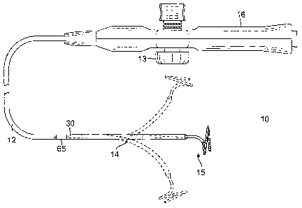
[0020] FIG. 1 is a top plan view of a catheter in accordance with one
embodiment of the
present invention.
[0021] FIG. 2 is a side cross-sectional view of a transition section
between a catheter body
and a deflectable section of the catheter of FIG. 1 in accordance with one
embodiment of the
present invention.
100221 FIG. 2A is an end cross-sectional view of the transition
section of FIG. 2, taken
along line A--A.
[0023] FIG. 2B is an end cross-sectional view of the transition
section of FIG. 2, taken
along line B¨B.
[0024] FIG. 2C is an end cross-sectional view of the catheter body of
FIG. 2, taken along
line C¨C.
[0025] FIG. 3 is a perspective view of the deflectable section of
FIG. 1, shown partially
broken away.
-7-
CA 02833093 2013-11-13
. .
1
[0026] FIG. 3A is a top view of a joint bracket in accordance with
one embodiment.
[0027] FIG. 4 is a perspective view of a joint bracket pair in
accordance with another
embodiment, as mounted on a deflection beam.
[0028] FIG. 4A is a perspective view of one bracket of FIG. 4.
[0029] FIG. 4B is a perspective view of another bracket of FIG. 4.
[0030] FIG. 4C is an end cross-sectional view of a transition section
employing the joint
bracket pair of FIG. 4.
[0031] FIG. 5 is a side cross-sectional view of a junction between
the deflectable section
and a distal assembly of the catheter of FIG. 1, in accordance with an
embodiment.
[0032] FIG. 5A is an end-cross sectional view of the deflectable
section of FIG. 5, taken
along line A¨A.
[0033] FIG. 5B is an end-cross sectional view of the deflectable
section of FIG. 5, taken
along line B¨B.
[0034] FIG. 5C is an end-cross sectional view of the deflectable
section of FIG. 5, taken
along line C¨C.
[0035] FIG. 6A is a top plan view of a distal end of the deflection
beam according to one
embodiment.
[0036] FIG. 68 is a top plan view of a distal end of the deflection
beam of FIG. 6A, in an
original configuration.
[0037] FIG. 6C is a top plan view of a distal end of the deflection
beam of FIG. 6A, as
attached to components of the distal assembly, according to one embodiment.
[0038] FIG. 7 is a top plan view of a distal end of the deflection
beam according to another
embodiment.
[0039] FIG. 8 is a perspective view of a deflection beam with beam
stiffeners in accordance
with one embodiment.
[0040] FIG. 8A is an end cross-sectional view of a deflection beam
with beam stiffeners.
-8-
CA 02833093 2013-11-13
1
[0041] FIG. 8B is an end cross-sectional view of a deflection beam
with a beam stiffener
with a channel.
[0042] FIG. 8C is a side elevational view of a deflection beam with
beam stiffeners affixed
to the beam at their proximal ends.
[0043] FIG. 9 is a top plan view of a tapered deflection beam, in
accordance with one
embodiment.
[0044] FIG. 10 is a perspective view of deflectable section with a
tapered deflection beam
with parts broken away, in accordance with one embodiment.
[0045] FIG. 11 is a top plan view of a tapered deflection beam with
sloped sections, in
accordance with one embodiment.
[0046] FIG. 11A is a top plan view of the deflection beam of FIG. 11
with tapered brackets
mounted thereon.
[0047] FIG. 11B is a side elevational view of the deflection beam and
brackets of FIG. 11A
with a reflowed tubular structure shown partially broken away, according to
one embodiment.
[0048] FIG. 11C is a side elevational view of the deflection beam,
brackets and tubular
structure of FIG. 11B, with heat shrinking tubings prior to recovery and
reflowing.
[0049] FIG. 12 is a top plan view of a tapered deflection beam
without sloped sections, in
accordance with one embodiment.
[0050] FIG. 12A is a top plan view of the deflection beam of FIG. 12
with brackets
mounted thereon.
DETAILED DESCRIPTION OF THE INVENTION
[0051] The present invention is directed to a catheter having a
catheter body (or shaft) and
a deflectable distal portion having an elongated flat beam or "blade" to
effectuate precise on-
plane bi-directional deflection while maximizing space within the catheter for
components
including lead wires, puller wires, cables, tubings and any other support
members for advanced
-9-
CA 02833093 2013-11-13
1
distal tip designs. With reference to FIG. 1, a catheter 10 in accordance with
an embodiment of
the present invention includes a catheter body 12, a deflectable distal
section 14 distal of the
catheter body, and a control handle 16 proximal of the catheter shaft. The
deflectable section
14 has a tip assembly 15 having, for example, a lasso design with a generally
circular main
portion extending and oriented transversely from a distal end of the
deflectable section 14. Bi-
directional deflection is effectuated by user manipulation of an actuator 13
provided on the
control handle 16 which moves a puller wire that extends along the catheter
from the control
handle 16 through the catheter body 12, and into the deflectable section 14.
[0052] With reference to FIGS. 2 and 2A, the catheter body 12 is an
elongated tubular
structure 11 comprising a single, central or axial lumen 18. The catheter body
12 is flexible,
i.e., bendable, but substantially non-compressible along its length. The
catheter body 12 may be
of any suitable construction and made of any suitable materials. In one
embodiment, the
catheter body 12 is multi-layered comprising at least an inner coat or layer
20, and an outer coat
or layer 22 with an imbedded braided mesh 21 of stainless steel or the like to
increase torsional
stiffness of the catheter body 12 so that, when the control handle 16 is
rotated, the deflectable
section 14 of the catheter 10 rotates in a corresponding manner. The outer
diameter of the
catheter body 12 is not critical, but is preferably no more than about 8
French. Likewise the
thicknesses of the layers 20 and 22 are not critical.
[0053] The deflectable section 14 has a tubular structure 17 with
construction similar to the
tubular structure 11 of the catheter body 12 except with greater flexibility.
In the embodiment
of FIGS. 2 and 2B, the deflectable section 14 has a central lumen 19 and a
multi-layered
construction comprising at least an inner coat or layer 24, and an outer coat
or layer 26 with an
imbedded braided mesh 25 of stainless steel or the like. The outer diameter of
the deflectable
section 14 is similar to the catheter body 12, at preferably no more than
about 8 French.
[0054] Suitable materials for the layers of the catheter body 12 and
the deflectable section
14 include materials with moderate heat deflection temperatures so stiffness
of the deflectable
-10-
CA 02833093 2013-11-13
1
section 14 and thus its deflection characteristics are not modified by
introduction into the
patient's body due to temperature variations. Suitable materials for the inner
and outer layers
20 and 22 of the catheter body 12 include Pebax and Pellethane. Materials
particularly suitable
for both the inner and outer layers 20 and 24 include lower shore hardness
plastics ranging
from about 25-55D.
[0055] Suitable materials for the inner and outer layers 24 and 26 of
deflectable section 14
include polyurethane or Pebax. In one embodiment, the tubular structure 17 of
the deflectable
section 14 includes an extruded braided structure, with the inner layer 24
having a thickness
ranging between about 0.002 inch to 0.003 inch of natural "sticky" 2533-SA-01
PEBAX, then
braided with 0.0016 inch diameter, PEN braid (50-80 pics per inch), and the
outer layer 26
including extruded PEBAX 5533-SA-01 or 4033-SA-01 PEBAX with about 25% barium
sulfate added for radiopacity.
[0056] Extending through the length of the deflectable section 14 is
an elongated support
structure configured as a flat beam or "blade" 30 with a rectangular cross-
section R having a
greater width W and a lesser thickness T, as shown in FIG. 2B, defining two
opposing
rectangular face surfaces FA and FB (or sides, used interchangeably herein)
that are flat and
smooth, and two outer longitudinal side edge surfaces El and E2 that are
friction-inducing,
e.g., uneven, rough, textured and/or serrated. The beam 30 may be constructed
of any suitable
high yield strength material that can be straightened or bent out of its
original shape upon
exertion of a force and is capable of substantially returning to its original
shape upon removal
of the force. Suitable materials for the beam include full hard, cold worked
stainless steel
alloys (304 or 316 full hard condition), nickel/titanium alloys (nitinol) or
phosphor bronze
alloys. Nitinol alloys typically comprise about 55% nickel and 45% titanium,
but may
comprise from about 54% to about 57% nickel with the balance being titanium. A
suitable
nickel/titanium alloy is nitinol, which has excellent shape memory, together
with ductility,
strength, corrosion resistance, electrical resistivity and temperature
stability. The width W of
-11-
CA 02833093 2013-11-13
1
the beam generally equals the inner diameter of the deflectable section 14.
Accordingly, the
beam 30 is situated inside the deflectable section 14 to effectively divide or
bisect the central
lumen 19 into two sub-lumens, e.g., equal half cylindrical spaces 19A and 19B,
with
components such as lead wires, cables, and tubings passing through either
space.
[0057] The catheter 10 has exceptional torque transmission capability
provided by a joint or
transition section 65 between the catheter shaft 12 and the deflectable
section 14, as shown in
FIGS. 2, 2A and 2B. The transition section 65 transfers torsional forces from
the control
handle 16 to the distal assembly 15 with high fidelity and low hysteresis, to
provide a user with
a means to accurately place and control the distal assembly 15 within the
patient. The
transition section 65 includes a pair of opposing, elongated half-cylindrical
members or
brackets 66A, 66B, e.g., formed by die cutting or acid etching, with circular
perforations or
punched through-holes 68 arranged in a predetermined pattern. In one
embodiment, there are
11 through-holes and the pattern includes a plurality of transverse rows, with
adjacent rows
offset by a predetermined distance, although it is understood that other
alternating or offset
patterns would be suitable, as well. In the illustrated embodiment of FIG. 3A,
the pattern has
rows R1, R3, R5 and R7 with two through-holes each, and rows R2, R4 and R6
with one
through-hole each, where rows R2, R4 and R6 are offset from rows 1, 3, 5 and 7
by about the
diameter of a perforation. The brackets 66A, 66B can be constructed of the
same material as
the beam 30 and may be pre-coated with an adhesive for higher bond strength
during heat
fusion.
[0058] In the illustrated embodiment, each bracket has a uniform semi-
circular or "C"
shape cross section along its length and is affixed at its outer side edges
69, e.g., by laser
welding 73, to a respective side of the beam 30. Having a curved or semi-
circular cross-
section, the C brackets 66A, 66B provide structural support to abutting ends
of the tubular
structures 11 and 17 at the transition section 65. In the illustrated
embodiment, the brackets
66A and 66B are affixed to the beam 30 near the proximal end 30P (which
extends a short
-12-
CA 02833093 2013-11-13
1
distance proximally past the joint 65 between the catheter body 12 and the
deflectable section
14). So affixed, the members 66A and 66B along with the side edges El and E2
form a full
cylindrical hollow body 66 (FIG. 3) with a circumferential contour
substantially encircling the
beam 30 at the transition section 65. As best shown in FIG. 2B, the full
cylindrical body 66
(used interchangeably with the brackets 66A, 66B) defines a central lumen 67
that is bisected
by the beam 30 into two semi-circular cavities 67A and 67B through which
components, such
as lead wires, cables, etc., can pass.
[0059] With reference to FIGS. 2 and 3, in assembling the catheter
and the transition
section 65, a distal end 11D of the tubular structure 11 of the catheter shaft
12 is slid onto
proximal end 66P of the cylindrical body 66. A proximal end 17P of the tubular
structure 17
deflectable section 14 is slid onto distal end 66D of the cylindrical body 66,
with the beam 30
extending through the lumen 19 of the deflectable section 14. Accordingly,
distal end of the
tubular structure 17 and proximal end of the tubular structure 11 cover the
body 66 from
opposite directions such that they abut each other at or near a mid-location
along the length of
the body 66, which can range between about 5 mm and 12 mm, preferably about
6.5 mm and
10 mm.
[0060] The inner coatings 20 and 24 of the tubular structures 11 and
17, respectively, are
then fused to the body 66, with application of sufficient heat and pressure so
as to melt and
flow into the perforations 68 forming nodes 20N and 24N. The fusion creates a
very strong
interlocking bond between the tubular structures of the catheter shaft 12 and
the deflectable
section 14. The nodes 20N and 24N increase the axial load capacity to the
joint 65. In fact, the
resulting torque transmission bond joint can be stronger in torsion and
tensile force loading
than the braided catheter body 12 and deflection section 14 that are bonded to
it. The friction-
inducing edges El and E2 of the beam 30 within and in contact with the body 66
also help grab
the inner layers 20 and 24 and prevent slippage between the beam 30 and the
tubular structures
11 and 17.
-13-
CA 02833093 2013-11-13
1
[0061] To facilitate the application of heat and pressure to the
transition section 65, one or
more protective heat-shrink tubing 70 (FIG. 2), e.g., fluorinated ethylene
propylene (FEP) or
polyethylene terephthalate (PET), is placed and shrunken (or "recovered") over
the transition
section (e.g., by a heat gun or oven). The transition section 65 covered by
the heat-shrink
tubing(s) 70 is then placed in a two-piece heat fusing die head (not shown)
for heating to melt
(or "reflow") the inner layers 20 and 24 into the perforations 68, followed by
cooling. The
shrink tubing 70 can be used as a process aid to prevent the melted layers
from contacting the
heated die and create a uniform transition between mating ends of the
deflectable section 14
and the catheter body 12. Thus, the shrink tubing 70 is removed from the
transition section 65
after the fusing process.
[0062] The heat fusing die head utilizes a highly accurate fusing die
height measurement
indicator (LVDT) to sense fusing die head movement during the heating/fusing
process. Since
the construction materials of the layers of the shaft 12 and the deflection
section 14 may
include extruded raw thermoplastic polymers with a wide range of heat
histories ( 25 F)
between material lots, monitoring the softening of the polymers and the
resultant die head
movement is another means besides temperature measurement to achieve process
control while
reducing the influence of polymer heat history during the heating/fusing
process. Moreover,
the transition section can be created in minimal duration (e.g., less than
about 60 seconds) using
a thermal fusing machine that is water-cooled to provide fast cycle times. The
resulting
transition section is advantageously homogenous and seamless. The structure is
nondiversified
once heat-pressure fuse operation is completed.
[0063] In an alternate embodiment as shown in FIGS. 4-4C, each
elongated half-cylindrical
bracket 66'A, 66'B includes a planar portion 63A, 63B that is flat and
rectangular. The planar
portion is adjoined to the respective half-cylindrical bracket along a
longitudinal side edge 69 at
a nontangential angle O (FIG. 4A) of about 90 degrees measured between the
planar portion
and a tangent T off side edge 69. Accordingly, each member 66'A, 66'B has a
uniform cross-
-14-
CA 02833093 2013-11-13
1
section along its length resembling a horizontal letter "G". The G brackets
66'A and 66'B with
their respective planar portions 63A and 63B can be formed from a single
rectangular piece of
die cut sheet that is bent along the longitudinal side edge 69A or 69B. In the
illustrated
embodiment, the G brackets 66'A and 66'B are affixed to the beam 30 near its
proximal end
30P (FIG. 4) with each member opposing each other from opposite sides FA and
FB of the
beam 30. Each portion 63A, 63B is affixed to a respective surface FA, FB,
e.g., by weld 73,
leaving free edge 61A and 61B unattached and free floating. In the illustrated
embodiment, the
width of each planar portion 63A, 63B is about half the diameter of a half-
cylindrical bracket
66'A, 66'B.
[0064]
Opposing and upside down from each other, the G brackets 66'A and 66'B
jointly
form nearly a full cylindrical body 66' (with the exception of the unattached
edge 61A and 61B)
substantially encircling the beam 30 at the transition section 65, with the
planar portions 63A,
63B extending diametrically toward each other sandwiching the beam 30
therebetween. The
portions 63A, 63B are thus parallel to each other, and parallel and coplanar
with the beam. The
body 66' (used interchangeably with the half-cylindrical brackets 66'A, 66'B)
defines a central
lumen that is bisected by the beam 30 (and the portions 63A, 63B) into two
semi-circular
cavities 67'A and 67'B through which components, such as lead wires, cables,
etc., can pass.
So joined, the members 66A', 66'B and the beam 30 have a cross-section
resembling the letter
"S". Because only the planar portions 63A, 63B are affixed to the beam leaving
edges 61A,
61B free floating, each half-cylindrical bracket 66'A, 66'B acts as a "spring"
to provide an
outward force when pressed on by the inner layers 20 and 24 during heat
recovery of the heat
shrinking tubing 70 and the reflowing of the inner layers 20 and 24. The
outward force ensures
larger and deeper nodes 20N and 24N and therefore a better bond between the G
brackets 66'A
and 66'B and the tubular structures 11 and 17 of the catheter body 12 and
deflectable section
14. The planar portions 63A and 63B provide large flat surface areas for
clamping the G
brackets 66'A and 66'B and the beam 30 together to provide a better setup in
preparation for
-15-
CA 02833093 2013-11-13
1
resistance or laser welding these components together in terms of minimizing
the gap between
the welded surfaces and enabling axial alignment between the beam and the
brackets. The
large flat surfaces also ensure better contact between contact surfaces of the
planar portions
63A and 63B and the beam 30 for a better and stronger weld.
[0065] An accordance with a feature of the present invention, the
catheter 10 provides bi-
directional deflection with a single continuous puller wire 28 that
advantageously requires less
actuation force by a user and imposes less shear stress on the puller wire.
The puller wire 28
has a U-bend mid-portion 28M being a distal-most portion of the puller wire in
the catheter. As
shown in FIG. 5, the U-bend mid-portion 28M divides the puller wire into two
longitudinal
portions 28A and 28B of generally equal length, each with a proximal end that
is anchored in
the control handle 16. With reference to FIGS. 6A, 6B and 6C, to anchor the U-
bend portion
28M at a distal location on the catheter, a distal end of the beam 30 has a
receiving formation
32 e.g., either an on-axis slit 32S or an on-axis through-hole 32H, which
securely receives the
mid-portion 28M so that each long portion 28A and 28B extends longitudinally
centered on the
beam along a respective face surface FA, FB of the beam 30. This arrangement
advantageously avoids the use of conventional T-bars, crimp type connections,
soldering or
welding as a means to anchor a distal end of the puller wire to the beam 30.
And, because the
puller wire is not rigidly attached to the beam 30, this arrangement provides
smooth bi-
directional steering.
[0066] As illustrated in FIGS. 6A and 6B, the distal end 30D of the
beam 30 has an original
configuration prior to assembly of the catheter and attachment of the puller
wire 28, which
includes an elongated longitudinal closed slit 32S with a distal end 31 and a
proximal end 33.
The slit 32S is disposed immediately proximal of a distal end portion 30D of
the beam 30. The
through-hole 32H is disposed in the distal end portion 30D. The U-bend mid
portion 28M of
the puller wire may be inserted and hooked through the hole 32H, or
alternatively in the slit
32S at its proximal end 33. In the latter regard, the slit 32S is adapted into
an open
-16-
CA 02833093 2013-11-13
1
configuration (FIG. 6C) from a closed configuration (FIG. 6A) for receiving
the U-bend mid-
portion 28M when the distal end portion 30D of the beam is detached by a user
bending or
cutting along a transverse "pre-cut" groove 52 (FIG. 6A) provided on the face
FA of the beam
30 proximal of the hole 32H. In the illustrated embodiment, a first transverse
groove 52a is
aligned with the distal end 31 of the slot 32 and a second (half width)
transverse groove 52b is
aligned at or near a midpoint along the length of the slot 32S. Thus, the
distal end portion 30D
can be readily broken off or otherwise detached from the beam along the groove
52a. For
easier access to the open slit 32S, portion 30a can be detached from the beam
30 along the
groove 52b, as shown in FIG. 6C. The puller wire portions 28A and 28B extend
proximally
along opposites sides FA and FB of the beam 30 through the deflectable section
14, the central
lumen 18 of the catheter body, and into the control handle 16.
[0067] As shown in FIG. 6C, the slit 32S is generally longitudinally
centered and on-axis
with the longitudinal axis of the beam 30 such that the slit divides the beam
into two generally
equal elongated sections or prongs 54a, 54b. In the illustrated embodiment, a
hollow tube or
ferrule 60 (e.g., of stainless steel) is affixed e.g., by laser welding, to
face FA of the prong 54b
(although it is understood that the tube 60 may be alternatively affixed to
prong 54a, with the
portion 30b detached from the beam). A proximal end of a support member 72
supporting the
distal assembly 15 is inserted and anchored in the tube 60, e.g., by crimping,
to create an
interference fit between the tube 60 and the support member 72 to transmit
torque and
tension/compression forces from the beam 30 to the distal assembly 15. A
mechanical crimp
process eliminates problematic adhesive bonding that can loosen or fail
causing the distal
assembly 15 to spin. A servo process with precision force control is used to
detect a defined
force slope so that acceptable interference between the support member 72 and
the tube 60 is
created without damaging the puller wire 28.
[0068] Proximal ends of the portions 28A and 28B are anchored in the
control handle 16
and deflection mechanism in the control handle 16 responsive to the actuator
13 manipulated
-17-
CA 02833093 2013-11-13
. .
1
by a user is configured to draw or otherwise act on a proximal end of puller
wire portion 28A
or 28 to deflect the distal section 14 with a distinct curvature on side FA or
FB of the beam 30.
Throughout the catheter body 12, each puller wire portion extends through a
respective
compression coil 62A and 65B (FIGS. 2, 2C and 8) which is flexible but resists
compression so
that deflection of the catheter initiates at or near distal ends of the
compression coils. Along the
beam 30 in the deflectable section 14, each puller wire portion may be coated
with PTFE or
Teflon so the puller wires can slide smoothly inside a respective protective
spacer tube 36
provided on a respective side of the beam 30 as discussed in further detail
below.
[0069] As understood by one of ordinary skill in the art, the puller wire
28 is in tension to
create a bending moment to deflect the beam 30 in the desired direction.
Conventional catheter
with a flat beam may use a puller wire with a rectangular cross-section that
is welded and
tightly constrained to the beam to prevent adhesion failure. While this design
may be simple
and compact in certain respects, the puller wire is under significant force
due because of its
close proximity to the beam, which in pure bending requires a substantial
bending moment
stress during deflection. In contrast, as illustrated in the drawings,
including FIG. 2B, the
catheter of the present invention is configured to provide a spacer of a
predetermined thickness
to separate the puller wire 28 and a neutral bending axis NA of the beam 30 by
a predetermined
distance so as to lower the force on the puller wire, including the bending
moment. Moreover,
the catheter 10 includes a puller wire 28 with a circular (or at least round)
cross section to
reduce the area moment of inertia, as an otherwise rectangular puller wire
with the same cross-
sectional area separated from the neutral axis by a comparable spacer would
unduly increase
the size/diameter of the catheter and the area moment of inertia to result in
an unacceptably stiff
catheter.
[0070] As shown in FIG. 2B, the spacer on each side of the beam 30 may
include a spacer
adhesive layer 34 and a wall of a lumened elastomeric puller wire spacer tube
36. The adhesive
layer 34 may be provided by an ultra high temperature adhesive transfer tape
sold by 3M under
-18-
CA 02833093 2013-11-13
1
the model 100HT. The adhesive layer may have a thickness of about 0.001 inch
and require
about 72 hours to achieve full adhesive bond strength. The spacer tube 36,
which may be
constructed of polyimide, thin wall polyetheretherketone (PEEK), nylon or
other thin wall
thermoplastic tubing, is affixed to the adhesive layer 34, and a respective
puller wire proximal
portion 28A or 28B extends through lumen 37. An interior surface of the lumen
37
surrounding the puller wire may be coated with polytetrafluoroethylene (PTFE),
e.g. TEFLON
or TEFLON composite, to reduce static and dynamic friction with the puller
wire. On each
side FA and FB of the beam 30, the spacer runs longitudinally generally
between the receiving
formation 32S or 32H (FIG. 6A) and a proximal end 30P of the beam 30 (FIG. 2).
Alternatively, the spacer may include an extrusion surrounding each puller
wire portion. The
extrusion may be made of PEEK.
[0071] The round puller wire 28 has a diameter D ranging between
about 0.007 inch and
0.009 inch, and preferably about 0.008 inch. The beam 30 has a thickness T of
about 0.004
inch and 0.007 inch, and preferably between about 0.005 inch and 0.006 inch.
The puller wire
and the neutral axis are separated by a distance d, ranging between about
0.008 inch and 0.025
inch, and preferably between about 0.010 inch and 0.015 inch. In one
embodiment, the puller
wire diameter D is 0.008 inch and a Nitinol 304V wire, and the beam thickness
is 0.005 inch.
[0072] To constrain and secure the puller wire 28 on the beam 30 and
as an additional
means to prevent adhesive failure and detachment, at least a first inner heat
shrink tubing 38 is
placed on the beam 30, covering and surrounding the spacers on both sides FA
and FB of the
beam 30, inclusive of the puller wire portions 28A, 28B trained through the
spacers (hereinafter
referred to as "the beam assembly"). In the illustrated embodiments, including
FIGS. 2B and
6A, the first inner heat shrink tubing 38 is followed by a second outer heat
shrink tubing 39 that
is placed over the beam assembly to surround and seal the components and the
first heat shrink
tubing 38. The first heat shrink tubing 38 may constructed of high temperature
resistant
polyester (PET) or fluorinated ethylene propylene (FEP) and have a wall
thickness ranging
-19-
CA 02833093 2013-11-13
1
between about 0.0005 inch and 0.004 inch, and preferably between about 0.00015
inch and
0.001 inch, in an expanded state. Another suitable material is polyester in
terms of thin wall
and high strength. The first heat shrink tubing 38 is recovered by heating
with a hot air-based
heating system thus providing a second bonding structure for the spacer tubes
36, as well as a
first sealing structure for the adhesive layers 34 and spacer tubes 36. The
uneven longitudinal
edges El and E2 of the beam 30 help grab and secure the first heat shrink
tubing 38 so it do not
migrate or slip during deflection.
[0073] The second heat shrink tubing 39 may be constructed of
extruded natural PEBAX,
e.g., 2533-SA-01 (22D shore hardness), thin wall with a thickness ranging
between about
0.002 inch and 0.003 inch, or natural PELLETHANE (e.g., 80A shore hardness).
The second
heat shrink tubing 39 may be a layer of "sticky" low shore hardness
thermoplastic elastomer
which is heated and recovered over the first heat shrink tubing 38, thus
creating a second layer
sealing structure and a "sticky" heat bondable outer layer surrounding the
beam assembly. The
sticky outer layer provided by the second heat shrink tubing 39 is well suited
to bond with the
tubular structure 17 of the deflectable section 14 through a resistive heating
process with clamp
members to heat the beam 30.
[0074] The heat shrink tubings 38 and 39 extend from the distal end
30D of the beam to
near the distal end of the brackets 66A, 66B, so as not to interfere with the
weld 73 between the
66A and 66B and the beam 30. Depending on the length of the beam proximal of
the brackets
66A, 66B, heat shrink tubings may be provided there as well.
[0075] In another embodiment as illustrated in FIG. 7, a pair of
tensile fibers 29, e.g.,
VECTRAN cords are utilized instead of puller wires. A crimped metal tube 31,
e.g., of
stainless steel or other alloys) is attached to the distal end of each fiber
29. Each tube 31 is
affixed, e.g., by resistance- or laser-welded, to the longitudinal center of a
respective side of the
distal portion 30D of the beam. A respective spacer tube 36 surrounds each
fiber and is bonded
to a respective surface FA or FB of the beam by a spacer adhesive layer 34.
The fibers 29 may
-20-
CA 02833093 2013-11-13
1
be coated with low density polyethylene or TEFLON, e.g., DUPONT TRASYS 9825 or
TRASYS 426 and MCLUBE 1829 TEFLON based coatings, to damp out noise and
prevent
stick-slip type non-uniform motion created by variations in dynamic and static
friction
coefficients during deflection. Food grade damping gel (e.g., Nye Lubricants
fluorocarbon Gel
835C-FG// 874//880FG) having synthetic hydrocarbon and PTFE or silicone and
PTFE to coat
the fibers 29 and interior of the spacer tubes 36.
[0076] Where the deflectable section 14 has a length greater than
about 90mm, one or more
elongated flat beam stiffeners 80 may be mounted to either or both sides FA
and FB of the
beam 30 to modify and obtain desired curve geometry when the puller wire or
tensile fibers
(collectively referred to as "puller members") are activated via the control
handle 16. As
shown in FIGS. 8, 8A, 8B, 8C, one or more stiffeners 80 may be adhesively
bonded to the
beam 30. The stiffeners 80 are generally parallel with the beam 30 and can
have similar or
different lengths relative to each other and the beam. The stiffeners may be
bonded by a layer
of adhesive 81 (FIGS. 8A and 8B), e.g., applied via ultra high temperature
adhesive transfer
tape sold by 3M under the name 110HT. The adhesive may have a layer thickness
of about 1.0
mm. The adhesive provides a viscoelastic bond between the stiffeners and the
beam.
Alternatively, the stiffeners 80 may be spot welded by laser to the beam at
selected locations 82
as shown in FIG 8. It is understood that these two different bonding methods
provide different
degrees of stiffness despite employing beams and stiffeners of the same
thicknesses due to the
viscoelastic behavior of the adhesive bond compared to the metal-to-metal
fusing of the spot
welding bonds.
[0077] In yet another alternate embodiment of FIG. 8C, proximal ends
80P of the stiffener
beams 80 may be bonded to and rigidly supported by the beam 30 at or near the
brackets 66 of
the transition section 65, leaving distal ends 80D of the stiffener beams free
floating and
unattached, to create another type of curve. Moreover, depending on the shape
and size of the
stiffeners 80, a longitudinal channel 84 (FIG. 8B) to accommodate the puller
wire and the
-21-
CA 02833093 2013-11-13
1
spacer.
[0078] The cross section of the beam itself may change along its
length. As illustrated in
FIGS. 9, 11 and 12, each of beams 130, 230, 330 have a tapered configuration
with the width
W decreasing (continuously or discontinuously) from their proximal ends 130P,
230P, 330P to
their distal ends 130D, 230D, 330D. The narrower distal end facilitates
cannulation and
tracking through smaller tubular regions, such as the great cardiac vein, and
the larger proximal
end provides more stability near larger tubular regions, such as the coronary
sinus ostium when
tracked inside the coronary sinus. The width of the beam may be gradually
tapered, for
example, in a linear manner, for a generally smooth profile along its side
edges El and E2
(FIG. 9), or it may step-tapered in a manner along its length, with linearly
sloped portions (FIG.
11) or without sloped portions (FIG. 12). It is understood that the beam may
be constructed
from multiple beam segments fused together end to end or as a single
continuous elongated
body. In one embodiment, the distal section 14 supported by the beam may have
a proximal
section with a 7 french diameter, a mid-section with a 6 french diameter and a
distal section
with a 5 french diameter.
[0079] In the embodiment of FIG. 9, where the beam 130 has the
gradually tapered width,
one or more pairs of brackets 166A and 166B are affixed to the beam 130 at
selected locations
forming generally a generally full cylindrical body encircling the selected
locations. The
selected locations for affixation of brackets (C or G brackets) may be a joint
between beam
segments and/or a joint where the width of the beam changes. The diameters of
the brackets
along their lengths vary correspondingly with the changing widths of the beam
at those selected
locations.
[0080] In the embodiment of FIGS. 10 and 11, the beam 230 has a step-
tapered
configuration with rectangular sections 230D, 230M, 230P adjoined by sloped
sections 231
therebetween. Each rectangular section has a respective width which is uniform
throughout
that section. However, the more distal rectangular sections have smaller
widths than the more
-22-
CA 02833093 2013-11-13
1
proximal rectangular sections such that WD < WM < WP where WD is the width of
the most
distal section 230D, WM is the width of the mid section 230M and WP is the
width of the most
proximal section 230P. Between each rectangular section is a sloped section
231 whose width
changes linearly (by decreasing in the distal direction or increasing in the
proximal direction)
along its length so that the sloped section 231 bridges the adjacent
rectangular sections 230
without sharp bends or corners on side edges El and E2 of the beam. The slope
of each section
as measured relative to the longitudinal axis of the beam ranges between 0 and
less than 90
degrees, preferably between about 15 and 30 degrees. In the illustrated
embodiment, the beam
230 includes three rectangular sections 230 and two sloped sections 231 in
between and
alternating with the rectangular sections 230. As shown in FIG. 11A and 11B, a
pair of
brackets 266A and 266B (266B not shown) are mounted on each tapered section
231 forming
generally a full cylindrical body encircling each tapered section. Each pair
of bracket conform
with their respective tapered section 231 with a diameter that also changes
along its length so as
to be similarly tapered as the respective section 231.
100811 Alternatively, in the embodiment of FIGS. 12 and 12A, the beam
330 has a
nonlinear or step-tapered configuration having rectangular sections 330
directly adjoined to
each other without sloped sections. Thus, the side edges El and E2 have a
"step" profile with
corners 331. Rectangular sections 330D, 330M and 330P have uniform widths WD,
WM or
WP, respectively, wherein WD < WM < WP. A pair of brackets 366A and 366B (366B
not
shown) are mounted at or near each corner 331 forming a generally full
cylindrical body
overlapping the distal end and proximal end of adjacent pairs of rectangular
sections 230. The
diameter of each bracket may be uniform along its length and conform with the
narrower width
of the adjacent pairs of rectangular sections 231.
100821 A method of assembling a tapered beam, for example, the beam
230 is described
below in reference to FIG. 10, although it is understood that the method may
be used for any
beam, including the beam 130 or the beam 330. A continuous section of tubing
217
-23-
CA 02833093 2013-11-13
1
(comprising, e.g., extruded inner layer 24 and outer layer 26 with an embedded
braided mesh
25, as described above) is placed over the beam, as illustrated in FIG. 11B.
The tubing 217 has
a sufficient length to cover the beam longitudinally and a suitable diameter
that is large enough
to accommodate all widths of the beam. Where the beam has a plurality N of
cylindrical bodies
266, a plurality of at least (N+1) heat shrink tubings 270 are placed over the
tubing 217 with
ends of adjacent tubes 270 abutting at or near a midpoint of each cylindrical
body 266. The
heat-shrink tubings 270 may be fluorinated ethylene propylene (FEP) or
polyethylene
terephthalate (PET). Each heat shrink tube 270 may have a distinct diameter
that corresponds
with the width(s) of the section(s) 230 or 231 it covers. In the illustrated
embodiment of FIG.
10, there are three heat shrink tubings 270D, 270M and 270P, with respective
diameters DD,
DM and DP wherein DD < DM < DP.
[0083] The heat shrink tubings 270 are recovered by application of
heat (e.g., by a heat
gun) and then placed in a two-piece heat fusing die head (not shown) for
heating to reflow the
tubing 217 of the deflectable section 14, which conforms the tubing 217 to the
cylindrical
brackets 266A and 266B and fuses the inner layer 24 to the brackets by means
of melted
material flowing into perforations 268 to form nodes interlocking the tubing
217 and the
brackets 266A and 266B. Textured side edges El and E2 of the beam 230 also
help minimize
slippage between the beam 230 and the tubing 217. Thereafter, the heat shrink
tubings 270 can
be removed from the tubing 217.
[0084] Alternatively, the tubular structure 17 of the deflectable
section 14 may be
constructed by injection molding, instead of extrusion and reflow.
[0085] In the illustrated embodiment of FIG. 1, the distal assembly
15 comprises a
generally straight proximal region and a generally circular main region having
at least one loop
circling about 360 degrees, if not two loops circling about 720 degrees. The
proximal region is
mounted on the deflectable section 14 and the main region carries a plurality
of electrodes (ring
and/or tip) for mapping and/or ablation. With reference to FIG. 5, the distal
assembly 15
-24-
CA 02833093 2013-11-13
1
includes the shape memory support member 72, lead wires 140 for the electrodes
carried on the
distal assembly 15, and a cover 120 extending the length of the distal
assembly. The lead wires
140 attached to the electrodes on the distal assembly 15 extend through a
nonconductive sheath
141 which extends from the distal assembly through the lumen half 19B of the
deflectable
section 14, through the cavity half 67B of the transition section 65, through
the lumen 18 of the
catheter shaft 12, and into the control handle 16. Ring electrodes may also be
carried on the
deflectable section 14, as shown in FIG. 3.
[0086] An electromagnetic position sensor 134 (FIG. 5) is mounted in
or near the distal end
of the deflectable section 14 or the proximal end of the distal assembly 15. A
sensor cable 136
extends from the sensor 134 into the half lumen 19A of the deflectable section
14, the cavity
half 67B of the transition section 65, the central lumen 18 of the catheter
body 12 and into the
control handle 16 where it terminates in a suitable connector (not shown).
[0087] The catheter 10 may also be adapted for irrigation at the
distal assembly 15, for
example, to supply fluid at or near the electrodes of the distal assembly. To
that end, an
irrigation tubing 150 may be provided to pass fluid to the distal assembly 15
from the control
handle 16. In the illustrated embodiment of FIG. 2, the tubing 150 passes
through the central
lumen 18 of the catheter body 12, the lumen 19b of the deflectable section 14,
and into the
distal assembly 15.
[0088] In use, a suitable guiding sheath is inserted into the patient
with its distal end
positioned at a desired location. An example of a suitable guiding sheath for
use in connection
with the present invention is the Preface.TM. Braiding Guiding Sheath,
commercially available
from Biosense Webster, Inc. (Diamond Bar, Calif.). The distal end of the
sheath is guided into
one of the chamber, for example, the atria. A catheter in accordance with an
embodiment of the
present invention is fed through the guiding sheath until its distal end
extends out of the distal
end of the guiding sheath. As the catheter is fed through the guiding sheath,
the distal assembly
15 is straightened to fit through the sheath. Once the distal end of the
catheter is positioned at
-25-
CA 02833093 2013-11-13
. .
1
the desired location, the guiding sheath is pulled proximally, allowing the
deflectable section
14 and distal assembly 15 to extend outside the sheath, and the distal
assembly 17 returns to its
original shape due to its shape-memory.
[0089] The user manipulating the actuator 13 on the control handle 16
actuates deflection
mechanism inside the control handle 16 to draw puller wire proximal portion
28A or 28B to
deflect the distal section 14 on-plane to one or the other side of the beam
30. The user may
then rotate the distal assembly 15 by rotating the control handle 16 which
transfers torque to the
catheter body 12 and the deflectable section 14 through the transition
section(s) 65. The
brackets 66A and 66B to which the tubular structures 11 and 17 of the catheter
body 12 and the
deflectable section 14 are bonded by means of interlocking nodes formed in the
perforations 68
of the brackets 66A and 66B under heat fusion.
[0090] Suitable materials for construction of the beam, the beam stiffeners
and/or the half-
cylindrical brackets include 50/50NiTi, titanium (Ti-6A1-4V), phosphor bronze
510, beryllium
copper, monel alloy K-500 or MP35N (a non-magnetic nickel-cobalt-chromium-
molybdenum
alloy). Suitable materials for the puller wire include preformed, heat treated
and TEFLON
coated NiTi wire, monel alloy K-500 or dual VECTRAN fibers.
[0091] Suitable materials for imbedded braided mesh for the tubular
structures of the
catheter body and/or the deflectable section include stainless steel (304V or
316), phosphor
bronze, monel K-500, PEN or other synthetic fibers that can readily bond with
PEBAX or
PELLETHANE extruded thermoplastics during the secondary/outer extrusion coat
or layer.
[0092] The preceding description has been presented with reference to
presently preferred
embodiments of the invention. Workers skilled in the art and technology to
which this
invention pertains will appreciate that alterations and changes in the
described structure may be
practiced without meaningfully departing from the principal, spirit and scope
of this invention.
As understood by one of ordinary skill in the art, the drawings are not
necessarily to scale.
Accordingly, the foregoing description should not be read as pertaining only
to the precise
-26-
CA 02833093 2013-11-13
. .
1
structures described and illustrated in the accompanying drawings, but rather
should be read
consistent with and as support to the following claims which are to have their
fullest and fair
scope.
10
20
-27-