Note: Descriptions are shown in the official language in which they were submitted.
ACCOMMODATIVE INTRAOCULAR LENS AND METHOD OF IMPLANTATION
Field of the Invention
This invention relates generally to the field of intraocular lenses (I0L) and,
more
particularly, to accommodative IOLs.
Background of the Invention
The human eye in its simplest terms functions to provide vision by
transmitting light
through a clear outer portion called the cornea, and focusing the image by way
of a crystalline
lens onto a retina. The quality of the focused image depends on many factors
including the size
and shape of the eye, and the transparency of the cornea and the lens. The
lens is held in place
within the posterior chamber of the eye by a membrane known as the capsular
bag or posterior
capsule, immersed in the aqueous humor. The shape of the lens and the
refractive index of the
lens relative to the aqueous humor determine where light rays are focused onto
the retina.
When age or disease causes the lens to become less transparent, vision
deteriorates
because of the diminished light which can be transmitted to the retina. This
deficiency in the lens
of the eye is medically known as a cataract. An accepted treatment for this
condition is surgical
removal of the lens and replacement of the lens function by an artificial
intraocular lens (I0L).
Cataractous lenses are often removed by a surgical technique called
phacoemulsification. During
this procedure, an opening is made in the anterior side of the capsular bag, a
thin membrane
enclosing the natural lens. A thin phacoemulsification cutting tip is inserted
into the diseased
lens and vibrated ultrasonically. The vibrating cutting tip liquefies or
emulsifies the lens so that
the lens may be aspirated out of the eye. The diseased lens, once removed, is
replaced by an
artificial lens.
In the natural lens, multifocality of distance and near vision is provided by
a mechanism
known as accommodation. The natural lens, early in life, is soft and contained
within the
capsular bag. The bag is suspended from the ciliary muscle by the zonules.
Relaxation of the
ciliary muscle tightens the zonules, and stretches the capsular bag. As a
result, the natural lens
tends to flatten. Tightening of the ciliary muscle relaxes the tension on the
zonules, allowing the
capsular bag and the natural lens to assume a more rounded shape. In this way,
the natural lens
can be focused alternatively on near and far objects. As the lens ages, it
becomes harder and is
1
CA 2833317 2018-10-09
less able to change shape in reaction to the tightening of the ciliary muscle.
This makes it harder
for the lens to focus on near objects, a medical condition known as
presbyopia. Presbyopia
affects nearly all adults over the age of 45 or 50.
When a cataract or other disease requires the removal of the natural lens and
replacement
with an artificial intraocular lens ("IOL"), the IOL used to replace the
natural lens has often been
a monofocal lens. These lenses do not change power in response to the movement
of the capsular
bag, requiring that the patient use a pair of spectacles or contact lenses for
near vision. However,
there are several examples in the prior art of bladder or bag-like intraocular
lenses that consist of
an outer flexible skin filled with a viscous gel. The resulting lens
completely fills the capsular
bag and is very soft and pliable, much like the natural lens. See for example,
U.S. Patent Nos.
4,373,218 (Schachar), 4,585,457 (Kalb), 4,685,921 (Peyman), 4,693,717
(Michelson), 5,275,623
(Sarfarazi), 4,822,360 (Deacon), 5,489,302 (Skottun) and 6,217,612 (Woods).
But in order to
provide accommodation, movement of the ciliary muscle must be adequately
transmitted to the
lens system through the capsular bag, and none of these references disclose a
mechanism for
ensuring that there is a tight connection or fixation between the capsular bag
and the lens system.
Therefore, a need continues to exist for a safe and stable accommodative
intraocular lens
system and method for implantation that provides accommodation over a broad
and useful range.
Brief Summary of the Invention
An accommodating intraocular lens (AIOL) adapted for implantation into a
capsular bag
includes an outer shell, a valve, and a force transfer assembly. The outer
shell includes at least
one surface modification on at least a periphery of the outer shell to promote
bonding with the
capsular bag. The valve is configured to permit injection of a fill material.
The force transfer
assembly in the outer shell is adapted to transfer forces from the capsular
bag to change the
shape of the filled outer shell in response to changes in capsular bag shape.
Certain exemplary embodiments can provide an accommodating intraocular lens
(AIOL)
adapted for implantation into a capsular bag, comprising: an outer shell
having an anterior
surface, a posterior surface, and a living hinge at a periphery of the outer
shell, the anterior
surface, posterior surface, and living hinge defining an internal void that is
fillable with a fill
material, the outer shell including at least one surface modification at a
contact region on at least
the portion of the periphery of the outer shell the surface modification
configured to promote cell
2
CA 2833317 2018-10-09
growth and adhesion with the capsular bag at the contact region; a valve
configured to permit
injection of a fill material; and a force transfer assembly comprising a
plurality of stiffening ribs,
at least a portion of each stiffening rib comprising a region of the anterior
surface of the outer
shell having a greater thickness than adjacent regions of the anterior surface
of the outer shell
.. and extending into the internal void only a portion of the way between the
anterior surface and
the posterior surface, the force transfer assembly adapted to transfer forces
from the capsular bag
at the contact region to change the shape of the filled outer shell in
response to changes in
capsular bag shape.
Brief Description of the Drawings
FIG. I is an enlarged cross-sectional view of the lens according to a
particular
embodiment of the present invention.
FIG. 2 is an enlarged cross-sectional view of the lens of FIG. 1 showing the
lens
implanted in a capsular bag.
2a
CA 2833317 2018-10-09
CA 02833317 2013-10-15
WO 2012/166435
PCMJS2012/038973
FIG. 3 is an enlarged cross-sectional view of the lens of FIG. I showing the
lens implanted in a capsular bag and material being injected into the lens to
approximate the unaccommodated state.
FIG. 4 is an enlarged cross-sectional view of the lens of FIG. 1 showing the
lens implanted in a capsular bag and material being removed from the lens.
FIG. 5 is an enlarged cross-sectional view of the lens of FIG. I showing the
lens implanted in a capsular bag and being in the accommodated state.
FIG. 6 is flowchart showing a method of implanting an inflatable
accommodating lens according to particular embodiments of the present
invention;
io FIG. 7
illustrates a dual-optic accommodating IOL according to a particular
embodiment of the present invention;
FIGs. 8A, 8B, and 8C illustrates various embodiments of a dual-optic AIOL
according to particular embodiments of the present invention as viewed along
the
optical axis;
FIG. 9 is a flowchart showing an example method of implanting a dual-optic
AIOL according to particular embodiments of the present invention; and
FIG. 10 illustrates a peripheral band usable in conjunction with various
embodiments of the present invention.
Detailed Description of the Invention
Various embodiments of the present invention may provide an improved
accommodating lens by promoting adhesion of the capsular bag around mechanical
features of the accommodating 10L. This provides a more robust mechanical
connection between the bag and the IOL to allow the flattening and relaxing of
the
bag, as opposed to the force of the ciliary muscles, to move the lens. The
changes in
shape of the capsular bag are in turn used either to deform the lens to
produce a power
change (akin to the accommodation of the natural lens) or to produce a change
in IOL
power by separating two optical elements.
Various embodiments of the present invention also include mechanical
structures for translating the force produced by movement of the capsular bag
into
forces producing either deformation of the lens or separation of optical
elements of the
lens. By combining this with strong adhesion of the capsular bag to the IOL at
particular points along the mechanical structure, particular embodiments of
IOLs
according to the present invention advantageously provide increased mechanical
efficiency and a greater degree of accommodative change in the optical power
of the
IOL.
3
CA 02833317 2013-10-15
WO 2012/166435
PCMJS2012/038973
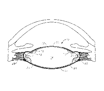
As best seen in FIG. 1, a lens 10 according to a particular embodiment of the
present invention generally consists of an outer shell 12 defining internal
void 13
which contains and interior fill material 14. The outer shell 12 is preferably
formed in
any suitable overall diameter or length, for example, around 10 millimeters,
for
implantation in the capsular bag of the eye. The outer shell 12 preferably is
made
from a soft, foldable material that is inherently resistant to the formation
of posterior
capsular opacification ("PCO"), such as a soft acrylic. In certain
embodiments, the
material of the outer shell 12 may be relatively more elastic than the
capsular bag, so
that outer shell 12 can be moved by the capsular bag with relative ease. The
outer
io shell 12 contains a fill valve 16 allowing fill material 14 to be
injected into or
removed from void 13.
The outer shell 12 may also include a force transfer structure, such as a
plurality of stiffening radial ribs 18 having an appropriate spacing, e.g., 30
, and/or
may be attached to a peripheral band surrounding the lens 10 (as described in
greater
is detail below). In the depicted embodiment, the lens 10 also includes
living hinges 21
at the periphery, so as to facilitate the shape change of the surfaces. In
particular
embodiments, a peripheral band may be coupled to the living hinge assembly to
transfer forces from the capsular bag into actuation of the living hinges. The
outer
shell 12 may also contain sharp peripheral corners 20 designed to prevent
equatorial
20 cell proliferation on the optical surfaces of the lens 10, but cell
adhesion is preferably
encouraged around the hinges 21 at the periphery of the lens to improve the
mechanical efficiency of force transfer between the capsular bag and lens 10.
At least part of the outer shell 12 is coated with surface modification 22,
which
may include coatings, texturing, or other suitable variation designed to
promote
25 protein adhesion. Examples of such coatings include complementary
proteins, growth
factors for the capsular bag, chitin or other organic chemicals used in
signaling cell
growth conditions. The polymer structure used to form lens 10 may be protein-
fortified, so that the lens material itself has a surface that encouraged
protein bonding.
Other suitable surface modifications include nano-channels or other structures
30 allowing cellular interpenetration into the lens structure. Such
structures may also
include coatings or treatments to promote cell growth and binding within the
interpenetrating cell/lens network. Still other suitable surface modifications
include
the use of biocompatible adhesives.
Fill material 14 preferably is a liquid, gel, or low molecular weight polymer
35 with a refractive index greater than that of the surrounding aqueous
humor. Such
materials may include (but are not limited to) silicone oil, perfluoron, and
cross-linked
or non-cross-linked polymer gels. It is also preferable to minimize losses of
fill
4
CA 02833317 2013-10-15
WO 2012/166435
PCMJS2012/038973
material 14 due to diffusion, so outer shell 12 should preferably be
relatively
impermeable to fill material 14 and the surrounding aqueous humor. As best
seen in
FIG. 2, lens 10 may be implanted in capsular bag 24 in an unfilled state. As
seen in
FIG. 3, internal void 13 is then filled with fill material 14 through valve 16
using an
appropriate instrument, such as cannula 26 so that lens 10 approximates the
shape of
the natural lens in a disaccommodated state, which results in anterior surface
30 of
outer shell 12 being relatively flat.. In the depicted embodiment of FIG. 2,
the anterior
surface 30 changes shape considerably during accommodation while the posterior
surface remains relatively unchanged in shape, but alternative embodiments
could
io have both the anterior and the posterior surfaces changing shape to a
lesser or greater
degree. One advantage of thickening the posterior surface or making the
posterior
surface relatively more rigid is that the posterior surface could be
relatively fixed in
order to more easily allow aspheric and/or tone correction to be used in lens
10.
Diffractive and/or multifocal optics could be incorporated into the surface as
well.
When lens 10 is over-filled, zonules 28 are in a relaxed position. Lens 10 is
left in this over-filled condition for a period of time sufficient for protein
adhesions to
form between outer shell 12 and capsular bag 24, e.g., 2-4 weeks. As best seen
in
FIG. 4, after protein adhesions have formed between outer shell 12 and
capsular bag
24, sufficient filler material 14 is removed from void 13 through valve 16 for
lens 10
zo to adopt the shape of a disaccommodated lens, as best seen in FIG. 5,
with zonules 28
in tension and anterior surface 30 having a more rounded shape relative to the
overfilled state, as shown by arrows 32. The lens 10 may be also mechanically
biased
toward the accommodated state, so that when the capsular bag relaxes, the
default
tendency of the lens 10 is to restore to the accommodated state. For example,
the
living hinges 21 may have a spring action that tends to urge the anterior
surface of the
lens 30 into the accommodated shape.
FIG. 6 is a flowchart 100 showing an example implantation method according
to particular embodiments of the present invention. At step 102, an inflatable
accommodating lens ("AIOL") is provided with a surface modification to promote
protein adhesion with the capsular bag. At step 104, the AIOL is implanted in
an
unfilled state. At step 106, the AIOL is overfilled to facilitate contact with
the
capsular bag. At step 109, the capsular bag is allowed to heal around the AIOL
for
sufficient time to allow bonding between the capsular bag and the AIOL. At
step 110,
fill material is removed from the AIOL to reach a disaccommodated state for
the
AIOL.
FIG. 7 is a cross sectional view of a dual-optic AIOL 200 according to another
embodiment of the present invention. For purposes of this specification, "dual-
optic"
5
refers to an AIOL including at least two optical elements, but such a dual-
optic AIOL could
include additional optical elements as well. The dual-optic AIOL 200 includes
an anterior optical
element 202 and a posterior optical element 204. Preferably, one of the
anterior optical element
202 or posterior optical element 204 has a positive power, and the other has a
negative power so
that the difference between the powers is relatively large and a change in
spacing between the
optical elements 202 and 204 produces a significant change in overall optical
power. One or both
surfaces may also include aspheric, tonic, diffractive, and/or multifocal
correction as well. While
both optical elements 202 and 204 are shown within the capsular bag, the AIOL
200 could
include a sulcus-fixated anterior optical element 202.
The AIOL 200 also includes interlocking features 206 between the optical
elements 202
and 204. Interlocking features 206 are located peripherally around the optical
elements 202 and
204, and the interlocking features 206 also include surface modifications,
such as the ones
described above, to promote bonding of the capsular bag to the interlocking
features 206.
Interlocking features 206 may be formed integrally, so that the entire AIOL
200 is a single piece,
or they can alternatively be complementary features attached to their
respective optical elements
202 and 204 so that the interlocking features 206 are connected to one another
before or during
implantation. While the interlocking features 206 are illustrated in an
integrated living hinge
configuration, other arrangements could function suitably as well, including
arrangements using
a hook-and-clasp or hinge pin. In the illustrated embodiment, the interlocking
features 206 are
configured to hold the optical elements 202 and 204 spaced apart from one
another in the
disaccommodated state to prevent adhesion.
Because the capsular bag is firmly attached to the interlocking features 206,
the capsular
bag pulls the interlocking features 206 outwardly when flattened. The
interlocking features 206
are shaped such that the optical elements 202 and 204 are pulled together when
the interlocking
features 206 are pulled outward. The interlocking features 206 are also shaped
to store
mechanical energy when the optical elements 202 and 204 are pulled together.
For example, the
interlocking features 206 may include spring windings that are twisted by
pulling outwardly on
the interlocking features 206. Thus, when the capsular bag is flattened, the
AIOL 200 will be
pulled into a disaccommodated (lower power) state. When the capsular bag
relaxes, the
interlocking features 206 release the stored mechanical energy to force the
optical elements 202
and 204 apart, increasing the optical power of the AIOL 200 to provide
accommodation.
6
CA 2833317 2018-10-09
CA 02833317 2013-10-15
WO 2012/166435
PCMJS2012/038973
In order for the capsular bag to have adequate tension to pull the
interlocking
features 206, the capsular bag should be bonded firmly to the interlocking
features 206
in the disaccommodated state. To facilitate this bonding, the AIOL 200 may
include
retaining features, such as clips, that hold the optical elements 202 and 204
in the
disaccommodated state with mechanical energy being stored in the interlocking
features 206. The retaining features may be left in place for two or more
weeks while
postsurgical healing and bonding of the capsular bag is taking place.
Following the
bonding process, the retaining features can be removed or otherwise disabled,
such as
by directing laser pulses to sever the retaining features. The retaining
features could
w also be made
biodegradable, so that they would erode over time and eventually
dissolve after the capsular bag was well-bonded. Once the retaining features
are no
longer holding the optical elements 202 and 204 together, the mechanical
energy
stored in spring members can be released when the tension on the capsular bag
is
released, providing accommodation as described above.
FIGs. 8A, 8B, and 8C illustrate several different embodiments of interlocking
features 206 as viewed along the optical axis. In the embodiment shown in FIG.
8A,
the interlocking features 206 form a continuous circle with surface
modification at the
periphery of the AIOL 200 to facilitate attachment to the capsular bag. In the
embodiment shown in FIG. 8B, the interlocking features 206 include
fenestrations to
facilitate interpenetration of capsular cells into the interlocking features
206. In the
embodiment shown in FIG. 8C, the interlocking features 206 are joined at six T-
shaped junctions that have surface modifications to promote capsular cell
growth and
bonding to the interlocking features 206. The illustrated embodiments are only
examples, and any structure capable of storing mechanical energy that has
appropriate
surface modifications to promote bonding with the capsular bag can be suitable
for
interlocking features 206.
FIG. 9 is a flowchart 300 illustrating an example method for implanting a
dual-optic AlOL like the one illustrated in FIG. 7. At step 302, a dual-optic
AIOL
with interlocking features having surface modifications to promote bonding to
the
capsular bag is provided. At step 304, retaining features holding the AIOL,
200 in a
disaccommodated state are provided. At step 306, the AIOL is implanted. At
step
308, the capsular bag is allowed to heal and to bond to the AIOL. At step 308,
the
retaining features are disabled to allow the AIOL 200 to move freely in
response to the
movement of the capsular bag.
FIG. 10 illustrates a peripheral band 400 suitable for use with any of the
foregoing embodiments described above, although it is illustrated particularly
with the
lens 10 of FIG. 1. Peripheral band 400 serves to improve the mechanical
connection
7
CA 02833317 2013-10-15
WO 2012/166435
PCMJS2012/038973
with the capsular bag by preserving the tension in the anterior and posterior
zonules as
the capsular bag heals around the lens 10. One difficulty that can arise with
accommodating IOLs generally is that the AIOL can be somewhat flatter than the
natural lens. This causes the more anterior and posterior zonules to be in
greater
tension as the capsular bag heals around the IOL than they would be around the
natural lens. The peripheral band 400 has sufficient width to span the area of
the
capsular bag where the zonules are attached, thus preventing the capsular bag
from
flattening in this area and preserving the zonular tension. This
advantageously
improves the force transfer from the capsular bag. The peripheral band 400 can
also
io be attached to
the living hinges 21 illustrated, for example, in FIG. 1, to provide
additional leverage for the capsular bag forces to change the shape of the
lens 10. The
peripheral band 400 can also be made of an elastic material that is
mechanically
biased toward the accommodated state, allowing the lens 10 to more easily
restore to
an accommodated position when tension in the capsular bag is relaxed.
As with other embodiments described above, the peripheral band 400 has
surface modifications that promote bonding of the capsular bag to the
peripheral band
400. The peripheral band 400 is mechanically connected to lens 10 or to the
interlocking features 206 of dual-optic AIOL 200 so as to preserve a robust
mechanical connection between the capsular bag and the movement of the AIOL.
This mechanical connection can be made, for example, by sizing the peripheral
band
400 so that it fits snugly around the A1OL, by adhering the peripheral band
400 to the
AIOL using adhesive, or by co-polymerizing or otherwise integrally forming the
peripheral band 400 with the AIOL. The peripheral band 400 can also include a
sharp
corner for prevention of PCO.
Various embodiments of the present invention, including examples of AIOLs
that promote bonding to the capsular bag and facilitate mechanical response of
the
AIOL to changes in the capsular bag in order to produce accommodation, have
been
provided. This description is given for purposes of illustration and
explanation. It
will be apparent to those skilled in the relevant art that changes and
modifications may
be made to the invention described above without departing from the scope of
the
invention as claimed. Such modifications include, for example, the adaptation
of any
of the described embodiments for drug delivery or the modifications of such
features
as the peripheral band to reduce positive or negative dysphotopsia.
8