Note: Descriptions are shown in the official language in which they were submitted.
CA 02834584 2013-10-29
- 1 -
FGFR-Fc FUSION PROTEIN AND USE THEREOF
FIELD OF INVENTION
The present invention belongs to the field of biotechnology and relates to the
treatment of diseases, especially the treatment of FGF overexpression-related
diseases.
Particularly, the present invention relates to FGFR-Fc fusion proteins and the
use thereof
in the treatment of angiogenesis regulation-related diseases. More
particularly, the present
invention relates to isolated soluble FGFR-Fc fusion proteins and their
applications in
manufacture of the medicament for the treatment of angiogenesis regulation-
related
diseases.
BACKGROUND OF THE INVENTION
Angiogenesis is one of the primary factors resulting in the growth and
metastasis of
malignant tumor [1]. The process of angiogenesis is regulated by many factors,
among
which some factors promote angiogenesis, while some factors inhibit
angiogenesis, and as
a result, the regulation of angiogenesis is a very complicated dynamic
equilibrium process
[2]. The anti-angiogenesis treatment is intended to control the growth of
tumor by
blocking angiogenic stimulating factors or preventing angiogenesis in the
tumor using
angiogenesis inhibitors. At present, a large amount of angiogenic stimulating
factors are
known, such as, for example, vascular endothelial growth factor (VEGF),
fibroblast
growth factor (FGF), hepatocyte growth factor (HGF) etc., which may stimulate
the
division and differentiation of vascular endothelial cells and the
morphogenesis of blood
vessels. Among these factors mentioned above, it is now known that VEGF is the
most
angiogenesis-specific and the most effective growth factor [3, 4].
In a hypoxic environment inside the tumor tissue, a plenty of VEGFs are
secreted by
the tumor cells, which induce the division and migration of vascular
endotheliocytes,
finally resulting in the establishment of tumor vascular network. It has been
demonstrated
CA 02834584 2013-10-29
- 2 -
by a lot of animal experiments that the inhibition of VEGF may prevent
angiogenesis, and
further inhibit the growth of tumor. For this reason, VEGF and its receptors
become
important targets for anti-angiogenesis medicaments. At present, anti-
angiogenesis
medicaments demonstrated in clinical trials to have remarkable efficacy
include
Bevacizumab (under the trade name of Avastin), which is able to block VEGF
directly and
inhibit the tumor angiogenesis. Bevacizumab was approved for marketing by FDA
in 2004,
and as a first-line drug for rectal cancer, it is the first marketing-approved
drug that plays a
role in anticarcinogenesis by inhibiting angiogenesis. Avastin is a humanized
anti-VEGF
monoclonal antibody, which is produced by a famous US biotechnology company
Genentech. In a large-scale Phase III clinical trial, the combined therapy by
Avastin and
chemotherapy may significantly extend the survival time of the patients
suffered from
many kinds of cancers, including rectal cancer, lung cancer, breast cancer and
renal cancer,
etc. [5, 6] The clinical success of Avastin is a landmark, demonstrating that
the
anti-angiogenesis treatment using tumor vascular system as the target is a
clinically
effective measure and provide a new path for the tumor treatment. In western
countries,
Avastin has already been widely used for tumor therapy and is one of the drugs
holding
the largest global market share.
Besides Avastin, several drugs for anti-VEGF signaling are also under the late
phase
of human clinical trial in foreign countries, which are expected for clinical
application in
the next several years. Among others, Aflibercept (also called as VEGF-Trap),
developed
by the cooperation between US biotechnology company Regeneron and Sanofi-
Aventis, is
now under the large-scale Phase III clinical trial [7]. An anti-VEGF receptor
II (VEGFR2)
monoclonal antibody drug IMC-1121B (Imclone) is also under the Phase III
clinical trial
[8]. In China, the development of new medicaments now also focuses on the anti-
tumor
medicament using anti-angiogenesis as the target. Medicaments using VEGF and
its
receptor, or other angiogenic targets are under the development by several
Chinese
companies or research institutions. These new drugs will definitely provide
new choices
for cancer therapy and new hope for the patients.
CA 02834584 2013-10-29
- 3 -
Great progress has been achieved in the clinical treatment of tumor using anti-
VEGF
medicament, however, it has also been shown by the clinical trial that the
anti-VEGF
treatment are also considerably limited. From the point of the effect of tumor
treatment,
Avastin may extend the half survival time of the colon cancer patient for
about 3-4 months
[9, 10], and extend the half survival time of the breast cancer patient for
about 7-8 months
[11], and thus, Avastin cannot effectively inhibit the growth of tumor blood
vessel in a
long term. Therefore, the problem how to further improve the effect of
clinical treatment
using anti-angiogenesis method need to be solved by tumor investigators and is
also the
main point of the research and development of the next generation anti-
angiogenesis
medicament.
The primary causes resulting in the failure of anti-VEGF treatment or the
appearance
of resistance may depend on the regulation of tumor angiogenesis by a
plurality of factors.
Although VEGF plays an important role in angiogenesis, it is not the only
angiogenesis
stimulating factor. Meanwhile, owing to the heterogeneity of tumor cells, the
complexity
of tumor microenvironment and the compensatory response mechanism of body,
when the
activity of VEGF is inhibited for a long period of time, other angiogenesis
stimulating
factors would be expressed [12], and thus the growth of tumor blood vessel is
no longer
dependent on VEGF signaling path. The variation of angiogenesis factors
expressed by the
tumor was studied during anti-VEGFR2 treatment for pancreatic tumor by Prof.
Hanahan's
group (University of California, San Francisco, US), indicating that the
expression of
several genes changed during anti-VEGF treatment, in which the expression of
FGF-2
significantly increased. It has been shown that the expression of FGF,
especially FGF-2,
increased significantly in the tumor resistant to anti-VEGF treatment so that
angiogenesis
was activated again and the tumor repopulation was inhibited after blocking
FGF signal
pathway [13]. It may be seen that the over-expression of FGF-2 is closely
related to the
ability of tumor to escape from anti-VEGF treatment. Therefore, we believe the
angiogenesis of tumor may be efficiently prevented and the tumor growth may be
inhibited by blocking FGF pathway, so that angiogenesis-related diseases can
be treated
CA 02834584 2013-10-29
- 4 -
alone by anti-FGF treatment or by a combination therapy of anti-FGF and anti-
VEGF
treatment.
Fibroblast growth factor (FGF) is a growth factor family for heparin-binding,
and
there are 22 family members (FGF 1-14, 16-23) in the mammals. FGF plays an
important
role in many biological functions, for example, cell proliferation,
differentiation, migration,
angiogenesis and tumorigenesis etc. Fibroblast growth factor receptor (FGFR)
is the
receptor which binds the family members of fibroblast growth factor. FGF may
bind
FGFR and activate the downstream signal pathway, which plays an important role
in a
physiological and pathological process, such as embryogenesis, development,
vasculogenesis, vasodilatation, neuroregulation, ischemia protection, wound
healing and
tumorigenesis etc. [14, 15]1t has already been demonstrated that
overexpression of
FGF/FGFR in vivo is closely related to many diseases including tumors (such as
fibroma,
neuroglioma, melanoma, prostate carcinoma, lymphomata, leukaemia, urinary
system
cancer etc.), skeletal system diseases (dwarfism, craniosynostosis,
achondroplasia,
acanthosis nigricans) and renal failure etc. It has already been reported that
increased
expression level of FGF and its receptor may directly promote the survival and
proliferation of tumor cells, and the survival of hepatic carcinoma cells is
significantly
reduced by down-regulation of FGF by siRNA [22]. Therefore, it is expected to
block
FGF pathway by constructing an FGFR-Fc fusion protein that is able to
antagonize FGF,
which is of the potential for treating FGF overexpression-related diseases.
At present, few researches focus on the development of new anti-angiogenesis
medicament using FGF and its receptor as the target in clinical trials. For
example,
FP-1039, a fusion protein composed of whole extracellular domain of human
FGFR1 and
human IgG1 Fe fragment, is developed by a US company Five Prime and now in
volunteer recruitment stage of Phase I clinical trail. However, it has been
suggested by
researches of Wang and Olsen that the first Ig-like domain of the
extracellular domain of
human FGFR1 and the linking fragment between the first and the second Ig-like
domain
of the extracellular domain of human FGFR1 may inhibit binding of FGFR1 and
FGF [20,
21].
CA 02834584 2013-10-29
- 5 -
Therefore, it is expected to block FGF pathway by constructing an FGFR-Fc
fusion
protein that is able to antagonize FGF so that angiogenesis may be efficiently
inhibited or
it may act on tumor cells directly and inhibit their growth, and it is of the
potential for
treating FGF overexpression-related diseases to cure angiogenesis-related
diseases such as
tumors.
SUMMARY
The space structure of protein is closely related to its biological function.
The FGF
binding capacity is directly influenced by difference among the conformations
of each
Ig-like domain of the extracellular domain of FGFR and the linking fragment.
Different
fusion proteins, composed of the FGFR extracellular domain fragments of
various lengths
and IgG Fc, are constructed by means of genetic engineering to obtain fusion
proteins with
different conformations, so that the fusion protein with high efficiency of
FGF binding and
biological activity can be screened.
There are four FGFR genes in the mammals: fgfR1 -fgfR4. Fibroblast growth
factor
receptor is composed of the extracellular domain, transmembrane domain and
intracellular
domain. There are many members in FGFR family, which have similar
extracellular
domain but vary in the ligand binding property and kinase domain. Their
extracellular
domains include three immunoglobulin-like (Ig-like) domains: the first Ig-like
domain, the
second Ig-like domain and the third Ig-like domain, and there is a sequence
between the
first and the second Ig-like domain, which is referred as the intermediate
functional
sequence of the Ig-like domain of FGFR (IFS for short herein) in this
specification. The
intermediate functional sequence may comprise one acidic amino acid segment,
which is
referred as acidic box (AB).
The present invention relates to an isolated soluble fusion protein of
fibroblast growth
factor receptor (FGFR), which comprises: the part derived from the
intermediate
functional sequence (also referred as IFS herein) of the Ig-like domain of
FGFR, the
CA 02834584 2013-10-29
- 6 -
second Ig-like domain (also referred as D2 herein) of FGFR, the third Ig-like
domain (also
referred as D3 herein) of FGFR and immunoglobulin Fc region.
The present invention relates to a fusion protein, which comprises or consists
of: the
part derived from the intermediate functional sequence region of the Ig-like
domain of
FGFR, the second Ig-like domain of FGFR, the third Ig-like domain of FGFR and
immunoglobulin Fc region. In some embodiments, the part derived from IFS
contains no
acidic box. In some other embodiments, the part of IFS has the amino acid
sequence of
position 134 to position 162, position 145 to position 162 or position 151 to
position 162
of SEQ ID NO: 1, or has the amino acid sequence sharing at least 90% identity,
preferably
93%, 95%, 97%, 98% or 99% identity, with the amino acid sequence of position
134 to
position 162, position 145 to position 162 or position 151 to position 162 of
SEQ ID NO:
1.
The present invention further relates to a fusion protein, which comprises or
consists
of: the first Ig-like domain (also referred as D1 herein) of FGFR or a moiety
thereof, the
part derived from the intermediate functional sequence region of the Ig-like
domain of
FGFR, the second Ig-like domain of FGFR, the third Ig-like domain of FGFR and
immunoglobulin Fc region. Preferably, said D1 domain or a moiety thereof
possesses:
the amino acid sequence corresponding to position 40 to position 118 of SEQ ID
NO:
1, or the amino acid sequence sharing at least 70% identity, preferably at
least 80%, 90%,
93%, 95%, 97%, 98% or 99% identity with the sequence of position 40 to
position 118 of
SEQ ID NO: 1; or
the amino acid sequence corresponding to position 77 to position 118 of SEQ ID
NO:
1, or the amino acid sequence sharing at least 70% identity, preferably at
least 80%, 90%,
93%, 95%, 97%, 98% or 99% identity with the amino acid sequence of position 77
to
position 118 of SEQ ID NO: I.
In one aspect, the present invention relates to a fusion protein, which
comprises or
consists of: the intermediate functional sequence region of the Ig-like domain
of FGFR or
CA 02834584 2014-10-30
- 7 -
a moiety thereof, the second Ig-like domain of FGFR, the third Ig-like domain
of FGFR
and immunoglobulin Fc region, wherein:
the second Ig-like domain of FGFR has the amino acid sequence corresponding to
position 163 to position 247 of SEQ ID NO: 1, or the amino acid sequence
sharing at least
70% identity, preferably at least 80%, 90%, 93%, 95%, 97%, 98% or 99% identity
with
the amino acid sequence of position 163 to position 247 of SEQ ID NO: 1;
and/or
the third Ig-like domain of FGFR has the amino acid sequence corresponding to
position 270 to position 359 of SEQ ID NO: 1, or the amino acid sequence
sharing at least
70% identity, preferably at least 80%, 90%, 93%, 95%, 97%, 98% or 99% identity
with
the amino acid sequence of position 270 to position 359 of SEQ ID NO: 1.
The present invention further relates to a fusion protein, which comprises a
region
derived from the extracellular domain of FGFR and immunoglobulin Fc region or
composed thereof, wherein the region derived from the extracellular domain of
FGFR:
(1) has the amino acid sequence indicated by any one of positions 1-353 of SEQ
ID
NO: 9, positions 1-299 of SEQ ID NO: 10, positions 1-273 of SEQ ID NO: 11,
positions
1-241 of SEQ ID NO: 12, positions 1-230 of SEQ ID NO: 13, positions 1-224 of
SEQ ID
NO: 14 and positions 1-219 of SEQ ID NO: 15, or the amino acid sequence
encoded by
the nucleotide sequence indicated by any one of positions 1-1059 of SEQ ID NO:
16,
positions 1-897 of SEQ ID NO: 17, positions 1-819 of SEQ ID NO: 18, positions
1-723 of
SEQ ID NO: 19, positions 1-690 of SEQ ID NO: 20, positions 1-672 of SEQ ID NO:
21
and positions 1-657 of SEQ ID NO: 22;
(2) comprises or consists of the amino acid sequence sharing at least 70%
identity,
preferably at least 80%, 90%, 93%, 95%, 97%, 98% or 99% identity with the
amino acid
sequence indicated by any one of positions 1-353 of SEQ ID NO: 9, positions 1-
299 of
SEQ ID NO: 10, positions 1-273 of SEQ ID NO: 11, positions 1-241 of SEQ ID NO:
12,
positions 1-230 of SEQ ID NO: 13, positions 1-224 of SEQ ID NO: 14 and
positions
1-219 of SEQ ID NO: is; or
CA 02834584 2014-10-30
- 8 -
(3) comprises or consists of the amino acid sequence encoded by the nucleotide
sequence sharing at least 70% identity, preferably at least 80%, 90%, 93%,
95%, 97%,
98% or 99% identity with the nucleotide sequence indicated by any one of
position 1-1059
of SEQ ID NO: 16, position 1-897 of SEQ ID NO: 17, position 1-819 of SEQ ID
NO: 18,
position 1-723 of SEQ ID NO: 19, position 1-690 of SEQ ID NO: 20, position 1-
672 of
SEQ ID NO: 21 and position 1-657 of SEQ ID NO: 22.
The present invention further relates to a fusion protein, said protein:
(1) comprises the amino acid sequence indicated by any one of SEQ ID NOs: 9-
15, or
the amino acid sequence encoded by the nucleotide sequence indicated by any
one of SEQ
ID NOs: 16-22;
(2) comprises or consists of the amino acid sequence sharing at least 70%
identity,
preferably at least 80%, 90%, 93%, 95%, 97%, 98% or 99% identity, with the
amino acid
sequence indicated by any one of SEQ ID NOs: 9-15; or
(3) comprises or consists of the amino acid sequence encoded by the nucleotide
sequence sharing at least 70% identity, preferably at least 80%, 90%, 93%,
95%, 97%,
98% or 99% identity, with the nucleotide sequence indicated by any one of SEQ
ID NOs:
16-22.
Preferably, in the fusion protein of the present invention, the immunoglobulin
Fc
region is human IgG1 Fe region, and more preferably, it comprises:
the amino acid sequence corresponding to SEQ ID NO: 7, or the amino acid
sequence
sharing at least 70% identity, preferably at least 80%, 90%, 93%, 95%, 97%,
98% or 99%
identity, with the amino acid sequence of SEQ ID NO: 7; or
the amino acid sequence encoded by the nucleotide sequence corresponding to
SEQ
ID NO: 8, or the amino acid sequence encoded by the nucleotide sequence
sharing at least
70% identity, preferably at least 80%, 90%, 93%, 95%, 97%, 98% or 99%
identity, with
the nucleotide sequence of SEQ ID NO: 8.
CA 02834584 2013-10-29
- 9 -
In one embodiment of the present invention, the immunoglobulin Fe region is
located
at the C-terminus of the fusion protein.
Preferably, the present invention further relates to a fusion protein
precursor
comprising a secretory signal peptide region, for example, VEGFR1 signal
peptide region,
and preferably, the secretory signal peptide region has the amino acid
sequence of position
1 to position 26 of SEQ ID NO: 2 or the amino acid sequence encoded by the
nucleotide
sequence of SEQ ID NO: 23. Preferably, the signal peptide region is located at
the
N-terminus of the precursor.
In another aspect, the present invention relates to a fusion protein which
sequentially
comprises from the N-terminus to the C-terminus: the part derived from IFR,
D2, D3 and
immunoglobulin Fe region.
In another aspect, the domains and/or regions involved in the fusion protein
of the
present invention are linked directly and/or by a linker. In one embodiment,
the region
derived from the extracellular domain of FGFR and immunoglobulin Fe region are
linked
directly. In another embodiment, the region derived from the extracellular
domain of
FGFR and immunoglobulin Fe region are linked by a linker.
In one aspect, the fusion protein of the present invention inhibits
angiogenesis. In
another aspect, the fusion protein of the present invention binds FGF,
preferably FGF2, in
vivo and/or in vitro. In another aspect, the fusion protein of the present
invention inhibits
tumor cells directly.
The present invention further relates to an FGFR-Fc fusion protein, which
comprises a
part derived from the extracellular domain of FGFR and a part derived from
immunoglobulin Fe region. Particularly, the part derived from the
extracellular domain of
FGFR is the part derived from the extracellular domain of FGFR1. Preferably,
the
immunoglobulin Fe region is human immunoglobulin Fe region, for example, human
IgG1 Fe region. In one aspect of the present invention, the FGFR-Fc fusion
protein of the
present invention has the capacity of binding and/or antagonizing FGF, and
thus, may
inhibit angiogenesis.
CA 02834584 2013-10-29
- 10 -
In the FGFR-Fc fusion protein of the present invention, the part derived from
the
extracellular domain of FGFR may comprise one or more selected from the group
consisting of: D1 domain or a moiety thereof, the part derived from IFS, D2
domain or a
moiety thereof and D3 domain or a moiety thereof. In one embodiment, the part
derived
from the extracellular domain of FGFR may comprise D1 or a moiety thereof, the
part
derived from IFS, D2 domain and D3 domain. In another embodiment, the part
derived
from the extracellular domain of FGFR may comprise the part derived from IFS,
D2
domain and D3 domain, and preferably, the part derived from IFS has the amino
acid
sequence corresponding to position 134 to position 162, position 145 to
position 162 or
position 151 to position 162 of SEQ ID NO: 1. In some preferable embodiments,
the
FGFR-Fc fusion protein of the present invention contains no D1 or a moiety
thereof. In
some other preferable embodiments, the FGFR-Fc fusion protein of the present
invention
contains no part from IFS other than the amino acid sequence corresponding to
position
134 to position 162, position 145 to position 162 or position 151 to position
162 of SEQ
ID NO: 1.
In some embodiments of the present invention, the order from the N-terminus to
the
C-terminus of each region and/or each domain involved in the FGFR-Fc fusion
protein
may be any order. In some other embodiments, said order can be as shown in
Fig. 1. In
some other embodiments, said order may be different from the order shown in
Fig. 1.
In some embodiments, the FGFR-Fc fusion protein of the present invention
further
comprises one or more intrachain disulfide bonds, and preferably, comprises
one or more
intra-chain disulfide bonds in the Ig-like domain.
In one aspect of the present invention, the FGFR-Fc fusion protein can be
produced by
expression of the nucleic acid comprising the nucleotide sequence indicated by
any one of
SEQ ID NOs: 16-22 in a mammalian cell line. Particularly, the mammalian cell
line is
CHO cell line.
CA 02834584 2013-10-29
- 11 -
Additionally, a FGFR-Fc fusion protein is also provided in the present
invention, in
which domains and/or regions involved in the fusion protein are operatively
linked and/or
by a linker.
In another aspect of the present invention, an isolated nucleic acid molecule
which
encodes the fusion protein or the precursor of the fusion protein of the
present invention is
provided. Preferably, the nucleic acid molecule comprises the nucleotide
sequence
indicated by any one of SEQ ID NOs: 16-22.
In another aspect of the present invention, a vector comprising the nucleic
acid
molecule of the present invention is provided.
In another aspect of the present invention, cells, preferably CHO cells,
transfected by
the vector are provided.
In the present invention, a composition comprising the fusion protein of the
present
invention, which is mixed with a pharmaceutically acceptable carrier, is
provided.
In the present invention, a pharmaceutical composition, which comprises the
fusion
protein, the nucleic acid molecule, the vector or the cells of the present
invention, as well
as a pharmaceutically acceptable carrier, is also provided.
In another aspect of the present invention, a method for producing the
angiogenesis-inhibitory fusion protein, which is carried out by expressing the
fusion
protein of the present invention in prokaryotic cells or eukaryotic cells,
especially, in
mammalian cell lines, is provided.
The present invention further relates to a method for producing the
angiogenesis-inhibitory fusion protein, which is carried out by expressing the
nucleic acid
molecule of the present invention in mammalian cell lines. Preferably, the
mammalian cell
line is CHO cell line.
In another aspect of the present invention, a method for inhibition of
angiogenesis is
provided, which comprises administrating angiogenesis-inhibiting effective
amount of the
FGFR-Fc fusion protein, the nucleic acid molecule encoding the protein, the
vector
CA 02834584 2013-10-29
- 12 -
comprising the nucleic acid molecule and/or a pharmaceutical composition
comprising
any one mentioned above according to the present invention to the subject in
need thereof.
Preferably, the method is carried out in the mammals.
In another aspect of the present invention, a method for the treatment or
prevention of
tumors in the mammals is provided, which comprises administrating
therapeutically or
preventively effective amount of the FGFR-Fc fusion protein, the nucleic acid
molecule
encoding the protein, the vector comprising the nucleic acid molecule and/or a
pharmaceutical composition comprising any one mentioned above according to the
present
invention to the subject in need thereof, and preferably, the tumor is a solid
tumor.
In another aspect of the present invention, a method for the treatment or
prevention of
ophthalmic angiogenesis-related diseases in the mammals is provided, which
comprises
administrating therapeutically or preventively effective amount of the FGFR-Fc
fusion
protein, the nucleic acid molecule encoding the protein, the vector comprising
the nucleic
acid molecule and/or a pharmaceutical composition comprising any one mentioned
above
according to the present invention to the subject in need thereof, and
preferably, the
ophthalmic angiogenesis-related disease is age-related macular degeneration.
The present invention further relates to use of the FGFR-Fc fusion protein,
the nucleic
acid molecule encoding the protein, the vector comprising the nucleic acid
molecule
and/or a pharmaceutical composition comprising any one mentioned above
according to
the present invention in manufacture of a medicament for inhibiting
angiogenesis.
Furthermore, the present invention further relates to use of the FGFR-Fc
fusion protein,
the nucleic acid molecule encoding the protein, the vector comprising the
nucleic acid
molecule and/or a phaimaceutical composition comprising any one mentioned
above
according to the present invention in manufacture of a medicament for the
treatment or
prevention of angiogenesis-related diseases, and preferably, the angiogenesis-
related
disease is a tumor or ophthalmic angiogenesis-related disease.
In view of different provisions for the subject protected in the patent
systems of
different countries, the disclosure has further provided the pharmaceutical
uses
CA 02834584 2015-09-15
- 13 -
corresponding to the methods mentioned above and the medicines for the
intended uses.
These various pharmaceutical uses and medicines are also covered in the
protection scope
of the present invention, as if they were already specifically described in
the present
disclosure.
In the disclosure, only some specific embodiments claimed for protection are
illustrated by way of example. It will be apparent that the technical features
described in
one or more technical proposals can be combined with any one or more technical
proposals.
With reference to the accompanying figures and the description in more detail
below,
the present invention will be illustrated by way of example only. It should be
understood
that the description below is intended to be illustrative of example technical
solutions, and
is not limiting. The scope of the claims should not be limited by the
preferred
embodiments described herein, but should be given the broadest interpretation
consistent
with the specification as a whole.
BRIEF DESCRIPTION OF THE DRAWINGS
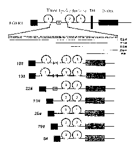
Fig. 1 is a structural representation of FGFR1-Fc fusion protein. FGFR1-Fc
fusion
protein is represented by a solid line, and the deleted amino acid is
represented by a dash
line; the antibody-like domain is represented by a circle; different antibody-
like domains
are represented by number 1-3; the disulfide bond is represented by s s; human
IgG1 Fc is
represented by a grey box; VEGFR1 signal peptide is represented by SP; the
acidic box
sequence is represented by a box with letter AB.
Fig. 2 shows the comparison of FGF-2 binding among various FGFR1-Fc fusion
proteins. Binding of heparin (100 ng/mL) containing FGF-2 (50 ng/mL) or FGF-2
(50
ng/mL) alone to each FGFR1-Fc fusion protein (20 ng/mL) is detected by ELISA.
Fig. 3 shows SDS-PAGE of 26# FGFR1-Fc fusion protein.
CA 02834584 2015-09-15
- 14 -
Fig. 4 shows the binding of FGF-2 to a gradient concentration of 26# FGFR1-Fc
fusion protein.
Fig. 5 shows the affinity between 26# FGFR1-Fc fusion protein and FGF-2.
Fig. 6 shows the effect of 26# FGFR1-Fc fusion protein on the HUVEC cell
division
induced by FGF-2.
DESCRIPTION OF THE PREFERRED EMBODIMENTS
Definitions
Unless otherwise defined, all scientific terms used herein have the same
meaning as
commonly understood by those skilled in the art. With regard to the
definitions and terms
in the art, reference may be made to Current Protocols in Molecular Biology
(Ausubel) by
the skilled one. Standard three- and/or one-letter code used for expressing
one of 20
common L-amino acids in the art are adopted as the abbreviation of amino acid
residues.
Although the number ranges and approximate parameter values are given in a
broad
range in the present invention, all numbers in the specific examples are
described as
precise as possible. However, certain errors exist in any numerical values
essentially,
which may be resulted from the standard deviation during the measurement for
each of
them. Additionally, it should be understood that all ranges disclosed herein
encompass any
and all possible subranges contained therein. For example, it should be
understood that the
range from 1 to 10" as described herein encompasses any and all possible
subranges
between the minimum 1 and the maximum 10 (including the endpoints); i.e., all
subranges
started from the minimum 1 or more, for example 1 to 6.1, and all subranges
ended at the
maximum 10 or less, for example 5.5 to 10.
Additionally, it should be noted that unless otherwise clearly and explicitly
stated, the
singular form includes the plural referent, as used in the present invention.
The term "or"
and the term "and/or" are used interchangeably, unless otherwise clearly
indicated in the
context.
CA 02834584 2013-10-29
- 15 -
As used herein, the term "Fe", "Fe region", "Fe fragment" or "immunoglobulin
Fe
region" refers to the crystallizable fragment of immunoglobulin, and in the
present
invention, said Fe region is preferably the human IgG1 Fe region.
The teiiii "Fe fusion protein" refers to the antibody-like molecule which
incorporates
the binding specificity of a heterologous protein and the effector function of
a constant
region of an immunoglobulin. In the term of molecular structure, a Fe fusion
protein
comprises the amino acid sequence having the required binding specificity and
the
sequence of a constant region of an immunoglobulin. A Fe fusion protein
molecule
generally comprises a binding site of a receptor or a ligand. The sequence of
immunoglobulin constant region may be derived from any immunoglobulin, for
example,
IgG-1, IgG-2, IgG-3 or IgG-4 subtype, IgA (including IgA-1 and IgA-2), IgE,
IgD or IgM.
The term "soluble" protein as used herein refers to the protein which may be
dissolved in an aqueous solution at a biologically relevant temperature, pH
level and
osmotic pressure. The "soluble fusion protein" as used herein is intended to
mean that the
fusion protein does not contain a transmembrane region or an intracellular
region.
As used herein, the term "isolated" refers to the following substance and/or
entity: (1)
which is isolated from at least some components which is present when
initially produced
(in natural environment and/or in a experiment device) and related thereto
and/or (2)
which is produced, prepared and/or manufactured artificially. The isolated
substance
and/or entity may be isolated from at least about 10%, about 20%, about 30%,
about 40%,
about 50%, about 60%, about 70%, about 80%, about 90%, about 95%, about 98%,
about
99%, substantially 100% or 100% other components related to it initially.
The term "part" and "fragment" interchangeably refer to a part of polypeptide,
nucleic
acid or other molecular constructs.
The term "Ig-like domain" as used herein refers to immunoglobulin-like domain,
which may be found in a plurality of protein families and involved in many
biological
functions, including cell-cell recognition, cell surface receptor, immune
function and the
like.
CA 02834584 2013-10-29
- 16 -
Fibroblast growth factor (FGF) is a heparin-binding growth factor family,
which has
22 family members in the mammals (FGF 1-14, 16-23). FGF has many important
biological functions, such as cell multiplication, differentiation, migration,
angiogenesis
and tumorigenesis. FGF exerts many biological functions by binding and
activating the
cell surface FGF receptor (FGFR). (See, for example, Eswarakumar et al.
Cytokine
Growth Factor Rev. 16: 139-149, 2005). Fibroblast growth factor receptor
(FGFR) is the
receptor that binds the family members of fibroblast growth factor. A part of
fibroblast
growth factor receptor is involved in the disease process. In the mammals,
there are 4
FGFR genes: fgfR 1 -fgfR4. The fibroblast growth factor receptor is composed
of
extracellular domain, transmembrane domain and intracellular domain. There are
many
members in FGFR family, which are different from each other in the term of
ligand
binding properties and kinase domains. However, the extracellular domains
thereof are
similar. There are three immunoglobulin-like (Ig-like) domains contained in
their
extracellular domains: the first Ig-like domain, the second Ig-like domain and
the third
Ig-like domain, and there is also a sequence contained between the first and
the second
Ig-like domain. Said sequence contained between the first and the second Ig-
like domain
is referred herein as the intermediate functional sequence region of the Ig-
like domain of
FGFR. Said intermediate regulation sequence comprises a region of acidic amino
acids,
referred as acidic box (AB).
As used herein, the term "the first Ig-like domain of FGFR" or "the first Ig-
like
domain" refers to the first Ig-like domain in the protein FGFR from the N-
tenninus, which
has for example the amino acid sequence corresponding to position 40 to
position 118 of
SEQ ID NO: 1. Similarly, the term "the second Ig-like domain of FGFR" or "the
second
Ig-like domain" refers to the second Ig-like domain in the protein FGFR from
the
N-terminus, which has for example the amino acid sequence corresponding to
position 163
to position 247 of SEQ ID NO: 1; the term "the third Ig-like domain of FGFR"
or "the
third Ig-like domain" refers to the first Ig-like domain in the protein FGFR
from the
N-terminus, which has for example the amino acid sequence corresponding to
position 270
to position 359 of SEQ ID NO: 1. Preferably, the FGFR is FGFR1, and the first
Ig-like
CA 02834584 2013-10-29
- 17 -
domain of FGFR is the first Ig-like domain of FGFR1, and the second Ig-like
domain of
FGFR is the second Ig-like domain of FGFR1, and the third Ig-like domain of
FGFR is the
third Ig-like domain of FGFR1.
A part of sequence of hFGFR1 is given as follows, in which each Ig-like domain
is
shown in shaded area sequentially, see
http://www.ncbi.nlm.nih.gov/protein/AAH15035.1
MWSWKCLLFWAVLVTATLCTARP SPTLPEQAQPWGAPVEVESFLVHPGDLLQLR
CRLRDDVQSINWLRDGVQLAESNRTRITGEEVEVQDSVPADSGLYACVTSSPSGS
DTTYFSVNVSDALP S SEDDDDDDDSSSEEKETDNTKPNPVAPYWTSPEKMEKK
rAVPAAKTVKIKCPSSG 180
N1(4iliP LTRIGGYKVRKATWSTIVIDS
VPS DKUNYTC IVEN HYGSINI-ITYQLDV E RSPHRPILQAGLPANKTVAL(i NVF F
M CK V YSDPQP Li IQ WLKHIEVNGSKICiP300DN LPYVQILKTAGVN-I LDKEIVIEVL4
R N V SFEDAGEYTCLAGNS1GLSHHSAWLTVLEA LEER
The amino acid sequence of FGFR1 may be found in SEQ ID NO: 1, and its
encoding nucleotide sequence may be found in SEQ ID NO: 4.
As used herein, the term "the intermediate functional sequence region of the
Ig-like
domain of FGFR" or "the intermediate functional sequence of the Ig-like domain
of
FGFR" or "IFS" refers to the sequence between the first Ig-like domain and the
second
Ig-like domain in the protein FGFR, and preferably, IFS sequence has the amino
acid
sequence corresponding to position 118 to position 162 of SEQ ID NO: 1.
Unexpectedly,
it has been found by the present inventor that there is a significant effect
of the
intermediate functional sequence region on the function of the Ig-like domain.
In some
embodiments of the present invention a FGFR fusion protein, which comprises a
plurality
of parts of various lengths derived from the intermediate functional sequence
region, and
particularly preferably, the part derived from the intermediate functional
sequence region
contains no acidic box. More preferably, the part derived from IFS has the
amino acid
sequence corresponding to position 134 to position 162, position 145 to
position 162 or
position 151 to position 162 of SEQ ID NO: 1. The protein FGFR is preferably
FGFR1
(SEQ ID NO: 1), especially the protein human FGFR1. The amino acid sequence of
the
CA 02834584 2013-10-29
- 18 -
protein human FGFR1 may be found in SEQ ID NO: 1, and its cDNA sequence may be
found in SEQ ID NO: 4.
The term "FGFR" as used herein refers to fibroblast growth factor receptor,
which
may be FGFR1, FGFR2, FGFR3 and/or FGFR4. Preferably, the FGFR in the present
invention is FGFR1, more preferably, human FGFR1.
As used herein, the term "degenerate variant" is intended to mean that the
degenerate
variant comprises a degenerate change at the third position of the amino acid
codon so that
the degenerate variants encode the same amino acid, for example the wobble
position of a
triplet code comprising one or more changed variants (also referred as
synonymous
variant).
As used herein, the term "subject" refers to mammals, such as human. However,
it
may also be other animals, such as domesticated animals (such as dog and cat
etc.),
livestocks (such as cattle, sheep, pig and horse etc.) or experimental animals
(such as
monkey, rat, mouse, rabbit and guinea pig etc.).
As used herein, the term "percentage identity", "homology" or "identity"
refers to the
sequence identity between two amino acid sequences or nucleic acid sequences.
The
percentage identity may be determined by alignment between two sequences, and
the
percentage identity refers to the amount of the same residue (i.e., amino acid
or nucleotide)
at the same position in the sequence aligned. Sequence alignment and
comparison may be
performed using standard algorithms in the art (for example Smith and
Waterman, 1981,
Adv. Appl . Math. 2: 482; Needleman and Wunsch, 1970, 1 MoI. Biol. 48: 443;
Pearson
and Lipman, 1988, Proc. Natl. Acad. Sci., USA, 85: 2444) or by the
computerized
versions of these algorithms (Wisconsin Genetics Software Package Release 7.0,
Genetics
Computer Group, 575 Science Drive, Madison, WI). Said computerized versions
publicly
available are BLAST and FASTA. Additionally, ENTREZ available through National
Institutes of Health (Bethesda MD) may be used for sequence alignment. When
BLAST
and GAP-BLAST are used, default parameters for each program (for example,
BLASTN,
available on the website of National Center for Biotechnology Information) may
be used.
CA 02834584 2015-09-15
- 19 -
In one embodiment, the percentage identity between two sequences may be
determined using GCG with a gap-weight of 1 so that the giving weight of each
amino
acid gap seems as if it is a single amino acid mismatch between two sequences.
Alternatively, ALIGN (version 2.0), which is a part of GCG (Accelrys, San
Diego, CA)
Sequence Alignment Software Package, may be used.
As used herein, the term "hybridization" refers to the process by which a
stable
double-stranded polynucleotide is formed by non-covalent bonding between two
single
stranded polynucleotides. The term "hybridization" also may refer to triple-
stranded
hybridization. The double stranded polynucleotide (generally) produced is the
"hybrid" or
"duplex". "The condition for hybridization" generally includes a salt
concentration lower
than about 1 M, and more generally, lower than about 500 mM, and lower than
about 200
mM. The hybridization temperature may be as low as 5 C, but it usually higher
than about
22 C, and more usually higher than about 30 C, and preferably higher than
about 37 C.
Hybridization is usually carried out under strict conditions (i.e., the
conditions under
which the probe will hybridize to its target sequence). Strict hybridization
conditions are
dependent on the sequence and will be varied under different conditions.
Higher
hybridization temperature will be probably required by longer segments for
specific
hybridization. Since the hybridization stringency may be influenced by other
factors
(including base composition and length of the complementary strand, the
presence of
organic solvent and the degree of base mismatch), the combination of
parameters is more
important than the absolute value of any single parameter. Generally, the
strict condition is
selected as 5 C lower than the Tm of the sequence under certain ionic strength
and pH.
Exemplary strict conditions include pH 7.0 to 8.3, sodium ion (or other salts)
concentration of at least 0.01 M to no more than 1 M and temperature of at
least 25 C. For
strict conditions, see, for example Sambrook, Fritsche and Maniatis.
"Molecular Cloning
A laboratory Manual", 21ud edition, Cold Spring Harbor Press (1989) and
Anderson
"Nucleic Acid Hybridization", 1st edition, BIOS Scientific Publishers Limited
(1999).
CA 02834584 2013-10-29
- 20 -
As used herein, the term "linker", "peptide linker", "linking sequence" or
"linker
sequence" refers to a short amino acid sequence by which individual domain
and/or region
involved in the present fusion protein are linked together, and the length of
the short
amino acid sequence is generally 0-20 amino acids, and preferably, 2-10 amino
acids.
As used herein, the term of "the amino acid sequence corresponding to SEQ ID
NO:
N" in a fusion protein or part or domain is intended to mean said fusion
protein or part or
domain has the amino acid sequence substantially as indicated by SEQ ID NO: N,
and
preferably, containing no more than 1, 2, 3, 4, 5, 10 or 20 substitutions,
additions and
deletions of amino acids, and yet preferably, said fusion protein or part or
domain shares at
least 80%, 90%, 93%, 95%, 97%, 98% or 99% identity with the amino acid
sequence of
SEQ ID NO: N, and more preferably, said fusion protein or part or domain has
the amino
acid sequence as indicated by SEQ ID NO: N.
As used herein, the term "FGFR-Fc fusion protein" refers to a fusion protein
which
comprises the part derived from the extracellular domain of FGFR and the part
derived
from the immunoglobulin Fe region, wherein the part derived from the
extracellular
domain of FGFR may: (1) comprise the amino acid sequence sharing at least 70%
identity,
preferably at least 80%, 90%, 93%, 95%, 97%, 98% or 99% identity, with the
amino acid
sequence indicated by any one of SEQ ID NOs: 9-15 or composed thereof; (2)
comprise
the amino acid sequence encoded by the nucleotide sequence sharing at least
70% identity,
preferably at least 80%, 90%, 93%, 95%, 97%, 98% or 99% identity, with the
nucleotide
sequence indicated by any one of SEQ ID NOs: 16-22 or composed thereof; or (3)
possess
the amino acid sequence indicated by any one of SEQ ID NOs: 9-15, or the amino
acid
sequence encoded by the nucleotide sequence indicated by any one of SEQ ID
NOs:
16-22.
In some preferable embodiments, the FGFR-Fc fusion protein may be encoded by
the
nucleic acid, in which the nucleotide sequence encoding the part derived from
the
extracellular domain of FGFR comprises the sequence of which the complementary
sequence is hybridized with the nucleotide sequence as indicated by any one of
SEQ ID
CA 02834584 2013-10-29
-21 -
NOs: 16-22 under stringent conditions, or comprises the degenerative variant
of the
nucleotide sequence as indicated by any one of SEQ ID NOs: 16-22. In some
preferable
embodiments, the nucleotide sequence encoding the immunoglobulin Fe region
comprises
the sequence of which the complementary sequence is hybridized with the
nucleotide
sequence indicated by SEQ ID NO: 8 under stringent conditions, or comprises
the
degenerative variant of the nucleotide sequence indicated by SEQ ID NO: 8.
In other preferable embodiments, the FGFR-Fc fusion protein includes the FGFR-
Fc
fusion protein variant. In one embodiment, the variant includes the variant
which contains
no more than 2, 3, 4, 5 or 10 substitutions, additions or deletions of amino
acid in the part
derived from IFS corresponding to the amino acid sequence indicated by
position 134 to
position 162, position 145 to position 162 or position 151 to position 162 of
SEQ ID NO:
1, and preferably, the variant retains the angiogenesis-inhibitory capacity.
In another
embodiment, the variant includes the variant which contains no more than 2, 3,
4, 5, 10 or
20 substitutions, additions or deletions of amino acid in D2 domain
corresponding to the
amino acid sequence indicated by position 163 to position 247 of SEQ ID NO: 1,
and
preferably, the variant retains the angiogenesis-inhibitory capacity. In
another embodiment,
the variant includes the variant which contains no more than 2, 3, 4, 5, 10 or
20
substitutions, additions or deletions of amino acid in D3 domain corresponding
to the
amino acid sequence indicated by position 270 to position 359 of SEQ ID NO: 1,
and
preferably, the variant retains the angiogenesis-inhibitory capacity. In
another embodiment,
the substitution, addition or deletion is located at the linker or the linking
part.
In addition to the naturally occurring modifications in the part derived from
the
extracellular domain of FGFR and the part derived from immunoglobulin Fe
region, other
post-translational modifications may also be comprised in the FGFR-Fc fusion
protein.
Such modifications include, but are not limited to, acetylation,
carboxylation,
glycosylation, phosphorylation, esterification and acylation. As a result, non-
amino acid
component may be comprised in the modified FGFR-Fc fusion protein, for example
polyethylene glycol, lipid, polysaccharide or monosaccharide, and phosphoric
acid. Effect
CA 02834584 2013-10-29
- 22 -
of such non-amino acid components on the function of the FGFR-Fc fusion
protein may
be tested as described for other FGFR-Fc fusion protein variants herein. When
FGFR-Fc
fusion protein is produced in a cell, post-translational processing is also
possibly important
for correct folding and/or protein function. Special cell machines and unique
mechanisms
exist in different cells (for example CHO, HeLa, MDCK, 293, WI38, NIH-3T3 or
HEK293) for these post-translational activities, and different cells may be
selected to
make sure correct modification and processing of FGFR-Fc fusion protein.
The fusion protein as described herein may be produced by any method known in
the
art. For example, it may be produced by chemical synthesis or from nucleic
acid
expression. The peptides used in the present invention may be easily prepared
according to
the established standard liquid, or preferably, solid phase peptide synthesis
method known
in the art (see, for example J. M. Stewart and J. D. Young, Solid Phase
Peptide Synthesis,
2nd edition, Pierce Chemical Company, Rockford, Illinois (1984), in M.
Bodanzsky, and A.
Bodanzsky, The Practice of Peptide Synthesis, Springer Verlag, New York
(1984)). The
fusion protein may be produced by the techniques known in the art so that one
or more
intramolecular crosslinkings may be formed between the cysteine residues
located in the
polypeptide sequence expected to be comprised in the protein (see, for example
US patent
No. 5478925). In addition, general modifications may be performed to the
protein
described herein by adding cysteine or biotin to the C-terminus or N-terminus
of the
protein.
As used herein, "therapeutically effective amount" or "effective amount"
refers to the
dosage which is sufficient to show the benefit to the subject administrated.
The actually
administrated dosage, the rate and the time course of administration are
dependent on the
condition of the patient and the severity of the disease. Finally, the
physician is
responsible for the prescription (for example decision on the dosage etc.) and
will make a
decision for the treatment, usually by considering the disease treated,
individual condition
of the patient, position of delivery, the method for administration and other
factors known
to the physician.
CA 02834584 2013-10-29
- 23 -
A series of isolated soluble FGFR-Fc fusion proteins are constructed by the
present
inventor, which may bind FGF and effectively inhibit the cell division induced
by FGF.
The fusion protein preferably comprises: the part derived from IFS, D2, D3 and
immunoglobulin Fe region.
Unexpectedly, it has also been found by the present inventor that the binding
of FGF
by the fusion protein is significantly influenced by the length of the part
derived from IFS.
Therefore, in some embodiments of the present invention fusion proteins
comprising the
parts derived from IFS with various lengths. Preferably, the part derived from
IFS
comprises no acidic box, and more preferably, it has the amino acid sequence
corresponding to position 134 to position 162, position 145 to position 162 or
position 151
to position 162 of SEQ ID NO: 1. In some preferable embodiments, the part
derived from
IFS comprises the fusion protein corresponding to the amino acid sequence
indicated by
position 145 to position 162 of SEQ ID NO: 1, which has extremely high FGF
affinity and
may particularly effectively inhibit the cell division induced by FGF.
In some embodiments of the present invention, a soluble FGFR-Fc fusion protein
is
provided, which comprises: D1, a part derived from IFS, D2, D3 and
immunoglobulin Fe
region. Preferably, the part derived from IFS comprises no acidic box, and
more
preferably, it has the amino acid sequence corresponding to position 134 to
position 162,
position 145 to position 162 or position 151 to position 162 of SEQ ID NO: 1.
In some other embodiments of the present invention, a soluble FGFR-Fc fusion
protein is provided, which comprises: a part of D1, a part derived from IFS,
D2, D3 and
immunoglobulin Fe region. Preferably, the part derived from IFS comprises no
acidic box,
and more preferably, it has the amino acid sequence corresponding to position
134 to
position 162, position 145 to position 162 or position 151 to position 162 of
SEQ ID NO:
1.
In some other embodiments of the present invention, a soluble FGFR-Fc fusion
protein is provided, which is composed of: a part derived from IFS, D2, D3 and
immunoglobulin Fe region. Preferably, the part derived from IFS comprises no
acidic box,
CA 02834584 2013-10-29
- 24 -
and more preferably, it has the amino acid sequence corresponding to position
134 to
position 162, position 145 to position 162 or position 151 to position 162 of
SEQ ID NO:
1.
In some other embodiments of the present invention, a soluble FGFR-Fc fusion
protein is provided, which is sequentially composed of, from the N-tenninus to
the
C-terminus, a part derived from IFS, D2, D3 and immunoglobulin Fe region.
Preferably,
the part derived from IFS comprises no acidic box, and more preferably, it has
the amino
acid sequence corresponding to position 134 to position 162, position 145 to
position 162
or position 151 to position 162 of SEQ ID NO: 1.
In some other embodiments of the present invention, an FGFR-Fc fusion protein
is
provided, which may inhibit tumor cells directly or indirectly. Preferably,
the FGFR-Fc
fusion protein of the present invention inhibits tumor cells directly. More
preferably, the
growth of tumor cells is inhibited by the FGFR-Fc fusion protein of the
present invention
by at least 10%, 20%, 30%, 40%, 50%, 80%, 90% and 95% etc. The tumor cells may
be
any tumor cells, for example, leukaemia, lung cancer, liver cancer, head and
neck cancer,
stomach cancer, bladder cancer, carcinoma of uterine cervix etc. Particularly,
the
inhibition is achieved by direct binding to tumor cells.
In some embodiments, the present invention includes use of (i) FGFR-Fc fusion
protein, or (ii) the polynucleotide encoding such fusion protein, in the
preparation of the
compositions or medicaments for the treatment of diseases mediated by or
related to
angiogenesis. For example, in one embodiment, the present invention includes
use of (i)
FGFR-Fc fusion protein, or (ii) the polynucleotide encoding such fusion
protein in the
preparation of the medicaments as an angiogenesis inhibitor.
In some embodiments, the FGFR-Fc fusion protein according to the present
invention
may be produced by the expression of the nucleotide sequence as indicated by
any one of
SEQ ID NOs: 16-22 in a mammalian cell line. In particular, the mammalian cell
line is
CHO cell line.
CA 02834584 2013-10-29
- 25 -
Additionally, in the present invention, the FGFR-Fc fusion protein as
described below
is provided, in which a part derived from the extracellular domain of FGFR may
be fused
with the immunoglobulin Fc region with or without a linker.
In some other embodiments, the present invention includes the isolated nucleic
acid
molecules encoding the FGFR-Fc fusion protein, and the present invention also
includes
use of these molecules in manufacture of a medicament. The nucleic acid may be
recombinant, synthetic or produced by any available methods in the art, and
the method
includes cloning by means of using a standard technique.
In some other embodiments, the present invention includes a vector comprising
the
isolated nucleic acid molecule of the present invention. The vector may be an
expression
vector, in which the nucleic acid is operatively linked to a control sequence
which is able
to provide the expression of the nucleic acid in a host cell. A plurality of
vectors may be
used. For example, suitable vectors may include virus (for example poxvirus,
adenovirus,
baculovirus etc.); yeast vector, bacteriophage, chromosome, artificial
chromosome,
plasmid, cosmid.
In some embodiments, the present invention further includes the cells
transfected by
these vectors so that the FGFR-Fc fusion protein is expressed. The host cell
suitable for
the present invention may be prokaryotic cell or eukaryotic cell. They include
bacteria, for
example E. coli, yeast, insect cell and mammalian cell. The mammalian cell
lines that may
be used include, but are not limited to, Chinese Hamster Ovary (CHO) cell,
baby hamster
kidney cell, NSO mouse myeloma cell, monkey and human cell lines, and derivate
cell
lines thereof, etc.
In another aspect of the present invention, a method for angiogenesis
inhibition is
provided, comprising administrating the FGFR-Fc fusion protein of the present
invention
to the subject in need thereof Preferably, the method is carried out in the
mammals.
In another aspect of the present invention, a method for binding FGF in vitro
or in vivo
is provided, which comprises contacting FGF to the fusion protein according to
the present
invention.
CA 02834584 2013-10-29
- 26 -
In another aspect of the present invention, a method for the treatment or
prevention of
tumors in the mammals is provided, which comprises administrating the FGFR-Fc
fusion
protein of the present invention to the subject in need thereof, and
preferably, the tumor is
a solid tumor.
In another aspect of the present invention, a method for the treatment or
prevention of
ophthalmic angiogenesis-related diseases in the mammals is provided, which
comprises
administrating the FGFR-Fc fusion protein of the present invention to the
subject in need
thereof, and preferably, the ophthalmic angiogenesis-related disease is age-
related macular
degeneration.
The present invention also relates to use of the FGFR-Fc fusion protein in the
preparation of medicaments for angiogenesis inhibition. Additionally, the
present
invention also relates to use of the FGFR-Fc fusion protein in the preparation
of
medicaments for the treatment or prevention of angiogenesis-related diseases,
and
preferably, angiogenesis-related diseases are tumors or ophthalmic
angiogenesis-related
disease.
The angiogenesis-related diseases as described in the present invention
include, but
are not limited to, angiogenesis-dependent cancers, comprising, for example,
solid tumor,
hematogenic tumor (for example leukaemia) and tumor metastasis; benign tumor,
for
example, angioma, acoustic neuroma, neurofibroma, trachoma and pyogenic
granuloma;
rheumatoid arthritis; psoriasis; rubeosis; Osler-Webber Syndrome; myocardial
angiogenesis; plaque neovascularization; telangiectasia; hemophiliac joint and
angiofibroma.
In some embodiments of the methods described, one or more FGFR-Fc fusion
proteins
may be administrated together (simultaneously) or at a different time
(sequentially).
Additionally, the fusion protein may be administrated together with additional
medicament
used for cancer treatment or angiogenesis inhibition.
In some embodiments, the method disclosed in the present invention may be used
alone. Alternatively, the subject method may be combined with other
conventional
CA 02834584 2013-10-29
- 27 -
anticancer therapies for the treatment or prevention of proliferative diseases
(for example
tumor). For example, these methods may be used for the prevention of cancers,
the
prevention of cancer relapse and postoperative metastasis, and may be used as
a
supplement for other cancer therapies. As disclosed in the present invention,
the
effectiveness of conventional cancer therapies (for example, chemotherapy,
radiotherapy,
phototherapy, immunotherapy and operation) may be enhanced by using target
polypeptide therapeutic agents.
In ophthalmology, angiogenesis is related to, for example, diabetic
retinopathy,
retinopathy of prematurity, age-related macular degeneration, corneal
transplantation
rejection, neovascular glaucoma and RLF (retrolental fibroplasia). The FGFR-Fc
fusion
protein disclosed herein may be administrated inside the eye or by other
routes. Other
diseases related to angiogenesis in ophthalmology include, but not limited to,
epidemic
keratoconjunctivitis, Vitamin A deficiency, contact lens overwear, atopic
keratitis,
superior limbic keratitis, pterygium keratitis sicca, sjogren, acne rosacea,
phlyctenosis,
syphilis, Mycobacteria infection, lipid degeneration, chemical burn, bacterial
ulcer, fungal
ulcer, Herpes simplex infection, Herpes zoster infection, protozoan infection,
Kaposi
sarcoma, Mooren ulcer, Terrien's marginal degeneration, mariginal keratolysis,
rheumatoid arthritis, systemic lupus, polyarteritis, trauma, Wegeners
sarcoidosis, Scleritis,
Steven's Johnson disease, periphigoid radial keratotomy and comeal graph
rejection, sickle
cell anemia, sarcoid, pseudoxanthoma elasticum, Pagets disease, vein
occlusion, artery
occlusion, carotid obstructive disease, chronic uveitis/vitritis,
mycobacterial infections,
Lyme's disease, systemic lupus erythematosis, retinopathy of prematurity,
Eales disease,
Bechets disease, infection resulting in retinitis or choroiditis, presumed
ocular
histoplasmosis, Bests disease, myopia, optic pit, Stargarts disease, pars
planitis, chronic
retinal detachment, hyperviscosity syndromes, toxoplasmosis, trauma and post-
laser
complication.Other diseases include, but not limited to, rubeosis
(neovasculariation of the
angle) related diseases and diseases induced by abnormal hyperplasia of the
fibrous blood
vessel or fibrous tissue, including all kinds of proliferative
vitreoretinopathy.
CA 02834584 2013-10-29
- 28 -
Administration
The fusion protein of the present invention may be administrated alone, but
preferably, as a pharmaceutical composition which usually comprises a suitable
pharmaceutical excipient, diluent or carrier selected according to the
intended
administration route. The fusion protein may be administrated to the patient
in need
thereof by any suitable route. A precise dosage will be dependent on many
factors,
including exact properties of the fusion protein.
Some suitable administration routes include (but are not limited to) oral,
rectal, nasal,
topical (including buccal and sublingual), subcutaneous, vaginal or parenteral
(including
subcutaneous, intramuscular, intravenous, intracutaneous, intrathecal and
extradural)
administration.
For intravenous injection and injection at the focal site, active ingredients
are present
in the form of a parenterally-acceptable aqueous solution, which is free of
pyrogen and has
appropriate pH value, isotonicity and stability.
A suitable solution may be well formulated by the skilled one in the art
using, for
example, isotonic excipients such as sodium chloride injection, Ringer's
injection, Ringer's
lactate injection. As required, preservative, stabilizer, buffering agent,
antioxidant and/or
some other additives may be added. The pharmaceutical composition orally
administrated
may be in a form of tablet, capsule, powder or oral liquid etc. Solid carrier,
such as gelatin
or adjuvant, may be comprised in a tablet. Liquid pharmaceutical composition
usually
comprises liquid carrier, such as water, petroleum, animal or vegetable oil,
mineral oil or
synthetic oil. Also included may be non-nal saline solution, glucose or other
sugar
solutions or glycols such as ethylene glycol, propylene glycol or polyethylene
glycol.
Examples of the techniques and schemes as mentioned above and other techniques
and schemes as used according to the present invention may be found in
Remington's
Pharmaceutical Sciences, 16th edition, Oslo, A. (ed), 1980.
CA 02834584 2013-10-29
- 29 -
Cloning of the fusion protein and construction of the expression plasmid
The FGF receptor fragment are obtained from the amplification of the cDNA
template of corresponding receptor through PCR, and IgG1 Fc fragment is
obtained from
the cDNA amplification of the human-derived IgG1 through PCR. When PCR primers
are
designed, linking sequences are introduced between different fragments so that
these
different fragments may be finally linked by overlap PCR to form reading
frames for
different fusion proteins, and endonuclease BspE I and Pst I site are added to
both ends of
the cDNA. The cDNAs for different fusion proteins may be cloned to the
expression
plasmid after digestion by BspE I and Pst I. The plasmid after cloning may be
determined
by endonuclease digestion, electrophoresis and finally DNA sequencing.
Expression and purification of the fusion protein
The present fusion protein may be expressed and purified by techniques
commonly
used in the art. DNA from corresponding fusion protein plasmid was purified
using
plasmid purification kit (MAX) available from Qiagen, and the concentration of
plasmid
DNA was determined using UV spectrophotometry, and the plasmid was transfected
to
CHO cell using FUGENE 6 liposome (Roche). Specific method for transfection was
performed according to the specification of the product. Based on the
expression amount
required for the proteins, two methods were employed in the present invention
for protein
expression: (1) transient expression, in which the fusion protein contained
culture
supernatant was usually harvested 48-72 h after transfection, and the relative
content of
the fusion protein was then determined using human IgG ELISA so that the
fusion protein
may be rapidly and efficiently obtained; (2) establishing a stable cell line
and producing
the common DHFR-defective CHO cell expression system using the recombinant
protein
medicament expression, the basic process of which includes cell transfection,
selection of
stably transfected cell, clone screening, stress amplification, culture medium
and process
optimization etc., and finally realizing a large-scale suspension culture of
CHO
CA 02834584 2013-10-29
- 30 -
engineering cell strain in a serum free culture medium. The culture product
was collected
and the fusion protein was purified using Protein A affinity column. The
purified protein
was analyzed by sodium dodecyl sulfate polyacrylamide gel electrophoresis (SDS-
PAGE),
and subsequently all eluates in which the required expression product was
contained were
combined and filtered using a 0.22 tm filter, and then protein quantification
was carried
out according to a plurality of methods such as Lowry protein assay. The
volume of CHO
cell culture in the present invention was at a level of 10 L bioreactor,
through which the
fusion protein obtained after purification could satisfy the protein amount
required in the
animal experiments, and also a basis was established for future scaling-up.
Neutralization of FGF by the fusion protein was validated at a level of
protein
After the fusion protein expressed by CHO was obtained, the binding capacity
of the
fusion protein to FGF is evaluated in the present invention at a level of
protein. Binding
experiment and affinity experiment were performed for validation in the
present invention,
in which steps of the binding experiment included: after initially coated by
FGF-2 on a
96-well ELISA plate, the coated well was blocked by BSA followed by adding
each fusion
protein at the same concentration, and then a secondary antibody to human IgG
Fc-HRP
was added after washing, and the samples were developed, stopped and read at
450nm on
a ELISA plate, and finally the fusion protein which had binding capacity to
FGF-2 was
screened based on the signal strength. The affinity experiment was performed
in order to
determine the affinity of the fusion protein to FGF-2 in the solution system,
which
comprised the following steps: FGF-2 was initially coated on a 96-well ELISA
plate to
capture the antibody, and then the coated well was blocked by BSA, and
subsequently a
mxiture of the fusion protein and FGF-2 which was previously prepared and
incubated
were added with a gradient of diluted standards, and after incubation, an HRP-
labeled
detection antibody was added (using antibody 2 which specifically detected
free VEGF or
FGF-2), and subsequently the samples were developed, stopped and read at 450nm
on a
ELISA plate, and finally the relative concentration of free FGF-2 was detected
in the
CA 02834584 2013-10-29
- 31 -
mixture of the fusion protein and FGF-2. Through the experiments above, the
fusion
protein having a blocking effect on FGF-2 was screened.
Neutralization of FGF by the fusion protein was validated at a cellular level
After the binding capacity of the fusion protein to FGF-2 was determined at a
level of
protein, its angiogenesis-inhibiting effect will be further validated at a
cellular level in the
present invention. The inhibition capacity of the fusion protein on the
division and
migration of the vascular endotheliocyte is examined by the division test
using human
umbilical vein endothelial cell (HUVEC) and the HUEVC cell migration test. The
inhibition capacity of the fusion protein on the division of HUVEC cell can be
examined
by the HUVEC cell division test, which comprises the following steps during
the
experiment: 3000 HUVEC cells/well were inoculated to a 96-well plate and
cultured at
37 C in an incubator supplemented with 5% CO2, and then FGF-2 as well as a
mixture of
the fusion protein at different concentrations with FGF-2 are added
respectively, and after
culturing for another 3-4 days, 10% CCK-8 is added and cultured for 2 h before
the
sample is read at 450 nm on a EL1SA plate. The inhibition capacity of the
fusion protein
on the division of vascular endotheliocyte induced by FGF-2 was evaluated
based on the
difference of absorbance, and the median effective concentration of the fusion
protein was
obtained for FGF-2 inhibition. The inhibition capacity of the fusion protein
on HUVEC
cell migration was examined by the HUVEC cell migration test, which comprises
the
following steps during the experiment: 50000 HUVEC cells as well as the fusion
protein
at various concentrations were initially inoculated in the upper chamber,
while 600 [IL
FGF-2 containing culture liquid was added into the lower chamber, and
subsequently, the
sample was cultured at 37 C in an incubator supplemented with 5% CO2 for 20-24
h
before cells on the face side of the membrane of the upper chamber were
removed, and
then cells on the back side of the membrane were fixed, stained and washed
with PBS
before observed and counted under an inverted microscope. The migration of
HUVEC
cells induced by the stimulation of FGF-2 was demonstrated by counting the
HUVEC
CA 02834584 2013-10-29
- 32 -
cells on the back side of the membrane, and the inhibition capacity of the
fusion protein on
the migration of the vascular endotheliocyte was tested by adding the fusion
protein at
various concentrations into the culture liquid. Through the experiments
mentioned above,
the inhibition capacity of the new fusion protein constructed in the present
invention was
validated on the division and migration of the vascular endotheliocyte induced
by FGF-2,
which also provided a basis for future animal experiments.
Tumor growth-inhibiting capacity of the fusion protein was validated by the
tumor model
After the blocking effect of the new fusion protein in the present invention
on FGF-2
signal was demonstrated by experiments at a protein level and a cellular
level, its
anti-tumor capacity would be tested in animal tumor models in the present
invention. In
the present invention, the anti-angiogenesis and anti-tumor effect of the
fusion protein
would be validated by models commonly used in searching medicaments for
angiogenesis
and tumor, for example, LLC mouse lung cancer, U87 gliocytoma, B16 melanoma
and so
on. In animal experiments, in addition to conventional control groups, control
medicaments, such as VEGF-Trap, FP-1039, would also be included so as to
obtain
comparative data for anti-tumor capacity. During experiments, 100 ttL tumor
cell liquid
with appropriate amount was subcutaneously injected into C57 mouse on one side
of the
back, and the tumor volume was measured with a vernier caliper twice a week.
Upon the
tumor grew to about 200 mm3, the fusion protein at various concentrations was
subcutaneously injected and the mice were sacrificed after 2-3 weeks.
Subsequently, the
tumor volume was measured with a vernier caliper, and the anti-tumor effect of
the fusion
protein was validated by the size of the tumor. Furthermore, individual tumor
tissue was
analyzed using methods such as immunohistochemistry to investigate the
regulation
mechanism of angiogenesis.
Examples
CA 02834584 2013-10-29
- 33 -
Example 1: Construction of recombinant expression plasmid for FGFR1-Fc fusion
protein
The FGF receptor fragment is obtained from the amplification of the cDNA
templet
of FGF receptor through PCR, and IgG1 Fe fragment is obtained from the cDNA
amplification of the human-derived IgG1 through PCR. A commercially available
cDNA
(PCR Ready First Strand cDNA, derived from human adult colon cancer tissue,
BioChain)
was used as the template for FGFR1 fragment. Total RNA was extracted from the
blood of
healthy human subjects using human blood RNA extraction kit (QIAGEN).
According to
the manufacturer's instruction of reverse transcription kit (Promega), RT-PCR
was
performed using M-MLV reverse transcriptase (Promega) so that RNA was
reversely
transcripted to cDNA which was used as the template for IgG1 Fe fragment. RT-
PCR was
performed according to the manufacturer's instruction of reverse transcription
kit, which
has the following steps: Oligo dT, dNTP, total RNA and DEPC H20 were mixed
homogeneously and reacted at 70 C for 10 min before placed on ice for 5 min,
and
subsequently RNase inhibitor, M-MLV reverse transcriptase and reaction buffer
were
added. The mixture was reacted at 42 C for 1 h and subsequently at 70 C for 15
min, and
the cDNA obtained may be used as the template.
Various FGFR1 fragments were individually amplified by PCR using the cDNA from
human adult colon cancer tissue as the template (the primers were listed in
table 1), and
IgG1 Fe fragment was amplified by PCR using human blood cDNA as the template
(the
primers were listed in table 1 and 2). The reaction conditions for the PCR
were as follows:
min of pre-denaturalization at 98 C, total 30 cycles of 30 s of
denaturalization at 98 C,
45 s of annealing at 56 C and 2 min of extension at 72 C, and finally another
10 min of
extension. When PCR primers were designed, 20 or more complementary base
sequences
were introduced as the linking sequence between FGFR1 fragment and IgG1 Fe
fragment
so that the FGFR1 fragment and IgG1 Fe fragment may be subsequently linked by
overlap
PCR to form reading frames for different fusion proteins, and at the same
time, restriction
endonuclease BspE I and Pst I site were added at both ends of the PCR product.
CA 02834584 2013-10-29
- 34 -
Subsequently, overlap PCR was carried out to obtain each FGFR1-Fc fusion
protein
fragment by amplification. The process of the overlap PCR reaction may be
divided into
two rounds, in which the fragment required for linking and containing no
primer was
included in the first round with reaction conditions as follows: 5 min of
pre-denaturalization at 98 C, 6 cycles of 30 s of denaturalization at 98 C, 45
s of
annealing at 56 C and 5 min of extension at 72 C, and finally another 10 min
of extension
at 72 C; after the first round, the second round of PCR was carried out by
adding the
primers for both ends with reaction conditions as follows: 5 min of pre-
denaturalization at
98 C, 30 cycles of 30 s of denaturalization at 98 C, 45 s of annealing at 56 C
and 2 min of
extension at 72 C, and finally another 10 min of extension at 72 C; through
the process
above, reading frames for different fusion proteins were spliced, and at the
same time,
restriction endonuclease BspE I and Pst I site were added at both ends of the
cDNA.
After amplification, the fragments amplified by PCR were purified using
QIAquick
PCR purification kit (QIAGEN). cDNAs of various fusion proteins and the
eucaryotic
expression plasmid pSV2-dhfr (ATCC) were digested by BspE I and Pst I,
respectively.
Subsequently, 1% agarose gel electrophoresis was performed on the digested
samples
under a voltage of 90 V. Target fragments were recovered using QIAquick gel
extraction
kit (QIAGEN) before ligating at 16 C for 1 h using a ligase (NEB). The mixture
for
ligation reaction was transformed to the competent Top10 E. coli under the
conditions of
90 s of reaction at 42 C followed by 3 min of standing on ice. After the
sterile LB culture
broth (free of antibody) added, the mixture was shaken at 250 rpm in a shaker
at 37 C for
1 h before coating on a LB plate supplemented with ampicillin. The plate was
cultured
overnight in a thermostated incubator at 37 C, and then single colonies were
picked out
and transferred to an ampicillin-containing LB culture broth. The inoculated
culture broth
was shaken at 250 rpm in a shaker at 37 C overnight before the plasmid was
extracted
using alkaline lysis. Subsequently, the sample was digested by restriction
endonuclease
before evaluated by 1% agarose gel electrophoresis under a voltage of 90 V.
The
recombinant plasmid with correct endonuclease digestion was confirmed by DNA
sequencing. Based on the steps above, 19#, 13#, 22#, 23#, 26#, 29# and 8#
expression
CA 02834584 2013-10-29
- 35 -
plasmid for FGFR1-Fc fusion protein were constructed. The protein sequence of
FGFR1-Fc in each fusion protein and its encoding nucleotide sequence were
listed in
Table 3. The schematic diagram of the fusion protein structure was shown in
Fig. 1.
Table 1: Primers used for amplification of FGFR1 fragment
Fusion Upstream primer Downstream primer
protein
19# 19#-FGFR1For ( SEQ ID NO: 24) FGFR1Rev ( SEQ ID NO: 31)
TAGTTCCGGAAGGCCGTCCCCGACCTTGCCTG GTTTTGTCCTCCAGGTAC
AGGGGCGAGGTC
13# 13#-FGFR1For ( SEQ ID NO: 25) FGFR1Rev
TAGTTCCGGAAAAAATCGCACCCGCATCACAG
22# 22#-FGFR1For ( SEQ ID NO: 26) FGFR1Rev
TAGTTCCGGAGTAACCAGCAGCCCCTCGGGC
23# 23#-FGFR I For ( SEQ ID NO: 27) FGFR1Rev
TAGTTCCGGATCCTCTTCAGAGGAGAAAGAAAC
26# 26#-FGFR1For ( SEQ ID NO: 28) FGFR1Rev
TAGTTCCGGAAAACCTAACCCCGTAGCTCCAT
29# 29#-FGFR1For ( SEQ ID NO: 29) FGFR1Rev
TAGTTCCGGACCATATTGGACATCCCCAGAAAAG
8# 8#-FGFR I For ( SEQ ID NO: 30) FGFR1Rev
CTAGCTCCGGACCAGAAAAGATGGAAAAGAAATTGC
Table 2: Primers used for amplification of IgG1 Fe fragment
Upstream primer Downstream primer
IgGI Fe fragment FcFor ( SEQ ID NO: 32)
FcRev ( SEQ ID NO: 33)
CA 02834584 2013-10-29
- 36 -
CTGTACCTGGAGGACAAAACTCACACATGC GATATCTGCAGTCATTT
ACCCGGAGACAGG
Table 3: Protein sequences and nucleotide sequences for FGFR1-Fc fusion
proteins
Fusion protein Upstream primer
Downstream primer
19# SEQ ID NO: 9 SEQ ID NO: 16
13# SEQ ID NO: 10 SEQ ID NO: 17
22# SEQ ID NO: 11 SEQ ID NO: 18
23# SEQ ID NO: 12 SEQ ID NO: 19
26# SEQ ID NO: 13 SEQ ID NO: 20
29# SEQ ID NO: 14 SEQ ID NO: 21
8# SEQ ID NO: 15 SEQ ID NO: 22
Example 2: Transient expression and quantification of the fusion proteins
The DNA of individual fusion protein plasmid was purified using MAX Plasmid
Purification Kit (Qiagen). The concentration of the plasmid DNA was determined
by UV
spectrophotometry. 1 jig recombinant plasmid and 6 uL liposome (FuGENE 6
Transfection Reagent, Roche) were homogeneously mixed into 100 fit fresh IMDM
culture broth (GIBC0); after standing for 15 min, the mixture was added to the
CHO cells
(ATCC) cultured overnight after inoculation at a cell density of 3x10⁵/mL into
a 6-well
plate; the mixture was cultured at 37 C in an incubator supplemented with 5%
CO2 for 48
h with a cell complete culture broth (IMDM medium containing 10% FBS, 1% HT
and
1% glutamine, all supplied by GIBC0); subsequently, the supernatant was
collected and
determined for the relative content of the fusion protein using human IgG
ELISA kit for
protein quantification (BETHYL). The relative content of the fusion protein
expressed and
secreted by CHO was determined with the following steps: 100 uL anti-human IgG-
Fc
CA 02834584 2013-10-29
- 37 -
protein (10 g/mL) purified by affinity was coated to a 96-well ELISA plate
(IMMULON)
and subsequently washed for 5 times using 300 L PBST washing solution; each
coated
well was blocked with 200 tL freshly prepared blocking working solution
(blocking stock
solution : PBS=1: 19) and incubated at 37 C for 1 h; after washed in 300 lit
PBST
washing solution for 5 times, 100 [IL IgG solution diluted in a gradient (200
ng/mL
original concentration and diluted by PBS in the multiple proportion of 1: 2)
as a standard
and 100 !IL culture supernatant of each fusion protein diluted in a gradient
(starting with
the concentration of each culture supernatant, and diluted by PBS in the
multiple
proportion of 1: 5) were added to each well and incubated at 37 C for 2h;
after washed in
300 pt PBST washing solution for 5 times, 100 tL anti-human IgG Fc-HRP
secondary
antibodies diluted with PBS in a ratio of 1: 10000 was added and incubated at
37 C for lh;
after washed, the well was developed by adding 100 lit developing solution
(KPL);
finally, after the development was stopped by adding 100 L stopping solution
(KPL), the
absorbance of the ELISA plate was read at a wavelength of 450 nm on a ELISA
reader.
The concentrations of various fusion proteins may thereby be determined
according to the
standard curve.
Example 3: Binding of the fusion proteins
The binding capacity of 19#, 13#, 22#, 23#, 26#, 29# and 8# fusion protein
constructed above to FGF-2 was detected by ELISA. Initially, a 96-well ELISA
plate
(IMMULON Company) was coated by 100 !AL solution containing 50 ng/mL FGF-2
(R&D Systems) as well as containing 100 ng/mL heparin (Sigma Company) and 50
ng/mL FGF-2. Subsequently, the plate was washed by 300 it.L PBST washing
solution for
times before each coated well was blocked by 2001AL freshly prepared blocking
working
solution (KPL Company) (blocking stock solution : PBS = 1:19) and incubated at
37 C for
1 h. After washed in 300 p.L PBST washing solution for 5 times, 100 1,t1_,
solutions of
various fusion proteins (dissolve in PBS, pH=7.2, concentration of 20 ng/ml)
were added
and incubated at 37 C for 2 h. After washed in 3001..t1_, PBST washing
solution for 5 times,
CA 02834584 2013-10-29
- 38 -
100 uL secondary antibody to human IgG Fc-HRP (BETHYL Company) diluted with
PBS
in a ratio of 1:10000 was added and incubated at 37 C for 1 h. After washed in
300 uL
PBST washing solution for 5 times, the well was developed to the presence of
color at
room temperature in a dark place by adding 100 [IL developing solution (KPL
Company),
and finally the development was stopped by adding 100 ti,L stopping solution
(KPL
Company) before the absorbance of the ELISA plate was read at a wavelength of
450 nm
on a ELISA reader. The higher the binding capacity of the fusion protein to
FGF2 was, the
larger the absorbance was and the stronger the signal was. Based on the
strength of the
signal, 26# fusion protein was determined to have the highest binding capacity
to FGF-2.
Comparison of FGF-2 binding among various fusion proteins was shown in Fig. 2.
It can
be seen from Fig. 2 that 19#, 13#, 22#, 23#, 26# and 29# fusion protein
constructed in the
present invention bound to FGF at different extents in the presence of
heparin, and
particularly, the binding extent of 23#, 26# and 29# was extremely higher than
control,
and higher than that of 19#, 13# and 22#, indicating that the fusion proteins
containing no
acidic box according to the present invention had excellent effect. Among
others,
especially high binding extent was demonstrated by 26#, which presented us a
clue that
the fusion protein of the present invention had significantly better binding
effect when
comprises a part of certain length derived from the intermediate functional
sequence of the
Ig-like domain of FGFR.
Example 4: Stable expression and purification of the fusion proteins
DHFR-defective CHO cells (ATCC) were transfected by the recombinant expression
plasmid of 26# fusion protein (possessing a high FGF-2 binding capacity)
through a
liposome (Roche). Particularly, 5 ug recombinant plasmid and 30 uL liposome
(FuGENE
6 Transfection Reagent, Roche) were homogeneously mixed into 100 uL fresh IMDM
culture broth (GIBC0); after standing for 15 min, the mixture was added to the
DHFR-defective CHO cells (ATCC) cultured overnight after inoculation at a cell
density
of 3x10⁵/mL in a 10 cm culture dish (Corning); the mixture was cultured at 37
C in an
CA 02834584 2013-10-29
- 39 -
incubator supplemented with 5% CO2 for 2-3 days with a cell complete culture
broth
containing 10% FBS, 1% HT and 1% glutamine in a IMDM culture medium (all
supplied
by GIBC0); subsequently, the cells were digested by trypsin (GIBCO),
inoculated at a cell
density of 3x10⁵/mL in 30 mL serum-free 302 culture medium (SAFC) in a flask,
and
selectively cultured at 37 C in an incubator supplemented with 5% CO2 at 100
rpm to a
cell density of 106/mL. Subsequently, 3000 cells were inoculated into a 10 cm
culture dish
(Corning) (the culture broth containing 10% FBS and 1% glutamine in an IMDM
culture
medium) and cultured at 37 C in an incubator supplemented with 5% CO2 to form
single
clones. These single clones were picked out and cultured in a 96-well plate
(Corning). The
relative content of the fusion protein expressed and secreted by each
individual single
clone was determined using a human IgG ELISA kit for protein quantification
(BETHYL)
under the same conditions and steps as described in Example 2 for the
determination of the
relative content of the fusion protein. The clone with the highest expression
amount was
screened out and transferred to a 6-well plate for culturing to a confluence
rate of about
70%. The cells were digested by trypsin and transferred to a 10 cm culture
dish.
Subsequently, gradual stress amplification was carried out by adding
methotrexate (MTX,
Sigma) with various concentrations (10 nM, 20 nM, 50 nM, 100 nM, 200 nM and
500 nM).
After stress amplification, the cells were digested by trypsin and inoculated
at a cell
density of 3x10⁵/mL in a flask. The expression amount of a single cell was
determined so
that genetically engineered stains of CHO were obtained for expressing a
particular fusion
protein. Finally, large-scale suspension culture (volume of 10L) of the
genetically
engineered stain of CHO was carried out at 37 C, 5% CO2, 40% dissolved oxygen
and 80
rpm in a serum-free 302 culture medium (pH 7.0, SAFC). The culture product was
collected by centrifugation. After the supernatant was filtered using 0.45 [im
filter
membrane (Millipore), affinity chromatography was performed according to the
instruction manual of Protein A affinity column (GE) with the specific steps
as follows:
initially, a protein A affinity column was equilibrated by a PBS buffer (pH
7.0);
subsequently, the supernatant was loaded on the column and washed again with
the PBS
buffer; finally, the column was eluted with a citric acid buffer (pH 3.0), and
the eluent was
CA 02834584 2015-09-15
- 40 -
collected and filtered by a 0.45 pm filter membrane. After virus inactivation
by adding
S/D (0.3% tributyl phosphate/1% TweenTm 80) at 24 C for 6 h, the target
protein was
further purified by a molecular sieve chromatography with the following steps:
first, the
eluent obtained from the Protein A affinity chromatography was dialyzed in a
dialysis bag
against a PBS buffer; subsequently, the sample was concentrated in a 10 KD
ultrafiltration
cup (Millipore); the sample concentrated using the ultrafiltration cup was
then loaded on a
molecular sieve chromatography column Superdex 200 (GE) equilibrated by a PBS
buffer,
and subsequently the column was eluted with a PBS buffer and the eluting peak
was
collected. The purified protein was analyzed by SDS-PAGE (Fig. 3); and
subsequently,
the eluates containing the required expression product was combined and
filtered with a
0.22 pm filter membrane (Millipore) before the protein content was determined
using
many methods such Lowry protein assay.
Example 5: Gradient-binding experiment of the fusion proteins
The binding capacities of the fusion proteins as constructed above to FGF-2
were
detected by ELISA, similarly as in Example 3. Initially, a 96-well ELISA plate
was coated
by 100 pL solution containing 50 ng/mL FGF-2 (R&D Systems). Subsequently, the
plate
was washed in 300 L PBST washing solution for 5 times before each coated well
was
blocked by 200 [IL freshly prepared blocking working solution (KPL) (blocking
stock
solution : PBS = 1: 19) and incubated at 37 C for 1 h. After washed in 300 I.
PBST
washing solution for 5 times, 100 pL solutions containing various fusion
proteins at
different concentrations (the starting content of protein was 16000 pM, and
was diluted in
a ratio of 1: 3) were added and incubated at 37 C for 2 h. After washed in 300
p,L PBST
washing solution for 5 times, 100 pL anti-human IgG Fc-HRP secondary antibody
(BETHYL) diluted with PBS in a ratio of 1: 10000 was added and incubated at 37
C for 1
h. After washed in 300 !IL PBST washing solution for 5 times, the well was
developed by
adding 100 !AL developing solution (KPL), and finally the development was
stopped by
adding 100 pl stopping solution (KPL) before the absorbance of the ELISA plate
was
CA 02834584 2013-10-29
-41 -
read at a wavelength of 450 nm on a ELISA reader. Based on the intensity of
the signal,
the gradient binding capacities of the fusion proteins to FGF-2 were
determined. In the
experiment procedure mentioned above, specific conditions and steps may be
found in
Example 3. Gradient binding of 26# fusion protein to FGF-2 was compared in
Fig. 4. It
can be seen that the binding capacity of 26# fusion protein to FGF-2 was dose-
dependent.
It has been suggested by this example that the binding capacity to FGF-2
increased with an
enhanced molar concentration of 26# fusion protein, manifested by a stronger
signal at a
wavelength of 450 nm; while the binding capacity to FGF-2 decreased
correspondingly
with a gradient dilution of the molar concentration of 26# fusion protein.
Example 6: Affinity experiment of the fusion proteins
The affinity of the fusion protein to FGF-2 in a solution system was
determined by an
affinity experiment. Initially, a 96-well ELISA plate was coated by 100 uL
solution
containing 2.0 pg/mL FGF-2 capture antibody (R&D Systems). Subsequently, the
plate
was washed in 300 uL PBST washing solution for 5 times before each coated well
was
blocked by a blocking working solution (KPL) (as seen in Example 3) and
incubated at
37 C for 1 h. After washed in 300 uL PBST washing solution for 5 times,
previously
prepared and incubated (4 C overnight) mixture of the fusion proteins and FGF-
2 as well
as the standard (R&D Systems) diluted in a gradient were added, in which the
specific
preparation procedure was as follows: the starting concentration of 26# fusion
protein was
400 pM (dissolved in PBS) and diluted in a gradient ratio of 2-fold, and the
solutions of
the fusion protein were 1: 1 mixed with 20 pM FGF-2 solution (dissolved in
PBS), and
that is, the starting final concentration of each fusion protein was 200pM,
and the final
concentration of FGF-2 was 10 pM in the mixture solution prepared. The plate
was
incubated at 37 C for 2 h and washed in 300 uL PBST washing solution for 5
times before
100 uL FGF-2 detection antibody solution (250 ng/mL) was added (R&D systems,
which
may specifically detect free antibodies against FGF-2). The plate was
incubated at 37 C
for 2 h and washed in 3004 PBST washing solution for 5 times, and
subsequently, HRP
CA 02834584 2013-10-29
- 42 -
labeled streptavidin (R&D systems) was added (diluted by PBS in 1: 200). The
plate was
incubated at 37 C for 2 h and washed in 300 [IL PBST washing solution for 5
times before
the well was developed at room temperature in a dark place for an appropriate
duration
(about 15-30 min) by adding 100 !IL developing solution (KPL). Finally, after
the
development was stopped by adding 100 iaL stopping solution (KPL), the
absorbance of
the ELISA plate was read at a wavelength of 450 nm on a ELISA reader. The
relative
concentration of free FGF-2 in the mixture of the fusion protein and FGF-2 was
determined. The affinity between 26# fusion protein and FGF-2 in a solution
system can
be seen in Fig. 5. As demonstrated in this Example, 26# fusion protein had
high affinity to
FGF-2 in a solution system. The affinity increased with an enhanced
concentration, which
is manifested as a decreased amount of free FGF-2 with an enhanced
concentration of the
fusion protein. The affinity between 26# fusion protein and FGF-2 in a
solution system
can be seen in Fig. 5. As demonstrated in this Example, 26# fusion protein had
affinity to
FGF-2 in a solution system. The affinity increased with an enhanced
concentration, which
is manifested as a decreased amount of free FGF-2.
Example 7: Inhibitory test for division on human umbilical vein endothelial
cell
The inhibitory ability of the fusion proteins on the division of vascular
endothelial
cells was examined in a division test for human umbilical vein endothelial
cell (HUVEC).
HUVEC cells (AllCells) were cultured to the exponential growth phase in an
HUVEC
complete medium (AllCells) at 37 C in an incubator supplemented with 5% CO2.
HUVEC
cells were counted after digested by trypsin. 3000 HUVEC cells were inoculated
per well
in an HUVEC basal medium containing 1% FBS (AllCells) in a 96-well plate. The
plate
was cultured overnight at 37 C in an incubator supplemented with 5% CO2.
100 pL FGF-2 (R&D Systems) solution (final concentration of 5 ng/mL) diluted
by
an HUVEC basal medium containing 1% FBS, as well as 100 111_, mixture of
various
amount of 26# fusion protein and FGF-2 (in which the final concentration of
the fusion
protein was 40 pM, diluted in an HUVEC basal medium containing 1% FBS with a
ratio
CA 02834584 2013-10-29
- 43 -
of 1:10, and the final concentration of FGF-2 was 5 ng/mL) were added and
cultured for
another 3-4 days. Subsequently, the culture medium was taken out and a culture
medium
containing 10% CCK-8 (DOJINDO) was added for another 2 h of culture before the
absorbance of the 96-well plate was read directly at a wavelength of 450 nm on
an ELISA
reader. Based on the difference of the absorbance, the inhibitory ability of
the fusion
protein on the division of vascular endothelial cells induced by FGF-2 was
determined.
The effect of the fusion protein on HUVEC cell division induced by FGF-2 was
shown in
Fig. 6. As demonstrated in this Example, 26# fusion protein has biological
activity and
function at the cellular level, which can inhibit HUVEC cell division induced
by FGF-2,
and has the binding capacity to FGF-2. Such binding capacity increases as the
molar
concentration of 26# fusion protein increases, which is indicated by the
inhibition of
HUVEC cell division induced by FGF-2.
The present invention has already been illustrated by specific examples.
However, it
will be appreciated by a person of ordinary skill in the art that the present
invention is not
limited to each specific embodiments. Various changes and modifications may be
made by
a person of ordinary skill under the scope of the present invention, and each
technical
feature mentioned in the specification may be combined without departing from
the spirit
and scope of the invention. Such changes and modifications fall into the scope
of the
present invention.
References
[1] Hanahan D, Weinberg RA. The hallmarks of cancer. Cell, 2000, 100(1):57-70.
[2] Hanahan D, Folkman J. Patterns and emerging mechanisms of the angiogenic
switch
during tumorigenesis. Cell. 1996, 86:353-64.
[3] Ferrara N, Gerber HP, LeCouter J. The biology of VEGF and its receptors.
Nat Med.
2003, 9:669-76.
CA 02834584 2013-10-29
- 44 -
[4] Ferrara N. Vascular endothelial growth factor as a target for anticancer
therapy.
Oncologist. 2004, 1:2-10.
[5] Jenab-Wolcott J, Giantonio BJ. Bevacizumab: current indications and future
development for management of solid tumors. Expert Opin Biol Ther. 2009,
9(4):507-17.
[6] Summers J, Cohen MH, Keegan P, Pazdur R. FDA drug approval summary:
bevacizumab plus interferon for advanced renal cell carcinoma. Oncologist.
2010,
15(1):104-11.
[7] Hsu JY, Wakelee HA. Monoclonal antibodies targeting vascular endothelial
growth
factor: current status and future challenges in cancer therapy. BioDrugs.
2009,
23(5):289-304.
[8] Krupitskaya Y, Wakelee HA. Ramucirumab, a fully human mAb to the
transmembrane
signaling tyrosine kinase VEGFR-2 for the potential treatment of cancer. Curr
Opin
Investig Drugs. 2009, 10(6):597-605.
[9] Hurwitz H, Fehrenbacher L, Novotny W, Cartwright T, Hainsworth J, Heim W,
Berlin
J, Baron A, Griffing S, Holmgren E, Ferrara N, Fyfe G, Rogers B, Ross R,
Kabbinavar F.
Bevacizumab plus irinotecan, fluorouracil, and leucovorin for metastatic
colorectal cancer.
N Engl J Med. 2004, 350(23):2335-42.
[10] Sandler A, Gray R, Perry MC, Brahmer J, Schiller JH, Dowlati A, Lilenbaum
R,
Johnson DH. Paclitaxel-carboplatin alone or with bevacizumab for non-small-
cell lung
cancer. N Engl J Med. 2006, 355(24):2542-50.
[11] Jenab-Wolcott J, Giantonio BJ. Bevacizumab: current indications and
future
development for management of solid tumors. Expert Opin Biol Ther, 2009,
9(4):507-17.
[12] Dorrell MI, Aguilar E, Scheppke L, Barnett FH, Friedlander M. Combination
angiostatic therapy completely inhibits ocular and tumor angiogenesis. Proc
Natl Acad Sci
U S A. 2007, 104(3):967-72.
[13] Casanovas 0, Hicklin DJ, Bergers G, Hanahan D. Drug resistance by evasion
of
antiangiogenic targeting of VEGF signaling in late-stage pancreatic islet
tumors. Cancer
Cell. 2005, 8(4):299-309.
[14] A Beenken, M Mohammadi. Nature Rev., 2009, 8(3): 235-53.
CA 02834584 2013-10-29
- 45 -
[15] M Mohammadi, S K Olsen, 0 A Ibrahimi. Cytokine, 2005, 16: 107-437.
[16] M Presta, P D Era, S Mitola et al. Cytokine, 2005, 16(2): 159-178.
[17] R Grose, C Dickson. Cytokine, 2005, 16(2): 179-186.
[18] Y H Cao, R H Cao, E M Hedlund. J. Mol. Med., 2008, 86(7): 785-789.
[19] M J Cross, L C Welsh. Trends Pharmacol. Sci., 2001, 22(4): 201-207.
[20] Wang F. et al., J Biol Chem. 1995, 270:10231-10235
[21]Olsen SK. et al., Proc Natl Acad Sci US A. 2004, 101:935-940
[22]Gauglhofer C, Sagmeister S, Schrottmaier W, Fischer C, Rodgarkia-Dara C,
Mohr T,
Stattner S, Bichler C, Kandioler D, Wrba F, Schulte-Hermann R, Holzmann K,
Grusch M,
Marian B, Berger W, Grasl-Kraupp B. Hepatology. 2011 Mar;53(3):854-64.