Note: Descriptions are shown in the official language in which they were submitted.
1
METHOD AND APPARATUS FOR MEASUREMENT OF NEURAL RESPONSE - A
Cross-Reference to Related Applications
This application claims the benefit of Australian Provisional Patent
Application No. 2011901817
filed 13 May 2011.
Technical Field
The present invention relates to measurement of a neural response to a
stimulus, and in particular
relates to measurement of a compound action potential by using one or more
electrodes
implanted proximal to the neural pathway.
Background of the Invention
Neuromodulation is used to treat a variety of disorders including chronic
pain, Parkinson's
disease, and migraine. A neuromodulation system applies an electrical pulse to
tissue in order to
generate a therapeutic effect. When used to relieve chronic pain, the
electrical pulse is applied to
the dorsal column (DC) of the spinal cord or dorsal root ganglion (DRG). Such
a system
typically comprises an implanted electrical pulse generator, and a power
source such as a battery
that may be rechargeable by transcutaneous inductive transfer. An electrode
array is connected
to the pulse generator, and is positioned in the dorsal epidural space above
the dorsal column.
An electrical pulse applied to the dorsal column by an electrode causes the
depolarisation of
neurons, and generation of propagating action potentials. The fibres being
stimulated in this way
inhibit the transmission of pain from that segment in the spinal cord to the
brain.
While the clinical effect of spinal cord stimulation (SCS) is well
established, the precise
mechanisms involved are poorly understood. The DC is the target of the
electrical stimulation,
as it contains the afferent A13 fibres of interest. Al3 fibres mediate
sensations of touch, vibration
and pressure from the skin. The prevailing view is that SCS stimulates only a
small number of
A[3 fibres in the DC. The pain relief mechanisms of SCS are thought to include
evoked
antidromic activity of AP fibres having an inhibitory effect, and evoked
orthodromic activity of
AP fibres playing a role in pain suppression. It is also thought that SCS
recruits AP nerve fibres
primarily in the DC, with antidromic propagation of the evoked response from
the DC into the
dorsal horn thought to synapse to wide dynamic range neurons in an inhibitory
manner.
CA 2835486 2018-07-19
CA 02835486 2013-11-08
WO 2012/155183 PCT/A1J2012/000511
2
Neuromodulation may also be used to stimulate efferent fibres, for example to
induce motor
functions. In general, the electrical stimulus generated in a neuromodulation
system triggers a
neural action potential which then has either an inhibitory or excitatory
effect. Inhibitory effects
can be used to modulate an undesired process such as the transmission of pain,
or to cause a
desired effect such as the contraction of a muscle.
The action potentials generated among a large number of fibres sum to form a
compound action
potential (CAP). The CAP is the sum of responses from a large number of single
fibre action
potentials. The CAP recorded is the result of a large number of different
fibres depolarising.
The propagation velocity is determined largely by the fibre diameter and for
large myelinated
fibres as found in the dorsal root entry zone (DREZ) and nearby dorsal column
the velocity can
be over 60 ms 1. The CAP generated from the firing of a group of similar
fibres is measured as a
positive peak potential P1, then a negative peak Ni, followed by a second
positive peak P2. This
is caused by the region of activation passing the recording electrode as the
action potentials
propagate along the individual fibres.
To better understand the effects of neuromodulation and/or other neural
stimuli, it is desirable to
record a CAP resulting from the stimulus. However, this can be a difficult
task as an observed
CAP signal will typically have a maximum amplitude in the range of microvolts,
whereas a
stimulus applied to evoke the CAP is typically several volts. Electrode
artefact usually results
from the stimulus, and manifests as a decaying output of several millivolts
throughout the time
that the CAP occurs, presenting a significant obstacle to isolating the CAP of
interest. Some
neuromodulators use monophasic pulses and have capacitors to ensure there is
no DC flow to the
tissue. In such a design, current flows through the electrodes at all times,
either stimulation
current or equilibration current, hindering spinal cord potential (SCP)
measurement attempts.
Moreover, high-pass filter poles in measurement circuitry generate increased
electrical artefact
with mono-phasic pulses. The capacitor recovers charge at the highest rate
immediately after the
stimulus, undesirably causing greatest artefact at the same time that the
evoked response occurs.
To resolve a lOuV SCP with luV resolution in the presence of an input 5V
stimulus, for
example, requires an amplifier with a dynamic range of 134dB, which is
impractical in implant
systems. As the neural response can be contemporaneous with the stimulus
and/or the stimulus
artefact, CAP measurements present a difficult challenge of amplifier design.
In practice, many
non-ideal aspects of a circuit lead to artefact, and as these mostly have a
decaying exponential
3
appearance that can be of positive or negative polarity, their identification
and elimination can be
laborious.
A number of approaches have been proposed for recording a CAP. King (US Patent
No.
5,913,882) measures the spinal cord potential (SCP) using electrodes which are
physically
spaced apart from the stimulus site. To avoid amplifier saturation during the
stimulus artefact
period, recording starts at least 1 ¨ 2.5 ms after the stimulus. At typical
neural conduction
velocities, this requires that the measurement electrodes be spaced around 10
cm or more away
from the stimulus site, which is undesirable as the measurement then
necessarily occurs in a
different spinal segment and may be of reduced amplitude.
Nygard (US Patent No. 5,758,651) measures the evoked CAP upon an auditory
nerve in the
cochlea, and aims to deal with artefacts by a sequence which comprises: (1)
equilibrating
electrodes by short circuiting stimulus electrodes and a sense electrode to
each other; (2)
applying a stimulus via the stimulus electrodes, with the sense electrode
being open circuited
from both the stimulus electrodes and from the measurement circuitry; (3) a
delay, in which the
stimulus electrodes are switched to open circuit and the sense electrode
remains open circuited;
and (4) measuring, by switching the sense electrode into the measurement
circuitry. Nygard also
teaches a method of nulling the amplifier following the stimulus. This sets a
bias point for the
amplifier during the period following stimulus, when the electrode is not in
equilibrium. As the
bias point is reset each cycle, it is susceptible to noise. The Nygard
measurement amplifier is a
differentiator during the nulling phase which makes it susceptible to pickup
from noise and input
transients when a non-ideal amplifier with finite gain and bandwidth is used
for implementation.
Daly (US Patent Application No. 2007/0225767) utilizes a biphasic stimulus
plus a third phase
"compensatory" stimulus which is refined via feedback to counter stimulus
artefact. As for
Nygard, Daly's focus is the cochlea. Daly's measurement sequence comprises (1)
a quiescent
phase where stimulus and sense electrodes are switched to Kid; (2) applying
the stimulus and
then the compensatory phase, while the sense electrodes are open circuited
from both the
stimulus electrodes and from the measurement circuitry; (3) a load settling
phase of about 1 is in
which the stimulus electrodes and sense electrodes are shorted to Vda; and (4)
measurement, with
stimulus electrodes open circuited from Kid and from the current source, and
with sense
electrodes switched to the measurement circuitry. However a 1 As load settling
period is too
short for equilibration of electrodes which typically have a time constant of
around 100 us.
CA 2835486 2019-09-12
CA 02835486 2013-11-08
WO 2012/155183 PCT/AU2012/000511
4
Further, connecting the sense electrodes to Vdd pushes charge onto the sense
electrodes,
exacerbating the very problem the circuit is designed to address.
Evoked responses are less difficult to detect when they appear later in time
than the artifact, or
when the signal-to-noise ratio is sufficiently high. The artifact is often
restricted to a time of 1 ¨
2 ms after the stimulus and so, provided the neural response is detected after
this time window,
data can be obtained. This is the case in surgical monitoring where there arc
large distances
between the stimulating and recording electrodes so that the propagation time
from the stimulus
site to the recording electrodes exceeds 2 ms. Because of the unique anatomy
and tighter
coupling in the cochlea, cochlear implants use small stimulation currents
relative to the tens of
mA sometimes required for SCS, and thus measured signals in cochlear systems
present a
relatively lower artifact. However to characterize the responses from the
dorsal columns, high
stimulation currents and close proximity between electrodes are required, and
therefore the
measurement process must overcome artifact directly, in contrast to existing
"surgical
monitoring" techniques.
Any discussion of documents, acts, materials, devices, articles or the like
which has been
included in the present specification is solely for the purpose of providing a
context for the
present invention. It is not to be taken as an admission that any or all of
these matters form part
of the prior art base or were common general knowledge in the field relevant
to the present
invention as it existed before the priority date of each claim of this
application.
Throughout this specification the word "comprise", or variations such as
''comprises" or
"comprising", will be understood to imply the inclusion of a stated element,
integer or step, or
group of elements, integers or steps, but not the exclusion of any other
element, integer or step,
or group of elements, integers or steps.
Summary of the Invention
According to a first aspect the present invention provides a method for
measuring a neural
response to a stimulus, the method comprising:
settling measurement circuitry prior to a stimulus, by connecting a sense
electrode to the
measurement circuitry to allow the measurement circuitry to settle towards a
bio-electrically
defined steady state;
CA 02835486 2013-11-08
WO 2012/155183 PCT/AU2012/000511
recovering charge on stimulus electrodes by short circuiting the stimulus
electrodes to
each other;
applying an electrical stimulus from the stimulus electrodes to neural tissue,
while
keeping the sense electrode disconnected from the measurement circuitry;
5 imposing a delay during which the stimulus electrodes are open
circuited and the sense
electrode is disconnected from the measurement circuitry and from the stimulus
electrodes; and
after the delay, measuring a neural response signal present at the sense
electrode by
connecting the sense electrode to the measurement circuitry.
According to a second aspect the present invention provides an implantable
device for measuring
a neural response to a stimulus, the device comprising:
a plurality of electrodes including one or more nominal stimulus electrodes
and one or
more nominal sense electrodes;
a stimulus source for providing a stimulus to be delivered from the one or
more stimulus
electrodes to neural tissue;
measurement circuitry for amplifying a neural signal sensed at the one or more
sense
electrodes; and
a control unit configured to control application of a stimulus to the neural
tissue and
measurement of an evoked neural response, the control unit configured to
settle the measurement
circuitry prior to a stimulus by connecting the or each sense electrode to the
measurement
circuitry to allow the measurement circuitry to settle towards a bio-
electrically defined steady
state, the control unit further configured to recover charge on the stimulus
electrodes by short
circuiting the stimulus electrodes to each other, the control unit further
configured to cause the
stimulus source to apply an electrical stimulus from the stimulus electrodes
to neural tissue while
keeping the or each sense electrode disconnected from the measurement
circuitry, the control
unit further configured to impose a delay during which the stimulus electrodes
are open circuited
and the sense electrode is disconnected from the measurement circuitry and
from the stimulus
electrodes, and the control unit further configured to measure a neural
response signal present at
the sense electrode by connecting the or each sense electrode to the
measurement circuitry after
the delay.
It is to be understood herein that open circuiting of an electrode involves
ensuring that the
electrode is disconnected from other electrodes, the stimulus source, the
measurement circuitry
and from voltage rails. Ensuring that the sense electrode is disconnected from
the stimulus
CA 02835486 2013-11-08
WO 2012/155183 PCT/AU2012/000511
6
electrodes during the delay period avoids charge transfer onto the sense
electrode(s) and
associated artefact. The present invention recognizes that connecting the
sense electrodes to the
stimulus electrodes during a post-stimulus delay period can undesirably give
rise to such charge
transfer and associated artefact, particularly if the delay is short relative
to the time constant of
the stimulus electrodes, the latter typically being around 100 gs. The sense
electrode is
preferably open circuited during the post-stimulus delay so as to be
disconnected from all other
electrodes of the array, to prevent such charge transfer to the sense
electrode from other non-
stimulus electrodes. With particular regard to the case of spinal cord
response measurement, the
present invention recognizes that in the spinal cord, the stimulation
electrodes may never reach
equilibrium at the stimulation rates used for chronic pain, so that connecting
them to the
stimulating electrodes at any time would increase artefact. This lack of
equilibrium is due to the
nature of the Helmholtz layer which causes fractional pole variation in the
electrode impedance
with frequency, with time constants as long as tens of milliseconds.
The present invention recognizes that it is beneficial to provide for pre-
stimulus settling of the
measurement circuitry towards a bio-electrically defined steady state. This
ensures that charge
recovery occurs in the settling stage prior to the stimulus and not during or
immediately after the
stimulus and thus does not give rise to artefact during or immediately after
the stimulus. Thus,
the present invention captures the bio-electrically defined steady state as
reference point voltage
at the end of the measurement cycle, when the system is in its most stable
state. The system then
amplifies the difference between the captured voltage and the reference point
voltage. Where
repeated measurement cycles are undertaken, the present invention further
permits the
measurement amplifier to accumulate a bias point over multiple cycles rather
than re-setting the
bias point each cycle. The settle period is preferably sufficiently long to
permit the electrodes
and circuitry to reach an equilibrium, and for example the settle period may
be around 1 ms or
greater, as permitted by a stimulus rate. For example if therapeutic stimuli
are applied to a dorsal
column at about 100 Hz and do not give rise to a slow neural response, then
after the
approximately 2 ms duration of an evoked fast response up to about 8 ms would
be available for
the settling period. However, this is generally longer than required and the
settling period may
be substantially less than 8 ms.
The delay may be in the range of substantially zero to 1 ms, and for example
may be about 0.3
ms. Such embodiments permit onset of the neural response to be observed, this
typically
occurring about 0.3 ms after the stimulus for an electrode 3 cm away from the
stimulus site. In
CA 02835486 2013-11-08
WO 2012/155183 PCT/AU2012/000511
7
embodiments in which an amplifier of the measurement circuitry has a very high
dynamic range,
and/or if using a measurement electrode closer to the stimulus electrode, the
delay may be set to
a smaller value for example in the range of 50 ¨ 200 is. The delay is
preferably set to a value
which ensures the measurement amplifier is not saturated and therefore
performs linearly at all
times when connected without experiencing clipping, and for example a feedback
loop may be
implemented to determine a suitable delay which avoids amplifier saturation
for a given
stimulus.
In preferred embodiments of the invention, the signal from the or each sense
electrode is passed
to a sample-and-hold circuit at the input of a measurement amplifier. In such
embodiments
measurements of a single evoked response may be obtained from a plurality of
sense electrodes,
even if the measurement circuitry of each electrode is connected to the
control unit only by a two
wire bus or the like, as is commonly required in implanted electrode arrays.
Additionally or alternatively, a buffer or follower amplifier is preferably
provided in some
embodiments, between the sense electrode and the measurement amplifier. The
buffer is
preferably connected to the sense electrode without interposed switches, so
that the high reverse
impedance of the buffer effectively prevents switching transients from being
conveyed to the
sense electrode, thereby avoiding artefact which may arise upon the sense
electrode if subjected
to such transients. The buffer amplifier is also preferably configured to give
current gain to drive
a storage capacitor of a sample and hold circuit. A series capacitor may be
interposed between
the sense electrode and the buffer to avoid DC transfer with the tissue in the
event where the
amplifier malfunctions. This capacitor also allows the bias voltage of the
amplifier to equilibrate
as the electrode voltage can drift over time periods of several tens of
seconds..
In preferred embodiments of the invention, the stimulus and sense electrodes
are selected from
an implanted electrode array. The electrode array may for example comprise a
linear array of
electrodes arranged in a single column along the array. Alternatively the
electrode array may
comprise a two dimensional array having two or more columns of electrodes
arranged along the
array. Preferably, each electrode of the electrode array is provided with an
associated
measurement amplifier, to avoid the need to switch the sense electrode(s) to a
shared
measurement amplifier, as such switching can add to measurement artefact.
Providing a
dedicated measurement amplifier for each sense electrode is further
advantageous in permitting
recordings to be obtained from multiple sense electrodes simultaneously.
CA 02835486 2013-11-08
WO 2012/155183 PCT/AU2012/000511
8
The measurement may be a single-ended measurement obtained by passing a signal
from a
single sense electrode to a single-ended amplifier. Alternatively, the
measurement may be a
differential measurement obtained by passing signals from two sense electrodes
to a differential
amplifier.
While recovering charge by short circuiting the stimulus electrodes together,
it may in some
embodiments be advantageous to disconnect the sense electrode from the
measurement circuitry,
for example by setting a sample-and-hold circuit to "hold".
Embodiments of the invention may prove beneficial in obtaining a CAP
measurement which has
lower dynamic range and simpler morphology as compared to systems more
susceptible to
artefact. Such embodiments of the present invention may thus reduce the
dynamic range
requirements of implanted amplifiers, and may avoid or reduce the complexity
of signal
processing systems for feature extraction, simplifying and miniaturizing an
implanted integrated
circuit. Such embodiments may thus be particularly applicable for an automated
implanted
evoked response feedback system for stimulus control. Thus, in a further
aspect, the present
invention provides a method for feedback control of a neural stimulus, the
method comprising an
implanted control unit obtaining a CAP measurement in accordance with the
method of the first
aspect, and the implanted control unit using the obtained CAP measurement to
control the
delivery of subsequent neural stimuli by the implant.
in some embodiments of the invention, an averaged CAP measurement may be
obtained by (i)
delivering a first biphasic stimulus which starts with a pulse of a first
polarity and then delivers a
pulse of a second polarity opposite to the first polarity, and obtaining a
first measurement of a
CAP evoked by the first stimulus; (ii) delivering a second biphasic stimulus
which starts with a
pulse of the second polarity and then delivers a pulse of the first polarity,
and obtaining a second
measurement of a CAP evoked by the second stimulus; and (iii) taking an
average of the first
measurement and the second measurement to obtain an averaged measurement. Such
embodiments exploit the observation that artefact polarity usually reflects
the stimulus polarity,
whereas the CAP polarity is independent of the stimulus polarity and is
instead determined by
the anatomy and physiology of the spinal cord membrane, so that averaging the
first and second
measurements will tend to selectively cancel out artefact. Further noting that
for some electrode
polarity configurations, such as monopolar, an "anodic first" biphasic
stimulus usually has a
CA 02835486 2013-11-08
WO 2012/155183 PCT/AU2012/000511
9
lower stimulus threshold for neural recruitment than a "cathodic first"
biphasic stimulus, the
averaged measurement may have a morphology of either (i) a typical CAP of half
amplitude if
only the anodic-first stimulus exceeds the stimulus threshold; (ii) the
average of two CAPs of
different amplitude if both stimuli exceed the stimulus threshold but the
cathodic first stimulus
does not cause saturation recruitment; or (iii) a typical CAP if both stimuli
exceed saturation
recruitment. Some embodiments may therefore obtain a curve of the averaged
measurement NS.
stimulus amplitude in order to obtain information regarding the recruitment
effected by each
stimulus, and such information may be used for feedback control by the
implant.
In some embodiments, the method of the present invention may be applied
contemporaneously
with administration of a drug, in order to gauge efficacy of drug delivery.
For example, the
implant may comprise or be operatively connected to a drug reservoir and drug
delivery pump,
with the pump being controlled by feedback based on CAP measurements.
According to another aspect the present invention provides a computer program
product
comprising computer program code means to make an implanted processor execute
a procedure
for measuring a neural response to a stimulus, the computer program product
comprising
computer program code means for carrying out the method of the first aspect.
The present invention recognises that when considering spinal cord
stimulation, obtaining
information about the activity within the spinal segment where stimulation is
occurring is highly
desirable. Observing the activity and extent of propagation both above
(rostrally of) and below
(caudally of) the level of stimulation is also highly desirable. The present
invention recognises
that in order to record the evoked activity within the same spinal segment as
the stimulus
requires an evoked potential recording system which is capable of recording an
SCP within
approximately 3cm of its source, i.e. within approximately 0.3 ms of the
stimulus, and further
recognises that in order to record the evoked activity using the same
electrode array as applied
the stimulus requires an evoked potential recording system which is capable of
recording an SCP
within approximately 7 cm of its source, i.e. within approximately 0.7 ms of
the stimulus.
In preferred embodiments the stimulus comprises a bi-phasic pulse, and the
stimulus electrodes
have no capacitors. In contrast to a monophasic pulse and capacitor
arrangement, such
embodiments permit the stimulus electrode current to be interrupted, or forced
to zero, at those
CA 02835486 2013-11-08
WO 2012/155183 PCT/AU2012/000511
times where it would interfere with measurement. Omitting capacitors is also
desirable in order
to minimise the size of the implanted device.
Brief Description of the Drawings
5 An example of the invention will now be described with reference to the
accompanying
drawings, in which:
Figurc 1 illustrates currents and voltages which can contribute to SCP
measurements;
Figure 2 illustrates the circuitry of one embodiment of the present invention,
throughout
five phases of a measurement cycle;
10 Figure 3 illustrates idealised waveforms arising in the circuit of
Figure 2 during each
phase of the measurement cycle;
Figure 4 illustrates SCP measurements made using the embodiment of Figure 2;
Figure 5 illustrates the circuitry of an alternative embodiment of the
invention
implementing differential CAP measurements;
Figure 6 illustrates delayed activation of a measurement amplifier to avoid
clipping;
Figure 7 illustrates an embodiment in which alternate phased stimuli are used
to obtain an
averaged CAP measurement;
Figure 8a illustrates the "anodic first" and "cathodic first" CAP responses
induced by the
method of Figure 7, while Figure 8b illustrates the averaged measurement
obtained therefrom;
Figure 9 illustrates the CAP response to anodic-first and cathodic-first
stimuli,
respectively, with increasing stimulus amplitude;
Figure 10 illustrates the nature of differential CAP measurements in the
spinal cord;
Figure 11 illustrates a model of a metal electrode in a conductive solution;
Figure 12 illustrates segmented electrodes which may be used to reduce
artefact without
sacrificing noise, impedance or current carrying capacity;
Figures 13a and 13b illustrate the effect of epidural administration of
Lignocaine on
suppression of the spinal evoked responses; and
Figure 14a is a plot showing the artefact arising when electrode shorting is
performed,
and Figure 14b is a plot showing the artefact arising when the sense electrode
is disconnected
from the measurement circuitry and from the stimulus electrodes after the
stimulus.
Description of the Preferred Embodiments
Figure 1 shows the currents and voltages that contribute to SCP measurements.
These signals
include the stimulus current 102 applied by two stimulus electrodes, which is
a charge-balanced
CA 02835486 2013-11-08
WO 2012/155183 PCT/AU2012/000511
11
biphasic pulse to provide low artefact. Alternative embodiments may instead
use three
electrodes to apply a tripolar charge balanced stimulus. In the case of spinal
cord stimulation,
the stimulus currents 102 used to provide paraesthesia and pain relief
typically consist of pulses
in the range of 3-30 mA amplitude, with pulse width typically in the range of
100-400 las, or
alternatively may be paraesthesia-free such as neuro or escalator style
stimuli. The stimuli can
comprise monophasic or biphasic pulses.
The stimulus 102 induces a voltage on adjacent electrodes, referred to as
stimulus crosstalk 104.
Where the stimuli 102 are SCP stimuli they typically induce a voltage 104 in
the range of about
1-5 Von a SCP sense electrode.
The stimulus 102 also induces electrode artefact, which is a residual voltage
on an electrode
resulting from uneven charge distribution on its surface. The electrode
artefact is indicated in
the voltage waveform 104 after cessation of stimulus crosstalk. The stimulus
102 disturbs the
galvanic interface between the sense electrode and the tissue, so that after
stimulus crosstalk in
voltage 104 concludes, a voltage known as the electrode artefact continues on
the electrode, as
indicated in waveform 104 in Figure 1. Electrode artefact is very difficult to
measure, and
depends on factors such as the stimulation pulse, the geometry of the
electrodes and the bio-
electrical nature of the tissue surrounding the electrodes. Electrode artefact
can have a typical
value of 500 ILLY at a time 50 las after stimulation ceases. Electrode
artefact is difficult to
measure because it is indistinguishable from electrical artefact, the latter
being caused by the
amplifier's exposure to the high stimulation voltages. Further, the causes of
electrical artefact
can be subtle, and therefore hard to identify and eliminate.
An appropriate stimulus 102 will also induce nerves to fire, and thereby
produces an evoked
neural response 106. In the spinal cord, the neural response 106 has two major
components: a
fast response lasting ¨2 ms and a slow response lasting ¨15 ms. The slow
response only appears
at stimulation amplitudes which are larger than the minimum stimulus required
to elicit a fast
response. The amplitude of the evoked response seen by epidural electrodes is
typically no more
than hundreds of microvolts, but in some clinical situations can be only tens
of microvolts.
In practical implementation a measurement amplifier used to measure the evoked
response does
not have infinite bandwidth, and will normally have infinite impulse response
filter poles, and so
12
the stimulus crosstalk 104 will produce an output 108 during the evoked
response 106, this
output being referred to as electrical artefact.
Electrical artefact can be in the hundreds of millivolts as compared to a SCP
of interest in the
tens of microvolts. Electrical artefact can however be reduced by suitable
choice of a high-pass
filter pole frequency.
The measurement amplifier output 110 will therefore contain the sum of these
various
contributions 102-108. Separating the evoked response of interest (106) from
the artefacts 104
and 108 is a major technical challenge. For example, to resolve a 10 V SCP
with 1 V
resolution, and have at the input a 5V stimulus, requires an amplifier with a
dynamic range of
134dB. As the response can overlap the stimulus this represents a difficult
challenge of
amplifier design.
.. Figures 2a ¨ 2e are schematic diagrams of the five phases of operation of a
sample and hold
(S/H) measurement amplifier in accordance with one embodiment of the present
invention. The
stimulus and measurement circuitry 200 comprises a front end buffer amplifier
206 that is
always connected to the sense electrode 202 such that there is no switch
between the sense
electrode 202 and the front end buffer amplifier 206. The output of the front
end buffer
amplifier 206 drives a sample and hold circuit 208, followed by a high gain
measurement
amplifier 210 with unity gain at DC. The front end buffer amplifier 206 has
sufficiently wide
bandwidth that it can follow the voltage induced on the sense electrodes 202
by the stimulus
pulse, and settle before the SCP begins. A current source 212 can be
selectively connected to
stimulus electrodes 204 to deliver a stimulus. The stimulus electrodes 204 and
sense electrode
202 are in the same electrode array of a single implanted device.
The stimulus and measurement circuitry 200 operates to obtain a SC measurement
using five
phases. The first phase shown in Figure 2a open circuits the stimulus
electrodes 204 and
connects the sense electrode 202 to the high gain measurement amplifier 210 by
setting the
sample and hold circuit to "sample". The first phase shown in Figure 2a allows
the amplifier
chain 206, 210 to settle, with no disturbance from the stimulating electrodes
204.
In the second phase shown in Fig 2b, the stimulus electrodes 204 are short
circuited to each
other. This allows the stimulating electrodes 204 to recover charge, so as to
avoid DC injection
CA 2835486 2019-05-02
13
to the tissue as is required for electrical implants. During this phase, the
sample-and-hold 208 is
set to "hold" so that charge transfer on the stimulus electrodes 204 does not
disrupt the high gain
measurement amplifier 210.
In the third phase shown in Figure 2c, the stimulation is applied. The
stimulus electrodes 204 are
switched to the current source 212, and the sample-and-hold 208 is set to
"hold" so that the large
stimulus crosstalk seen on electrode 202 is not presented to the high gain
measurement amplifier
210.
The fourth phase shown in Figure 2d provides for a post-stimulus delay. In
this phase the
stimulus electrodes 204 are open circuited, and the sample-and-hold remains in
the "hold"
position, to allow the electrodes 202, 204 settle towards equilibrium, as
defined by bio-electrical
conditions.
Finally, in the fifth phase shown in Figure 2e, the SCP present at sense
electrode 202 is measured
by switching the sample-hold 208 to "sample-.
When performing repeated measurement cycles in this fashion, it is noted that
the switch
positions are the same in the phase 1 "settling" and the phase 5 "measuring"
states. Thus, the
state of phase 5 is maintained, by virtue of a subsequent phase 1, until the
electrodes and
circuitry are in equilibrium, even after the time that useful SCP data is no
longer present or being
captured. Such embodiments thus provide a greater length of the "settle"
state.
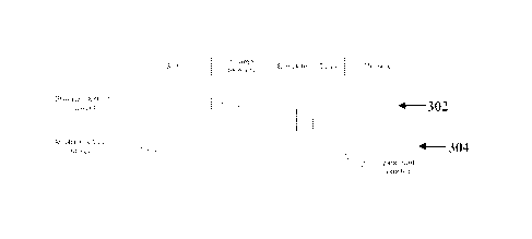
Figure 3 shows idealised waveforms arising during the SCP measurement process
of Figure2.
Figure 3 illustrates the current 302 of stimulus electrodes 204, and the
output voltage 304 of high
gain measurement amplifier 210, during each of the five phases of the
measurement cycle.
Importantly, it can be seen that phase 1 permits the amplifier bias point to
settle to a steady state
as defined by bio-electrical conditions at the sense electrode, while phases 2-
4 do not disrupt the
high gain measurement amplifier 210 bias point.
An advantage of this circuit is that in the phase 2 equilibration, the
circuitry around high gain
measurement amplifier 210 is a low-pass filter, and is therefore relatively
immune to noise and
input transients. This also allows the high gain measurement amplifier 210 to
accumulate its
bias point over successive measurement cycles, as it does not need to be reset
for each cycle.
CA 2835486 2019-05-02
14
Moreover, because of the front end buffer amplifier 206 before the sample/hold
208, the input-
referred effect (i.e. the effect upon sense electrode 202) of the charge
injection into the
sample/hold 208 is lower.
In the embodiment of Figure 2, the sense electrode 202 is never shorted to the
stimulus
electrodes 204, recognising that this creates dis-equilibrium in the sense
electrodes and adds
artefact, rather than having the effect of creating equilibrium as previously
thought. In some
embodiments, it may be possible to overlap the "settle" (equilibrate) phase of
Figure 2a, and the
"charge recovery" phase of Fig 2b, although it would be expected that the
artefact would be
higher, and the time taken to reach equilibrium longer.
Figure 4 is a plot of 22 separate measurements of ovine SCP made using the
embodiment of
Figure 2. The measurements were obtained sequentially for differing stimuli,
the stimuli
comprising biphasic current pulses of 40 us pulse width and a current
amplitude which varied
from 0-10 mA. The measurements were then plotted on a single chart to produce
Figure 4. The
recorded signals consist of the neural response and a small electrode
artefact. The neural
response is tri-phasic, consisting of a first phase with a positive PI peak
followed by a second
phase with negative Ni peak and then a third phase with a secondary positive
P2 peak. The
neural response morphology in Figure 4 is characteristic of extracellular
recordings of axonal
compound action potentials. The first phase is dominated by the capacitive
current due to the
initial membrane depolarization. The second phase is dominated by Na- ion
current and is
negative due to the influx of Na ions during the neuronal membrane action
potential. The third
phase is positive due to the 1{ ion conduction during repolarization.
The waveforms of Figure 4 have lower dynamic range and simpler morphology than
measurements produced by previous approaches, due to the absence of stimulus
crosstalk and
reduced artefact. When wishing to provide a system built on an implanted
integrated circuit,
wide dynamic range amplifiers are difficult to design, as are signal
processing systems for
feature extraction. Beneficially, the nature of the measured waveforms shown
in Figure 4
permits. for example, a circuit for extracting the peak-to-peak SCP amplitude
to have fewer
components than would be required to operate upon the waveform produced by
previous
approaches. Thus the techniques of the present invention for artefact
reduction greatly assist in
building a practical implanted, evoked response feedback system.
CA 2835486 2019-05-02
15
Moreover, it is notable that in this case of a 40 us pulse width the
measurement system is settled
and ready to record prior to onset of the evoked CAP. The sense electrode was
less than 50mm
from the stimulus electrode, and a post-stimulus delay of 50 us was observed
before the
measurement amplifier was switched in to obtain the recordings shown in Figure
4. As can be
seen in Figure 4 the largest peak to peak response was about 2.4 mV,
significantly less than the
voltage present when applying a 10 mA stimulus. Moreover, the epidural space
is much smaller
in sheep than in humans, and so the electrode is expected to be closer to the
ovine neural tissue
and the magnitude of the sensed tri-phasic potentials is correspondingly
higher in the sheep than
is expected for humans, emphasizing the difficulty of making such recordings.
Figure 5 illustrates the circuitry of an alternative embodiment of the
invention in which a
differential measurement amplifier is used, and charge recovery is via a
voltage rail Vdd. As can
be seen, in accordance with the present invention the measurement phases are
carried out in a
corresponding manner despite the use of different hardware.
In the embodiments of either figure 2 or figure 5, artefact can cause the high
gain measurement
amplifier 210 to clip, and the amplifier can subsequently be slow to recover.
However, in
preferred embodiments the sample point, being the transition from the
"stimulate" to "measure'
phases, is delayed, allowing clipping to be avoided. Figure 6 illustrates the
manner of
determining a suitable delay 602, which is often in the range of 50-200 pis,
noting that the fast
response typically concludes within about 2 ms. Such embodiments may permit
use of a higher
amplifier gain than would otherwise be the case. In particular, a variable
delay and increased
amplifier gain may be particularly apt in circumstances where high-gain is
desired, and parts of
the SCP of interest do not immediately follow the stimulation. Thus, delaying
the start of
measurement will avoid the side effects of clipping.
In another embodiment of the invention shown in figure 7, a method to
eliminate artefact from
an SCP measurement is to alternate the phase of stimulus waveforms and take an
average of
obtained measurements. This method is effective when the stimulus electrodes
have different
area. For example, in tripolar stimulation a central electrode is driven
anodically in the first
phase and consists of a single electrode of the array, whereas the electrode
driven cathodically in
the first phase consists of two electrodes of the array connected in parallel.
The electrodes in
parallel would usually be on either side of the other stimulating electrode.
Similarly, if
stimulation were between one electrode in the epidural space and one electrode
elsewhere, such
CA 2835486 2019-05-02
16
as being attached to an implant body, then a mode of stimulation referred to
as "monopolar"
stimulation is obtained.
Figure 7 shows the stimulus current for a positive "anodic-first" stimulus
702, and the stimulus
current for a negative "cathodic first" stimulus 704. In this embodiment these
are applied in
succession with respective CAP measurements obtained after each stimulus. The
respective
measurement electrode voltages 706 and 708 arising from each such stimulus are
also shown. It
will be observed where indicated in waveforms 706, 708 that the artefacts from
each of the two
stimuli are of substantially identical magnitude, but opposite sign. In most
situations it will be
found that the artefact polarity depends on the stimulus polarity. An example
of this would be
electrical artefact caused by the high-pass poles of the front end amplifier
206. Clearly, either
phase could be used for stimulating nervous tissue, though their effects will
differ.
In contrast, the positive and negative phase stimuli 702, 704 produce SCPs of
differing
amplitudes, but approximately similar shape and importantly of similar
polarity, as this is
determined by the anatomy and physiology of the spinal cord nerve fibre
membranes. Thus,
when the voltages 706, 708 resulting from the positive and negative phase
stimuli 702, 704 are
recorded, and averaged, the opposite phase stimulation artefacts substantially
cancel, leaving the
SCP or a combination of the two SCPs 710. Note that in practical situations,
the artefact can
have much higher amplitude than the SCP, making it much harder to detect the
SCP than is
apparent from Fig 7.
The response of the spinal cord to these two polarities of stimulation are
referred to as the
"anodic" and "cathodic" SCP responses, as referred to the electrode considered
to be that closest
to the recording electrode. I.e. anodic tripolar stimulation makes the central
stimulating
electrode anodic in the first phase of stimulus. Usually cathodic stimulation
has a lower threshold
for neural activation than is the case for anodic stimulation. Nevertheless,
the SCP polarity is
independent of whether the stimulus is anodic 702 or cathodic 704.
Figure 8a illustrates spinal cord measurements obtained in response to anodic
and cathodic
monophasic stimulations, respectively, the stimuli being of equal amplitude.
Note that the
measurement obtained in response to the anodic stimulation lacks the
characteristic P1 -N I -P2
form, indicating that the anodic stimulation did not evoke a neural response
in this case. In
CA 2835486 2019-05-02
16A
contrast, the measurement obtained in response to the cathodic stimulus
exhibits a significant
evoked neural response.
CA 2835486 2019-05-02
CA 02835486 2013-11-08
WO 2012/155183 PCT/AU2012/000511
17
Figure 8b shows an average of the two responses in Fig. 10a. As can be seen,
while the
characteristic form of the SCP has been altered, the artefact is essentially
removed as stimuli of
opposite polarity and equal amplitude produce artefact of opposite polarity
and equal amplitude,
which cancel when averaged.
This embodiment of the invention further recognises that the averaged waveform
of Figure 8b
can be used to obtain a range of information despite the atypical SCP form. In
this regard,
Figure 9 illustrates SCP growth curves against stimulus amplitude, for both
anodic and cathodic
monophasic stimuli. Figure 9 also shows the growth behaviour of the average
SCP against
stimulus amplitude. It can be seen from Fig 9 that the threshold of the
average response is
identical to the threshold of the more sensitive response for cathodic
stimulation.
When the stimulus amplitude is in the range 902 such that only the cathodic
stimulus produces
an SCP, then the averaged SCP waveform would have a normal SCP morphology but
would be
half the amplitude compared to a true cathodic SCP due to the averaging. In
the region 904
where both the anodic and cathodic responses contribute to the averaged SCP,
the resultant
averaged SCP waveform will have morphology in between the two measurements. It
would not
directly represent an SCP, but rather the average of two different SCPs.
Nevertheless, this
waveform could still be valuable for example in implementing an automatic
control loop for
stimulation adjustment, as it gives a value proportional to neural
recruitment.
It is further to be noted that the principle portrayed by Figure 9 applies in
a similar manner to
other stimulus polarities. For example, some embodiments may stimulate with a
tripolar
arrangement having a centre electrode operating as a cathode and having two
edge electrodes,
being those immediately to each side of the centre electrode, operating as
anodes. This tripolar
arrangement means that the recovery charge is shared between the two edge
electrodes. For a
biphasic tripolar stimulus the cathodic charge on the 2nd phase is shared
between two electrodes
and thus is half that on the first phase. Thus the principle shown in Figure 9
is true for tripolar
stimulation, at least up to the point where the current is twice the threshold
current at which point
the edge electrodes' currents are each at the threshold and will thus start to
generate action
potentials.
Some embodiments of the invention, such as the embodiment of Figure 5, may use
differential
amplifiers so as to detect the voltage difference between two sense
electrodes. Differential
CA 02835486 2013-11-08
WO 2012/155183 PCT/AU2012/000511
18
amplifiers simplify the task of separating electrode artefact. If they are
connected to electrodes
with similar area, and separated from the stimulation electrodes in a similar
manner, then they
receive similar levels of electrode artefact and this will be removed when
their difference voltage
is obtained. However, in such a system the voltage recorded by the amplifier
is the difference
between the voltages at two points along a bundle of neurons, and can thus be
difficult to
interpret. When making SCP measurements, it is preferable to use single-ended
amplifiers as
they more accurately measure the SCP, and they arc more sensitive in measuring
the SCP.
Differential amplifiers are often used because they provide a means to reduce
electrode artefact,
when other means have been insufficient. However, Figure 10 illustrates a
problem of
measuring SCPs with differential amplifiers. It shows a spinal cord potential.
As this potential
travels along the spine at a velocity, which can be as high as 80 m.s1, it can
also be considered as
a spatial wave. Given that a peak-to-peak cycle of the fast response of an SCP
typically lasts for
1 ms, the wave will travel 8 cm in this time. Using this 1 ms = 8 cm scale, a
5cm electrode array
is drawn alongside the SCP in Figure 10. Connected to this electrode array are
two amplifiers
configured to make differential SCP measurements from separate pairs of sense
electrodes. As
can be seen from Figure 10, the difference between the voltages on the
adjacent electrodes will
be quite small and significantly smaller than the peak to peak amplitude of
the SCP, and thus
more susceptible to electrical noise generated by the amplifier. The output of
the amplifier will
approximate the differential of the SCP, and thus be harder to interpret than
a simple measure of
the SCP itself. If measuring evoked SCPs with a micro-package stimulator
design, for example
in a system using a two-wire bus, differential measurements between non-
adjacent electrodes are
not possible. Further, if wishing to measure the slow response of the SCP,
which has a period of
about 6 ms and correspondingly reduced signal gradients, differential
measurements are even
more difficult to effect. Thus it will be appreciated that single-ended
measurements are
preferable, as long as artefact can be kept at a sufficiently low level.
With the measurement sequence of the present invention, the artefact is
reduced so that some
embodiments may instead use a single-ended amplifier, even in situations where
previously they
would have suffered from too much electrode artefact. Moreover, trials to date
show that
recording can be initiated with an extremely short time interval from
cessation of the stimulus,
permitting the same electrode array to be used for recording and stimulation,
and even permitting
recordings to be made on the electrode immediately adjacent to the stimulus
electrode in an
electrode array with electrode spacings of less than 10 mm.
CA 02835486 2013-11-08
WO 2012/155183 PCT/AU2012/000511
19
Single ended amplifiers have the further advantage that they consist of fewer
capacitors and
amplifier components than differential amplifiers, so will take up less space
on a silicon chip,
which is a significant benefit when intended for use in an implanted system
with many electrodes
and where the silicon area for each amplifier is limited.
Preferred embodiments of the invention may comprise a separate amplifier chain
(e.g. 206, 208,
210, see Fig 2) for every electrode, organised in parallel manner, permitting
simultaneous
recording of a single CAP from multiple sense electrodes in parallel, and also
eliminating the
switching noise arising in systems which switch the sense electrode to a
shared measurement
amplifier.
Further embodiments of the invention may employ divisible electrodes, as
discussed below with
reference to Figures 11 and 12. When considering electrode artefact in
particular, the sources of
electrode artefact are relatively poorly understood. The surface of a metal
electrode can be
modelled as an RC network. For an accurate model, an infinite-phase element is
required, but
for the explanation of artefact a simple RC model will suffice, as shown in
Figure 11a. A
conductive solution can be modelled as a mesh of resistors. Where a conductive
solution meets a
piece of metal of finite dimensions, the metal provides an alternative
conduction path to the
solution. This charges the electrode-to-tissue capacitances at the "ends" of
the electrodes, with
opposite polarities. The electrode does not acquire net charge, but it does
cease to be in
equilibrium. After the external current ceases, then the electrode will pass
current through the
solution as it re-equilibrates for a short time after the stimulus. This
current will affect the
potential of another electrode in the solution, and in the case of multi-
electrode arrays a unique
such current will arise at every electrode in response to local conditions
experienced at that
electrode. The cumulative impact of such re-equilibration currents is seen by
a sense electrode
as electrode artefact.
A similar effect happens when current flows between two electrodes, as shown
in Figure 11b.
During application of a stimulus, the current preferentially flows between the
parts of the
electrodes where they are closest. When the current is interrupted, the charge
on the surface of
the electrodes must re-equilibrate; this also leads to a residual current and
contributes to
electrode artefact seen by a sense electrode.
CA 02835486 2013-11-08
WO 2012/155183 PCT/AU2012/000511
The model of Figure 11 predicts that using smaller electrodes will reduce
artefact. However,
smaller electrodes will have higher noise when used as measurement electrodes,
and higher
resistance and lower current carrying capacity when used as stimulus
electrodes. Two means to
reduce artefact without sacrificing noise, impedance or current carrying
capacity are shown in
5 Figures 12a and 12b. The electrode configuration of Figure 12a reduces
artefact induced in a
single metallic electrode; the electrode is composed of two or more smaller
electrodes that can be
disconnected during a stimulation phase, and reconnected during a measurement
phase. In the
configuration of Figure 12b, an electrode is segmented, and individual current
sources are
provided for each segment. This forces the current in the segments to match,
and so reduces
10 artefact.
The evoked response telemetry of the present invention may in some embodiments
be used to
monitor the effect of a delivered compound. The administration of compounds
(drugs or other
chemical therapeutics) to effect a change in the nervous system is common for
treatment of a
15 wide number of diseases and disorders. Anaesthetics of various types are
administered to the
spinal cord for the relief of pain. Perhaps the most common form is
administration of
anaesthetics in the epidural space for pain relief during child birth.
In such embodiments, a catheter comprising a drug delivery tube may be fitted
with electrode
20 elements and configured to obtain neural response measurements in
accordance with the present
invention in order to monitor drug-induced effects on the neural response.
Alternatively an
electrode array may be temporarily or permanently implanted and used to apply
neural stimuli
and monitor the neural response. The neural response measurements may be
obtained repeatedly
during administration of the drug in order to directly measure the effect of
the administered drug
and control the dosage delivered.
Figures 15a and 15b illustrate the effect of administration of anaesthetic to
the spinal cord, with a
neural response being present prior to administration and largely being absent
subsequent to
administration. As can be seen, there is a direct correlation between the
measured evoked
response and the dosage of the anaesthetic. A "partial block" may be effected
by ceasing
administration of the anaesthetic once the neural response amplitude reduces
to a desired level.
The technology described herein is suitable for full implantation within the
body of a subject and
as a result the evoked potential monitoring could be used in the
administration of an active
CA 02835486 2013-11-08
WO 2012/155183 PCT/AU2012/000511
21
compound to produce a therapeutic benefit. The system could be integrated
within an
implantable pump to control the administration of the compound.
Figure 14 shows two plots which compare the artefact arising when electrode
shorting is
performed, to the artefact arising when the sense electrode is disconnected
from the
measurement circuitry and from the stimulus electrodes after the stimulus.
The plots of Figure 14 were obtained from an array placed in a saline bath,
and were taken under
the following conditions. A stimulation comprising a biphasic pulse of
amplitude 10mA and
duration 400 !Is was applied using a tripolar configuration, with electrodes
El and E3 grounded
and electrode E2 stimulating, at a stimulus rate of 40Hz. The artefact
measurement of interest
(1502, 1512) was obtained on electrode 4 for each plot. Measurements were also
obtained on
electrodes 5 to 7 using the method of the present invention in both plots,
these measurements
indicated collectively at 1504, 1512. The measurement parameters for each plot
included
recovering charge on the stimulus electrodes by short circuiting the stimulus
electrodes to each
other for 100 ius before stimulation. As shown in Figure 14a, when the sense
electrodes were
shorted as taught by prior art methods, the artefact in the measurement 1502
was considerably
larger than the artefact present in measurements 1504. In contrast, when the
sense electrode E4
was disconnected from the measurement circuitry and from the stimulus
electrodes after the
stimulus, as taught by the present invention, the artefact in the measurement
1512 from electrode
E4 was considerably reduced. The effect of this benefit in preferred
embodiments is that an
evoked response can be recorded in a single measurement with sufficient signal
to noise ratio to
permit analysis of the individual evoked response measurement. Moreover, such -
single shot"
measurements can in some embodiments be obtained in response to normal
therapeutic stimuli.
This avoids wasting battery power to deliver a train of high power stimuli
having parameters
which are well outside normal therapeutic settings and thus not of therapeutic
benefit, to enable
an averaged response to be extracted over a large number of measurements, as
is required in
systems having poor artefact performance.
It will be appreciated by persons skilled in the art that numerous variations
and/or modifications
may be made to the invention as shown in the specific embodiments without
departing from the
spirit or scope of the invention as broadly described. For example in the
measurements stages of
charge recovery (Figure 2b), stimulate (Figure 2c) and delay (Figure 2d), the
sense electrodes are
described as being disconnected from the sense circuitry. In the embodiment of
Figure 2 this is
CA 02835486 2013-11-08
WO 2012/155183
PCT/AU2012/000511
22
effected by setting the sample and hold 208 to "hold", and it is noted that in
alternative
embodiments the sample and hold 208 may be positioned elsewhere in the
measurement chain.
Such embodiments are all to be understood to be within the scope of the phrase
"disconnecting
the sense electrode from the measurement circuitry" or similar as used herein.
The present
embodiments are, therefore, to be considered in all respects as illustrative
and not restrictive.