Note: Descriptions are shown in the official language in which they were submitted.
CA 02835862 2013-11-12
WO 2012/166549 PCT/US2012/039441
EXTRACELLULAR MATRIX MATERIAL VALVE CONDUIT AND
METHODS OF MAKING THEREOF
Cross-Reference to Related Applications
[0001] This application claims the benefit of the filing dates of U.S.
Provisional Patent
Application Number 61/490,693, filed on May 27, 2011, U.S. Provisional Patent
Application
No. 61/490,873, filed on May 27, 2011, U.S. Provisional Patent Application No.
61/491,723,
filed on May 31, 2011, and U.S. Provisional Patent Application No. 61/650,911,
filed on May
23, 2012, each of which is hereby incorporated by reference herein in its
entirety.
Field
[0002] The invention generally relates to extracellular matrix material
valve conduits
and methods of making such valve conduits. More particularly, the invention
relates to
methods of forming valve conduits from sheets or conduits of extracellular
matrix materials,
as well as the extracellular matrix material valve conduits resulting from
such methods.
Background
[0003] Cardiac surgeons currently employ a variety of techniques to
accomplish
valvular reconstruction within the hearts of patients. For example, cryo-
preserved allografts,
bovine jugular vein grafts, porcine valves, and autologous pericardium have
all been used in
such valvular reconstruction procedures. However, these known techniques all
suffer from
several major limitations. More specifically, cryo-preserved allografts are
prone to
calcification and failure over time, and the high costs and low availability
of allografts limit
the utility of allografts in developing countries. These grafts also increase
the likelihood that
the anti-human antibodies of a patient will react with, and ultimately reject,
a future heart
transplant due to prior antigen exposure. Jugular vein grafts, although widely
available, can
only be provided in a narrow range of sizes, and the jugular vein grafts are
prone to undesired
calcification and aneurysmal dilatation. Similarly, porcine valves calcify
over time, leading
to a significant decrease in the integrity of the valves, particularly in
children. Autologous
pericardium has been used with short-term success; however, the procedures
employing
autologous pericardium are typically complicated and time-consuming, and are,
therefore,
unsuited for use in most countries. Moreover, autologous pericardium calcifies
over time,
and a patient's own pericardium cannot be used as a replacement valve material
when the
patient has had previous heart surgeries.
1
CA 02835862 2013-11-12
WO 2012/166549 PCT/US2012/039441
[0004] Additionally, known valve conduits that are employed in valvular
reconstruction procedures are typically formed from multiple pieces, such as,
for example, a
graft portion and a valve portion. Thus, before these valve conduits can be
used, the valve
portion must be properly secured within the graft portion. This limitation
adds significant
complexity and time to the overall procedure, and the two-part structure of
the resulting valve
conduits can contribute to failure of the device.
[0005] Furthermore, at a fundamental level, known valve conduits are used
to replace
a defective valve rather than to regenerate a native valve. Thus, following
implantation, these
valve conduits are incapable of achieving formation of a physiologically and
anatomically
correct replacement valve.
[0006] In developing countries, cost and supply constraints limit the
widespread use of
alternative conduits for valvular reconstruction operations. Thus, there is a
need for a readily
available, low-cost valve replacement material that can easily be used during
surgical
procedures in developing countries.
[0007] Accordingly, there is a need in the art for a heart valve conduit
that, upon
implantation within the heart of a subject, is configured to promote
regeneration of a
replacement heart valve, including leaflets and sinus portions that are
identical or
substantially identical to the leaflets and sinus portions of a native valve.
There is a further
need for a unitary, implantable heart valve conduit that distally integrates
into a native artery
such that, over time, the synthetic material of the heart valve conduit is
undetectable. There
is still a further need for a sterile, acellular, and low-cost heart valve
conduit that can be
quickly and efficiently constructed using readily available materials or that
is pre-constructed
for rapid implantation.
SUMMARY
[0008] Methods for regenerating semi-lunar valves to replace defective
semi-lunar
valves within the heart of a subject are disclosed. In one disclosed method, a
defective semi-
lunar valve is removed from the heart of the subject. A sheet of extracellular
matrix (ECM)
material is positioned in a folded position, in which a bottom edge of the
sheet is folded
toward a top edge of the sheet such that the bottom edge of the sheet is
spaced a selected
distance from the top edge of the sheet. The sheet of ECM material is secured
in the folded
position at a first attachment point and a second attachment point, thereby
forming a folded
ECM material construct. The folded ECM material construct is positioned in an
aligned
2
CA 02835862 2013-11-12
WO 2012/166549 PCT/US2012/039441
position, in which a first side edge of the folded ECM material construct is
in substantial
alignment with a second side edge of the folded ECM material construct. With
the folded
ECM material construct in the aligned position, the first side edge is secured
to the second
side edge, thereby forming an ECM material valve conduit.
[0009] The ECM material valve conduit has a lumen, an inlet portion
defining an inlet
and having an inner layer and an outer layer, and an outlet portion defining
an outlet. The
inner layer of the inlet portion is positioned within the lumen, while the
outer layer of the
inlet portion cooperates with the outlet portion to define an outer wall of
the ECM material
valve conduit. The ECM material valve conduit is attached to an annular region
or outlet of
the heart of the subject and to an artery of the subject such that the inlet
portion of the ECM
material valve conduit is positioned proximate the annular region. The inner
layer of the
ECM material valve conduit includes leaflet-promoting portions for
regenerating leaflets, and
the outer layer of the ECM material valve conduit includes sinus-promoting
formations for
regenerating sinus portions of the replacement semi-lunar valve. ECM material
valve
conduits that are formed and used according to the described methods are also
disclosed.
BRIEF DESCRIPTION OF THE DRAWINGS
[0010] These and other features of the preferred embodiments of the
invention will
become more apparent in the detailed description in which reference is made to
the appended
drawings wherein:
[0011] Figure 1 depicts an exemplary sheet of extracellular matrix
material, as
described herein.
[0012] Figures 2A is a top view of the sheet of Figure 1 in a folded
position, as
described herein. Figure 2B is a side perspective view of the sheet in the
folded position.
[0013] Figure 3A is a side perspective view of an extracellular matrix
material valve
conduit formed from the sheet of Figures 1-2B, as described herein. Figure 3B
is a top view
of the extracellular matrix material valve conduit.
[0014] Figure 4 is a schematic depiction of the leaflet-promoting portions
and sinus-
promoting portions of the inner layer of the extracellular matrix material
valve conduit
depicted in Figures 3A and 3B.
[0015] Figure 5 is a perspective view of an extracellular matrix material
conduit, as
described herein.
3
CA 02835862 2013-11-12
WO 2012/166549 PCT/US2012/039441
[0016] Figure 6A is a side perspective view of the extracellular matrix
conduit of
Figure 5 in a reflected position, thereby forming an extracellular matrix
material valve
conduit. Figure 6B is a top view of the extracellular matrix valve conduit.
[0017] Figures 7-11 are images of a regenerated pulmonary valve taken at
three
months following implantation of an extracellular matrix material valve
conduit for purposes
of regenerating the pulmonary valve in the heart of the sheep. Figure 7 is an
image of the
right ventricular outflow tract of the regenerated pulmonary valve. Figure 8
is an image of
the leaflets of the regenerated pulmonary valve. Figure 9 is an image of the
regenerated
pulmonary valve, as observed from the right ventricle of the heart of the
sheep. Figure 10 is
an image depicting the progress of leaflet formation in the regenerated
pulmonary valve.
Figure 11 is an image depicting the progress of sinus formation in the
regenerated pulmonary
valve.
[0018] Figures 12-14 are images depicting exemplary extracellular matrix
valve
conduits, as described herein. Figures 12 and 13 are images of exemplary
extracellular
matrix valve conduits prior to hydration. Figure 14 is an image of an
exemplary extracellular
matrix valve conduit following hydration.
[0019] Figures 15-23 are sketches and images associated with a patient
study that was
performed using concepts as described herein. Figures 15, 16, and 23 depict
valve conduits
that were implanted into the heart of a patient during the study, while
Figures 17-22 are
images of echocardiograms that were recorded during the study.
[0020] Figure 24 is a diagram of an exemplary extracellular matrix valve
conduit
construction, which depicts a sewing seam allowance (s), a sewing cuff (sc), a
leaflet height
(hl), a leaflet width (1w), and an ECM sheet width (w).
[0021] Figure 25 depicts Doppler echocardiography images taken
postoperatively for
an exemplary extracellular matrix material valve conduit as described herein.
Figure 25(a)
depicts the ECM material valve conduit during opening. Figure 25(b) depicts
the ECM
material valve conduit during closure. Figure 25(c) depicts the ECM material
valve conduit
radially at closure.
[0022] Figure 26 includes images of a regenerated pulmonary valve at
various time
points following implantation of an exemplary extracellular matrix material
valve conduit as
described herein. Figure 26(a) shows regeneration at 3 months. Figure 26(b)
shows
4
CA 02835862 2013-11-12
WO 2012/166549 PCT/US2012/039441
regeneration at 5 months. Figure 26(c) shows regeneration at 6 months. Figure
26(d) shows
regeneration at 12 months.
[0023] Figures 27-28 depict the results of an experiment in which DNA
content was
measured for small intestinal submucosa (SIS) compositions following various
sterilization
methods, including the sterilization methods described herein. Figure 27 shows
the DNA
content of each SIS composition following sterilization. Figure 28 shows the
percentage of
DNA that was removed from each SIS composition following sterilization, as
compared to
raw, unprocessed SIS.
[0024] Figures 29-30 depict the results of an experiment in which native
growth factor
content was measured for SIS compositions following various sterilization
methods,
including the sterilization methods described herein. Figure 29 shows the bFGF
content of
each SIS composition (normalized by dry weight of samples) following
sterilization. Figure
30 shows the active TGF-I3 content of each SIS composition (normalized by dry
weight of
samples) following sterilization.
[0025] Figure 31 depicts the results of an experiment in which bFGF was
incorporated
into SIS compositions during rapid depressurization, as described herein.
Figure 31 shows
the bFGF content for each SIS composition (normalized by dry weight of
samples) following
rapid depressurization.
[0026] Figure 32 depicts the results of an experiment in which the tensile
strength of
two-ply SIS compositions was measured following various sterilization methods,
including
the sterilization methods described herein. Figure 32 shows the tensile
strength measured for
each SIS composition following sterilization.
[0027] Figure 33 depicts the results of an experiment in which native
growth factor
content was measured for SIS compositions following various sterilization
and/or
decellularization methods, including the sterilization and decellularization
methods described
herein. Figure 33 shows the bFGF enzyme-linked immunosorbent assay (ELISA)
results for
each SIS composition (normalized by dry weight of samples) following
sterilization and/or
decellularization.
[0028] Figure 34 shows the DNA content in SIS after it is processed in
various ways.
The baseline measurement is raw. The tissue was then exposed to supercritical
CO2 followed
by rapid depressurization (RDP) to facilitate enhanced removal of DNA and
cellular debris.
After the RDP, the tissue was placed in supercritical CO2 with peracetic acid
(PAA) for
CA 02835862 2013-11-12
WO 2012/166549 PCT/US2012/039441
sterilization. The comparison is to processed SIS either unsterilized or
sterilized with
ethylene oxide (ETO).
[0029] Figure 35 shows the Percent removal of DNA from SIS after it is
processed in
various ways. The baseline measurement is raw. The tissue was then exposed to
supercritical
CO2 followed by rapid depressurization (RDP) to facilitate enhanced removal of
DNA and
cellular debris. After the RDP, the tissue was placed in supercritical CO2
with peracetic acid
(PAA) for sterilization. The comparison is to processed SIS either
unsterilized or sterilized
with ethylene oxide (ETO).
[0030] Figure 36 shows the variable active Transforming Growth Factor (TGF-
beta)
content in SIS after it is processed in various ways. The baseline measurement
is raw, or
unprocessed SIS followed by processing with only Triton X-100 (TX-100)
detergent. The
tissue was then exposed to supercritical CO2 followed by rapid
depressurization (RDP) to
facilitate enhanced removal of DNA and cellular debris. After the RDP, the
tissue was placed
in supercritical CO2 with peracetic acid (PAA) for sterilization. The
comparison is to
processed SIS either unsterilized or sterilized with ethylene oxide (ETO).
[0031] Figure 37 shows the variable basic Fibroblast Growth Factor (bFGF)
content in
SIS after it is processed in various ways. The baseline measurement is raw, or
unprocessed
SIS followed by processing with only Triton X-100 (TX-100) detergent. The
tissue was then
exposed to supercritical CO2 followed by rapid depressurization (RDP) to
facilitate enhanced
removal of DNA and cellular debris. After the RDP, the tissue was placed in
supercritical
CO2 with peracetic acid (PAA) for sterilization. The comparison is to
processed SIS either
unsterilized or sterilized with ethylene oxide (ETO).
[0032] Figure 38 shows the addition of basic Fibroblast Growth Factor
(bFGF) content
to SIS using rapid depressurization. The baseline measurement is raw, or
unprocessed SIS.
The comparison is to processed SIS either unsterilized or sterilized with
ethylene oxide
(ETO).
[0033] Figure 39 is a cut-away view of the human heart.
DETAILED DESCRIPTION
[0034] The present invention may be understood more readily by reference
to the
following detailed description, examples, drawings, and claims, and their
previous and
following description. However, before the present devices, systems, and/or
methods are
disclosed and described, it is to be understood that this invention is not
limited to the specific
6
CA 02835862 2013-11-12
WO 2012/166549 PCT/US2012/039441
devices, systems, and/or methods disclosed unless otherwise specified, as such
can, of course,
vary. It is also to be understood that the terminology used herein is for the
purpose of
describing particular aspects only and is not intended to be limiting.
[0035] As used in the specification and the appended claims, the singular
forms "a,"
"an" and "the" include plural referents unless the context clearly dictates
otherwise. Thus,
for example, reference to an "attachment point" can include two or more such
attachment
points unless the context indicates otherwise.
[0036] Ranges may be expressed herein as from "about" one particular
value, and/or
to "about" another particular value. When such a range is expressed, another
aspect includes
from the one particular value and/or to the other particular value. Similarly,
when values are
expressed as approximations, by use of the antecedent "about," it will be
understood that the
particular value forms another aspect. It will be further understood that the
endpoints of each
of the ranges are significant both in relation to the other endpoint, and
independently of the
other endpoint.
[0037] As used herein, the terms "optional" and "optionally" mean that the
subsequently described event or circumstance may or may not occur, and that
the description
includes instances where said event or circumstance occurs and instances where
it does not.
[0038] The word "or" as used herein means any one member of a particular
list and
also includes any combination of members of that list.
[0039] Unless otherwise expressly stated, it is in no way intended that
any method or
aspect set forth herein be construed as requiring that its steps be performed
in a specific
order. Accordingly, where a method claim does not specifically state in the
claims or
descriptions that the steps are to be limited to a specific order, it is in no
way intended that an
order be inferred, in any respect. This holds for any possible non-express
basis for
interpretation, including matters of logic with respect to arrangement of
steps or operational
flow, plain meaning derived from grammatical organization or punctuation, or
the number or
type of aspects described in the specification.
[0040] Without the use of such exclusive terminology, the term
"comprising" in the
claims shall allow for the inclusion of any additional element¨irrespective of
whether a
given number of elements is enumerated in the claim or the addition of a
feature could be
regarded as transforming the nature of an element set forth in the claims.
Except as
7
CA 02835862 2013-11-12
WO 2012/166549 PCT/US2012/039441
specifically defined herein, all technical and scientific terms used herein
are to be given as
broad a commonly understood meaning as possible while maintaining claim
validity.
[0041] As used herein, a "subject" is an individual and includes, but is
not limited to, a
mammal (e.g., a human, horse, pig, rabbit, dog, sheep, goat, non-human
primate, cow, cat,
guinea pig, or rodent), a fish, a bird, a reptile or an amphibian. The term
does not denote a
particular age or sex. Thus, adult and newborn subjects, as well as fetuses,
whether male or
female, are intended to be included. A "patient" is a subject afflicted with a
disease or
disorder. The term "patient" includes human and veterinary subjects. As used
herein, the
term "subject" can be used interchangeably with the term "patient."
[0042] As used herein, the term "circumference" refers to the perimeter
of, or length
of the boundary defined by, a closed planar figure. Optionally, as used
herein, a
"circumference" can correspond to the perimeter of a closed planar circle.
However, it is
contemplated that a "circumference" can correspond to the perimeter of any
closed planar
figure, such as, for example and without limitation, an oval, square,
rectangular, trapezoidal,
or nonsymmetrical closed planar figure. For example, as used herein, an outer
"circumference" of a conduit corresponds to the perimeter of the closed planar
figure defined
by an outer surface of the conduit at a particular location along the
longitudinal axis of the
conduit.
[0043] As used herein, the term "acellular" is meant to describe
extracellular matrix
compositions that are at least 80 % decellularized such that the extracellular
matrix
composition is at least 80 % without cells and/or cellular remnants. In some
exemplary
aspects described herein, the term "acellular" can refer to extracellular
matrix compositions
that are at least 90 % decellularized such that the extracellular matrix
composition is at least
90 % without cells and/or cellular remnants. In other exemplary aspects
described herein, the
term "acellular" can refer to extracellular matrix compositions that are at
least 95 %
decellularized such that the extracellular matrix composition is at least 95 %
without cells
and/or cellular remnants. In other exemplary aspects described herein, the
term "acellular"
can refer to extracellular matrix compositions that are at least 96 %
decellularized such that
the extracellular matrix composition is at least 96 % without cells and/or
cellular remnants.
In still other exemplary aspects described herein, the term "acellular" can
refer to
extracellular matrix compositions that are at least 97 % decellularized such
that the
extracellular matrix composition is at least 97 % without cells and/or
cellular remnants. In
further exemplary aspects described herein, the term "acellular" can refer to
extracellular
8
CA 02835862 2013-11-12
WO 2012/166549 PCT/US2012/039441
matrix compositions that are at least 98 % decellularized such that the
extracellular matrix
composition is at least 98 % without cells and/or cellular remnants. In still
further exemplary
aspects described herein, the term "acellular" can refer to extracellular
matrix compositions
that are at least 99 % decellularized such that the extracellular matrix
composition is at least
99 % without cells and/or cellular remnants. Thus, as used herein, the term
"acellular" can
refer to extracellular matrix compositions that are decellularized at levels
of 80 %, 81 %, 82
%, 83 %, 84 %, 85 %, 86 %, 87 %, 88 %, 89 %, 90 %, 91 %, 92 %, 93 %, 94 %, 95
%, 96 %,
97 %, 98 %, 99 %, 100 %, and any percentages falling between these values.
[0044] As used herein, the term "additive" refers to materials that can be
selectively
incorporated into the disclosed ECM materials to impart predetermined
properties to the
sterilized, acellular ECM compositions disclosed herein. Such additives can
include, for
example and without limitation, growth factors, cytokines, proteoglycans,
glycosaminoglycans (GAGs), proteins, peptides, nucleic acids, small molecules,
cells and
pharmaceutical agents, such as statin drugs, corticosterioids, anti-arrhythmic
drugs,
nonsteroidal anti-inflammatory drugs, other anti-inflammatory compounds,
nanoparticles,
and metallic compounds.
[0045] As used herein, the term "contemporaneously" refers to the
simultaneous
and/or overlapping occurrence of events, as well as the sequential occurrence
of events within
about thirty minutes before or after one another. Thus, if a first event
occurs, then a second
event can be said to have occurred contemporaneously with the first event if
it occurred
concurrently with the first event or within thirty minutes before or after the
first event. For
example, if a first method step is performed, then a second method step
performed five
minutes after the first method step can be said to be performed
"contemporaneously" with the
first method step. Similarly, if the second method step was performed ten
minutes before
performance of a third method step, then the second method step can be said to
be performed
"contemporaneously" with the third method step.
[0046] As used herein, the term "supercritical" refers to a fluid state of
a material
when it is held at or above its critical temperature and critical pressure.
When a material is
held at or above its critical temperature and critical pressure, then it
typically adopts
functional properties of both a gas and a liquid and is said to function as a
supercritical fluid.
Thus, for example, when carbon dioxide is held at or above its critical
temperature (31.1 C)
and its critical pressure (1,071 psi), it behaves as a supercritical carbon
dioxide fluid and can,
for example, exhibit the expansion properties of a gas while having the
density of a liquid.
9
CA 02835862 2013-11-12
WO 2012/166549 PCT/US2012/039441
[0047] Described herein are valve conduits made from extracellular matrix
(ECM)
material. In exemplary aspects, the ECM material valve conduits regenerate a
semi-lunar
(tri-leaflet) valve, such as a pulmonary valve or an aortic valve within a
heart of a subject. In
these aspects, the ECM material valve conduits can regenerate a semi-lunar
valve to replace a
defective semi-lunar valve within the heart of the subject. It is contemplated
that such
defective semi-lunar valves can be attached at an annular region between a
ventricle of the
heart of the subject and an artery of the subject. As used herein, the term
"annular region"
refers to the portion of the heart of a subject that is proximate to the
native position of an
annulus between a ventricle within the heart of the subject and an artery of
the subject. When
an annulus is positioned within the heart of the subject, the annular region
includes the
annulus as well as the heart muscle proximate the annulus. When the annulus
has been
removed from the heart of the subject, the annular region includes the heart
muscle proximate
the former position of the annulus within the heart of the subject.
[0048] In exemplary aspects, a disclosed ECM material valve conduit can
comprise
any known ECM component or material, including, for example and without
limitation,
mucosal layers and components, submucosal layers and components, muscularis
layers and
components, and/or basement membrane layers and components. It is contemplated
that a
disclosed ECM material valve conduit can comprise an ECM material obtained
from any
mammalian tissue source, including, for example and without limitation,
stomach tissue (e.g.,
stomach submucosa (SS)), small intestinal tissue (e.g., small intestinal
submucosa (SIS)),
large intestinal tissue, bladder tissue (e.g., urinary bladder submucosa
(UBS)), liver tissue
(e.g., liver basement membrane (LBM)), heart tissue (e.g., pericardium), lung
tissue, kidney
tissue, pancreatic tissue, prostate tissue, mesothelial tissue, fetal tissue,
a placenta, a ureter,
veins, arteries, heart valves with or without their attached vessels, tissue
surrounding the roots
of developing teeth, and tissue surrounding growing bone. It is further
contemplated that a
disclosed ECM material valve conduit can comprise an ECM material obtained
from ECM
components or materials of one or more mammals including, for example and
without
limitation, humans, cows, pigs, dogs, sheep, cats, horses, rodents, and the
like. Thus, it is
contemplated that a disclosed ECM material valve conduit can comprise ECM
components or
materials from two or more of the same mammalian species, such as, for example
and
without limitation, two or more cows, two or more pigs, two or more dogs, or
two or more
sheep. It is further contemplated that a disclosed ECM material valve conduit
can comprise
ECM components or materials from two or more different mammalian species, such
as, for
CA 02835862 2013-11-12
WO 2012/166549 PCT/US2012/039441
example and without limitation, a pig and a cow, a pig and a dog, a pig and a
sheep, or a cow
and a sheep. It is still further contemplated that a disclosed ECM material
valve conduit can
comprise ECM components or materials obtained from a first tissue source, such
as, for
example and without limitation, SIS, from a first mammal, as well as ECM
components or
materials obtained from a second tissue source, such as, for example and
without limitation,
SS, from a second mammal.
[0049] In one aspect, and with reference to Figures 3A-4 and 6A-6B, a
disclosed ECM
material valve conduit 40, 140 can have a longitudinal axis 41, 141 and can
comprise a lumen
42, 142, an inlet portion 44, 144, and an outlet portion 56, 156. In this
aspect, it is
contemplated that the lumen 42, 142 can have an inner diameter. Optionally,
the inner
diameter of the lumen 42, 142 can be substantially constant along the
longitudinal axis 41,
141 of the ECM material valve conduit 40, 140. In exemplary aspects, it is
contemplated that
the inner diameter of the lumen 42, 142 can range from about 15 mm to about 30
mm. In a
further aspect, it is contemplated that the ECM material valve conduit 40, 140
can have a
longitudinal length (along longitudinal axis 41, 141) ranging from about 20 mm
to about 40
mm, and more preferably, from about 22 mm to about 34 mm.
[0050] In another aspect, the outlet portion 56, 156 can define an outlet
58, 158 in
communication with the lumen 42, 142 of a disclosed ECM material valve conduit
40, 140.
In an additional aspect, the inlet portion 44, 144 can define an inlet 46, 146
in communication
with the lumen 42, 142 of a disclosed ECM material valve conduit 40, 140 and
comprise an
outer layer and an inner layer 48, 148 positioned within the lumen of the ECM
material valve
conduit. In this aspect, it is contemplated that the inner layer 48, 148 and
the outer layer of
the inlet portion 44, 144 can be of unitary, continuous construction, with the
inner layer being
inwardly reflected within the lumen 42, 142 of the ECM material valve conduit
40, 140.
Thus, it is contemplated that, due to the unitary and continuous construction
of the inner layer
48, 148 and the outer layer, the inner layer and the outer layer do not have
to be secured to
one another proximate the inlet 46, 146 of the ECM material valve conduit 40,
140.
[0051] In a further aspect, the inner layer 48, 148 of the inlet portion
44, 144 of a
disclosed ECM material conduit 40, 140 can be attached to the outer layer of
the inlet portion
44, 144 of the ECM material conduit at a plurality of attachment points 34,
134, such as, for
example, two or three attachment points. In this aspect, it is contemplated
that the plurality
of attachment points 34, 134 can be substantially equally spaced along an
outer
circumference of the ECM material valve conduit 40, 140. It is further
contemplated that the
11
CA 02835862 2013-11-12
WO 2012/166549 PCT/US2012/039441
plurality of attachment points 34, 134 can be positioned substantially within
a common plane
that is substantially perpendicular to the longitudinal axis 41, 141 of the
ECM material valve
conduit 40, 140. In another aspect, the inner layer 48, 148 of the outlet
portion 56, 156 can
be attached to the outer layer at the plurality of attachment points 34, 134
using any
conventional surgical attachment means, including, for example and without
limitation, non-
absorbable sutures, absorbable sutures, surgical pastes, surgical glues,
staples, and the like.
In this aspect, it is contemplated that, when non-absorbable sutures are used
to secure the
inner layer 48, 148 to the outer layer, the knots of each suture can be
positioned in contact
with the outer wall such that the outer wall is positioned between the inner
layer and the
knots, thereby ensuring that the knots will not extend into the lumen 42, 142
following
implantation of the ECM material valve conduit 40, 140. In exemplary aspects,
the inner
layer and outer layers 48, 148 can be secured to one another using a cruciate
suture pattern.
In still a further aspect, the outer layer of the inlet portion of a disclosed
ECM material valve
conduit 40, 140 can cooperate with the outlet portion of the ECM material
valve conduit to
define an outer wall 52, 152 of the ECM material valve conduit.
[0052] In an additional aspect, and with reference to Figures 3B and 4,
the inner layer
48, 148 of the inlet portion 44, 144 of a disclosed ECM material valve conduit
40, 140 can
comprise leaflet-promoting portions 50, 150. In this aspect, it is
contemplated that, following
attachment of the ECM material valve conduit 40, 140 to an annular region of
the heart of the
subject and an artery of the subject such that the inlet portion 44, 144 of
the ECM material
valve conduit is positioned proximate the annular region, the leaflet-
promoting portions 50,
150 of the inner layer 48, 148 can be configured to regenerate three leaflets
of a replacement
semi-lunar valve. In exemplary aspects, each leaflet-promoting portion 50, 150
of the inner
layer 48, 148 can have a longitudinal length. In these aspects, it is
contemplated that the
longitudinal length of the leaflet-promoting portions 50, 150 can optionally
be greater than or
equal to the length of the regenerated leaflets of the replacement semi-lunar
valve, as
measured by the elongate length of the regenerated leaflets extending from the
valve conduit
wall. For example, it is contemplated that the ratio between the longitudinal
length of the
leaflet-promoting portions 50, 150 and the length of the regenerated leaflets
can be 1.0:1,
1.1:1, 1.2:1, 1.3:1, 1.4:1, 1.5:1, 1.6:1, 1.7:1, 1.8:1, 1.9:1, 2.0:1, 2.1:1,
2.2:1, 2.3:1, 2.4:1,
2.5:1, 2.6:1, 2.7:1, 2.8:1, 2.9:1, 3.0:1, 3.1:1, 3.2:1, 3.3:1, 3.4:1, 3.5:1,
3.6:1, 3.7:1, 3.8:1,
3.9:1, 4.0:1, and any ratios falling between these values. In other optional
aspects, it is
12
CA 02835862 2013-11-12
WO 2012/166549 PCT/US2012/039441
contemplated that the longitudinal length of the leaflet-promoting portions
50, 150 can be less
than the length of the regenerated leaflets of the replacement semi-lunar
valve.
[0053] In another aspect, and with reference to Figure 3B, it is
contemplated that at
least a portion of the outer wall 52, 152 (i.e., the outer layer of the inlet
portion 44, 144) of a
disclosed ECM material valve conduit 40, 140 can comprise sinus-promoting
portions 54,
154. In this aspect, it is contemplated that, following attachment of the ECM
material valve
conduit 40, 140 to the annular region and the artery, the sinus-promoting
portions 54, 154 of
the outer wall 52, 152 of the ECM material valve conduit can be configured to
fuse with the
inner layer 48, 148 of the ECM material valve conduit to regenerate sinus
portions of the
replacement semi-lunar valve.
[0054] In a further aspect, and with reference to Figures 3B and 4, it is
contemplated
that the inner layer 48, 148 of the inlet portion 44, 144 of a disclosed ECM
material valve
conduit 40, 140 can further comprise commissure-promoting portions 51, 151. In
this aspect,
it is contemplated that, following attachment of the ECM material valve
conduit 40, 140 to
the annular region and the artery, the commissure-promoting portions 51, 151
can be
configured to fuse with at least a portion of the outer wall 52, 152 (e.g.,
the outer layer of the
inlet portion 44, 144). Thus, it is contemplated that the continuity of the
inner layer 48, 148
and the outer wall 52, 152 can permit the inner layer and the outer wall to
cooperate in
promoting the regeneration of the replacement semi-lunar valve.
[0055] Optionally, the ECM material valve conduit 40, 140 can have a multi-
laminate
structure. In exemplary aspects, the ECM material valve conduit 40, 140 can
comprise
between 2 and 10 layers laminated together. In an exemplary aspect, the ECM
material valve
conduit can be a four-ply (four layer) multi-laminate structure. It is
contemplated that such a
multi-laminate structure can increase the structural integrity of the ECM
material valve
conduit.
Methods of Forming the ECM Material Valve Conduits from a Sheet of ECM
Material
[0056] In exemplary aspects, a disclosed ECM material valve conduit can be
formed
from a sheet of ECM material. In these aspects, the sheet of ECM material can
comprise any
known ECM component or material, including, for example and without
limitation, mucosal
layers and components, submucosal layers and components, muscularis layers and
components, and/or basement membrane layers and components. Optionally, the
sheet of
ECM material can have a multi-laminate structure that is produced by
conventional methods.
13
CA 02835862 2013-11-12
WO 2012/166549 PCT/US2012/039441
It is contemplated that a disclosed ECM material valve conduit can comprise an
ECM
material obtained from any mammalian tissue source, including, for example and
without
limitation, stomach tissue (e.g., stomach submucosa (SS)), small intestinal
tissue (e.g., small
intestinal submucosa (SIS)), large intestinal tissue, bladder tissue (e.g.,
urinary bladder
submucosa (UBS)), liver tissue (e.g., liver basement membrane (LBM)), heart
tissue (e.g.,
pericardium), lung tissue, kidney tissue, pancreatic tissue, prostate tissue,
mesothelial tissue,
fetal tissue, a placenta, a ureter, veins, arteries, heart valves with or
without their attached
vessels, tissue surrounding the roots of developing teeth, and tissue
surrounding growing
bone. In one aspect, the sheet of ECM material can have a width ranging from
about 20 mm
to about 150 mm. In an additional aspect, the sheet of ECM material can have a
thickness
ranging from about 0.02 mm to about 3 mm. It is contemplated that the sheet of
ECM
material can have any length that is appropriate for desired folding of the
sheet and for
desired attachment of a disclosed ECM material valve conduit within the heart
of a subject.
[0057] In one aspect, and with reference to Figures 1-4, a method of
forming a
disclosed ECM material valve conduit 40 from a sheet 10 of ECM material can
comprise
positioning the sheet of ECM material in a folded position. In this aspect, it
is contemplated
that the sheet of ECM material 10 can have a top portion 12 comprising a top
edge 14 of the
sheet and a bottom portion 16 comprising a bottom edge 18 of the sheet. It is
further
contemplated that, in the folded position, the bottom edge 18 of the sheet 10
of ECM material
can be spaced a selected distance 20 from the top edge 14 of the sheet,
thereby forming a
sewing cuff In one aspect, in the folded position, the selected distance 20 by
which the
bottom edge 18 is spaced from the top edge 14 can range from about 0 mm to
about 150 mm.
In this aspect, it is contemplated that the selected distance 20 can be any
distance that permits
desired attachment of the ECM material valve conduit 40 to an artery of a
subject. In
exemplary aspects, the selected distance 20 can be about 10 mm.
[0058] In another aspect, as shown in Figures 2A and 2B, the method of
forming a
disclosed ECM material valve conduit can comprise securing the sheet 10 of ECM
material in
the folded position, thereby forming a folded ECM material construct. In this
aspect, the
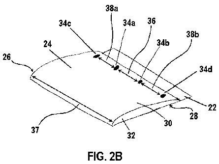
folded ECM material construct can have an upper portion 22, a lower portion
24, a first side
edge 26, and a second side edge 28. In one aspect, the lower portion 24 of the
folded ECM
material construct can comprise a first layer 30 and a second layer 32. In
this aspect, the first
layer 30 of the lower portion 24 can correspond to the folded bottom portion
16 of the sheet
of ECM material. In an additional aspect, as depicted in Figures 2A and 2B,
the first layer
14
CA 02835862 2013-11-12
WO 2012/166549 PCT/US2012/039441
30 of the lower portion 24 of the folded ECM material construct can be
attached to the
second layer 32 of the lower portion 24 of the folded ECM material construct
at a first
attachment point 34a and a second attachment point 34b. In this aspect, it is
contemplated
that the first attachment point 34a can be spaced from the second attachment
point 34b by a
selected distance 36. It is contemplated that the selected distance 36 by
which the first
attachment point 34a is spaced from the second attachment point 34b can range
from about 1/4
to about 1/2 the width 37 of the sheet 10 of ECM material. Thus, it is
contemplated that the
selected distance 36 by which the first attachment point 34a is spaced from
the second
attachment point 34b can range from about 5 mm to about 75 mm. In exemplary
aspects, the
selected distance 36 by which the first attachment point 34a is spaced from
the second
attachment point 34b can be about 1/3 the width 37 of the sheet 10 of ECM
material. In a
further aspect, it is contemplated that the first layer 30 of the lower
portion 24 of the folded
ECM material construct can be attached to the second layer 32 of the lower
portion using any
conventional surgical attachment means, including, for example and without
limitation, non-
absorbable sutures, absorbable sutures, surgical pastes, surgical glues,
staples, and the like.
In this aspect, it is contemplated that, when non-absorbable sutures are used
to secure the first
layer 30 to the second layer 32, the knots of each suture can be positioned in
contact with the
second layer such that the second layer is positioned between the first layer
and the knots. In
exemplary aspects, the first and second layers 30, 32 can be secured to one
another using a
cruciate suture pattern.
[0059] In another aspect, the first layer 30 of the lower portion 24 of
the folded ECM
material construct can optionally be further attached to the second layer 32
of the lower
portion of the folded ECM material construct at a third attachment point 34c
intermediate the
first side edge 26 of the folded ECM material construct and the first
attachment point 34a. In
this aspect, it is contemplated that the distance 38a by which the third
attachment point 34c is
spaced from the first attachment point 34a can be substantially equal to the
selected distance
36 by which the first attachment point is spaced from the second attachment
point 34b. It is
further contemplated that the third attachment point 34c can be spaced from
the first side
edge 26 of the folded ECM material construct by a selected distance ranging
from about 1
mm to about 2 mm and, more preferably, being about 1.5 mm.
[0060] In still another aspect, the first layer 30 of the lower portion 24
of the folded
ECM material construct can optionally be further attached to the second layer
32 of the lower
portion of the folded ECM material construct at a fourth attachment point 34d
intermediate
CA 02835862 2013-11-12
WO 2012/166549 PCT/US2012/039441
the second side edge 28 of the folded ECM material construct and the second
attachment
point 34b. In this aspect, it is contemplated that the distance 38b by which
the fourth
attachment point 34d is spaced from the second attachment point 34b can be
substantially
equal to the selected distance 36 by which the first attachment point 34a is
spaced from the
second attachment point. It is further contemplated that the fourth attachment
point 34d can
be spaced from the second side edge 28 of the folded ECM material construct by
a selected
distance ranging from about 1 mm to about 2 mm and, more preferably, being
about 1.5 mm.
[0061] In a further aspect, and with reference to Figure 3A, the method of
forming a
disclosed ECM material valve conduit can comprise positioning the folded ECM
material
construct in an aligned position. In this aspect, it is contemplated that, in
the aligned position,
the first side edge 26 of the folded ECM material construct can be in
substantial alignment
with the second side edge 28 of the folded ECM material construct. In
exemplary aspects, it
is contemplated that the aligned position can correspond to a position in
which the first side
edge 26 and the second side edge 28 are rolled or otherwise advanced toward
one another
until the first and second side edges are substantially adjacent to one
another. In these
aspects, the first side edge 26 and the second side edge 28 can be advanced
toward one
another such that the second layer 32 of the lower portion 24 and the upper
portion 22 of the
folded ECM material construct cooperate to define a substantially cylindrical
ECM material
construct, with the first layer of the lower portion of the folded ECM
material construct being
positioned within and extending from a periphery of the substantially
cylindrical ECM
material construct. Optionally, it is contemplated that the aligned position
can correspond to
a position in which the first and second side edges 26, 28 are in an
overlapping configuration.
It is further contemplated that the aligned position can correspond to a
position in which the
first and second side edges are everted relative to the lumen 42 of the ECM
material valve
conduit.
[0062] In an additional aspect, and with reference to Figures 3A and 3B,
with the
folded ECM material construct in the aligned position, the first side edge 26
of the folded
ECM material construct can be secured to the second side edge 28 of the folded
ECM
material construct, thereby forming an ECM material valve conduit 40
comprising a lumen
42 and having a longitudinal axis 41. In this aspect, it is contemplated that
the first side edge
26 and the second side edge 28 of the folded ECM material construct can be
secured such
that the first side edge 26 and the second side edge 28 are everted relative
to the lumen 42 of
the resulting ECM material valve conduit 40. In exemplary aspects, the first
and second
16
CA 02835862 2013-11-12
WO 2012/166549
PCT/US2012/039441
attachment points 34a, 34b can be positioned substantially within a common
plane that is
substantially perpendicular to the longitudinal axis 41 of the ECM material
valve conduit 40.
In these aspects, it is further contemplated that the third attachment point
34c and/or fourth
attachment point 34d, when present, can also be positioned within the common
plane. In a
further aspect, it is contemplated that the first side edge 26 can be secured
to the second side
edge 28 using any conventional surgical attachment means, including, for
example and
without limitation, non-absorbable sutures, absorbable sutures, surgical
pastes, surgical glues,
staples, and the like. In an exemplary aspect, it is contemplated that the
attachment means
used to secure the first side edge to the second side edge can form a seam 60
along the
longitudinal length of the ECM material valve conduit. In this aspect, when
two attachment
points 34 have been used to attach the first layer 30 of the folded ECM
material construct to
the second layer 32 of the folded ECM material construct, it is contemplated
that the seam 60
can function as a third attachment point that, in exemplary configurations,
can be
substantially equally radially spaced from the first and second attachment
points.
Alternatively, when three or four attachment points 34 have been used to
attach the first layer
30 of the folded ECM material construct to the second layer 32 of the folded
ECM material
construct, it is contemplated that the third and/or fourth attachment points
34c, 34d can be
positioned proximate the first and/or second side edges 26, 28 such that,
after the first side
edge is secured to the second side edge as described herein, the seam 60 can
be positioned
proximate the third and/or fourth attachment point(s). In exemplary aspects,
as shown in
Figure 3A, the seam 60 can be formed from a plurality of sutures 62 spaced
along the
longitudinal axis 41 of the ECM material valve conduit 40. In other exemplary
aspects, the
seam 60 can comprise a continuous suture, such as, for example and without
limitation, a
continuous 6-0 polypropylene suture.
[0063] In
one aspect, the lower portion 24 of the folded ECM material construct can
correspond to an inlet portion 44 of the ECM material valve conduit 40. In
this aspect, the
inlet portion 44 of the ECM material valve conduit 40 can define an inlet 46
in fluid
communication with the lumen 42 of the ECM material valve conduit. In another
aspect, the
first layer 30 of the lower portion 24 of the folded ECM material construct
can correspond to
an inner layer 48 positioned within the lumen 42 of the ECM material valve
conduit 40. In
still another aspect, the second layer 32 of the lower portion 24 of the
folded ECM material
construct can cooperate with the upper portion 22 of the folded ECM material
construct to
define an outer wall 52 of the ECM material valve conduit 40. In yet another
aspect, the
17
CA 02835862 2013-11-12
WO 2012/166549 PCT/US2012/039441
upper portion 22 of the folded ECM material construct can correspond to an
outlet portion 56
of the ECM material valve conduit 40. In this aspect, the outlet portion 56 of
the ECM
material valve conduit 40 can define an outlet 58 in fluid communication with
the lumen 42
of the ECM material valve conduit.
Methods of Forming the ECM Material Valve Conduits from an ECM Material
Conduit
[0064] In exemplary aspects, and with reference to Figures 5-6B, a
disclosed ECM
material valve conduit 140 can be formed from an ECM material conduit 100. In
these
aspects, the ECM material conduit 100 can comprise any known ECM component or
material, including, for example and without limitation, mucosal layers and
components,
submucosal layers and components, muscularis layers and components, and/or
basement
membrane layers and components. Optionally, the ECM material conduit 100 can
have a
multi-laminate structure that is produced by conventional methods. It is
contemplated that a
disclosed ECM material valve conduit 140 can comprise an ECM material obtained
from any
mammalian tissue source, including, for example and without limitation,
stomach tissue (e.g.,
stomach submucosa (SS)), small intestinal tissue (e.g., small intestinal
submucosa (SIS)),
large intestinal tissue, bladder tissue (e.g., urinary bladder submucosa
(UBS)), liver tissue
(e.g., liver basement membrane (LBM)), heart tissue (e.g., pericardium), lung
tissue, kidney
tissue, pancreatic tissue, prostate tissue, mesothelial tissue, fetal tissue,
a placenta, a ureter,
veins, arteries, heart valves with or without their attached vessels, tissue
surrounding the roots
of developing teeth, and tissue surrounding growing bone. In one aspect, the
ECM material
conduit 100 that is used to form the ECM material valve conduit 140 can be
obtained by
resecting an intact, lumenal portion of a mammalian tissue source, such as,
for example and
without limitation, an intact, lumenal portion of the small intestine of a
mammal. In this
aspect, it is contemplated that selected layers of the intact portion of the
mammalian tissue
source can be removed following resection.
[0065] In an additional aspect, it is contemplated that a disclosed ECM
material valve
conduit 140 can be formed from ECM that is produced using known in vitro
methods. For
example, a disclosed ECM material conduit 100 can be formed by growing cells
on an outer
surface of a cylindrical mandrel using known in vitro methods. It is
contemplated that the
growth of cells on the outer surface of the mandrel can lead to production of
one or more
ECM materials.
18
CA 02835862 2013-11-12
WO 2012/166549 PCT/US2012/039441
[0066] In one aspect, and with reference to Figures 5-6B, a method of
forming a
disclosed ECM material valve conduit 140 from an ECM material conduit 100 can
comprise
positioning the ECM material conduit in a reflected position. In this aspect,
it is
contemplated that the ECM material conduit 100 can define a lumen 102 and have
a top
portion 104 and a bottom portion 108. It is further contemplated that the top
portion 104 of
the ECM material conduit 100 can comprise a top end 106 of the ECM material
conduit,
while the bottom portion 108 of the ECM material conduit can comprise a bottom
end 110 of
the ECM material conduit. In an additional aspect, it is contemplated that the
step of
positioning the ECM material conduit 100 in the reflected position can
comprise inwardly
reflecting the bottom end 110 of the ECM material conduit within the lumen 102
of the ECM
material conduit toward the top end 106 of the ECM material conduit. It is
further
contemplated that, in the reflected position, the bottom end 110 of the ECM
material conduit
100 can be spaced a selected distance 112 from the top end 106 of the ECM
material conduit.
In one aspect, in the folded position, the selected distance 112 by which the
bottom end 110
is spaced from the top end 106 can range from about 0 mm to about 150 mm. In
this aspect,
it is contemplated that the selected distance 112 can be any distance that
permits desired
attachment of the ECM material valve conduit 140 to an artery of a subject.
[0067] In another aspect, the method of forming a disclosed ECM material
valve
conduit can comprise securing the ECM material conduit 100 in the reflected
position,
thereby forming an ECM material valve conduit 140. In this aspect, the ECM
material valve
conduit 140 comprises a lumen 142, an inlet portion 144, and an outlet portion
156 and can
have a longitudinal axis 141 and an outer circumference. In one aspect, the
outlet portion 156
can define an outlet 158 in communication with the lumen 142 of a disclosed
ECM material
valve conduit 140. In an additional aspect, the inlet portion 144 can define
an inlet 146 in
communication with the lumen 142 of a disclosed ECM material valve conduit 140
and can
comprise an outer layer and an inner layer 148 positioned within the lumen of
the ECM
material valve conduit. In this aspect, it is contemplated that the inner
layer 148 of the inlet
portion 144 of the ECM material valve conduit 140 can correspond to the
reflected bottom
end 110 of the ECM material conduit 100. In a further aspect, the inner layer
148 can be
attached to the outer layer at three attachment points 134. In this aspect, it
is contemplated
that the three attachment points 134 can be substantially equally spaced along
the outer
circumference of the ECM material valve conduit 140. For example, it is
contemplated that
the three attachment points 134 can be spaced from adjacent attachment points
by a distance
19
CA 02835862 2013-11-12
WO 2012/166549 PCT/US2012/039441
ranging from about 5 mm to about 75 mm along the outer circumference of the
ECM material
valve conduit 140. It is further contemplated that the three attachment points
134 can be
positioned substantially within a common plane that is substantially
perpendicular to the
longitudinal axis 141 of the ECM material valve conduit 140. In still another
aspect, the
outer layer of the inlet portion 144 of the ECM material valve conduit 100 can
cooperate with
the outlet portion 156 of the ECM material valve conduit to define an outer
wall 152 of the
ECM material valve conduit.
[0068] In an additional aspect, the method of forming a disclosed ECM
material valve
conduit can comprise lyophilizing the ECM material valve conduit using known
methods. In
a further aspect, when a disclosed ECM material valve conduit has been
lyophilized, the
method of forming the ECM material valve conduit can further comprise
hydrating the ECM
material valve conduit using known methods. In this aspect, it is contemplated
that the
lyophilized ECM material valve conduit can be hydrated in sterile water,
saline solution, or a
balanced salt solution for a period ranging from about 5 minutes to about 30
minutes.
[0069] In exemplary aspects, it is contemplated that the ECM material
valve conduits
40, 140 described herein can be sterilized and/or decellularized using known
methods or as
disclosed herein. In these aspects, such sterilization and/or
decellularization steps can be
performed at any stage in the construction of the ECM material valve conduit
prior to
implantation of the ECM material valve conduit within a subject. In one
aspect, it is
contemplated that the ECM material valve conduits 40, 140 described herein can
be sterilized
using ethylene oxide gas.
[0070] In one aspect, a disclosed ECM material valve conduit can comprise
a sterile,
acellular ECM composition. In exemplary aspects, such a sterile, acellular ECM
composition
can be formed by contemporaneously sterilizing and decellularizing an isolated
ECM
material. More particularly, as disclosed in the following methods, desired
sterilization and
decellularization of the isolated ECM material can occur contemporaneously
such that the
native properties of the tissue composition are maintained and the ECM
material is rendered
sterile and acellular.
Sterilization/Decellularization of ECM Compositions for Use in ECM Valve
Conduits
[0071] As described herein, the disclosed methods make use of rapid
depressurization
of an isolated ECM material to render the ECM material acellular. This rapid
depressurization of the ECM material occurs at depressurization rates that are
significantly
CA 02835862 2013-11-12
WO 2012/166549 PCT/US2012/039441
higher than the depressurization rates applied in previously known methods. In
addition to
rendering acellular the ECM material as described herein, the rapid
depressurization of the
ECM material also can be used to enhance the incorporation of desired
sterilants and
additives into the ECM material. Further, it is contemplated that the rapid
depressurization of
the ECM material can render the ECM material acellular while also improving
retention of
native growth factors, as compared to previously known decellularization
methods. Still
further, it is contemplated that the rapid depressurization of the ECM
material can be used to
improve retention of the tensile strength of the ECM material, as compared to
previously
known decellularization methods.
[0072] The disclosed methods not only do not significantly weaken the
mechanical
strength and bioptric properties of the ECM compositions, but also the methods
are more
effective in decellularizing the ECM compositions and in enhancing the
incorporation of
various additives into the ECM compositions. Thus, the disclosed sterilization
and
decellularization methods provide ECM compositions that are more
decellularized and have a
greater capacity to incorporate and then deliver more additives than ECM
compositions
known in the art. Moreover, the disclosed sterilization and decellularization
methods provide
ECM compositions that have greater amounts and/or concentrations of retained
native growth
factors and that have greater tensile strength than sterilized and
decellularized ECM
compositions known in the art.
[0073] Optionally, it is contemplated that the ECM material of a disclosed
ECM
material valve conduit can be sterilized using a known sterilization system,
such as, for
example and without limitation, the system described in U.S. Patent No.
7,108,832, assigned
to NovaSterilis, Inc., which patent is expressly incorporated herein by
reference in its
entirety. Thus, in some aspects, the system used to perform the disclosed
methods can
comprise a standard compressed storage cylinder and a standard air compressor
used in
operative association with a booster (e.g., a Haskel Booster AGT 7/30). In
other aspects, the
air compressor and booster can be replaced with a single compressor. In
exemplary aspects,
the compressed storage cylinder can be configured to receive carbon dioxide,
and the booster
can be a carbon dioxide booster.
[0074] The system can further comprise an inlet port, which allows one or
more
additives contained in a reservoir to be added to a reactor vessel through a
valve and an
additive line. As used herein, the term "reactor vessel" refers to any
container having an
interior space that is configured to receive an ECM material and permit
exposure of the ECM
21
CA 02835862 2013-11-12
WO 2012/166549 PCT/US2012/039441
material to one or more sterilants and additives, as disclosed herein. In
exemplary aspects,
the reactor vessel can be, without limitation, a basket, a bucket, a barrel, a
box, a pouch, and
other known containers. In one aspect, it is contemplated that the reactor
vessel can be a
syringe that is filled with an ECM material.
[0075] It is contemplated that a selected primary sterilant, such as, for
example and
without limitation, carbon dioxide, can be introduced to the reactor vessel
from a header line
via a valve and a supply line. It is further contemplated that a filter, such
as, for example and
without limitation, a 0.5 um filter, can be provided in the supply line to
prevent escape of
material from the vessel. In exemplary aspects, a pressure gauge can be
provided
downstream of a shut-off valve in the header line to allow the pressure to be
visually
monitored. A check valve can be provided in the header line upstream of the
valve to prevent
reverse fluid flow into the booster. In order to prevent an overpressure
condition existing in
the header line, a pressure relief valve can optionally be provided.
[0076] In one aspect, depressurization of the reactor vessel can be
accomplished using
an outlet line and a valve in communication with the reactor vessel. In this
aspect, it is
contemplated that the depressurized fluid can exit the vessel via the supply
line, be filtered by
a filter unit, and then be directed to a separator, where filtered fluid, such
as carbon dioxide,
can be exhausted via an exhaust line. It is further contemplated that valves
can be
incorporated into the various lines of the apparatus to permit fluid isolation
of upstream
components.
[0077] In one exemplary aspect, the reactor vessel can comprise stainless
steel, such
as, for example and without limitation, 316 gauge stainless steel. In another
exemplary
aspect, the reactor vessel can have a total internal volume sufficient to
accommodate the
materials being sterilized, either on a laboratory or commercial scale. For
example, it is
contemplated that the reactor vessel can have a length of about 8 inches, an
inner diameter of
about 2.5 inches, and an internal volume of about 600 mL. In additional
aspects, the reactor
vessel can comprise a vibrator, a temperature control unit, and a mechanical
stirring system
comprising an impeller and a magnetic driver. In one optional aspect, it is
contemplated that
the reactor vessel can contain a basket comprising 316 gauge stainless steel.
In this aspect, it
is contemplated that the basket can be configured to hold materials to be
sterilized while also
protecting the impeller and directing the primary sterilant in a predetermined
manner.
22
CA 02835862 2013-11-12
WO 2012/166549 PCT/US2012/039441
[0078] It is contemplated that the reactor vessel can be operated at a
constant pressure
or under continual pressurization and depressurization (pressure cycling)
conditions without
material losses due to splashing or turbulence, and without contamination of
pressure lines
via back-diffusion. It is further contemplated that the valves within the
system can permit
easy isolation and removal of the reactor vessel from the other components of
the system. In
one aspect, the top of the reactor vessel can be removed when depressurized to
allow access
to the interior space of the reactor vessel.
[0079] Optionally, the system can comprise a temperature control unit that
permits a
user to adjustably control the temperature within the reactor vessel.
[0080] In use, the disclosed apparatus can be employed in a method of
producing a
sterilized, acellular ECM composition, such as disclosed herein. However, it
is understood
that the disclosed apparatus is merely exemplary, and that any apparatus
capable of
performing the disclosed method steps can be employed to produce the
sterilized, acellular
ECM composition. Thus, the claimed method is in no way limited to a particular
apparatus.
[0081] It is contemplated that significant reductions in colony-forming
units (CFUs)
can be achieved in accordance with the disclosed methods by subjecting an
isolated ECM
material to sterilization temperature and pressure conditions using a primary
sterilant.
Optionally, it is contemplated that the primary sterilant can be combined with
one or more
secondary sterilants to achieve desired sterilization. Optionally, it is
further contemplated
that selected additives can be incorporated into an ECM material to impart
desired
characteristics to the resulting ECM composition. It is still further
contemplated that the
disclosed methods can be employed to produce sterilized, acellular ECM
compositions for
implantation within the body of a subject.
[0082] As described herein, the disclosed methods make use of rapid
depressurization
of an isolated ECM material to render the ECM material acellular. This rapid
depressurization of the ECM material occurs at depressurization rates that are
significantly
higher than the depressurization rates applied in previously known methods. In
addition to
rendering acellular the ECM material as described herein, the rapid
depressurization of the
ECM material also can be used to enhance the incorporation of desired
sterilants and
additives into the ECM material. Further, it is contemplated that the rapid
depressurization of
the ECM material can render the ECM material acellular while also improving
retention of
native growth factors, as compared to previously known decellularization
methods. Still
23
CA 02835862 2013-11-12
WO 2012/166549 PCT/US2012/039441
further, it is contemplated that the rapid depressurization of the ECM
material can be used to
improve retention of the tensile strength of the ECM material, as compared to
previously
known decellularization methods.
[0083] The disclosed methods not only do not significantly weaken the
mechanical
strength and bioptric properties of the ECM compositions, but also the methods
are more
effective in decellularizing the ECM compositions and in enhancing the
incorporation of
various additives into the ECM compositions. Thus, the disclosed sterilization
and
decellularization methods provide ECM compositions that are more
decellularized and have a
greater capacity to incorporate and then deliver more additives than ECM
compositions
known in the art. Moreover, the disclosed sterilization and decellularization
methods provide
ECM compositions that have greater amounts and/or concentrations of retained
native growth
factors and that have greater tensile strength than sterilized and
decellularized ECM
compositions known in the art.
[0084] In exemplary aspects, the primary sterilant can be carbon dioxide
at or near its
supercritical pressure and temperature conditions. However, it is contemplated
that any
conventional sterilant, including, for example, gas, liquid, or powder
sterilants that will not
interfere with the native properties of the ECM material can be used as the
primary sterilant.
[0085] In one exemplary aspect, the disclosed sterilization process can be
practiced
using carbon dioxide as a primary sterilant at pressures ranging from about
1,000 to about
3,500 psi and at temperatures ranging from about 25 C to about 60 C. More
preferably,
when supercritical carbon dioxide is used, it is contemplated that the
sterilization process can
use carbon dioxide as a primary sterilant at pressures at or above 1,071 psi
and at
temperatures at or above 31.1 C. In this aspect, the ECM material to be
sterilized can be
subjected to carbon dioxide at or near such pressure and temperature
conditions for times
ranging from about 10 minutes to about 24 hours, more preferably from about 15
minutes to
about 18 hours, and most preferably, from about 20 minutes to about 12 hours.
Preferably,
the carbon dioxide employed in the disclosed systems and methods can be pure
or,
alternatively, contain only trace amounts of other gases that do not impair
the sterilization
properties of the carbon dioxide. For ease of further discussion below, the
term "supercritical
carbon dioxide" will be used, but it will be understood that such a term is
non-limiting in that
carbon dioxide within the pressure and temperature ranges as noted above can
be employed
satisfactorily in the practice of the disclosed methods. Within the disclosed
pressure and
24
CA 02835862 2013-11-12
WO 2012/166549 PCT/US2012/039441
temperature ranges, it is contemplated that the carbon dioxide can be
presented to the ECM
material in a gas, liquid, fluid or plasma form.
[0086] The secondary sterilants employed in the disclosed methods can, in
some
aspects, include chemical sterilants, such as, for example and without
limitation, peroxides
and/or carboxylic acids. Preferred carboxylic acids include alkanecarboxylic
acids and/or
alkanepercarboxylic acids, each of which can optionally be substituted at the
alpha carbon
with one or more electron-withdrawing substituents, such as halogen, oxygen
and nitrogen
groups. Exemplary species of chemical sterilants employed in the practice of
the disclosed
methods include, for example and without limitation, hydrogen peroxide (H202),
acetic acid
(AcA), peracetic acid (PAA), trifluoroacetic acid (TFA), and mixtures thereof.
In one
exemplary aspect, the chemical sterilants can include Sporeclenz0 sterilant,
which is a
mixture comprising acetic acid, hydrogen peroxide, and peracetic acid.
[0087] It is contemplated that the secondary sterilants can be employed in
a
sterilization-enhancing effective amount of at least about 0.001 vol. % and
greater, based on
the total volume of the primary sterilant. It is further contemplated that the
amount of
secondary sterilant can be dependent upon the particular secondary sterilant
that is employed.
Thus, for example, it is contemplated that peracetic acid can be present in
relatively small
amounts of about 0.005 vol. % and greater, while acetic acid can be employed
in amounts of
about 1.0 vol. % and greater. Thus, it is contemplated that the concentration
of the secondary
sterilants can range from about 0.001 vol. % to about 2.0 vol. % and can
typically be used as
disclosed herein to achieve a sterilization-enhancing effect in combination
with the disclosed
primary sterilants, such as, for example and without limitation, supercritical
carbon dioxide.
[0088] In one aspect, the method of producing a sterilized, acellular ECM
composition
can comprise harvesting a selected tissue from a mammal and rinsing the
selected tissue in
sterile saline or other biocompatible liquid known to a person of skill in the
art, such as, for
example and without limitation, Ringer's solution and a balanced biological
salt solution. In
this aspect, the selected tissue can be, for example and without limitation,
stomach tissue
(e.g., stomach submucosa (SS)), small intestinal tissue (e.g., small
intestinal submucosa
(SIS)), large intestinal tissue, bladder tissue (e.g., urinary bladder
submucosa (UBS)), liver
tissue (e.g., liver basement membrane (LBM)), heart tissue (e.g., pericardium,
epicardium,
endocardium, myocardium), lung tissue, kidney tissue, pancreatic tissue,
prostate tissue,
mesothelial tissue, fetal tissue, a placenta, a ureter, veins, arteries, heart
valves with or
without their attached vessels, tissue surrounding the roots of developing
teeth, and tissue
CA 02835862 2013-11-12
WO 2012/166549 PCT/US2012/039441
surrounding growing bone. In another aspect, the method can comprise freezing
the selected
tissue for a period ranging from about 12 to about 36 hours, more preferably,
from about 18
to about 30 hours, and most preferably, from about 22 to about 26 hours. For
example, it is
contemplated that the period during which the selected tissue is frozen can be
12 hours, 13
hours, 14 hours, 15 hours, 16 hours, 17 hours, 18 hours, 19 hours, 20 hours,
21 hours, 22
hours, 23 hours, 24 hours, 25 hours, 26 hours, 27 hours, 28 hours, 29 hours,
30 hours, 31
hours, 32 hours, 33 hours, 34 hours, 35 hours, 36 hours, and any other period
of time falling
between the preceding values. In an additional aspect, the method can comprise
thawing the
selected tissue in cold hypotonic tris buffer. Optionally, in this aspect, the
method can
comprise thawing the selected tissue in cold hypotonic tris buffer on ice with
5 mM
ethylenediaminetetraacetic acid (EDTA). In exemplary aspects, it is
contemplated that the
steps of freezing and thawing the selected tissue can be cyclically repeated
up to six times.
[0089] In another aspect, the method can comprise isolating an ECM
material from the
selected tissue. In this aspect, the ECM material can be any material
comprising known
extracellular matrix components, including, for example and without
limitation, stomach
tissue (e.g., stomach submucosa (SS)), small intestinal tissue (e.g., small
intestinal submucosa
(SIS)), large intestinal tissue, bladder tissue (e.g., urinary bladder
submucosa (UBS)), liver
tissue (e.g., liver basement membrane (LBM)), heart tissue (e.g., pericardium,
epicardium,
endocardium, myocardium), lung tissue, kidney tissue, pancreatic tissue,
prostate tissue,
mesothelial tissue, fetal tissue, a placenta, a ureter, veins, arteries, heart
valves with or
without their attached vessels, tissue surrounding the roots of developing
teeth, and tissue
surrounding growing bone. In one exemplary, non-limiting aspect, the step of
isolating an
ECM material can comprise isolating SIS from a mammalian tissue source. In
this aspect,
the method can comprise: incising a wall of a small intestine along a path
that is substantially
parallel to the longitudinal axis of the small intestine; opening the small
intestine along the
path of the incision such that the small intestine lies flat on a surface;
rinsing the small
intestine with sterile saline or other biocompatible fluid; mechanically
stripping the SIS of the
small intestine from the surrounding smooth muscle and serosal layers and from
the tunica
mucosa, leaving essentially the submucosal and basement membrane layers.
However, it is
contemplated that the ECM material can be isolated using any conventional
technique,
including those described in: U.S. Patent No. 4,902,508; U.S. Patent No.
5,275,826; U.S.
Patent No. 5,281,422; U.S. Patent No. 5,554,389; U.S. Patent No. 6,579,538;
U.S. Patent No.
6,933,326; U.S. Patent No. 7,033,611; Voytik-Harbin et al., "Identification of
Extractable
26
CA 02835862 2013-11-12
WO 2012/166549 PCT/US2012/039441
Growth Factors from Small Intestinal Submucosa," J. Cell. Biochem., Vol. 67,
pp. 478-491
(1997); Hodde et al., "Virus Safety of a Porcine-Derived Medical Device:
Evaluation of a
Viral Inactivation Method," Biotech. & Bioeng., Vol. 79, No. 2, pp. 211-216
(2001); Badylak
et al., "The Extracellular Matrix as a Scaffold for Tissue Reconstruction,"
Cell &
Developmental Biology, Vol. 13, pp. 377-383 (2002); Robinson et al.,
"Extracelular Matrix
Scaffold for Cardiac Repair," Circulation, Vol. 112, pp. I-135-1-143 (2005);
Hodde et al.,
"Effects of Sterilization on an Extracellular Matrix Scaffold: Part I.
Composition and Matrix
Architecture," J. Mater. Sci.: Mater. Med., Vol. 18, pp. 537-543 (2007); and
Hodde et al.,
"Effects of Sterilization on an Extracellular Matrix Scaffold: Part II.
Bioactivity and Matrix
Interaction," J. Mater. Sci.: Mater. Med., Vol. 18, pp. 545-550 (2007), each
of which is
expressly incorporated herein by reference in its entirety.
[0090] In an additional aspect, the method can comprise incubating the
isolated ECM
material for 24 to 48 hours in 0.5-1% Triton X-100/0.5-1% Deoxycholic acid
with 5 mM
EDTA in Dulbecco's Phosphate Buffered Saline (DPBS) (Lonza Walkersville,
Inc.). In this
aspect, it is contemplated that flat or sheet-like ECM materials, such as
stomach submucosa
(SS), small intestinal submucosa (SIS), and bladder submucosa (UBS), can be
incubated in a
stretched configuration. It is further contemplated that ECM material conduits
or other
lumenal ECM materials, such as intact ureters, arteries, veins, and small
intestines or formed
ECM conduits, can be perfused with the various disclosed solutions through
soaking and by
use of a peristaltic pump.
[0091] In a further aspect, after incubation, the method can comprise
rinsing the ECM
material with DPBS. In this aspect, it is contemplated that the step of
rinsing the ECM
material can comprise rinsing the ECM material up to six times, including one,
two, three,
four, five, or six times, with each rinse lasting for about thirty minutes. In
an exemplary
aspect, it is contemplated that the step of rinsing the ECM material can
comprise rinsing the
ECM material three times, with each rinse lasting for about thirty minutes.
[0092] Optionally, in exemplary aspects, the method can further comprise a
second
incubation procedure. In these aspects, the second incubation procedure can
comprise
incubating the ECM material in isotonic tris buffer containing 10-50 [ig/mL of
RNAase/0.2-
0.5 [tg/mL DNAase with 5 mM EDTA. It is contemplated that the step of
incubating the
ECM material in isotonic tris buffer can be performed at a temperature of
about 37 C,
substantially corresponding to the temperature of a human body. It is further
contemplated
that the step of incubating the ECM material in isotonic tris buffer can be
performed for a
27
CA 02835862 2013-11-12
WO 2012/166549 PCT/US2012/039441
period ranging from about 30 minutes to about 24 hours, more preferably, from
about 1 hour
to about 18 hours, and most preferably, from about 2 hours to about 12 hours.
In an
additional aspect, the second incubation procedure can further comprise
rinsing the ECM
material with DPBS. In this aspect, it is contemplated that the step of
rinsing the ECM
material can comprise rinsing the ECM material three times, with each rinse
lasting for about
thirty minutes.
[0093] In yet another aspect, whether or not the second incubation
procedure is
performed, the method can comprise storing the ECM material at a temperature
ranging from
about 1 C to about 10 C, more preferably, from about 2 C to about 6 C, and,
most
preferably, from about 3 C to about 5 C. In an exemplary aspect, the ECM
material can be
stored at 4 C.
[0094] In an additional aspect, the method can comprise introducing the
ECM material
into the interior space of the reactor vessel. Optionally, in this aspect, one
or more secondary
sterilants from the reservoir can be added into the interior space of the
reactor vessel along
with the ECM material. In these aspects, it is contemplated that the one or
more secondary
sterilants from the reservoir can be added into the interior space of the
reactor vessel before,
after, or contemporaneously with the ECM material. It is further contemplated
that the
temperature control unit can be selectively adjusted to produce a desired
temperature within
the interior space of the reactor vessel. In a further aspect, the method can
comprise
equilibrating the pressure within the reactor vessel and the pressure within
the storage
cylinder. For example, in this aspect, it is contemplated that the pressure
within the reactor
vessel and the pressure within the storage cylinder can be substantially equal
to atmospheric
pressure. In yet another aspect, after equilibration of the pressures within
the apparatus, the
method can comprise operating the magnetic driver to activate the impeller of
the reactor
vessel. In still a further aspect, the method can comprise selectively
introducing the primary
sterilant from the storage cylinder into the reactor vessel until a desired
pressure within the
reactor vessel is achieved. In this aspect, it is contemplated that the step
of selectively
introducing the primary sterilant into the reactor vessel can comprise
selectively activating
the air compressor and the booster to increase flow of the primary sterilant
into the reactor
vessel. In exemplary aspects, the air compressor and booster can be activated
to subject the
ECM material to supercritical pressures and temperatures, such as, for example
and without
limitation, the pressures and temperatures necessary to produce supercritical
carbon dioxide,
for a time period ranging from about 20 minutes to about 60 minutes.
28
CA 02835862 2013-11-12
WO 2012/166549 PCT/US2012/039441
[0095] In a further aspect, the method can comprise rapidly depressurizing
the reactor
vessel. In this aspect, a predetermined amount of primary sterilant, such as,
for example and
without limitation, supercritical carbon dioxide, can be released from the
reactor vessel
through the depressurization line. It is contemplated that the primary
sterilant can be released
from the reactor vessel through opening of the valve coupled to the reactor
vessel to thereby
reduce the pressure within the reactor vessel. As used herein, the term "rapid
depressurization" refers to depressurization of the reactor vessel at a rate
greater than or equal
to 400 psi/min. For example, it is contemplated that the reactor vessel can be
rapidly
depressurized at a depressurization rate ranging from about 2.9 MPa/min. to
about 18.0
MPa/min. (about 400 psi/min. to about 2,600 psi/min.), more preferably, from
about 5.0
MPa/min. to about 10.0 MPa/min. (700 psi/min. to about 1,500 psi/min.), and,
most
preferably, from about 7.0 MPa/min. to about 8.0 MPa/min. (about 1,000
psi/min. to about
1,200 psi/min.). Thus, these rapid depressurizations are significantly greater
than the 300
psi/min. depressurization rate disclosed in U.S. Patent No. 7,108,832. Without
being bound
by any particular theory, it is believed that the disclosed rapid
depressurization rates increase
the level of decellularization achieved in the ECM material. For example, it
is contemplated
that the rapid depressurization of a disclosed ECM material can lead to levels
of
decellularization in the ECM material of greater than about 96%, including
96.1%, 96.2%,
96.3%, 96.4%, 96.5%, 96.6%, 96.7%, 96.8%, 96.9%, 97.0%, 97.1%, 97.2%, 97.3%,
97.4%,
97.5%, 97.6%, 97.7%, 97.8%, 97.9%, 98.0%, 98.1%, 98.2%, 98.3%, 98.4%, 98.5%,
98.6%,
98.7%, 98.8%, 98.9%, 99.0%, 99.1%, 99.2%, 99.3%, 99.4%, 99.5%, 99.6%, 99.7%,
99.8%,
99.9%, and 100%.
[0096] In exemplary aspects, the method can further comprise the step of
incorporating one or more additives into the ECM material. In these aspects,
it is
contemplated that the one or more additives can be provided in either a powder
or a liquid
form. In one optional aspect, the step of incorporating the one or more
additives can
comprise introducing the one or more additives into the reactor vessel during
the step of
rapidly depressurizing the reactor vessel. In this aspect, it is contemplated
that the
introduction of the one or more additives can be characterized as a
conventional foaming
process. In another optional aspect, the step of incorporating the one or more
additives can
comprise introducing the one or more additives into the reactor vessel after
the step of rapidly
depressurizing the reactor vessel. In this aspect, it is contemplated that the
one or more
additives can be added to the ECM material after the rapid depressurization of
the reactor
29
CA 02835862 2013-11-12
WO 2012/166549 PCT/US2012/039441
vessel has caused the ECM material to swell and/or expand, thereby permitting
improved
penetration of the additives into the ECM material. It is further contemplated
that, in an
exemplary aspect, the one or more additives can be added to the ECM material
within about
thirty minutes after the rapid depressurization of the reactor vessel. In a
further optional
aspect, the step of incorporating the one or more additives can comprise
introducing the one
or more additives into the reactor vessel both during and after the step of
rapidly
depressurizing the reactor vessel. In this aspect, it is contemplated that the
one or more
additives can be released into the reactor vessel in both a quick manner and a
slow, extended
manner. In still a further optional aspect, the step of incorporating the one
or more additives
can comprise introducing the one or more additives into the reactor vessel
before the step of
rapidly depressurizing the reactor vessel.
[0097] The disclosed additives can be incorporated into the ECM material
to impart
selected properties to the resulting sterilized, acellular ECM composition.
Thus, it is
contemplated that the one or more additives can be selected to replace or
supplement
components of the ECM material that are lost during processing of the ECM
material as
described herein. For example, and as described below, the one or more
additives can
comprise growth factors, cytokines, proteoglycans, glycosaminoglycans (GAGs),
proteins,
peptides, nucleic acids, small molecules, drugs, or cells. It is further
contemplated that the
one or more additives can be selected to incorporate non-native components
into the ECM
material. For example, the one or more additives can comprise, for example and
without
limitation, growth factors for recruiting stem cells, angiogenic cytokines,
and anti-
inflammatory cytokines. It is still further contemplated that the one or more
additives can be
pharmaceutical agents, such as statins, corticosteroids, non-steroidal anti-
inflammatory drugs,
anti-inflammatory compounds, anti-arrhythmic agents, and the like. It is still
further
contemplated that the one or more additives can be nanoparticles, such as, for
example and
without limitation, silver nanoparticles, gold nanoparticles, platinum
nanoparticles, iridium
nanoparticles, rhodium nanoparticles, palladium nanoparticles, copper
nanoparticles, zinc
nanoparticles, and other metallic nanoparticles. It is still further
contemplated that the one or
more additives can be metallic compounds. In one exemplary aspect, the one or
more
additives can be selected to pharmaceutically suppress the immune response of
a subject
following implantation of the resulting ECM composition into the body of a
subject.
[0098] In one aspect, the one or more additives can comprise one or more
growth
factors, including, for example and without limitation, transforming growth
factor-I3 1, 2, or 3
CA 02835862 2013-11-12
WO 2012/166549 PCT/US2012/039441
(TGF-I3 1, 2, or 3), fibroblast growth factor-2 (FGF-2), also known as basic
fibroblast growth
factor (bFGF), vascular endothelial growth factor (VEGF), placental growth
factor (PGF),
connective tissue growth factor (CTGF), hepatocyte growth factor (HGF),
Insulin-like growth
factor (IGF), macrophage colony stimulating factor (M-CSF), platelet derived
growth factor
(PDGF), epidermal growth factor (EGF), and transforming growth factor-a (TGF-
a).
[0099] In another aspect, the one or more additives can comprise one or
more
cytokines, including, for example and without limitation, stem cell factor,
stromal cell-
derived factor-1 (SDF-1), granulocyte macrophage colony-stimulating factor (GM-
CSF),
interferon gamma (IFN-gamma), Interleukin-3, Interleukin-4, Interleukin-10,
Interleukin-13,
Leukemia inhibitory factor (LIF), amphiregulin, thrombospondin 1,
thrombospondin 2,
thrombospondin 3, thrombospondin 4, thrombospondin 5, and angiotensin
converting enzyme
(ACE).
[00100] In an additional aspect, the one or more additives can comprise one
or more
proteoglycans, including, for example and without limitation, heparan sulfate
proteoglycans,
betaglycan, syndecan, decorin, aggrecan, biglycan, fibromodulin, keratocan,
lumican,
epiphycan, perlecan, agrin, testican, syndecan, glypican, serglycin, selectin,
lectican,
versican, neurocan, and brevican.
[00101] In a further aspect, the one or more additives can comprise one or
more
glycosaminoglycans, including, for example and without limitation, heparan
sulfate,
hyaluronic acid, heparin, chondroitin sulfate B (dermatan sulfate), and
chondroitin sulfate A.
[00102] In still a further aspect, the one or more additives can comprise
one or more
proteins, peptides, or nucleic acids, including, for example and without
limitation, collagens,
elastin, vitronectin, versican, laminin, fibronectin, fibrillin-1, fibrillin-
2, plasminogen, small
leucine-rich proteins, cell-surface associated protein, cell adhesion
molecules (CAMs), a
matrikine, a matrix metalloproteinase (MMP), a cadherin, an immunoglobin, a
multiplexin,
cytoplasmic domain-44 (CD-44), amyloid precursor protein, tenascin,
nidogen/entactin,
fibulin I, fibulin II, integrins, transmembrane molecules, and osteopontin.
[00103] In yet another aspect, the one or more additives can comprise one
or more
pharmaceutical agents, including, for example and without limitation, statin
drugs, for
example, cerevastatin, atorvastatin, fluvastatin, lovastatin, mevastatin,
pitavastatin,
pravastatin, rosuvastatin, and simvastatin, corticosteroids, non-steroidal
anti-inflammatory
31
CA 02835862 2013-11-12
WO 2012/166549 PCT/US2012/039441
drugs, anti-inflammatory compounds, anti-arrhythmic agents, antimicrobials,
antibiotics, and
the like.
[00104] In exemplary aspects, the steps of introducing the one or more
additives into
the reactor vessel can comprise opening the valve to allow the one or more
additives to flow
from the reservoir into the inlet port. Prior to pressurization, it is
contemplated that the one
or more additives can be introduced directly into the reactor vessel prior to
sealing and/or via
the inlet port.
[00105] It is contemplated that the disclosed rapid depressurization and
repressurization
of the reactor vessel, with or without the addition of the one or more
additives, can be
repeated for any desired number of cycles. It is further contemplated that the
cycles of rapid
depressurization and repressurization, as well as the introduction of the
primary sterilants
and/or secondary sterilants and/or additives, can be automatically controlled
via a controller
that is configured to selectively open and/or close the various valves of the
system to achieve
desired pressure conditions and cycles.
[00106] In some aspects, the disclosed methods can further comprise the
step of
agitating the contents of the reactor vessel. In these aspects, it is
contemplated that the step
of agitating the contents of the reactor vessel can comprise periodically
agitating the contents
of the reactor vessel using a vibrator. It is further contemplated that the
agitation of the
reactor vessel can be intermittent, continual, or continuous. In exemplary
aspects, the step of
agitating the contents of the reactor vessel can occur during the step of
introducing the
primary sterilant into the reactor vessel. It is contemplated that the
agitation of the contents
of the reactor vessel can enhance the mass transfer of the sterilants and/or
additives by
eliminating voids in the fluids within the reactor vessel to provide for more
complete contact
between the ECM material and the sterilants and/or additives. It is further
contemplated that
the step of agitating the contents of the reactor vessel can comprise
selectively adjusting the
intensity and duration of agitation so as to optimize sterilization times,
temperatures, and
pressurization/depressurization cycles.
[00107] In a further aspect, after the sterilization and decellularization
of the ECM
material is complete, the method can further comprise depressurizing the
reactor vessel and
deactivating the magnetic drive so as to cease movement of the stirring
impeller. Finally, the
method can comprise the step of removing the resulting sterilized, acellular
ECM
composition through the top of the reactor vessel.
32
CA 02835862 2013-11-12
WO 2012/166549 PCT/US2012/039441
Methods of Regenerating Heart Valves Using the ECM Material Valve Conduits
[00108] Also disclosed herein are methods of regenerating heart valves. In
an
exemplary aspect, a method of regenerating a semi-lunar valve to replace a
defective semi-
lunar valve within a heart of a subject is disclosed. In this aspect, and with
reference to
Figure 24, it is contemplated that the defective semi-lunar valve is attached
at an annulus of
an annular region therebetween a ventricle of the heart of the subject and an
artery of the
subject. As used herein, the term "semi-lunar valve" can refer to either a
pulmonary valve or
an aortic valve within the heart of the subject. It is contemplated that, if
the defective semi-
lunar valve is a pulmonary valve, then the defective semi-lunar valve is
attached at an
annulus of an annular region between the right ventricle of the heart of the
subject and the
pulmonary artery of the subject. It is further contemplated that, if the
defective semi-lunar
valve is an aortic valve, then the defective semi-lunar valve is attached at
an annulus of an
annular region between the left ventricle of the heart of the subject and the
aorta of the
subject.
[00109] In one aspect, a disclosed method of regenerating a semi-lunar
valve can
comprise removing the defective semi-lunar valve from the heart of the
subject, thereby
exposing the annular region. In this aspect, it is contemplated that the step
of removing the
defective semi-lunar valve can optionally comprise removing a portion of an
artery that was
coupled to the defective semi-lunar valve, such as an aorta or a pulmonary
artery. It is further
contemplated that the step of removing the defective semi-lunar valve can
optionally
comprise removing the annulus of the annular region.
[00110] It is contemplated that the step of removing the defective semi-
lunar valve can
further comprise placing the subject on cardiopulmonary bypass. It is further
contemplated
that the step of removing the defective semi-lunar valve can further comprise
arresting and/or
fibrillating the heart of the subject and exposing the defective valve through
an incision in the
heart of the subject. Alternatively, the defective valve can be accessed
percutaneously using
known methods.
[00111] In an additional aspect, a disclosed method of regenerating a semi-
lunar valve
can further comprise implanting an ECM material valve conduit, such as those
disclosed
herein. In this aspect, and as further disclosed herein, the ECM material
valve conduit can
define a lumen and have an inlet portion and an outlet portion. In a further
aspect, the step of
implanting an ECM material valve conduit can comprise securing the inlet
portion of the
33
CA 02835862 2013-11-12
WO 2012/166549 PCT/US2012/039441
ECM material valve conduit to the annular region and securing the outlet
portion of the ECM
material valve conduit to the artery of the subject such that the inlet
portion is positioned
proximate the annular region. In this aspect, it is contemplated that the ECM
material valve
conduit can be secured to the annular region and/or the artery using any
conventional surgical
attachment means, including, for example and without limitation, non-
absorbable sutures,
absorbable sutures, surgical pastes, surgical glues, staples, and the like.
Optionally, in one
aspect, the ECM material valve conduit can be secured to the annular region
before it is
secured to the artery. In this aspect, it is contemplated that, after the ECM
material valve
conduit has been properly secured to the annulus, the length of the ECM
material valve
conduit can be trimmed as necessary to eliminate any excess length while
retaining adequate
tissue for proper attachment of the ECM material valve conduit to the artery.
Alternatively,
in another aspect, the ECM material valve conduit can be secured to the artery
before it is
secured to the annular region. In this aspect, it is contemplated that, after
the ECM material
valve conduit has been properly secured to the artery, the length of the ECM
material valve
conduit can be trimmed as necessary to eliminate any excess length while
retaining adequate
tissue for proper attachment of the ECM material valve conduit to the annular
region.
[00112] In exemplary aspects, it is contemplated that one or more pledgets
can be
added to the outer wall of the ECM material valve conduit at locations
proximate the
attachment of the ECM material valve conduit to the annular region and/or the
artery. In
these aspects, it is contemplated that the pledgets can be configured to
shield the ECM
material valve conduit from direct contact with the sutures between the ECM
material valve
conduit and the annular region and/or artery, thereby minimizing the risk of
the suture cutting
through the ECM material valve conduit and providing additional structural
integrity to the
ECM material valve conduit. It is contemplated that any conventional pledget,
such as, for
example and without limitation, Teflon pledgets, can be employed for these
purposes. In an
exemplary aspect, it is contemplated that the one or more pledgets can
comprise at least one
ECM material. It is contemplated that the one or more pledgets can have any
suitable shape
and dimensions. However, in exemplary aspects, it is contemplated that the
pledget can
comprise a substantially rectangular sheet having a length ranging from about
2 mm to about
4 mm and a width ranging from about 2 mm to about 4 mm.
[00113] After the ECM material valve conduit is properly secured to the
annular region
and to the artery, and after any necessary trimming or sculpting of the ECM
material valve
34
CA 02835862 2013-11-12
WO 2012/166549 PCT/US2012/039441
conduit has been completed, the heart of the subject can be closed as
necessary and the heart
of the subject can be restarted.
[00114] It is further contemplated that anti-coagulant and/or anti-
thrombotic agents
and/or therapies can be delivered to the subject before, during, and/or after
the ECM material
valve conduit is properly secured to the annular region and to the artery. It
is contemplated
that these agents and/or therapies can prevent thrombus formation on the
leaflets of the
regenerated valve in the subject.
[00115] In additional aspects, and as shown in Figure 4, the inner layer of
a disclosed
ECM material valve conduit can comprise leaflet-promoting portions and
commissure-
promoting portions, and the outer layer of the disclosed ECM material valve
conduit can
comprise sinus-promoting portions. In these aspects, it is contemplated that
the leaflet-
promoting portions can regenerate a plurality of leaflets (i.e., three
leaflets) of a replacement
semi-lunar valve proximate the annulus. It is further contemplated that the
sinus-promoting
portions of the outer layer of the ECM material valve conduit can fuse with
the inner layer of
the ECM material valve conduit to regenerate sinus portions of the replacement
semi-lunar
valve. It is still further contemplated that the commissure-promoting portions
of the ECM
material valve conduit can fuse with the outer layer of the ECM material valve
conduit to
regenerate commissures of the replacement semi-lunar valve. Figures 7-11
display
regeneration of a pulmonary valve following implantation of an exemplary ECM
material
valve conduit according to the methods described herein.
[00116] It is contemplated that, following implantation of the ECM material
valve
conduit as disclosed herein, the implanted ECM material valve conduit can
attract stem cells
of the subject that will remodel the ECM material to form the replacement semi-
lunar valve.
The stem cells can remodel the inner layer of the ECM material valve conduit
and the
supporting arterial wall into leaflets and commissures of the replacement semi-
lunar valve
and can remodel the outer layer of the ECM material valve conduit into sinus
portions of the
replacement semi-lunar valve. During this remodeling process, it is
contemplated that the
implanted ECM material valve conduit can provide normal valvular function. It
is further
contemplated that the implanted ECM material valve conduit can be gradually
degraded and
replaced with host tissue that is indistinguishable (i.e., identical or
substantially identical) to
normal, native tissue.
CA 02835862 2013-11-12
WO 2012/166549 PCT/US2012/039441
Experimental Examples
[00117] The following examples are put forth so as to provide those of
ordinary skill in
the art with a complete disclosure and description of how the compounds,
compositions,
articles, devices and/or methods claimed herein are made and evaluated, and
are intended to
be purely exemplary of the invention and are not intended to limit the scope
of what the
inventors regard as their invention. Efforts have been made to ensure accuracy
with respect
to numbers (e.g., amounts, temperature, etc.), but some errors and deviations
should be
accounted for. Unless indicated otherwise, parts are parts by weight,
temperature is in C or
is at ambient temperature, and pressure is at or near atmospheric.
Example One
[00118] An ECM material valve conduit was constructed as disclosed herein.
A sheep
was anesthetized. The left chest of the sheep was opened, and then the heart
of the sheep was
exposed. The sheep was placed on cardiopulmonary bypass to support the sheep
while the
pulmonary valve and portions of the pulmonary artery were excised. During the
excision, the
pulmonary valve (including leaflets) and the annulus at which the pulmonary
valve was
secured were removed. Additionally, several centimeters of the pulmonary
artery distal to
the pulmonary valve complex were removed. The ECM material valve conduit was
then
sutured into place, with the inlet portion sutured to the heart and the outlet
portion sutured to
the remaining pulmonary artery.
[00119] Figures 7-14 depict findings at harvest after the conduit had been
in the animal
for three months of remodeling. Figure 7 demonstrates how the ECM material
valve conduit
has taken on the appearance of a normal pulmonary artery. Figure 8 shows the
remodeled
leaflets from the perspective of the distal, pulmonary artery. Figure 9 shows
the inlet portion
of the ECM material valve conduit from the perspective of the heart and the
ventricular side
of the leaflets. Figure 10 shows the three leaflets with the conduit opened
longitudinally.
Figure 11 shows the three leaflets from the perspective of the pulmonary
artery with the ECM
material valve conduit opened.
[00120] Figures 12-14 show exemplary ECM material valve conduits prior to
implantation. Figure 12 shows a manufactured ECM material valve conduit in a
closed
position, while Figure 13 shows the ECM material valve conduit in an open
position. Figure
14 shows an exemplary ECM material valve conduit that has been hydrated in
sterile water
prior to implantation.
36
CA 02835862 2013-11-12
WO 2012/166549
PCT/US2012/039441
Example Two
[00121] As depicted in Figures 15-16, a large rectangular patch of
pericardium was
used to construct a tri-leaflet conduit through a process of folding and
suturing. The patch
was rolled over an appropriately sized dilator. After the patch was rolled
onto the dilator,
lengthwise sutures were used to form an ECM conduit. This ECM conduit, which
is shown
in Figure 23, was used to establish right ventricular to pulmonary continuity
in a congenital
reconstructive procedure performed on a patient.
[00122] The graft material came as a dried sheet in an easily stored
packet. The sheet
had the dimensions of 7cm x 10cm. The sheet was prepared in saline solution
for ten
minutes.
[00123] The patient was a 12 year-old female with a bicuspid aortic valve
causing
severe aortic stenosis. Approximately 18 months earlier, a retrograde balloon
valvuloplasty
had been performed. Subsequently, the patient developed severe aortic
regurgitation with
progressive left ventricular dilatation. The left ventricular end diastolic
dimension was 6.2
cm, and the patient was symptomatic with dyspnea on exertion.
[00124] With the patient anesthetized and draped for surgery, the ECM
conduit was
hydrated in normal saline solution for ten minutes and then trimmed to allow
for a final
conduit size of 20 mm. The size of the conduit was determined by pre-operative
echocardiographic measurements of the patient's pulmonary valve size and then
up-sizing to
create a valve Z score of 2. A 20 mm conduit was created by trimming the
rectangular patch
of pericardium to the dimensions of 65mm x 45mm. With reference to Figure 15,
the length
(L) and width (W) of the patch were determined by the formulas:
L = D*7r; and
W = (2*D) + C,
where D is the desired diameter of the conduit and C is the cuff length, which
corresponds to the portion of the conduit that is to be attached to the
pulmonary artery of the
patient.
[00125] After preparation of the conduit, a Ross procedure, using a full
root
replacement technique with continuous monofilament suture for each anastomosis
was
performed. The procedure was performed at 32 C, using bi-caval cannulation
and
37
CA 02835862 2013-11-12
WO 2012/166549 PCT/US2012/039441
intermittent retrograde blood cardioplegia every 15 to 20 minutes. The conduit
implantation
was performed using a continuous monofilament suture after the proximal aortic
root
reconstruction and before the distal aortic suture line was completed. The
distal conduit
anastomosis was completed before the proximal conduit anastomosis. Both
anastomoses
were performed using continuous 5-0 prolene sutures before the aortic cross
clamp was
removed. The branch pulmonary arteries were mobilized extensively to allow for
a tension-
free proximal anastomosis and to avoid the use of additional patch material
proximally. The
conduit was created with a 5 mm distal cuff extension. After release of the
cross clamp, the
heart of the patient regained vigorous contractility with minimal inotropic
support.
[00126] The procedure was completed and the patient was easily separated
from
bypass. The patient's cardiac performance and hemostasis were satisfactory.
She was
awakened and extubated in the operating room. Her recovery was uneventful and
after one
day in the intensive care unit was transferred to the ward. Five days after
the operation, she
underwent a complete echocardiogram prior to hospital discharge. The results
of this are
shown in Figures 17-19. Six days after the operation, she was discharged from
the hospital in
excellent condition.
[00127] Initial follow-up echocardiogram studies were obtained one month
after
hospital discharge. These echocardiogram studies showed no stenosis or
insufficiency of the
conduit and showed mild autograft insufficiency with well-preserved left
ventricular
function. The ventricular dimensions of the patient returned to normal. The
patient did not
show any symptoms and was taking aspirin and an ACE inhibitor. The
echocardiograms
indicated that the patient was in New York Heart Association (NYHA) heart
failure
classification 1.
[00128] Subsequent follow-up echocardiogram studies were obtained five
months after
hospital discharge. At the time of these follow-up echocardiogram studies, the
patient had
gained weight and had returned to full physical activity without limitations.
Auscultation
revealed no systolic or diastolic murmurs. The follow-up echocardiograms,
which are shown
in Figures 20-22, indicated that the patient remained in NYHA classification
1. The
echocardiograms also demonstrated that the patient had completely normal right
and left
ventricular function as well as normal conduit function. Minor conduit
insufficiency was
observed, and leaflet mobility was unchanged from the echocardiograms recorded
at five
days after the operation. Peak echo velocity within the conduit was 2
meters/second and
38
CA 02835862 2013-11-12
WO 2012/166549
PCT/US2012/039441
color flow did not exhibit any turbulence. The left ventricular end diastolic
diameter was
measured as 4.0 cm.
Example Three
[00129] Various measurements of exemplary ECM material valve conduits, as
depicted
in Figure 24, are provided in Table 1 below:
Table 1
side sewing
Annulus D, Width (w), Sewing Cuff height leaflet leaflet width
allowance (s),
mm mm height, mm .(sc), mm :(01)s, mm (iw), mm
mm
18 59.5 38.8 10.0 . 14.4 18.8 1.5
28 91.0 54.8 10.0 22.4 29.3 1.5
30 97.2 58.0 10.0 24.0 31.4 1.5
Example Four
[00130] In vitro mechanical evaluation was performed on the seam of
exemplary ECM
material valve conduits as described herein. The maximum tensile break force
for the sewn
seam was found to be 52.1 14.1 N (11.7 3.16 lbf), with a minimum and maximum
of 34.8 N
(7.82 lbf) and 72.3 N (16.25 lbf), respectively. The tensile force, ball
burst, and suture pull-
out forces for exemplary 4-ply ECM material valve conduits as described herein
was
determined to be19.35 5.51 N (4.35 1.24 lbf), 126.6 30.2N (6699 1598 mmHg),
and
11.12 2.08 N (2.50 0.47 lbf), respectively. These results indicate that the
structural integrity
of the ECM material valve conduits described herein are more than adequate to
meet the
force requirements of the pulmonary valve in the low-pressure environment of
the right heart.
Example Five
[00131] Exemplary ECM material valve conduits were evaluated in an animal
study
model. The ECM material valve conduits were implanted in a pulmonary valve
position
following removal of the native pulmonary valve. The results have shown
physiologically
normal hemodynamic results for the ECM material valve conduits in the
immediate
postoperative period prior to tissue remodeling and also out to twelve months.
At 12 months,
much of the ECM was remodeled into the animal's native tissue.
Echocardiography has
shown good hemodynamics for the regenerated valves out to 12 months with
complete
coaptation of the leaflets and no leaflet prolapse (Figure 25).
39
CA 02835862 2013-11-12
WO 2012/166549 PCT/US2012/039441
[00132] Gross necropsy and histology results showed appropriate remodeling
of the
valve with increased replacement by host tissue at each subsequent time point.
The
replacement valves appeared grossly similar to the native valve that was
replaced. Figure 26
shows 3-, 5-, 6-, and 12-month explants from sheep implanted with the ECM
material
Pulmonary Valve Conduit as part of the non-GLP study. These images
demonstrated the
progressive remodeling that is occurring over time in the sheep. As shown, at
the 3-month
time point (Figure 26(a)), remodeling has already occurred at the valve
annulus and is
extending to the leaflets to regenerate apparently normal valve tissue. At 12
months (Figure
26(d)), the leaflets are remodeled and appear similar to native valve tissue.
[00133] H&E histology of the explanted ECM Pulmonary Valve Conduit after 3,
5, 6
and 12 months was analyzed. Cells were distributed throughout the valve and
even into the
tip of the leaflet by 3 months, and by 6 months the remodeled tissue has
formed a three-layer
structure similar to the native valve tissue with a ventricularis, spongiosa,
and fibrosa.
Example Six
[00134] In exemplary applications of the disclosed sterilization and
decellularization
methods, selected tissues were harvested and rinsed in sterile saline. The
selected tissues
were then frozen for 24 hours. The frozen tissues were thawed in cold
hypotonic tris buffer
on ice with 5 mM ethylenediaminetetraacetic acid (EDTA). An extracellular
matrix material
was then isolated from each selected tissue, as described herein.
[00135] The isolated extracellular matrix materials were incubated for 24
to 48 hours in
0.5-1% Triton X-100/0.5-1% Deoxycholic acid with 5 mM EDTA in Dulbecco's
Phosphate
Buffered Saline (DPBS) (Lonza Walkersville, Inc.). Flat extracellular matrix
materials, such
as stomach submucosa (SS), small intestinal submucosa (SIS), and bladder
submucosa
(UBS), were incubated in a stretched configuration. Tubular extracellular
matrix materials,
such as ureters, arteries, veins, and tubular SIS, were perfused with the
solutions through
soaking and by use of a peristaltic pump.
[00136] After incubation, each extracellular matrix material was rinsed
three times with
DPBS. Each rinsing with DPBS lasted 30 minutes. Some extracellular matrix
materials were
then incubated for 2 to 12 hours at 37 C in isotonic tris buffer containing 10-
50 [tg/mL of
RNAase/0.2-0.5 [ig/mL DNAase with 5 mM EDTA. Following this incubation step,
the
extracellular matrix materials were again rinsed three times with DPBS. Each
rinsing with
DPBS lasted 30 minutes. The extracellular matrix materials were stored at 4 C.
CA 02835862 2013-11-12
WO 2012/166549 PCT/US2012/039441
[00137] Within 48 hours of storage, the extracellular matrix materials were
processed in
supercritical carbon dioxide as disclosed herein for 20-60 minutes at
temperatures at or
greater than 31.1 C and pressures at or greater than 1,071 psi. After this
sterilization step, the
extracellular matrix materials were rapidly depressurized at a rate of 2.7
MPa/10 sec. (391.6
psi/10 sec.) for a minute and 19 seconds. During this time, the pressure
applied to the
extracellular matrix materials rapidly decreased from 9.9 MPa to 0.69 MPa.
[00138] The extracellular matrix materials were then processed in
supercritical carbon
dioxide and peracetic acid (PAA) as disclosed herein for 30 minutes to 6 hours
to achieve
terminal sterilization. In this processing step, the pressure applied to the
extracellular matrix
materials was increased to 9.9 MPa. The resulting sterilized, acellular
extracellular matrix
materials were then packaged in Tyvek0 (E.I. du Pont de Nemours & Company)
pouches
that were sealed within plastic pouches to prevent fluid leakage.
[00139] Table 2 summarizes the sterilization and decellularization of
porcine ureter,
bovine pericardium, and porcine mesothelium.
Table 2
Material Triton X-100 Deoxycholic TX- RNAse/ Supercritical
Conc. Acid Conc. 100/Deoxy DNAse CO2/PAA
incubation incubation time
Porcine 0.5% 0.5% 24 hours 2 hours 120
minutes
ureters
Bovine 0.5% 0.5% 24 hours 2 hours 180
minutes
pericardium
Porcine 0.5% 0.5% 24 hours 2 hours 120
minutes
mesothelium
Example Seven
[00140] The DNA content of ECM material samples was measured as an
indicator of
decellularization of the respective ECM material samples using various
sterilization and
decellularization techniques. The measured DNA content was evaluated with a
pico green
assay in which DNA was labeled with a fluorescent label that was detected with
a
spectrophotometer. The measured DNA content was normalized by the dry weight
of the
samples. DNA content was measured and evaluated for the following treatment
groups: (1)
Lyophilized, non-sterile SIS; (2) Ethylene Oxide (Et0)-sterilized SIS; (3)
Lyophilized, non-
sterile SIS that was sterilized through a 60 minute treatment with PAA and
supercritical CO2,
as disclosed herein; (4) Lyophilized, non-sterile SIS that was sterilized
through a 20 minute
41
CA 02835862 2013-11-12
WO 2012/166549 PCT/US2012/039441
treatment with PAA and supercritical CO2, as disclosed herein; and (5) Raw,
unprocessed
SIS.
[00141] Figure 27 shows the total DNA content for the respective samples,
as
normalized by dry weight. Figure 28 shows the percent of DNA that was removed
from each
respective sample, as compared to raw, unprocessed SIS. These results
indicated that by
sterilizing the non-sterile SIS using a 60 minute treatment with PAA and
supercritical CO2, as
disclosed herein, over 96% of the DNA found in raw SIS was removed, as
compared to only
94% when the SIS was sterilized by Et0 and only 93% when the SIS was not
sterilized by
any method.
Example Eight
[00142] Ureters were processed with a gentle detergent (0.5% Triton X-
100/0.5%
Sodium Deoxycholate in 5mM EDTA in DPBS) for 24 hours and then rinsed three
times in
DPBS as disclosed herein. After this pretreatment, the ureters were
decellularized and
sterilized using rapid depressurization and treatment with PAA and
supercritical CO2, as
disclosed herein. Hematoxylin and Eosin (H&E) Stains were prepared for one
sample ureter
at the following stages of treatment: (A) native ureter; (B) pretreated
ureter; and (C)
pretreated ureter with rapid depressurization and treatment with PAA and
supercritical CO2,
as disclosed herein. These stains indicated that DNA content was significantly
reduced with
rapid depressurization.
Example Nine
[00143] The growth factor content of ECM material samples was measured.
Enzyme-
linked immunosorbent (ELISA) assays were performed on the ECM material samples
to
quantify the content of bFGF and the active form of TGF-I3 in each respective
sample. The
following treatment groups were evaluated: (1) Lyophilized, non-sterile SIS;
(2) Ethylene
Oxide (Et0)-sterilized SIS; (3) Lyophilized, non-sterile SIS that was
sterilized through a 60
minute treatment with PAA and supercritical CO2, as disclosed herein; (4)
Lyophilized, non-
sterile SIS that was sterilized through a 20 minute treatment with PAA and
supercritical CO2,
as disclosed herein; and (5) Raw, unprocessed SIS. The bFGF content and TGF-I3
content
measurements were normalized by dry weight of each respective sample. These
results are
shown in Figures 29 and 30. These results indicated that the concentration of
both growth
factors was reduced by exposure to Et0. However, the concentration of the
growth factors
was not affected by sterilization with PAA and supercritical CO2.
42
CA 02835862 2013-11-12
WO 2012/166549 PCT/US2012/039441
Example Ten
[00144] Using the methods disclosed herein, supercritical CO2 was used as a
primary
sterilant and as a carrier for adding bFGF into SIS sheets. First, the
respective SIS sheets
were placed into Tyvek0 pouches along with varying amounts of bFGF. The
pouches were
exposed to supercritical CO2 for 60 minutes at 9.6 MPa. The pouches were
rapidly
depressurized at a rate of 7.20 MPa/min. Samples were directly processed in 16
mL PAA in
supercritical CO2 for 20 minutes. The following treatment groups were
evaluated: (1) No
bFGF added; (2) 5 uL, bFGF added; and (3) 15 uL, bFGF added. Each uL, of bFGF
contained
0.1 [tg of bFGF. Thus, since each SIS sheet weighed approximately 0.5 g, the
maximum
concentrations of bFGF for the 5 uL, and 15 uL, groups were about 4170 pg/mg
dry weight
and about 12,500 pg/mg dry weight, respectively. The bFGF content for these
groups is
shown in Figure 31, as measured with respect to the dry weight of the
respective samples.
These results indicated that the measured concentrations of bFGF did not reach
the maximum
concentrations and that the sample to which 15 uL, of bFGF was added did not
have a
measured concentration of bFGF that was three times greater than the measured
concentration of bFGF in the sample to which 5 uL, of bFGF was added.
Example Eleven
[00145] The tensile strengths of two-ply SIS samples were measured. The
following
treatment groups were evaluated: (1) Et0 Treatment; (2) PAA/supercritical CO2
treatment for
20 minutes; (3) PAA/supercritical CO2 treatment for 60 minutes; and (4)
PAA/supercritical
CO2 treatment for 120 minutes. The tensile strength test results are shown in
Figure 32.
These results indicated that the SIS samples that were processed with
PAA/supercritical CO2
for 20 or 120 minutes, as disclosed herein, were significantly stronger than
the SIS samples
that were processed with Et0.
Example Twelve
[00146] Rapid depressurization was used following gentle detergent soaks or
perfusion
of the ECM materials listed in Table 3 (below) at the noted concentrations and
for the noted
time periods. Tissues were harvested and rinsed in saline. The tissues were
frozen for at
least 24 hours. The tissues were thawed in cold hypotonic tris buffer on ice
with 5 mM
EDTA. The ECM of interest was isolated. For flat tissues (e.g., stomach
submucosa, small
intestine submucosa, and bladder submucosa), the tissue was stretched on a
tissue stretching
device and incubated in solutions in a stretched configuration. For tubular
tissues (e.g.,
43
CA 02835862 2013-11-12
WO 2012/166549 PCT/US2012/039441
ureters, arteries, veins, and tubular SIS), the tissue was perfused with
solutions using a
peristaltic pump and were soaked during incubation. The tissues were incubated
for 2 to 24
hours in 0.5% Triton X-100/0.5% Deoxycholic acid with 5 mM EDTA in DPBS. The
tissues
were rinsed 3 times for 15-30 minutes each time in DPBS. The tissues were
stored at 4 C.
Within 48 hours of tissue storage, the tissues were processed in supercritical
CO2 for 20-120
minutes followed by rapid depressurization (RDP)(decrease in pressure from 9.9
MPa to 0.69
MPa in 1 min 19 sec, corresponding to a depressurization of 2.7 MPa/lOsec).
Table 3
Material Triton X-100 Deoxycholic TX-100/Deoxy
Supercritical CO2
Conc. Acid Conc. incubation time
Porcine ureters 0.5% 0.5% 24 hours 60 minutes
Bovine 0.5% 0.5% 24 hours 60 minutes
pericardium
Porcine 0.5% 0.5% 2 hours 60 minutes
mesothelium
SIS 0.5% 0.5% 2 hours 60 minutes
[00147] The results showed that supercritical CO2 exposure followed by
rapid
depressurization (5CCO2+RDP) did aid in the removal of cell remnants and DNA
while
preserving growth factors in the ECMs.
Example Thirteen
[00148] The growth factor content of various ECM compositions was analyzed
using
basic fibroblast growth factor (bFGF) as a representative growth factor. bFGF
was selected
because it is a prevalent growth factor in native ECM tissues. An enzyme-
linked
immunosorbent assay (ELISA, R&D Systems, Minneapolis, MN) was used to measure
the
bFGF content in the following samples: (1) Unprocessed (Raw) SIS; (2) SIS
after detergent
soak (TX-deoxy) only; (3) SIS after TX-deoxy and RDP (includes SCCO2); (4) SIS
after TX-
deoxy, RDP, and PAA (SCCO2 with PAA for sterilization); (5) SIS after TX-
deoxy, and
PAA; (6) SIS sterilized by Et0 (supplied by Cook Biotech, Inc.); and (7) non-
sterile SIS
(supplied by Cook Biotech, Inc.).
[00149] In these studies, SIS was used to compare an ECM composition
processed with
and without RDP to SIS provided by Cook Biotech, Inc. Some of the processed
SIS was also
sterilized using the described SCCO2+ PAA method after decellularization. The
measured
growth factor content of the respective ECM compositions is shown in Figure
33.
44
CA 02835862 2013-11-12
WO 2012/166549 PCT/US2012/039441
[00150] These results indicate that the rapid depressurization process was
more
effective than other decellularization processes at preserving the bFGF
content and that the
additional RDP processing to remove residual DNA and cell fragments results in
only a small
loss of bFGF. By comparison, the PAA sterilization process appeared to remove
almost all
of the remaining bFGF, even in the absence of RDP. Additionally, the rapid
depressurization
process preserved more of the bFGF content in the native SIS than the Cook
decellularization
methods. For purposes of these results, when the bFGF content was reduced, it
is assumed
that all other growth factor content was similarly reduced since the growth
factors are all
bound to the ECM compositions in a similar manner.
[00151] Throughout this application, various publications are referenced.
The
disclosures of these publications in their entireties are hereby incorporated
by reference into
this application in order to more fully describe the state of the art to which
this invention
pertains.
[00152] It will be apparent to those skilled in the art that various
modifications and
variations can be made in the present invention without departing from the
scope or spirit of
the invention. Other embodiments of the invention will be apparent to those
skilled in the art
from consideration of the specification and practice of the invention
disclosed herein. It is
intended that the specification and examples be considered as exemplary only,
with a true
scope and spirit of the invention being indicated by the following claims.