Note: Descriptions are shown in the official language in which they were submitted.
CA 02838400 2013-12-27
HUMANIZED ANTI-EGFL7 ANTIBODIES AND METHODS USING SAME
10
FIELD OF THE INVENTION
The present invention relates generally to the field of molecular biology.
More
specifically, the invention concerns anti-EGFL7 antibodies, and uses of same.
BACKGROUND OF THE INVENTION
The development of a vascular supply is a fundamental requirement for many
physiological and pathological processes. Actively growing tissues such as
embryos and
tumors require adequate blood supply. They satisfy this need by producing pro-
angiogenic
factors, which promote new blood vessel formation from existing vessels via a
process
called angiogenesis; or from progenitor cells through a process called
vasculogenesis.
Tubulogenesis is an essential step in vascular development. Vascular tube
foimation is a
complex but orderly biological event involving all or many of the following
steps: a)
endothelial cells (EC) proliferate from existing ECs or differentiate from
progenitor cells; b)
EC migration; c) ECs coalesce to form cord-like structures; d) vascular cords
then undergo
tubulogenesis to form vessels with a central lumen e) existing cords or
vessels send out
sprouts to form secondary vessels (angiogenesis); 0 primitive vascular plexus
undergo
further remodeling and reshaping; and g) peri-endothelial cells are recruited
to encase the
endothelial tubes, providing maintenance and modulatory functions to the
vessels; such cells
including pericytes for small capillaries, smooth muscle cells for larger
vessels, and
myocardial cells in the heart. Hanahan, D. Science 277, 48-50 (1997); Hogan,
B. L. &
Kolodziej, P. A. Nature Reviews Genetics. 3, 513-23 (2002); Lubarsky, B. &
Krasnow, M.
A. Cell. 112, 19-28 (2003).
It is now well established that angiogenesis, which involves the formation of
new
blood vessels from preexisting endothelium, is implicated in the pathogenesis
of a variety of
disorders. These include solid tumors and metastasis, atherosclerosis,
retrolental fibroplasia,
CA 02838400 2013-12-27
WO 2010/129904 PCT/US2010/034097
hemangiomas, chronic inflammation, intraocular neovascular syndromes such as
proliferative retinopathies, e.g., diabetic retinopathy, age-related macular
degeneration
(AMD), ncovascular glaucoma, immune rejection of transplanted corneal tissue
and other
tissues, rheumatoid arthritis, and psoriasis. Folkman et al., J. Biol. Chem.,
267: 10931-
10934 (1992); Klagsbrun et al., Annu. Rev. Physiol., 53: 217-239 (1991); and
Garner A.,
"Vascular diseases", In: Pathobiology of Ocular Disease. A Dynamic Approach,
Garner A.,
Klintworth GK, eds., 2nd Edition (Marcel Dekker, NY, 1994), pp 1625-1710.
In the case of tumor growth, angiogenesis appears to be crucial for the
transition
from hyperplasia to neoplasia, and for providing nourishment for the growth
and metastasis
of the tumor. Folkman et al., Nature, 339: 58 (1989). The neovascularization
allows the
tumor cells to acquire a growth advantage and proliferative autonomy compared
to normal
cells. A tumor usually begins as a single aberrant cell which can proliferate
only to a size of
a few cubic millimeters due to the distance from available capillary beds, and
it can stay
'dormant' without further growth and dissemination for a long period of time.
Some tumor
cells then switch to the angiogenic phenotype to activate endothelial cells,
which proliferate
and mature into new capillary blood vessels. These newly formed blood vessels
not only
allow for continued growth of the primary tumor, but also for the
dissemination and
recolonization of metastatic tumor cells. Accordingly, a correlation has been
observed
between density of microvessels in tumor sections and patient survival in
breast cancer as
well as in several other tumors. Weidner et al., N. Engl. J. Med, 324: 1-6
(1991); Horak et
al., Lancet, 340: 1120-1124 (1992); Macchiatini et al., Lancet, 340: 145-146
(1992). The
precise mechanisms that control the angiogenic switch is not well understood,
but it is
believed that neovascularization of tumor mass results from the net balance of
a multitude of
angiogcncsis stimulators and inhibitors (Folkman, 1995, Nat Mcd 1(1):27-31).
The process of vascular development is tightly regulated. To date, a
significant
number of molecules, mostly secreted factors produced by surrounding cells,
have been
shown to regulate EC differentiation, proliferation, migration and coalescence
into cord-like
structures. For example, vascular endothelial growth factor (VEGF) has been
identified as
the key factor involved in stimulating angiogencsis and in inducing vascular
permeability.
Ferrara et al., Endocr. Rev., 18: 4-25 (1997). The finding that the loss of
even a single
VEGF allele results in embryonic lethality points to an irreplaceable role
played by this
factor in the development and differentiation of the vascular system.
Furthermore, VEGF
has been shown to be a key mediator of neovascularization associated with
tumors and
intraocular disorders. Ferrara et al., Endocr. Rev., supra. The VEGF mRNA is
2
CA 02838400 2013-12-27
overexpressed by the majority of human tumors examined. Berkman et al., J.
Clin. Invest.,
91: 153-159 (1993); Brown et al., Human Pathol., 26: 86-91 (1995); Brown et
al., Cancer
Res., 53: 4727-4735 (1993); Mattem et al., Brit. J. Cancer, 73: 931-934
(1996); Dvorak et
al., Am. J. Pathol., 146: 1029-1039 (1995).
Some of the steps during vessel tube formation are still poorly defined.
Particularly,
little is know about how tubulogenesis is regulated -- how vascular cords
progress to
become tubes, and what factors regulate this transition. In view of the role
of
vasculogenesis and angiogenesis in many diseases and disorders, it is
desirable to have a
means of reducing or inhibiting one or more of the biological effects causing
these
processes.
SUMMARY OF THE INVENTION
The invention is in part based on a variety of antibodies to EGFL7. EGFL7
presents
as an important and advantageous therapeutic target, and the invention
provides antibodies
as therapeutic and diagnostic agents for use in targeting pathological
conditions associated
with expression and/or activity of EGFL7. Accordingly, the invention provides
methods,
compositions, kits and articles of manufacture related to EGFL7.
For example, in some embodiments, the invention provides anti-EGFL7
antibodies.
In some embodiments, the invention provides an anti-EGFL7 antibody comprising
a
variable domain comprising at least one, two, three, four or five
hypervariable region (HVR)
sequences selected from the group consisting of: (i) HVR-Li comprising
KX1SX2SX3DYX4GDSYX5S, wherein X1 is A or R; X2 is H or Q; X3 is G or V; X4 is
selected from the group consisting of D, L, R, S, and W; and Xs is M or V (SEQ
ID NO:
210); (ii) HVR-L2 comprising GASX1X2FX3, wherein X1 is N or Y; X2 is selected
from the
group consisting of L, R and Y; and X3 is Q or S (SEQ ID NO: 211); (iii) HVR-
L3
comprising QQNNEX1PX2T, wherein X1 is D or E; and X2 is F or Y (SEQ ID NO:
212);
(iv) HVR-Hl comprising GX1X2X3X4TYGX5S, wherein X1 is H or V; X2 is R or T; X3
is
selected from the group consisting of F, G, R, and S; X4 is selected from the
group
consisting of D, G, R, and T; and X5 IS M or Y (SEQ ID NO: 213); (v) HVR-H2
comprising
GWINX1X2SGVPTX3AX4X5X6X7X8, wherein X1 is selected from the group consisting
of I,
M, T, and W; X2 is H or R; X3 is selected from group consisting of I, M, T,
and Y; X4 is D
or H; X5 is selected from group consisting of D, M and T; X6 is F or Y; X7 is
K or S; and X8
3
CA 02838400 2013-12-27
WO 2010/129904 PCT/US2010/034097
is G or R (SEQ ID NO: 214, and (vi) HVR-H3 comprising AXILGSX2AVDX3, wherein
Xi
is N or R; X2 is selected from the group consisting of C, S, and Y; and X3 is
A or Y (SEQ ID
NO: 215). In some embodiments, the anti-EGFL7 antibody comprises all six of
the
aforementioned HVRs. In some embodiments, HVR-Ll comprises an amino acid
sequence
selected from SEQ ID NOs: 31 and 37-43, HVR-L2 comprises an amino acid
sequence
selected from the group consisting of SEQ ID NOs: 32 and 44-47, HVR-L3
comprises an
amino acid sequence selected from the group consisting of SEQ ID NOs: 33 and
48, HVR-
H1 comprises an amino acid sequence selected from the group consisting of SEQ
ID NOs:
34 and 49-57, HVR-H2 comprises an amino acid sequence selected from the group
consisting of SEQ ID NOs: 35 and 58-73, and HVR-H3 comprises an amino acid
sequence
selected from the group consisting of SEQ ID NOs: 36 and 74-77. In some
embodiments
the heavy chain comprises the following framework sequences: FR-H1 comprises
EXIQLVESGGGLVQPGGSLRLSCAAS, wherein X1 is I or V (SEQ ID NO: 216); FR-H2
comprises WVRQAPGKGLEWX1, wherein Xi is I or V (SEQ ID NO: 217); FR-H3
comprises RFTX1SX2DX3SX4X5TX6YLQMNSLRAEDTAVYX7CAR, wherein Xi is F or I;
X2 is L or R; X3 is N or T, X4 is selected from the group consisting of A, E,
K and T; X5 is
N or S; X6 is selected from the group consisting of A, L, M, T and V; and X7
is F or Y (SEQ
ID NO: 218); and FR-H4 comprises WGQGTLVTVSS (SEQ ID NO: 219). In some
embodiments, the heavy chain comprises the following framework sequences: FR-
H1
comprises EVQLVESGGGLVQPGGSLRLSCAAS (SEQ ID NO: 197); FR-H2 comprises
WVRQAPGKGLEWV (SEQ ID NO: 198); FR-H3 comprises
RFTISX1DNSKNTX2YLQMNSLRAEDTAVYYCAR, wherein X1 L or R; X2 is selected
from the group consisting of A, L, M, T and V (SEQ ID NO: 220); and FR-H4
comprises
WGQGTLVTVSS (SEQ ID NO: 200). In some embodiments, the light chain comprises
the
following framework sequences: FR-L1 comprises DIQMTQSPSSLSASVGDRVTITC
(SEQ ID NO: 201), FR-L2 comprises WYQQKPGKAPKLLIY (SEQ ID NO: 202), FR-L3
comprises GVPSRFSGSGSGTDFTLTISSLQPEDFATYYC (SEQ ID NO: 203), FR-L4
comprises FGQGTKVEIK (SEQ ID NO: 221) or FGQGTKVEIKR (SEQ ID NO: 204). In
some embodiments, the light chain comprises the variable domain sequence of
4F11.v17 or
4F11.v22 as shown in Figure 15 (SEQ ID NOs: 82 and 83). In some embodiments,
the
heavy chain comprises the variable domain sequence of 4F11.v17 or 4F11.v22 as
shown in
Figure 16 (SEQ ID NOs: 84 and 85). In some embodiments, the invention provides
an
antibody wherein the light chain comprises the variable domain sequence of
4F11.v17 as
shown in Figure 15 (SEQ ID NO: 82) and the heavy chain comprises the variable
domain
4
CA 02838400 2013-12-27
WO 2010/129904
PCT/US2010/034097
sequence of 4F11.v17 as shown in Figure 16 (SEQ ID NO: 84). In some
embodiments, the
invention provides an antibody wherein the light chain comprises the variable
domain
sequence of 4F11.v22 as shown in Figure 15 (SEQ ID NO: 83) and the heavy chain
comprises the variable domain sequence of 4F11.v22 as shown in Figure 16 (SEQ
ID NO:
85).
In some embodiments, the invention provides a anti-EGFL7 antibody comprising a
variable domain comprising at least one, two, three, four or five HVR
sequences selected
from the group consisting of: (i) HVR-L1 comprising
X1X2X3X4X5X6VX7X8X9X101TYLX11, wherein X1 is selected from the group
consisting of
L, Q, R, S, and T; X2 is selected from the group consisting of P, T, and W; X3
is H or S; X4
is D or Q; X5 is G or S; X6 is L or V; X7 is H or P; X8 is selected from the
group consisting
of I, L, P, T, and Y; X9 is selected from the group consisting of N, Q or S;
Xio is selected
from the group consisting of A, G, and S; and Xii is G or H (SEQ ID NO: 222);
(ii) 1{VR-
L2 comprising RVSNX1X2S, wherein Xi is D or R; and X2 is selected from the
group
consisting of A, G, F, I, and T (SEQ ID NO: 223); (iii) HVR-L3 comprising
X1QSX2X3VPLT, wherein Xi is selected from the group consisting of A, G, I, K,
L, N, S, T,
and V; X2 is C or T; and X3 is F or H (SEQ ID NO: 224); (iv) HVR-H1 comprising
GYX1X2X3DX4YX5N, wherein X1 is N or T; X2 is F or V; X3 is selected from the
group
consisting of I, M, R, and S; X4 is selected from the group consisting of Y,
Q, and K; and X5
is I or M (SEQ ID NO: 225); (v) HVR-H2 comprising
GDINX1X2X3X4X5X6HX7X8X9XioXiiXi2X13, wherein X1 is selected from the group
consisting of A, L, N, and P; X2 is selected from the group consisting of D,
L, and R; X3 is
selected from the group consisting of G, K, N, R, S, and Y; X4 is G or S; X5
is selected from
the group consisting of G, I, K, R, S, T, and V; X6 is selected from the group
consisting of
G, R, and T; X7 is selected from the group consisting of I, V, and Y; Xi is N
or S; X9 is
selected from the group consisting of A, N, and Q; X10 is K or V; X11 is F or
Q; X12 is K or
T; and X13 is selected from the group consisting of G, H, R, and S (SEQ ID NO:
226), and
(vi) HVR-H3 comprising X1REGVYHX2YDDYAX3DY, wherein X1 is selected from the
group consisting of A, N, and T; X2 is D or P; and X3 is M or W (SEQ ID NO:
227). In
some embodiments, the anti-EGFL7 antibody comprises all six of the
aforementioned
HVRs. In some embodiments, HVR-L1 comprises an amino acid sequence selected
from
SEQ ID NOs: 100 and 106-124, HVR-L2 comprises an amino acid sequence selected
from
the group consisting of SEQ ID NOs: 101 and 125-129, HVR-L3 comprises an amino
acid
sequence selected from the group consisting of SEQ ID NOs: 102 and 130-145,
HVR-Hl
5
CA 02838400 2013-12-27
WO 2010/129904
PCT/US2010/034097
comprises an amino acid sequence selected from the group consisting of SEQ ID
NOs: 103
and 146-153, HVR-H2 comprises an amino acid sequence selected from the group
consisting of SEQ ID NOs: 104 and 154-187, and HVR-H3 comprises an amino acid
sequence selected from the group consisting of SEQ ID NOs: 105 and 188-192. In
some
embodiments, the heavy chain comprises the following framework sequences: FR-
H1
comprises EVQLVESGGGLVQPGGSLRLSCAAS (SEQ ID NO: 197); FR-H2 comprises
WVRQAPGKGLEWX1, wherein X1 is I or V (SEQ ID NO: 228); FR-H3 comprises
RXITX2SX3DX4SX5X6TX7YX8QMNSLRAEDTAVYYC, wherein Xi is F or V; X2 is I or
L; X3 is selected from the group consisting of L, R, and V; X4 is K or N; X5
is selected from
the group consisting of K, N, R, and S; X6 is N or S; X7 is selected from the
group
consisting of A, L, and V; and X8 is L or M (SEQ ID NO: 229); and FR-H4
comprises
WGQGTLVTVSS (SEQ ID NO: 200). In some embodiments, the heavy chain comprises
the following framework sequences: FR-H1 comprises
EVQLVESGGGLVQPGGSLRLSCAAS (SEQ ID NO: 197); FR-H2 comprises
WVRQAPGKGLEWV (SEQ ID NO: 198); FR-H3 comprises
RFTISRDXISICNTX2YLQMNSLRAEDTAVYYCAR, wherein X1 is N or K; and X2 is
selected from the group consisting of A, L, and V (SEQ ID NO: 230); and FR-H4
comprises
WGQGTLVTVSS (SEQ ID NO: 200). In some embodiments, the light chain comprises
the
following framework sequences: FR-L1 comprises DIQMTQSPSSLSASVGDRVTITC
(SEQ ID NO: 201), FR-L2 comprises WYQQKPGKAPKLLIY (SEQ ID NO: 202), FR-L3
comprises GVPSRFSGSGSGTDFTLTISSLQPEDFA'TYYC (SEQ ID NO: 203), FR-L4
comprises FGQGTKVEIK (SEQ ID NO: 221) or FGQGTKVEIKR (SEQ ID NO: 204). In
some embodiments, the light chain comprises the variable domain sequence of
18F7.v6 or
18F7.v6k as shown in Figure 27 (SEQ ID NOs: 193 and 194). In some embodiments,
the
heavy chain comprises the variable domain sequence of 18F7.v6 or 18F7v6k as
shown in
Figure 28 (SEQ ID NOs: 195 and 196). In some embodiments, the invention
provides an
antibody wherein the light chain comprises the variable domain sequence of
18F7.v6 as
shown in Figure 27 (SEQ ID NO: 193) and the heavy chain comprises the variable
domain
sequence of 18F7.v6 as shown in Figure 28 (SEQ ID NO: 195). In some
embodiments, the
invention provides an antibody wherein the light chain comprises the variable
domain
sequence of 18F7.v6k as shown in Figure 27 (SEQ ID NO: 194) and the heavy
chain
comprises the variable domain sequence of 18F7.v6k as shown in Figure 28 (SEQ
ID NO:
196).
6
CA 02838400 2013-12-27
Various embodiments of this invention relate to an anti-EGFL7 antibody
comprising the following
HVR sequences: (i) HVR-L1 comprising the sequence of SEQ ID NO: 242 or
RTSQSLVHINXIITYLH, wherein X1 is A, G, or S; (ii) FIVR-L2 comprising the
sequence of SEQ ID
NO: 101 or RVSNRFS; (iii) HVR-L3 comprising the sequence of SEQ ID NO: 131 or
GQSTHVPLT;
(iv) HVR-H1 comprising the sequence of SEQ ID NO: 103 or GYTFIDYYMN; (v) HVR-
H2
comprising the sequence of SEQ ID NO: 243 or GDINLDNXiGTHYNQKFKG, wherein X1
is G or S;
and (vi) HVR-H3 comprising the sequence of SEQ ID NO: 105 or AREGVYHDYDDYAMDY.
The
HVR-L1 may comprise SEQ ID NO:100, 237 or 238. The HVR-H2 may comprise SEQ ID
NO:104 or
240. The heavy chain may comprise the variable domain sequence defined in SEQ
ID NO:195 or 196.
The light chain may comprise the variable domain sequence defined in SEQ ID
NO:193 or 194. Such
an antibody may be used for binding EGFL7 as described herein. Suitable
antibodies may be used as
described herein for treating a tumor, a cancer or a cell proliferative
disorder; for enhancing efficacy of
an anti-angiogenesis agent; and/or in reducing or inhibiting profusion and
permeability of a tumor; or,
in preparation of a medicament for such treating, enhancing, reducing or
inhibiting.
Various embodiments of this invention relate to an anti-EGFL7 antibody
comprising variable
domain(s) comprising from one to six hypervariable region (HVR) sequences
selected from the group
consisting of: (i) HVR-L1 comprising KX1SX2SX3DYX4GDSYX5S, wherein X1 is A or
R; X2 is H or
Q; X3 is G or V; X4 is selected from the group consisting of D, L, R, S, and
W; and X5 is M or V (SEQ
ID NO: 210); (ii) HVR-L2 comprising GASX1X2EX3, wherein X1 is N or Y; X2 is
selected from the
group consisting of L, R and Y; and X3 is Q or S (SEQ ID NO: 211); (iii) HVR-
L3 comprising
QQNNEXIPX2T, wherein X1 is D or E; and X2 is F or Y (SEQ ID NO: 212); (iv) HVR-
Hl comprising
GX1X2X3X4TYGX5S, wherein X1 is H or V; X2 is R or T; X3 is selected from the
group consisting of
F, G, R, and S; X4 is selected from the group consisting of D, G, R, and T;
and X5 is M or Y (SEQ ID
NO: 213); (v) HVR-H2 comprising GWINXIX2SGVPTX3AX4X5X6X7X8, wherein Xi is
selected from
the group consisting of I, M, T, and W; X2 is H or R; X3 is selected from
group consisting of I, M, T,
and Y; X4 is D or H; X5 is selected from group consisting of D, M and T; X6 is
F or Y; X7 is K or S;
and Xg is G or R (SEQ ID NO: 214, and (vi) HVR-H3 comprising AXILGSX2AVDX3,
wherein X1 is
N or R; X2 is selected from the group consisting of C, S, and Y; and X3 is A
or Y (SEQ ID NO: 215);
as well as use of such an antibody as an inhibitor of cellular proliferation
(including cancer cell
proliferation), as an anti-angiogenesis agent and/or for reducing or
inhibiting perfusion or permeability
(or both) of a tumor, as described in this application.
6a
CA 02838400 2013-12-27
WO 2010/129904 PCT/US2010/034097
In some embodiments, the invention provides an antibody where at least a
portion of
the framework sequence is a human consensus framework sequence. In some
embodiments,
the antibody comprises human x subgroup 1 consensus framework sequence. In
some
embodiments, the antibody comprises heavy chain human subgroup III consensus
framework sequence.
In some embodiments, the invention provides an anti-EGFL7 antibody that is a
bispecific antibody. In some embodiments, the bispecific antibody binds to
vascular
endothelial growth factor (VEGF), e.g. to the same VEGF epitope as bevacizumab
or
ranibizumab.
In some embodiments, the invention provides a nucleic acid encoding an
antibody of
the invention. In some embodiments, the invention provides a vector comprising
such a
nucleic acid. In some embodiments, the invention provides a host cell
comprising the
nucleic acid or vector.
In some embodiments, the invention provides a composition comprising an
antibody
of the invention. In some embodiments, the composition comprises a carrier. In
some
embodiments, the composition in a pharmaceutical composition.
In some embodiments, the invention provides a method for making an anti-EGFL7
antibody by expressing in a suitable host cell a vector comprising a nucleic
acid encoding an
antibody of the invention and recovering the antibody. In some embodiments,
the host cell
is prokaryotic. In some embodiments, the host cell is eukaryotic.
In some embodiments, the invention provides a method for treating a tumor, a
cancer, or a cell proliferative disorder, the method comprising administering
an effective
amount of an anti-EGFL7 antibody of the invention to an individual in need of
such
treatment. In some embodiments, the invention provides an anti-EGFL7 antibody
for use in
the treatment of a tumor, a cancer, or a cell proliferative disorder. In some
embodiments,
the cancer is selected from the group consisting of breast cancer, colorectal
cancer, lung
cancer, esophageal cancer, bladder cancer, ovarian cancer, pancreatic cancer,
and
hepatocellular carcinoma. In some embodiments, the cancer is breast cancer,
colorectal
cancer or lung cancer. In some embodiments, the cell proliferative disorder is
cancer.
In some embodiments, the treatment also comprises an effective amount of a
second
medicament, wherein the anti-EGFL7 antibody is a first medicament. In some
embodiments, the second medicament is another antibody, a chemotherapeutic
agent, a
cytotoxic agent, an anti-angiogenic agent, an immunosuppressive agent, a
prodrug, a
cytokine, a cytokine antagonist, cytotoxic radiotherapy, a corticosteroid, an
anti-emetic, a
7
CA 02838400 2013-12-27
WO 2010/129904
PCT/1JS2010/034097
cancer vaccine, an analgesic, or a growth-inhibitory agent. In some
embodiments, the
second medicament is an anti-VEGF antibody, e.g. bevacizumab. In some
embodiments,
the second medicament is administered prior to or subsequent to the
administration of the
anti-EGFL7 antibody. In some embodiments, the second medicament is
administered
concurrently with the anti-EGFL7 antibody.
In some embodiments, the invention provides a method of reducing or inhibiting
angiogenesis in a subject having a pathological condition associated with
angiogenesis,
comprising administering to the subject an antibody of the invention, thereby
reducing or
inhibiting angiogenesis in the subject. In some embodiments, the invention
provides an
antibody of the invention for use in the treatment of a pathological condition
associated with
angiogenesis. In some embodiments, the pathological condition is a neoplastic
condition.
In some embodiments, the pathological condition in a non-neoplastic condition.
In some
embodiments, the non-neoplastic condition is selected from the group
consisting of diabetic
and other proliferative retinopathies, retinopathy of prematurity, neovascular
glaucoma, age-
related macular degeneration, diabetic macular edema, corneal
neovascularization, corneal
graft neovascularization, retinal/choroidal neovascularization.
In some embodiments, the invention provides a method of enhancing efficacy of
an
anti-angiogenesis agent in a subject having a pathological condition
associated with
angiogenesis, comprising administering to the subject an effective amount of
an antibody of
the invention in combination with the anti-angiogenesis agent, thereby
enhancing said anti-
angiogenesis agent's inhibitory activity. In some embodiments, the invention
provides an
antibody of the invention for use in enhancing efficacy of an anti-
angiogenesis agent in a
subject having a pathological condition associated with angiogenesis. In some
embodiments, the pathological condition associated with angiogenesis is a
tumor, cancer or
cell proliferative disorder. In some embodiments, the pathological condition
associated with
angiogenesis in a non-neoplastic condition. In some embodiments, the non-
neoplastic
condition is selected from the group consisting of diabetic and other
proliferative
retinopathies, retinopathy of prematurity, neovascular glaucoma, age-related
macular
degeneration, diabetic macular edema, corneal neovascularization, corneal
graft
neovascularization, retinal/choroidal neovascularization. In some embodiments,
the anti-
angiogenesis agent is administered prior to or subsequent to the
administration of the anti-
EGFL7 antibody. In some embodiments, the anti-angiogenesis agent is
administered
concurrently with the anti-EGFL7 antibody. In some embodiments, the anti-
antigenesis
agent is an anti-VEGF agent, an anti-VEGF antibody, e.g. bevacizumab or
ranibizumab.
8
CA 02838400 2013-12-27
WO 2010/129904
PCT/US2010/034097
In some embodiments, the invention provides a method of reducing or inhibiting
perfusion and permeability of a tumor in a subject, comprising administering
to the subject
an antibody of the invention. In some embodiments, the invention provides an
antibody of
the invention for use in reducing or inhibiting perfusion and permeability of
a tumor in a
subject. In some embodiments, the method or use further comprises
administering an anti-
angiogenesis agent, e.g. an anti-VEGF agent (e.g. an anti-VEGF antibody such
as
bevacizumab.
BRIEF DESCRIPTION OF THE FIGURES
FIGURE 1 depicts amino acid sequences of EGFL7 from mouse (SEQ ID NO: 1)
and human (SEQ ID ON: 2). The locations of the EMI1, EMI2, EGF and coiled-
coiled
domains are indicated. Truncated EGFL7 lacks the coiled-coiled domains. The
sequence of
peptides EMI1 (SEQ ID NO: 3), EMI2 (SEQ ID NO: 4) and p2 (SEQ ID NO: 5), P4
(SEQ
ID NO: 6), p5 (SEQ ID NO: 7), and p6 (SEQ ID NO: 8) are underlined.
FIGURE 2 depicts the amino acid sequence of the variable light domain of the
human Kappa I consensus (SEQ ID NO: 9) and 4F11.v1 (SEQ ID NO: 10). Positions
are
numbered according to Kabat and hypervariable regions are boxed.
FIGURE 3 depicts the amino acid sequence of the variable heavy domain of the
human subgroup III consensus (SEQ ID NO: 11) and 4F11.v1 (SEQ ID NO: 12).
Positions
are numbered according to Kabat and hypervariable regions are boxed.
FIGURE 4 depicts oligonucleotides used to toggle positions in the Framework
Toggle Library. The DNA sequence of 4F11.v1 and the oligonucleotides used to
generate
the framework toggle are shown. The amino acid sequences of the original and
some
resulting framework toggle regions are also shown. In some cases additional
amino acid
residues were also incorporated based on how the degenerate codons were
designed.
Sequence identifiers are shown in parentheses to the right of the
corresponding sequence
(SEQ ID NOs: 13-28).
FIGURE 5 depicts results demonstrating that mu4F11 binding to EGFL7 can be
blocked by Peptide 2 (SEQ ID NO: 5), but not by overlapping Peptides 1 or 3
(SEQ ID
NOs: 29 and 30, respectively) or a random control peptide.
FIGURE 6 depicts binding of phage displaying 4F11.v1 Fab to truncated EGFL7
immobilized on a microtiter plate. Both samples of 4F11.v1 phage show
increased binding
to immobilized EGFL7 as a function of increasing phage concentration. A
control phage
9
CA 02838400 2013-12-27
WO 2010/129904
PCT/US2010/034097
shows background binding similar to levels of 4F11.v1 phage at low phage
concentrations
suggesting some non-specific phage-EGFL7 interaction.
FIGURE 7 depicts binding of phage displaying 4F11.v1 Fab to EM12 domain or p2
peptide biotinylated either through a free amino or free thiol.
FIGURE 8 depicts the abundance of residues found at each framework position
during the Framework Toggle. Amino acids introduced at each framework position
during
the Framework Toggle are listed.
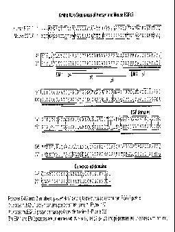
FIGURE 9 depicts CDR sequence changes observed in each of the 6 "single
position
libraries" (SPLs) for each of the 3 frameworks. Libraries were separated by
framework used
(4F11.v1, 4F11.v2 and 4F11.v3). Changes obtained from each of the SPLs versus
the
particular CDR sequence are highlighted. The VL and VH sequences outside of
these
changes were identical to the corresponding framework and are now shown.
Sequence
identifiers are shown in parentheses to the right of the corresponding
sequence (SEQ ID
NOs: 31-77). Individual sequences that appear more than once may not always
have a
corresponding sequence identifier.
FIGURE 10 depicts the framework and library design for the variable light
domain
of limited libraries 1 and 2. The amino acid sequence of the human Kappa I
consensus
(SEQ ID NO: 9) and 4F11.v1 (SEQ ID NO: 10) are shown compared to the template
used
for library 1 (SEQ ID NO: 78) and library 2 (SEQ ID NO: 79). Positions that
were
randomized to all 20 amino acids are shown with slash through the amino acid.
FIGURE 11 depicts the framework and library design for the variable heavy
domain
of limited libraries 1 and 2. The amino acid sequence of the human subgroup
III consensus
(SEQ ID NO: 11) and 4F11.v 1 (SEQ ID NO: 12) arc shown compared to the
template used
for library 1 (SEQ ID NO: 80) and library 2 (SEQ ID NO: 81). Positions that
were
randomized to all 20 amino acids are shown with slash through the amino acid.
FIGURE 12 depicts the frequency of changes observed at randomized positions in
limited libraries 1 and 2. The preference of amino acids selected at positions
53 and 54 in
the light chain and 29, 52, and 98 in the heavy chain is shown. The preference
(Sigma) for
any amino acid is reported as the number of standard deviations above a random
chance
occurrence of a given residue in the library assuming a binomial distribution
of amino acids.
Scoring by this method accounts for the expected codon bias and sampling
statistics when
establishing a consensus.
FIGURES 13 & 14 depict inhibition of HUVEC adhesion to immobilized human or
mouse EGFL7 in vitro by humanized 4F11 variants. HUVECs (20,000 cells/well)
were
CA 02838400 2013-12-27
WO 2010/129904
PCT/US2010/034097
allowed to adhere to 96 well plates coated with 51.4g/m1 human or murine EGFL7
in the
presence of increasing concentrations of antibody. The number of cells that
still adhered to
the plates after washing were counted and calculated as percent of the total
cells plated into
each well.
FIGURE 15 depicts the amino acid sequence of the variable light domain of the
human Kappa I consensus (SEQ ID NO: 9), 4F11.v1 (SEQ ID NO: 10), 4F11.v17 (SEQ
ID
NO: 82), and 4F11.v22 (SEQ ID NO: 83). Positions are numbered according to
Kabat and
hypervariable regions are boxed.
FIGURE 16 depicts the amino acid sequence of the variable heavy domain of the
human subgroup III consensus (SEQ ID NO: 11), 4F11.v1 (SEQ ID NO: 12),
4F11.v17
(SEQ ID NO: 84), and 4F11.v22 (SEQ ID NO: 85). Positions are numbered
according to
Kabat and hypervariable regions are boxed.
FIGURE 17 depicts the amino acid sequence of the variable light domain of the
human Kappa I consensus (SEQ ID NO: 9) and the 18F7-graft (SEQ ID NO: 86).
Positions
are numbered according to Kabat and hypervariable regions are boxed.
FIGURE 18 depicts the amino acid sequence of the variable heavy domain of the
human subgroup III consensus (SEQ ID NO: 11) and the 18F7-graft (SEQ ID NO:
87).
Positions are numbered according to Kabat and hypervariable regions are boxed.
FIGURE 19 depicts oligonucleotides used to toggle positions in the Framework
Toggle Library. The DNA sequence of 18F7-graft and the oligonucleotides used
to generate
the framework toggle are shown. The amino acid sequences of the original and
some
resulting framework toggle regions are also shown. In some cases additional
amino acid
residues were also incorporated based on how the degenerate codons were
designed.
Sequence identifiers are shown in parentheses to the right of the
corresponding sequence
(SEQ ID NOs: 88-99).
FIGURE 20 depicts results demonstrating that mul8F7 binding to EGFL7 can be
blocked by EMI1 (SEQ ID NO: 3) or Peptide P5 (SEQ ID NO: 7), but not by
Peptides P4 or
P6 (SEQ ID NOs: 6 and 8, respectively). Chicken embryonic fibroblasts were
transfected
with a plasmid containing the HA-tagged full-length human EGFL7 cDNA. Cell
lysatc
prepared from transfected cells was immunoprecipitated with mul8F7 in the
presence of
200-fold excess competitive peptides. Immunoprecipitates were analyzed by
western blot
using an anti-HA antibody.
FIGURE 21 depicts binding of phage displaying 18F7-graft Fab to truncated
huEGFL7 immobilized on a microtiter plate. Both samples of 18F7-graft phage
show
11
CA 02838400 2013-12-27
WO 2010/129904
PCT/US2010/034097
increased binding to immobilized EGFL7 as a function of increasing phage
concentration.
A control phage shows background binding similar to levels of 18F7-graft phage
at low
phage concentrations suggesting some non-specific phage-EGFL7 interaction.
FIGURE 22 depicts binding of phage displaying 18F7-graft Fab to EMI1 domain or
p5 peptide biotinylated either through a free amino or free thiol.
FIGURE 23 depicts the abundance of residues found at each framework position
during the Framework Toggle. Amino acids introduced at each framework position
during
the Framework Toggle are listed.
FIGURE 24 depicts CDR sequence changes observed in each of the 6 SPLs for each
of the 3 frameworks. Libraries were separated by framework used (18F7-graft,
18F7-K,
18F7-KV, and 18F7-KA). Changes obtained from each of the SPLs versus the
particular
CDR sequence are highlighted. The VL and VH sequences outside of these changes
were
identical to the corresponding framework and are now shown. Sequence
identifiers are
shown in parentheses to the right of the corresponding sequence (SEQ ID NOs:
100-192).
Individual sequences that appear more than once may not always have a
corresponding
sequence identifier.
FIGURE 25 depicts inhibition of HUVEC adhesion to immobilized human or mouse
EGFL7 in vitro by humanized 18F7 variants. HUVECs (20,000 cells/well) were
allowed to
adhere to 96 well plates coated with .4.1g/m1 human or murine EGFL7 in the
presence of
increasing concentrations of antibody. The number of cells that still adhered
to the plates
after washing were counted and calculated as percent of the total cells plated
into each well.
FIGURE 26 depicts inhibition of HUVEC transwell migration. HUVECs (50,000
cells per well) were grown for 16 hours in the top chambers of transwell
plates, and the
membranes in the top chamber were coated with 5ps/m1 recombinant human EGFL7
protein. Various concentrations of control antibody (anti-IgE) or different
variants of 18F7
were added to the culture medium in the top and bottom chambers, whereas 20
ng/ml of
recombinant human VEGF-165 was added in the bottom wells to stimulate HUVEC
migration. Cells migrated to the undersides of the top chambers were counted
and plotted
against the treatments (antibodies and concentrations).
FIGURE 27 depicts the amino acid sequence of the variable light domain of the
human Kappa I consensus (SEQ ID NO: 9), 18F7.v6 (SEQ ID NO: 193), and 18F7.v6k
(SEQ ID NO: 194). Positions are numbered according to Kabat and hypervariable
regions
are boxed. Where sequence for 18F7.v6k is not shown (in the second and third
parts of the
alignment), the corresponding sequence is identical to the sequence for
18F7.v6.
12
CA 02838400 2013-12-27
WO 2010/129904 PCT/US2010/034097
FIGURE 28 depicts the amino acid sequence of the variable heavy domain of the
human subgroup III consensus (SEQ ID NO: 11), 18F7.v6 (SEQ ID NO: 195), and
18F7.v6k (SEQ ID NO: 196). Positions are numbered according to Kabat and
hypervariable
regions are boxed. Where sequence for 18F7.v6k is not shown (in the first and
third parts of
the alignment), the corresponding sequence is identical to the sequence for
18F7.v6.
FIGURE 29 depicts inhibition of H1299 xenograft tumor growth using hul8F7.v6k
alone and in combination with an anti-VEGF antibody.
FIGURE 30 depicts inhibition of LXFL 1674 xenograft tumor growth using
hul8F7.v6k alone and in combination with an anti-VEGF antibody.
FIGURE 31 depicts inhibition of neonatal trachea vascularization using
hul8F7.v6k
alone and in combination with an anti-VEGF antibody.
DETAILED DESCRIPTION OF THE INVENTION
The invention provides methods, compositions, kits and articles of manufacture
for anti-
EGFL7 antibodies.
Details of these methods, compositions, kits and articles of manufacture are
provided
herein.
General techniques
The techniques and procedures described or referenced herein are generally
well
understood and commonly employed using conventional methodology by those
skilled in
the art, such as, for example, the widely utilized methodologies described in
Sambrook et
al., Molecular Cloning: A Laboratory Manual 3rd. edition (2001) Cold Spring
Harbor
Laboratory Press, Cold Spring Harbor, N.Y. CURRENT PROTOCOLS IN MOLECULAR
BIOLOGY (F. M. Ausubel, et al. eds., (2003)); the series METHODS IN ENZYMOLOGY
(Academic Press, Inc.): PCR 2: A PRACTICAL APPROACH (M. J. MacPherson, B. D.
Hames and G. R. Taylor eds. (1995)), Harlow and Lane, eds. (1988) ANTIBODIES,
A
LABORATORY MANUAL, and ANIMAL CELL CULTURE (R. I. Freshncy, ed. (1987)).
Definitions
An "isolated" antibody is one which has been identified and separated and/or
recovered from a component of its natural environment. Contaminant components
of its
natural environment are materials which would interfere with diagnostic or
therapeutic uses
for the antibody, and may include enzymes, hormones, and other proteinaceous
or
nonproteinaceous solutes. In preferred embodiments, the antibody will be
purified (1) to
greater than 95% by weight of antibody as determined by the Lowry method, and
most
13
CA 02838400 2013-12-27
WO 2010/129904
PCT/US2010/034097
preferably more than 99% by weight, (2) to a degree sufficient to obtain at
least 15 residues
of N-terminal or internal amino acid sequence by use of a spinning cup
sequenator, or (3) to
homogeneity by SDS-PAGE under reducing or nonreducing conditions using
Coomassie
blue or, preferably, silver stain. Isolated antibody includes the antibody in
situ within
recombinant cells since at least one component of the antibody's natural
environment will
not be present. Ordinarily, however, isolated antibody will be prepared by at
least one
purification step.
An "isolated" nucleic acid molecule is a nucleic acid molecule that is
identified and
separated from at least one contaminant nucleic acid molecule with which it is
ordinarily
associated in the natural source of the antibody nucleic acid. An isolated
nucleic acid
molecule is other than in the form or setting in which it is found in nature.
Isolated nucleic
acid molecules therefore are distinguished from the nucleic acid molecule as
it exists in
natural cells. However, an isolated nucleic acid molecule includes a nucleic
acid molecule
contained in cells that ordinarily express the antibody where, for example,
the nucleic acid
molecule is in a chromosomal location different from that of natural cells.
The term "anti-EGFL7 antibody" or "an antibody that binds to EGFL7" refers to
an
antibody that is capable of binding EGFL7 with sufficient affinity such that
the antibody is
useful as a diagnostic and/or therapeutic agent in targeting EGFL7. In certain
embodiments,
an antibody that binds to EGFL7 has a dissociation constant (Kd) of < 1KM, <
100 nM, < 10
nM, < 1 nM, or < 0.1 nM.
"Binding affinity" generally refers to the strength of the sum total of
noncovalent
interactions between a single binding site of a molecule (e.g., an antibody)
and its binding
partner (e.g., an antigen). Unless indicated otherwise, as used herein,
"binding affinity"
refers to intrinsic binding affinity which reflects a 1:1 interaction between
members of a
binding pair (e.g., antibody and antigen). The affinity of a molecule X for
its partner Y can
generally be represented by the dissociation constant (Kd). Affinity can be
measured by
common methods known in the art, including those described herein. Low-
affinity
antibodies generally bind antigen slowly and tend to dissociate readily,
whereas high-
affinity antibodies generally bind antigen faster and tend to remain bound
longer. A variety
of methods of measuring binding affinity are known in the art, any of which
can be used for
purposes of the present invention. Specific illustrative embodiments are
described in the
following.
In one embodiment, the "Kd" or "Kd value" according to this invention is
measured
by a radiolabeled antigen binding assay (RIA) performed with the Fab version
of an
14
CA 02838400 2013-12-27
WO 2010/129904
PCT/US2010/034097
antibody of interest and its antigen as described by the following assay that
measures
solution binding affinity of Fabs for antigen by equilibrating Fab with a
minimal
concentration of (1251)-labeled antigen in the presence of a titration series
of unlabeled
antigen, then capturing bound antigen with an anti-Fab antibody-coated plate
(Chen, et al.,
(1999) J. Mol Biol 293:865-881). To establish conditions for the assay,
microtiter plates
(Dynex) are coated overnight with 5 14/m1 of a capturing anti-Fab antibody
(Cappel Labs)
in 50 mM sodium carbonate (pH 9.6), and subsequently blocked with 2% (w/v)
bovine
serum albumin in PBS for two to five hours at room temperature (approximately
23 C). In
a non-adsorbant plate (Nunc #269620), 100 pM or 26 pM ['251]-antigen are mixed
with
serial dilutions of a Fab of interest (e.g., consistent with assessment of an
anti-VEGF
antibody, Fab-12, in Presta et al., (1997) Cancer Res. 57:4593-4599). The Fab
of interest is
then incubated overnight; however, the incubation may continue for a longer
period (e.g., 65
hours) to insure that equilibrium is reached. Thereafter, the mixtures are
transferred to the
capture plate for incubation at room temperature (e.g., for one hour). The
solution is then
removed and the plate washed eight times with 0.1% TweenTm-20 in PBS. When the
plates
have dried, 150 i.11/well of scintillant (MicroSeintTm-20; Packard) is added,
and the plates are
counted on a Top Count gamma counter (Packard) for ten minutes. Concentrations
of each
Fab that give less than or equal to 20% of maximal binding are chosen for use
in
competitive binding assays. According to another embodiment the Kd or Kd value
is
measured by using surface plasmon resonance assays using a BlAcore-2000 or a
BIAcoreTm-3000 (BlAcore, Inc., Piscataway, NJ) at 25 C with immobilized
antigen CM5
chips at ¨10 response units (RU). Briefly, carboxymethylated dextran biosensor
chips
(CM5, BlAcore Inc.) are activated with N-ethyl-N'- (3-dimethylaminopropy1)-
carbodiimide
hydrochloride (EDC) and N-hydroxysuccinimide (NHS) according to the supplier's
instructions. Antigen is diluted with 10mM sodium acetate, pH 4.8, into
51.1g/m1 (-0.21AM)
before injection at a flow rate of 5111/minute to achieve approximately 10
response units
(RU) of coupled protein. Following the injection of antigen, 1M ethanolamine
is injected to
block unreacted groups. For kinetics measurements, two-fold serial dilutions
of Fab (0.78
nM to 500 nM) are injected in PBS with 0.05% TweenTm 20 (PBST) at 25 C at a
flow rate
of approximately 251.d/rain. Association rates (kon) and dissociation rates
(koff) are calculated
using a simple one-to-one Langmuir binding model (BIAcoreTM Evaluation
Software
version 3.2) by simultaneous fitting the association and dissociation
sensorgram. The
equilibrium dissociation constant (Kd) is calculated as the ratio koff/kon.
See, e.g., Chen, Y.,
et al., (1999) J. Mol. Biol. 293:865-881. If the on-rate exceeds 106 M-1 S-1
by the surface
CA 02838400 2013-12-27
WO 2010/129904
PCT/US2010/034097
plasmon resonance assay above, then the on-rate can be determined by using a
fluorescent
quenching technique that measures the increase or decrease in fluorescence
emission
intensity (excitation = 295 nm; emission = 340 nm, 16 nm band-pass) at 25 C of
a 20nM
anti-antigen antibody (Fab form) in PBS, pH 7.2, in the presence of increasing
concentrations of antigen as measured in a spectrometer, such as a stop-flow
equipped
spectrophotometer (Aviv Instruments) or a 8000-series SLM Amine
spectrophotometer
(ThermoSpectronic) with a stirred cuvette.
The term "vector," as used herein, is intended to refer to a nucleic acid
molecule
capable of transporting another nucleic acid to which it has been linked. One
type of vector
is a "plasmid", which refers to a circular double stranded DNA loop into which
additional
DNA segments may be ligated. Another type of vector is a phage vector. Another
type of
vector is a viral vector, wherein additional DNA segments may be ligated into
the viral
genome. Certain vectors are capable of autonomous replication in a host cell
into which
they are introduced (e.g., bacterial vectors having a bacterial origin of
replication and
episomal mammalian vectors). Other vectors (e.g., non-episomal mammalian
vectors) can
be integrated into the genome of a host cell upon introduction into the host
cell, and thereby
are replicated along with the host genome. Moreover, certain vectors are
capable of
directing the expression of genes to which they are operatively linked. Such
vectors are
referred to herein as "recombinant expression vectors" (or simply,
"recombinant vectors" or
"expression vectors"). In general, expression vectors of utility in
recombinant DNA
techniques are often in the form of plasmids. In the present specification,
"plasmid" and
"vector" may be used interchangeably.
"Polynucleotide," or "nucleic acid," as used interchangeably herein, refer to
polymers of nucleotides of any length, and include DNA and RNA. The
nucleotides can be
deoxyribonucleotides, ribonucleotides, modified nucleotides or bases, and/or
their analogs,
or any substrate that can be incorporated into a polymer by DNA or RNA
polymerase, or by
a synthetic reaction. A polynucleotide may comprise modified nucleotides, such
as
methylated nucleotides and their analogs. If present, modification to the
nucleotide structure
may be imparted before or after assembly of the polymer. The sequence of
nucleotides may
be interrupted by non-nucleotide components. A polynucleotide may be further
modified
after synthesis, such as by conjugation with a label. Other types of
modifications include, for
example, "caps", substitution of one or more of the naturally occurring
nucleotides with an
analog, internucleotide modifications such as, for example, those with
uncharged linkages
(e.g., methyl phosphonates, phosphotriesters, phosphoamidates, carbamates,
etc.) and with
16
CA 02838400 2013-12-27
WO 2010/129904
PCT/US2010/034097
charged linkages (e.g., phosphorothioates, phosphorodithioates, etc.), those
containing
pendant moieties, such as, for example, proteins (e.g., nucleases, toxins,
antibodies, signal
peptides, poly-L-lysine, etc.), those with intercalators (e.g., acridine,
psoralen, etc.), those
containing chelators (e.g., metals, radioactive metals, boron, oxidative
metals, etc.), those
25 "Oligonucleotide," as used herein, generally refers to short, generally
single
stranded, generally synthetic polynucleotides that are generally, but not
necessarily, less
than about 200 nucleotides in length. The terms "oligonucleotide" and
"polynucleotide" are
not mutually exclusive. The description above for polynucleotides is equally
and fully
applicable to oligonucleotides.
30 The term "EGFL7" (interchangeably termed "EGF-like-domain, multiple 7"),
as
used herein, refers, unless specifically or contextually indicated otherwise,
to any native or
variant (whether native or synthetic) EGFL7 polypeptide. The term "native
sequence"
specifically encompasses naturally occurring truncated or secreted forms
(e.g., an
extracellular domain sequence), naturally occurring variant forms (e.g.,
alternatively spliced
17
CA 02838400 2013-12-27
WO 2010/129904
PCT/US2010/034097
forms) and naturally-occurring allelic variants. The term "wild type EGFL7"
generally
refers to a polypeptide comprising the amino acid sequence of a naturally
occurring EGFL7
protein. The term "wild type EGFL7 sequence" generally refers to an amino acid
sequence
found in a naturally occurring EGFL7.
The terms "antibody" and "inununoglobulin" are used interchangeably in the
broadest sense and include monoclonal antibodies (for e.g., full length or
intact monoclonal
antibodies), polyclonal antibodies, multivalent antibodies, multispecific
antibodies (e.g.,
bispecific antibodies so long as they exhibit the desired biological activity)
and may also
include certain antibody fragments (as described in greater detail herein). An
antibody can
be human, humanized and/or affinity matured.
The term "variable" refers to the fact that certain portions of the variable
domains
differ extensively in sequence among antibodies and are used in the binding
and specificity
of each particular antibody for its particular antigen. However, the
variability is not evenly
distributed throughout the variable domains of antibodies. It is concentrated
in three
segments called complementarity-determining regions or hypervariable regions
(CDRs or
HVRs, used interchangeably herein) both in the light-chain and the heavy-chain
variable
domains. The more highly conserved portions of variable domains are called the
framework
(FR). The variable domains of native heavy and light chains each comprise four
FR regions,
largely adopting a13-sheet configuration, connected by three HVRs, which form
loops
connecting, and in some cases forming part of, the 13-sheet structure. The
HVRs in each
chain are held together in close proximity by the FR regions and, with the
HVRs from the
other chain, contribute to the formation of the antigen-binding site of
antibodies (see Kabat
et al., Sequences of Proteins ofimmunological Interest, Fifth Edition,
National Institute of
Health, Bethesda, MD (1991)). The constant domains are not involved directly
in binding
an antibody to an antigen, but exhibit various effector functions, such as
participation of the
antibody in antibody-dependent cellular toxicity.
Papain digestion of antibodies produces two identical antigen-binding
fragments,
called "Fab" fragments, each with a single antigen-binding site, and a
residual "Fe"
fragment, whose name reflects its ability to crystallize readily. Pepsin
treatment yields an
F(aby)2 fragment that has two antigen-combining sites and is still capable of
cross-linking
antigen.
'Tv" is the minimum antibody fragment which contains a complete antigen-
recognition and -binding site. In a two-chain Fv species, this region consists
of a dimer of
one heavy- and one light-chain variable domain in tight, non-covalent
association. In a
18
CA 02838400 2013-12-27
WO 2010/129904
PCT/US2010/034097
single-chain Fv species, one heavy- and one light-chain variable domain can be
covalently
linked by a flexible peptide linker such that the light and heavy chains can
associate in a
"dimeric" structure analogous to that in a two-chain Fv species. It is in this
configuration
that the three HVRs of each variable domain interact to define an antigen-
binding site on the
surface of the VH-VL dimer. Collectively, the six HVRs confer antigen-binding
specificity
to the antibody. However, even a single variable domain (or half of an Fv
comprising only
three HVRs specific for an antigen) has the ability to recognize and bind
antigen, although
at a lower affinity than the entire binding site.
The Fab fragment also contains the constant domain of the light chain and the
first
constant domain (CH1) of the heavy chain. Fab' fragments differ from Fab
fragments by the
addition of a few residues at the carboxy terminus of the heavy chain CH1
domain including
one or more cysteines from the antibody hinge region. Fab'-SH is the
designation herein for
Fab' in which the cysteine residue(s) of the constant domains bear a free
thiol group. F(a1:02
antibody fragments originally were produced as pairs of Fab' fragments which
have hinge
cysteines between them. Other chemical couplings of antibody fragments are
also known.
The "light chains" of antibodies (immunoglobulins) from any vertebrate species
can
be assigned to one of two clearly distinct types, called kappa (lc) and lambda
(X), based on
the amino acid sequences of their constant domains.
Depending on the amino acid sequence of the constant domain of their heavy
chains,
immunoglobulins can be assigned to different classes. There are five major
classes of
immunoglobulins: IgA, IgD, IgE, IgG, and IgM, and several of these can be
further divided
into subclasses (isotypes), e.g., IgGi, IgG2, IgG3, Igat, IgAi, and IgA2. The
heavy-chain
constant domains that correspond to the different classes of immunoglobulins
are called a, 6,
c, y, and 11, respectively. The subunit structures and three-dimensional
configurations of
different classes of immunoglobulins are well known.
"Antibody fragments" comprise only a portion of an intact antibody, wherein
the
portion preferably retains at least one, preferably most or all, of the
functions normally
associated with that portion when present in an intact antibody. Examples of
antibody
fragments include Fab, Fab', F(ab')2, and Fv fragments; diabodies; linear
antibodies; single-
chain antibody molecules; and multispecific antibodies formed from antibody
fragments. In
one embodiment, an antibody fragment comprises an antigen binding site of the
intact
antibody and thus retains the ability to bind antigen. In another embodiment,
an antibody
fragment, for example one that comprises the Fe region, retains at least one
of the biological
functions normally associated with the Fc region when present in an intact
antibody, such as
19
CA 02838400 2013-12-27
WO 2010/129904 PCT/US2010/034097
FcRn binding, antibody half life modulation, ADCC function and complement
binding. In
one embodiment, an antibody fragment is a monovalent antibody that has an in
vivo half life
substantially similar to an intact antibody. For e.g., such an antibody
fragment may
comprise on antigen binding arm linked to an Fe sequence capable of conferring
in vivo
stability to the fragment.
The term "hypervariable region," "HVR," or "HV," when used herein refers to
the
regions of an antibody variable domain which are hypervariable in sequence
and/or form
structurally defined loops. Generally, antibodies comprise six HVRs; three in
the VH (H1,
H2, H3), and three in the VL (L1, L2, L3). In native antibodies, H3 and L3
display the most
diversity of the six HVRs, and H3 in particular is believed to play a unique
role in
conferring fine specificity to antibodies. See, e.g., Xu et al., Immunity
13:37-45 (2000);
Johnson and Wu, in Methods in Molecular Biology 248:1-25 (Lo, ed., Human
Press,
Totowa, NJ, 2003). Indeed, naturally occurring camelid antibodies consisting
of a heavy
chain only are functional and stable in the absence of light chain. See, e.g.,
Hamers-
Casterman et at., Nature 363:446-448 (1993); Sheriff et at., Nature Struct.
Biol. 3:733-736
(1996).
A number of HVR delineations are in use and are encompassed herein. The Kabat
Complementarity Determining Regions (CDRs) are based on sequence variability
and are
the most commonly used (Kabat et al., Sequences of Proteins of Immunological
Interest, 5th
Ed. Public Health Service, National Institutes of Health, Bethesda, MD.
(1991)). Chothia
refers instead to the location of the structural loops (Chothia and Lesk J.
MoL Biol. 196:901-
917 (1987)). The AbM HVRs represent a compromise between the Kabat HVRs and
Chothia structural loops, and are used by Oxford Molecular's AbM antibody
modeling
software. The "contact" HVRs are based on an analysis of the available complex
crystal
structures. The residues from each of these HVRs are noted below.
CA 02838400 2013-12-27
WO 2010/129904
PCT/US2010/034097
Loop Kabat AbM Chothia Contact
Li L24-L34 L24-L34 L26-L32 L30-L36
L2 L50-L56 L50-L56 L50-L52 L46-L55
L3 L89-L97 L89-L97 L91-L96 L89-L96
H1 H31-H35B H26-H35B H26-H32 H30-H35B
(Kabat Numbering)
H1 H31-H35 H26-H35 H26-H32 H30-H35
(Chothia Numbering)
H2 H50-H65 H50-H58 H53-H55 H47-H58
H3 H95-H102 H95-H102 H96-H101 H93-H101
HVRs may comprise "extended HVRs" as follows: 24-36 or 24-34 (L1), 46-56 or
50-56 (L2) and 89-97 or 89-96 (L3) in the VL and 26-35 (H1), 50-65 or 49-65
(H2) and 93-
102, 94-102, or 95-102 (113) in the VH. The variable domain residues are
numbered
according to Kabat et al., supra, for each of these definitions.
"Framework" or "FR" residues are those variable domain residues other than the
hypervariable region residues as herein defined.
"Humanized" forms of non-human (e.g., murine) antibodies are chimeric
antibodies
that contain minimal sequence derived from non-human immunoglobulin. In one
embodiment, a humanized antibody is a human immunoglobulin (recipient
antibody) in
which residues from a HVR of the recipient are replaced by residues from a HVR
of a non-
human species (donor antibody) such as mouse, rat, rabbit, or nonhuman primate
having the
desired specificity, affinity, and/or capacity. In some instances, FR residues
of the human
immunoglobulin are replaced by corresponding non-human residues. Furthermore,
humanized antibodies may comprise residues that are not found in the recipient
antibody or
in the donor antibody. These modifications may be made to further refine
antibody
performance. In general, a humanized antibody will comprise substantially all
of at least
one, and typically two, variable domains, in which all or substantially all of
the
hypervariable loops correspond to those of a non-human immunoglobulin, and all
or
substantially all of the FRs are those of a human immunoglobulin sequence. The
humanized
antibody optionally will also comprise at least a portion of an immunoglobulin
constant
region (Fc), typically that of a human immunoglobulin. For further details,
see, e.g., Jones
etal., Nature 321:522-525 (1986); Riechmann etal., Nature 332:323-329 (1988);
and
21
CA 02838400 2013-12-27
WO 2010/129904
PCT/US2010/034097
Presta, Curr. Op. Struct. Biol. 2:593-596 (1992). See also, e.g., Vaswani and
Hamilton,
Ann. Allergy, Asthma & Immunol. 1:105-115 (1998); Harris, Biochem. Soc.
Transactions
23:1035-1038 (1995); Hurle and Gross, Curr. Op. Biotech. 5:428-433 (1994); and
U.S. Pat.
Nos. 6,982,321 and 7,087,409.
The term "monoclonal antibody" as used herein refers to an antibody obtained
from
a population of substantially homogeneous antibodies, i.e., the individual
antibodies
comprising the population are identical except for possible mutations, e.g.,
naturally
occurring mutations, that may be present in minor amounts. Thus, the modifier
"monoclonal" indicates the character of the antibody as not being a mixture of
discrete
antibodies. In certain embodiments, such a monoclonal antibody typically
includes an
antibody comprising a polypeptide sequence that binds a target, wherein the
target-binding
polypeptide sequence was obtained by a process that includes the selection of
a single target
binding polypeptide sequence from a plurality of polypeptide sequences. For
example, the
selection process can be the selection of a unique clone from a plurality of
clones, such as a
pool of hybridoma clones, phage clones, or recombinant DNA clones. It should
be
understood that a selected target binding sequence can be further altered, for
example, to
improve affinity for the target, to humanize the target binding sequence, to
improve its
production in cell culture, to reduce its irnmunogenicity in vivo, to create a
multispecific
antibody, etc., and that an antibody comprising the altered target binding
sequence is also a
monoclonal antibody of this invention. In contrast to polyclonal antibody
preparations,
which typically include different antibodies directed against different
determinants
(epitopes), each monoclonal antibody of a monoclonal antibody preparation is
directed
against a single determinant on an antigen. In addition to their specificity,
monoclonal
antibody preparations are advantageous in that they arc typically
uncontaminated by other
immunoglobulins.
The modifier "monoclonal" indicates the character of the antibody as being
obtained
from a substantially homogeneous population of antibodies, and is not to be
construed as
requiring production of the antibody by any particular method. For example,
the
monoclonal antibodies to be used in accordance with the present invention may
be made by
a variety of techniques, including, for example, the hybridoma method (e.g.,
Kohler and
Milstein, Nature, 256:495-97 (1975); Hongo etal., Hybridoma, 14 (3): 253-260
(1995),
Harlow et al., Antibodies: A Laboratory Manual, (Cold Spring Harbor Laboratory
Press,
2nd ed. 1988); Hammerling et al., in: Monoclonal Antibodies and T-Cell
Hybridomas 563-
681 (Elsevier, N.Y., 1 9 8 1)), recombinant DNA methods (see, e.g., U.S.
Patent No.
22
CA 02838400 2013-12-27
WO 2010/129904
PCT/US2010/034097
4,816,567), phage-display technologies (see, e.g., Clackson et al., Nature,
352: 624-628
(1991); Marks etal., J. Mol. Biol. 222: 581-597 (1992); Sidhu etal., J. Mol.
Biol. 338(2):
299-310 (2004); Lee etal., J. Mol. Biol. 340(5): 1073-1093 (2004); Fellouse,
Proc. Natl.
Acad. Sc!. USA 101(34): 12467-12472 (2004); and Lee etal., J. Immunol. Methods
284(1-
2): 119-132(2004), and technologies for producing human or human-like
antibodies in
animals that have parts or all of the human immunoglobulin loci or genes
encoding human
immunoglobulin sequences (see, e.g., WO 1998/24893; WO 1996/34096; WO
1996/33735;
WO 1991/10741; Jakobovits etal., Proc. Natl. Acad. Sci. USA 90: 2551 (1993);
Jakobovits
etal., Nature 362: 255-258 (1993); Bruggemann etal., Year in Immunol. 7:33
(1993); U.S.
to Patent Nos. 5,545,807; 5,545,806; 5,569,825; 5,625,126; 5,633,425; and
5,661,016; Marks
et al., Bio/Technology 10: 779-783 (1992); Lonberg et al., Nature 368: 856-859
(1994);
Morrison, Nature 368: 812-813 (1994); Fishwild etal., Nature Biotechnol. 14:
845-851
(1996); Neuberger, Nature Biotechnol. 14: 826 (1996); and Lonberg and Huszar,
Intern.
Rev. Immunol. 13: 65-93 (1995).
The monoclonal antibodies herein specifically include "chimeric" antibodies in
which a portion of the heavy and/or light chain is identical with or
homologous to
corresponding sequences in antibodies derived from a particular species or
belonging to a
particular antibody class or subclass, while the remainder of the chain(s) is
identical with or
homologous to corresponding sequences in antibodies derived from another
species or
belonging to another antibody class or subclass, as well as fragments of such
antibodies, so
long as they exhibit the desired biological activity (see, e.g.,U.S. Patent
No. 4,816,567; and
Morrison et al., Proc. Natl. Acad. Sc!. USA 81:6851-6855 (1984)). Chimeric
antibodies
include PR1MATIZEDO antibodies wherein the antigen-binding region of the
antibody is
derived from an antibody produced by, e.g., immunizing macaque monkeys with
the antigen
of interest.
"Single-chain Fv" or "scFv" antibody fragments comprise the VH and VL domains
of antibody, wherein these domains are present in a single polypeptide chain.
Generally, the
scFv polypeptide further comprises a polypeptide linker between the VH and VL
domains
which enables the scFv to form the desired structure for antigen binding. For
a review of
scFv see Pluckthun, in The Pharmacology of Monoclonal Antibodies, vol. 113,
Rosenburg
and Moore eds., Springer-Verlag, New York, pp. 269-315 (1994).
An "antigen" is a predetermined antigen to which an antibody can selectively
bind.
The target antigen may be polypeptide, carbohydrate, nucleic acid, lipid,
hapten or other
naturally occurring or synthetic compound.
23
CA 02838400 2013-12-27
WO 2010/129904
PCT/US2010/034097
The term "diabodies" refers to small antibody fragments with two antigen-
binding
sites, which fragments comprise a heavy-chain variable domain (VII) connected
to a light-
chain variable domain (VL) in the same polypeptide chain (VH - VL). By using a
linker
that is too short to allow pairing between the two domains on the same chain,
the domains
are forced to pair with the complementary domains of another chain and create
two antigen-
binding sites. Diabodies are described more fully in, for example, EP 404,097;
WO
93/11161; and Hollinger etal., Proc. Natl. Acad. Sc!. USA, 90:6444-6448
(1993).
Triabodies and tetrabodies are also described in Hudson et at., Nat. Med.
9:129-134 (2003).
A "human antibody" is one which possesses an amino acid sequence which
corresponds to that of an antibody produced by a human and/or has been made
using any of
the techniques for making human antibodies as disclosed herein. This
definition of a human
antibody specifically excludes a humanized antibody comprising non-human
antigen-
binding residues. Human antibodies can be produced using various techniques
known in the
art, including phage-display libraries. Hoogenboom and Winter, J. Mot Biol.,
227:381
(1991); Marks et al., J. Mot Biol., 222:581 (1991). Also available for the
preparation of
human monoclonal antibodies are methods described in Cole et al., Monoclonal
Antibodies
and Cancer Therapy, Alan R. Liss, p. 77 (1985); Boerner et al., I Immunot,
147(1):86-95
(1991). See also van Dijk and van de Winkel, Curr. Opin. Pharmacol., 5: 368-74
(2001).
Human antibodies can be prepared by administering the antigen to a transgenic
animal that
has been modified to produce such antibodies in response to antigenic
challenge, but whose
endogenous loci have been disabled, e.g., immunized xenomice (see, e.g., U.S.
Pat. Nos.
6,075,181 and 6,150,584 regarding XENOMOUSErm technology). See also, for
example,
Li etal., Proc. Natl. Acad. Sc!. USA, 103:3557-3562 (2006) regarding human
antibodies
generated via a human 8-cell hybridoma technology.
The term "variable domain residue numbering as in Kabat" or "amino acid
position
numbering as in Kabat," and variations thereof, refers to the numbering system
used for
heavy chain variable domains or light chain variable domains of the
compilation of
antibodies in Kabat et al., supra. Using this numbering system, the actual
linear amino acid
sequence may contain fewer or additional amino acids corresponding to a
shortening of, or
insertion into, a FR or HVR of the variable domain. For example, a heavy chain
variable
domain may include a single amino acid insert (residue 52a according to Kabat)
after
residue 52 of H2 and inserted residues (e.g. residues 82a, 82b, and 82c, etc.
according to
Kabat) after heavy chain FR residue 82. The Kabat numbering of residues may be
24
CA 02838400 2013-12-27
WO 2010/129904
PCT/US2010/034097
determined for a given antibody by alignment at regions of homology of the
sequence of the
antibody with a "standard" Kabat numbered sequence.
The Kabat numbering system is generally used when referring to a residue in
the
variable domain (approximately residues 1-107 of the light chain and residues
1-113 of the
heavy chain) (e.g, Kabat et al., Sequences of Immunological Interest. 5th Ed.
Public Health
Service, National Institutes of Health, Bethesda, Md. (1991)). The "EU
numbering system"
or "EU index" is generally used when referring to a residue in an
immunoglobulin heavy
chain constant region (e.g., the EU index reported in Kabat etal., supra). The
"EU index as
in Kabat" refers to the residue numbering of the human IgG1 EU antibody.
Unless stated
otherwise herein, references to residue numbers in the variable domain of
antibodies means
residue numbering by the Kabat numbering system. Unless stated otherwise
herein,
references to residue numbers in the constant domain of antibodies means
residue
numbering by the EU numbering system (e.g., see United States Provisional
Application
No. 60/640,323, Figures for EU numbering).
A "blocking" antibody or an "antagonist" antibody is one which inhibits or
reduces
biological activity of the antigen it binds. Certain blocking antibodies or
antagonist
antibodies substantially or completely inhibit the biological activity of the
antigen.
The term "substantially similar" or "substantially the same," as used herein,
denotes
a sufficiently high degree of similarity between two numeric values (for
example, one
associated with an antibody of the invention and the other associated with a
reference/comparator antibody), such that one of skill in the art would
consider the
difference between the two values to be of little or no biological and/or
statistical
significance within the context of the biological characteristic measured by
said values (e.g.,
Kd values). The difference between said two values is, for example, less than
about 50%,
less than about 40%, less than about 30%, less than about 20%, and/or less
than about 10%
as a function of the reference/comparator value.
The phrase "substantially reduced," or "substantially different," as used
herein,
denotes a sufficiently high degree of difference between two numeric values
(generally one
associated with a molecule and the other associated with a
reference/comparator molecule)
such that one of skill in the art would consider the difference between the
two values to be
of statistical significance within the context of the biological
characteristic measured by said
values (e.g., Kd values). The difference between said two values is, for
example, greater
than about 10%, greater than about 20%, greater than about 30%, greater than
about 40%,
CA 02838400 2013-12-27
WO 2010/129904
PCT/US2010/034097
and/or greater than about 50% as a function of the value for the
reference/comparator
molecule.
Antibody "effector functions" refer to those biological activities
attributable to the
Fc region (a native sequence Fc region or amino acid sequence variant Fc
region) of an
antibody, and vary with the antibody isotype. Examples of antibody effector
functions
include: Clq binding and complement dependent cytotoxicity (CDC); Fc receptor
binding;
antibody-dependent cell-mediated cytotoxicity (ADCC); phagocytosis; down
regulation of
cell surface receptors (e.g. B cell receptor); and B cell activation.
The term "Fc region" herein is used to define a C-terminal region of an
immunoglobulin heavy chain, including native sequence Fc regions and variant
Fc
regions. Although the boundaries of the Fc region of an immunoglobulin heavy
chain might
vary, the human IgG heavy chain Fc region is usually defined to stretch from
an amino acid
residue at position Cys226, or from Pro230, to the carboxyl-terminus thereof.
The C-
terminal lysine (residue 447 according to the EU numbering system) of the Fc
region may
be removed, for example, during production or purification of the antibody, or
by
recombinantly engineering the nucleic acid encoding a heavy chain of the
antibody. Accordingly, a composition of intact antibodies may comprise
antibody
populations with all K447 residues removed, antibody populations with no K447
residues
removed, and antibody populations having a mixture of antibodies with and
without the
K447 residue.
A "functional Fc region" possesses an "effector function" of a native sequence
Fc
region. Exemplary "effector functions" include Clq binding; CDC; Fc receptor
binding;
ADCC; phagocytosis; down regulation of cell surface receptors (e.g. B cell
receptor; BCR),
etc. Such effcctor functions generally require the Fc region to be combined
with a binding
domain (e.g., an antibody variable domain) and can be assessed using various
assays as
disclosed, for example, in definitions herein.
A "native sequence Fc region" comprises an amino acid sequence identical to
the
amino acid sequence of an Fc region found in nature. Native sequence human Fc
regions
include a native sequence human IgG1 Fc region (non-A and A allotypes); native
sequence
human IgG2 Fc region; native sequence human IgG3 Fc region; and native
sequence human
IgG4 Fc region as well as naturally occurring variants thereof.
A "variant Fc region" comprises an amino acid sequence which differs from that
of a
native sequence Fc region by virtue of at least one amino acid modification,
preferably one
or more amino acid substitution(s). Preferably, the variant Fc region has at
least one amino
26
CA 02838400 2013-12-27
WO 2010/129904
PCMJS2010/034097
acid substitution compared to a native sequence Fc region or to the Fe region
of a parent
polypeptide, e.g. from about one to about ten amino acid substitutions, and
preferably from
about one to about five amino acid substitutions in a native sequence Fe
region or in the Fe
region of the parent polypeptide. The variant Fe region herein will preferably
possess at
least about 80% homology with a native sequence Fe region and/or with an Fe
region of a
parent polypeptide, and most preferably at least about 90% homology therewith,
more
preferably at least about 95% homology therewith.
"Fe receptor" or "FcR" describes a receptor that binds to the Pc region of an
antibody. In some embodiments, an FcR is a native human FcR. In some
embodiments, an
FcR is one which binds an IgG antibody (a gamma receptor) and includes
receptors of the
FcyRI, FcyRII, and FcyRIII subclasses, including allelic variants and
alternatively spliced
forms of those receptors. FcyRII receptors include FcyRIIA (an "activating
receptor") and
FcyRIIB (an "inhibiting receptor"), which have similar amino acid sequences
that differ
primarily in the cytoplasmic domains thereof. Activating receptor FcyRIIA
contains an
immunorcceptor tyrosine-based activation motif (ITAM) in its cytoplasmic
domain.
Inhibiting receptor FcyRIIB contains an immunoreceptor tyrosine-based
inhibition motif
(ITIM) in its cytoplasmic domain. (see, e.g., Daeron, Anntt. Rev. Immunol.
15:203-234
(1997)). FcRs are reviewed, for example, in Ravetch and Kinet, Annu. Rev.
Immunol 9:457-
92 (1991); Capel et al., Immunomethods 4:25-34 (1994); and de Haas etal., J.
Lab. Clin.
Med. 126:330-41 (1995). Other FcRs, including those to be identified in the
future, are
encompassed by the term "FcR" herein.
The term "Pc receptor" or "FcR" also includes the neonatal receptor, FeRn,
which is
responsible for the transfer of maternal IgGs to the fetus (Guyer etal., J.
Immunol. 117:587
(1976) and Kim etal., J. Immunol. 24:249 (1994)) and regulation of homeostasis
of
immunoglobulins. Methods of measuring binding to FcRn are known (see, e.g.,
Ghetie and
Ward., Immunol. Today 18(12):592-598 (1997); Ghetie etal., Nature
Biotechnology,
15(7):637-640 (1997); Hinton et al., J. Biol. Chem. 279(8):6213-6216 (2004);
WO
2004/92219 (Hinton et al.).
Binding to human FcRn in vivo and serum half life of human FcRn high affmity
binding polypeptides can be assayed, e.g., in transgenic mice or transfected
human cell lines
expressing human FcRn, or in primates to which the polyp eptides with a
variant Fe region
are administered. WO 2000/42072 (Presta) describes antibody variants with
improved or
27
CA 02838400 2013-12-27
WO 2010/129904
PCT/US2010/034097
diminished binding to FcRs. See also, e.g., Shields et al. J. Biol. Chem.
9(2):6591-6604
(2001).
"Human effector cells" are leukocytes which express one or more FcRs and
perform
effector functions. In certain embodiments, the cells express at least FcyRIII
and perform
ADCC effector function(s). Examples of human leukocytes which mediate ADCC
include
peripheral blood mononuclear cells (PBMC), natural killer (NK) cells,
monocytes, cytotoxic
T cells, and neutrophils. The effector cells may be isolated from a native
source, e.g., from
blood.
"Antibody-dependent cell-mediated cytotoxicity" or "ADCC" refers to a form of
cytotoxicity in which secreted Ig bound onto Fc receptors (FcRs) present on
certain
cytotoxic cells (e.g. NK cells, neutrophils, and macrophages) enable these
cytotoxic effector
cells to bind specifically to an antigen-bearing target cell and subsequently
kill the target
cell with cytotoxins. The primary cells for mediating ADCC, NK cells, express
FcyRIII
only, whereas monocytes express FcyRI, FcyRII, and FcyRIII. FcR expression on
hematopoietic cells is summarized in Table 3 on page 464 of Ravetch and Kinet,
Annu. Rev.
Immunol 9:457-92 (1991). To assess ADCC activity of a molecule of interest, an
in vitro
ADCC assay, such as that described in US Patent No. 5,500,362 or 5,821,337 or
U.S. Patent
No. 6,737,056 (Presta), may be performed. Useful effector cells for such
assays include
PBMC and NK cells. Alternatively, or additionally, ADCC activity of the
molecule of
interest may be assessed in vivo, e.g., in an animal model such as that
disclosed in Clynes et
al. PNAS (USA) 95:652-656 (1998).
"Complement dependent cytotoxicity" or "CDC" refers to the lysis of a target
cell in
the presence of complement. Activation of the classical complement pathway is
initiated by
the binding of the first component of the complement system (Cl q) to
antibodies (of the
appropriate subclass), which are bound to their cognate antigen. To assess
complement
activation, a CDC assay, e.g., as described in Gazzano-Santoro et al., J.
ImmunoL Methods
202:163 (1996), may be performed. Polypeptide variants with altered Fc region
amino acid
sequences (polypeptides with a variant Fc region) and increased or decreased
Clq binding
capability are described, e.g., in US Patent No. 6,194,551 B1 and WO
1999/51642. See
also, e.g., Idusogie etal. J. ImmunoL 164: 4178-4184 (2000).
The term "Fc region-comprising antibody" refers to an antibody that comprises
an
Fc region. The C-terminal lysine (residue 447 according to the EU numbering
system) of
the Fc region may be removed, for example, during purification of the antibody
or by
28
CA 02838400 2013-12-27
WO 2010/129904
PCT/US2010/034097
recombinant engineering of the nucleic acid encoding the antibody.
Accordingly, a
composition comprising an antibody having an Fc region according to this
invention can
comprise an antibody with K447, with all K447 removed, or a mixture of
antibodies with
and without the K447 residue.
An "acceptor human framework" for the purposes herein is a framework
comprising
the amino acid sequence of a VL or VH framework derived from a human
immunoglobulin
framework, or from a human consensus framework. An acceptor human framework
"derived from" a human immunoglobulin framework or human consensus framework
may
comprise the same amino acid sequence thereof, or may contain pre-existing
amino acid
sequence changes. Where pre-existing amino acid changes are present,
preferably no more
than 5 and preferably 4 or less, or 3 or less, pre-existing amino acid changes
are present.
Where pre-existing amino acid changes are present in a VH, preferably those
changes are
only at three, two or one of positions 71H, 73H and 78H; for instance, the
amino acid
residues at those positions may be 71A, 73T and/or 78A. In one embodiment, the
VL
acceptor human framework is identical in sequence to the VL human
immunoglobulin
framework sequence or human consensus framework sequence.
A "human consensus framework" is a framework which represents the most
commonly occurring amino acid residue in a selection of human immunoglobulin
VL or VH
framework sequences. Generally, the selection of human immunoglobulin VL or VH
sequences is from a subgroup of variable domain sequences. Generally, the
subgroup of
sequences is a subgroup as in Kabat et al. In one embodiment, for the VL, the
subgroup is
subgroup kappa I as in Kabat et al. In one embodiment, for the VH, the
subgroup is
subgroup III as in Kabat et al.
A "VH subgroup III consensus framework" comprises the consensus sequence
obtained from the amino acid sequences in variable heavy subgroup III of Kabat
et aL In
one embodiment, the VH subgroup III consensus framework amino acid sequence
comprises at least a portion or all of each of the following sequences:
EVQLVESGGGLVQPGGSLRLSCAAS (SEQ ID NO: 197)-H1-WVRQAPGKGLEWV
(SEQ ID NO: 198)-H2-RFTISRDNSKNTLYLQMNSLRAEDTAVYYC (SEQ ID NO: 199-
H3-WGQGTLVTVSS (SEQ ID NO: 200).
A "VL subgroup I consensus framework" comprises the consensus sequence
obtained from the amino acid sequences in variable light kappa subgroup I of
Kabat et al.
In one embodiment, the VH subgroup I consensus framework amino acid sequence
comprises at least a portion or all of each of the following sequences:
29
CA 02838400 2013-12-27
WO 2010/129904
PCT/US2010/034097
DIQMTQSPSSLSASVGDRVTITC (SEQ ID NO: 201)-L1-WYQQKPGKAPI(LLIY (SEQ
ID NO: 202)-L2-GVPSRFSGSGSGTDFTLTISSLQPEDFATYYC (SEQ ID NO: 203)-L3-
FGQGTKVEIK (SEQ ID NO: 221).
A "biological sample" (interchangeably termed "sample" or "tissue or cell
sample")
encompasses a variety of sample types obtained from an individual and can be
used in a
diagnostic or monitoring assay. The definition encompasses blood and other
liquid samples
of biological origin, solid tissue samples such as a biopsy specimen or tissue
cultures or
cells derived therefrom, and the progeny thereof. The definition also includes
samples that
have been manipulated in any way after their procurement, such as by treatment
with
reagents, solubilization, or enrichment for certain components, such as
proteins or
polynucleotides, or embedding in a semi-solid or solid matrix for sectioning
purposes. The
term "biological sample" encompasses a clinical sample, and also includes
cells in culture,
cell supernatants, cell lysates, serum, plasma, biological fluid, and tissue
samples. The
source of the biological sample may be solid tissue as from a fresh, frozen
and/or preserved
organ or tissue sample or biopsy or aspirate; blood or any blood constituents;
bodily fluids
such as cerebral spinal fluid, amniotic fluid, peritoneal fluid, or
interstitial fluid; cells from
any time in gestation or development of the subject. In some embodiments, the
biological
sample is obtained from a primary or metastatic tumor. The biological sample
may contain
compounds which are not naturally intermixed with the tissue in nature such as
preservatives, anticoagulants, buffers, fixatives, nutrients, antibiotics, or
the like.
For the purposes herein a "section" of a tissue sample is meant a single part
or piece
of a tissue sample, e.g. a thin slice of tissue or cells cut from a tissue
sample. It is
understood that multiple sections of tissue samples may be taken and subjected
to analysis
according to the present invention. In some embodiments, the same section of
tissue sample
is analyzed at both morphological and molecular levels, or is analyzed with
respect to both
protein and nucleic acid.
The word "label" when used herein refers to a compound or composition which is
conjugated or fused directly or indirectly to a reagent such as a nucleic acid
probe or an
antibody and facilitates detection of the reagent to which it is conjugated or
fused. The label
may itself be detectable (e.g., radioisotope labels or fluorescent labels) or,
in the case of an
enzymatic label, may catalyze chemical alteration of a substrate compound or
composition
which is detectable.
A "medicament" is an active drug to treat the disorder in question or its
symptoms,
or side effects.
CA 02838400 2013-12-27
WO 2010/129904
PCT/US2010/034097
A "disorder" or "disease" is any condition that would benefit from treatment
with a
substance/molecule or method of the invention. This includes chronic and acute
disorders
or diseases including those pathological conditions which predispose the
mammal to the
disorder in question. Non-limiting examples of disorders to be treated herein
include
malignant and benign tumors; carcinoma, blastoma, and sarcoma.
The terms "cell proliferative disorder" and "proliferative disorder" refer to
disorders
that are associated with some degree of abnormal cell proliferation. In one
embodiment, the
cell proliferative disorder is cancer.
"Tumor", as used herein, refers to all neoplastic cell growth and
proliferation,
whether malignant or benign, and all pre-cancerous and cancerous cells and
tissues. The
terms "cancer", "cancerous", "cell proliferative disorder", "proliferative
disorder" and
"tumor" are not mutually exclusive as referred to herein.
The terms "cancer" and "cancerous" refer to or describe the physiological
condition
in mammals that is typically characterized by unregulated cell
growth/proliferation.
Examples of cancer include but are not limited to, carcinoma, lymphoma,
blastoma,
sarcoma, and leukemia. More particular examples of such cancers include
squamous cell
cancer, small-cell lung cancer, pituitary cancer, esophageal cancer,
astrocytoma, soft tissue
sarcoma, non-small cell lung cancer, adenocarcinoma of the lung, squamous
carcinoma of
the lung, cancer of the peritoneum, hepatocellular cancer, gastrointestinal
cancer, pancreatic
cancer, glioblastoma, cervical cancer, ovarian cancer, liver cancer, bladder
cancer,
hepatoma, breast cancer, colon cancer, colorectal cancer, endometrial or
uterine carcinoma,
salivary gland carcinoma, kidney cancer, liver cancer, prostate cancer, vulval
cancer, thyroid
cancer, hepatic carcinoma, brain cancer, endometrial cancer, testis cancer,
cholangiocarcinoma, gallbladder carcinoma, gastric cancer, melanoma, and
various types of
head and neck cancer. Dysregulation of angiogenesis can lead to many disorders
that can be
treated by compositions and methods of the invention. These disorders include
both non-
neoplastic and neoplastic conditions. Neoplastics include but are not limited
those described
above. Non-neoplastic disorders include but are not limited to undesired or
aberrant
hypertrophy, arthritis, rheumatoid arthritis (RA), psoriasis, psoriatic
plaques, sarcoidosis,
atherosclerosis, atherosclerotic plaques, diabetic and other proliferative
retinopathics
including retinopathy of prematurity, retrolental fibroplasia, neovascular
glaucoma, age-
related macular degeneration, diabetic macular edema, corneal
neovascularization, corneal
graft neovascularization, corneal graft rejection, retinal/choroidal
neovascularization,
neovascularization of the angle (rubeosis), ocular neovascular disease,
vascular restenosis,
31
CA 02838400 2013-12-27
WO 2010/129904
PCT/US2010/034097
artcriovenous malformations (AVM), meningioma, hemangioma, angiofibroma,
thyroid
hyperplasias (including Grave's disease), corneal and other tissue
transplantation, chronic
inflammation, lung inflammation, acute lung injury/ARDS, sepsis, primary
pulmonary
hypertension, malignant pulmonary effusions, cerebral edema (e.g., associated
with acute
stroke/ closed head injury/ trauma), synovial inflammation, pannus formation
in RA,
myositis ossificans, hypertropic bone formation, osteoarthritis (OA),
refractory ascites,
polycystic ovarian disease, endometriosis, 3rd spacing of fluid diseases
(pancreatitis,
compartment syndrome, bums, bowel disease), uterine fibroids, premature labor,
chronic
inflammation such as IBD (Crohn's disease and ulcerative colitis), renal
allograft rejection,
inflammatory bowel disease, nephrotic syndrome, undesired or aberrant tissue
mass growth
(non-cancer), hemophilic joints, hypertrophic scars, inhibition of hair
growth, Osler-Weber
syndrome, pyogenic granuloma retrolental fibroplasias, scleroderma, trachoma,
vascular
adhesions, synovitis, dermatitis, preeclampsia, ascites, pericardial effusion
(such as that
associated with pericarditis), and pleural effusion.
The term "wasting" disorders (e.g., wasting syndrome, cachexia, sarcopenia)
refers
to a disorder caused by undesirable and/or unhealthy loss of weight or loss of
body cell
mass. In the elderly as well as in AIDS and cancer patients, wasting disease
can result in
undesired loss of body weight, including both the fat and the fat-free
compartments.
Wasting diseases can be the result of inadequate intake of food and/or
metabolic changes
related to illness and/or the aging process. Cancer patients and AIDS
patients, as well as
patients following extensive surgery or having chronic infections, immunologic
diseases,
hyperthyroidism, Crohn's disease, psychogenic disease, chronic heart failure
or other severe
trauma, frequently suffer from wasting disease which is sometimes also
referred to as
cachexia, a metabolic and, sometimes, an eating disorder. Cachexia is
additionally
characterized by hypermetabolism and hypercatabolism. Although cachexia and
wasting
disease are frequently used interchangeably to refer to wasting conditions,
there is at least
one body of research which differentiates cachexia from wasting syndrome as a
loss of fat-
free mass, and particularly, body cell mass (Mayer, 1999, J. Nutr. 129(1S
Suppl.):256S-
259S). Sarcopenia, yet another such disorder which can affect the aging
individual, is
typically characterized by loss of muscle mass. End stage wasting disease as
described
above can develop in individuals suffering from either cachexia or sarcopenia.
As used herein, "treatment" refers to clinical intervention in an attempt to
alter the
natural course of the individual or cell being treated, and can be performed
either for
32
CA 02838400 2013-12-27
WO 2010/129904
PCT/US2010/034097
prophylaxis or during the course of clinical pathology. Desirable effects of
treatment
include preventing occurrence or recurrence of disease, alleviation of
symptoms,
diminishment of any direct or indirect pathological consequences of the
disease, decreasing
the rate of disease progression, amelioration or palliation of the disease
state, and remission
or improved prognosis. In some embodiments, antibodies of the invention are
used to delay
development of a disease or disorder.
An "anti-angiogenesis agent" or "angiogenesis inhibitor" refers to a small
molecular
weight substance, a polynucleotide, a polypeptide, an isolated protein, a
recombinant
protein, an antibody, or conjugates or fusion proteins thereof, that inhibits
angiogenesis,
vasculogenesis, or undesirable vascular permeability, either directly or
indirectly. For
example, an anti-angiogenesis agent is an antibody or other antagonist to an
angiogenic
agent as defined above, e.g., antibodies to VEGF, antibodies to VEGF
receptors, small
molecules that block VEGF receptor signaling (e.g., PTK787/ZI(2284, SU6668,
SUTENTS/SU11248 (sunitinib malate), AMG706). Anti-angiogenesis agents also
include
native angiogenesis inhibitors, e.g., angiostatin, endostatin, etc. See, e.g.,
Klagsbrun and
D'Amore, Annu. Rev. Physiol., 53:217-39 (1991); Streit and Detmar, Oncogene,
22:3172-
3179 (2003) (e.g., Table 3 listing anti-angiogenic therapy in malignant
melanoma); Ferrara
& Alitalo, Nature Medicine 5(12):1359-1364 (1999); Tonini et al., Oncogene,
22:6549-6556
(2003) (e.g., Table 2 listing antiangiogenic factors); and, Sato Int. J. Clin.
Oncol., 8:200-206
(2003) (e.g., Table 1 lists Anti-angiogenic agents used in clinical trials).
An "individual," "subject," or "patient" is a vertebrate. In certain
embodiments, the
vertebrate is a mammal. Mammals include, but are not limited to, farm animals
(such as
cows), sport animals, pets (such as cats, dogs, and horses), primates, mice
and rats. In
certain embodiments, a mammal is a human.
An "effective amount" refers to an amount effective, at dosages and for
periods of
time necessary, to achieve the desired therapeutic or prophylactic result.
A "therapeutically effective amount" of a substance/molecule of the invention,
agonist or antagonist may vary according to factors such as the disease state,
age, sex, and
weight of the individual, and the ability of the substance/molecule, agonist
or antagonist to
elicit a desired response in the individual. A therapeutically effective
amount is also one in
which any toxic or detrimental effects of the substance/molecule, agonist or
antagonist are
outweighed by the therapeutically beneficial effects. A "prophylactically
effective amount"
refers to an amount effective, at dosages and for periods of time necessary,
to achieve the
desired prophylactic result. Typically but not necessarily, since a
prophylactic dose is used
33
CA 02838400 2013-12-27
WO 2010/129904
PCT/US2010/034097
in subjects prior to or at an earlier stage of disease, the prophylactically
effective amount
will be less than the therapeutically effective amount.
The term "cytotoxic agent" as used herein refers to a substance that inhibits
or
prevents the function of cells and/or causes destruction of cells. The term is
intended to
j131, 1125, ¨", Rem, smI53, Bi212, p32 and
include radioactive isotopes (e.g., At211, Y90, Rel
radioactive isotopes of Lu), chemotherapeutic agents e.g. methotrexate,
adriamicin, vinca
alkaloids (vincristine, vinblastine, etoposide), doxorubicin, melphalan,
mitomycin C,
chlorambucil, daunorubicin or other intercalating agents, enzymes and
fragments thereof
such as nucleolytic enzymes, antibiotics, and toxins such as small molecule
toxins or
enzymatically active toxins of bacterial, fungal, plant or animal origin,
including fragments
and/or variants thereof, and the various antitumor or anticancer agents
disclosed below.
Other cytotoxic agents are described below. A tumoricidal agent causes
destruction of tumor
cells.
A "toxin" is any substance capable of having a detrimental effect on the
growth or
proliferation of a cell.
A "chemotherapeutic agent" is a chemical compound useful in the treatment of
cancer. Examples of chemotherapeutic agents include alkylating agents such as
thiotepa and
cyclosphosphamide (CYTOXAN8); alkyl sulfonates such as busulfan, improsulfan
and
piposulfan; aziridines such as benzodopa, carboquone, meturedopa, and uredopa;
ethylenimines and methylamelamines including altretamine, triethylenemelamine,
triethylenephosphoramide, triethylenethiophosphoramide and trimethylomelamine;
acetogenins (especially bullatacin and bullatacinone); delta-9-
tetrahydrocannabinol
(cironabinol, MARINOLS); beta-lapachone; lapachol; colchicines; betulinic
acid; a
camptothecin (including the synthetic analogue topotecan (HYCAMTINC), CPT-11
(irinotecan, CAMPTOSARO), acetylcamptothecin, scopolectin, and 9-
aminocamptothecin);
bryostatin; callystatin; CC-1065 (including its adozelesin, carzelesin and
bizelesin synthetic
analogues); podophyllotoxin; podophyllinic acid; teniposide; cryptophycins
(particularly
cryptophycin 1 and cryptophycin 8); dolastatin; duocarmycin (including the
synthetic
analogues, KW-2189 and CB1-TM1); eleutherobin; pancratistatin; a sarcodictyin;
spongistatin; nitrogen mustards such as chlorambucil, chlomaphazine,
chlorophosphamide,
estramustine, ifosfamide, mechlorethamine, mechlorethamine oxide
hydrochloride,
melphalan, novembichin, phenesterine, prednimustine, trofosfamide, uracil
mustard;
nitrosoureas such as carmustine, chlorozotocin, fotemustine, lomustine,
nimustine, and
ranimnustine; antibiotics such as the enediyne antibiotics (e. g.,
calicheamicin, especially
34
CA 02838400 2013-12-27
WO 2010/129904 PCT/US2010/034097
calicheamicin gammal I and calicheamicin omegaIl (see, e.g., Nicolaou etal.,
Angew. Chem
Intl. Ed. EngL, 33: 183-186 (1994)); CDP323, an oral alpha-4 integrin
inhibitor; dynemicin,
including dynemicin A; an esperamicin; as well as neocarzinostatin chromophore
and
related chromoprotein enediyne antibiotic chromophores), aclacinomysins,
actinomycin,
authramycin, azaserine, bleomycins, cactinomycin, carabicin, carminomycin,
carzinophilin,
chromomycins, dactinomycin, daunorubicin, detorubicin, 6-diazo-5-oxo-L-
norleucine,
doxorubicin (including ADRIAMYCINO, morpholino-doxorubicin, cyanomorpholino-
doxorubicin, 2-pyrrolino-doxorubicin, doxorubicin HC1 liposome injection
(DOXILO),
liposomal doxorubicin TLC D-99 (MYOCETO), peglylated liposomal doxorubicin
(CAELYX6), and deoxydoxorubicin), epirubicin, esorubicin, idarubicin,
marcellomycin,
mitomycins such as mitomycin C, mycophenolic acid, nogalamycin, olivomycins,
peplomycin, porfiromycin, puromycin, quelamycin, rodorubicin, streptonigrin,
streptozocin,
tubercidin, ubenimex, zinostatin, zorubicin; anti-metabolites such as
methotrexate,
gemcitabine (GEMZARt), tegafur (UFTORALS), capecitabine (XELODAO), an
epothilone, and 5-fluorouracil (5-FU); folic acid analogues such as
denopterin,
methotrexate, pteropterin, trimetrexate; purine analogs such as fludarabine, 6-
mercaptopurine, thiamiprine, thioguanine; pyrimidine analogs such as
ancitabine,
azacitidine, 6-azauridine, carmofur, cytarabine, dideoxyuridine,
doxifluridine, enocitabine,
floxuridine; androgens such as calusterone, dromostanolone propionate,
epitiostanol,
mcpitiostane, testolactone; anti-adrenals such as aminoglutethimidc, mitotane,
trilostanc;
folic acid replenisher such as frolinic acid; aceglatone; aldophosphamide
glycoside;
aminolevulinic acid; eniluracil; amsacrine; bestrabucil; bisantrene;
edatraxate; defofamine;
demecolcine; diaziquone; elfomithine; elliptinium acetate; an epothilone;
etoglucid; gallium
nitrate; hydroxyurea; lentinan; lonidaininc; maytansinoids such as maytansine
and
ansamitocins; mitoguazone; mitoxantrone; mopidanmol; nitraerine; pentostatin;
phenamet;
pirarubicin; losoxantrone; 2-ethylhydrazide; procarbazine; PSKO polysaccharide
complex
(IRS Natural Products, Eugene, OR); razoxane; rhizoxin; sizofiran;
spirogcrmanium;
tenuazonic acid; triaziquone; 2,2',2'-trichlorotriethylamine; trichothecenes
(especially T-2
toxin, verracurin A, roridin A and anguidine); urethan; vindesinc (ELDISINEO,
FILDES1N0); dacarbazine; mannomustine; mitobronitol; mitolactol; pipobroman;
gacytosine; arabinoside ("Ara-C"); thiotepa; taxoid, e.g., paclitaxel
(TAXOLO), albumin-
engineered nanoparticle formulation of paclitaxel (ABRAXANETN4), and docetaxel
(TAXOTERE0); chloranbucil; 6-thioguanine; mercaptopurine; methotrexate;
platinum
agents such as cisplatin, oxaliplatin (e.g., ELOXATIN*), and carboplatin;
vincas, which
CA 02838400 2013-12-27
WO 2010/129904
PCT/US2010/034097
prevent tubulin polymerization from forming mierotubules, including
vinblastine
(VELBANO), vincristine (ONCOVINO), vindesine (ELDISINE , FILDESINt), and
vinorelbine (NAVELBINE0); etoposidc (VP-16); ifosfamide; mitoxantrone;
leucovorin;
novantrone; edatrex ate; daunomycin; aminopterin; ibandronate; topoisomerase
inhibitor
RFS 2000; difluoromethylomithine (DMF0); retinoids such as retinoic acid,
including
bexarotene (TARGRETINR); bisphosphonates such as clodronate (for example,
BONEFOSO or OSTACO), etidronate (DIDROCAL0), NE-58095, zoledronic
acid/zoledronate (ZOMETAO), alendronate (FOSAMAX0), pamidronate (AREDIAt),
tiludronate (SKELIDO), or risedronate (ACTONEL0); troxacitabine (a 1,3-
dioxolane
nucleoside cytosine analog); antisense oligonucleotides, particularly those
that inhibit
expression of genes in signaling pathways implicated in aberrant cell
proliferation, such as,
for example, PKC-alpha, Raf, H-Ras, and epidermal growth factor receptor (EGF-
R);
vaccines such as THERATOPE8 vaccine and gene therapy vaccines, for example,
ALLOVECTINO vaccine, LEUVECTIN vaccine, and VAXIDO vaccine; topoisomerase 1
inhibitor (e.g., LURTOTECANO); rmRH (e.g., ABARELIXO); BAY439006 (sorafenib;
Bayer); SU-11248 (sunitinib, SUTENT , Pfizer); perifosine, COX-2 inhibitor
(e.g.
celecoxib or etoricoxib), proteosome inhibitor (e.g. PS341); bortezomib
(VELCADEC);
CCI-779; tipifamib (R11577); orafenib, ABT510; Bc1-2 inhibitor such as
oblimersen
sodium (GENASENSEt); pixantrone; EGFR inhibitors (see definition below);
tyrosine
kinase inhibitors (see definition below); scrine-threonine kinase inhibitors
such as
rapamycin (sirolimus, RAPAMUNE0); farnesyltransferase inhibitors such as
lonafarnib
(SCH 6636, SARASARTm); and pharmaceutically acceptable salts, acids or
derivatives of
any of the above; as well as combinations of two or more of the above such as
CHOP, an
abbreviation for a combined therapy of cyclophosphamide, doxorubicin,
vincristine, and
prednisolone; and FOLFOX, an abbreviation for a treatment regimen with
oxaliplatin
(ELOXATINTm) combined with 5-FU and leucovorin.
Chemotherapeutic agents as defined herein include "anti-hormonal agents" or
"endocrine therapeutics" which act to regulate, reduce, block, or inhibit the
effects of
hormones that can promote the growth of cancer. They may be hormones
themselves,
including, but not limited to: anti-estrogens with mixed agonist/antagonist
profile, including,
tamoxifen (NOLVADEX0), 4-hydroxytamoxifen, toremifene (FARESTONO), idoxifene,
droloxifene, raloxifene (EVISTAt), trioxifene, keoxifene, and selective
estrogen receptor
modulators (SERMs) such as SERM3; pure anti-estrogens without agonist
properties, such
as fulvestrant (FASLODEX0), and EM800 (such agents may block estrogen receptor
(ER)
36
CA 02838400 2013-12-27
WO 2010/129904
PCT/US2010/034097
dimerization, inhibit DNA binding, increase ER turnover, and/or suppress ER
levels);
aromatase inhibitors, including steroidal aromatase inhibitors such as
formestane and
exemestane (AROMASINO), and nonsteroidal aromatase inhibitors such as
anastrazole
(ARIMIDEX0), letrozole (FEMARAt) and aminoglutethimide, and other aromatase
inhibitors include vorozole (RIVISORO), megestrol acetate (MEGASEC),
fadrozole, and
4(5)-imidazoles; lutenizing hormone-releaseing hormone agonists, including
leuprolide
(LUPRON and ELIGARDO), goserelin, buserelin, and tripterelin; sex steroids,
including
progestines such as megestrol acetate and medroxyprogesterone acetate,
estrogens such as
diethylstilbestrol and premarin, and androgens/retinoids such as
fluoxymesterone, all
transretionic acid and fenretinide; onapristone; anti-progesterones; estrogen
receptor down-
regulators (ERDs); anti-androgens such as flutamide, nilutamide and
bicalutamide; and
pharmaceutically acceptable salts, acids or derivatives of any of the above;
as well as
combinations of two or more of the above.
A "growth inhibitory agent" when used herein refers to a compound or
composition
which inhibits growth of a cell (such as a cell expressing EGFL7) either in
vitro or in vivo.
Thus, the growth inhibitory agent may be one which significantly reduces the
percentage of
cells (such as a cell expressing EGFL7) in S phase. Examples of growth
inhibitory agents
include agents that block cell cycle progression (at a place other than S
phase), such as
agents that induce G1 arrest and M-phase arrest. Classical M-phase blockers
include the
vincas (vincristinc and vinblastine), taxanes, and topoisomerase II inhibitors
such as
doxorubicin, epirubicin, daunorubicin, etoposide, and bleomycin. Those agents
that arrest
G1 also spill over into S-phase arrest, for example, DNA alkylating agents
such as
tamoxifen, prednisone, dacarbazinc, mechlorethaminc, cisplatin, methotrexate,
5-
fluorouracil, and ara-C. Further information can be found in Mendelsohn and
Israel, eds.,
The Molecular Basis of Cancer, Chapter 1, entitled "Cell cycle regulation,
oncogenes, and
antineoplastic drugs" by Murakami et al. (W.B. Saunders, Philadelphia, 1995),
e.g., p. 13.
The taxanes (paclitaxel and docetaxel) arc anticancer drugs both derived from
the yew tree.
Docetaxel (TAXOTERE , Rhone-Poulenc Rorer), derived from the European yew, is
a
semisynthetic analogue of paclitaxel (TAXOLO, Bristol-Myers Squibb).
Paclitaxel and
docetaxel promote the assembly of microtubules from tubulin dimers and
stabilize
microtubules by preventing depolymerization, which results in the inhibition
of mitosis in
cells.
37
CA 02838400 2013-12-27
WO 2010/129904 PCT/US2010/034097
"Doxorubicin" is an anthracycline antibiotic. The full chemical name of
doxorubicin
is (8S-cis)-10-[(3-amino-2,3,6-trideoxy-a-L-lyxo-hexapyranosypoxy]-7,8,9,10-
tetrahydro-
6,8,11-trihydroxy-8-(hydroxyacety1)-1-methoxy-5,12-naphthacenedione.
The term "Fe region-comprising polypeptide" refers to a polypeptide, such as
an
antibody or immunoadhesin (see definitions below), which comprises an Fe
region. The C-
terminal lysine (residue 447 according to the EU numbering system) of the Fe
region may
be removed, for example, during purification of the polypeptide or by
recombinant
engineering the nucleic acid encoding the polypeptide. Accordingly, a
composition
comprising a polypeptide having an Fe region according to this invention can
comprise
polypeptides with K447, with all K447 removed, or a mixture of polypeptides
with and
without the K447 residue.
Throughout this specification and claims, the word "comprise," or variations
such as
"comprises" or "comprising," will be understood to imply the inclusion of a
stated integer or
group of integers but not the exclusion of any other integer or group of
integers.
Generating variant antibodies exhibiting reduced or absence of HAMA response
Reduction or elimination of a HAMA response is a significant aspect of
clinical
development of suitable therapeutic agents. See, e.g., Khaxzaeli et al., J.
Natl. Cancer Inst.
(1988), 80:937; Jaffers et al., Transplantation (1986), 41:572; Shawler et
at., J. Immunol.
(1985), 135:1530; Sears et al., J. Biol. Response Mod. (1984), 3:138; Miller
etal., Blood
(1983), 62:988; Hakimi etal., J. Immunol. (1991), 147:1352; Reichmann et at.,
Nature
(1988), 332:323; Junghans et al., Cancer Res. (1990), 50:1495. As described
herein, the
invention provides antibodies that are humanized such that HAMA response is
reduced or
eliminated. Variants of these antibodies can further be obtained using routine
methods
known in the art, some of which are further described below.
For example, an amino acid sequence from an antibody as described herein can
serve as a starting (parent) sequence for diversification of the framework
and/or
hypervariable sequence(s). A selected framework sequence to which a starting
hypervariable sequence is linked is referred to herein as an acceptor human
framework.
While the acceptor human frameworks may be from, or derived from, a human
immunoglobulin (the VL and/or VH regions thereof), preferably the acceptor
human
frameworks are from, or derived from, a human consensus framework sequence as
such
frameworks have been demonstrated to have minimal, or no, immunogenicity in
human
patients.
38
CA 02838400 2013-12-27
WO 2010/129904
PCT/1JS2010/034097
Where the acceptor is derived from a human immunoglobulin, one may optionally
select a human framework sequence that is selected based on its homology to
the donor
framework sequence by aligning the donor framework sequence with various human
framework sequences in a collection of human framework sequences, and select
the most
homologous framework sequence as the acceptor.
In one embodiment, human consensus frameworks herein are from, or derived
from,
VH subgroup III and/or VL kappa subgroup I consensus framework sequences.
Thus, the VH acceptor human framework may comprise one, two, three or all of
the
following framework sequences:
FR1 comprising EVQLVESGGGLVQPGGSLRLSCAAS (SEQ ID NO: 197),
FR2 comprising WVRQAPGKGLEWV (SEQ ID NO: 198),
FR3 comprising FR3 comprises RFTISX1DX2SKNTX3YLQMNSLRAEDTAVYYC (SEQ
ID NO: 205), wherein X1 is A or R, X2 is T or N, and X3 is A or L,
FR4 comprising WGQGTLVTVSS (SEQ ID NO: 200).
In one embodiment, the VH acceptor human framework comprises one, two, three
or
all of the following framework sequences:
FRI comprising EVQLVESGGGLVQPGGSLRLSCAAS (SEQ ID NO: 197),
FR2 comprising WVRQAPGKGLEWV (SEQ ID NO: 198),
FR3 comprising RFTISADTSKNTAYLQMNSLRAEDTAVYYC (SEQ ID NO: 231),
RFTISADTSKNTAYLQMNSLRAEDTAVYYCA (SEQ ID NO: 206),
RFTISADTSKNTAYLQMNSLRAEDTAVYYCAR (SEQ ID NO: 207),
RFTISADTSKNTAYLQMNSLRAEDTAVYYCS (SEQ ID NO: 208), or
RFTISADTSKNTAYLQMNSLRAEDTAVYYCSR (SEQ ID NO: 209)
FR4 comprising WGQGTLVTVSS (SEQ ID NO: 200).
The VL acceptor human framework may comprise one, two, three or all of the
following framework sequences:
FR1 comprising DIQMTQSPSSLSASVGDRVTITC (SEQ ID NO: 201),
FR2 comprising WYQQKPGICAPKLLIY (SEQ ID NO: 202),
FR3 comprising GVPSRFSGSGSGTDFTLTISSLQPEDFATYYC (SEQ ID NO: 203),
FR4 comprising FGQGTKVEIK (SEQ ID NO: 221).
39
CA 02838400 2013-12-27
WO 2010/129904
PCT/US2010/034097
While the acceptor may be identical in sequence to the human framework
sequence
selected, whether that is from a human immunoglobulin or a human consensus
framework,
the present invention contemplates that the acceptor sequence may comprise pre-
existing
amino acid substitutions relative to the human immunoglobulin sequence or
human
consensus framework sequence. These pre-existing substitutions are preferably
minimal;
usually four, three, two or one amino acid differences only relative to the
human
immunoglobulin sequence or consensus framework sequence.
Hypervariable region residues of the non-human antibody are incorporated into
the
VL and/or VH acceptor human frameworks. For example, one may incorporate
residues
corresponding to the Kabat CDR residues, the Chothia hypervariable loop
residues, the
Abm residues, and/or contact residues. Optionally, the extended hypervariable
region
residues as follows are incorporated: 24-34 (Li), 50-56 (L2) and 89-97 (L3),
26-35 (H1),
50-65 or 49-65 (H2) and 93-102, 94-102, or 95-102 (H3).
While "incorporation" of hypervariable region residues is discussed herein, it
will be
appreciated that this can be achieved in various ways, for example, nucleic
acid encoding
the desired amino acid sequence can be generated by mutating nucleic acid
encoding the
mouse variable domain sequence so that the framework residues thereof are
changed to
acceptor human framework residues, or by mutating nucleic acid encoding the
human
variable domain sequence so that the hypervariable domain residues are changed
to non-
human residues, or by synthesizing nucleic acid encoding the desired sequence,
etc.
In the examples herein, hypervariable region-grafted variants were generated
by
Kunkel mutagenesis of nucleic acid encoding the human acceptor sequences,
using a
separate oligonucleotide for each hypervariable region. Kunkel et al., Methods
Enzymol.
154:367-382 (1987). Appropriate changes can be introduced within the framework
and/or
hypervariable region, using routine techniques, to correct and re-establish
proper
hypervariable region-antigen interactions.
Phage(mid) display (also referred to herein as phage display in some contexts)
can
be used as a convenient and fast method for generating and screening many
different
potential variant antibodies in a library generated by sequence randomization.
However,
other methods for making and screening altered antibodies are available to the
skilled
person.
Phage(mid) display technology has provided a powerful tool for generating and
selecting novel proteins which bind to a ligand, such as an antigen. Using the
techniques of
phage(mid) display allows the generation of large libraries of protein
variants which can be
CA 02838400 2013-12-27
WO 2010/129904
PCT/US2010/034097
rapidly sorted for those sequences that bind to a target molecule with high
affinity. Nucleic
acids encoding variant polypeptides are generally fused to a nucleic acid
sequence encoding
a viral coat protein, such as the gene III protein or the gene VIII protein.
Monovalent
phagemid display systems where the nucleic acid sequence encoding the protein
or
polypeptide is fused to a nucleic acid sequence encoding a portion of the gene
III protein
have been developed. (Bass, S., Proteins, 8:309 (1990); Lowman and Wells,
Methods: A
Companion to Methods in Enzymology, 3:205 (1991)). In a monovalent phagemid
display
system, the gene fusion is expressed at low levels and wild type gene III
proteins are also
expressed so that infectivity of the particles is retained. Methods of
generating peptide
libraries and screening those libraries have been disclosed in many patents
(e.g. U.S. Patent
No. 5,723,286, U.S. Patent No. 5,432, 018, U.S. Patent No. 5,580,717, U.S.
Patent No.
5,427,908 and U.S. Patent No. 5,498,530).
Libraries of antibodies or antigen binding polypeptides have been prepared in
a
number of ways including by altering a single gene by inserting random DNA
sequences or
by cloning a family of related genes. Methods for displaying antibodies or
antigen binding
fragments using phage(mid) display have been described in U.S. Patent Nos.
5,750,373,
5,733,743, 5,837,242, 5,969,108, 6,172,197, 5,580,717, and 5,658,727. The
library is then
screened for expression of antibodies or antigen binding proteins with the
desired
characteristics.
Methods of substituting an amino acid of choice into a template nucleic acid
are well
established in the art, some of which are described herein. For example,
hypervariable
region residues can be substituted using the Kunkel method. See, e.g., Kunkel
et al.,
Methods Enzymol. 154:367-382 (1987).
The sequence of oligonucicotides includes one or more of the designed codon
sets
for the hypervariable region residues to be altered. A codon set is a set of
different
nucleotide triplet sequences used to encode desired variant amino acids. Codon
sets can be
represented using symbols to designate particular nucleotides or equimolar
mixtures of
nucleotides as shown in below according to the TUB code.
TUB CODES
G Guanine
A Adenine
T Thymine
C Cytosine
R (A or G)
41
CA 02838400 2013-12-27
WO 2010/129904
PCT/US2010/034097
Y (C or T)
M (A or C)
K (G or T)
S (C or G)
W (A or T)
H (A or C or T)
B (C or G or T)
V (A or C or G)
D (A or G or T)
N (A or C or G or T)
For example, in the codon set DVK, D can be nucleotides A or G or T; V can be
A
or G or C; and K can be G or T. This codon set can present 18 different codons
and can
encode amino acids Ala, Trp, Tyr, Lys, Thr, Asn, Lys, Ser, Arg, Asp, Glu, Gly,
and Cys.
Oligonucleotide or primer sets can be synthesized using standard methods. A
set of
oligonucleotides can be synthesized, for example, by solid phase synthesis,
containing
sequences that represent all possible combinations of nucleotide triplets
provided by the
codon set and that will encode the desired group of amino acids. Synthesis of
oligonucleotides with selected nucleotide "degeneracy" at certain positions is
well known in
that art. Such sets of nucleotides having certain codon sets can be
synthesized using
commercial nucleic acid synthesizers (available from, for example, Applied
Biosystems,
Foster City, CA), or can be obtained commercially (for example, from Life
Technologies,
Rockville, MD). Therefore, a set of oligonucleotides synthesized having a
particular codon
set will typically include a plurality of oligonucleotides with different
sequences, the
differences established by the codon set within the overall sequence.
Oligonucleotides, as
used according to the invention, have sequences that allow for hybridization
to a variable
domain nucleic acid template and also can include restriction enzyme sites for
cloning
purposes.
In one method, nucleic acid sequences encoding variant amino acids can be
created
by oligonucleotide-mediated mutagenesis. This technique is well known in the
art as
described by Zoller et al. Nucleic Acids Res. 10:6487-6504(1987). Briefly,
nucleic acid
sequences encoding variant amino acids are created by hybridizing an
oligonucicotide set
encoding the desired codon sets to a DNA template, where the template is the
single-
stranded form of the plasmid containing a variable region nucleic acid
template sequence.
After hybridization, DNA polymerase is used to synthesize an entire second
complementary
42
CA 02838400 2013-12-27
WO 2010/129904
PCT/US2010/034097
strand of the template that will thus incorporate the oligonucleotide primer,
and will contain
the codon sets as provided by the oligonucleotide set.
Generally, oligonucleotides of at least 25 nucleotides in length are used. An
optimal
oligonucleotide will have 12 to 15 nucleotides that are completely
complementary to the
template on either side of the nucleotide(s) coding for the mutation(s). This
ensures that the
oligonucleotide will hybridize properly to the single-stranded DNA template
molecule. The
oligonucleotides are readily synthesized using techniques known in the art
such as that
described by Crea et al., Proc. Nat'l. Acad. Sci. USA, 75:5765 (1978).
The DNA template is generated by those vectors that are either derived from
bacteriophage M13 vectors (the commercially available M13mp18 and M13mp19
vectors
are suitable), or those vectors that contain a single-stranded phage origin of
replication as
described by Viera et aL, Meth. EnzymoL, 153:3 (1987). Thus, the DNA that is
to be
mutated can be inserted into one of these vectors in order to generate single-
stranded
template. Production of the single-stranded template is described in sections
4.21-4.41 of
Sambrook et al., above.
To alter the native DNA sequence, the oligonucleotide is hybridized to the
single
stranded template under suitable hybridization conditions. A DNA polymerizing
enzyme,
usually T7 DNA polymerase or the Klenow fragment of DNA polymerase I, is then
added to
synthesize the complementary strand of the template using the oligonucleotide
as a primer
for synthesis. A heteroduplex molecule is thus formed such that one strand of
DNA encodes
the mutated form of gene 1, and the other strand (the original template)
encodes the native,
unaltered sequence of gene 1. This heteroduplex molecule is then transformed
into a
suitable host cell, usually a prokaryote such as E. coil JM101. After growing
the cells, they
are plated onto agamse plates and screened using the oligonucleotide primer
radiolabellcd
with a 32-Phosphate to identify the bacterial colonies that contain the
mutated DNA.
The method described immediately above may be modified such that a homoduplex
molecule is created wherein both strands of the plasmid contain the
mutation(s). The
modifications are as follows: The single stranded oligonucleotide is annealed
to the single-
stranded template as described above. A mixture of three deoxyribonucleotides,
deoxyriboadenosine (dATP), deoxyriboguanosine (dGTP), and deoxyribothymidine
(dTT),
is combined with a modified thiodeoxyribocytosine called dCTP-(aS) (which can
be
obtained from Amersham). This mixture is added to the template-oligonucleotide
complex.
Upon addition of DNA polymerase to this mixture, a strand of DNA identical to
the
template except for the mutated bases is generated. In addition, this new
strand of DNA will
43
CA 02838400 2013-12-27
WO 2010/129904
PCITLTS2010/034097
contain dCTP-(aS) instead of dCTP, which serves to protect it from restriction
endonuclease
digestion. After the template strand of the double-stranded heteroduplex is
nicked with an
appropriate restriction enzyme, the template strand can be digested with
ExoIII nuclease or
another appropriate nuclease past the region that contains the site(s) to be
mutagenized. The
reaction is then stopped to leave a molecule that is only partially single-
stranded. A
complete double-stranded DNA homoduplex is then formed using DNA polymerase in
the
presence of all four deoxyribonucleotide triphosphates, ATP, and DNA ligase.
This
homoduplex molecule can then be transformed into a suitable host cell.
As indicated previously the sequence of the oligonucleotide set is of
sufficient length
1.0 to hybridize to the template nucleic acid and may also, but does not
necessarily, contain
restriction sites. The DNA template can be generated by those vectors that are
either
derived from bacteriophage M13 vectors or vectors that contain a single-
stranded phage
origin of replication as described by Viera etal. Meth. Enzymol., 153:3
(1987). Thus, the
DNA that is to be mutated must be inserted into one of these vectors in order
to generate
single-stranded template. Production of the single-stranded template is
described in
sections 4.21-4.41 of Sambrook etal., supra.
According to another method, a library can be generated by providing upstream
and
downstream oligonucleotide sets, each set having a plurality of
oligonucleotides with
different sequences, the different sequences established by the codon sets
provided within
the sequence of the oligonucleotides. The upstream and downstream
oligonucleotide sets,
along with a variable domain template nucleic acid sequence, can be used in a
polymerase
chain reaction to generate a "library" of PCR products. The PCR products can
be referred
to as "nucleic acid cassettes", as they can be fused with other related or
unrelated nucleic
acid sequences, for example, viral coat proteins and dimerization domains,
using established
molecular biology techniques.
The sequence of the PCR primers includes one or more of the designed codon
sets
for the solvent accessible and highly diverse positions in a hypervariable
region. As
described above, a codon set is a set of different nucleotide triplet
sequences used to encode
desired variant amino acids.
Antibody selectants that meet the desired criteria, as selected through
appropriate
screening/selection steps can be isolated and cloned using standard
recombinant techniques.
Antibody fragments
The present invention encompasses antibody fragments. Antibody fragments may
be
generated by traditional means, such as enzymatic digestion, or by recombinant
techniques.
44
CA 02838400 2013-12-27
WO 2010/129904
PCT/US2010/034097
In certain circumstances there are advantages of using antibody fragments,
rather than whole
antibodies. The smaller size of the fragments allows for rapid clearance, and
may lead to
improved access to solid tumors. For a review of certain antibody fragments,
see Hudson et
al. (2003) Nat. Med. 9:129-134.
Various techniques have been developed for the production of antibody
fragments.
Traditionally, these fragments were derived via proteolytic digestion of
intact antibodies
(see, e.g., Morimoto et al., Journal of Biochemical and Biophysical Methods
24:107-117
(1992); and Brennan et al., Science, 229:81 (1985)). However, these fragments
can now be
produced directly by recombinant host cells. Fab, Fv and ScFv antibody
fragments can all
be expressed in and secreted from E. coli, thus allowing the facile production
of large
amounts of these fragments. Antibody fragments can be isolated from the
antibody phage
libraries discussed above. Alternatively, Fab'-SH fragments can be directly
recovered from
E. coli and chemically coupled to form F(ab'), fragments (Carter et al.,
Rio/Technology
10:163-167 (1992)). According to another approach, F(abl)2 fragments can be
isolated
directly from recombinant host cell culture. Fab and F(ab')2 fragment with
increased in vivo
half-life comprising salvage receptor binding epitope residues are described
in U.S. Pat. No.
5,869,046. Other techniques for the production of antibody fragments will be
apparent to the
skilled practitioner. In certain embodiments, an antibody is a single chain Fv
fragment
(scFv). See WO 93/16185; U.S. Pat. Nos. 5,571,894; and 5,587,458. Fv and scFv
are the
only species with intact combining sites that are devoid of constant regions;
thus, they may
be suitable for reduced nonspecific binding during in vivo use. scFv fusion
proteins may be
constructed to yield fusion of an effector protein at either the amino or the
carboxy terminus
of an scFv. See Antibody Engineering, ed. Borrebaeck, supra. The antibody
fragment may
also be a "linear antibody", e.g., as described in U.S. Pat. No. 5,641,870,
for example. Such
linear antibodies may be monospecific or bispecific.
Humanized Antibodies
The invention encompasses humanized antibodies. Various methods for humanizing
non-human antibodies are known in the art. For example, a humanized antibody
can have
one or more amino acid residues introduced into it from a source which is non-
human.
These non-human amino acid residues are often referred to as "import"
residues, which are
typically taken from an "import" variable domain. Humanization can be
essentially
performed following the method of Winter and co-workers (Jones et al. (1986)
Nature
321:522-525; Riechmann et al. (1988) Nature 332:323-327; Verhoeyen et al.
(1988) Science
239:1534-1536), by substituting hypervariable region sequences for the
corresponding
CA 02838400 2013-12-27
WO 2010/129904
PCT/US2010/034097
sequences of a human antibody. Accordingly, such "humanized" antibodies are
chimeric
antibodies (U.S. Patent No. 4,816,567) wherein substantially less than an
intact human
variable domain has been substituted by the corresponding sequence from a non-
human
species. In practice, humanized antibodies are typically human antibodies in
which some
hypervariable region residues and possibly some FR residues are substituted by
residues
from analogous sites in rodent antibodies.
The choice of human variable domains, both light and heavy, to be used in
making
the humanized antibodies can be important to reduce antigenicity. According to
the so-
called "best-fit" method, the sequence of the variable domain of a rodent
antibody is
screened against the entire library of known human variable-domain sequences.
The human
sequence which is closest to that of the rodent is then accepted as the human
framework for
the humanized antibody. See, e.g., Sims et al. (1993)J. ImmunoL 151:2296;
Chothia etal.
(1987) J. Mot Biol. 196:901. Another method uses a particular framework
derived from the
consensus sequence of all human antibodies of a particular subgroup of light
or heavy
chains. The same framework may be used for several different humanized
antibodies. See,
e.g., Carter etal. (1992) Proc. Natl. Acad. ScL USA, 89:4285; Presta et al.
(1993)J.
ImmunoL,151:2623.
It is further generally desirable that antibodies be humanized with retention
of high
affinity for the antigen and other favorable biological properties. To achieve
this goal,
according to one method, humanized antibodies are prepared by a process of
analysis of the
parental sequences and various conceptual humanized products using three-
dimensional
models of the parental and humanized sequences. Three-dimensional
immunoglobulin
models are commonly available and are familiar to those skilled in the art.
Computer
programs are available which illustrate and display probable three-dimensional
conformational structures of selected candidate immunoglobulin sequences.
Inspection of
these displays permits analysis of the likely role of the residues in the
functioning of the
candidate immunoglobulin sequence, i.e., the analysis of residues that
influence the ability
of the candidate immunoglobulin to bind its antigen. In this way, FR residues
can be
selected and combined from the recipient and import sequences so that the
desired antibody
characteristic, such as increased affinity for the target antigen(s), is
achieved. In general, the
hypervariable region residues are directly and most substantially involved in
influencing
antigen binding.
46
CA 02838400 2013-12-27
WO 2010/129904
PCT/US2010/034097
Human Antibodies
Human antibodies of the invention can be constructed by combining Fv clone
variable domain sequence(s) selected from human-derived phage display
libraries with
known human constant domain sequences(s) as described above. Alternatively,
human
It is now possible to produce transgenic animals (e.g. mice) that are capable,
upon
immunization, of producing a full repertoire of human antibodies in the
absence of
endogenous immunoglobulin production. For example, it has been described that
the
homozygous deletion of the antibody heavy-chain joining region (JH) gene in
chimeric and
20 Gene shuffling can also be used to derive human antibodies from non-
human, e.g.
rodent, antibodies, where the human antibody has similar affinities and
specificities to the
starting non-human antibody. According to this method, which is also called
"epitope
imprinting", either the heavy or light chain variable region of a non-human
antibody
fragment obtained by phage display techniques as described herein is replaced
with a
47
CA 02838400 2013-12-27
WO 2010/129904
PCT/US2010/034097
Bispecific Antibodies
Bispecific antibodies are monoclonal antibodies that have binding
specificities for at
least two different antigens. In certain embodiments, bispecific antibodies
are human or
humanized antibodies. In certain embodiments, one of the binding specificities
is for
EGFL7 and the other is for any other antigen. In certain embodiments, the
other antigen is
vascular endothelial growth factor (VEGF), e.g. the epitope bound by the
antibodies
bevacizumab and ranibizumab. In certain embodiments, the bispecific antibody
has a first
arm comprising the HVR sequences of an antibody of the invention and a second
arm
comprising the HVR sequences of bevacizumab or ranibizumab. In certain
embodiments,
the bispecific antibody comprises the VH and VL sequences of bevacizumab or
ranibizumab. In certain embodiments, bispecific antibodies may bind to two
different
epitopes of EGFL7. Bispecific antibodies may also be used to localize
cytotoxic agents to
cells which express EGFL7. These antibodies possess a EGFL7-binding arm and an
arm
which binds a cytotoxic agent, such as, e.g., saporin, anti-interferon-a,
vinca alkaloid, ricin
A chain, methotrexate or radioactive isotope hapten. Bispecific antibodies can
be prepared
as full length antibodies or antibody fragments (e.g. F(ab)2bispecific
antibodies).
Methods for making bispecific antibodies are known in the art. Traditionally,
the
recombinant production of bispecific antibodies is based on the co-expression
of two
immunoglobulin heavy chain-light chain pairs, where the two heavy chains have
different
specificities (Milstein and Cuello, Nature, 305: 537 (1983)). Because of the
random
assortment of immunoglobulin heavy and light chains, these hybridomas
(quadromas)
produce a potential mixture of 10 different antibody molecules, of which only
one has the
correct bispecific structure. The purification of the correct molecule, which
is usually done
by affinity chromatography steps, is rather cumbersome, and the product yields
arc low.
Similar procedures are disclosed in WO 93/08829 published May 13, 1993, and in
Traunecker etal., EMBO J., 10: 3655 (1991).
According to a different approach, antibody variable domains with the desired
binding specificities (antibody-antigen combining sites) are fused to
immunoglobulin
constant domain sequences. The fusion, for example, is with an immunoglobulin
heavy
chain constant domain, comprising at least part of the hinge, CH2, and CH3
regions. In
certain embodiments, the first heavy-chain constant region (CH1), containing
the site
necessary for light chain binding, is present in at least one of the fusions.
DNAs encoding
the immunoglobulin heavy chain fusions and, if desired, the immunoglobulin
light chain, are
inserted into separate expression vectors, and are co-transfected into a
suitable host
48
CA 02838400 2013-12-27
WO 2010/129904
PCT/US2010/034097
organism. This provides for great flexibility in adjusting the mutual
proportions of the three
polypeptide fragments in embodiments when unequal ratios of the three
polypeptide chains
used in the construction provide the optimum yields. It is, however, possible
to insert the
coding sequences for two or all three polypeptide chains in one expression
vector when the
expression of at least two polypeptide chains in equal ratios results in high
yields or when
the ratios are of no particular significance.
In one embodiment of this approach, the bispecific antibodies are composed of
a
hybrid immunoglobulin heavy chain with a first binding specificity in one arm,
and a hybrid
immunoglobulin heavy chain-light chain pair (providing a second binding
specificity) in the
other arm. It was found that this asymmetric structure facilitates the
separation of the
desired bispecific compound from unwanted immunoglobulin chain combinations,
as the
presence of an immunoglobulin light chain in only one half of the bispecific
molecule
provides for a facile way of separation. This approach is disclosed in WO
94/04690. For
further details of generating bispecific antibodies see, for example, Suresh
etal., Methods in
Enzymology, 121:210 (1986).
According to another approach, the interface between a pair of antibody
molecules
can be engineered to maximize the percentage of heterodimers which are
recovered from
recombinant cell culture. The interface comprises at least a part of the CH3
domain of an
antibody constant domain. In this method, one or more small amino acid side
chains from
the interface of the first antibody molecule are replaced with larger side
chains (e.g. tyrosine
or tryptophan). Compensatory "cavities" of identical or similar size to the
large side
chain(s) are created on the interface of the second antibody molecule by
replacing large
amino acid side chains with smaller ones (e.g. alanine or threoninc). This
provides a
mechanism for increasing the yield of the heterodimer over other unwanted end-
products
such as homodimers.
Bispecific antibodies include cross-linked or "heteroconjugate" antibodies.
For
example, one of the antibodies in the heteroconjugate can be coupled to
avidin, the other to
biotin. Such antibodies have, for example, been proposed to target immune
system cells to
unwanted cells (US Patent No. 4,676,980), and for treatment of HIV infection
(WO
91/00360, WO 92/00373, and EP 03089). Heteroconjugate antibodies may be made
using
any convenient cross-linking method. Suitable cross-linking agents are well
known in the
art, and are disclosed in US Patent No. 4,676,980, along with a number of
cross-linking
techniques.
49
CA 02838400 2013-12-27
WO 2010/129904
PCT/US2010/034097
Techniques for generating bispecific antibodies from antibody fragments have
also
been described in the literature. For example, bispecific antibodies can be
prepared using
chemical linkage. Brennan et al., Science, 229: 81(1985) describe a procedure
wherein
intact antibodies are proteolytically cleaved to generate F(ab1)2 fragments.
These fragments
are reduced in the presence of the dithiol complexing agent sodium arsenite to
stabilize
vicinal dithiols and prevent intermolecular disulfide formation. The Fab'
fragments
generated are then converted to thionitrobenzoate (NB) derivatives. One of the
Fab'-TNB
derivatives is then reconverted to the Fab'-thiol by reduction with
mercaptoethylamine and
is mixed with an equimolar amount of the other Fab'-TNB derivative to form the
bispecific
antibody. The bispecific antibodies produced can be used as agents for the
selective
immobilization of enzymes.
Recent progress has facilitated the direct recovery of Fab'-SH fragments from
E.
coil, which can be chemically coupled to form bispecific antibodies. Shalaby
et al., .I. Exp.
Med., 175: 217-225 (1992) describe the production of a fully humanized
bispecific antibody
F(ab')2 molecule. Each Fab' fragment was separately secreted from E. coil and
subjected to
directed chemical coupling in vitro to form the bispecific antibody. The
bispecific antibody
thus formed was able to bind to cells overexpressing the HER2 receptor and
normal human
T cells, as well as trigger the lytic activity of human cytotoxic lymphocytes
against human
breast tumor targets.
Various techniques for making and isolating bispecific antibody fragments
directly
from recombinant cell culture have also been described. For example,
bispecific antibodies
have been produced using leucine zippers. Kostelny et
Immunol., 148(5):1547-1553
(1992). The leucine zipper peptides from the Fos and Jun proteins were linked
to the Fab'
portions of two different antibodies by gene fusion. The antibody homodimers
were
reduced at the hinge region to form monomers and then re-oxidized to form the
antibody
heterodimers. This method can also be utilized for the production of antibody
homodimers.
The "diabody" technology described by Hollinger et al., Proc. Natl. Acad. Sci.
USA,
90:6444-6448 (1993) has provided an alternative mechanism for making
bispecific antibody
fragments. The fragments comprise a heavy-chain variable domain (VH) connected
to a
light-chain variable domain (VL) by a linker which is too short to allow
pairing between the
two domains on the same chain. Accordingly, the VH and VL domains of one
fragment are
forced to pair with the complementary VL and VH domains of another fragment,
thereby
forming two antigen-binding sites. Another strategy for making bispecific
antibody
CA 02838400 2013-12-27
WO 2010/129904
PCT/US2010/034097
fragments by the use of single-chain Fv (sFv) dimers has also been reported.
See Gruber et
al., J. Immunol., 152:5368 (1994).
Antibodies with more than two valencies arc contemplated. For example,
trispecific
antibodies can be prepared. Tuft et al. J. Immunol. 147: 60 (1991).
Multivalent Antibodies
A multivalent antibody may be internalized (and/or catabolized) faster than a
bivalent antibody by a cell expressing an antigen to which the antibodies
bind. The
antibodies of the present invention can be multivalent antibodies (which are
other than of
the IgM class) with three or more antigen binding sites (e.g. tetravalent
antibodies), which
can be readily produced by recombinant expression of nucleic acid encoding the
polypeptide
chains of the antibody. The multivalent antibody can comprise a dimerization
domain and
three or more antigen binding sites. In certain embodiments, the dimerization
domain
comprises (or consists of) an Fc region or a hinge region. In this scenario,
the antibody will
comprise an Fc region and three or more antigen binding sites amino-terminal
to the Fc
region. In certain embodiments, a multivalent antibody comprises (or consists
of) three to
about eight antigen binding sites. In one such embodiment, a multivalent
antibody
comprises (or consists of) four antigen binding sites. The multivalent
antibody comprises at
least one polypeptide chain (for example, two polypeptide chains), wherein the
polypeptide
chain(s) comprise two or more variable domains. For instance, the polypeptide
chain(s) may
comprise VD1-(X 1)n -VD2-(X2)n -Fc, wherein VD1 is a first variable domain,
VD2 is a
second variable domain, Fc is one polypeptide chain of an Fc region, X1 and X2
represent
an amino acid or polypeptide, and n is 0 or 1. For instance, the polypeptide
chain(s) may
comprise: VH-CH1-flexible linker-VH-CH1-Fc region chain; or VH-CHI-VH-CH1-Fc
region chain. The multivalent antibody herein may further comprise at least
two (for
example, four) light chain variable domain polypeptides. The multivalent
antibody herein
may, for instance, comprise from about two to about eight light chain variable
domain
polypeptides. The light chain variable domain polypeptides contemplated here
comprise a
light chain variable domain and, optionally, further comprise a CL domain.
Single-Domain Antibodies
In some embodiments, an antibody of the invention is a single-domain antibody.
A
single-domain antibody is a single polyeptide chain comprising all or a
portion of the heavy
chain variable domain or all or a portion of the light chain variable domain
of an antibody.
In certain embodiments, a single-domain antibody is a human single-domain
antibody
(Domantis, Inc., Waltham, MA; see, e.g., U.S. Patent No. 6,248,516 B1). In one
51
CA 02838400 2013-12-27
WO 2010/129904
PCT/US2010/034097
embodiment, a single-domain antibody consists of all or a portion of the heavy
chain
variable domain of an antibody.
Antibody Variants
In some embodiments, amino acid sequence modification(s) of the antibodies
described herein are contemplated. For example, it may be desirable to improve
the binding
affinity and/or other biological properties of the antibody. Amino acid
sequence variants of
the antibody may be prepared by introducing appropriate changes into the
nucleotide
sequence encoding the antibody, or by peptide synthesis. Such modifications
include, for
example, deletions from, and/or insertions into and/or substitutions of,
residues within the
amino acid sequences of the antibody. Any combination of deletion, insertion,
and
substitution can be made to arrive at the final construct, provided that the
final construct
possesses the desired characteristics. The amino acid alterations may be
introduced in the
subject antibody amino acid sequence at the time that sequence is made.
A useful method for identification of certain residues or regions of the
antibody that
are preferred locations for mutagenesis is called "alanine scanning
mutagenesis" as
described by Cunningham and Wells (1989) Science, 244:1081-1085. Here, a
residue or
group of target residues are identified (e.g., charged residues such as arg,
asp, his, lys, and
glu) and replaced by a neutral or negatively charged amino acid (e.g., alanine
or
polyalanine) to affect the interaction of the amino acids with antigen. Those
amino acid
locations demonstrating functional sensitivity to the substitutions then are
refined by
introducing further or other variants at, or for, the sites of substitution.
Thus, while the site
for introducing an amino acid sequence variation is predetermined, the nature
of the
mutation per se need not be predetermined. For example, to analyze the
performance of a
mutation at a given site, ala scanning or random mutagenesis is conducted at
the target
codon or region and the expressed immunoglobulins are screened for the desired
activity.
Amino acid sequence insertions include amino- and/or carboxyl-terminal fusions
ranging in length from one residue to polypeptides containing a hundred or
more residues,
as well as intrasequence insertions of single or multiple amino acid residues.
Examples of
terminal insertions include an antibody with an N-terminal methionyl residue.
Other
insertional variants of the antibody molecule include the fusion to the N- or
C-terminus of
the antibody to an enzyme (e.g. for ADEPT) or a polypeptide which increases
the serum
half-life of the antibody.
In certain embodiments, an antibody of the invention is altered to increase or
decrease the extent to which the antibody is glycosylated. Glycosylation of
polypeptides is
52
CA 02838400 2013-12-27
WO 2010/129904
PCT/US2010/034097
typically either N-linked or 0-linked. N-linked refers to the attachment of a
carbohydrate
moiety to the side chain of an asparagine residue. The tripeptide sequences
asparagine-X-
serine and asparaginc-X-threonine, where X is any amino acid except proline,
are the
recognition sequences for enzymatic attachment of the carbohydrate moiety to
the
asparagine side chain. Thus, the presence of either of these tripeptide
sequences in a
polypeptide creates a potential glycosylation site. 0-linked glycosylation
refers to the
attachment of one of the sugars N-aceylgalactosamine, galactose, or xylose to
a
hydroxyamino acid, most commonly serine or threonine, although 5-
hydroxyproline or 5-
hydroxylysine may also be used.
Addition or deletion of glycosylation sites to the antibody is conveniently
accomplished by altering the amino acid sequence such that one or more of the
above-
described tripeptide sequences (for N-linked glycosylation sites) is created
or removed. The
alteration may also be made by the addition, deletion, or substitution of one
or more serine
or threonine residues to the sequence of the original antibody (for 0-linked
glycosylation
sites).
Where the antibody comprises an Fc region, the carbohydrate attached thereto
may
be altered. Native antibodies produced by mammalian cells typically comprise a
branched,
biantennary oligosaccharide that is generally attached by an N-linkage to
Asn297 of the
CH2 domain of the Fc region. See, e.g., Wright et al. (1997) TIB TECH 15:26-
32. The
oligosaccharide may include various carbohydrates, e.g., mannose, N-acetyl
glucosamine
(G1cNAc), galactose, and sialic acid, as well as a fucose attached to a GlcNAc
in the "stem"
of the biantennary oligosaccharide structure. In some embodiments,
modifications of the
oligosaccharide in an antibody of the invention may be made in order to create
antibody
variants with certain improved properties.
For example, antibody variants are provided having a carbohydrate structure
that
lacks fucose attached (directly or indirectly) to an Fc region. Such variants
may have
improved ADCC function. See, e.g., US Patent Publication Nos. US 2003/0157108
(Presta,
L.); US 2004/0093621 (Kyowa Haldco Kogyo Co., Ltd). Examples of publications
related
to "defucosylated" or "fucose-deficient" antibody variants include: US
2003/0157108; WO
2000/61739; WO 2001/29246; US 2003/0115614; US 2002/0164328; US 2004/0093621;
US 2004/0132140; US 2004/0110704; US 2004/0110282; US 2004/0109865; WO
2003/085119; WO 2003/084570; WO 2005/035586; WO 2005/035778; W02005/053742;
W02002/031140; Okazaki et al. J. Mol. Biol. 336:1239-1249 (2004); Yamane-
Ohnuki etal.
Biotech. Bioeng. 87: 614 (2004). Examples of cell lines capable of producing
defucosylated
53
CA 02838400 2013-12-27
WO 2010/129904
PCT/US2010/034097
antibodies include Lec13 CHO cells deficient in protein fucosylation (Ripka et
al. Arch.
Biochem. Biophys. 249:533-545 (1986); US Pat Appl No US 2003/0157108 Al,
Presta, L;
and WO 2004/056312 Al, Adams etal., especially at Example 11), and knockout
cell lines,
such as alpha-1,6-fucosyltransferase gene, FUT8, knockout CHO cells (see,
e.g., Yamane-
Ohnuki etal. Biotech. Bioeng. 87: 614 (2004); Kanda, Y. etal., BiotechnoL
Bioeng.,
94(4):680-688 (2006); and W02003/085107).
Antibodies variants are further provided with bisected oligosaccharides, e.g.,
in
which a biantennary oligosaccharide attached to the Fc region of the antibody
is bisected by
GlcNAc. Such antibody variants may have reduced fucosylation and/or improved
ADCC
function. Examples of such antibody variants are described, e.g., in WO
2003/011878
(Jean-Mairet etal.); US Patent No. 6,602,684 (Umana etal.); and US
2005/0123546
(Umana et al.). Antibody variants with at least one galactose residue in the
oligosaccharide
attached to the Fc region are also provided. Such antibody variants may have
improved
CDC function. Such antibody variants are described, e.g., in WO 1997/30087
(Patel et al.);
WO 1998/58964 (Raju, S.); and WO 1999/22764 (Raju, S.).
In certain embodiments, an antibody variant comprises an Fc region with one or
more amino acid substitutions which further improve ADCC, for example,
substitutions at
positions 298, 333, and/or 334 of the Fc region (Eu numbering of residues).
Such
substitutions may occur in combination with any of the variations described
above.
In certain embodiments, the invention contemplates an antibody variant that
possesses some but not all effector functions, which make it a desirable
candidate for many
applications in which the half life of the antibody in vivo is important yet
certain effector
functions (such as complement and ADCC) are unnecessary or deleterious. In
certain
embodiments, the Fc activities of the antibody are measured to ensure that
only the desired
properties are maintained. In vitro and/or in vivo cytotoxicity assays can be
conducted to
confirm the reduction/depletion of CDC and/or ADCC activities. For example, Fe
receptor
(FcR) binding assays can be conducted to ensure that the antibody lacks FcyR
binding
(hence likely lacking ADCC activity), but retains FcRn binding ability. The
primary cells
for mediating ADCC, NK cells, express FcyRIII only, whereas monocytes express
FcyRI,
FcyRII and FcyRIII. FcR expression on hematopoietic cells is summarized in
Table 3 on
page 464 of Ravetch and Kinet, Annu. Rev. Immunot 9:457-92 (1991). Non-
limiting
examples of in vitro assays to assess ADCC activity of a molecule of interest
is described in
U.S. Patent No. 5,500,362 (see, e.g. Hellstrom, I., et al. Proc. Nat'l Acad.
Sci. USA 83:7059-
54
CA 02838400 2013-12-27
WO 2010/129904
PCT/US2010/034097
7063 (1986)) and Hellstrom, let al., Proc. Nati Acad. ScL USA 82:1499-1502
(1985);
5,821,337 (see Bruggemann, M. et al., J. Exp. Med. 166:1351-1361 (1987)).
Alternatively,
non-radioactive assays methods may be employed (see, for example, ACTITm non-
radioactive cytotoxicity assay for flow cytometry (CellTechnology, Inc.
Mountain View,
CA; and CytoTox 96 non-radioactive cytotoxicity assay (Promega, Madison, WI).
Useful
effector cells for such assays include peripheral blood mononuclear cells
(PBMC) and
Natural Killer (NK) cells. Alternatively, or additionally, ADCC activity of
the molecule of
interest may be assessed in vivo, e.g., in a animal model such as that
disclosed in Clynes et
al. Proc. Nat'l Acad. Sci. USA 95:652-656 (1998). Clq binding assays may also
be carried
out to confirm that the antibody is unable to bind Clq and hence lacks CDC
activity. To
assess complement activation, a CDC assay may be performed (see, for example,
Gazzano-
Santoro et al., J. ImmunoL Methods 202:163 (1996); Cragg, M.S. et al., Blood
101:1045-
1052 (2003); and Cragg, M.S. and M.J. Glennie, Blood 103:2738-2743 (2004)).
FcRn
binding and in vivo clearance/half life determinations can also be performed
using methods
known in the art (see, for example, Petkova, S.B. et al., Int 1 Immunol.
18(12):1759-1769
(2006)).
Other antibody variants having one or more amino acid substitutions are
provided.
Sites of interest for substitutional mutagenesis include the hypervariable
regions, but FR
alterations are also contemplated. Conservative substitutions are shown in
Table 1 under the
heading of "preferred substitutions." More substantial changes, denominated
"exemplary
substitutions" are provided in the "Amino Acid Substitution Table", or as
further described
below in reference to amino acid classes. Amino acid substitutions may be
introduced into
an antibody of interest and the products screened, e.g., for a desired
activity, such as
improved antigen binding, decreased immunogcnicity, improved ADCC or CDC, etc.
CA 02838400 2013-12-27
WO 2010/129904 PCT/US2010/034097
Amino Acid Substitution Table
Original Exemplary Preferred
Residue Substitutions Substitutions
Ala (A) Val; Leu; Ile Val
Arg (R) Lys; Gin; Asn Lys
Mn (N) Gin; His; Asp, Lys; Arg Gin
Asp (D) Glu; Mn Glu
Cys (C) Ser; Ala Ser
Gin (Q) Asn; Glu Asn
Glu (E) Asp; Gin Asp
Gly (G) Ala Ala
His (H) Asn; Gin; Lys; Arg Arg
Ile (I) Leu; Val; Met; Ala; Leu
Phe; Norleucine
Leu (L) Norleucine; Ile; Val; Ile
Met; Ala; Phc
Lys (K) Arg; Gin; Asn Arg
Met (M) Leu; Phe; Ile Leu
Phe (F) Trp; Leu; Val; Ile; Ala; Tyr Tyr
Pro (P) Ala Ala
Ser (S) Thr Thr
Thr (T) Val; Ser Ser
Trp (W) Tyr; Phe Tyr
Tyr (Y) Trp; Phe; Thr; Ser Phe
Val (V) Ile; Leu; Met; Phe; Leu
Ala; Norleucine
Modifications in the biological properties of an antibody may be accomplished
by
selecting substitutions that affect (a) the structure of the polypeptide
backbone in the area of
the substitution, for example, as a sheet or helical conformation, (b) the
charge or
hydrophobicity of the molecule at the target site, or (c) the bulk of the side
chain. Amino
acids may be grouped according to similarities in the properties of their side
chains (in A. L.
Lehninger, in Biochemistry, second ed., pp. 73-75, Worth Publishers, New York
(1975)):
(1) non-polar: Ala (A), Val (V), Leu (L), Ile (I), Pro (P), Phe (F), Trp (W),
Met (M)
(2) uncharged polar: Gly (G), Ser (S), Thr (T), Cys (C), Tyr (Y), Asn (N), Gin
(Q)
(3) acidic: Asp (D), Glu (E)
56
CA 02838400 2013-12-27
WO 2010/129904
PCT/US2010/034097
(4) basic: Lys (K), Arg (R), His(H)
Alternatively, naturally occurring residues may be divided into groups based
on
common side-chain properties:
(1) hydrophobic: Norleucine, Met, Ala, Val, Leu, Ile;
(2) neutral hydrophilic: Cys, Ser, Thr, Asn, Gin;
(3) acidic: Asp, Glu;
(4) basic: His, Lys, Arg;
(5) residues that influence chain orientation: Gly, Pro;
(6) aromatic: Trp, Tyr, Phe.
Non-conservative substitutions will entail exchanging a member of one of these
classes for another class. Such substituted residues also may be introduced
into the
conservative substitution sites or, into the remaining (non-conserved) sites.
One type of substitutional variant involves substituting one or more
hypervariable
region residues of a parent antibody (e.g. a humanized or human antibody).
Generally, the
resulting variant(s) selected for further development will have modified
(e.g., improved)
biological properties relative to the parent antibody from which they are
generated. An
exemplary substitutional variant is an affinity matured antibody, which may be
conveniently
generated using phage display-based affinity maturation techniques. Briefly,
several
hypervariable region sites (e.g. 6-7 sites) are mutated to generate all
possible amino acid
substitutions at each site. The antibodies thus generated are displayed from
filamentous
phage particles as fusions to at least part of a phage coat protein (e.g., the
gene III product of
M13) packaged within each particle. The phage-displayed variants are then
screened for
their biological activity (e.g. binding affinity). In order to identify
candidate hypervariable
region sites for modification, scanning mutagcnesis (e.g., alanine scanning)
can be
performed to identify hypervariable region residues contributing significantly
to antigen
binding. Alternatively, or additionally, it may be beneficial to analyze a
crystal structure of
the antigen-antibody complex to identify contact points between the antibody
and antigen.
Such contact residues and neighboring residues are candidates for substitution
according to
techniques known in the art, including those elaborated herein. Once such
variants are
generated, the panel of variants is subjected to screening using techniques
known in the art,
including those described herein, and variants with superior properties in one
or more
relevant assays may be selected for further development.
Nucleic acid molecules encoding amino acid sequence variants of the antibody
are
prepared by a variety of methods known in the art. These methods include, but
are not
57
CA 02838400 2013-12-27
WO 2010/129904
PCT/US2010/034097
limited to, isolation from a natural source (in the case of naturally
occurring amino acid
sequence variants) or preparation by oligonucleotide-mediated (or site-
directed)
mutagenesis, PCR mutagenesis, and cassette mutagenesis of an earlier prepared
variant or a
non-variant version of the antibody.
It may be desirable to introduce one or more amino acid modifications in an Fc
region of antibodies of the invention, thereby generating an Fc region
variant. The Fc
region variant may comprise a human Fc region sequence (e.g., a human IgGl,
IgG2, IgG3
or IgG4 Fc region) comprising an amino acid modification (e.g. a substitution)
at one or
more amino acid positions including that of a hinge cysteine.
In accordance with this description and the teachings of the art, it is
contemplated
that in some embodiments, an antibody of the invention may comprise one or
more
alterations as compared to the wild type counterpart antibody, e.g. in the Fc
region. These
antibodies would nonetheless retain substantially the same characteristics
required for
therapeutic utility as compared to their wild type counterpart. For example,
it is thought that
certain alterations can be made in the Fc region that would result in altered
(i.e., either
improved or diminished) Clq binding and/or Complement Dependent Cytotoxicity
(CDC),
e.g., as described in W099/51642. See also Duncan & Winter, Nature 322:738-40
(1988);
U.S. Patent No. 5,648,260; U.S. Patent No. 5,624,821; and W094/29351
concerning other
examples of Fc region variants. W000/42072 (Presta) and WO 2004/056312
(Lowman)
describe antibody variants with improved or diminished binding to FcRs. See,
also, Shields
et al. J. Biol. Chem. 9(2): 6591-6604 (2001). Antibodies with increased half
lives and
improved binding to the neonatal Fc receptor (FcRn), which is responsible for
the transfer of
maternal IgGs to the fetus (Guyer et al., J. Immunol. 117:587 (1976) and Kim
et al., J.
Immunol. 24:249 (1994)), are described in US2005/0014934A1 (Hinton et al.).
These
antibodies comprise an Fc region with one or more substitutions therein which
improve
binding of the Fc region to FcRn. Polypeptide variants with altered Fc region
amino acid
sequences and increased or decreased C lq binding capability are described in
US patent No.
6,194,551B1, W099/51642. See, also, Idusogie etal. J. Immunol. 164: 4178-4184
(2000).
In another aspect, the invention provides antibodies comprising modifications
in the
interface of Fc polypeptides comprising the Fc region, wherein the
modifications facilitate
and/or promote heterodimerization. These modifications comprise introduction
of a
protuberance into a first Fc polypeptide and a cavity into a second Fc
polypeptide, wherein
the protuberance is positionable in the cavity so as to promote complexing of
the first and
58
CA 02838400 2013-12-27
WO 2010/129904 PCT/IJS2010/034097
second Fc polypeptides. Methods of generating antibodies with these
modifications are
known in the art, e.g., as described in U.S. Pat. No. 5,731,168.
In yet another aspect, it may be desirable to create cysteinc engineered
antibodies,
e.g., "thioMAbs," in which one or more residues of an antibody are substituted
with cysteine
residues. In particular embodiments, the substituted residues occur at
accessible sites of the
antibody. By substituting those residues with cysteine, reactive thiol groups
are thereby
positioned at accessible sites of the antibody and may be used to conjugate
the antibody to
other moieties, such as drug moieties or linker-drug moieties, as described
further herein. In
certain embodiments, any one or more of the following residues may be
substituted with
cysteine: V205 (Kabat numbering) of the light chain; A118 (EU numbering) of
the heavy
chain; and S400 (EU numbering) of the heavy chain Fe region.
Antibody derivatives
The antibodies of the present invention can be further modified to contain
additional
nonproteinaceous moieties that are known in the art and readily available.
Preferably, the
moieties suitable for derivatization of the antibody are water soluble
polymers. Non-
limiting examples of water soluble polymers include, but are not limited to,
polyethylene
glycol (PEG), copolymers of ethylene glycol/propylene glycol,
carboxymethylcellulose,
dextran, polyvinyl alcohol, polyvinyl pyrrolidone, poly-1, 3-dioxolane, poly-
1,3,6-trioxane,
ethylene/maleic anhydride copolymer, polyaminoacids (either homopolymers or
random
copolymers), and dextran or poly(n-vinyl pyrrolidone)polyethylene glycol,
propropylene
glycol homopolymers, prolypropylene oxide/ethylene oxide co-polymers,
polyoxyethylated
polyols (e.g., glycerol), polyvinyl alcohol, and mixtures thereof.
Polyethylene glycol
propionaldehyde may have advantages in manufacturing due to its stability in
water. The
polymer may be of any molecular weight, and may be branched or unbranched. The
number
of polymers attached to the antibody may vary, and if more than one polymer
are attached,
they can be the same or different molecules. In general, the number and/or
type of polymers
used for derivatization can be determined based on considerations including,
but not limited
to, the particular properties or functions of the antibody to be improved,
whether the
antibody derivative will be used in a therapy under defined conditions, etc.
In another embodiment, conjugates of an antibody and nonproteinaceous moiety
that
may be selectively heated by exposure to radiation are provided. In one
embodiment, the
nonproteinaceous moiety is a carbon nanotube (Kam et al., Proc. NatL Acad.
Sci. USA 102:
11600-11605 (2005)). The radiation may be of any wavelength, and includes, but
is not
limited to, wavelengths that do not harm ordinary cells, but which heat the
nonproteinaceous
59
CA 02838400 2013-12-27
WO 2010/129904
PCT/IJS2010/034097
moiety to a temperature at which cells proximal to the antibody-
nonprotcinaceous moiety
are killed.
Activity Assays
The antibodies of the present invention can be characterized for their
physical/chemical properties and biological functions by various assays known
in the art.
In one aspect, assays are provided for identifying anti-EGFL7 antibodies
thereof
having biological activity. Biological activity may include, e.g., the
modulation of one or
more aspects of EGFL7-associated effects, including but not limited to EGFL7
binding,
EGFL7-mediated protection of endothelial cells under hypoxic stress, and the
ability of
EGFL7 to mediate endothelial cell adhesion.
In certain embodiments of the invention, the immunoglobulins produced herein
are
analyzed for their biological activity. In some embodiments, the
immunoglobulins of the
present invention are tested for their antigen binding activity. The antigen
binding assays
that are known in the art and can be used herein include without limitation
any direct or
competitive binding assays using techniques such as western blots,
radioimmunoassays,
ELISA (enzyme linked immnosorbent assay), "sandwich" immunoassays,
immunoprecipitation assays, fluorescent immunoassays, and protein A
immunoassays. An
illustrative antigen binding assay is provided below in the Examples section.
The purified antibodies can be further characterized by a series of assays
including,
but not limited to, N-terminal sequencing, amino acid analysis, non-denaturing
size
exclusion high pressure liquid chromatography (HPLC), mass spectrometry, ion
exchange
chromatography and papain digestion.
In some embodiments, the present invention contemplates altered antibodies
that
possess some but not all effector functions, which make it a desired candidate
for many
applications in which the half life of the antibody in vivo is important yet
certain effector
functions (such as complement and ADCC) are unnecessary or deleterious. In
certain
embodiments, the Fc activities of the produced immunoglobulin are measured to
ensure that
only the desired properties are maintained. In vitro and/or in vivo
cytotoxicity assays can be
conducted to confirm the reduction/depletion of CDC and/or ADCC activities.
For
example, Fe receptor (FcR) binding assays can be conducted to ensure that the
antibody
lacks FcyR binding (hence likely lacking ADCC activity), but retains FcRn
binding ability.
The primary cells for mediating ADCC, NK cells, express FcyRIII only, whereas
monocytes express FcyRI, FcyRII and FcyRIII. FcR expression on hematopoietic
cells is
CA 02838400 2013-12-27
WO 2010/129904 PCT/US2010/034097
summarized in Table 3 on page 464 of Ravetch and Kinct, Annu. Rev. Imrnunol
9:457-92
(1991). An example of an in vitro assay to assess ADCC activity of a molecule
of interest is
described in US Patent No. 5,500,362 or 5,821,337. Useful effector cells for
such assays
include peripheral blood mononuclear cells (PBMC) and Natural Killer (NK)
cells.
Alternatively, or additionally, ADCC activity of the molecule of interest may
be assessed in
vivo, e.g., in a animal model such as that disclosed in Clynes et al. PNAS
(USA) 95:652-656
(1998). CI q binding assays may also be carried out to confirm that the
antibody is unable to
bind Clq and hence lacks CDC activity. To assess complement activation, a CDC
assay,
e.g. as described in Gazzano-Santoro et al., J. Immunol. Methods 202:163
(1996), may be
performed. FcRn binding and in vivo clearance/half life determinations can
also be
performed using methods known in the art.
In some embodiments, the invention provides altered antibodies that possess
increased effector functions and/or increased half-life.
Vectors, Host Cells and Recombinant Methods
For recombinant production of an antibody of the invention, the nucleic acid
encoding it is isolated and inserted into a replicable vector for further
cloning (amplification
of the DNA) or for expression. DNA encoding the antibody is readily isolated
and
sequenced using conventional procedures (e.g., by using oligonucleotide probes
that are
capable of binding specifically to genes encoding the heavy and light chains
of the
antibody). Many vectors are available. The choice of vector depends in part on
the host cell
to be used. Generally, preferred host cells are of either prokaryotic or
eukaryotic (generally
mammalian) origin. It will be appreciated that constant regions of any isotype
can be used
for this purpose, including IgG, IgM, IgA, IgD, and IgE constant regions, and
that such
constant regions can be obtained from any human or animal species.
a. Generating antibodies using prokaryotic host cells:
i. Vector Construction
Polynucicotide sequences encoding polypeptide components of the antibody of
the
invention can be obtained using standard recombinant techniques. Desired
polynucleotide
sequences may be isolated and sequenced from antibody producing cells such as
hybridoma
cells. Alternatively, polynucleotides can be synthesized using nucleotide
synthesizer or
PCR techniques. Once obtained, sequences encoding the polypeptides are
inserted into a
recombinant vector capable of replicating and expressing heterologous
polynucleotides in
prokaryotic hosts. Many vectors that are available and known in the art can be
used for the
purpose of the present invention. Selection of an appropriate vector will
depend mainly on
61
CA 02838400 2013-12-27
WO 2010/129904
PCT/US2010/034097
the size of the nucleic acids to be inserted into the vector and the
particular host cell to be
transformed with the vector. Each vector contains various components,
depending on its
function (amplification or expression of heterologous polynucleotidc, or both)
and its
compatibility with the particular host cell in which it resides. The vector
components
generally include, but are not limited to: an origin of replication, a
selection marker gene, a
promoter, a ribosome binding site (RBS), a signal sequence, the heterologous
nucleic acid
insert and a transcription termination sequence.
In general, plasmid vectors containing replicon and control sequences which
are
derived from species compatible with the host cell are used in connection with
these hosts.
The vector ordinarily carries a replication site, as well as marking sequences
which are
capable of providing phenotypic selection in transformed cells. For example,
E. coli is
typically transformed using pBR322, a plasmid derived from an E. coli species.
pBR322
contains genes encoding ampicillin (Amp) and tetracycline (Tet) resistance and
thus
provides easy means for identifying transformed cells. pBR322, its
derivatives, or other
microbial plasmids or bacteriophage may also contain, or be modified to
contain, promoters
which can be used by the microbial organism for expression of endogenous
proteins.
Examples of pBR322 derivatives used for expression of particular antibodies
are described
in detail in Carter et al., U.S. Patent No. 5,648,237.
In addition, phage vectors containing replicon and control sequences that are
compatible with the host microorganism can be used as transforming vectors in
connection
with these hosts. For example, bacteriophage such as XGEMTm-11 may be utilized
in
making a recombinant vector which can be used to transform susceptible host
cells such as
E. coli LE392.
The expression vector of the invention may comprise two or more promoter-
cistron
pairs, encoding each of the polypeptide components. A promoter is an
untranslated
regulatory sequence located upstream (5') to a cistron that modulates its
expression.
Prokaryotic promoters typically fall into two classes, inducible and
constitutive. Inducible
promoter is a promoter that initiates increased levels of transcription of the
cistron under its
control in response to changes in the culture condition, e.g. the presence or
absence of a
nutrient or a change in temperature.
A large number of promoters recognized by a variety of potential host cells
are well
known. The selected promoter can be operably linked to cistron DNA encoding
the light or
heavy chain by removing the promoter from the source DNA via restriction
enzyme
62
CA 02838400 2013-12-27
WO 2010/129904
PCT/US2010/034097
digestion and inserting the isolated promoter sequence into the vector of the
invention. Both
the native promoter sequence and many heterologous promoters may be used to
direct
amplification and/or expression of the target genes. In some embodiments,
heterologous
promoters are utilized, as they generally permit greater transcription and
higher yields of
expressed target gene as compared to the native target polypeptide promoter.
Promoters suitable for use with prokaryotic hosts include the PhoA promoter,
the 13-
galactamase and lactose promoter systems, a tryptophan (trp) promoter system
and hybrid
promoters such as the tac or the trc promoter. However, other promoters that
are functional
in bacteria (such as other known bacterial or phage promoters) are suitable as
well. Their
nucleotide sequences have been published, thereby enabling a skilled worker
operably to
ligate them to cistrons encoding the target light and heavy chains (Siebenlist
et al. (1980)
Cell 20: 269) using linkers or adaptors to supply any required restriction
sites.
In one aspect of the invention, each cistron within the recombinant vector
comprises
a secretion signal sequence component that directs translocation of the
expressed
polypeptides across a membrane. In general, the signal sequence may be a
component of
the vector, or it may be a part of the target polypeptide DNA that is inserted
into the vector.
The signal sequence selected for the purpose of this invention should be one
that is
recognized and processed (i.e. cleaved by a signal peptidase) by the host
cell. For
prokaryotic host cells that do not recognize and process the signal sequences
native to the
heterologous polypeptides, the signal sequence is substituted by a prokaryotic
signal
sequence selected, for example, from the group consisting of the alkaline
phosphatase,
pcnicillinase, Ipp, or heat-stable enterotoxin H (STII) leaders, LamB, PhoE,
Pc1B, OmpA
and MBP. In one embodiment of the invention, the signal sequences used in both
cistrons
of the expression system are STII signal sequences or variants thereof.
In another aspect, the production of the immunoglobulins according to the
invention
can occur in the cytoplasm of the host cell, and therefore does not require
the presence of
secretion signal sequences within each cistron. In that regard, immunoglobulin
light and
heavy chains are expressed, folded and assembled to form functional
immunoglobulins
within the cytoplasm. Certain host strains (e.g., the E. coil trxB- strains)
provide cytoplasm
conditions that are favorable for disulfide bond formation, thereby permitting
proper folding
and assembly of expressed protein subunits. Proba and Pluckthun Gene, 159:203
(1995).
Prokaryotic host cells suitable for expressing antibodies of the invention
include
Archaebacteria and Eubacteria, such as Gram-negative or Gram-positive
organisms.
63
CA 02838400 2013-12-27
WO 2010/129904
PCT/US2010/034097
Examples of useful bacteria include Escherichia (e.g., E. coli), Bacilli
(e.g., B. subtilis),
Enterobacteria, Pseudomonas species (e.g., P. aeruginosa), Salmonella
typhimurium,
Serratia marcescans, Klebsiella, Protcus, Shigella, Rhizobia, Vitreoscilla, or
Paracoccus. In
one embodiment, gram-negative cells are used. In one embodiment, E. coli cells
are used as
hosts for the invention. Examples of E. coli strains include strain W3110
(Bachmann,
Cellular and Molecular Biology, vol. 2 (Washington, D.C.: American Society for
Microbiology, 1987), pp. 1190-1219; ATCC Deposit No. 27,325) and derivatives
thereof,
including strain 33D3 having genotype W3110 AfhuA (AtonA) ptr3 lac Iq lacL8
AompTA(nmpc-fepE) degP41 lora (U.S. Pat. No. 5,639,635). Other strains and
derivatives thereof, such as E. coli 294 (ATCC 31,446), E. coli B, E. cola
1776 (ATCC
31,537) and E. coli RV308(ATCCO 31,608) are also suitable. These examples are
illustrative rather than limiting. Methods for constructing derivatives of any
of the above-
mentioned bacteria having defined genotypes are known in the art and described
in, for
example, Bass et al., Proteins, 8:309-314 (1990). It is generally necessary to
select the
appropriate bacteria taking into consideration replicability of the replicon
in the cells of a
bacterium. For example, E. coli, Serratia, or Salmonella species can be
suitably used as the
host when well known plasmids such as pBR322, pBR325, pACYC177, or pKN410 are
used to supply the replicon. Typically the host cell should secrete minimal
amounts of
proteolytic enzymes, and additional protease inhibitors may desirably be
incorporated in the
cell culture.
Antibody Production
Host cells are transformed with the above-described expression vectors and
cultured
in conventional nutrient media modified as appropriate for inducing promoters,
selecting
transformants, or amplifying the genes encoding the desired sequences.
Transformation means introducing DNA into the prokaryotic host so that the DNA
is
replicable, either as an extrachromosomal element or by chromosomal integrant.
Depending
on the host cell used, transformation is done using standard techniques
appropriate to such
cells. The calcium treatment employing calcium chloride is generally used for
bacterial cells
that contain substantial cell-wall barriers. Another method for transformation
employs
polyethylene glycol/DMSO. Yet another technique used is electroporation.
Prokaryotic cells used to produce the polypeptides of the invention are grown
in
media known in the art and suitable for culture of the selected host cells.
Examples of
suitable media include luria broth (LB) plus necessary nutrient supplements.
In some
64
CA 02838400 2013-12-27
WO 2010/129904 PCT/US2010/034097
embodiments, the media also contains a selection agent, chosen based on the
construction of
the expression vector, to selectively permit growth of prokaryotic cells
containing the
expression vector. For example, ampicillin is added to media for growth of
cells expressing
ampicillin resistant gene.
Any necessary supplements besides carbon, nitrogen, and inorganic phosphate
sources may also be included at appropriate concentrations introduced alone or
as a mixture
with another supplement or medium such as a complex nitrogen source.
Optionally the
culture medium may contain one or more reducing agents selected from the group
consisting
of glutathione, cysteine, cystamine, thiog,lycollate, dithioerythritol and
dithiothreitol.
The prokaryotic host cells are cultured at suitable temperatures. For E. coli
growth,
for example, the preferred temperature ranges from about 20 C to about 39 C,
more
preferably from about 25 C to about 37 C, even more preferably at about 30 C.
The pH of
the medium may be any pH ranging from about 5 to about 9, depending mainly on
the host
organism. For E. coli, the pH is preferably from about 6.8 to about 7.4, and
more preferably
about 7Ø
If an inducible promoter is used in the expression vector of the invention,
protein
expression is induced under conditions suitable for the activation of the
promoter. In one
aspect of the invention, PhoA promoters are used for controlling transcription
of the
polypeptides. Accordingly, the transformed host cells are cultured in a
phosphate-limiting
medium for induction. Preferably, the phosphate-limiting medium is the C.R.A.P
medium
(see, e.g., Simmons et al., J. Immunol. Methods (2002), 263:133-147). A
variety of other
inducers may be used, according to the vector construct employed, as is known
in the art.
In one embodiment, the expressed polypeptides of the present invention are
secreted
into and recovered from the periplasm of the host cells. Protein recovery
typically involves
disrupting the microorganism, generally by such means as osmotic shock,
sonication or
lysis. Once cells are disrupted, cell debris or whole cells may be removed by
centrifugation
or filtration. The proteins may be further purified, for example, by affinity
resin
chromatography. Alternatively, proteins can be transported into the culture
media and
isolated therein. Cells may be removed from the culture and the culture
supernatant being
filtered and concentrated for further purification of the proteins produced.
The expressed
polypeptides can be further isolated and identified using commonly known
methods such as
polyacrylamide gel electrophoresis (PAGE) and Western blot assay.
CA 02838400 2013-12-27
WO 2010/129904
PCT/US2010/034097
In one aspect of the invention, antibody production is conducted in large
quantity by
a fermentation process. Various large-scale fed-batch fermentation procedures
are available
for production of recombinant proteins. Large-scale fermentations have at
least 1000 liters
of capacity, preferably about 1,000 to 100,000 liters of capacity. These
fermentors use
agitator impellers to distribute oxygen and nutrients, especially glucose (the
preferred
carbon/energy source). Small scale fermentation refers generally to
fermentation in a
fermentor that is no more than approximately 100 liters in volumetric
capacity, and can
range from about 1 liter to about 100 liters.
In a fermentation process, induction of protein expression is typically
initiated after
the cells have been grown under suitable conditions to a desired density,
e.g., an 0D550 of
about 180-220, at which stage the cells are in the early stationary phase. A
variety of
inducers may be used, according to the vector construct employed, as is known
in the art
and described above. Cells may be grown for shorter periods prior to
induction. Cells are
usually induced for about 12-50 hours, although longer or shorter induction
time may be
used.
To improve the production yield and quality of the polypeptides of the
invention,
various fermentation conditions can be modified. For example, to improve the
proper
assembly and folding of the secreted antibody polypeptides, additional vectors
overexpressing chaperone proteins, such as Dsb proteins (DsbA, DsbB, DsbC,
DsbD and or
DsbG) or FkpA (a peptidylprolyl cis,trans-isomerase with chaperone activity)
can be used to
co-transform the host prokaryotic cells. The chaperone proteins have been
demonstrated to
facilitate the proper folding and solubility of heterologous proteins produced
in bacterial
host cells. Chen et al. (1999) J Bio Chem 274:19601-19605; Georgiou et al.,
U.S. Patent
No. 6,083,715; Georgiou et al., U.S. Patent No. 6,027,888; Bothmann and
Pluckthun (2000)
J. Biol. Chem. 275:17100-17105; Ramm and Pluckthun (2000) J. Biol. Chem.
275:17106-
17113; Arie et al. (2001) Mol. Microbiol. 39:199-210.
To minimize protcolysis of expressed hetcrologous proteins (especially those
that are
proteolytically sensitive), certain host strains deficient for proteolytic
enzymes can be used
for the present invention. For example, host cell strains may be modified to
effect genetic
mutation(s) in the genes encoding known bacterial proteases such as Protease
III, OmpT,
DegP, Tsp, Protease I, Protease Mi, Protease V, Protease VI and combinations
thereof.
Some E. coli protease-deficient strains are available and described in, for
example, Joly et
al. (1998), supra; Georgiou et al., U.S. Patent No. 5,264,365; Georgiou et
al., U.S. Patent
No. 5,508,192; Hara et al., Microbial Drug Resistance, 2:63-72 (1996).
66
CA 02838400 2013-12-27
WO 2010/129904
PCT/US2010/034097
In one embodiment, E. coli strains deficient for proteolytic enzymes and
transformed
with plasmids overexpressing one or more chaperone proteins are used as host
cells in the
expression system of the invention.
Antibody Purification
Standard protein purification methods known in the art can be employed. The
following procedures are exemplary of suitable purification procedures:
fractionation on
immunoaffinity or ion-exchange columns, ethanol precipitation, reverse phase
HPLC,
chromatography on silica or on a cation-exchange resin such as DEAE,
chromatofocusing,
SDS-PAGE, ammonium sulfate precipitation, and gel filtration using, for
example,
Sephadex G-75.
In one aspect, Protein A immobilized on a solid phase is used for
immunoaffinity
purification of the full length antibody products of the invention. Protein A
is a 41kD cell
wall protein from Staphylococcus aureas which binds with a high affinity to
the Fc region of
antibodies. Lindmark et al (1983) J. Immunol. Meth. 62:1-13. The solid phase
to which
Protein A is immobilized is preferably a column comprising a glass or silica
surface, more
preferably a controlled pore glass column or a silicic acid column. In some
applications, the
column has been coated with a reagent, such as glycerol, in an attempt to
prevent
nonspecific adherence of contaminants.
As the first step of purification, the preparation derived from the cell
culture as
described above is applied onto the Protein A immobilized solid phase to allow
specific
binding of the antibody of interest to Protein A. The solid phase is then
washed to remove
contaminants non-specifically bound to the solid phase. Finally the antibody
of interest is
recovered from the solid phase by elution.
b. Generating antibodies using eukaryotic host cells:
The vector components generally include, but are not limited to, one or more
of the
following: a signal sequence, an origin of replication, one or more marker
genes, an
enhancer element, a promoter, and a transcription termination sequence.
(i) Signal sequence component
A vector for use in a cukaryotic host cell may also contain a signal sequence
or other
polypeptide having a specific cleavage site at the N-terminus of the mature
protein or
polypeptide of interest. The heterologous signal sequence selected preferably
is one that is
recognized and processed (i.e., cleaved by a signal peptidase) by the host
cell. In
mammalian cell expression, mammalian signal sequences as well as viral
secretory leaders,
for example, the herpes simplex gD signal, are available.
67
CA 02838400 2013-12-27
WO 2010/129904
PCT/US2010/034097
The DNA for such precursor region is ligated in reading frame to DNA encoding
the
antibody.
(ii) Origin of replication
Generally, an origin of replication component is not needed for mammalian
expression vectors. For example, the SV40 origin may typically be used only
because it
contains the early promoter.
(iii) Selection gene component
Expression and cloning vectors may contain a selection gene, also termed a
selectable marker. Typical selection genes encode proteins that (a) confer
resistance to
antibiotics or other toxins, e.g., ampicillin, neomycin, methotrexate, or
tetracycline, (b)
complement auxotrophic deficiencies, where relevant, or (c) supply critical
nutrients not
available from complex media.
One example of a selection scheme utilizes a drug to arrest growth of a host
cell.
Those cells that are successfully transformed with a heterologous gene produce
a protein
conferring drug resistance and thus survive the selection regimen. Examples of
such
dominant selection use the drugs neomycin, mycophenolic acid and hygromycin.
Another example of suitable selectable markers for mammalian cells are those
that
enable the identification of cells competent to take up the antibody nucleic
acid, such as
DHFR, thymidine kinase, metallothionein-I and -II, preferably primate
metallothionein
genes, adenosine deaminasc, omithine decarboxylase, etc.
For example, cells transformed with the DHFR selection gene are first
identified by
culturing all of the transformants in a culture medium that contains
methotrexate (Mtx), a
competitive antagonist of DHFR. An appropriate host cell when wild-type DHFR
is
employed is the Chinese hamster ovary (CHO) cell line deficient in DHFR
activity (e.g.,
ATCC CRL-9096).
Alternatively, host cells (particularly wild-type hosts that contain
endogenous
DHFR) transformed or co-transformed with DNA sequences encoding an antibody,
wild-
type DHFR protein, and another selectable marker such as aminoglycoside 3'.
phosphotransferase (APH) can be selected by cell growth in medium containing a
selection
agent for the selectable marker such as an aminoglycosidic antibiotic, e.g.,
kanamycin,
neomycin, or G418. See U.S. Patent No. 4,965,199.
(iv) Promoter component
Expression and cloning vectors usually contain a promoter that is recognized
by the
host organism and is operably linked to the antibody polypeptide nucleic acid.
Promoter
68
CA 02838400 2013-12-27
WO 2010/129904
PCT/US2010/034097
sequences are known for eukaryotcs. Virtually alleukaryotic genes have an AT-
rich region
located approximately 25 to 30 bases upstream from the site where
transcription is initiated.
Another sequence found 70 to 80 bases upstream from the start of transcription
of many
genes is a CNCAAT (SEQ ID NO: 232) region where N may be any nucleotide. At
the 3'
end of most eukaryotic genes is an AATAAA (SEQ ID NO: 233) sequence that may
be the
signal for addition of the poly A tail to the 3' end of the coding sequence.
All of these
sequences are suitably inserted into eukaryotic expression vectors.
Antibody polypeptide transcription from vectors in mammalian host cells is
controlled, for example, by promoters obtained from the genomes of viruses
such as
polyoma virus, fowlpox virus, adenovirus (such as Adenovirus 2), bovine
papilloma virus,
avian sarcoma virus, cytomegalovirus, a retrovirus, hepatitis-B virus and
Simian Virus 40
(SV40), from heterologous mammalian promoters, e.g., the actin promoter or an
immunoglobulin promoter, from heat-shock promoters, provided such promoters
are
compatible with the host cell systems.
The early and late promoters of the SV40 virus are conveniently obtained as an
SV40 restriction fragment that also contains the SV40 viral origin of
replication. The
immediate early promoter of the human cytomegalovirus is conveniently obtained
as a
Hind.111 E restriction fragment. A system for expressing DNA in mammalian
hosts using the
bovine papilloma virus as a vector is disclosed in U.S. Patent No. 4,419,446.
A
modification of this system is described in U.S. Patent No. 4,601,978.
Alternatively, the
Rous Sarcoma Virus long terminal repeat can be used as the promoter.
(v) Enhancer element component
Transcription of DNA encoding the antibody polypeptide of this invention by
higher
eukaryotes is often increased by inserting an enhancer sequence into the
vector. Many
enhancer sequences are now known from mammalian genes (globin, elastase,
albumin, a-
fetoprotein, and insulin). Typically, however, one will use an enhancer from a
eukaryotic
cell virus. Examples include the SV40 enhancer on the late side of the
replication origin (bp
100-270), the cytomegalovirus early promoter enhancer, the polyoma enhancer on
the late
side of the replication origin, and adenovirus enhancers. See also Yaniv,
Nature 297:17-18
(1982) on enhancing elements for activation of eukaryotic promoters. The
enhancer may be
spliced into the vector at a position 5' or 3' to the antibody polypeptide-
encoding sequence,
but is preferably located at a site 5' from the promoter.
69
CA 02838400 2013-12-27
WO 2010/129904 PCT/US2010/034097
(w) Transcription termination component
Expression vectors used in eukaryotic host cells will typically also contain
sequences
necessary for the termination of transcription and for stabilizing the mRNA.
Such
sequences are commonly available from the 5' and, occasionally 3',
untranslated regions of
eukaryotic or viral DNAs or cDNAs. These regions contain nucleotide segments
transcribed
as polyadenylated fragments in the untranslated portion of the mRNA encoding
an antibody.
One useful transcription termination component is the bovine growth hormone
polyadenylation region. See W094/11026 and the expression vector disclosed
therein.
(viz) Selection and transformation of host cells
Suitable host cells for cloning or expressing the DNA in the vectors herein
include
higher eukaryote cells described herein, including vertebrate host cells.
Propagation of
vertebrate cells in culture (tissue culture) has become a routine procedure.
Examples of
useful mammalian host cell lines are monkey kidney CV1 line transformed by
SV40 (COS-
7, ATCC CRL 1651); human embryonic kidney line (293 or 293 cells subcloned
for
growth in suspension culture, Graham et al., J. Gen Virol. 36:59 (1977)) ;
baby hamster
kidney cells (BHK, ATCC CCL 10); Chinese hamster ovary cells/-DH1FR (CHO,
Urlaub
et al., Proc. Natl. Acad. Sci. USA 77:4216 (1980)) ; mouse sertoli cells (TM4,
Mather, Biol.
Reprod. 23:243-251 (1980) ); monkey kidney cells (CV1 ATCC CCL 70); African
green
monkey kidney cells (VERO-76, ATCC CRL-1587); human cervical carcinoma cells
(HELA, ATCC CCL 2); canine kidney cells (MDCK, ATCC CCL 34); buffalo rat
liver
cells (BRL 3A, ATCC CRL 1442); human lung cells (W138, ATCC CCL 75); human
liver cells (Hep G2, HB 8065); mouse mammary tumor (MMT 060562, ATCC CCL51);
TRI cells (Mather et al., Annals N.Y. Acad. Sci. 383:44-68 (1982)); MRC 5
cells; FS4 cells;
and a human hcpatoma line (Hcp 02).
Host cells are transformed with the above-described expression or cloning
vectors
for antibody production and cultured in conventional nutrient media modified
as appropriate
for inducing promoters, selecting transformants, or amplifying the genes
encoding the
desired sequences.
(viii) Culturing the host cells
The host cells used to produce an antibody of this invention may be cultured
in a
variety of media. Commercially available media such as Ham's F10 (Sigma),
Minimal
Essential Medium ((MEM), (Sigma), RPMI-1640 (Sigma), and Dulbecco's Modified
Eagle's
Medium ((DMEM), Sigma) are suitable for culturing the host cells. In addition,
any of the
CA 02838400 2013-12-27
WO 2010/129904 PCT/US2010/034097
media described in Ham et al., Meth. Enz. 58:44 (1979), Barnes et al., Anal.
Biochem.102:255 (1980), U.S. Pat. Nos. 4,767,704; 4,657,866; 4,927,762;
4,560,655; or
5,122,469; WO 90/03430; WO 87/00195; or U.S. Patent Re. 30,985 may be used as
culture
media for the host cells. Any of these media may be supplemented as necessary
with
hormones and/or other growth factors (such as insulin, transferrin, or
epidermal growth
factor), salts (such as sodium chloride, calcium, magnesium, and phosphate),
buffers (such
as HEPES), nucleotides (such as adenosine and thymidine), antibiotics (such as
GENTAMYCINTm drug), trace elements (defined as inorganic compounds usually
present
at final concentrations in the micromolar range), and glucose or an equivalent
energy source.
Any other necessary supplements may also be included at appropriate
concentrations that
would be known to those skilled in the art. The culture conditions, such as
temperature, pH,
and the like, are those previously used with the host cell selected for
expression, and will be
apparent to the ordinarily skilled artisan.
(ix) Purification of antibody
When using recombinant techniques, the antibody can be produced
intracellularly, or
directly secreted into the medium. If the antibody is produced
intracellularly, as a first step,
the particulate debris, either host cells or lysed fragments, are removed, for
example, by
centrifugation or ultrafiltration. Where the antibody is secreted into the
medium,
supernatants from such expression systems are generally first concentrated
using a
commercially available protein concentration filter, for example, an Amicon or
Millipore
Pellicon ultrafiltration unit. A protease inhibitor such as PMSF may be
included in any of
the foregoing steps to inhibit proteolysis and antibiotics may be included to
prevent the
growth of adventitious contaminants.
The antibody composition prepared from the cells can be purified using, for
example, hydroxylapatite chromatography, gel electrophoresis, dialysis, and
affinity
chromatography, with affinity chromatography being the preferred purification
technique.
The suitability of protein A as an affinity ligand depends on the species and
isotype of any
immunoglobulin Fe domain that is present in the antibody. Protein A can be
used to purify
antibodies that are based on human yl, y2, or y4 heavy chains (Lindmark et
al., J. Immunol.
Meth. 62:1-13 (1983)). Protein G is recommended for all mouse isotypes and for
human y3
(Guss et al., EMBO J. 5:15671575 (1986)). The matrix to which the affinity
ligand is
attached is most often agarose, but other matrices are available. Mechanically
stable
matrices such as controlled pore glass or poly(styrenedivinyl)benzene allow
for faster flow
71
CA 02838400 2013-12-27
WO 2010/129904
PCT/US2010/034097
rates and shorter processing times than can be achieved with agarose. Where
the antibody
comprises a CH3 domain, the Bakerbond ABXTmresin (J. T. Baker, Phillipsburg,
NJ) is
useful for purification. Other techniques for protein purification such as
fractionation on an
ion-exchange column, ethanol precipitation, Reverse Phase HPLC, chromatography
on
silica, chromatography on heparin SEPHAROSETM chromatography on an anion or
cation
exchange resin (such as a polyaspartic acid column), chromatofocusing, SDS-
PAGE, and
ammonium sulfate precipitation are also available depending on the antibody to
be
recovered.
Following any preliminary purification step(s), the mixture comprising the
antibody
of interest and contaminants may be subjected to low pH hydrophobic
interaction
chromatography using an elution buffer at a pH between about 2.5-4.5,
preferably
performed at low salt concentrations (e.g., from about 0-0.25M salt).
Immunoconjugates
The invention also provides immunoconjugates (interchangeably referred to as
"antibody-drug conjugates," or "ADCs") comprising an antibody conjugated to
one or more
cytotoxic agents, such as a chemotherapeutic agent, a drug, a growth
inhibitory agent, a
toxin (e.g., a protein toxin, an enzymatically active toxin of bacterial,
fungal, plant, or
animal origin, or fragments thereof), or a radioactive isotope (i.e., a
radioconjugate).
Immunoconjugates have been used for the local delivery of cytotoxic agents,
i.e.,
drugs that kill or inhibit the growth or proliferation of cells, in the
treatment of cancer
(Lambert, J. (2005) Curr. Opinion in Pharmacology 5:543-549; Wu et al (2005)
Nature
Biotechnology 23(9):1137-1146; Payne, G. (2003) i 3:207-212; Syrigos and
Epenetos
(1999) Anticancer Research 19:605-614; Niculescu-Duvaz and Springer (1997)
Adv. Drug
Deliv. Rev. 26:151-172; U.S. Pat. No. 4,975,278). Immunoconjugates allow for
the targeted
delivery of a drug moiety to a tumor, and intracellular accumulation therein,
where systemic
administration of unconjugated drugs may result in unacceptable levels of
toxicity to normal
cells as well as the tumor cells sought to be eliminated (Baldwin et al.,
Lancet (Mar. 15,
1986) pp. 603-05; Thorpe (1985) "Antibody Carriers Of Cytotoxic Agents In
Cancer
Therapy: A Review," in Monoclonal Antibodies '84: Biological And Clinical
Applications
(A. Pinchera et al., eds) pp. 475-506. Both polyclonal antibodies and
monoclonal antibodies
have been reported as useful in these strategies (Rowland et al., (1986)
Cancer Immunol.
Immunother. 21:183-87). Drugs used in these methods include daunomycin,
doxorubicin,
methotrexate, and vindesine (Rowland et al., (1986) supra). Toxins used in
antibody-toxin
conjugates include bacterial toxins such as diphtheria toxin, plant toxins
such as ricin, small
72
CA 02838400 2013-12-27
WO 2010/129904
PCT/US2010/034097
molecule toxins such as geldanamycin (Mandler et al (2000)J. Nat. Cancer Inst.
92(19):1573-1581; Mandler et at (2000) Bioorganic & Med. Chem. Letters 10:1025-
1028;
Mandler et at (2002) Bioconjugate Chem. 13:786-791), maytansinoids (EP
1391213; Liu et
al., (1996) Proc. Natl. Acad. Sci. USA 93:8618-8623), and calicheamicin (Lode
et al (1998)
Cancer Res. 58:2928; Hinman et al (1993) Cancer Res. 53:3336-3342). The toxins
may
exert their cytotoxic effects by mechanisms including tubulin binding, DNA
binding, or
topoisomerase inhibition. Some cytotoxic drugs tend to be inactive or less
active when
conjugated to large antibodies or protein receptor ligands.
ZEVALIN (ibritumomab tiuxetan, Biogen/Idec) is an antibody-radioisotope
conjugate composed of a murine IgG1 kappa monoclonal antibody directed against
the
CD20 antigen found on the surface of normal and malignant B lymphocytes and
111In or
90Y radioisotope bound by a thiourea linker-chelator (Wiseman et al (2000)
Eur. Jour.
Nucl. Med. 27(7):766-77; Wiseman et al (2002) Blood 99(12):4336-42; Witzig et
at (2002)
Clin. OncoL 20(10):2453-63; Witzig et at (2002) J. Clin. OncoL 20(15):3262-
69).
Although ZEVALIN has activity against B-cell non-Hodgkin's Lymphoma (NHL),
administration results in severe and prolonged cytopenias in most patients.
MYLOTARGTm
(gemtuzumab ozogamicin, Wyeth Pharmaceuticals), an antibody-drug conjugate
composed
of a huCD33 antibody linked to calicheamicin, was approved in 2000 for the
treatment of
acute myeloid leukemia by injection (Drugs of the Future (2000) 25(7):686; US
Patent Nos.
4970198; 5079233; 5585089; 5606040; 5693762; 5739116; 5767285; 5773001).
Cantuzumab mertansine (Immunogen, Inc.), an antibody-drug conjugate composed
of the
huC242 antibody linked via the disulfide linker SPP to the maytansinoid drug
moiety, DM1,
is advancing into Phase II trials for the treatment of cancers that express
CanAg, such as
colon, pancreatic, gastric, and other cancers. MLN-2704 (Millennium Pharm.,
BZL
Biologics, Immunogen Inc.), an antibody-drug conjugate composed of the anti-
prostate
specific membrane antigen (PSMA) monoclonal antibody linked to the
maytansinoid drug
moiety, DM1, is under development for the potential treatment of prostate
tumors. The
auristatin peptides, auristatin E (AE) and monomethylauristatin (MMAE),
synthetic analogs
of dolastatin, were conjugated to chimeric monoclonal antibodies cBR96
(specific to Lewis
Y on carcinomas) and cAC10 (specific to CD30 on hematological malignancies)
(Doronina
et al (2003) Nature BiotechnoL 21(7):778-784) and are under therapeutic
development.
In certain embodiments, an immunoconjugate comprises an antibody and a
chemotherapeutic agent or other toxin. Chemotherapeutic agents useful in the
generation of
73
CA 02838400 2013-12-27
WO 2010/129904
PCT/US2010/034097
immtmoconjugatcs are described herein (e.g., above). Enzymatically active
toxins and
fragments thereof that can be used include diphtheria A chain, nonbinding
active fragments
of diphtheria toxin, exotoxin A chain (from Pseudomonas aeruginosa), ricin A
chain, abrin
A chain, modeccin A chain, alpha-sarcin, Aleurites fordii proteins, dianthin
proteins,
Phytolaca americana proteins (PAPI, PAPII, and PAP-S), momordica charantia
inhibitor,
curcin, crotin, sapaonaria officinalis inhibitor, gelonin, mitogellin,
restrictocin, phenomycin,
enomycin, and the tricothecenes. See, e.g., WO 93/21232 published October 28,
1993. A
variety of radionuclides are available for the production of radioconjugated
antibodies.
Bi, 131/, 131/n 9Y
, 0,
Examples include 212 , and 186Re. Conjugates of the antibody and
cytotoxic
agent are made using a variety of bifunctional protein-coupling agents such as
N-
succinimidy1-3-(2-pyridyldithiol) propionate (SPDP), iminothiolane (IT),
bifunctional
derivatives of imidoesters (such as dimethyl adipimidate HCI), active esters
(such as
disuccinimidyl suberate), aldehydes (such as glutaraldehyde), bis-azido
compounds (such as
bis (p-azidobenzoyl) hexanediamine), bis-diazonium derivatives (such as bis-(p-
diazoniumbenzoy1)-ethylenediamine), diisocyanates (such as toluene 2,6-
diisocyanate), and
bis-active fluorine compounds (such as 1,5-difluoro-2,4-dinitrobenzene). For
example, a
ricin immunotoxin can be prepared as described in Vitetta et al., Science,
238: 1098 (1987).
Carbon-14-labeled 1-isothiocyanatobenzy1-3-methyldiethylene
triaminepentaacetic acid
(MX-DTPA) is an exemplary chelating agent for conjugation of radionucleotide
to the
antibody. See W094/11026.
Conjugates of an antibody and one or more small molecule toxins, such as a
calicheamicin, maytansinoids, dolastatins, aurostatins, a trichothecene, and
CC1065, and the
derivatives of these toxins that have toxin activity, are also contemplated
herein.
Maytansine and maytansinoids
In some embodiments, the immunoconjugate comprises an antibody (full length or
fragments) conjugated to one or more maytansinoid molecules.
Maytansinoids are mitototic inhibitors which act by inhibiting tubulin
polymerization. Maytansine was first isolated from the east African shrub
Maytenus serrata
(U.S. Patent No. 3,896,111). Subsequently, it was discovered that certain
microbes also
produce maytansinoids, such as maytansinol and C-3 maytansinol esters (U.S.
Patent No.
4,151,042). Synthetic maytansinol and derivatives and analogues thereof are
disclosed, for
example, in U.S. Patent Nos. 4,137,230; 4,248,870; 4,256,746; 4,260,608;
4,265,814;
4,294,757; 4,307,016; 4,308,268; 4,308,269; 4,309,428; 4,313,946; 4,315,929;
4,317,821;
74
CA 02838400 2013-12-27
WO 2010/129904 PCT/US2010/034097
4,322,348; 4,331,598; 4,361,650; 4,364,866; 4,424,219; 4,450,254; 4,362,663;
and
4,371,533.
Maytansinoid drug moieties are attractive drug moieties in antibody drug
conjugates
because they are: (i) relatively accessible to prepare by fermentation or
chemical
modification, derivatization of fermentation products, (ii) amenable to
derivatization with
functional groups suitable for conjugation through the non-disulfide linkers
to antibodies,
(iii) stable in plasma, and (iv) effective against a variety of tumor cell
lines.
Inununoconjugates containing maytansinoids, methods of making same, and their
therapeutic use are disclosed, for example, in U.S. Patent Nos. 5,208,020,
5,416,064 and
European Patent EP 0 425 235 B1 . Liu et al., Proc. Natl. Acad. Sci. USA
93:8618-8623
(1996) described immunoconjugates comprising a maytansinoid designated DM1
linked to
the monoclonal antibody C242 directed against human colorectal cancer. The
conjugate
was found to be highly cytotoxic towards cultured colon cancer cells, and
showed antitumor
activity in an in vivo tumor growth assay. Chari et al., Cancer Research
52:127-131 (1992)
describe immunoconjugates in which a maytansinoid was conjugated via a
disulfide linker
to the murine antibody A7 binding to an antigen on human colon cancer cell
lines, or to
another murine monoclonal antibody TA.1 that binds the HER-2/neu oncogene. The
cytotoxicity of the TA.1-maytansinoid conjugate was tested in vitro on the
human breast
cancer cell line SK-BR-3, which expresses 3 x 10⁵ HER-2 surface antigens per
cell. The
drug conjugate achieved a degree of cytotoxicity similar to the free
maytansinoid drug,
which could be increased by increasing the number of maytansinoid molecules
per antibody
molecule. The A7-maytansinoid conjugate showed low systemic cytotoxicity in
mice.
Antibody-maytansinoid conjugates are prepared by chemically linking an
antibody
to a maytansinoid molecule without significantly diminishing the biological
activity of
either the antibody or the maytansinoid molecule. See, e.g., U.S. Patent No.
5,208,020. An
average of 3-4 maytansinoid molecules conjugated per antibody molecule has
shown
efficacy in enhancing cytotoxicity of target cells without negatively
affecting the function or
solubility of the antibody, although even one molecule of toxin/antibody would
be expected
to enhance cytotoxicity over the use of naked antibody. Maytansinoids are well
known in
the art and can be synthesized by known techniques or isolated from natural
sources.
Suitable maytansinoids are disclosed, for example, in U.S. Patent No.
5,208,020 and in the
other patents and nonpatent publications referred to hereinabove. Preferred
maytansinoids
are maytansinol and maytansinol analogues modified in the aromatic ring or at
other
positions of the maytansinol molecule, such as various maytansinol esters.
CA 02838400 2013-12-27
WO 2010/129904
PCT/US2010/034097
There are many linking groups known in the art for making antibody-
maytansinoid
conjugates, including, for example, those disclosed in U.S. Patent No.
5,208,020 or EP
Patent 0 425 235 Bl, Chari et al., Cancer Research 52:127-131 (1992), and U.S.
Patent
Application No. 10/960,602, filed Oct. 8, 2004. Antibody-maytansinoid
conjugates
comprising the linker component SMCC may be prepared as disclosed in U.S.
Patent
Application No. 10/960,602, filed Oct. 8, 2004. The linking groups include
disulfide
groups, thioether groups, acid labile groups, photolabile groups, peptidase
labile groups, or
esterase labile groups, as disclosed in the above-identified patents,
disulfide and thioether
groups being preferred. Additional linking groups are described and
exemplified herein.
Conjugates of the antibody and maytansinoid may be made using a variety of
bifunctional protein coupling agents such as N-succinimidy1-3-(2-
pyridyldithio) propionate
(SPDP), succinimidy1-4-(N-maleimidomethyl) cyclohexane-l-carboxylate (SMCC),
iminothiolane (IT), bifunctional derivatives of imidoesters (such as dimethyl
adipimidate
HC1), active esters (such as disuccinimidyl suberate), aldehydes (such as
glutaraldehyde),
bis-azido compounds (such as bis (p-azidobenzoyl) hexanediamine), bis-
diazonium
derivatives (such as bis-(p-diazoniumbenzoy1)-ethylenediamine), diisocyanates
(such as
toluene 2,6-diisocyanate), and bis-active fluorine compounds (such as 1,5-
difluoro-2,4-
dinitrobenzene). Particularly preferred coupling agents include N-succinimidy1-
3-(2-
pyridyldithio) propionate (SPDP) (Carlsson et al., Biochem. J. 173:723-737
(1978)) and N-
succinimidy1-4-(2-pyridylthio)pentanoate (SPP) to provide for a disulfide
linkage.
The linker may be attached to the maytansinoid molecule at various positions,
depending on the type of the link. For example, an ester linkage may be formed
by reaction
with a hydroxyl group using conventional coupling techniques. The reaction may
occur at
the C-3 position having a hydroxyl group, the C-14 position modified with
hydroxymethyl,
the C-15 position modified with a hydroxyl group, and the C-20 position having
a hydroxyl
group. In a preferred embodiment, the linkage is formed at the C-3 position of
maytansinol
or a maytansinol analogue.
Auristatins and dolastatins
In some embodiments, the immunoconjugate comprises an antibody conjugated to
dolastatMs or dolostatin pcptidic analogs and derivatives, the auristatins (US
Patent Nos.
5635483; 5780588). Dolastatins and auristatins have been shown to interfere
with
microtubule dynamics, GTP hydrolysis, and nuclear and cellular division (Woyke
et al
(2001) Antimicrob. Agents and Chemother. 45(12):3580-3584) and have anticancer
(US
5663149) and antifungal activity (Pettit et al (1998) Antimicrob. Agents
Chemother.
76
CA 02838400 2013-12-27
WO 2010/129904 PCT/US2010/034097
42:2961-2965). The dolastatin or auristatin drug moiety may be attached to the
antibody
through the N (amino) terminus or the C (carboxyl) terminus of the peptidic
drug moiety
(WO 02/088172).
Exemplary auristatin embodiments include the N-terminus linked
monomethylauristatin drug moieties DE and DF, disclosed in "Monomethylvaline
Compounds Capable of Conjugation to Ligands", US Ser. No. 10/983,340, filed
Nov. 5,
2004.
Typically, peptide-based drug moieties can be prepared by forming a peptide
bond
between two or more amino acids and/or peptide fragments. Such peptide bonds
can be
prepared, for example, according to the liquid phase synthesis method (see E.
Schroder and
K. Liibke, "The Peptides", volume 1, pp 76-136, 1965, Academic Press) that is
well known
in the field of peptide chemistry. The auristatin/dolastatin drug moieties may
be prepared
according to the methods of: US 5635483; US 5780588; Pettit et al (1989) J.
Am. Chem.
Soc. 111:5463-5465; Pettit et al (1998) Anti-Cancer Drug Design 13:243-277;
Pettit, G.R.,
et al. Synthesis, 1996, 719-725; and Pettit et at (1996) J. Chem. Soc. Perkin
Trans. 1 5:859-
863. See also Doronina (2003) Nat Biotechnol 21(7):778-784; "Monomethylvaline
Compounds Capable of Conjugation to Ligands", US Ser. No. 10/983,340, filed
Nov. 5,
2004 (disclosing, e.g., linkers and methods of preparing monomethylvaline
compounds such
as MMAE and MMAF conjugated to linkers).
Calicheamicin
In other embodiments, the immunoconjugate comprises an antibody conjugated to
one or more calicheamicin molecules. The calicheamicin family of antibiotics
are capable
of producing double-stranded DNA breaks at sub-picomolar concentrations. For
the
preparation of conjugates of the calicheamicin family, see U.S. patents
5,712,374,
5,714,586, 5,739,116, 5,767,285, 5,770,701, 5,770,710, 5,773,001, 5,877,296
(all to
American Cyanamid Company). Structural analogues of calicheamicin which may be
used
include, but arc not limited to, ylI, a21, a31, N-acetyl-y1I, PSAG and Oil
(Hinman et al.,
Cancer Research 53:3336-3342 (1993), Lode et al., Cancer Research 58:2925-2928
(1998)
and the aforementioned U.S. patents to American Cyanamid). Another anti-tumor
drug that
the antibody can be conjugated is QFA which is an antifolate. Both
calicheamicin and QFA
have intracellular sites of action and do not readily cross the plasma
membrane. Therefore,
cellular uptake of these agents through antibody mediated internalization
greatly enhances
their cytotoxic effects.
77
CA 02838400 2013-12-27
WO 2010/129904
PCT/US2010/034097
Other cytotoxic agents
Other antitumor agents that can be conjugated to the antibodies include BCNU,
streptozoicin, vincristine and 5-fluorouracil, the family of agents known
collectively LL-
E33288 complex described in U.S. patents 5,053,394, 5,770,710, as well as
esperamicins
(U.S. patent 5,877,296).
Enzymatically active toxins and fragments thereof which can be used include
diphtheria A chain, nonbinding active fragments of diphtheria toxin, exotoxin
A chain (from
Pseudomonas aeruginosa), ricin A chain, abrin A chain, modeccin A chain, alpha-
sarcin,
Aleurites fordii proteins, dianthin proteins, Phytolaca americana proteins
(PAPI, PAPII, and
PAP-S), momordica charantia inhibitor, curcin, crotin, sapaonaria officinalis
inhibitor,
gelonin, mitogellin, restrictocin, phenomycin, enomycin and the tricothecenes.
See, for
example, WO 93/21232 published October 28, 1993.
The present invention further contemplates an immunoconjugate formed between
an
antibody and a compound with nucleolytic activity (e.g., a ribonuclease or a
DNA
endonuclease such as a deoxyribonuclease; DNase).
For selective destruction of the tumor, the antibody may comprise a highly
radioactive atom. A variety of radioactive isotopes are available for the
production of
,
radioconjugated antibodies. Examples include At211, i's', /125 y90, Re186,
Re188, sm153,
Bi212, P32, p 212
and radioactive isotopes of Lu. When the conjugate is used for detection, it
may comprise a radioactive atom for scintigraphic studies, for example tc99m
or 1123, or a
spin label for nuclear magnetic resonance (NMR) imaging (also known as
magnetic
resonance imaging, tnri), such as iodine-123 again, iodine-131, indium-111,
fluorine-19,
carbon-13, nitrogen-15, oxygen-17, gadolinium, manganese or iron.
The radio- or other labels may be incorporated in the conjugate in known ways.
For
example, the peptide may be biosynthesized or may be synthesized by chemical
amino acid
synthesis using suitable amino acid precursors involving, for example,
fluorine-19 in place
of hydrogen. Labels such as tc"m or 1123, Reim, Reiss and
I can be attached via a
cysteine residue in the peptide. Yttrium-90 can be attached via a lysine
residue. The
IODOGEN method (Fraker et al (1978) Biochem. Biophys. Res. Commun. 80: 49-57)
can
be used to incorporate iodine-123. "Monoclonal Antibodies in
Immunoscintigraphy"
(Chatal,CRC Press 1989) describes other methods in detail.
Conjugates of the antibody and cytotoxic agent may be made using a variety of
bifunctional protein coupling agents such as N-succinimidy1-3-(2-
pytidyldithio) propionate
(SPDP), succinimidy1-4-(N-maleimidomethyl) cyclohexane-l-carboxylate (SMCC),
78
CA 02838400 2013-12-27
WO 2010/129904 PCT/US2010/034097
iminothiolane (IT), bifunctional derivatives of imidocsters (such as dimethyl
adipimidate
HC1), active esters (such as disuccinimidyl suberate), aldehydes (such as
glutaraldehyde),
bis-azido compounds (such as bis (p-azidobenzoyl) hexanediamine), bis-
diazonium
derivatives (such as bis-(p-diazoniumbenzoy1)-ethylenediamine), diisocyanates
(such as
toluene 2,6-diisocyanate), and bis-active fluorine compounds (such as 1,5-
difluoro-2,4-
dinitrobenzene). For example, a ricin immunotoxin can be prepared as described
in Vitetta
et al., Science 238:1098 (1987). Carbon-14-labeled 1-isothiocyanatobenzy1-3-
methyldiethylene triaminepentaacetic acid (MX-DTPA) is an exemplary chelating
agent for
conjugation of radionucleotide to the antibody. See W094/11026. The linker may
be a
"cleavable linker" facilitating release of the cytotoxic drug in the cell. For
example, an acid-
labile linker, peptidase-sensitive linker, photolabile linker, dimethyl linker
or disulfide-
containing linker (Chari et al., Cancer Research 52:127-131 (1992); U.S.
Patent No.
5,208,020) may be used.
The compounds expressly contemplate, but are not limited to, ADC prepared with
cross-linker reagents: BMPS, EMCS, GMBS, HBVS, LC-SMCC, MBS, MPBH, SBAP,
SIA, STAB, SMCC, SMPB, SMPH, sulfo-EMCS, sulfo-GMBS, sulfo-KMUS, sulfo-MBS,
sulfo-SIAB, sulfo-SMCC, and sulfo-SMPB, and SVSB (succinimidy1-(4-
vinylsulfone)benzoate) which are commercially available (e.g., from Pierce
Biotechnology,
Inc., Rockford, IL., U.S.A). See pages 467-498, 2003-2004 Applications
Handbook and
Catalog.
Preparation of antibody drug conjugates
In the antibody drug conjugates (ADC), an antibody (Ab) is conjugated to one
or
more drug moieties (D), e.g. about 1 to about 20 drug moieties per antibody,
through a
linker (L). The ADC of Formula I may be prepared by several routes, employing
organic
chemistry reactions, conditions, and reagents known to those skilled in the
art, including:
(1) reaction of a nucleophilic group of an antibody with a bivalent linker
reagent, to form
Ab-L, via a covalent bond, followed by reaction with a drug moiety D; and (2)
reaction of a
nucleophilic group of a drug moiety with a bivalent linker reagent, to form D-
L, via a
covalent bond, followed by reaction with the nucleophilic group of an
antibody. Additional
methods for preparing ADC are described herein.
Ab¨(L¨D)p
The linker may be composed of one or more linker components. Exemplary linker
components include 6-maleimidocaproyl ("MC"), maleimidopropanoyl ("MP"),
valine-
79
CA 02838400 2013-12-27
WO 2010/129904
PCT/US2010/034097
citrulline ("val-cit"), alanine-phenylalanine ("ala-phe"), p-
aminobenzyloxycarbonyl
("PAB"), N-Succinimidyl 4-(2-pyridylthio) pentanoate ("SPP"), N-Succinimidyl 4-
(N-
maleimidomethyl) cyclohexane-1 carboxylate ("SMCC'), and N-Succinimidyl (4-
iodo-
acetyl) aminobenzoate ("SIAB"). Additional linker components are known in the
art and
some are described herein. See also "Monomethylvaline Compounds Capable of
Conjugation to Ligands", US Ser. No. 10/983,340, filed Nov. 5, 2004.
In some embodiments, the linker may comprise amino acid residues. Exemplary
amino acid linker components include a dipeptide, a tripeptide, a tetrapeptide
or a
pentapeptide. Exemplary dipeptides include: valine-citrulline (vc or val-cit),
alanine-
phenylalaninc (af or ala-phe). Exemplary tripeptides include: glycine-valine-
citrulline (gly-
val-cit) and glycine-glycine-glycine (gly-gly-gly). Amino acid residues which
comprise an
amino acid linker component include those occurring naturally, as well as
minor amino
acids and non-naturally occurring amino acid analogs, such as citrulline.
Amino acid linker
components can be designed and optimized in their selectivity for enzymatic
cleavage by a
particular enzymes, for example, a tumor-associated protease, cathepsin B, C
and D, or a
plasmin protease.
Nucleophilic groups on antibodies include, but are not limited to: (i) N-
terminal
amine groups, (ii) side chain amine groups, e.g. lysine, (iii) side chain
thiol groups, e.g.
cysteine, and (iv) sugar hydroxyl or amino groups where the antibody is
glycosylated.
Amine, thiol, and hydroxyl groups are nucleophilic and capable of reacting to
form covalent
bonds with electrophilic groups on linker moieties and linker reagents
including: (i) active
esters such as NHS esters, HOBt esters, haloformates, and acid halides; (ii)
alkyl and benzyl
halides such as haloacetamides; (iii) aldehydes, ketones, carboxyl, and
maleimide groups.
Certain antibodies have reducible interchain disulfides, i.e. cysteine
bridges. Antibodies
may be made reactive for conjugation with linker reagents by treatment with a
reducing
agent such as DTT (dithiothreitol). Each cysteine bridge will thus form,
theoretically, two
reactive thiol nucleophiles. Additional nucleophilic groups can be introduced
into
antibodies through the reaction of lysines with 2-iminothiolane (Traut's
reagent) resulting in
conversion of an amine into a thiol. Reactive thiol groups may be introduced
into the
antibody (or fragment thereof) by introducing one, two, three, four, or more
cysteine
residues (e.g., preparing mutant antibodies comprising one or more non-native
cysteine
amino acid residues).
Antibody drug conjugates may also be produced by modification of the antibody
to
introduce electrophilic moieties, which can react with nucleophilic
substituents on the linker
CA 02838400 2013-12-27
WO 2010/129904
PCT/US2010/034097
reagent or drug. The sugars of glycosylated antibodies may be oxidized, e.g.
with periodate
oxidizing reagents, to form aldehyde or ketone groups which may react with the
amine
group of linker reagents or drug moieties. The resulting imine Schiff base
groups may form
a stable linkage, or may be reduced, e.g. by borohydride reagents to form
stable amine
linkages. In one embodiment, reaction of the carbohydrate portion of a
glycosylated
antibody with either glactose oxidase or sodium meta-periodate may yield
carbonyl
(aldehyde and ketone) groups in the protein that can react with appropriate
groups on the
drug (Hermanson, Bioconjugate Techniques). In another embodiment, proteins
containing
N-terminal serine or threonine residues can react with sodium meta-periodate,
resulting in
production of an aldehyde in place of the first amino acid (Geoghegan & Stroh,
(1992)
Bioconjugate Chem. 3:138-146; US 5362852). Such aldehyde can be reacted with a
drug
moiety or linker nucleophile.
Likewise, nucleophilic groups on a drug moiety include, but are not limited
to:
amine, thiol, hydroxyl, hydrazide, oxime, hydrazine, thiosemicarbazone,
hydrazine
carboxylate, and arylhydrazide groups capable of reacting to form covalent
bonds with
electrophilic groups on linker moieties and linker reagents including: (i)
active esters such as
NHS esters, HOBt esters, haloformates, and acid halides; (ii) alkyl and benzyl
halides such
as haloacetamides; (iii) aldehydes, ketones, carboxyl, and maleimide groups.
Alternatively, a fusion protein comprising the antibody and cytotoxic agent
may be
made, e.g., by recombinant techniques or peptide synthesis. The length of DNA
may
comprise respective regions encoding the two portions of the conjugate either
adjacent one
another or separated by a region encoding a linker peptide which does not
destroy the
desired properties of the conjugate.
In yet another embodiment, the antibody may be conjugated to a "receptor"
(such
streptavidin) for utilization in tumor pre-targeting wherein the antibody-
receptor conjugate
is administered to the patient, followed by removal of unbound conjugate from
the
circulation using a clearing agent and then administration of a "ligand"
(e.g., avidin) which
is conjugated to a cytotoxic agent (e.g., a radionucleotide).
Pharmaceutical Formulations
Therapeutic formulations comprising an antibody of the invention arc prepared
for
storage by mixing the antibody having the desired degree of purity with
optional
physiologically acceptable carriers, excipients or stabilizers (Remington: The
Science and
Practice of Pharmacy 20th edition (2000)), in the form of aqueous solutions,
lyophilized or
other dried formulations. Acceptable carriers, excipients, or stabilizers are
nontoxic to
81
CA 02838400 2013-12-27
WO 2010/129904
PCT/US2010/034097
recipients at the dosages and concentrations employed, and include buffers
such as
phosphate, citrate, histidine and other organic acids; antioxidants including
ascorbic acid
and methionine; preservatives (such as octadecyldimethylbenzyl ammonium
chloride;
hexamethonium chloride; benzalkonium chloride, benzethonium chloride; phenol,
butyl or
benzyl alcohol; alkyl parabens such as methyl or propyl paraben; catechol;
resorcinol;
cyclohexanol; 3-pentanol; and m-cresol); low molecular weight (less than about
10 residues)
polypeptides; proteins, such as serum albumin, gelatin, or immunoglobulins;
hydrophilic
polymers such as polyvinylpyrrolidone; amino acids such as glycine, glutamine,
asparagine,
histidine, arginine, or lysine; monosaccharides, disaccharides, and other
carbohydrates
including glucose, mannose, or dextrins; chelating agents such as EDTA; sugars
such as
sucrose, mannitol, trehalose or sorbitol; salt-forming counter-ions such as
sodium; metal
complexes (e.g., Zn-protein complexes); and/or non-ionic surfactants such as
TWEENTm,
PLURONICSTm or polyethylene glycol (PEG).
The formulation herein may also contain more than one active compound as
necessary for the particular indication being treated, preferably those with
complementary
activities that do not adversely affect each other. Such molecules are
suitably present in
combination in amounts that are effective for the purpose intended.
The active ingredients may also be entrapped in microcapsule prepared, for
example,
by coacervation techniques or by interfacial polymerization, for example,
hydroxymethylcellulose or gelatin-microcapsule and poly-(methylmethacylate)
microcapsule, respectively, in colloidal drug delivery systems (for example,
liposomes,
albumin microspheres, microemulsions, nano-particles and nanocapsules) or in
macroemulsions. Such techniques are disclosed in Remington: The Science and
Practice of
Pharmacy 20th edition (2000).
The formulations to be used for in vivo administration must be sterile. This
is
readily accomplished by filtration through sterile filtration membranes.
Sustained-release preparations may be prepared. Suitable examples of sustained-
release preparations include semipermeable matrices of solid hydrophobic
polymers
containing the immunoglobulin of the invention, which matrices are in the form
of shaped
articles, e.g., films, or microcapsule. Examples of sustained-release matrices
include
polyesters, hydrogels (for example, poly(2-hydroxyethyl-methacrylate), or
poly(vinylalcohol)), polylactides (U.S. Pat. No. 3,773,919), copolymers of L-
glutamic acid
and y ethyl-L-glutamate, non-degradable ethylene-vinyl acetate, degradable
lactic acid-
82
CA 02838400 2013-12-27
WO 2010/129904
PCT/US2010/034097
glycolic acid copolymers such as the LUPRON DEPOT' d (injectable microspheres
composed of lactic acid-glycolic acid copolymer and leuprolide acetate), and
poly-D-(-)-3-
hydroxybutyric acid. While polymers such as ethylene-vinyl acetate and lactic
acid-glycolic
acid enable release of molecules for over 100 days, certain hydrogels release
proteins for
shorter time periods. When encapsulated immunoglobulins remain in the body for
a long
time, they may denature or aggregate as a result of exposure to moisture at 37
C, resulting
in a loss of biological activity and possible changes in immunogenicity.
Rational strategies
can be devised for stabilization depending on the mechanism involved. For
example, if the
aggregation mechanism is discovered to be intermolecular S-S bond formation
through thio-
disulfide interchange, stabilization may be achieved by modifying sulfhydryl
residues,
lyophilizing from acidic solutions, controlling moisture content, using
appropriate additives,
and developing specific polymer matrix compositions.
Uses
An antibody of the present invention may be used in, for example, in vitro, ex
vivo
and in vivo therapeutic methods.
The invention provides methods and compositions useful for modulating disease
states associated with expression and/or activity of EGFL7, such as increased
expression
and/or activity or undesired expression and/or activity, said methods
comprising
administration of an effective dose of an anti-EGFL7 antibody to an individual
in need of
such treatment.
In one aspect, the invention provides methods for treating or preventing a
tumor, a
cancer, and/or a cell proliferative disorder, the methods comprising
administering an
effective amount of an anti-EGFL7 antibody to an individual in need of such
treatment.
In one aspect, the invention provides methods for inhibiting angiogenesis, the
methods comprising administering an effective amount of an anti-EGFL7 antibody
to an
individual in need of such treatment.
In one aspect, the invention provides methods for enhancing the efficacy of
another
anti-angiogenic agent, the methods comprising administering an effective
amount of an anti-
EGFL7 antibody to an individual in need of such treatment. In some
embodiments, the
individual has a tumor, a cancer, and/or a cell proliferative disorder. In
some embodiments,
the other anti-angiogenic agent targets VEGF, e.g. an anti-VEGF antibody.
It is understood that any suitable anti-EGFL7 antibody may be used in methods
of
treatment, including monoclonal and/or polyclonal antibodies, a human
antibody, a chimeric
83
CA 02838400 2013-12-27
WO 2010/129904
PCT/US2010/034097
antibody, an affinity-matured antibody, a humanized antibody, and/or an
antibody fragment.
In some embodiments, any anti-EGFL7 antibody described herein is used for
treatment.
In any of the methods herein, one may administer to the subject or patient
along with
the antibody herein an effective amount of a second medicament (where the
antibody herein
is a first medicament), which is another active agent that can treat the
condition in the
subject that requires treatment. For instance, an antibody of the invention
may be co-
administered with another antibody, chemotherapeutic agent(s) (including
cocktails of
chemotherapeutic agents), anti-angiogenic agent(s), immunosuppressive
agents(s),
cytokine(s), cytokine antagonist(s), and/or growth-inhibitory agent(s). The
type of such
to second medicament depends on various factors, including the type of
disorder, the severity
of the disease, the condition and age of the patient, the type and dose of
first medicament
employed, etc.
Where an antibody of the invention inhibits tumor growth, for example, it may
be
particularly desirable to combine it with one or more other therapeutic agents
that also
inhibit tumor growth. For instance, an antibody of the invention may be
combined with an
anti-angiogenic agent, such as an anti-VEGF antibody (e.g., AVASTINC) and/or
anti-ErbB
antibodies (e.g. HERCEPTIN trastuzumab anti-HER2 antibody or an anti-HER2
antibody
that binds to Domain II of HER2, such as OMNITARGTm pertuzumab anti-HER2
antibody)
in a treatment scheme, e.g. in treating any of the disease described herein,
including
colorectal cancer, lung cancer, hepatocellular carcinoma, breast cancer and/or
pancreatic
cancer. In some instances, the previous combinations may be accomplished using
a
bispecific antibody. Alternatively, or additionally, the patient may receive
combined
radiation therapy (e.g. external beam irradiation or therapy with a
radioactive labeled agent,
such as an antibody). Such combined therapies noted above include combined
administration (where the two or more agents are included in the same or
separate
formulations), and separate administration, in which case, administration of
the antibody of
the invention can occur prior to, and/or following, administration of the
adjunct therapy or
therapies. In addition, combining an antibody of this invention with a
relatively non-
cytotoxic agent such as another biologic molecule, e.g., another antibody is
expected to
reduce cytotoxicity versus combining the antibody with a chemotherapeutic
agent of other
agent that is highly toxic to cells.
Treatment with a combination of the antibody herein with one or more second
medicaments preferably results in an improvement in the signs or symptoms of
cancer. For
instance, such therapy may result in an improvement in survival (overall
survival and/or
84
CA 02838400 2013-12-27
WO 2010/129904
PCT/US2010/034097
progression-free survival) relative to a patient treated with the second
medicament only
(e.g., a chemotherapeutic agent only), and/or may result in an objective
response *(partial or
complete, preferably complete). Moreover, treatment with the combination of an
antibody
herein and one or more second medicament(s) preferably results in an additive,
and more
preferably synergistic (or greater than additive), therapeutic benefit to the
patient.
Preferably, in this combination method the timing between at least one
administration of the
second medicament and at least one administration of the antibody herein is
about one
month or less, more preferably, about two weeks or less.
For treatment of cancers, the second medicament is preferably another
antibody,
chemotherapeutic agent (including cocktails of chemotherapeutic agents), anti-
angiogenic
agent, itnmunosuppressive agent, prodrug, cytokine, cytokine antagonist,
cytotoxic
radiotherapy, corticosteroid, anti-emetic, cancer vaccine, analgesic, anti-
vascular agent,
and/or growth-inhibitory agent. The cytotoxic agent includes an agent
interacting with
DNA, the antimetabolites, the topoisomerase I or II inhibitors, or the spindle
inhibitor or
stabilizer agents (e.g., preferably vinca alkaloid, more preferably selected
from vinblastine,
deoxyvinblastine, vincristine, vindesine, vinorelbine, vinepidine,
vinfosiltine, vinzolidine
and vinfimine), or any agent used in chemotherapy such as 5-FU, a taxane,
doxorubicin, or
dexamethasone.
In some embodiments, the second medicament is another antibody used to treat
cancers such as those directed against the extracellular domain of the
HER2/neu receptor,
e.g., trastuzumab, or one of its functional fragments, pan-HER inhibitor, a
Src inhibitor, a
MEK inhibitor, or an EGFR inhibitor (e.g., an anti-EGFR antibody (such as one
inhibiting
the tyrosine kinase activity of the EGFR), which is preferably the mouse
monoclonal
antibody 225, its mouse-man chimeric derivative C225, or a humanized antibody
derived
from this antibody 225 or derived natural agents, dianilinophthalimides,
pyrazolo- or
pyrrolopyridopyrimidines, quinazilines, gefitinib, erlotinib, cetuximab, ABX-
EFG,
cancrtinib, EKB-569 and PKI-166), or dual-EGFR/HER-2 inhibitor such as
lapatanib.
Additional second medicaments include alemtuzumab (CAMPATHTm), FavID (IDKLH),
CD20 antibodies with altered glycosylation, such as GA-101/GLYCARTTm,
oblimersen
(GENASENSETm), thalidomide and analogs thereof, such as lenalidomide
(REVLIMIDTm),
imatinib, sorafenib, ofatumumab (HUMAX-CD20Tm), anti-CD40 antibody, e.g. SGN-
40,
and anti-CD-80 antibody, e.g. galiximab.
The anti-emetic agent is preferably ondansetron hydrochloride, granisetron
hydrochloride, metroelopramide, domperidone, haloperidol, cyclizine,
lorazepam,
CA 02838400 2013-12-27
WO 2010/129904
PCT/US2010/034097
prochlorperazine, dcxamethasonc, levomepromazine, or tropisetron. The vaccine
is
preferably GM-CSF DNA and cell-based vaccines, dendritic cell vaccine,
recombinant viral
vaccines, heat shock protein (HSP) vaccines, allogeneic or autologous tumor
vaccines. The
analgesic agent preferably is ibuprofen, naproxen, choline magnesium
trisalicylate, or
oxycodone hydrochloride. The anti-vascular agent preferably is bevacizumab, or
rhuMAb-
VEGF. Further second medicaments include anti-proliferative agents such a
farnesyl
protein transferase inhibitors, anti-VEGF inhibitors, p53 inhibitors, or PDGFR
inhibitors.
The second medicament herein includes also biologic-targeted therapy such as
treatment
with antibodies as well as small-molecule-targeted therapy, for example,
against certain
receptors.
Many anti-angiogenic agents have been identified and are known in the art,
including those listed herein, e.g., listed under Definitions, and by, e.g.,
Carmeliet and Jain,
Nature 407:249-257 (2000); Ferrara et al., Nature Reviews:Drug Discovery,
3:391-400
(2004); and Sato Int. J. Clin. Oncol., 8:200-206 (2003). See also, US Patent
Application
US20030055006. In one embodiment, an anti-EGFL7 antibody is used in
combination with
an anti-VEGF neutralizing antibody (or fragment) and/or another VEGF
antagonist or a
VEGF receptor antagonist including, but not limited to, for example, soluble
VEGF receptor
(e.g., VEGFR-1, VEGFR-2, VEGFR-3, neuropilins (e.g., NRP I, NRP2)) fragments,
aptamers capable of blocking VEGF or VEGFR, neutralizing anti-VEGFR
antibodies, low
molecule weight inhibitors of VEGFR tyrosine kinases (RTK), antisense
strategies for
VEGF, ribozymes against VEGF or VEGF receptors, antagonist variants of VEGF;
and any
combinations thereof. Alternatively, or additionally, two or more angiogenesis
inhibitors
may optionally be co-administered to the patient in addition to VEGF
antagonist and other
agent. In certain embodiment, one or more additional therapeutic agents, e.g.,
anti-cancer
agents, can be administered in combination with anti-EGFL7 antibody, the VEGF
antagonist, and an anti-angiogenesis agent.
Chemotherapeutic agents useful herein arc described supra, e.g., in the
definition of
"chemotherapeutic agent".
Such second medicaments may be administered within 48 hours after the
antibodies
herein are administered, or within 24 hours, or within 12 hours, or within 3-
12 hours after
said agent, or may be administered over a pre-selected period of time, which
is preferably
about 1 to 2 days. Further, the dose of such agent may be sub-therapeutic.
The antibodies herein can be administered concurrently, sequentially, or
alternating
with the second medicament or upon non-responsiveness with other therapy.
Thus, the
86
CA 02838400 2013-12-27
WO 2010/129904
PCT/US2010/034097
combined administration of a second medicament includes co-administration
(concurrent
administration), using separate formulations or a single pharmaceutical
formulation, and
consecutive administration in either order, wherein preferably there is a time
period while
both (or all) medicaments simultaneously exert their biological activities.
All these second
medicaments may be used in combination with each other or by themselves with
the first
medicament, so that the express "second medicament" as used herein does not
mean it is the
only medicament besides the first medicament, respectively. Thus, the second
medicament
need not be one medicament, but may constitute or comprise more than one such
drug.
These second medicaments as set forth herein are generally used in the same
dosages
and with administration routes as the first medicaments, or about from 1 to
99% of the
dosages of the first medicaments. If such second medicaments are used at all,
preferably,
they are used in lower amounts than if the first medicament were not present,
especially in
subsequent dosings beyond the initial dosing with the first medicament, so as
to eliminate or
reduce side effects caused thereby.
The invention also provides methods and compositions for inhibiting or
preventing
relapse tumor growth or relapse cancer cell growth. Relapse tumor growth or
relapse cancer
cell growth is used to describe a condition in which patients undergoing or
treated with one
or more currently available therapies (e.g., cancer therapies, such as
chemotherapy, radiation
therapy, surgery, hormonal therapy and/or biological therapy/immunotherapy,
anti-VEGF
antibody therapy, particularly a standard therapeutic regimen for the
particular cancer) is not
clinically adequate to treat the patients or the patients are no longer
receiving any beneficial
effect from the therapy such that these patients need additional effective
therapy. As used
herein, the phrase can also refer to a condition of the "non-
responsive/refractory" patient,
e.g., which describe patients who respond to therapy yet suffer from side
effects, develop
resistance, do not respond to the therapy, do not respond satisfactorily to
the therapy, etc. In
various embodiments, a cancer is relapse tumor growth or relapse cancer cell
growth where
the number of cancer cells has not been significantly reduced, or has
increased, or tumor
size has not been significantly reduced, or has increased, or fails any
further reduction in
size or in number of cancer cells. The determination of whether the cancer
cells are relapse
tumor growth or relapse cancer cell growth can be made either in vivo or in
vitro by any
method known in the art for assaying the effectiveness of treatment on cancer
cells, using
the art-accepted meanings of "relapse" or "refractory" or "non-responsive" in
such a
context. A tumor resistant to anti-VEGF treatment is an example of a relapse
tumor growth.
87
CA 02838400 2013-12-27
WO 2010/129904
PCT/US2010/034097
The invention provides methods of blocking or reducing relapse tumor growth or
relapse cancer cell growth in a subject by administering anti-EGFL7 antibody
to block or
reduce the relapse tumor growth or relapse cancer cell growth in subject. In
certain
embodiments, the antagonist can be administered subsequent to the other cancer
therapeutic.
In certain embodiments, the anti-EGFL7 antibody is administered simultaneously
with
cancer therapy. Alternatively, or additionally, the anti-EGFL7 antibody
therapy alternates
with another cancer therapy, which can be performed in any order. The
invention also
encompasses methods for administering one or more inhibitory antibodies to
prevent the
onset or recurrence of cancer in patients predisposed to having cancer.
Generally, the
subject was or is concurrently undergoing cancer therapy. In one embodiment,
the cancer
therapy is treatment with an anti-angiogenesis agent, e.g., a VEGF antagonist.
The anti-
angiogenesis agent includes those known in the art and those found under the
Definitions
herein. In one embodiment, the anti-angiogenesis agent is an anti-VEGF
neutralizing
antibody or fragment (e.g., humanized A4.6.1, AVASTIN (Genentech, South San
Francisco, CA), Y0317, M4, G6, B20, 2C3, etc.). See, e.g., U.S. Patents
6,582,959,
6,884,879, 6,703,020; W098/45332; WO 96/30046; W094/10202; EP 0666868B1; US
Patent Applications 20030206899, 20030190317, 20030203409, and 20050112126;
Popkov
et at., Journal of Immunological Methods 288:149-164 (2004); and,
W02005012359.
Additional agents can be administered in combination with VEGF antagonist and
an anti-
EGFL7 antibody for blocking or reducing relapse tumor growth or relapse cancer
cell
growth.
The antibodies of the invention (and adjunct therapeutic agent) is/are
administered
by any suitable means, including parenteral, subcutaneous, intraperitoneal,
intrapulmonary,
and intranasal, and, if desired for local treatment, intralcsional
administration. Parenteral
infusions include intramuscular, intravenous, intraarterial, intraperitoneal,
or subcutaneous
administration. In addition, the antibodies are suitably administered by pulse
infusion,
particularly with declining doses of the antibody. Dosing can be by any
suitable route, e.g.
by injections, such as intravenous or subcutaneous injections, depending in
part on whether
the administration is brief or chronic.
The location of the binding target of an antibody of the invention may be
taken into
consideration in preparation and administration of the antibody. When the
binding target is
an intracellular molecule, certain embodiments of the invention provide for
the antibody or
antigen-binding fragment thereof to be introduced into the cell where the
binding target is
located. In one embodiment, an antibody of the invention can be expressed
intracellularly
88
CA 02838400 2013-12-27
WO 2010/129904
PCT/US2010/034097
as an intrabody. The term "intrabody," as used herein, refers to an antibody
or antigen-
binding portion thereof that is expressed intracellularly and that is capable
of selectively
binding to a target molecule, as described, e.g., in Marasco, Gene Therapy 4:
11-15 (1997);
Kontermann, Methods 34: 163-170 (2004); U.S. Patent Nos. 6,004,940 and
6,329,173; U.S.
Patent Application Publication No. 2003/0104402, and PCT Publication No.
W02003/077945. See also, for example, W096/07321 published March 14, 1996,
concerning the use of gene therapy to generate intracellular antibodies.
Intracellular expression of an intrabody may be effected by introducing a
nucleic
acid encoding the desired antibody or antigen-binding portion thereof (lacking
the wild-type
leader sequence and secretory signals normally associated with the gene
encoding that
antibody or antigen-binding fragment) into a target cell. One or more nucleic
acids
encoding all or a portion of an antibody of the invention can be delivered to
a target cell,
such that one or more intrabodies are expressed which are capable of binding
to an
intracellular target polypeptide and modulating the activity of the target
polypeptide. Any
standard method of introducing nucleic acids into a cell may be used,
including, but not
limited to, microinjection, ballistic injection, electroporation, calcium
phosphate
precipitation, liposomes, and transfection with retroviral, adenoviral, adeno-
associated viral
and vaccinia vectors carrying the nucleic acid of interest.
In certain embodiments, nucleic acid (optionally contained in a vector) may be
introduced into a patient's cells by in vivo and ex vivo methods. In one
example of in vivo
delivery, nucleic acid is injected directly into the patient, e.g., at the
site where therapeutic
intervention is required. In a further example of in vivo delivery, nucleic
acid is introduced
into a cell using transfection with viral vectors (such as adenovirus, Herpes
simplex I virus,
or adeno-associated virus) and lipid-based systems (useful lipids for lipid-
mediated transfer
of the gene are DOTMA, DOPE and DC-Chol, for example). For review of certain
gene
marking and gene therapy protocols, see Anderson et al., Science 256:808-813
(1992), and
WO 93/25673 and the references cited therein. In an example of ex vivo
treatment, a
patient's cells are removed, nucleic acid is introduced into those isolated
cells, and the
modified cells are administered to the patient either directly or, for
example, encapsulated
within porous membranes which are implanted into the patient (see, e.g., U.S.
Patent Nos.
4,892,538 and 5,283,187). A commonly used vector for ex vivo delivery of a
nucleic acid is
a retroviral vector.
In another embodiment, internalizing antibodies are provided. Antibodies can
possess certain characteristics that enhance delivery of antibodies into
cells, or can be
89
CA 02838400 2013-12-27
WO 2010/129904
PCT/US2010/034097
modified to possess such characteristics. Techniques for achieving this are
known in the art.
For example, cationization of an antibody is known to facilitate its uptake
into cells (see,
e.g., U.S. Patent No. 6,703,019). Lipofections or liposomes can also be used
to deliver the
antibody into cells. Where antibody fragments are used, the smallest
inhibitory fragment
that specifically binds to the target protein may be advantageous. For
example, based upon
the variable-region sequences of an antibody, peptide molecules can be
designed that retain
the ability to bind the target protein sequence. Such peptides can be
synthesized chemically
and/or produced by recombinant DNA technology. See, e.g., Marasco et al.,
Proc. Natl.
Acad. Sci. USA, 90: 7889-7893 (1993).
Entry of antibodies into target cells can be enhanced by other methods known
in the
art. For example, certain sequences, such as those derived from HIV Tat or the
Antennapedia homeodomain protein are able to direct efficient uptake of
heterologous
proteins across cell membranes. See, e.g., Chen et al., Proc. Natl. Acad. Sci.
USA (1999),
96:4325-4329.
When the binding target of an antibody is located in the brain, certain
embodiments
of the invention provide for the antibody to traverse the blood-brain barrier.
Several art-
known approaches exist for transporting molecules across the blood-brain
barrier, including,
but not limited to, physical methods, lipid-based methods, stem cell-based
methods, and
receptor and channel-based methods.
Physical methods of transporting an antibody across the blood-brain barrier
include,
but are not limited to, circumventing the blood-brain barrier entirely, or by
creating
openings in the blood-brain barrier. Circumvention methods include, but are
not limited to,
direct injection into the brain (see, e.g., Papanastassiou et al., Gene
Therapy 9: 398-406
(2002)), interstitial infusion/convection-enhanced delivery (see, e.g., Bobo
et al., Proc. Natl.
Acad. Sci. USA 91: 2076-2080 (1994)), and implanting a delivery device in the
brain (see,
e.g., Gill et at., Nature Med. 9: 589-595 (2003); and Gliadel WafersTM,
Guildford
Pharmaceutical). Methods of creating openings in the barrier include, but arc
not limited to,
ultrasound (see, e.g., U.S. Patent Publication No. 2002/0038086), osmotic
pressure (e.g., by
administration of hypertonic mannitol (Neuwelt, E. A., Implication of the
Blood-Brain
Barrier and its Manipulation,V ols 1 & 2, Plenum Press, N.Y. (1989)),
permeabilization by,
e.g., bradykinin or permeabilizer A-7 (see, e.g., U.S. Patent Nos. 5,112,596,
5,268,164,
5,506,206, and 5,686,416), and transfection of neurons that straddle the blood-
brain barrier
with vectors containing genes encoding the antibody (see, e.g., U.S. Patent
Publication No.
2003/0083299).
CA 02838400 2013-12-27
WO 2010/129904 PCT/US2010/034097
Lipid-based methods of transporting an antibody across the blood-brain barrier
include, but are not limited to, encapsulating the antibody in liposomes that
are coupled to
antibody binding fragments that bind to receptors on the vascular endothelium
of the blood-
brain barrier (see, e.g., U.S. Patent Application Publication No.
20020025313), and coating
the antibody in low-density lipoprotein particles (see, e.g., U.S. Patent
Application
Publication No. 20040204354) or apolipoprotein E (see, e.g., U.S. Patent
Application
Publication No. 20040131692).
Stem-cell based methods of transporting an antibody across the blood-brain
barrier
entail genetically engineering neural progenitor cells (NPCs) to express the
antibody of
interest and then implanting the stem cells into the brain of the individual
to be treated. See
Behrstock et al. (2005) Gene Ther. 15 Dec. 2005 advanced online publication
(reporting that
NPCs genetically engineered to express the neurotrophic factor GDNF reduced
symptoms of
Parkinson disease when implanted into the brains of rodent and primate
models).
Receptor and channel-based methods of transporting an antibody across the
blood-
brain barrier include, but are not limited to, using glucocorticoid blockers
to increase
permeability of the blood-brain barrier (see, e.g., U.S. Patent Application
Publication Nos.
2002/0065259, 2003/0162695, and 2005/0124533); activating potassium channels
(see, e.g.,
U.S. Patent Application Publication No. 2005/0089473), inhibiting ABC drug
transporters
(see, e.g., U.S. Patent Application Publication No. 2003/0073713); coating
antibodies with a
transferrin and modulating activity of the one or more transferrin receptors
(see, e.g., U.S.
Patent Application Publication No. 2003/0129186), and cationizing the
antibodies (see, e.g.,
U.S. Patent No. 5,004,697).
Antibodies of the invention would be formulated, dosed, and administered in a
fashion consistent with good medical practice. Factors for consideration in
this context
include the particular disorder being treated, the particular mammal being
treated, the
clinical condition of the individual patient, the cause of the disorder, the
site of delivery of
the agent, the method of administration, the scheduling of administration, and
other factors
known to medical practitioners. The antibody need not be, but is optionally
formulated with
one or more agents currently used to prevent or treat the disorder in
question. The effective
amount of such other agents depends on the amount of antibody present in the
formulation,
the type of disorder or treatment, and other factors discussed above. These
are generally
used in the same dosages and with administration routes as described herein,
or about from
1 to 99% of the dosages described herein, or in any dosage and by any route
that is
empirically/clinically determined to be appropriate.
91
CA 02838400 2013-12-27
WO 2010/129904
PCT/US2010/034097
For the prevention or treatment of disease, the appropriate dosage of an
antibody of
the invention (when used alone or in combination with one or more other
additional
therapeutic agents) will depend on the type of disease to be treated, the type
of antibody, the
severity and course of the disease, whether the antibody is administered for
preventive or
therapeutic purposes, previous therapy, the patient's clinical history and
response to the
antibody, and the discretion of the attending physician. The antibody is
suitably
administered to the patient at one time or over a series of treatments.
Depending on the type
and severity of the disease, about 1 g/kg to 15 mg/kg (e.g. 0.1mg/kg-10mg/kg)
of antibody
can be an initial candidate dosage for administration to the patient, whether,
for example, by
one or more separate administrations, or by continuous infusion. One typical
daily dosage
might range from about 1 !lag to 100 mg/kg or more, depending on the factors
mentioned
above. For repeated administrations over several days or longer, depending on
the
condition, the treatment would generally be sustained until a desired
suppression of disease
symptoms occurs. One exemplary dosage of the antibody would be in the range
from about
0.05 mg/kg to about 10 mg/kg. Thus, one or more doses of about 0.5 mg/kg, 2.0
mg/kg, 4.0
mg/kg or 10 mg/kg (or any combination thereof) may be administered to the
patient. Such
doses may be administered intermittently, e.g. every week or every three weeks
(e.g. such
that the patient receives from about two to about twenty, or e.g. about six
doses of the
antibody). An initial higher loading dose, followed by one or more lower doses
may be
administered. An exemplary dosing regimen comprises administering an initial
loading
dose of about 4 mg/kg, followed by a weekly maintenance dose of about 2 mg/kg
of the
antibody. However, other dosage regimens may be useful. The progress of this
therapy is
easily monitored by conventional techniques and assays.
Diagnostic methods and methods of detection
The anti-EGFL7 antibodies of the invention are useful in assays detecting
EGFL7
expression (such as diagnostic or prognostic assays) in specific cells or
tissues wherein the
antibodies are labeled as described below and/or are immobilized on an
insoluble matrix.
In another aspect, the invention provides methods for detection of EGFL7, the
methods comprising detecting EGFL7-anti-EGFL7 antibody complex in the sample.
The
term "detection" as used herein includes qualitative and/or quantitative
detection (measuring
levels) with or without reference to a control.
In another aspect, the invention provides any of the anti-EGFL7 antibodies
described
herein, wherein the anti-EGFL7 antibody comprises a detectable label.
92
CA 02838400 2013-12-27
WO 2010/129904
PCT/US2010/034097
In another aspect, the invention provides a complex of any of the anti-EGFL7
antibodies described herein and EGFL7. In some embodiments, the complex is in
vivo or in
vitro. In some embodiments, the complex comprises a cancer cell. In some
embodiments,
the anti-EGFL7 antibody is detectably labeled.
Anti-EGFL7 antibodies (e.g., any of the EGFL7 antibodies described herein) can
be
used for the detection of EGFL7 in any one of a number of well known detection
assay
methods.
For example, a biological sample may be assayed for EGFL7 by obtaining the
sample from a desired source, admixing the sample with anti-EGFL7 antibody to
allow the
93
CA 02838400 2013-12-27
WO 2010/129904
PCT/US2010/034097
the tissue sample may be sectioned. Alternatively, one may section the tissue
and fix the
sections obtained. By way of example, the tissue sample may be embedded and
processed
in paraffin by conventional methodology (See e.g., "Manual of Histological
Staining
Method of the Armed Forces Institute of Pathology", supra). Examples of
paraffin that may
be used include, but are not limited to, Paraplast, Broloid, and Tissuemay.
Once the tissue
sample is embedded, the sample may be sectioned by a microtome or the like
(See e.g.,
"Manual of Histological Staining Method of the Armed Forces Institute of
Pathology",
supra). By way of example for this procedure, sections may range from about
three microns
to about five microns in thickness. Once sectioned, the sections may be
attached to slides
by several standard methods. Examples of slide adhesives include, but are not
limited to,
silane, gelatin, poly-L-lysine and the like. By way of example, the paraffin
embedded
sections may be attached to positively charged slides and/or slides coated
with poly-L-
lysine. If paraffin has been used as the embedding material, the tissue
sections are generally
deparaffinized and rehydrated to water. The tissue sections may be
deparaffinized by
several conventional standard methodologies. For example, xylenes and a
gradually
descending series of alcohols may be used (See e.g., "Manual of Histological
Staining
Method of the Armed Forces Institute of Pathology", supra). Alternatively,
commercially
available deparaffinizing non-organic agents such as Hemo-De7 (CMS, Houston,
Texas)
may be used.
Analytical methods for EGFL7 all use one or more of the following reagents:
labeled EGFL7 analogue, immobilized EGFL7 analogue, labeled anti-EGFL7
antibody,
immobilized anti-EGFL7 antibody and steric conjugates. The labeled reagents
also are
known as "tracers."
The label used is any detectable functionality that does not interfere with
the binding
of EGFL7 and anti-EGFL7 antibody. Numerous labels are known for use in
immunoassay,
examples including moieties that may be detected directly, such as
fluorochrome,
chemiluminescent, and radioactive labels, as well as moieties, such as
enzymes, that must be
reacted or derivatized to be detected.
The label used is any detectable functionality that does not interfere with
the binding
of EGFL7 and anti-EGFL7 antibody. Numerous labels are known for use in
immunoassay,
examples including moieties that may be detected directly, such as
fluorochrome,
chemiluminescent, and radioactive labels, as well as moieties, such as
enzymes, that must be
reacted or derivatized to be detected. Examples of such labels include the
radioisotopes 32P,
14C, 125T, 3÷,
and 1311, fluorophores such as rare earth chelates or fluorescein and its
94
CA 02838400 2013-12-27
WO 2010/129904 PCT/US2010/034097
derivatives, rhodamine and its derivatives, dansyl, umbelliferone,
luceriferases, e.g., firefly
luciferase and bacterial luciferase (U.S. Pat. No. 4,737,456), luciferin, 2,3-
dihydrophthalazinediones, horseradish peroxidase (HRP), alkaline phosphatase,
0-
galactosidase, glucoamylase, lysozyrne, saccharide oxidases, e.g., glucose
oxidase, galactose
oxidase, and glucose-6-phosphate dehydrogenase, heterocyclic oxidases such as
unease and
xanthine oxidase, coupled with an enzyme that employs hydrogen peroxide to
oxidize a dye
precursor such as HRP, lactoperoxidase, or microperoxidase, biotin/avidin,
spin labels,
bacteriophage labels, stable free radicals, and the like.
Conventional methods are available to bind these labels covalently to proteins
or
polypeptides. For instance, coupling agents such as dialdehydes,
carbodiimides,
dimaleimides, bis-imidates, bis-diazotized benzidine, and the like may be used
to tag the
antibodies with the above-described fluorescent, chemiluminescent, and enzyme
labels.
See, for example, U.S. Pat. Nos. 3,940,475 (fluorimetry) and 3,645,090
(enzymes); Hunter
etal., Nature, 144: 945 (1962); David etal., Biochemistry, 13: 1014-1021
(1974); Pain et
al., J. Immunol. Methods, 40: 219-230 (1981); and Nygren, J. Histochem. and
Cytochem.,
30: 407-412 (1982). Preferred labels herein are enzymes such as horseradish
peroxidase and
alkaline phosphatase. The conjugation of such label, including the enzymes, to
the antibody
is a standard manipulative procedure for one of ordinary skill in immunoassay
techniques.
See, for example, O'Sullivan et al., "Methods for the Preparation of Enzyme-
antibody
Conjugates for Use in Enzyme Immunoassay," in Methods in Enzymology, ed. J.J.
Langone
and H. Van Vunakis, Vol. 73 (Academic Press, New York, New York, 1981), pp.
147-166.
Immobilization of reagents is required for certain assay methods.
Immobilization
entails separating the anti-EGFL7 antibody from any EGFL7 that remains free in
solution.
This conventionally is accomplished by either insolubilizing the anti-EGFL7
antibody or
EGFL7 analogue before the assay procedure, as by adsorption to a water-
insoluble matrix or
surface (Bennich etal.., U.S. 3,720,760), by covalent coupling (for example,
using
glutaraldehyde cross-linking), or by insolubilizing the anti-EGFL7 antibody or
EGFL7
analogue afterward, e.g., by immunoprecipitation.
The expression of proteins in a sample may be examined using
immunohistochemistry and staining protocols. Immunohistochemical staining of
tissue
sections has been shown to be a reliable method of assessing or detecting
presence of
proteins in a sample. Immunohistochemistry ("IHC") techniques utilize an
antibody to
probe and visualize cellular antigens in situ, generally by chromogenic or
fluorescent
methods. For sample preparation, a tissue or cell sample from a mammal
(typically a human
CA 02838400 2013-12-27
WO 2010/129904 PCT/US2010/034097
patient) may be used. The sample can be obtained by a variety of procedures
known in the
art including, but not limited to surgical excision, aspiration or biopsy. The
tissue may be
fresh or frozen. In one embodiment, the sample is fixed and embedded in
paraffin or the
like. The tissue sample may be fixed (i.e. preserved) by conventional
methodology. One of
ordinary skill in the art will appreciate that the choice of a fixative is
determined by the
purpose for which the sample is to be histologically stained or otherwise
analyzed. One of
ordinary skill in the art will also appreciate that the length of fixation
depends upon the size
of the tissue sample and the fixative used.
IHC may be performed in combination with additional techniques such as
morphological staining and/or fluorescence in-situ hybridization. Two general
methods of
IHC are available; direct and indirect assays. According to the first assay,
binding of
antibody to the target antigen (e.g., EGFL7) is determined directly. This
direct assay uses a
labeled reagent, such as a fluorescent tag or an enzyme-labeled primary
antibody, which can
be visualized without further antibody interaction. In a typical indirect
assay, unconjugated
primary antibody binds to the antigen and then a labeled secondary antibody
binds to the
primary antibody. Where the secondary antibody is conjugated to an enzymatic
label, a
chromogenic or fluorogenic substrate is added to provide visualization of the
antigen.
Signal amplification occurs because several secondary antibodies may react
with different
epitopes on the primary antibody.
The primary and/or secondary antibody used for immunohistochemistry typically
will be labeled with a detectable moiety. Numerous labels are available.
Aside from the sample preparation procedures discussed above, further
treatment of
the tissue section prior to, during or following IHC may be desired, For
example, epitope
retrieval methods, such as heating the tissue sample in citrate buffer may be
carried out (see,
e.g., Leong et al. AppL Inununohistochem. 4(3):201 (1996)).
Following an optional blocking step, the tissue section is exposed to primary
antibody for a sufficient period of time and under suitable conditions such
that the primary
antibody binds to the target protein antigen in the tissue sample. Appropriate
conditions for
achieving this can be determined by routine experimentation. The extent of
binding of
antibody to the sample is determined by using any one of the detectable labels
discussed
above. Preferably, the label is an enzymatic label (e.g. HRPO) which catalyzes
a chemical
alteration of the chromogenic substrate such as 3,3'-diaminobenzidine
chromogen.
Preferably the enzymatic label is conjugated to antibody which binds
specifically to the
96
CA 02838400 2013-12-27
WO 2010/129904 PCT/US2010/034097
primary antibody (e.g. the primary antibody is rabbit polyclonal antibody and
secondary
antibody is goat anti-rabbit antibody).
Specimens thus prepared may be mounted and coverslipped. Slide evaluation is
then
determined, e.g. using a microscope, and staining intensity criteria,
routinely used in the art,
may be employed.
Other assay methods, known as competitive or sandwich assays, are well
established
and widely used in the commercial diagnostics industry.
Competitive assays rely on the ability of a tracer EGFL7 analogue to compete
with
the test sample EGFL7 for a limited number of anti-EGFL7 antibody antigen-
binding sites.
The anti-EGFL7 antibody generally is insolubilized before or after the
competition and then
the tracer and EGFL7 bound to the anti-EGFL7 antibody are separated from the
unbound
tracer and EGFL7. This separation is accomplished by decanting (where the
binding partner
was preinsolubilized) or by centrifuging (where the binding partner was
precipitated after
the competitive reaction). The amount of test sample EGFL7 is inversely
proportional to the
amount of bound tracer as measured by the amount of marker substance. Dose-
response
curves with known amounts of EGFL7 are prepared and compared with the test
results to
quantitatively determine the amount of EGFL7 present in the test sample. These
assays are
called ELISA systems when enzymes are used as the detectable markers.
Another species of competitive assay, called a "homogeneous" assay, does not
require a phase separation. Here, a conjugate of an enzyme with the EGFL7 is
prepared and
used such that when anti-EGFL7 antibody binds to the EGFL7 the presence of the
anti-
EGFL7 antibody modifies the enzyme activity. In this case, the EGFL7 or its
immunologically active fragments are conjugated with a bifunctional organic
bridge to an
enzyme such as peroxidase. Conjugates are selected for use with anti-EGFL7
antibody so
that binding of the anti-EGFL7 antibody inhibits or potentiates the enzyme
activity of the
label. This method per se is widely practiced under the name of EMIT.
Steric conjugates arc used in steric hindrance methods for homogeneous assay.
These conjugates are synthesized by covalently linking a low-molecular-weight
hapten to a
small EGFL7 fragment so that antibody to hapten is substantially unable to
bind the
conjugate at the same time as anti-EGFL7 antibody. Under this assay procedure
the EGFL7
present in the test sample will bind anti-EGFL7 antibody, thereby allowing
anti-hapten to
bind the conjugate, resulting in a change in the character of the conjugate
hapten, e.g., a
change in fluorescence when the hapten is a fluorophore.
97
CA 02838400 2013-12-27
WO 2010/129904
PCT/US2010/034097
Sandwich assays particularly are useful for the determination of EGFL7 or anti-
EGFL7 antibodies. In sequential sandwich assays an immobilized anti-EGFL7
antibody is
used to adsorb test sample EGFL7, the test sample is removed as by washing,
the bound
EGFL7 is used to adsorb a second, labeled anti-EGFL7 antibody and bound
material is then
separated from residual tracer. The amount of bound tracer is directly
proportional to test
sample EGFL7. In "simultaneous" sandwich assays the test sample is not
separated before
adding the labeled anti-EGFL7. A sequential sandwich assay using an anti-EGFL7
monoclonal antibody as one antibody and a polyclonal anti-EGFL7 antibody as
the other is
useful in testing samples for EGFL7.
The foregoing are merely exemplary detection assays for EGFL7. Other methods
now or hereafter developed that use anti-EGFL7 antibody for the determination
of EGFL7
are included within the scope hereof, including the bioassays described
herein.
In one aspect, the invention provides methods to detect (e.g., presence or
absence of
or amount) a polynucleotide(s) (e.g., EGFL7 polynucleotides) in a biological
sample from
an individual, such as a human subject. A variety of methods for detecting
polynucleotides
can be employed and include, for example, RT-PCR, taqman, amplification
methods,
polynucleotide microarray, and the like.
Methods for the detection of polynucleotides (such as mRNA) are well known and
include, for example, hybridization assays using complementary DNA probes
(such as in situ
hybridization using labeled EGFL7 riboprobes), Northern blot and related
techniques, and
various nucleic acid amplification assays (such as RT-PCR using complementary
primers
specific for EGFL7, and other amplification type detection methods, such as,
for example,
branched DNA, SPIA, Ribo-SPIA, SISBA, TMA and the like).
Biological samples from mammals can be conveniently assayed for, e.g., EGFL7
mRNAs using Northern, dot blot or PCR analysis. For example, RT-PCR assays
such as
quantitative PCR assays are well known in the art. In an illustrative
embodiment of the
invention, a method for detecting EGFL7 mRNA in a biological sample comprises
producing cDNA from the sample by reverse transcription using at least one
primer;
amplifying the cDNA so produced using an EGFL7 polynucicotide as sense and
antisense
primers to amplify EGFL7 cDNAs therein; and detecting the presence or absence
of the
amplified EGFL7 cDNA. In addition, such methods can include one or more steps
that
allow one to determine the amount (levels) of EGFL7 mRNA in a biological
sample (e.g. by
simultaneously examining the levels a comparative control mRNA sequence of a
98
CA 02838400 2013-12-27
WO 2010/129904
PCT/US2010/034097
housekeeping gene such as an actin family member). Optionally, the sequence of
the
amplified EGFL7 cDNA can be determined.
Probes and/or primers may be labeled with a detectable marker, such as, for
example, a radioisotope, fluorescent compound, bioluminescent compound, a
chemiluminescent compound, metal chelator or enzyme. Such probes and primers
can be
used to detect the presence of EGFL7 polynucleotides in a sample and as a
means for detecting
a cell expressing EGFL7 proteins. As will be understood by the skilled
artisan, a great many
different primers and probes may be prepared (e.g., based on the sequences
provided in herein)
and used effectively to amplify, clone and/or detennine the presence or
absence of and/or
amount of EGFL7 mRNAs.
Optional methods of the invention include protocols comprising detection of
polynucleotides, such as EGFL7 polynucleotide, in a tissue or cell sample
using microarray
technologies. For example, using nucleic acid microarrays, test and control
mRNA samples
from test and control tissue samples are reverse transcribed and labeled to
generate cDNA
probes. The probes are then hybridized to an array of nucleic acids
immobilized on a solid
support. The array is configured such that the sequence and position of each
member of the
array is known. For example, a selection of genes that have potential to be
expressed in
certain disease states may be arrayed on a solid support. Hybridization of a
labeled probe
with a particular array member indicates that the sample from which the probe
was derived
expresses that gene. Differential gene expression analysis of disease tissue
can provide
valuable information. Microarray technology utilizes nucleic acid
hybridization techniques
and computing technology to evaluate the mRNA expression profile of thousands
of genes
within a single experiment. (see, e.g., WO 01/75166 published October 11,
2001; (See, for
example, U.S. 5,700,637, U.S. Patent 5,445,934, and U.S. Patent 5,807,522,
Lockart, Nature
Biotechnology, 14:1675-1680 (1996); Cheung, V.G. et al., Nature Genetics
21(Suppl):15-19
(1999) for a discussion of array fabrication). DNA microarrays are miniature
arrays
containing gene fragments that are either synthesized directly onto or spotted
onto glass or
other substrates. Thousands of genes are usually represented in a single
array. A typical
microarray experiment involves the following steps: 1. preparation of
fluorcscently labeled
target from RNA isolated from the sample, 2. hybridization of the labeled
target to the
microarray, 3. washing, staining, and scanning of the array, 4. analysis of
the scanned image
and 5. generation of gene expression profiles. Currently two main types of DNA
microarrays are being used: oligonucleotide (usually 25 to 70 mers) arrays and
gene
expression arrays containing PCR products prepared from cDNAs. In forming an
array,
99
CA 02838400 2013-12-27
WO 2010/129904
PCT/US2010/034097
oligonucleotides can be either prefabricated and spotted to the surface or
directly
synthesized on to the surface (in situ).
The Affymetrix GeneChip system is a commercially available microarray system
which comprises arrays fabricated by direct synthesis of oligonucleotides on a
glass surface.
Probe/Gene Arrays: Oligonucleotides, usually 25 mers, are directly synthesized
onto a glass
wafer by a combination of semiconductor-based photolithography and solid phase
chemical
synthesis technologies. Each array contains up to 400,000 different oligos and
each oligo is
present in millions of copies. Since oligonucleotide probes are synthesized in
known
locations on the array, the hybridization patterns and signal intensities can
be interpreted in
terms of gene identity and relative expression levels by the Affymetrix
Microarray Suite
software. Each gene is represented on the array by a series of different
oligonucleotide
probes. Each probe pair consists of a perfect match oligonucleotide and a
mismatch
oligonucleotide. The perfect match probe has a sequence exactly complimentary
to the
particular gene and thus measures the expression of the gene. The mismatch
probe differs
from the perfect match probe by a single base substitution at the center base
position,
disturbing the binding of the target gene transcript. This helps to determine
the background
and nonspecific hybridization that contributes to the signal measured for the
perfect match
oligo. The Microarray Suite software subtracts the hybridization intensities
of the mismatch
probes from those of the perfect match probes to determine the absolute or
specific intensity
value for each probe set. Probes are chosen based on current information from
GenBank
and other nucleotide repositories. The sequences are believed to recognize
unique regions of
the 3' end of the gene. A GeneChip Hybridization Oven ("rotisserie" oven) is
used to
carry out the hybridization of up to 64 arrays at one time. The fluidics
station performs
washing and staining of the probe arrays. It is completely automated and
contains four
modules, with each module holding one probe array. Each module is controlled
independently through Microarray Suite software using preprogrammed fluidics
protocols. The scanner is a confocal laser fluorescence scanner which measures
fluorescence
intensity emitted by the labeled cRNA bound to the probe arrays. The computer
workstation
with Microarray Suite software controls the fluidics station and the scanner.
Microarray
Suite software can control up to eight fluidics stations using preprogrammed
hybridization,
wash, and stain protocols for the probe array. The software also acquires and
converts
hybridization intensity data into a presence/absence call for each gene using
appropriate
algorithms. Finally, the software detects changes in gene expression between
experiments
100
CA 02838400 2013-12-27
WO 2010/129904
PCT/US2010/034097
by comparison analysis and formats the output into .txt files, which can be
used with other
software programs for further data analysis.
In some embodiments, the treatment is for a cancer selected from the group
consisting of colorectal cancer, lung cancer, ovarian cancer, pituitary
cancer, pancreatic
cancer, mammary fibroadenoma, prostate cancer, head and neck squamous cell
carcinoma,
soft tissue sarcoma, breast cancer, neuroblastomas, melanoma, breast
carcinoma, gastric
cancer, colorectal cancer (CRC), epithelial carcinomas, brain cancer,
endometrial cancer,
testis cancer, cholangiocarcinoma, gallbladder carcinoma, and hepatocellular
carcinoma.
Biological samples are described herein, e.g., in the definition of Biological
Sample.
in In some embodiment, the biological sample is scrum or of a tumor.
Articles opfanufacture
In another aspect of the invention, an article of manufacture containing
materials
useful for the treatment, prevention and/or diagnosis of the disorders
described above is
provided. The article of manufacture comprises a container and a label or
package insert on
or associated with the container. Suitable containers include, for example,
bottles, vials,
syringes, etc. The containers may be formed from a variety of materials such
as glass or
plastic. The container holds a composition which is by itself or when combined
with
another composition(s) effective for treating, preventing and/or diagnosing
the condition and
may have a sterile access port (for example the container may be an
intravenous solution
bag or a vial having a stopper pierceable by a hypodermic injection needle).
At least one
active agent in the composition is an antibody of the invention. The label or
package insert
indicates that the composition is used for treating the condition of choice,
such as cancer.
Moreover, the article of manufacture may comprise (a) a first container with a
composition
contained therein, wherein the composition comprises an antibody of the
invention; and (b)
a second container with a composition contained therein. The article of
manufacture in this
embodiment of the invention may further comprise a package insert indicating
that the first
and second antibody compositions can be used to treat a particular condition,
e.g. cancer.
Alternatively, or additionally, the article of manufacture may further
comprise a second (or
third) container comprising a pharmaceutically-acceptable buffer, such as
bacteriostatic
water for injection (BWFI), phosphate-buffered saline, Ringer's solution and
dextrose
solution. It may further include other materials desirable from a commercial
and user
standpoint, including other buffers, diluents, filters, needles, and syringes.
101
CA 02838400 2013-12-27
WO 2010/129904
PCT/US2010/034097
The following arc examples of the methods and compositions of the invention.
It is
understood that various other embodiments may be practiced, given the general
description
provided above.
EXAMPLES
Commercially available reagents referred to in the Examples were used
according to
manufacturer's instructions unless otherwise indicated.
Example 1: Generation of Humanized mu4F11 Antibodies
This example demonstrates the humanization of the murine antibody 4F11
(mu4F11)
directed against EGFL7. Residue numbers are according to Kabat (Kabat et al.,
Sequences
of proteins of immunological interest, 5th Ed., Public Health Service,
National Institutes of
Health, Bethesda, MD (1991)). Single letter amino acid abbreviations are used.
Materials and Methods
Full length human EGFL7 and a truncated form of human EGFL7 (residues 1-182)
containing the EMI and 2 EGF domains (lacking the 2 coiled-coiled domains)
were
expressed in CHO cells and purified by conventional means (Figure 1). Peptides
containing
the 4F11 epitope on EGFL7 called p2 (RPRYACCPGWKRT; SEQ ID NO: 5) and EMI2
(PARPRYACCPGWKRTSGLPGACGAAICQPP; SEQ ID NO: 4) were made synthetically.
A hybridoma expressing the murine antibody 4F11 was obtained by immunizing
Egf17 knockout mice with recombinant full length human EGFL7 protein expressed
in E.
coil and refolded. Antibodies were screened by ELISA using recombinant human
or murine
EGFL7 coated plates. A panel of function blocking antibodies were identified
by their
ability to block HUVEC adhesion to EGFL7 coated plates. Several antibodies
were
identified as cross-species function blocking antibodies, including one
designated 4F11 (see
co-owned International Patent Application WO 2007/106915, filed 16 March 2007
and
published 20 September 2007).
Cloning of murine 4F11 variable domains and generation of a chimeric 4F11
antibody ¨ Total RNA was extracted from hybridoma cells producing 4F11 using
standard
methods. The variable light (VL) and variable heavy (VH) domains were
amplified using
RT-PCR with degenerate primers to the heavy and light chains. The forward
primers were
specific for the N-terminal amino acid sequence of the VL and VH regions.
Respectively,
the LC and HC reverse primers were designed to anneal to a region in the
constant light
102
CA 02838400 2013-12-27
WO 2010/129904
PCT/US2010/034097
(CL) and constant heavy domain 1 (CH1), which are highly conserved across
species.
Amplified VL and VH were cloned into mammalian expression vectors to generate
a
chimeric antibody ch4F11. The polynucleotidc sequence of the inserts was
determined using
routine sequencing methods.
Direct hypervariable region grafts onto acceptor human consensus framework ¨
The phagemid used for this work is a monovalent Fab-g3 display vector and
consists of 2
open reading frames under control of a single phoA promoter. The first open
reading frame
consists of the stII signal sequence fused to the VL and CH1 domains of the
acceptor light
chain and the second consists of the stII signal sequence fused to the VH and
CH1 domains
of the acceptor heavy chain followed by the minor phage coat protein P3.
To make the CDR grafts, hypervariable regions from mu4F11 were grafted into
the
huKI and huIII consensus acceptor frameworks to generate the direct CDR-graft
(4F11.v1)
(Figures 2 and 3). In the VL domain the following regions were grafted to the
human
consensus acceptor: positions 24-34 (Li; SEQ ID NO: 31), 50-56 (L2; SEQ ID NO:
32) and
89-97 (L3; SEQ ID NO: 33). In the VH domain, positions 26-35 (H1; SEQ ID NO:
34), 49-
65 (H2; SEQ ID NO: 35) and 95-102 (H3; SEQ ID NO: 36) were grafted. MacCallum
et al.
(MacCallum et al. J. Mol. Biol. 262: 732-745 (1996)) have analyzed antibody
and antigen
complex crystal structures and found position 49 of the heavy chain to be part
of the contact
region thus it seems reasonable to include this position in the definition of
CDR-H2 when
humanizing antibodies.
4F11.v1 was generated by Kunkel mutagenesis as both a Fab displayed on phage
and
as an IgG using separate oligonucleotides for each hypervariable region.
Correct clones
were identified by DNA sequencing.
Framework Toggle ¨ To identify framework positions important for binding, a
framework toggle phage library was generated to offer either the murine or
human amino
acid at position 4 in VL, and positions 2, 48, 69, 71, 73, 75, 76, 78 and 91
in VH. These
positions were diversified as outlined in Figure 4, by Kunkel mutagenesis
using 5
oligonucleotides to mutate 4F11.v1 that was used as a template.
Randomization of the hypervariable regions ¨ Sequence diversity was introduced
at
positions in the hypervariable regions using Kunkel mutagenesis. To generate a
single
position library (SPL), each position in each hypervariable region was
individually
randomized to all possible 20 amino acids using oligonucleotides encoding NNS.
This
resulted in 76 sub-libraries, each having a diversity of 20 that were combined
into a pooled
103
CA 02838400 2013-12-27
WO 2010/129904 PCT/US2010/034097
"single position library" (SPL) encompassing variants with a single mutation
located within
one of the hypervariable regions. The six templates used for mutagenesis had
one stop
codon (TAA) in the middle of CDR Li, L2, L3 H1, H2 or H3 to avoid reselecting
the wild
type sequence. When generating the SPL, the oligonucleotides used to introduce
diversity
also repaired the stop codon in the corresponding template.
For the limited libraries, 4 oligonucleotides were incorporated simultaneously
which
repaired the stop codons (TAA) and introduced NNS at positions 53 and 54 in
CDR-L2, 29
in CDR-H1, 52 in CDR-H2 and 98 in CDR-H3.
Generation of phage libraries ¨Oligonucleotides designed to introduce
diversity
into framework positions or hypervariable regions as outlined above, were
phosphorylated
separately in 20 I reactions containing 660 ng of oligonucleotide, 50 mM Tris
pH 7.5, 10
mM MgCl2, 1 mM ATP, 20 mM DTT, and 5 U polynucleotide kinase for I h at 37 C.
To generate the framework toggle or limited libraries all phosphorylated
oligonucleotides directed to introduce diversity were added simultaneously to
the
mutagenesis reaction. For the SPL, 76 individual Kunkel mutagenesis reactions
were
performed in a 96-well PCR plate. From the phosphorylated oligonucleotides
reactions
(above), 2 0.1 was added to 300 ng Kunkel template containing the
corresponding stop codon
in 50 mM Tris pH 7.5, 10 mM MgC12 in a final volume of 10 O. The mixture was
annealed
at 90 C for 2 min, 50 C for 5 min and then cooled on ice. The annealed
template was then
filled in by adding 0.5 1 10 mM ATP, 0.5 p110 mM dNTPs (10 mM each of dATP,
dCTP,
dGTP and dTTP), 1 1 100 mM DTT, 1 gl 10X TM buffer (0.5 M Tris pH 7.5, 0.1 M
MgCl2), 80 U T4 ligase, and 4 U T7 polymerase in a total volume of 20 pi for 2
h at room
temperature. These filled-in and ligated products were then each transformed
into XL1-Blue
cells, grown in 0.5 ml of 2YT containing 5 g/ml of tetracycline and M13/K07
helper
phage (MOI 10) for 2 hr at 37 C and then pooled and transferred to 500 ml 2YT
containing
50 g/m1 carbenicillin and grown 16 h at 37 C.
Phage Selections ¨Multiple forms of antigen were used for phage selections.
Full
length or truncated EGFL7 (5 ug/m1) were immobilized in 50 mM sodium
bicarbonate pH
9.6 on MaxiSorpTM microtiter plates (Nunc) overnight at 4 C. EMI2 and p2
peptides were
also biotinylated either through their free cysteine (using maleimide PE02-
biotin; Pierce) or
through the free amine on their amino terminus (using NHS-LC-biotin, Pierce).
For
biotinylation reactions, a 2-fold molar excess of biotin reagent was used in
PBS.
Biotinylated EMI2 and p2 peptides were captured on NeutrAvidin (2 g/m1) that
had been
104
CA 02838400 2013-12-27
WO 2010/129904
PCT/US2010/034097
immobilized in 50 mM sodium bicarbonate pH 9.6 on MaxiSorpTM microtiter plates
(Nunc)
overnight at 4 C. All plates were blocked for at least 1 h using BlockerTM
Casein (Pierce).
Phage were harvested from the culture supernatant and suspended in PBS
containing
% powdered milk and 0.05 % TweenTm 20 (PBSBT). Following addition of the phage
5 library and a 1 hr incubation, microtiter wells were washed extensively
with PBS containing
0.05 % TweenTm 20 (PBST) and bound phage were eluted by incubating the wells
with 20
mM HC1, 500 mM KC1 for 30 min. Eluted phage were neutralized with 1 M Tris, pH
8 and
amplified using XL1-Blue cells and M13/K07 helper phage and grown overnight at
37 C in
2YT, 50 g/m1 carbenicillin. The titers of phage eluted from a target
containing well were
compared to titers of phage recovered from a non-target containing well to
assess
enrichment.
Selection stringency was increased by both capturing phage that bound to
decreasing
concentrations of biotinylated p2 peptide in solution followed by capture on
NeutrAvidin0
for 10 min (on rate selection) and by increasing the washing time and
temperature to allow
weak binding phage to be washed away (off rate selection).
IgG Production - For screening purposes, IgG variants were initially produced
in
293 cells. Vectors coding for VL and VH (25 g) were transfected into 293
cells using the
FuGENE0 system. 500 1 of FuGENE0 was mixed with 4.5 ml of DMEM media
containing no FBS and incubated at room temperature for 5 min. Each chain (25
pig) was
added to this mixture and incubated at room temperature for 20 min and then
transferred to
five T-150 flasks for transfection overnight at 37 C in 5% CO2. The following
day the
media containing the transfection mixture was removed and replaced with 23 ml
PS04
media with 0.1 ml/L trace elements (A0934) and 10 mg/L insulin (A0940). Cells
were
incubated for an additional 5 days after which the media was harvested at 1000
rpm for 5
min and sterile filtered using a 0.22 m low protein-binding filter. Samples
could be stored
at 4 C after addition of 2.5 ml 0.1% PMSF for every 125 ml of media.
Affinity determinations ¨Affinity determinations were performed by surface
plasmon resonance using a BIAcoreTm-2000. Truncated EGFL7 or p2 peptide was
immobilized (approximately 50 ¨ 200 RU) in 10 mM sodium acetate pH 4.8 on a
CM5
sensor chip. Purified IgG variants were injected (using a 2-fold serial
dilution of 0.5 to 1000
nM in PBST) at a flow rate of 30 L/min. Each sample was analyzed with 3-
minute
association and 3.5-minute disassociation. After each injection the chip was
regenerated
using 10 mM glycine pH 1.7.
105
CA 02838400 2013-12-27
WO 2010/129904
PCT/US2010/034097
Binding response was corrected by subtracting a control flow cell from IgG
variant
flow cells. A 1:1 Languir model of simultaneous fitting of Icon and koff was
used for kinetics
analysis.
Cell Adhesion Assay ¨ Mouse EGFL7 (mEGFL7-CB-His) or human EGFL7
(EGFL7-CB-His) were coated on microtiter plates at 5 !..tg/m1 in sodium
carbonate buffer
0/N at 4 C then blocked with 1% BSA in PBS. Anti-EGFL7 antibodies were added
(0.01
1.1g/m1 to 100 jig/m1), followed by the addition of 20,000 Human Umbilical
Vein Endothelial
Cells (HUVEC)/well in appropriate cell growth medium (EGM Lonza). Control
cells were
seeded in wells without antibody to calculate 100 % of seeded cells. Each
antibody
concentration was tested in triplicate. The plates were spun down for 5 min at
140 g to
synchronize contact of cells with substrate and then incubated in CO2
incubator for 30 min
and washed 3 times with PBS. Cells that adhered to the plates were counted
using
CyQUANTO buffer (Molecular Probes) and calculated as percent of the total
cells plated.
The percentages of cells that bound to the plate were plotted against the
concentrations of
each antibody.
Results and Discussion
Humanization of 4F11 - The human acceptor framework used for humanization of
mu4F11 is based on the consensus human kappa I VL domain and the consensus
human
subgroup III VH domain. Each complimentarity determining region (CDR) for
mu4F11 was
identified and grafted into the human acceptor framework to generate a CDR
graft
(4F11.v1) that could be displayed as an Fab on phage (Figures 2 and 3).
Antigen Evaluation for phage selection ¨ The 4F11 epitope on EGFL7 was mapped
to the second EMI domain and more specifically to peptide p2 using a
competition Western
blot analysis (Figure 1 and 5). Phage displaying 4F11.v1 bound to immobilized
full length
and truncated EGFL7, but significant non-specific phage binding was also
observed using a
control phage (Figure 6). For this reason, the p2 and EMI2 peptides that block
4F11 binding
to truncated EGFL7 were used for phage selections. The peptides were
biotinylated either
through their free cysteine to generate p2S and EMI2c (using maleimide PE02-
biotin;
Pierce) or through the free amine on their amino terminus to generate p2N and
EMI2.n
(using NHS-LC-biotin, Pierce). To assess binding, biotinylated peptides were
captured in
microtiter wells coated with NeutrAvidin . Phage displaying 4F11.v1 were used
to assess
binding to captured biotinylated EMI2 and p2 peptides. 4F11.v1 phage bound to
best to p2S
106
CA 02838400 2013-12-27
WO 2010/129904
PCT/US2010/034097
(Figure 7). The amount of phage captured was greatest when a concentration of
50 nM
biotinylated peptide was used for binding to the NeutrAvidin coated well.
Screening framework positions - The framework toggle library was panned for 4
rounds of selection on immobilized biotinylated p2S peptide. DNA sequence
analysis of 96
clones from the last round was used to evaluate the amino acid importance,
based on
abundance, at each toggled position (Figure 8). Amino acid abundance prior to
and after 4
rounds of selection suggested the replacement of L78 with Thr (L78T) or Val
(L78V) might
lead to improved binding.
Screening CDR positions - In order to identify further improvements, Single
Position Libraries were generated using 3 frameworks: the initial CDR graft
(4F11.v1),
4F11.v1 with L78T (4F11.v2), and 4F11.v1 with L78V (4F11.v3). For each SPL,
each
position in each CDR was individually randomized to all possible amino acids
(a total of 76
libraries, each containing 20 members, pooled into one SPL for each
framework). Six
4F11.v1 DNA templates (containing stop codons in the appropriate CDRs) were
used to
generate the three SPLs. The framework change at positions 78 in VH was
introduced
during SPL generation by using mutagenic oligonucleotides coding for the
appropriate
framework change (L78V or L78V). Thus, framework and individual CDR positions
were
mutated simultaneously. The SPLs were panned on soluble p2S peptide that was
captured
using immobilized NeutrAvidin0 as outlined in Table 1:
Table 1. SPL Phage Selection Conditions
Kon Selection Koff Selection
Antigen Binding Time Excess
Peptide Capture
Round 1 Immobilized 1 hour None
p2S (50 nM on
NeutrAvidinO)
Round 2 Immobilized 1 hour None
p2S (50 nM on
NeurAvidin)
Round 3 20 nM p2S in 30 min 3 hours 10 min
solution
Round 4 5 nM p2S in 30 min 4.5 hours; 37 C 10
min
solution
Round 5 10 nM p2S in 20 min 70 hours; 37 C 10
min
solution
107
CA 02838400 2013-12-27
WO 2010/129904 PCT/US2010/034097
Selection stringency was gradually increased by decreasing the concentration
of p2S
peptide, reducing the time allowed for binding and increasing the wash time
and
temperature. The highest phage recovery during the last 3 rounds of selection
was observed
with SPLs based on 4F11.v2 and 4F11.v3.
Clones from the last round were picked for DNA sequence analysis. Individual
sequence changes were identified in each CDR (Figure 9). The most abundant SPL
clones
had changes in VL at position N53Y in CDR-L2. Changes that appeared frequently
and in
more than one SPL were incorporated into 4F11.v3. These variants (.v4 through
.v12), the
4F11-graft (.v1) and changes to the VH framework (4F11.v2 and 4F11.v3), were
expressed
as IgG, purified and tested for binding to immobilized p2S by Biacore and
using the Cell
Adhesion Assay (Table 2). The weaker inhibition of cell adhesion observed for
4F11.v6 and
4F11.v10 compared to mu4F11 indicated further affinity improvements were
desirable.
Table 2. Humanized 4F11 Variants Expressed as IgG
Binding to p2S (BiacoreTM) Cell Adhesion
Assay
Hu4F11 Light Heavy ka (1/Ms) kd (1/s) KD Variant/ch4F11
Variant Chain Chain (nM) (fold)
chimera 2.21E+05 7.51E-4 3.44
1.00
.v1 Graft Graft 3.42E+04 3.31E-03 96.8
.v2 Graft Graft + 5.35E+04 2.91E-03 54.4
78T
.v3 Graft Graft + 8.45E+04 2.92E-03 34.6
78V
.v4 Graft + Graft + 2.31E+05 3.14E-03 13.6
Ll:D28R 78V
.v5 Graft + Graft + 7.11E+04 2.42E-03 34.0
L1:M33 V 78V
.v6 Graft + Graft + 1.23E+05 2.40E-03 19.5
4.01
L2:N53Y 78V
.v7 Graft + Graft + ND
L2:L54R 78V
.v8 Graft + Graft + ND
L3:Y96F 78V
.v9 Graft Graft + 2.64E+05 2.30E-03 9.0
78V +
108
CA 02838400 2013-12-27
WO 2010/129904
PCT/US2010/034097
111:F29R
.v10 Graft + Graft + 7.20E+04 1.97E-03 27.4
4.38
L2:N53Y 78V +
H3:S98Y
.v11 Graft Graft +
78V +
H3:S98Y
.v12 Graft Graft + 1.96E+05 2.18E-03 11.1
78V +
H2: T52aI
Reassessment offramework positions - Additional framework changes were
incorporated in sets. Variants were expressed as IgG (variants 13 through 16)
and tested by
BiacoreTM as outlined in Table 3:
Table 3. Humanized 4F11 Framework Variants Expressed as IgG
Binding to p2S (BiacoreTM)
Hu4F 11 Light Heavy ka (1/Ms) kd (1/s) KD (nM)
Variant Chain Chain
.v13 Graft + Graft + 1.49E+05 1.86E-03 12.5
M4L R71L,
L78A
.v14 Graft + Graft + 1.41E+05 9.84E-04 7.0
M4L V2I, V48M,
I69F, R71L,
N73T,
K75A,
N76S,
L78A,
Y91F
.v15 Graft Graft + 9.05E+04 2.76E-03 30.5
R71L,
L78A
.v16 Graft Graft + 9.81E+04 1.65E-03 16.8
V2I, V48M,
109
CA 02838400 2013-12-27
WO 2010/129904 PCT/US2010/034097
I69F, R71L,
N73T,
K75A,
N76S,
L78A,
Y91F
ch4F11 2.15E+05 7.92E-04 3.7
Of these, 4F11.v14 had the best affinity relative to mu4F11.
Limited Libraries - Two limited libraries were generated based on 4F11.v13 as
a
template. Library 1 contained the same framework changes as 4F11.V13 (LC:M4L
and HC:
R71L, L78A) while library 2 contained 2 additional changes in the heavy chain:
N76S and
Y91F (Figures 10 and 11). CDR changes in Ll and L3 (D28S and D94E) were also
incorporated into both libraries. Diversification was limited to positions
where changes had
been previously observed following selection of the SPLs (Figures 10 and 11).
These
positions (53 and 54 in the light chain and 29, 52, 98 in the heavy chain)
were diversified to
include all twenty amino acids and panned against immobilized p2S peptide that
was
captured using immobilized NeutrAvidin as outlined in Table 4:
Table 4. Limited Library Phage Selection Conditions
Kon Selection Koff Selection
Antigen Binding Time Excess
Peptide Capture
Round 1 Immobilized 1 hour None
b-p2S (50 nM
on
NeutrAvidin0)
Round 2 Immobilized 1 hour None
b-p2S (50 nM
on
NeurAvidint)
Round 3 50 nM b-p2S in 60 min 30 min, r.t. 10 min
solution
Round 4 10 nM b-p2S in 30 min 1 hr; 37 C 5 min
solution
Round 5 10 nM b-p2S in 15 min 2 hr; 37 C 5 min
solution
110
CA 02838400 2013-12-27
WO 2010/129904
PCT/US2010/034097
The preference for different amino acid residues at the randomized positions
for each
library is plotted in Figure 12. In light chain, both libraries preferentially
selected 53Y and
54R, while in the heavy chain, there was a preference for 29F and 52T. In CDR-
H3 of the
heavy chain, 3 amino acids were selected at position 98 (98Y, 98H, and 98R) in
both
libraries. To identify the best combination of these changes, variants 17
through 26 (Table
5) were constructed, expressed and characterized as IgG. Several variants had
affinities as
good or better than ch4F11. Variants were further characterized in the cell
migration assay
using human or mouse EGFL7 (Table 5 and Figure 13 and 14).
Table 5. Humanized 4F11 Variants Expressed as IgG
Hu4F11 Light Heavy VII Binding
to p2S Cell Adhesion
Variant Chain Chain Framework (BiacoreTM) Assay
Position Position used (human
EGFL7)
53 54 29 52 98 Fold
change in Variantich4F11
KD (fold)
(variant/chimera)
ch4F11 NLF TS 1 1.00
.v17 Y R F TY Library 1 0.93 0.86
.v18 Y R F TR Library 1 1.81 1.86
.v19 Y R GTR Library 1 1.43 12.87
.v20 Y R F TH Library 1 1.79 0.70
.v21 Y L R TR Library 1 1.31 3.12
.v22 Y R F TY Library 2 1 0.81
.v23 Y R F T R Library 2 3.88 nd
.v24 Y R G TR Library 2 0.48 2.52
.v25 Y R F TH Library 2 0.56 1.82
.v26 Y L R TR Library 2 0.66 1.38
The VL and VH domains from hu4F11.v17 and 4F11.v22 compared to mu4F11 and
4F11.v1 are shown in Figures 15 and 16, respectively.
Example 2: Generation of Humanized mul 8F7 Antibodies
This example demonstrates the humanization of the !mine antibody 18F7 (mul
8F7)
directed against EGFL7. Residue numbers are according to Kabat (Kabat et al.,
Sequences
111
CA 02838400 2013-12-27
WO 2010/129904
PCT/US2010/034097
of proteins of immunological interest, 5th Ed., Public Health Service,
National Institutes of
Health, Bethesda, MD (1991)). Single letter amino acid abbreviations are used.
Materials and Methods
Full length human EGFL7 and a truncated form of human EGFL7 (residues 1-182)
containing the EMI and 2 EGF domains (lacking the 2 coiled-coiled domains)
were
expressed in CHO cells and purified by conventional means (Figure 1). Peptides
containing
the 18F7 epitope on EGFL7 called p5 (RACSTYRTIYRTA; SEQ ID NO: 7) and EMI1
(LTTCDGHRACSTYRTIYRTAYRRSPG; SEQ ID NO: 3) were made synthetically.
A hybridoma expressing the murine antibody 18F7 was obtained by immunizing
Eg117
knockout mice with recombinant full length human EGFL7 protein expressed in E.
coil and
refolded. Antibodies were screened by ELISA using recombinant human or murine
EGFL7
coated plates. A panel of function blocking antibodies were identified by
their ability to
block HUVEC adhesion to EGFL7 coated plates. Several antibodies were
identified as
cross-species function blocking antibodies, including one designated 18F7 (see
co-owned
International Patent Application WO 2007/106915, filed 16 March 2007 and
published 20
September 2007).
Cloning of murine 18F7 variable domains and generation of a chimeric 18F7
antibody ¨ Total RNA was extracted from hybridoma cells producing 18F7 using
standard
methods. The variable light (VL) and variable heavy (VH) domains were
amplified using
RT-PCR with degenerate primers to the heavy and light chains. The forward
primers were
specific for the N-terminal amino acid sequence of the VL and VH regions.
Respectively,
the LC and HC reverse primers were designed to anneal to a region in the
constant light
(CL) and constant heavy domain 1 (CH1), which are highly conserved across
species.
Amplified VL and VH were cloned into mammalian expression vectors to generate
a
chimeric antibody chl8F7. The polynucleotide sequence of the inserts was
determined using
routine sequencing methods.
Direct hypervariable region grafts onto the acceptor human consensus framework
¨
The phagemid used for this work is a monovalent Fab-g3 display vector and
consists of 2
open reading frames under control of a single phoA promoter. The first open
reading frame
consists of the stII signal sequence fused to the VL and CH1 domains of the
acceptor light
chain and the second consists of the stII signal sequence fused to the VH and
CH1 domains
of the acceptor heavy chain followed by the minor phage coat protein P3.
112
CA 02838400 2013-12-27
WO 2010/129904
PCT/US2010/034097
To make the CDR grafts, hypervariable regions from mul8F7 were grafted into
the
huKI and hull consensus acceptor frameworks to generate the direct CDR-graft
(18F7-
graft) (Figures 17 and 18). In the VL domain the following regions were
grafted to the
human consensus acceptor: positions 24-34 (Ll; SEQ ID NO: 100), 50-56 (L2; SEQ
ID NO:
101) and 89-97 (L3; SEQ ID NO: 102). In the VH domain, positions 26-35 (H1;
SEQ ID
NO: 103), 49-65 (H2; SEQ ID NO: 104) and 95-102 (H3; SEQ ID NO: 105) were
grafted.
MacCallum et al. (MaeCallum et al. 1 Mol. Biol. 262: 732-745 (1996)) have
analyzed
antibody and antigen complex crystal structures and found position 49 of the
heavy chain to
be part of the contact region thus it seems reasonable to include this
position in the
definition of CDR-H2 when humanizing antibodies.
The 18F7-graft was generated by Kunkel mutagenesis as both a Fab displayed on
phage and as an IgG using separate oligonucleotides for each hypervariable
region. Correct
clones were identified by DNA sequencing.
Framework Toggle ¨ To identify framework positions important for binding, a
framework toggle phage library was generated to offer either the murine or
human amino
acid at positions 87 in VL, and positions 48, 67, 69, 71, 73, 75, 76, 78 and
80 in VH. These
positions were diversified as outlined in Figure 19, by Kunkel mutagenesis
using 3
oligonucleotides to mutate the 18F7-graft that was used as a template.
Randomization of the hypervariable regions ¨ Full sequence diversity was
introduced separately at each position in the hypervariable regions of the
18F7-graft using
Kunkel mutagenesis to generate single position libraries that were pooled
together. Each
position in each hypervariable region of 18F7-graft was fully randomized to
all possible 20
amino acids using oligonucleotides encoding NNS at the respective positions.
Multiple
libraries were made each consisting of 20 members having a single position
located within
one of the hypervariable regions fully randomized. To cover each position in
the
hypervariable regions, 76 libraries of this type were generated and combined
into a pooled
"single position library" (SPL) encompassing single mutations located
throughout each
hypervariable position. A stop codon (TAA) was introduced in the middle of
each CDR to
avoid reselecting the wild type CDR grafted sequence. This was accomplished by
Kunkel
mutagenesis and resulted in 6 different templates for the 18F7-graft - each
with a stop codon
introduced into a different CDR. When generating the library, the
oligonucleotides used to
introduce diversity also repaired the stop codon in the corresponding
template.
113
CA 02838400 2013-12-27
WO 2010/129904
PCT/US2010/034097
Generation of phage libraries ¨Oligonucleotides designed to introduce
diversity
into framework positions or each hypervariable region as outlined above, were
phosphorylated separately in 20 ul reactions containing 660 ng of
oligonucleotide, 50 mM
Tris pH 7.5, 10 mM MgC12, 1 mM ATP, 20 mM DTT, and 5 U polynucleotide kinase
for 1
hat 37 C.
To generate the framework toggle library all 3 phosphorylated oligonucleotides
directed to introduce diversity were added simultaneously to the mutagenesis
reaction. For
the SPL, 76 individual Kunkel mutagenesis reactions were performed in a 96-
well PCR
plate. From the phosphorylated oligonucleotides reactions (above), 2 p.1 was
added to 300 ng
Kunkel template containing the corresponding stop codon in 50 mM Tris pH 7.5,
10 mM
MgC12 in a final volume of 10 1. The mixture was annealed at 90 C for 2 min,
50 C for 5
mM and then cooled on ice. The annealed template was then filled in by adding
0.51u1 10
mM ATP, 0.5 pi 10 mM dNTPs (10 mM each of dATP, dCTP, dGTP and dTTP), 1
i.11100
mM DT!', 1 pl 10X TM buffer (0.5 M Tris pH 7.5, 0.1 M MgC12), 80 U T4 ligase,
and 4 U
T7 polymerase in a total volume of 20 p.1 for 2 h at room temperature. These
filled-in and
ligated products were then each transformed into XL1-Blue cells, grown in 0.5
ml of 2YT
containing 5 1.tg/m1 of tetracycline and Ml 3/K07 helper phage (MOI 10) for 2
hr at 37 C
and then pooled and transferred to 500 ml 2YT containing 50 lug/m1
carbenicillin and grown
16h at 37 C.
Phage Selections ¨Multiple forms of antigen were used for phage selections.
Full
length or truncated EGFL7 (5 gg/m1) were immobilized in 50 mM sodium
bicarbonate pH
9.6 on MaxiSorp TM
microtiter plates (Nunc) overnight at 4 C. EMI1 and p5 peptides were also
biotinylated either through their free eysteine (using malcimide PE02-biotin;
Pierce) or
through the free amine on their amino terminus (using NHS-LC-biotin, Pierce).
For
biotinylation reactions, a 2-fold molar excess of biotin reagent was used in
PBS.
Biotinylated EMI1 and p5 peptides were captured on NeutrAvidin (2 g/ml) that
had been
immobilized in 50 mM sodium bicarbonate pH 9.6 on MaxiSorpTM microtiter plates
(Nunc)
overnight at 4 C. All plates were blocked for at least 1 h using BlockerTM
Casein (Pierce).
Phage were harvested from the culture supernatant and suspended in PBS
containing
5 % powdered milk and 0.05 % TweenTm 20 (PBSBT). Following addition of the
phage
library and a 1 hr incubation, microtiter wells were washed extensively with
PBS containing
0.05 TweenTm 20 (PBST) and bound phage were eluted by incubating the wells
with 20
mM HC1, 500 mM KC1 for 30 mM. Eluted phage were neutralized with 1 M Tris, pH
8 and
114
CA 02838400 2013-12-27
WO 2010/129904
PCT/US2010/034097
amplified using XL1-Blue cells and M13/K07 helper phage and grown overnight at
37 C in
2YT, 50 p.g/m1 carbenicillin. The titers of phage eluted from a target
containing well were
compared to titers of phage recovered from a non-target containing well to
assess
enrichment.
Selection stringency was increased by both capturing phage that bound to
decreasing
concentrations of biotinylated p5 peptide in solution followed by capture on
NeutrAvidin
for 10 min (on rate selection) and by increasing the washing time and
temperature to allow
weak binding phage to be washed away (off rate selection).
IgG Production - For screening purposes, IgG variants were initially produced
in
293 cells. Vectors coding for VL and VH (25 tig) were transfected into 293
cells using the
FuGENE0 system. 500 jii of FuGENE was mixed with 4.5 ml of DMEM media
containing no FBS and incubated at room temperature for 5 mm. Each chain (25
ptg) was
added to this mixture and incubated at room temperature for 20 mm and then
transferred to
five T-150 flasks for transfection overnight at 37 C in 5% CO2. The following
day the
media containing the transfection mixture was removed and replaced with 23 ml
PS04
media with 0.1 ml/L trace elements (A0934) and 10 mg/L insulin (A0940). Cells
were
incubated for an additional 5 days after which the media was harvested at 1000
rpm for 5
mm and sterile filtered using a 0.22 pm low protein-binding filter. Samples
could be stored
at 4 C after addition of 15 ml 0.1% PMSF for every 125 ml of media.
Affinity determinations ¨Affinity determinations were performed by surface
plasmon
resonance using a BlAcoreTm-2000. Truncated EGFL7 or p5 peptide was
immobilized
(approximately 50 ¨ 200 RU) in 10 mM sodium acetate pH 4.8 on a CM5 sensor
chip.
Purified IgG variants were injected (using a 2-fold serial dilution of 0.5 to
1000 nM in
PBST) at a flow rate of 30 FL/mm. Each sample was analyzed with 3-minute
association
and 3.5-minute disassociation. After each injection the chip was regenerated
using 10 niM
glycine pH 1.7.
Binding response was corrected by subtracting a control flow cell from IgG
variant
flow cells. A 1:1 Languir model of simultaneous fitting of Icon and koff was
used for kinetics
analysis.
Results and Discussion
Humanization of 18F7 - The human acceptor framework used for humanization of
mul8F7 is based on the consensus human kappa I VL domain and the consensus
human
subgroup III VH domain. Each CDR for mul8F7 was identified and grafted into
the human
115
CA 02838400 2013-12-27
WO 2010/129904
PCT/US2010/034097
acceptor framework to generate a CDR graft that could be displayed as an Fab
on phage
(Figures 17 and 18).
Antigen Evaluation for phage selection ¨ The 18F7 epitope on EGFL7 was mapped
to the first EMI domain and more specifically to peptide p5 using a
competition Western
blot analysis (Figure 1 and 20). Phage displaying the 18F7-graft bound to
immobilized full
length and truncated EGFL7, but significant non-specific phage binding was
also observed
using a control phage (Figure 21). For this reason, the p5 and EMIl peptides
that block
18F7 binding to truncated EGFL7 were used for phage selections. The peptides
were
biotinylated either through their free cysteine to generate p5c and EMIlc
(using maleimide
PE02-biotin; Pierce) or through the free amine on their amino terminus to
generate p5n and
EMIln (using NHS-LC-biotin, Pierce). To assess binding, biotinylated peptides
were
captured in microtiter wells coated with NeutrAviding. Following 2 rounds of
selection on
immobilized truncated EGFL7, the framework toggle library phage pool was used
to assess
binding to captured biotinylated EMI1 and p5 peptides. The phage pool bound to
p5n and
EMIln, but not to p5c or EMI1 c (Figure 22). The amount of phage captured was
greatest
when a concentration of 50 nM biotinylated peptide was used for binding to the
NeutrAviding coated well.
After selection for 2 rounds against immobilized truncated EGFL7, the
framework
toggle library was further panned for 2 rounds of selection on immobilized
biotinylated p5n
peptide. DNA sequence analysis of 96 clones from the last round was used to
evaluate the
amino acid importance, based on abundance, at each toggled position (Figure
23). Amino
acid abundance prior to and after 4 rounds of selection suggested the changes
N73K and
L78A lead to improved binding. Although less prominent, L78V and V48I also do
so and
L78V was further studied.
SPLs were explored in an effort to identify further improvements using 4
frameworks derived from the framework toggle library results. The 4 frameworks
included
the initial CDR graft (18F7-graft), 18F7-graft with N73K (18F7.v2), 18F7-graft
with N73K
and L78A (18F7.v3), and 18F7-graft with N73K and L78V (18F7.v4). For each
framework,
an SPL was generated where each position in each CDR was individually
randomized to all
possible amino acids (a total of 76 libraries, each containing 20 members,
pooled into one
SPL). Six 18F7-graft DNA templates (containing stop codons in the appropriate
CDRs)
were used to generate all four SPLs. Framework changes at positions 73 and 78
in VH were
introduced during SPL generation by using mutagenic oligonucleotides coding
for the
116
CA 02838400 2013-12-27
WO 2010/129904
PCT/US2010/034097
appropriate framework changes. Thus, framework and individual CDR positions
were
mutated simultaneously. The four SPLs were panned for 2 rounds against
immobilized
truncated EGFL7 followed by 3 rounds of selection on soluble biotinylated p5n
peptide that
was captured using immobilized NeutrAvidine as outlined in Table 6:
Table 6. SPL Phage Selection Conditions
Kon Selection Koff Selection
Antigen Binding Time Excess
Peptide Capture
Round 1 Immobilized 1 hour None
b-p5N (50 nM
on
NeutrAvidinS)
Round 2 Immobilized 1 hour None
b-p5N (50 nM
on
NeurAvidine)
Round 3 20 nM b-p5N 30 min 3 hours 10 min
Round 4 5 nM b-p5N 30 min 4.5 hours; 37 C 10
min
Round 5 10 nM b-p5N 20 min 70 hours; 37 C 10
min
Selection stringency was gradually increased by decreasing the concentration
of biotinylated
p5n peptide, reducing the time allowed for binding and increasing the wash
time and
temperature. The highest phage recovery during the last 3 rounds of selection
was observed
with SPLs based on 18F7.v3 and 18F7.v4.
Clones from the last round were picked for DNA sequence analysis. Individual
sequence changes were identified in each CDR (Figure 24). The most abundant SP
library
clones had changes in VL at position S89. Changes that appeared frequently and
in more
than one SP library were incorporated into 18F7.v3. These variants (.v5
through .v10), the
18F7-graft (.v1) and changes to the VH framework (.v2, .v3 and .v4), were
expressed as IgG
for further analysis by BiacoreTM (Tables 7 and 8).
117
CA 02838400 2013-12-27
WO 2010/129904
PCT/US2010/034097
Table 7. 18F7-graft Variants Expressed as IgG
hul8F7grafted Light Chain Heavy Chain
variants
.v1 Graft Graft
.v2 Graft 73K
.v3 Graft 73K & 78A
.v4 Graft 73K & 78V
.v5 L2:F55G 73K & 78A
.v6 L3:S89G 73K & 78A
.v7 L3:S89A 73K & 78A
.v8 L3:S89V 73K & 78A
.v9 Graft 73K & 78A & H1:Y32K
.v10 Graft 73K & 78A &
H3:DlOOP
118
, Table 8. BiacoreTm Analysis of 18F7-graft Variants .
_ . a - -- -
.
B
0 iacore Analysis of 18F7 Variants t.)
1-,
0
---
0-,
t4
0
Over mEGFL7-CB at 37C 37C over immobolized P5
.
4-
ka (1/Ms/1e5) kd (1/sle4) KD (nM)
ka (1/Ms/1e5) kd1(1/e1e4) .KD (nM)
Chimeric 18F7=n= 5 0.36 0.16 =0.96 0.78 3.17 3.48 Chimeric
18F7 n=5 1.09 0.30 5.44 0.49 5.51 2.46
_
18F7 V1 . 0.97 2.74 2.82 18F7 V1
3.17 = 20 6.3
18F7 V2 1.10 1.98 1.81 18F7 V2
4.58 11.9 2.5'9
18F7 V3 0.55 0.905 1.64 18F7 V3
2.03 8.58 4.22 o
18F7 V4 0.50 2.08 4.15 18F7 V4
1.82 13.2 7.27 0
18F7 V5 0.73 1.17 1.61 18F7 V5
3.30 8.92 2.7 n.)
co
u)
co
.18F7 V6 0.50 0.204 0.408 18F7 V6
n=3 1.21 0.14 = 3.89 0.71 3.21 0.49 0.
0
1-.
0
tz.
18F7 V7 0.70 0.958 1.38 - 18F7 V7
2.51 .7.18 2.87 "
0
18F7 V8 0.98 1.99 2.04 18F7 V8
4.72 = 10.2 2.16
w
18F7 V9 0.59 0.932 1.58 18F7 V9
2.28 7.63 3.35 1
1-.
18F7 V10 1_60 1.95 1.22 18F7V10
8.25 8.11 0.982
I
I\ .)
-4
18F7 V6A n=2 0.55 0.01 4.04 0.00
7.29 0.17 18F7 V6A n=2 3.73 t 0.91 13.70 1.41 3.74
0.52
18F7 V6B =n=2 0.79 0.25 1.74 1.75
2.68 3.06 18F7 V6B n=2 1.96 0.25 6.38 1.82 3.22
0.52
18F7 V6C n=2 0.36 0.06 0.81 0.36
2.40 1.42 ' 18F7 V6C n=2 1.24 0.28 5.02 0.31 4.13
0.66
iv
n
18F7 V60 1.03 7.91 7.65 18F7 V6D
n=2 3.13 1.89 23.25 5.59 9.77 7.69
ra
18F7 V6E 0.48 4.74 9.9 18F7 V6E
3.03 15.4 . 5.1 c=
c,
7a
18F7 V61 0.23 4.88 21.3 18F7 V61
1.90 31.6 16.7 (...)
.6.
0
-..1
18F7 V6J 0.21 0.164 6.8 18F7 V6J
1.08 6.07 5.6
18F7 V6K 0.28 0.454 1.6 . 18F7 V6K
1.27 6.62 5.2
_
CA 02838400 2013-12-27
WO 2010/129904
PCT/US2010/034097
BiacoreTM analysis of these 10 variants indicated all bound quite well to
immobilized
mEGFL7 or the p5 peptide. 18F7.v6 had the slowest dissociation rate (kd) and
was effective
in the HUVEC adhesion assay (Figure 25).
Polishing of 18F7.v6 - Potential iso-aspartic acid forming sites (Asn-Gly) in
CDR-
Ll (N28,G29) and CDR-H2 (N54, G55) in 18F7.v6 were eliminated by testing
alternative
amino acids at these positions (Table 9).
Table 9. Variants Tested to Eliminate Potential Iso-aspartic Acid Forming
Sites
hul8F7.v6 variants Light Chain (N28 & G29) Heavy Chain (N54 & G55)
.v6A Ll:SG .v6
.v6B .v6 H2:SG
.v6C .v6 H2:NS
.v6D Ll:SG H2:SG
.v6E Ll:SG H2:NS
.v6I Ll:QG H2:NS
.v6J Li :NS H2:NS
.v6K Li :NA H2:NS
An SG sequence in H2 (.v6B and .v6D) expressed poorly, while this sequence in
Ll (.v6A)
increased the dissociation rate. In contrast, an NS sequence in H2 (.v6C) had
little affect on
the kinetics (Tables 8 and 9). Of the other changes sampled in Li in
conjunction with
H2:NS (.v6C), the changes NS (.v6J) and NA (.v6K) had a minimal affect on the
dissociation rate (Table 8 and 9, Figure 25). These changes can be used to
improve the
stability of 18F7.v6 while still maintaining desired biological properties
compared to murine
18F7 (Figures 25 and 26). The VL and VH domains from hul8F7.v6K are shown in
Figures 27 and 28, respectively.
Example 3: Treatment of tumor-bearing mice or neonatal mice with humanized
anti-
EGFL7 antibody
We investigated the ability of humanized anti-EGFL7 antibodies of the
invention
(alone and in combination with anti-VEGF therapy) to inhibit angiogenesis
and/or tumor
growth in a variety of models. We observed anti-EGFL7 antibodies enhance the
efficacy of
anti-VEGF therapy.
120
CA 02838400 2013-12-27
WO 2010/129904
PCT/US2010/034097
HRLN female nu/nu mice were injected subcutaneously with 1 x10⁷ H1299 human
non-small cell lung cancer tumor cells and allowed to develop tumors to 80-120
mm3.
Tumor-bearing mice were then randomly separated into four groups (12 mice
each) so that
the average tumor size in each group was 122 mm3. These mice were then treated
as
follows: Group 1: intraperitoneal (ip) injection of anti-VEGF antibody (B20-
4.1;
W02005/012359 and W02005/044853) once per week at 10 mg/kg; Group 2: ip
injection
of a negative control antibody anti-ragweed IgG once per week at 10 mg/kg and
anti-EGFL7
antibody (hul8F7.v6K) twice per week at 10 mg/kg; Group 3: ip injection of B20-
4.1 once
per week at 10 mg/kg and hul8F7.v6K twice per week at 10 mg/kg; Group 4: no
treatment.
Tumors were measured twice per week with a caliper and tumor volumes were
calculated as
(w2 x /)/2 (w = tumor width in mm, 1= tumor length in mm). Mice were
euthanized when
their tumors reached 1000 mm3 (defined as "mice reached end point"). Group
average
tumor volumes +/- SEM were plotted against time until? 50% of the mice reached
end
point. The data from this experiment showed that treatment with neither B20.4-
1 nor
hul8F7.v6K alone significantly reduced tumor growth over untreated control,
although
treatment with hul8F7.v6K exhibited a trend toward reduced growth (Figure 29).
In
contrast, treatment with both B20.4-1 and hul8F7.v6K significantly inhibited
tumor growth
(Figure 29).
Balb-c nude mice were injected subcutaneously in the right flank with 5 x 10⁶
HM7
carcinoma cells in 0.1 ml MatrigelTM. When mean tumor size reached 80-150 mm3,
animals
were separated into 4 groups of 10 mice each so that the average tumor sizes
in all the
groups were roughly equal and treated as follows: Group 1: ip injection of
anti-ragweed
IgG twice per week at 5 mg/kg; Group 2: ip injection of B20-4.1.1
(PCT/US2008/013248)
twice per week at 5 mg/kg; Group 3: ip injection of anti-EGFL7 antibody
(hul8F7.v6k)
twice per week at 10 mg/kg; Group 4: ip injection of B20-4.1.1 twice per week
at 5 mg/kg
and hul8F7.v6k twice per week at 10 mg/kg. Tumors were measured twice per week
with a
caliper and the width and length recorded. Mice were euthanized when tumors
were greater
than 1000 mm3 or when tumor growth or ulceration interfered with animal
health. We
observed that tumors in Groups 1 and 3 had similar growth rates, whereas
tumors in Group 2
grew more slowly than those in Group 1 and tumors in Group 4 exhibited a
negative growth.
Balb-c nude mice were injected subcutaneously in the right flank with primary
human large cell lung cancer tumor explants (LXFL 1674). The experiment
comprised a
reference group (Group 1) dosed with human IgG (hIgG) and murine IgG (mIgG)
control
antibodies, Group 2 that received hul8F7.v6k and mIgG, Group 3 that received
B20-4.1.1
121
CA 02838400 2013-12-27
WO 2010/129904
PCT/US2010/034097
and hIgG, and Group 4 that received hul8F7.v6K and B20-4.1.1. All treatments
were given
twice weekly ip with hul8F7.v6k (or hIgG) given at 10 mg/kg/dose four hours
prior to B20
(or mIgG) administered at 5 mg/kg/dose. Mice were sacrificed individually when
tumor
volume exceeded 2000 mm3 . Groups were evaluated for efficacy as long as more
than 50%
of Group 1 control mice were alive (day 14). Group size at the start of dosing
was 10 mice
bearing one tumor of 5-10 mm in diameter each per mouse. The advantage of the
combination therapy over the B20-4.1.1 monotherapy was statistically
significant (Figure
30).
We also tested the humanized anti-EGFL7 antibody hul8F7.v6k and its parental
antibody murine 18F7 in a murine neonatal organ angio genesis assay. Newborn
mice were
injected with antibodies at days 1 and 3 afer birth and organs were harvested
on day 5. The
vasculatures in multiple organs were stained with a vascular endothelial cell
marker CD31
and vascular densities were quantified. The groups were: Group 1 which
received 15 mg/m1
anti-ragweed antibody (n=3/experiment), Group 2 which received 5 mg/ml anti-
VEGF
antibody (G6.31; W02005/012359 and W02005/044853; n=3/experiment) and 10 mg/ml
ragweed antibody, Group 3 which received 5 mg/ml G6.31 and 10 mg/ml murine
18F7
(n=4/experiment), and Group 4 which received 5 mg/ml G6.31 and 10 mg/ml
hul8F7.v6k
(n=4/experiment). The pooled results from three independent experiments are
shown in
Figure 31, which demonstrated that the anti-VEGF antibody G6.31 has
significant anti-
angiogenesis activity, and the combination of G6.31 with either hul8F7.v6k or
murine 18F7
significantly enhanced the activity of G6.31. Similar anti-angiogenesis
activites were
observed in the intestinal villus vasculature. These results confirm that
hul8F7.v6k and
murine 18F7 have similar anti-angio genesis activities in this model.
Example 4: Inhibition of tumor perfusion and permeability by anti-EGFL7
antibodies in human subjects
We conducted dynamic contrast-magnetic resonance imaging (DCE-MRI)
assessments on human subjects who had been administered with two cycles of 3
mg/kg or
15 mg/kg hul8F7.v6k to explore changes in tumor vasculature in response to the
antibody.
DCE-MRI is an imaging modality that allows for the functional analysis of
tumor
microcirculation. Changes in vascular parameters such as Ve, the fractional
extravascular
and extracellular leakage volume, and Kt., the volume transfer constant,
reflect changes in
tumor perfusion and permability. Two baseline pre-treatment scans were
obtained
approximately 5-7 days apart (but at least 24 hours apart) prior to dosing in
Cycle 1 (for
122
CA 02838400 2013-12-27
WO 2010/129904
PCT/US2010/034097
example, Day -1 and Day -7 relative to administration of antibody). Post-
treatment scans
were obtained on Day 15 of Cycle 1 and on Day 8 of Cycle 2 ( 2 days to allow
for
scheduling difficulties). Evaluable metastatic lesions had to measure >3 cm in
the liver, or
>2 cm elsewhere in at least one dimension. In addition to DCE-MRI acquisition
sequences,
other MM acquisition sequences, such as diffusion-weighted imaging and T1- and
T2-
weighted imaging were acquired during the same image acquisition visit for
each subject.
As shown in Table 10, we observed that treatment with anti-EGFL7 antibodies
reduced
Ktrans in some solid tumors by up to approximately 40%.
Table 10. Median Ktrans
Patent Identifier, liver Base 1 Base 2 Cycle 1, Cycle 2,
tumor volume, and Day 15 Day 8
hul8F7.v6k dosage
3301, 82 cc (3 mg/kg) ND 0.021 0.0171 ND
3503, 7 cc (15 mg/kg) ND 0.0542 0.0435 0.0327
3505, 11 cc (15 0.0278 0.0288 0.0239 0.019
mg/kg)
3505, 142 cc (15 0.0239 0.022 0.0229 0.02
mg/kg)
3902, 235 cc (3 0.022 0.019 0.02 0.022
mg/kg)
123
CA 02838400 2013-12-27
SEQUENCE LISTING IN ELECTRONIC FORM
This description contains a sequence listing in electronic form in ASCII
text format. A copy of the sequence listing in electronic form is available
from the Canadian Intellectual Property Office. The sequences in the sequence
listing in electronic form are reproduced in the following table.
SEQUENCE TABLE
<110> GENENTECH, INC.
<120> HUMANIZED ANTI-EGFL7 ANTIBODIES AND METHODS USING SAME
<130> 81331-337D
<140> CA 2760246 DIV
<141> 2010-05-07
<150> 61/176,817
<151> 2009-05-08
<160> 244
<170> PatentIn version 3.5
<210> 1
<211> 278
<212> PRT
<213> Mus sp.
<400> 1
Met Gin Thr Met Trp Gly Ser Gly Glu Leu Leu Val Ala Trp Phe Leu
1 5 10 15
Val Leu Ala Ala Asp Gly Thr Thr Glu His Val Tyr Arg Pro Ser Arg
20 25 30
Arg Val Cys Thr Val Gly Ile Ser Gly Gly Ser Ile Ser Glu Thr Phe
35 40 45
Val Gin Arg Val Tyr Gin Pro Tyr Leu Thr Thr Cys Asp Gly His Arg
50 55 60
Ala Cys Ser Thr Tyr Arg Thr Ile Tyr Arg Thr Ala Tyr Arg Arg Ser
65 70 75 80
Pro Gly Val Thr Pro Ala Arg Pro Arg Tyr Ala Cys Cys Pro Gly Trp
85 90 95
Lys Arg Thr Ser Gly Leu Pro Gly Ala Cys Gly Ala Ala Ile Cys Gin
100 105 110
Pro Pro Cys Gly Asn Gly Gly Ser Cys Ile Arg Pro Gly His Cys Arg
115 120 125
Cys Pro Val Gly Trp Gin Gly Asp Thr Cys Gin Thr Asp Val Asp Glu
130 135 140
Cys Ser Thr Gly Glu Ala Ser Cys Pro Gin Arg Cys Val Asn Thr Val
145 150 155 160
Gly Ser Tyr Trp Cys Gin Gly Trp Glu Gly Gin Ser Pro Ser Ala Asp
165 170 175
Gly Thr Arg Cys Leu Ser Lys Glu Gly Pro Ser Pro Val Ala Pro Asn
124
CA 02838400 2013-12-27
180 185 190
Pro Thr Ala Gly Val Asp Ser Met Ala Arg Glu Glu Val Tyr Arg Leu
195 200 205
Gln Ala Arg Val Asp Val Leu Glu Gln Lys Leu Gln Leu Val Leu Ala
210 215 220
Pro Leu His Ser Leu Ala Ser Arg Ser Thr Glu His Gly Leu Gln Asp
225 230 235 240
Pro Gly Ser Leu Leu Ala His Ser Phe Gln Gln Leu Asp Arg Ile Asp
245 250 255
Ser Leu Ser Glu Gln Val Ser Phe Leu Glu Glu His Leu Gly Ser Cys
260 265 270
Ser Cys Lys Lys Asp Leu
275
<210> 2
<211> 274
<212> PRT
<213> Homo sapiens
<400> 2
Met Arg Gly Ser Gln Glu Val Leu Leu Met Trp Leu Leu Val Leu Ala
1 5 10 15
Val Gly Gly Thr Glu His Ala Tyr Arg Pro Gly Arg Arg Val Cys Ala
20 25 30
Val Arg Ala His Gly Asp Pro Val Ser Glu Ser Phe Val Gln Arg Val
35 40 45
Tyr Gln Pro Phe Leu Thr Thr Cys Asp Gly His Arg Ala Cys Ser Thr
50 55 60
Tyr Arg Thr Ile Tyr Arg Thr Ala Tyr Arg Arg Ser Pro Gly Leu Ala
65 70 75 80
Pro Ala Arg Pro Arg Tyr Ala Cys Cys Pro Gly Trp Lys Arg Thr Ser
85 90 95
Gly Leu Pro Gly Ala Cys Gly Ala Ala Ile Cys Gln Pro Pro Cys Arg
100 105 110
Asn Gly Gly Ser Cys Val Gin Pro Gly Arg Cys Arg Cys Pro Ala Gly
115 120 125
Trp Arg Gly Asp Thr Cys Gln Ser Asp Val Asp Glu Cys Ser Ala Arg
130 135 140
Arg Gly Gly Cys Pro Gln Arg Cys Ile Asn Thr Ala Gly Ser Tyr Trp
145 150 155 160
Cys Gln Cys Trp Glu Gly His Ser Leu Ser Ala Asp Gly Thr Leu Cys
165 170 175
Val Pro Lys Gly Gly Pro Pro Arg Val Ala Pro Asn Pro Thr Gly Val
180 185 190
Asp Ser Ala Met Lys Glu Glu Val Gln Arg Leu Gin Ser Arg Val Asp
195 200 205
Leu Leu Glu Glu Lys Leu Gln Leu Val Leu Ala Pro Leu His Ser Leu
210 215 220
Ala Ser Gln Ala Leu Glu His Gly Leu Pro Asp Pro Gly Ser Leu Leu
225 230 235 240
Val His Ser Phe Gln Gln Leu Gly Arg Ile Asp Ser Leu Ser Glu Gln
245 250 255
Ile Ser Phe Leu Glu Glu Gln Leu Gly Ser Cys Ser Cys Lys Lys Asp
260 265 270
Ser Gly
125
CA 02838400 2013-12-27
<210> 3
<211> 26
<212> PRT
<213> Artificial Sequence
<220>
<223> Description of Artificial Sequence: Synthetic peptide
<400> 3
Leu Thr Thr Cys Asp Gly His Arg Ala Cys Ser Thr Tyr Arg Thr Ile
1 5 10 15
Tyr Arg Thr Ala Tyr Arg Arg Ser Pro Gly
20 25
<210> 4
<211> 30
<212> PRT
<213> Artificial Sequence
<220>
<223> Description of Artificial Sequence: Synthetic polypeptide
<400> 4
Pro Ala Arg Pro Arg Tyr Ala Cys Cys Pro Gly Trp Lys Arg Thr Ser
1 5 10 15
Gly Leu Pro Gly Ala Cys Gly Ala Ala Ile Cys Gin Pro Pro
20 25 30
<210> 5
<211> 13
<212> PRT
<213> Artificial Sequence
<220>
<223> Description of Artificial Sequence: Synthetic peptide
<400> 5
Arg Pro Arg Tyr Ala Cys Cys Pro Gly Trp Lys Arg Thr
1 5 10
<210> 6
<211> 13
<212> PRT
<213> Artificial Sequence
<220>
<223> Description of Artificial Sequence: Synthetic peptide
<400> 6
Leu Thr Thr Cys Asp Gly His Arg Ala Cys Ser Thr Tyr
1 5 10
<210> 7
126
CA 02838400 2013-12-27
<211> 13
<212> PRT
<213> Artificial Sequence
<220>
<223> Description of Artificial Sequence: Synthetic peptide
<400> 7
Arg Ala Cys Ser Thr Tyr Arg Thr Ile Tyr Arg Thr Ala
1 5 10
<210> 8
<211> 13
<212> PRT
<213> Artificial Sequence
<220>
<223> Description of Artificial Sequence: Synthetic peptide
<400> 8
Arg Thr Ala Tyr Arg Arg Ser Pro Gly Val Thr Pro Ala
1 5 10
<210> 9
<211> 108
<212> PRT
<213> Artificial Sequence
<220>
<223> Description of Artificial Sequence: Synthetic polypeptide
<400> 9
Asp Ile Gln Met Thr Gln Ser Pro Ser Ser Leu Ser Ala Ser Val Gly
1 5 10 15
Asp Arg Val Thr Ile Thr Cys Arg Ala Ser Gln Ser Ile Ser Asn Tyr
20 25 30
Leu Ala Trp Tyr Gln Gln Lys Pro Gly Lys Ala Pro Lys Leu Leu Ile
35 40 45
Tyr Ala Ala Ser Ser Leu Glu Ser Gly Val Pro Ser Arg Phe Ser Gly
50 55 60
Ser Gly Ser Gly Thr Asp Phe Thr Leu Thr Ile Ser Ser Leu Gln Pro
65 70 75 80
Glu Asp Phe Ala Thr Tyr Tyr Cys Gln Gln Tyr Asn Ser Leu Pro Trp
85 90 95
Thr Phe Gly Gln Gly Thr Lys Val Glu Ile Lys Arg
100 105
<210> 10
<211> 112
<212> PRT
<213> Homo sapiens
<400> 10
Asp Ile Gln Met Thr Gln Ser Pro Ser Ser Leu Ser Ala Ser Val Gly
1 5 10 15
127
CA 02838400 2013-12-27
Asp Arg Val Thr Ile Thr Cys Lys Ala Ser Gin Ser Val Asp Tyr Asp
20 25 30
Gly Asp Ser Tyr Met Ser Trp Tyr Gin Gin Lys Pro Gly Lys Ala Pro
35 40 45
Lys Leu Leu Ile Tyr Gly Ala Ser Asn Leu Glu Ser Gly Val Pro Ser
50 55 60
Arg Phe Ser Gly Ser Gly Ser Gly Thr Asp Phe Thr Leu Thr Ile Ser
65 70 75 80
Ser Leu Gin Pro Glu Asp Phe Ala Thr Tyr Tyr Cys Gin Gin Asn Asn
85 90 95
Glu Asp Pro Tyr Thr Phe Gly Gin Gly Thr Lys Val Glu Ile Lys Arg
100 105 110
<210> 11
<211> 113
<212> PRT
<213> Artificial Sequence
<220>
<223> Description of Artificial Sequence: Synthetic polypeptide
<400> 11
Glu Val Gin Leu Val Glu Ser Gly Gly Gly Leu Val Gin Pro Gly Gly
1 5 10 15
Ser Leu Arg Leu Ser Cys Ala Ala Ser Gly Phe Thr Phe Ser Ser Tyr
20 25 30
Ala Met Ser Trp Val Arg Gin Ala Pro Gly Lys Gly Leu Glu Trp Val
35 40 45
Ser Val Ile Ser Gly Asp Gly Gly Ser Thr Tyr Tyr Ala Asp Ser Val
50 55 60
Lys Gly Arg Phe Thr Ile Ser Arg Asp Asn Ser Lys Asn Thr Leu Tyr
65 70 75 80
Leu Gin Met Asn Ser Leu Arg Ala Glu Asp Thr Ala Val Tyr Tyr Cys
85 90 95
Ala Arg Gly Phe Asp Tyr Trp Gly Gin Gly Thr Leu Val Thr Val Ser
100 105 110
Ser
<210> 12
<211> 117
<212> PRT
<213> Homo sapiens
<400> 12
Glu Val Gin Leu Val Glu Ser Gly Gly Gly Leu Val Gin Pro Gly Gly
1 5 10 15
Ser Leu Arg Leu Ser Cys Ala Ala Ser Gly His Thr Phe Thr Thr Tyr
20 25 30
Gly Met Ser Trp Val Arg Gin Ala Pro Gly Lys Gly Leu Glu Trp Val
35 40 45
Gly Trp Ile Asn Thr His Ser Gly Val Pro Thr Tyr Ala Asp Asp Phe
50 55 60
Lys Gly Arg She Thr Ile Ser Arg Asp Asn Ser Lys Asn Thr Leu Tyr
65 70 75 80
128
CA 02838400 2013-12-27
Leu Gin Met Asn Ser Leu Arg Ala Glu Asp Thr Ala Val Tyr Tyr Cys
85 90 95
Ala Arg Leu Gly Ser Ser Ala Val Asp Tyr Trp Gly Gin Gly Thr Leu
100 105 110
Val Thr Val Ser Ser
115
<210> 13
<211> 39
<212> DNA
<213> Artificial Sequence
<220>
<223> Description of Artificial Sequence: Synthetic oligonucleotide
<220>
<221> CDS
<222> (1)..(39)
<400> 13
gcc tat gca gat atc cag atg acc cag tcc ccg agc tcc 39
Ala Tyr Ala Asp Ile Gin Met Thr Gin Ser Pro Ser Ser
1 5 10
<210> 14
<211> 13
<212> PRT
<213> Artificial Sequence
<220>
<223> Synthetic Construct
<400> 14
Ala Tyr Ala Asp Ile Gin Met Thr Gin Ser Pro Ser Ser
1 5 10
<210> 15
<211> 10
<212> PRT
<213> Artificial Sequence
<220>
<223> Description of Artificial Sequence: Synthetic peptide
<400> 15
Asp Ile Val Leu Thr Gin Ser Pro Ala Ser
1 5 10
<210> 16
<211> 39
<212> DNA
<213> Artificial Sequence
<220>
<223> Description of Artificial Sequence: Synthetic oligonucleotide
129
CA 02838400 2013-12-27
<400> 16
gcctatcgag atatccagmt cacccagtcc ccgagctcc 39
<210> 17
<211> 39
<212> DNA
<213> Artificial Sequence
<220>
<223> Description of Artificial Sequence: Synthetic oligonucleotide
<220>
<221> CDS
<222> (1)..(39)
<400> 17
aca aac gcg tac gct gag gtt cag ctg gtg gag tct ggc 39
Thr Asn Ala Tyr Ala Glu Val Gln Leu Val Glu Ser Gly
1 5 10
<210> 18
<211> 13
<212> PRT
<213> Artificial Sequence
<220>
<223> Synthetic Construct
<400> 18
Thr Asn Ala Tyr Ala Glu Val Gln Leu Val Glu Ser Gly
1 5 10
<210> 19
<211> 8
<212> PRT
<213> Artificial Sequence
<220>
<223> Description of Artificial Sequence: Synthetic peptide
<400> 19
Gln Ile Gln Leu Val Gln Ser Gly
1 5
<210> 20
<211> 39
<212> DNA
<213> Artificial Sequence
<220>
<223> Description of Artificial Sequence: Synthetic oligonucleotide
<400> 20
acaaacgcgt acgctgagrt ccagctggtg gagtctggc 39
130
CA 02838400 2013-12-27
<210> 21
<211> 36
<212> DNA
<213> Artificial Sequence
<220>
<223> Description of Artificial Sequence: Synthetic oligonucleotide
<220>
<221> CDS
<222> (1)..(36)
<400> 21
ggt aag ggc ctg gaa tgg gtt gca agg att tat cct 36
Gly Lys Gly Leu Glu Trp Val Ala Arg Ile Tyr Pro
1 5 10
<210> 22
<211> 12
<212> PRT
<213> Artificial Sequence
<220>
<223> Synthetic Construct
<400> 22
Gly Lys Gly Leu Glu Trp Val Ala Arg Ile Tyr Pro
1 5 10
<210> 23
<211> 12
<212> PRT
<213> Artificial Sequence
<220>
<223> Description of Artificial Sequence: Synthetic peptide
<400> 23
Gly Lys Gly Leu Lys Trp Met Gly Trp Ile Asn Thr
1 5 10
<210> 24
<211> 36
<212> DNA
<213> Artificial Sequence
<220>
<223> Description of Artificial Sequence: Synthetic oligonucleotide
<400> 24
ggtaagggcc tggaatggrt ggcaaggatt tatcct 36
<210> 25
<211> 102
131
CA 02838400 2013-12-27
<212> DNA
<213> Artificial Sequence
<220>
<223> Description of Artificial Sequence: Synthetic polynucleotide
<220>
<221> CDS
<222> (1)..(102)
<400> 25
gtc aag ggc cgt ttc act ata agc cgc gac aac tcc aaa aac aca ctg 48
Val Lys Gly Arg She Thr Ile Ser Arg Asp Asn Ser Lys Asn Thr Leu
1 5 10 15
tac cta caa atg aac agc gag gac act gcc gtc tat tat tgt agc cgc 96
Tyr Leu Gin Met Asn Ser Glu Asp Thr Ala Val Tyr Tyr Cys Ser Arg
20 25 30
tgg gga 102
Trp Gly
<210> 26
<211> 34
<212> PRT
<213> Artificial Sequence
<220>
<223> Synthetic Construct
<400> 26
Val Lys Gly Arg Phe Thr Ile Ser Arg Asp Asn Ser Lys Asn Thr Leu
1 5 10 15
Tyr Leu Gin Met Asn Ser Glu Asp Thr Ala Val Tyr Tyr Cys Ser Arg
20 25 30
Trp Gly
<210> 27
<211> 34
<212> PRT
<213> Artificial Sequence
<220>
<223> Description of Artificial Sequence: Synthetic polypeptide
<400> 27
She Lys Gly Arg She Ala Phe Ser Leu Glu Thr Ser Ala Ser Thr Ala
1 5 10 15
His Leu Gin Ile Asn Asn Glu Asp Thr Ala Thr Tyr She Cys Ala Arg
20 25 30
Leu Gly
<210> 28
<211> 102
<212> DNA
<213> Artificial Sequence
132
CA 02838400 2013-12-27
<220>
<223> Description of Artificial Sequence: Synthetic polynucleotide
<400> 28
gtcaagggcc gtttcactwt cagcckcgac amctccrmaa rcacarygta cctacaaatg 60
aacagcgagg acactgccgt ctattwctgt gcgcgtctgg gt 102
<210> 29
<211> 13
<212> PRT
<213> Artificial Sequence
<220>
<223> Description of Artificial Sequence: Synthetic peptide
<400> 29
Arg Ser Pro Gly Leu Ala Pro Ala Arg Pro Arg Tyr Ala
1 5 10
<210> 30
<211> 13
<212> PRT
<213> Artificial Sequence
<220>
<223> Description of Artificial Sequence: Synthetic peptide
<400> 30
Gly Trp Lys Arg Thr Ser Gly Leu Pro Gly Ala Cys Gly
1 5 10
<210> 31
<211> 15
<212> PRT
<213> Artificial Sequence
<220>
<223> Description of Artificial Sequence: Synthetic peptide
<400> 31
Lys Ala Ser Gln Ser Val Asp Tyr Asp Gly Asp Ser Tyr met Ser
1 5 10 15
<210> 32
<211> 7
<212> PRT
<213> Artificial Sequence
<220>
<223> Description of Artificial Sequence: Synthetic peptide
<400> 32
Gly Ala Ser Asn Leu Glu Ser
1 5
133
CA 02838400 2013-12-27
<210> 33
<211> 9
<212> PRT
<213> Artificial Sequence
<220>
<223> Description of Artificial Sequence: Synthetic peptide
<400> 33
Gin Gin Asn Asn Glu Asp Pro Tyr Thr
1 5
<210> 34
<211> 10
<212> PRT
<213> Artificial Sequence
<220>
<223> Description of Artificial Sequence: Synthetic peptide
<400> 34
Gly His Thr She Thr Thr Tyr Gly Met Ser
1 5 10
<210> 35
<211> 18
<212> PRT
<213> Artificial Sequence
<220>
<223> Description of Artificial Sequence: Synthetic peptide
<400> 35
Gly Trp Ile Asn Thr His Ser Gly Val Pro Thr Tyr Ala Asp Asp She
1 5 10 15
Lys Gly
<210> 36
<211> 10
<212> PRT
<213> Artificial Sequence
<220>
<223> Description of Artificial Sequence: Synthetic peptide
<400> 36
Ala Arg Leu Gly Ser Ser Ala Val Asp Tyr
1 5 10
<210> 37
<211> 15
<212> PRT
<213> Artificial Sequence
<220>
134
CA 02838400 2013-12-27
<223> Description of Artificial Sequence: Synthetic peptide
<400> 37
Lys Arg Ser Gin Ser Val Asp Tyr Asp Gly Asp Ser Tyr Met Ser
1 5 10 15
<210> 38
<211> 15
<212> PRT
<213> Artificial Sequence
<220>
<223> Description of Artificial Sequence: Synthetic peptide
<400> 38
Lys Ala Ser His Ser Val Asp Tyr Asp Gly Asp Ser Tyr Met Ser
1 5 10 15
<210> 39
<211> 15
<212> PRT
<213> Artificial Sequence
<220>
<223> Description of Artificial Sequence: Synthetic peptide
<400> 39
Lys Ala Ser Gin Ser Gly Asp Tyr Asp Gly Asp Ser Tyr Met Ser
1 5 10 15
<210> 40
<211> 15
<212> PRT
<213> Artificial Sequence
<220>
<223> Description of Artificial Sequence: Synthetic peptide
<400> 40
Lys Ala Ser Gin Ser Val Asp Tyr Arg Gly Asp Ser Tyr Met Ser
1 5 10 15
<210> 41
<211> 15
<212> PRT
<213> Artificial Sequence
<220>
<223> Description of Artificial Sequence: Synthetic peptide
<400> 41
Lys Ala Ser Gin Ser Val Asp Tyr Asp Gly Asp Ser Tyr Val Ser
1 5 10 15
<210> 42
135
CA 02838400 2013-12-27
<211> 15
<212> PRT
<213> Artificial Sequence
<220>
<223> Description of Artificial Sequence: Synthetic peptide
<400> 42
Lys Ala Ser Gln Ser Val Asp Tyr Leu Gly Asp Ser Tyr Net Ser
1 5 10 15
<210> 43
<211> 15
<212> PRT
<213> Artificial Sequence
<220>
<223> Description of Artificial Sequence: Synthetic peptide
<400> 43
Lys Ala Ser Gln Ser Val Asp Tyr Trp Gly Asp Ser Tyr Net Ser
1 5 10 15
<210> 44
<211> 7
<212> PRT
<213> Artificial Sequence
<220>
<223> Description of Artificial Sequence: Synthetic peptide
<400> 44
Gly Ala Ser Tyr Leu Glu Ser
1 5
<210> 45
<211> 7
<212> PRT
<213> Artificial Sequence
<220>
<223> Description of Artificial Sequence: Synthetic peptide
<400> 45
Gly Ala Ser Asn Arg Glu Ser
1 5
<210> 46
<211> 7
<212> PRT
<213> Artificial Sequence
<220>
<223> Description of Artificial Sequence: Synthetic peptide
136
CA 02838400 2013-12-27
<400> 46
Gly Ala Ser Asn Tyr Glu Ser
1 5
<210> 47
<211> 7
<212> PRT
<213> Artificial Sequence
<220>
<223> Description of Artificial Sequence: Synthetic peptide
<400> 47
Gly Ala Ser Asn Leu Glu Gln
1 5
<210> 48
<211> 9
<212> PRT
<213> Artificial Sequence
<220>
<223> Description of Artificial Sequence: Synthetic peptide
<400> 48
Gln Gln Asn Asn Glu Asp Pro Phe Thr
1 5
<210> 49
<211> 10
<212> PRT
<213> Artificial Sequence
<220>
<223> Description of Artificial Sequence: Synthetic peptide
<400> 49
Gly His Thr Gly Thr Thr Tyr Gly Met Ser
1 5 10
<210> 50
<211> 10
<212> PRT
<213> Artificial Sequence
<220>
<223> Description of Artificial Sequence: Synthetic peptide
<400> 50
Gly His Thr Phe Thr Thr Tyr Gly Tyr Ser
1 5 10
<210> 51
<211> 10
<212> PRT
137
CA 02838400 2013-12-27
<213> Artificial Sequence
<220>
<223> Description of Artificial Sequence: Synthetic peptide
<400> 51
Gly His Thr She Asp Thr Tyr Gly Met Ser
1 5 10
<210> 52
<211> 10
<212> PRT
<213> Artificial Sequence
<220>
<223> Description of Artificial Sequence: Synthetic peptide
<400> 52
Gly His Thr She Arg Thr Tyr Gly Met Ser
1 5 10
<210> 53
<211> 10
<212> PRT
<213> Artificial Sequence
<220>
<223> Description of Artificial Sequence: Synthetic peptide
<400> 53
Gly Val Thr She Thr Thr Tyr Gly Met Ser
1 5 10
<210> 54
<211> 10
<212> PRT
<213> Artificial Sequence
<220>
<223> Description of Artificial Sequence: Synthetic peptide
<400> 54
Gly His Arg She Thr Thr Tyr Gly Met Ser
1 5 10
<210> 55
<211> 10
<212> PRT
<213> Artificial Sequence
<220>
<223> Description of Artificial Sequence: Synthetic peptide
<400> 55
Gly His Thr She Gly Thr Tyr Gly Met Ser
138
CA 02838400 2013-12-27
1 5 10
<210> 56
<211> 10
<212> PRT
<213> Artificial Sequence
<220>
<223> Description of Artificial Sequence: Synthetic peptide
<400> 56
Gly His Thr Arg Thr Thr Tyr Gly Met Ser
1 5 10
<210> 57
<211> 10
<212> PRT
<213> Artificial Sequence
<220>
<223> Description of Artificial Sequence: Synthetic peptide
<400> 57
Gly His Thr Ser Thr Thr Tyr Gly Met Ser
1 5 10
<210> 58
<211> 18
<212> PRT
<213> Artificial Sequence
<220>
<223> Description of Artificial Sequence: Synthetic peptide
<400> 58
Gly Trp Ile Asn Trp His Ser Gly Val Pro Thr Tyr Ala Asp Asp She
1 5 10 15
Lys Gly
<210> 59
<211> 18
<212> PRT
<213> Artificial Sequence
<220>
<223> Description of Artificial Sequence: Synthetic peptide
<400> 59
Gly Trp Ile Asn Met His Ser Gly Val Pro Thr Tyr Ala Asp Asp Phe
1 5 10 15
Lys Gly
<210> 60
<211> 18
139
CA 02838400 2013-12-27
<212> PRT
<213> Artificial Sequence
<220>
<223> Description of Artificial Sequence: Synthetic peptide
<400> 60
Gly Trp Ile Asn Thr His Ser Gly Val Pro Thr Met Ala Asp Asp Phe
1 5 10 15
Lys Gly
<210> 61
<211> 18
<212> PRT
<213> Artificial Sequence
<220>
<223> Description of Artificial Sequence: Synthetic peptide
<400> 61
Gly Trp Ile Asn Thr His Ser Gly Val Pro Thr Tyr Ala His Asp Phe
1 5 10 15
Lys Gly
<210> 62
<211> 18
<212> PRT
<213> Artificial Sequence
<220>
<223> Description of Artificial Sequence: Synthetic peptide
<220>
<221> MOD RES
<222> (15)..(15)
<223> Any amino acid
<400> 62
Gly Trp Ile Asn Thr His Ser Gly Val Pro Thr Tyr Ala Asp Xaa Phe
1 5 10 15
Lys Gly
<210> 63
<211> 18
<212> PRT
<213> Artificial Sequence
<220>
<223> Description of Artificial Sequence: Synthetic peptide
<400> 63
Gly Trp Ile Asn Ile His Ser Gly Val Pro Thr Tyr Ala Asp Asp Phe
1 5 10 15
Lys Gly
140
CA 02838400 2013-12-27
<210> 64
<211> 18
<212> PRT
<213> Artificial Sequence
<220>
<223> Description of Artificial Sequence: Synthetic peptide
<400> 64
Gly Trp Ile Asn Trp His Ser Gly Val Pro Thr Tyr Ala Asp Asp Phe
1 5 10 15
Lys Gly
<210> 65
<211> 18
<212> PRT
<213> Artificial Sequence
<220>
<223> Description of Artificial Sequence: Synthetic peptide
<400> 65
Gly Trp Ile Asn Thr Arg Ser Gly Val Pro Thr Tyr Ala Asp Asp Phe
1 5 10 15
Lys Gly
<210> 66
<211> 18
<212> PRT
<213> Artificial Sequence
<220>
<223> Description of Artificial Sequence: Synthetic peptide
<400> 66
Gly Trp Ile Asn Thr His Ser Gly Val Pro Thr Ile Ala Asp Asp Phe
1 5 10 15
Lys Gly
<210> 67
<211> 18
<212> PRT
<213> Artificial Sequence
<220>
<223> Description of Artificial Sequence: Synthetic peptide
<400> 67
Gly Trp Ile Asn Thr His Ser Gly Val Pro Thr Tyr Ala Asp Asp Phe
1 5 10 15
Ser Gly
<210> 68
<211> 18
<212> PRT
141
CA 02838400 2013-12-27
<213> Artificial Sequence
<220>
<223> Description of Artificial Sequence: Synthetic peptide
<400> 68
Gly Trp Ile Asn Thr His Ser Gly Val Pro Thr Thr Ala Asp Asp Phe
1 5 10 15
Lys Gly
<210> 69
<211> 18
<212> PRT
<213> Artificial Sequence
<220>
<223> Description of Artificial Sequence: Synthetic peptide
<400> 69
Gly Trp Ile Asn Thr His Ser Gly Val Pro Thr Tyr Ala Asp Thr Phe
1 5 10 15
Lys Gly
<210> 70
<211> 18
<212> PRT
<213> Artificial Sequence
<220>
<223> Description of Artificial Sequence: Synthetic peptide
<400> 70
Gly Trp Ile Asn Ile His Ser Gly Val Pro Thr Tyr Ala Asp Asp Phe
1 5 10 15
Lys Gly
<210> 71
<211> 18
<212> PRT
<213> Artificial Sequence
<220>
<223> Description of Artificial Sequence: Synthetic peptide
<400> 71
Gly Trp Ile Asn Thr His Ser Gly Val Pro Thr Tyr Ala Asp Met Phe
1 5 10 15
Lys Gly
<210> 72
<211> 18
<212> PRT
<213> Artificial Sequence
<220>
142
CA 02838400 2013-12-27
<223> Description of Artificial Sequence: Synthetic peptide
<400> 72
Gly Trp Ile Asn Thr His Ser Gly Val Pro Thr Tyr Ala Asp Asp Tyr
1 5 10 15
Lys Gly
<210> 73
<211> 18
<212> PRT
<213> Artificial Sequence
<220>
<223> Description of Artificial Sequence: Synthetic peptide
<400> 73
Gly Trp Ile Asn Thr His Ser Gly Val Pro Thr Tyr Ala Asp Asp Phe
1 5 10 15
Lys Arg
<210> 74
<211> 10
<212> PRT
<213> Artificial Sequence
<220>
<223> Description of Artificial Sequence: Synthetic peptide
<400> 74
Ala Asn Leu Gly Ser Ser Ala Val Asp Tyr
1 5 10
<210> 75
<211> 10
<212> PRT
<213> Artificial Sequence
<220>
<223> Description of Artificial Sequence: Synthetic peptide
<400> 75
Ala Arg Leu Gly Ser Cys Ala Val Asp Tyr
1 5 10
<210> 76
<211> 10
<212> PRT
<213> Artificial Sequence
<220>
<223> Description of Artificial Sequence: Synthetic peptide
<400> 76
Ala Arg Leu Gly Ser Tyr Ala Val Asp Tyr
1 5 10
143
CA 02838400 2013-12-27
<210> 77
<211> 10
<212> PRT
<213> Artificial Sequence
<220>
<223> Description of Artificial Sequence: Synthetic peptide
<400> 77
Ala Arg Leu Gly Ser Ser Ala Val Asp Ala
1 5 10
<210> 78
<211> 112
<212> PRT
<213> Artificial Sequence
<220>
<223> Description of Artificial Sequence: Synthetic polypeptide
<400> 78
Asp Ile Gin Leu Thr Gin Ser Pro Ser Ser Leu Ser Ala Ser Val Gly
1 5 10 15
Asp Arg Val Thr Ile Thr Cys Lys Ala Ser Gin Ser Val Asp Tyr Ser
20 25 30
Gly Asp Ser Tyr Met Ser Trp Tyr Gin Gin Lys Pro Gly Lys Ala Pro
35 40 45
Lys Leu Leu Ile Tyr Gly Ala Ser Asn Leu Glu Ser Gly Val Pro Ser
50 55 60
Arg Phe Ser Gly Ser Gly Ser Gly Thr Asp Phe Thr Leu Thr Ile Ser
65 70 75 80
Ser Leu Gin Pro Glu Asp Phe Ala Thr Tyr Tyr Cys Gin Gin Asn Asn
85 90 95
Glu Glu Pro Tyr Thr She Gly Gin Gly Thr Lys Val Glu Ile Lys Arg
100 105 110
<210> 79
<211> 112
<212> PRT
<213> Artificial Sequence
<220>
<223> Description of Artificial Sequence: Synthetic polypeptide
<400> 79
Asp Ile Gin Leu Thr Gin Ser Pro Ser Ser Leu Ser Ala Ser Val Gly
1 5 10 15
Asp Arg Val Thr Ile Thr Cys Lys Ala Ser Gin Ser Val Asp Tyr Ser
20 25 30
Gly Asp Ser Tyr Met Ser Trp Tyr Gin Gin Lys Pro Gly Lys Ala Pro
35 40 45
Lys Leu Leu Ile Tyr Gly Ala Ser Asn Leu Glu Ser Gly Val Pro Ser
50 55 60
Arg She Ser Gly Ser Gly Ser Gly Thr Asp She Thr Leu Thr Ile Ser
144
CA 02838400 2013-12-27
65 70 75 80
Ser Leu Gin Pro Glu Asp Phe Ala Thr Tyr Tyr Cys Gin Gin Asn Asn
85 90 95
Glu Glu Pro Tyr Thr Phe Gly Gin Gly Thr Lys Val Glu Ile Lys Arg
100 105 110
<210> 80
<211> 117
<212> PRT
<213> Artificial Sequence
<220>
<223> Description of Artificial Sequence: Synthetic polypeptide
<400> 80
Glu Val Gin Leu Val Glu Ser Gly Gly Gly Leu Val Gin Pro Gly Gly
1 5 10 15
Ser Leu Arg Leu Ser Cys Ala Ala Ser Gly His Thr Phe Thr Thr Tyr
20 25 30
Gly Met Ser Trp Val Arg Gin Ala Pro Gly Lys Gly Leu Glu Trp Val
35 40 45
Gly Trp Ile Asn Thr His Ser Gly Val Pro Thr Tyr Ala Asp Asp Phe
50 55 60
Lys Gly Arg Phe Thr Ile Ser Leu Asp Asn Ser Lys Asn Thr Ala Tyr
65 70 75 80
Leu Gin Met Asn Ser Leu Arg Ala Glu Asp Thr Ala Val Tyr Tyr Cys
85 90 95
Ala Arg Leu Gly Ser Ser Ala Val Asp Tyr Trp Gly Gin Gly Thr Leu
100 105 110
Val Thr Val Ser Ser
115
<210> 81
<211> 117
<212> PRT
<213> Artificial Sequence
<220>
<223> Description of Artificial Sequence: Synthetic polypeptide
<400> 81
Glu Val Gin Leu Val Glu Ser Gly Gly Gly Leu Val Gin Pro Gly Gly
1 5 10 15
Ser Leu Arg Leu Ser Cys Ala Ala Ser Gly His Thr Phe Thr Thr Tyr
20 25 30
Gly Met Ser Trp Val Arg Gin Ala Pro Gly Lys Gly Leu Glu Trp Val
35 40 45
Gly Trp Ile Asn Thr His Ser Gly Val Pro Thr Tyr Ala Asp Asp Phe
50 55 60
Lys Gly Arg Phe Thr Ile Ser Leu Asp Asn Ser Lys Ser Thr Ala Tyr
65 70 75 80
Leu Gin Met Asn Ser Leu Arg Ala Glu Asp Thr Ala Val Tyr Phe Cys
85 90 95
Ala Arg Leu Gly Ser Ser Ala Val Asp Tyr Trp Gly Gin Gly Thr Leu
100 105 110
145
CA 02838400 2013-12-27
Val Thr Val Ser Ser
115
<210> 82
<211> 112
<212> PRT
<213> Artificial Sequence
<220>
<223> Description of Artificial Sequence: Synthetic polypeptide
<400> 82
Asp Ile Gin Met Thr Gin Ser Pro Ser Ser Leu Ser Ala Ser Val Gly
1 5 10 15
Asp Arg Val Thr Ile Thr Cys Lys Ala Ser Gin Ser Val Asp Tyr Ser
20 25 30
Gly Asp Ser Tyr Met Ser Trp Tyr Gin Gin Lys Pro Gly Lys Ala Pro
35 40 45
Lys Leu Leu Ile Tyr Gly Ala Ser Tyr Arg Glu Ser Gly Val Pro Ser
50 55 60
Arg Phe Ser Gly Ser Gly Ser Gly Thr Asp Phe Thr Leu Thr Ile Ser
65 70 75 80
Ser Leu Gin Pro Glu Asp Phe Ala Thr Tyr Tyr Cys Gin Gin Asn Asn
85 90 95
Glu Glu Pro Tyr Thr Phe Gly Gin Gly Thr Lys Val Glu Ile Lys Arg
100 105 110
<210> 83
<211> 112
<212> PRT
<213> Artificial Sequence
<220>
<223> Description of Artificial Sequence: Synthetic polypeptide
<400> 83
Asp Ile Gin Met Thr Gin Ser Pro Ser Ser Leu Ser Ala Ser Val Gly
1 5 10 15
Asp Arg Val Thr Ile Thr Cys Lys Ala Ser Gln Ser Val Asp Tyr Ser
20 25 30
Gly Asp Ser Tyr Met Ser Trp Tyr Gin Gin Lys Pro Gly Lys Ala Pro
35 40 45
Lys Leu Leu Ile Tyr Gly Ala Ser Tyr Arg Glu Ser Gly Val Pro Ser
50 55 60
Arg She Ser Gly Ser Gly Ser Gly Thr Asp Phe Thr Leu Thr Ile Ser
65 70 75 80
Ser Leu Gin Pro Glu Asp Phe Ala Thr Tyr Tyr Cys Gin Gin Asn Asn
85 90 95
Glu Glu Pro Tyr Thr Phe Gly Gin Gly Thr Lys Val Glu Ile Lys Arg
100 105 110
<210> 84
<211> 117
<212> PRT
<213> Artificial Sequence
146
CA 02838400 2013-12-27
<220>
<223> Description of Artificial Sequence: Synthetic polypeptide
<400> 84
Glu Val Gin Leu Val Glu Ser Gly Gly Gly Leu Val Gin Pro Gly Gly
1 5 10 15
Ser Leu Arg Leu Ser Cys Ala Ala Ser Gly His Thr Phe Thr Thr Tyr
20 25 30
Gly Met Ser Trp Val Arg Gin Ala Pro Gly Lys Gly Leu Glu Trp Val
35 40 45
Gly Trp Ile Asn Thr His Ser Gly Val Pro Thr Tyr Ala Asp Asp Phe
50 55 60
Lys Gly Arg Phe Thr Ile Ser Leu Asp Asn Ser Lys Asn Thr Ala Tyr
65 70 75 80
Leu Gin Met Asn Ser Leu Arg Ala Glu Asp Thr Ala Val Tyr Tyr Cys
85 90 95
Ala Arg Leu Gly Ser Tyr Ala Val Asp Tyr Trp Gly Gin Gly Thr Leu
100 105 110
Val Thr Val Ser Ser
115
<210> 85
<211> 117
<212> PRT
<213> Artificial Sequence
<220>
<223> Description of Artificial Sequence: Synthetic polypeptide
<400> 85
Glu Ile Gin Leu Val Glu Ser Gly Gly Gly Leu Val Gin Pro Gly Gly
1 5 10 15
Ser Leu Arg Leu Ser Cys Ala Ala Ser Gly His Thr Phe Thr Thr Tyr
20 25 30
Gly Met Ser Trp Val Arg Gin Ala Pro Gly Lys Gly Leu Glu Trp Met
35 40 45
Gly Trp Ile Asn Thr His Ser Gly Val Pro Thr Tyr Ala Asp Asp Phe
50 55 60
Lys Gly Arg Phe Thr Ile Ser Leu Asp Asn Ser Lys Ser Thr Ala Tyr
65 70 75 80
Leu Gin Met Asn Ser Leu Arg Ala Glu Asp Thr Ala Val Tyr Phe Cys
85 90 95
Ala Arg Leu Gly Ser Tyr Ala Val Asp Tyr Trp Gly Gin Gly Thr Leu
100 105 110
Val Thr Val Ser Ser
115
<210> 86
<211> 113
<212> PRT
<213> Artificial Sequence
<220>
<223> Description of Artificial Sequence: Synthetic polypeptide
147
CA 02838400 2013-12-27
<400> 86
Asp Ile Gin Met Thr Gin Ser Pro Ser Ser Leu Ser Ala Ser Val Gly
1 5 10 15
Asp Arg Val Thr Ile Thr Cys Arg Thr Ser Gin Ser Leu Val His Ile
20 25 30
Asn Gly Ile Thr Tyr Leu His Trp Tyr Gin Gin Lys Pro Gly Lys Ala
35 40 45
Pro Lys Leu Leu Ile Tyr Arg Val Ser Asn Arg Phe Ser Gly Val Pro
50 55 60
Ser Arg Phe Ser Gly Ser Gly Ser Gly Thr Asp Phe Thr Leu Thr Ile
65 70 75 80
Ser Ser Leu Gin Pro Glu Asp Phe Ala Thr Tyr Tyr Cys Ser Gin Ser
85 90 95
Thr His Val Pro Leu Thr Phe Gly Gin Gly Thr Lys Val Glu Ile Lys
100 105 110
Arg
<210> 87
<211> 123
<212> PRT
<213> Artificial Sequence
<220>
<223> Description of Artificial Sequence: Synthetic polypeptide
<400> 87
Glu Val Gin Leu Val Glu Ser Gly Gly Gly Leu Val Gin Pro Gly Gly
1 5 10 15
Ser Leu Arg Leu Ser Cys Ala Ala Ser Gly Tyr Thr Phe Ile Asp Tyr
20 25 30
Tyr Met Asn Trp Val Arg Gin Ala Pro Gly Lys Gly Leu Glu Trp Val
35 40 45
Gly Asp Ile Asn Leu Asp Asn Gly Gly Thr His Tyr Asn Gin Lys Phe
50 55 60
Lys Gly Arg Phe Thr Ile Ser Arg Asp Asn Ser Lys Asn Thr Leu Tyr
65 70 75 80
Leu Gin Met Asn Ser Leu Arg Ala Glu Asp Thr Ala Val Tyr Tyr Cys
85 90 95
Ala Arg Glu Gly Val Tyr His Asp Tyr Asp Asp Tyr Ala Met Asp Tyr
100 105 110
Trp Gly Gin Gly Thr Leu Val Thr Val Ser Ser
115 120
<210> 88
<211> 13
<212> PRT
<213> Artificial Sequence
<220>
<223> Description of Artificial Sequence: Synthetic peptide
<400> 88
Glu Asp Phe Ala Thr Tyr Tyr Cys Ser Gin Ser Thr His
1 5 10
148
CA 02838400 2013-12-27
<210> 89
<211> 13
<212> PRT
<213> Artificial Sequence
<220>
<223> Description of Artificial Sequence: Synthetic peptide
<400> 89
Glu Asp Leu Gly Val Tyr Phe Cys Ser Gin Ser Thr His
1 5 10
<210> 90
<211> 39
<212> DNA
<213> Artificial Sequence
<220>
<223> Description of Artificial Sequence: Synthetic oligonucleotide
<400> 90
gaagacttcg caacttatta ctgtagccag agcacccac 39
<210> 91
<211> 39
<212> DNA
<213> Artificial Sequence
<220>
<223> Description of Artificial Sequence: Synthetic oligonucleotide
<400> 91
gaagacttcg caacttattw ctgtagccag agcacccac 39
<210> 92
<211> 13
<212> PRT
<213> Artificial Sequence
<220>
<223> Description of Artificial Sequence: Synthetic peptide
<400> 92
Gly Lys Gly Leu Glu Trp Val Gly Asp Ile Asn Leu Asp
1 5 10
<210> 93
<211> 13
<212> PRT
<213> Artificial Sequence
<220>
<223> Description of Artificial Sequence: Synthetic peptide
149
CA 02838400 2013-12-27
<400> 93
Gly Lys Ser Leu Glu Trp Ile Gly Asp Ile Asn Leu Asp
1 5 10
<210> 94
<211> 39
<212> DNA
<213> Artificial Sequence
<220>
<223> Description of Artificial Sequence: Synthetic oligonucleotide
<400> 94
ggtaagggcc tggaatgggt tggtgatatc aacctggat 39
<210> 95
<211> 39
<212> DNA
<213> Artificial Sequence
<220>
<223> Description of Artificial Sequence: Synthetic oligonucleotide
<400> 95
ggtaagggcc tggaatggrt cggtgatatc aacctggat 39
<210> 96
<211> 26
<212> PRT
<213> Artificial Sequence .
<220>
<223> Description of Artificial Sequence: Synthetic peptide
<400> 96
Gin Lys Phe Lys Gly Arg Phe Thr Ile Ser Arg Asp Thr Ser Lys Asn
1 5 10 15
Thr Leu Tyr Leu Gin Met Asn Ser Leu Arg
20 25
<210> 97
<211> 26
<212> PRT
<213> Artificial Sequence
<220>
<223> Description of Artificial Sequence: Synthetic peptide
<400> 97
Gin Lys Phe Lys Gly Lys Ala Thr Leu Thr Val Asp Lys Ser Ser Ser
1 5 10 15
Thr Ala Tyr Met Glu Leu His Ser Leu Thr
20 25
<210> 98
150
CA 02838400 2013-12-27
<211> 78
<212> DNA
<213> Artificial Sequence
<220>
<223> Description of Artificial Sequence: Synthetic oligonucleotide
<400> 98
cagaaattca aaggtcgttt cactataagc cgcgacacct ccaaaaacac actgtaccta 60
caaatgaaca gcttaaga 78
<210> 99
<211> 78
<212> DNA
<213> Artificial Sequence
<220>
<223> Description of Artificial Sequence: Synthetic oligonucleotide
<400> 99
cagaaattca aaggtcgtky cactmtcagc sktgacamgt ccarsarcac asygtacmtg 60
caaatgaaca gcttaaga 78
<210> 100
<211> 16
<212> PRT
<213> Artificial Sequence
<220>
<223> Description of Artificial Sequence: Synthetic peptide
<400> 100
Arg Thr Ser Gin Ser Leu Val His Ile Asn Gly Ile Thr Tyr Leu His
1 5 10 15
<210> 101
<211> 7
<212> PRT
<213> Artificial Sequence
<220>
<223> Description of Artificial Sequence: Synthetic peptide
<400> 101
Arg Val Ser Asn Arg Phe Ser
1 5
<210> 102
<211> 9
<212> PRT
<213> Artificial Sequence
<220>
<223> Description of Artificial Sequence: Synthetic peptide
151
CA 02838400 2013-12-27
<400> 102
Ser Gin Ser Thr His Val Pro Leu Thr
1 5
<210> 103
<211> 10
<212> PRT
<213> Artificial Sequence
<220>
<223> Description of Artificial Sequence: Synthetic peptide
<400> 103
Gly Tyr Thr Phe Ile Asp Tyr Tyr Met Asn
1 5 10
<210> 104
<211> 18
<212> PRT
<213> Artificial Sequence
<220>
<223> Description of Artificial Sequence: Synthetic peptide
<400> 104
Gly Asp Ile Asn Leu Asp Asn Gly Gly Thr His Tyr Asn Gin Lys Phe
1 5 10 15
Lys Gly
<210> 105
<211> 16
<212> PRT
<213> Artificial Sequence
<220>
<223> Description of Artificial Sequence: Synthetic peptide
<400> 105
Ala Arg Glu Gly Val Tyr His Asp Tyr Asp Asp Tyr Ala Met Asp Tyr
1 5 10 15
<210> 106
<211> 16
<212> PRT
<213> Artificial Sequence
<220>
<223> Description of Artificial Sequence: Synthetic peptide
<400> 106
Gin Thr Ser Gin Ser Leu Val His Ile Asn Gly Ile Thr Tyr Leu His
1 5 10 15
<210> 107
<211> 16
152
CA 02838400 2013-12-27
<212> PRT
<213> Artificial Sequence
<220>
<223> Description of Artificial Sequence: Synthetic peptide
<400> 107
Arg Thr Ser Gin Ser Leu Val His Tyr Asn Gly Ile Thr Tyr Leu His
1 5 10 15
<210> 108
<211> 16
<212> PRT
<213> Artificial Sequence
<220>
<223> Description of Artificial Sequence: Synthetic peptide
<400> 108
Arg Thr Ser Gin Ser Leu Val Pro Ile Asn Gly Ile Thr Tyr Leu His
1 5 10 15
<210> 109
<211> 16
<212> PRT
<213> Artificial Sequence
<220>
<223> Description of Artificial Sequence: Synthetic peptide
<400> 109
Ser Thr Ser Gin Ser Leu Val His Ile Asn Gly Ile Thr Tyr Leu His
1 5 10 15
<210> 110
<211> 16
<212> PRT
<213> Artificial Sequence
<220>
<223> Description of Artificial Sequence: Synthetic peptide
<400> 110
Arg Thr Ser Gin Ser Leu Val His Leu Asn Gly Ile Thr Tyr Leu His
1 5 10 15
<210> 111
<211> 16
<212> PRT
<213> Artificial Sequence
<220>
<223> Description of Artificial Sequence: Synthetic peptide
<400> 111
153
CA 02838400 2013-12-27
Leu Thr Ser Gin Ser Leu Val His Ile Asn Gly Ile Thr Tyr Leu His
1 5 10 15
<210> 112
<211> 16
<212> PRT
<213> Artificial Sequence
<220.>
<223> Description of Artificial Sequence: Synthetic peptide
<400> 112
Arg Trp Ser Gin Ser Leu Val His Ile Asn Gly Ile Thr Tyr Leu His
1 5 10 15
<210> 113
<211> 16
<212> PRT
<213> Artificial Sequence
<220>
<223> Description of Artificial Sequence: Synthetic peptide
<400> 113
Arg Pro Ser Gin Ser Leu Val His Ile Asn Gly Ile Thr Tyr Leu His
1 5 10 15
<210> 114
<211> 16
<212> PRT
<213> Artificial Sequence
<220>
<223> Description of Artificial Sequence: Synthetic peptide
<400> 114
Arg Thr His Gin Ser Leu Val His Ile Asn Gly Ile Thr Tyr Leu His
1 5 10 15
<210> 115
<211> 16
<212> PRT
<213> Artificial Sequence
<220>
<223> Description of Artificial Sequence: Synthetic peptide
<400> 115
Arg Thr Ser Gin Ser Val Val His Ile Asn Gly Ile Thr Tyr Leu His
1 5 10 15
<210> 116
<211> 16
<212> PRT
<213> Artificial Sequence
154
CA 02838400 2013-12-27
<220>
<223> Description of Artificial Sequence: Synthetic peptide
<400> 116
Arg Thr Ser Gin Ser Leu Val His Thr Asn Gly Ile Thr Tyr Leu His
1 5 10 15
<210> 117
<211> 16
<212> PRT
<213> Artificial Sequence
<220>
<223> Description of Artificial Sequence: Synthetic peptide
<400> 117
Arg Thr Ser Gin Ser Leu Val His Leu Asn Gly Ile Thr Tyr Leu His
1 5 10 15
<210> 118
<211> 16
<212> PRT
<213> Artificial Sequence
<220>
<223> Description of Artificial Sequence: Synthetic peptide
<400> 118
Arg Thr Ser Gin Ser Leu Val His Pro Asn Gly Ile Thr Tyr Leu His
1 5 10 15
<210> 119
<211> 16
<212> PRT
<213> Artificial Sequence
<220>
<223> Description of Artificial Sequence: Synthetic peptide
<400> 119
Arg Thr Ser Gin Ser Leu Val His Ile Asn Gly Ile Thr Tyr Leu Gly
1 5 10 15
<210> 120
<211> 16
<212> PRT
<213> Artificial Sequence
<220>
<223> Description of Artificial Sequence: Synthetic peptide
<400> 120
Thr Thr Ser Gin Ser Leu Val His Ile Asn Gly Ile Thr Tyr Leu His
1 5 10 15
155
CA 02838400 2013-12-27
<210> 121
<211> 16
<212> PRT
<213> Artificial Sequence
<220>
<223> Description of Artificial Sequence: Synthetic peptide
<400> 121
Leu Thr Ser Gin Ser Leu Val His Ile Asn Gly Ile Thr Tyr Leu His
1 5 10 15
<210> 122
<211> 16
<212> PRT
<213> Artificial Sequence
<220>
<223> Description of Artificial Sequence: Synthetic peptide
<400> 122
Arg Thr Ser Asp Ser Leu Val His Ile Asn Gly Ile Thr Tyr Leu His
1 5 10 15
<210> 123
<211> 16
<212> PRT
<213> Artificial Sequence
<220>
<223> Description of Artificial Sequence: Synthetic peptide
<400> 123
Arg Thr Ser Gin Gly Leu Val His Ile Asn Gly Ile Thr Tyr Leu His
1 5 10 15
<210> 124
<211> 16
<212> PRT
<213> Artificial Sequence
<220>
<223> Description of Artificial Sequence: Synthetic peptide
<400> 124
Arg Thr Ser Gin Ser Leu Val His Tyr Asn Gly Ile Thr Tyr Leu His
1 5 10 15
<210> 125
<211> 7
<212> PRT
<213> Artificial Sequence
<220>
156
CA 02838400 2013-12-27
<223> Description of Artificial Sequence: Synthetic peptide
<400> 125
Arg Val Ser Asn Asp Phe Ser
1 5
<210> 126
<211> 7
<212> PRT
<213> Artificial Sequence
<220>
<223> Description of Artificial Sequence: Synthetic peptide
<400> 126
Arg Val Ser Asn Arg Ile Ser
1 5
<210> 127
<211> 7
<212> PRT
<213> Artificial Sequence
<220>
<223> Description of Artificial Sequence: Synthetic peptide
<400> 127
Arg Val Per Asn Arg Thr Ser
1 5
<210> 128
<211> 7
<212> PRT
<213> Artificial Sequence
<220>
<223> Description of Artificial Sequence: Synthetic peptide
<400> 128
Arg Val Ser Asn Arg Gly Ser
1 5
<210> 129
<211> 7
<212> PRT
<213> Artificial Sequence
<220>
<223> Description of Artificial Sequence: Synthetic peptide
<400> 129
Arg Val Ser Asn Arg Ala Ser
1 5
157
CA 02838400 2013-12-27
<210> 130
<211> 9
<212> PRT
<213> Artificial Sequence
<220>
<223> Description of Artificial Sequence: Synthetic peptide
<400> 130
Ala Gin Ser Thr His Val Pro Leu Thr
1 5
<210> 131
<211> 9
<212> PRT
<213> Artificial Sequence
<220>
<223> Description of Artificial Sequence: Synthetic peptide
<400> 131
Gly Gin Ser Thr His Val Pro Leu Thr
1 5
<210> 132
<211> 9
<212> PRT
<213> Artificial Sequence
<220>
<223> Description of Artificial Sequence: Synthetic peptide
<400> 132
Leu Gin Ser Thr His Val Pro Leu Thr
1 5
<210> 133
<211> 9
<212> PRT
<213> Artificial Sequence
<220>
<223> Description of Artificial Sequence: Synthetic peptide
<400> 133
Ser Gin Ser Cys His Val Pro Leu Thr
1 5
<210> 134
<211> 9
<212> PRT
<213> Artificial Sequence
<220>
<223> Description of Artificial Sequence: Synthetic peptide
158
CA 02838400 2013-12-27
<400> 134
Ser Gin Ser Thr Phe Val Pro Leu Thr
1 5
<210> 135
<211> 9
<212> PRT
<213> Artificial Sequence
<220>
<223> Description of Artificial Sequence: Synthetic peptide
<400> 135
Ala Gin Ser Thr His Val Pro Leu Thr
1 5
<210> 136
<211> 9
<212> PRT
<213> Artificial Sequence
<220>
<223> Description of Artificial Sequence: Synthetic peptide
<400> 136
Gly Gin Ser Thr His Val Pro Leu Thr
1 5
<210> 137
<211> 9
<212> PRT
<213> Artificial Sequence
<220>
<223> Description of Artificial Sequence: Synthetic peptide
<400> 137
Leu Gin Ser Thr His Val Pro Leu Thr
1 5
<210> 138
<211> 9
<212> PRT
<213> Artificial Sequence
<220>
<223> Description of Artificial Sequence: Synthetic peptide
<400> 138
Ala Gin Ser Thr His Val Pro Leu Thr
1 5
<210> 139
159
CA 02838400 2013-12-27
<211> 9
<212> PRT
<213> Artificial Sequence
<220>
<223> Description of Artificial Sequence: Synthetic peptide
<400> 139
Asn Gin Ser Thr His Val Pro Leu Thr
1 5
<210> 140
<211> 9
<212> PRT
<213> Artificial Sequence
<220>
<223> Description of Artificial Sequence: Synthetic peptide
<400> 140
Ala Gin Ser Thr His Val Pro Leu Thr
1 5
<210> 141
<211> 9
<212> PRT
<213> Artificial Sequence
<220>
<223> Description of Artificial Sequence: Synthetic peptide
<400> 141
Ile Gin Ser Thr His Val Pro Leu Thr
1 5
<210> 142
<211> 9
<212> PRT
<213> Artificial Sequence
<220>
<223> Description of Artificial Sequence: Synthetic peptide
<400> 142
Leu Gin Ser Thr His Val Pro Leu Thr
1 5
<210> 143
<211> 9
<212> PRT
<213> Artificial Sequence
<220>
<223> Description of Artificial Sequence: Synthetic peptide
160
CA 02838400 2013-12-27
<400> 143
Val Gin Ser Thr His Val Pro Leu Thr
1 5
<210> 144
<211> 9
<212> PRT
<213> Artificial Sequence
<220>
<223> Description of Artificial Sequence: Synthetic peptide
<400> 144
Thr Gin Ser Thr His Val Pro Leu Thr
1 5
<210> 145
<211> 9
<212> PRT
<213> Artificial Sequence
<220>
<223> Description of Artificial Sequence: Synthetic peptide
<400> 145
Lys Gin Ser Thr His Val Pro Leu Thr
1 5
<210> 146
<211> 10
<212> PRT
<213> Artificial Sequence
<220>
<223> Description of Artificial Sequence: Synthetic peptide
<400> 146
Gly Tyr Thr Val Ile Asp Tyr Tyr Met Asn
1 5 10
<210> 147
<211> 10
<212> PRT
<213> Artificial Sequence
<220>
<223> Description of Artificial Sequence: Synthetic peptide
<400> 147
Gly Tyr Thr Phe Ile Asp Tyr Tyr Ile Asn
1 5 10
<210> 148
<211> 10
161
CA 02838400 2013-12-27
<212> PRT
<213> Artificial Sequence
<220>
<223> Description of Artificial Sequence: Synthetic peptide
<400> 148
Gly Tyr Asn Phe Ile Asp Tyr Tyr Met Asn
1 5 10
<210> 149
<211> 10
<212> PRT
<213> Artificial Sequence
<220>
<223> Description of Artificial Sequence: Synthetic peptide
<400> 149
Gly Tyr Thr Phe Met Asp Tyr Tyr Met Asn
1 5 10
<210> 150
<211> 10
<212> PRT
<213> Artificial Sequence
<220>
<223> Description of Artificial Sequence: Synthetic peptide
<400> 150
Gly Tyr Thr Phe Arg Asp Tyr Tyr Met Asn
1 5 10
<210> 151
<211> 10
<212> PRT
<213> Artificial Sequence
<220>
<223> Description of Artificial Sequence: Synthetic peptide
<400> 151
Gly Tyr Thr Phe Ser Asp Tyr Tyr Met Asn
1 5 10
<210> 152
<211> 10
<212> PRT
<213> Artificial Sequence
<220>
<223> Description of Artificial Sequence: Synthetic peptide
<400> 152
162
CA 02838400 2013-12-27
Gly Tyr Thr Phe Ile Asp Gin Tyr Met Asn
1 5 10
<210> 153
<211> 10
<212> PRT
<213> Artificial Sequence
<220>
<223> Description of Artificial Sequence: Synthetic peptide
<400> 153
Gly Tyr Thr Phe Ile Asp Lys Tyr Met Asn
1 5 10
<210> 154
<211> 18
<212> PRT
<213> Artificial Sequence
<220>
<223> Description of Artificial Sequence: Synthetic peptide
<400> 154
Gly Asp Ile Asn Leu Asp Gly Gly Gly Thr His Tyr Asn Gin Lys Phe
1 5 10 15
Lys Gly
<210> 155
<211> 18
<212> PRT
<213> Artificial Sequence
<220>
<223> Description of Artificial Sequence: Synthetic peptide
<400> 155
Gly Asp Ile Asn Leu Asp Asn Gly Lys Thr His Tyr Asn Gin Lys Phe
1 5 10 15
Lys Gly
<210> 156
<211> 18
<212> PRT
<213> Artificial Sequence
<220>
<223> Description of Artificial Sequence: Synthetic peptide
<400> 156
Gly Asp Ile Asn Leu Leu Asn Gly Gly Thr His Tyr Asn Gin Lys Phe
1 5 10 15
Lys Gly
<210> 157
163
CA 02838400 2013-12-27
<211> 18
<212> PRT
<213> Artificial Sequence
<220>
<223> Description of Artificial Sequence: Synthetic peptide
<400> 157
Gly Asp Ile Asn Leu Asp Asn Gly Arg Thr His Tyr Asn Gin Lys She
1 5 10 15
Lys Gly
<210> 158
<211> 18
<212> PRT
<213> Artificial Sequence
<220>
<223> Description of Artificial Sequence: Synthetic peptide
<400> 158
Gly Asp Ile Asn Leu Asp Asn Gly Ile Thr His Tyr Asn Gin Lys Phe
1 5 10 15
Lys Gly
<210> 159
<211> 18
<212> PRT
<213> Artificial Sequence
<220>
<223> Description of Artificial Sequence: Synthetic peptide
<400> 159
Gly Asp Ile Asn Leu Asp Asn Gly Gly Gly His Tyr Asn Gin Lys She
1 5 10 15
Lys Gly
<210> 160
<211> 18
<212> PRT
<213> Artificial Sequence
<220>
<223> Description of Artificial Sequence: Synthetic peptide
<400> 160
Gly Asp Ile Asn Leu Asp Asn Gly Gly Thr His Tyr Ser Gin Lys She
1 5 10 15
Lys Gly
<210> 161
<211> 18
<212> PRT
164
CA 02838400 2013-12-27
<213> Artificial Sequence
<220>
<223> Description of Artificial Sequence: Synthetic peptide
<400> 161
Gly Asp Ile Asn Leu Asp Asn Gly Gly Thr His Tyr Asn Asn Lys Phe
1 5 10 15
Lys Gly
<210> 162
<211> 18
<212> PRT
<213> Artificial Sequence
<220>
<223> Description of Artificial Sequence: Synthetic peptide
<400> 162
Gly Asp Ile Asn Leu Asp Asn Gly Gly Thr His Tyr Asn Gin Lys Gin
1 5 10 15
Lys Gly
<210> 163
<211> 18
<212> PRT
<213> Artificial Sequence
<220>
<223> Description of Artificial Sequence: Synthetic peptide
<400> 163
Gly Asp Ile Asn Leu Asp Asn Gly Gly Thr His Tyr Asn Gin Lys Phe
1 5 10 15
Thr Gly
<210> 164
<211> 18
<212> PRT
<213> Artificial Sequence
<220>
<223> Description of Artificial Sequence: Synthetic peptide
<400> 164
Gly Asp Ile Asn Leu Asp Asn Gly Gly Thr His Tyr Asn Gin Lys Phe
1 5 10 15
Lys His
<210> 165
<211> 18
<212> PRT
<213> Artificial Sequence
<220>
165
CA 02838400 2013-12-27
<223> Description of Artificial Sequence: Synthetic peptide
<400> 165
Gly Asp Ile Asn Leu Asp Asn Gly Gly Thr His Tyr Asn Gin Lys Phe
1 5 10 15
Lys Ser
<210> 166
<211> 18
<212> PRT
<213> Artificial Sequence
<220>
<223> Description of Artificial Sequence: Synthetic peptide
<400> 166
Gly Asp Ile Asn Ala Asp Asn Gly Gly Thr His Tyr Asn Gin Lys Phe
1 5 10 15
Lys Gly
<210> 167
<211> 18
<212> PRT
<213> Artificial Sequence
<220>
<223> Description of Artificial Sequence: Synthetic peptide
<400> 167
Gly Asp Ile Asn Leu Asp Asn Gly Thr Thr His Tyr Asn Gin Lys Phe
1 5 10 15
Lys Gly
<210> 168
<211> 18
<212> PRT
<213> Artificial Sequence
<220>
<223> Description of Artificial Sequence: Synthetic peptide
<400> 168
Gly Asp Ile Asn Leu Asp Asn Gly Gly Thr His Tyr Asn Ala Lys Phe
1 5 10 15
Lys Gly
<210> 169
<211> 18
<212> PRT
<213> Artificial Sequence
<220>
<223> Description of Artificial Sequence: Synthetic peptide
<400> 169
166
CA 02838400 2013-12-27
Gly Asp Ile Asn Leu Asp Asn Gly Gly Thr His Tyr Asn Asn Lys Phe
1 5 10 15
Lys Gly
<210> 170
<211> 18
<212> PRT
<213> Artificial Sequence
<220>
<223> Description of Artificial Sequence: Synthetic peptide
<400> 170
Gly Asp Ile Asn Leu Asp Asn Gly Gly Thr His Tyr Asn Gin Val Phe
1 5 10 15
Lys Gly
<210> 171
<211> 18
<212> PRT
<213> Artificial Sequence
<220>
<223> Description of Artificial Sequence: Synthetic peptide
<400> 171
Gly Asp Ile Asn Leu Asp Asn Gly Gly Thr His Tyr Asn Gin Lys Phe
1 5 10 15
Lys Arg
<210> 172
<211> 18
<212> PRT
<213> Artificial Sequence
<220>
<223> Description of Artificial Sequence: Synthetic peptide
<400> 172
Gly Asp Ile Asn Leu Asp Arg Gly Gly Thr His Tyr Asn Gin Lys Phe
1 5 10 15
Lys Gly
<210> 173
<211> 18
<212> PRT
<213> Artificial Sequence
<220>
<223> Description of Artificial Sequence: Synthetic peptide
<400> 173
Gly Asp Ile Asn Asn Asp Asn Gly Gly Thr His Tyr Asn Gin Lys Phe
1 5 10 15
Lys Gly
167
CA 02838400 2013-12-27
<210> 174
<211> 18
<212> PRT
<213> Artificial Sequence
<220>
<223> Description of Artificial Sequence: Synthetic peptide
<400> 174
Gly Asp Ile Asn Pro Asp Asn Gly Gly Thr His Tyr Asn Gin Lys She
1 5 10 15
Lys Gly
<210> 175
<211> 18
<212> PRT
<213> Artificial Sequence
<220>
<223> Description of Artificial Sequence: Synthetic peptide
<400> 175
Gly Asp Ile Asn Leu Arg Asn Gly Gly Thr His Tyr Asn Gin Lys Phe
1 5 10 15
Lys Gly
<210> 176
<211> 18
<212> PRT
<213> Artificial Sequence
<220>
<223> Description of Artificial Sequence: Synthetic peptide
<400> 176
Gly Asp Ile Asn Leu Asp Tyr Gly Gly Thr His Tyr Asn Gin Lys Phe
1 5 10 15
Lys Gly
<210> 177
<211> 18
<212> PRT
<213> Artificial Sequence
<220>
<223> Description of Artificial Sequence: Synthetic peptide
<400> 177
Gly Asp Ile Asn Leu Asp Ser Gly Gly Thr His Tyr Asn Gin Lys Phe
1 5 10 15
Lys Gly
<210> 178
<211> 18
168
CA 02838400 2013-12-27
<212> PRT
<213> Artificial Sequence
<220>
<223> Description of Artificial Sequence: Synthetic peptide
<400> 178
Gly Asp Ile Asn Leu Asp Arg Gly Gly Thr His Tyr Asn Gin Lys Phe
1 5 10 15
Lys Gly
<210> 179
<211> 18
<212> PRT
<213> Artificial Sequence
<220>
<223> Description of Artificial Sequence: Synthetic peptide
<400> 179
Gly Asp Ile Asn Leu Asp Lys Gly Gly Thr His Tyr Asn Gin Lys Phe
1 5 10 15
Lys Gly
<210> 180
<211> 18
<212> PRT
<213> Artificial Sequence
<220>
<223> Description of Artificial Sequence: Synthetic peptide
<400> 180
Gly Asp Ile Asn Leu Asp Asn Gly Val Thr His Tyr Asn Gin Lys Phe
1 5 10 15
Lys Gly
<210> 181
<211> 18
<212> PRT
<213> Artificial Sequence
<220>
<223> Description of Artificial Sequence: Synthetic peptide
<400> 181
Gly Asp Ile Asn Leu Asp Asn Gly Ser Thr His Tyr Asn Gin Lys Phe
1 5 10 15
Lys Gly
<210> 182
<211> 18
<212> PRT
<213> Artificial Sequence
169
CA 02838400 2013-12-27
<220>
<223> Description of Artificial Sequence: Synthetic peptide
<400> 182
Gly Asp Ile Asn Leu Asp Asn Gly Gly Arg His Tyr Asn Gin Lys Phe
1 5 10 15
Lys Gly
<210> 183
<211> 18
<212> PRT
<213> Artificial Sequence
<220>
<223> Description of Artificial Sequence: Synthetic peptide
<400> 183
Gly Asp Ile Asn Leu Asp Asn Gly Gly Thr His Val Asn Gin Lys Phe
1 5 10 15
Lys Gly
<210> 184
<211> 18
<212> PRT
<213> Artificial Sequence
<220>
<223> Description of Artificial Sequence: Synthetic peptide
<400> 184
Gly Asp Ile Asn Leu Asp Asn Gly Gly Thr His Ile Asn Gin Lys Phe
1 5 10 15
Lys Gly
<210> 185
<211> 18
<212> PRT
<213> Artificial Sequence
<220>
<223> Description of Artificial Sequence: Synthetic peptide
<400> 185
Gly Asp Ile Asn Leu Asp Asn Gly Gly Thr His Leu Asn Gin Lys Phe
1 5 10 15
Lys Gly
<210> 186
<211> 18
<212> PRT
<213> Artificial Sequence
<220>
<223> Description of Artificial Sequence: Synthetic peptide
170
CA 02838400 2013-12-27
<400> 186
Gly Asp Ile Asn Leu Asp Asn Gly Gly Thr His Tyr Asn Gin Lys Phe
1 5 10 15
Lys Arg
<210> 187
<211> 18
<212> PRT
<213> Artificial Sequence
<220>
<223> Description of Artificial Sequence: Synthetic peptide
<400> 187
Gly Asp Ile Asn Leu Asp Asn Gly Gly Thr His Tyr Asn Gin Lys Phe
1 5 10 15
Lys Ser
<210> 188
<211> 16
<212> PRT
<213> Artificial Sequence
<220>
<223> Description of Artificial Sequence: Synthetic peptide
<400> 188
Asn Arg Glu Gly Val Tyr His Asp Tyr Asp Asp Tyr Ala Met Asp Tyr
1 5 10 15
<210> 189
<211> 16
<212> PRT
<213> Artificial Sequence
<220>
<223> Description of Artificial Sequence: Synthetic peptide
<400> 189
Thr Arg Glu Gly Val Tyr His Asp Tyr Asp Asp Tyr Ala Met Asp Tyr
1 5 10 15
<210> 190
<211> 16
<212> PRT
<213> Artificial Sequence
<220>
<223> Description of Artificial Sequence: Synthetic peptide
<400> 190
Ala Arg Glu Gly Val Tyr His Pro Tyr Asp Asp Tyr Ala Met Asp Tyr
1 5 10 15
<210> 191
171
CA 02838400 2013-12-27
<211> 16
<212> PRT
<213> Artificial Sequence
<220>
<223> Description of Artificial Sequence: Synthetic peptide
<400> 191
Ala Arg Glu Gly Val Tyr His Pro Tyr Asp Asp Tyr Ala Met Asp Tyr
1 5 10 15
<210> 192
<211> 16
<212> PRT
<213> Artificial Sequence
<220>
<223> Description of Artificial Sequence: Synthetic peptide
<400> 192
Ala Arg Glu Gly Val Tyr His Asp Tyr Asp Asp Tyr Ala Trp Asp Tyr
1 5 10 15
<210> 193
<211> 113
<212> PRT
<213> Homo sapiens
<400> 193
Asp Ile Gin Met Thr Gin Ser Pro Ser Ser Leu Ser Ala Ser Val Gly
1 5 10 15
Asp Arg Val Thr Ile Thr Cys Arg Thr Ser Gin Ser Leu Val His Ile
20 25 30
Asn Gly Ile Thr Tyr Leu His Trp Tyr Gin Gin Lys Pro Gly Lys Ala
35 40 45
Pro Lys Leu Leu Ile Tyr Arg Val Ser Asn Arg Phe Ser Gly Val Pro
50 55 60
Ser Arg Phe Ser Gly Ser Gly Ser Gly Thr Asp Phe Thr Leu Thr Ile
65 70 75 80
Ser Ser Leu Gin Pro Glu Asp Phe Ala Thr Tyr Tyr Cys Gly Gin Ser
85 90 95
Thr His Val Pro Leu Thr Phe Gly Gln Gly Thr Lys Val Glu Ile Lys
100 105 110
Arg
<210> 194
<211> 113
<212> PRT
<213> Homo sapiens
<400> 194
Asp Ile Gin Met Thr Gin Ser Pro Ser Ser Leu Ser Ala Ser Val Gly
1 5 10 15
Asp Arg Val Thr Ile Thr Cys Arg Thr Ser Gin Ser Leu Val His Ile
20 25 30
172
CA 02838400 2013-12-27
Asn Ala Ile Thr Tyr Leu His Trp Tyr Gin Gin Lys Pro Gly Lys Ala
35 40 45
Pro Lys Leu Leu Ile Tyr Arg Val Ser Asn Arg Phe Ser Gly Val Pro
50 55 60
Ser Arg Phe Ser Gly Ser Gly Ser Gly Thr Asp Phe Thr Leu Thr Ile
65 70 75 80
Ser Ser Leu Gin Pro Glu Asp Phe Ala Thr Tyr Tyr Cys Gly Gin Ser
85 . 90 95
Thr His Val Pro Leu Thr Phe Gly Gin Gly Thr Lys Val Glu Ile Lys
100 105 110
Arg
<210> 195
<211> 123
<212> PRT
<213> Homo sapiens
<400> 195
Glu Val Gin Leu Val Glu Ser Gly Gly Gly Leu Val Gin Pro Gly Gly
1 5 10 15
Ser Leu Arg Leu Ser Cys Ala Ala Ser Gly Tyr Thr Phe Ile Asp Tyr
20 25 30
Tyr Met Asn Trp Val Arg Gin Ala Pro Gly Lys Gly Leu Glu Trp Val
35 40 45
Gly Asp Ile Asn Leu Asp Asn Gly Gly Thr His Tyr Asn Gin Lys Phe
50 55 60
Lys Gly Arg Phe Thr Ile Ser Arg Asp Lys Ser Lys Asn Thr Ala Tyr
65 70 75 80
Leu Gin Met Asn Ser Leu Arg Ala Glu Asp Thr Ala Val Tyr Tyr Cys
85 90 95
Ala Arg Glu Gly Val Tyr His Asp Tyr Asp Asp Tyr Ala Met Asp Tyr
100 105 110
Trp Gly Gin Gly Thr Leu Val Thr Val Ser Ser
115 120
<210> 196
<211> 123
<212> PRT
<213> Homo sapiens
<400> 196
Glu Val Gin Leu Val Glu Ser Gly Gly Gly Leu Val Gin Pro Gly Gly
1 5 10 15
Ser Leu Arg Leu Ser Cys Ala Ala Ser Gly Tyr Thr Phe Ile Asp Tyr
20 25 30
Tyr Met Asn Trp Val Arg Gin Ala Pro Gly Lys Gly Leu Glu Trp Val
35 40 45
Gly Asp Ile Asn Leu Asp Asn Ser Gly Thr His Tyr Asn Gin Lys Phe
50 55 60
Lys Gly Arg Phe Thr Ile Ser Arg Asp Lys Ser Lys Asn Thr Ala Tyr
65 70 75 80
Leu Gin Met Asn Her Leu Arg Ala Glu Asp Thr Ala Val Tyr Tyr Cys
85 90 95
Ala Arg Glu Gly Val Tyr His Asp Tyr Asp Asp Tyr Ala Met Asp Tyr
100 105 110
173
CA 02838400 2013-12-27
Trp Gly Gin Gly Thr Leu Val Thr Val Ser Ser
115 120
<210> 197
<211> 25
<212> PRT
<213> Homo sapiens
<400> 197
Glu Val Gin Leu Val Glu Ser Gly Gly Gly Leu Val Gin Pro Gly Gly
1 5 10 15
Ser Leu Arg Leu Ser Cys Ala Ala Ser
20 25
<210> 198
<211> 13
<212> PRT
<213> Homo sapiens
<400> 198
Trp Val Arg Gin Ala Pro Gly Lys Gly Leu Glu Trp Val
1 5 10
<210> 199
<211> 30
<212> PRT
<213> Homo sapiens
<400> 199
Arg Phe Thr Ile Ser Arg Asp Asn Ser Lys Asn Thr Leu Tyr Leu Gin
1 5 10 15
Met Asn Ser Leu Arg Ala Glu Asp Thr Ala Val Tyr Tyr Cys
20 25 30
<210> 200
<211> 11
<212> PRT
<213> Homo sapiens
<400> 200
Trp Gly Gin Gly Thr Leu Val Thr Val Ser Ser
1 5 10
<210> 201
<211> 23
<212> PRT
<213> Homo sapiens
<400> 201
Asp Ile Gin Met Thr Gln Ser Pro Ser Ser Leu Ser Ala Ser Val Gly
1 5 10 15
Asp Arg Val Thr Ile Thr Cys
<210> 202
174
CA 02838400 2013-12-27
<211> 15
<212> PRT
<213> Homo sapiens
<400> 202
Trp Tyr Gin Gin Lys Pro Gly Lys Ala Pro Lys Leu Leu Ile Tyr
1 5 10 15
<210> 203
<211> 32
<212> PRT
<213> Homo sapiens
<400> 203
Gly Val Pro Ser Arg Phe Ser Gly Ser Gly Ser Gly Thr Asp Phe Thr
1 5 10 15
Leu Thr Ile Ser Ser Leu Gin Pro Glu Asp Phe Ala Thr Tyr Tyr Cys
20 25 30
<210> 204
<211> 11
<212> PRT
<213> Homo sapiens
<400> 204
Phe Gly Gin Gly Thr Lys Val Glu Ile Lys Arg
1 5 10
<210> 205
<211> 30
<212> PRT
<213> Artificial Sequence
<220>
<223> Description of Artificial Sequence: Synthetic polypeptide
<220>
<221> MOD RES
<222> (6)..(6)
<223> Ala or Arg
<220>
<221> MOD RES
=
<222> (8)7.(8)
<223> Thr or Asn
<220>
<221> MOD RES
<222> (13)..(13)
<223> Ala or Leu
<400> 205
Arg Phe Thr Ile Ser Xaa Asp Xaa Ser Lys Asn Thr Xaa Tyr Leu Gin
1 5 10 15
Met Asn Ser Leu Arg Ala Glu Asp Thr Ala Val Tyr Tyr Cys
175
CA 02838400 2013-12-27
20 25 30
<210> 206
<211> 31
<212> PRT
<213> Homo sapiens
<400> 206
Arg Phe Thr Ile Ser Ala Asp Thr Ser Lys Asn Thr Ala Tyr Leu Gin
1 5 10 15
Met Asn Ser Leu Arg Ala Glu Asp Thr Ala Val Tyr Tyr Cys Ala
20 25 30
<210> 207
<211> 32
<212> PRT
<213> Homo sapiens
<400> 207
Arg She Thr Ile Ser Ala Asp Thr Ser Lys Asn Thr Ala Tyr Leu Gin
1 5 10 15
Met Asn Ser Leu Arg Ala Glu Asp Thr Ala Val Tyr Tyr Cys Ala Arg
20 25 30
<210> 208
<211> 31
<212> PRT
<213> Homo sapiens
<400> 208
Arg She Thr Ile Ser Ala Asp Thr Ser Lys Asn Thr Ala Tyr Leu Gin
1 5 10 15
Met Asn Ser Leu Arg Ala Glu Asp Thr Ala Val Tyr Tyr Cys Ser
20 25 30
<210> 209
<211> 32
<212> PRT
<213> Homo sapiens
<400> 209
Arg She Thr Ile Ser Ala Asp Thr Ser Lys Asn Thr Ala Tyr Leu Gin
1 5 10 15
Met Asn Ser Leu Arg Ala Glu Asp Thr Ala Val Tyr Tyr Cys Ser Arg
20 25 30
<210> 210
<211> 15
<212> PRT
<213> Artificial Sequence
<220>
<223> Description of Artificial Sequence: Synthetic peptide
<220>
176
CA 02838400 2013-12-27
<221> MOD RES
<222> (2)..(2)
<223> Ala or Arg
<220>
<221> MOD RES
<222> (4)..(4)
<223> His or Gln
<220>
<221> MOD RES
<222> (6)..(6)
<223> Gly or Val
<220>
<221> MOD RES
<222> (9)..(9)
<223> Asp, Leu, Arg, Ser or Trp
<220>
<221> MOD RES
<222> (14)..(14)
<223> Met or Val
<400> 210
Lys Xaa Ser Xaa Ser Xaa Asp Tyr Xaa Gly Asp Ser Tyr Xaa Ser
1 5 10 15
<210> 211
<211> 7
<212> PRT
<213> Artificial Sequence
<220>
<223> Description of Artificial Sequence: Synthetic peptide
<220>
<221> MOD RES
<222> (4)..(4)
<223> Asn or Tyr
<220>
<221> MOD RES
<222> (5)7.(5)
<223> Leu, Arg or Tyr
<220>
<221> MOD RES
<222> (7)..(7)
<223> Gln or Ser
<400> 211
Gly Ala Ser Xaa Xaa Glu Xaa
1 5
177
CA 02838400 2013-12-27
<210> 212
<211> 9
<212> PRT
<213> Artificial Sequence
<220>
<223> Description of Artificial Sequence: Synthetic peptide
<220>
<221> MOD RES
_
<222> (6)..(6)
<223> Asp or Glu
<220>
<221> MOD RES
_
<222> (8)..(8)
<223> Phe or Tyr
<400> 212
Gin Gin Asn Asn Glu Xaa Pro Xaa Thr
1 5
<210> 213
<211> 10
<212> PRT
<213> Artificial Sequence
<220>
<223> Description of Artificial Sequence: Synthetic peptide
<220>
<221> MOD RES
_
<222> (2)..(2)
<223> His or Val
<220>
<221> MOD RES
_
<222> (3)..(3)
<223> Arg or Thr
<220>
<221> MOD RES
<222> (4)7.(4)
<223> Phe, Gly, Arg or Ser
<220>
<221> MOD RES
_
<222> (5)..(5)
<223> Asp, Gly, Arg or Thr
<220>
<221> MOD RES
_
<222> (9)..(9)
<223> Met or Tyr
178
CA 02838400 2013-12-27
<400> 213
Gly Xaa Xaa Xaa Xaa Thr Tyr Gly Xaa Ser
1 5 10
<210> 214
<211> 18
<212> PRT
<213> Artificial Sequence
<220>
<223> Description of Artificial Sequence: Synthetic peptide
<220>
<221> MOD RES
_
<222> (5)..(5)
<223> Ile, Met, Thr or Trp
<220>
<221> MOD RES
_
<222> (6)..(6)
<223> His or Arg
<220>
<221> MOD RES
<222> (12)..(12)
<223> Ile, Met, Thr or Tyr
<220>
<221> MOD RES
_
<222> (14)..(14)
<223> Asp or His
<220>
<221> MOD RES
<222> (15)..(15)
<223> Asp, Met or Thr
<220>
<221> MOD RES
<222> (16)..(16)
<223> Phe or Tyr
<220>
<221> MOD RES
<222> (17) ..(17)
<223> Lys or Ser
<220>
<221> MOD RES
<222> (18)..(18)
<223> Gly or Arg
<400> 214
Gly Trp Ile Asn Xaa Xaa Her Gly Val Pro Thr Xaa Ala Xaa Xaa Xaa
1 5 10 15
179
CA 02838400 2013-12-27
Xaa Xaa
<210> 215
<211> 10
<212> PRT
<213> Artificial Sequence
<220>
<223> Description of Artificial Sequence: Synthetic peptide
<220>
<221> MOD RES
<222> (2)..(2)
<223> Asn or Arg
<220>
<221> MOD RES
<222> (6)..(6)
<223> Cys, Ser or Tyr
<220>
<221> MOD RES
<222> (10) ..(10)
<223> Ala or Tyr
<400> 215
Ala Xaa Leu Gly Ser Xaa Ala Val Asp Xaa
1 5 10
<210> 216
<211> 25
<212> PRT
<213> Artificial Sequence
<220>
<223> Description of Artificial Sequence: Synthetic peptide
<220>
<221> MOD RES
<222> (2)..(2)
<223> Ile or Val
<400> 216
Glu Xaa Gin Leu Val Glu Ser Gly Gly Gly Leu Val Gin Pro Gly Gly
1 5 10 15
Ser Leu Arg Leu Ser Cys Ala Ala Ser
20 25
<210> 217
<211> 13
<212> PRT
<213> Artificial Sequence
<220>
<223> Description of Artificial Sequence: Synthetic peptide
180
CA 02838400 2013-12-27
<220>
<221> MOD RES
<222> (13) ..(13)
<223> Ile or Val
<400> 217
Trp Val Arg Gin Ala Pro Gly Lys Gly Leu Glu Trp Xaa
1 5 10
<210> 218
<211> 32
<212> PRT
<213> Artificial Sequence
<220>
<223> Description of Artificial Sequence: Synthetic polypeptide
<220>
<221> MOD RES
<222> (4)..(4)
<223> Phe or Ile
<220>
<221> MOD RES
<222> (6)..(6)
<223> Leu or Arg
<220>
<221> MOD RES
<222> (8)7.(8)
<223> Asn or Thr
<220>
<221> MOD RES
<222> (10)..(10)
<223> Ala, Glu, Lys or Thr
<220>
<221> MOD RES
<222> (11) ..(11)
<223> Asn or Ser
<220>
<221> MOD RES
<222> (13) ..(13)
<223> Ala, Leu, Met, Thr or Val
<220>
<221> MOD RES
<222> (29)..(29)
<223> Phe or Tyr
<400> 218
Arg Phe Thr Xaa Ser Xaa Asp Xaa Ser Xaa Xaa Thr Xaa Tyr Leu Gin
181
CA 02838400 2013-12-27
1 5 10 15
Met Asn Ser Leu Arg Ala Glu Asp Thr Ala Val Tyr Xaa Cys Ala Arg
20 25 30
<210> 219
<211> 11
<212> PRT
<213> Artificial Sequence
<220>
<223> Description of Artificial Sequence: Synthetic peptide
<400> 219
Trp Gly Gin Gly Thr Leu Val Thr Val Ser Ser
1 5 10
<210> 220
<211> 32
<212> PRT
<213> Artificial Sequence
<220>
<223> Description of Artificial Sequence: Synthetic polypeptide
<220>
<221> MOD_RES
<222> (6)..(6)
<223> Leu or Arg
<220>
<221> MOD RES
<222> (13) ..(13)
<223> Ala, Leu, Met, Thr or Val
<400> 220
Arg Phe Thr Ile Ser Xaa Asp Asn Ser Lys Asn Thr Xaa Tyr Leu Gin
1 5 10 15
Met Asn Ser Leu Arg Ala Glu Asp Thr Ala Val Tyr Tyr Cys Ala Arg
20 25 30
<210> 221
<211> 10
<212> PRT
<213> Artificial Sequence
<220>
<223> Description of Artificial Sequence: Synthetic peptide
<400> 221
Phe Gly Gin Gly Thr Lys Val Glu Ile Lys
1 5 10
<210> 222
<211> 16
<212> PRT
182
CA 02838400 2013-12-27
<213> Artificial Sequence
<220>
<223> Description of Artificial Sequence: Synthetic peptide
<220>
<221> MOD RES
_
<222> (1)..(1)
<223> Leu, Gin, Arg, Ser or Thr
<220>
<221> MOD RES
_
<222> (2)..(2)
<223> Pro, Thr or Trp
<220>
<221> MOD RES
_
<222> (3)..(3)
<223> His or Ser
<220>
<221> MOD RES
_
<222> (4)..(4)
<223> Asp or Gin
<220>
<221> MOD RES
_
<222> (5)..(5)
<223> Gly or Ser
<220>
<221> MOD RES
_
<222> (6)..(6)
<223> Leu or Val
<220>
<221> MOD RES
_
<222> (8)..(8)
<223> His or Pro
<220>
<221> MOD RES
_
<222> (9)..(9)
<223> Ile, Leu, Pro, Thr or Tyr
<220>
<221> MOD RES
<222> (10) ..(10)
<223> Asn, Gin or Ser
<220>
<221> MOD RES
_
<222> (11)..(11)
<223> Ala, Gly or Ser
183
CA 02838400 2013-12-27
<220>
<221> MOD RES
<222> (16)..(16)
<223> Gly or His
<400> 222
Xaa Xaa Xaa Xaa Xaa Xaa Val Xaa Xaa Xaa Xaa Ile Thr Tyr Leu Xaa
1 5 10 15
<210> 223
<211> 7
<212> PRT
<213> Artificial Sequence
<220>
<223> Description of Artificial Sequence: Synthetic peptide
<220>
<221> MOD RES
<222> (5)..(5)
<223> Asp or Arg
<220>
<221> MOD RES
<222> (6)..(6)
<223> Ala, Gly, Phe, Ile or Thr
<400> 223
Arg Val Ser Asn Xaa Xaa Ser
1 5
<210> 224
<211> 9
<212> PRT
<213> Artificial Sequence
<220>
<223> Description of Artificial Sequence: Synthetic peptide
<220>
<221> MOD RES
<222> (1)..(1)
<223> Ala, Gly, Ile, Lys, Leu, Asn, Ser, Thr or Val
<220>
<221> MOD RES
<222> (4)..(4)
<223> Cys or Thr
<220>
<221> MOD RES
<222> (5)..(5)
<223> Phe or His
<400> 224
184
CA 02838400 2013-12-27
Xaa Gin Ser Xaa Xaa Val Pro Leu Thr
1 5
<210> 225
<211> 10
<212> PRT
<213> Artificial Sequence
<220>
<223> Description of Artificial Sequence: Synthetic peptide
<220>
<221> MOD RES
<222> (3)7.(3)
<223> Asn or Thr
<220>
<221> MOD RES
<222> (4)..(4)
<223> Phe or Val
<220>
<221> MOD RES
<222> (5)7.(5)
<223> Ile, Met, Arg or Ser
<220>
<221> MOD RES
<222> (7)..(7)
<223> Tyr, Gin or Lys
<220>
<221> MOD RES
<222> (9)..(9)
<223> Ile or Met
<400> 225
Gly Tyr Xaa Xaa Xaa Asp Xaa Tyr Xaa Asn
1 5 10
<210> 226
<211> 18
<212> PRT
<213> Artificial Sequence
<220>
<223> Description of Artificial Sequence: Synthetic peptide
<220>
<221> MOD RES
<222> (5)..(5)
<223> Ala, Leu, Asn or Pro
<220>
<221> MOD RES
185
CA 02838400 2013-12-27
<222> (6)..(6)
<223> Asp, Leu or Arg
<220>
<221> MOD RES
<222> (7)..(7)
<223> Gly, Lys, Asn, Arg, Ser or Tyr
<220>
<221> MOD RES
<222> (8)..(8)
<223> Gly or Ser
<220>
<221> MOD RES
<222> (9)..(9)
<223> Gly, Ile, Lys, Arg, Ser, Thr or Val
<220>
<221> MOD RES
<222> (10)..(10)
<223> Gly, Arg or Thr
<220>
<221> MOD RES
<222> (12)..(12)
<223> Ile, Val or Tyr
<220>
<221> MOD RES
<222> (13)..(13)
<223> Asn or Ser
<220>
<221> MOD RES
<222> (14)..(14)
<223> Ala, Asn or Gin
<220>
<221> MOD RES
<222> (157..(15)
<223> Lys or Val
<220>
<221> MOD RES
<222> (16)..(16)
<223> Phe or Gin
<220>
<221> MOD RES
<222> (17) ..(17)
<223> Lys or Thr
<220>
<221> MOD RES
186
CA 02838400 2013-12-27
<222> (18)..(18)
<223> Gly, His, Arg or Ser
<400> 226
Gly Asp Ile Asn Xaa Xaa Xaa Xaa Xaa Xaa His Xaa Xaa Xaa Xaa Xaa
1 5 10 15
Xaa Xaa
<210> 227
=
<211> 16
<212> PRT
<213> Artificial Sequence
<220>
<223> Description of Artificial Sequence: Synthetic peptide
<220>
<221> MOD RES
<222> (1)..(1)
<223> Ala, Asn or Thr
<220>
<221> MOD RES
<222> (8)..(8)
<223> Asp or Pro
<220>
<221> MOD RES
<222> (14)..(14)
<223> Met or Trp
<400> 227
Xaa Arg Glu Gly Val Tyr His Xaa Tyr Asp Asp Tyr Ala Xaa Asp Tyr
1 5 10 15
<210> 228
<211> 13
<212> PRT
<213> Artificial Sequence
<220>
<223> Description of Artificial Sequence: Synthetic peptide
<220>
<221> MOD RES
<222> (13)..(13)
<223> Ile or Val
<400> 228
Trp Val Arg Gin Ala Pro Gly Lys Gly Leu Glu Trp Xaa
1 5 10
<210> 229
<211> 30
<212> PRT
187
CA 02838400 2013-12-27
<213> Artificial Sequence
<220>
<223> Description of Artificial Sequence: Synthetic polypeptide
<220>
<221> MOD RES
_
<222> (2)..(2)
<223> She or Val
<220>
<221> MOD RES
_
<222> (4)..(4)
<223> Ile or Leu
<220>
<221> MOD RES
_
<222> (6)..(6)
<223> Leu, Arg or Val
<220>
<221> MOD RES
_
<222> (8)..(8)
<223> Lys or Asn
<220>
<221> MOD RES
<222> (10) ..(10)
<223> Lys, Asn, Arg or Ser
<220>
<221> MOD RES
<222> (11) ..(11)
<223> Asn or Ser
<220>
<221> MOD RES
_
<222> (13)..(13)
<223> Ala, Leu or Val
<220>
<221> MOD RES
<222> (15) ..(15)
<223> Leu or Met
<400> 229
Arg Xaa Thr Xaa Ser Xaa Asp Xaa Ser Xaa Xaa Thr Xaa Tyr Xaa Gln
1 5 10 15
Met Asn Ser Leu Arg Ala Glu Asp Thr Ala Val Tyr Tyr Cys
20 25 30
<210> 230
<211> 32
<212> PRT
<213> Artificial Sequence
188
CA 02838400 2013-12-27
<220>
<223> Description of Artificial Sequence: Synthetic polypeptide
<220>
<221> MOD_RES
<222> (8)..(8)
<223> Asn or Lys
<220>
<221> MOD RES
<222> (13)..(13)
<223> Ala, Leu or Val
<400> 230
Arg he Thr Ile Ser Arg Asp Xaa Ser Lys Asn Thr Xaa Tyr Leu Gin
1 5 10 15
Met Asn Ser Leu Arg Ala Glu Asp Thr Ala Val Tyr Tyr Cys Ala Arg
20 25 30
<210> 231
<211> 30
<212> PRT
<213> Homo sapiens
<400> 231
Arg Phe Thr Ile Ser Ala Asp Thr Ser Lys Asn Thr Ala Tyr Leu Gin
1 5 10 15
Met Asn Ser Leu Arg Ala Glu Asp Thr Ala Val Tyr Tyr Cys
20 25 30
<210> 232
<211> 6
<212> DNA
<213> Unknown
<220>
<223> Description of Unknown: Eukaryotic sequence
<220>
<221> modified base
<222> (2)..(2)
<223> a, c, t, g, unknown or other
<220>
<221> misc_feature
<222> (2)..(2)
<223> n is a, c, g, or t
<400> 232
cncaat 6
<210> 233
<211> 6
<212> DNA
189
CA 02838400 2013-12-27
<213> Unknown
<220>
<223> Description of Unknown: Eukaryotic sequence
<400> 233
aataaa 6
<210> 234
<211> 18
<212> PRT
<213> Artificial Sequence
<220>
<223> Description of Artificial Sequence: Synthetic peptide
<220>
<221> MOD RES
<222> (5)..(5)
<223> Ala, Leu, Asn or Pro
<220>
<221> MOD RES
<222> (6)..(6)
<223> Asp, Leu or Arg
<220>
<221> MOD RES
<222> (7)..(7)
<223> Gly, Lys, Asn, Arg, Ser or Tyr
<220>
<221> MOD RES
<222> (8)..(8)
<223> Gly or Ser
<220>
<221> MOD RES
<222> (9)..(9)
<223> Gly, Ile, Lys, Arg, Ser, Thr or Val
<220>
<221> MOD RES
<222> (10) ..(10)
<223> Gly, Arg or Thr
<220>
<221> MOD RES
<222> (12) ..(12)
<223> Ile, Val, Leu or Tyr
<220>
<221> MOD RES
<222> (13) ..(13)
<223> Asn or Ser
190
CA 02838400 2013-12-27
<220>
<221> MOD RES
<222> (14) ..(14)
<223> Ala, Asn or Gin
<220>
<221> MOD RES
<222> (15) ..(15)
<223> Lys or Val
<220>
<221> MOD RES
<222> (16) ..(16)
<223> Phe or Gin
<220>
<221> MOD RES
<222> (17)..(17)
<223> Lys or Thr
<220>
<221> MOD RES
<222> (18) ..(18)
<223> Gly, His, Arg or Ser
<400> 234
Gly Asp Ile Asn Xaa Xaa Xaa Xaa Xaa Xaa His Xaa Xaa Xaa Xaa Xaa
1 5 10 15
Xaa Xaa
<210> 235
<211> 16
<212> PRT
<213> Artificial Sequence
<220>
<223> Description of Artificial Sequence: Synthetic peptide
<400> 235
Arg Thr Ser Gin Ser Leu Val His Ile Ser Gly Ile Thr Tyr Leu His
1 5 10 15
<210> 236
<211> 16
<212> PRT
<213> Artificial Sequence
<220>
<223> Description of Artificial Sequence: Synthetic peptide
<400> 236
Arg Thr Ser Gin Ser Leu Val His Ile Gin Gly Ile Thr Tyr Leu His
1 5 10 15
191
CA 02838400 2013-12-27
<210> 237
<211> 16
<212> PRT
<213> Artificial Sequence
<220>
<223> Description of Artificial Sequence: Synthetic peptide
<400> 237
Arg Thr Ser Gln Ser Leu Val His Ile Asn Ser Ile Thr Tyr Leu His
1 5 10 15
<210> 238
<211> 16
<212> PRT
<213> Artificial Sequence
<220>
<223> Description of Artificial Sequence: Synthetic peptide
<400> 238
Arg Thr Ser Gln Ser Leu Val His Ile Asn Ala Ile Thr Tyr Leu His
1 5 10 15
<210> 239
<211> 18
<212> PRT
<213> Artificial Sequence
<220>
<223> Description of Artificial Sequence: Synthetic peptide
<400> 239
Gly Asp Ile Asn Leu Asp Ser Gly Gly Thr His Tyr Asn Gln Lys Phe
1 5 10 15
Lys Gly
<210> 240
<211> 18
<212> PRT
<213> Artificial Sequence
<220>
<223> Description of Artificial Sequence: Synthetic peptide
<400> 240
Gly Asp Ile Asn Leu Asp Asn Ser Gly Thr His Tyr Asn Gln Lys Phe
1 5 10 15
Lys Gly
<210> 241
<211> 9
<212> PRT
<213> Artificial Sequence
192
CA 02838400 2013-12-27
<220>
<223> Description of Artificial Sequence: Synthetic peptide
<220>
<221> MOD RES
<222> (1)7.(1)
<223> Ala, Gly, Ile, Lys, Leu, Asn, Thr or Val
<220>
<221> MOD RES
<222> (4)..(4)
<223> Cys or Thr
<220>
<221> MOD RES
<222> (5)..(5)
<223> Phe or His
<400> 241
Xaa Gin Ser Xaa Xaa Val Pro Leu Thr
1 5
<210> 242
<211> 16
<212> PRT
<213> Artificial Sequence
<220>
<223> Description of Artificial Sequence: Synthetic peptide
<220>
<221> MOD RES
<222> (11)..(11)
<223> Ala, Gly or Ser
<400> 242
Arg Thr Ser Gin Ser Leu Val His Ile Asn Xaa Ile Thr Tyr Leu His
1 5 10 15
<210> 243
<211> 18
<212> PRT
<213> Artificial Sequence
<220>
<223> Description of Artificial Sequence: Synthetic peptide
<220>
<221> MOD RES
<222> (8)..(8)
<223> Gly or Ser
<400> 243
Gly Asp Ile Asn Leu Asp Asn Xaa Gly Thr His Tyr Asn Gin Lys Phe
1 5 10 15
193
CA 02838400 2013-12-27
Lys Gly
<210> 244
<211> 32
<212> PRT
<213> Artificial Sequence
<220>
<223> Description of Artificial Sequence: Synthetic peptide
<400> 244
Arg Phe Thr Ile Ser Arg Asp Lys Ser Lys Asn Thr Ala Tyr Leu Gin
1 5 10 15
Met Asn Ser Leu Arg Ala Glu Asp Thr Ala Val Tyr Tyr Cys Ala Arg
20 25 30
194