Note: Descriptions are shown in the official language in which they were submitted.
CA 02839202 2013-12-12
1
DESCRIPTION
MEDICAL SUPPLY
TECHNICAL FIELD
[0001]
The present invention relates to a medical material.
BACKGROUND ART
[0002]
A blood coagulation reaction required for coagulating blood is an extremely
complicated reaction in which various blood coagulation factors are involved.
It is
thought that a primary hemostasis stage in which platelets are involved and a
coagulation thrombus formation stage in which blood coagulation factors such
as
thrombin are involved to stabilize and strengthen fibrins are particularly
important.
[0003]
The blood coagulation reaction is indispensable to lead bleeding due to injury
or the like to the hemostasis. On the other hand, however, in cases where the
blood
coagulation reaction proceeds due to contact between the blood and a medical
material, in hemodialysis or a procedure using a medical material such as
catheter,
stent or synthetic blood vessel, there is a risk that the formed blood clots
or
coagulation thrombus cause increased circulation pressure, impeded blood flow,
or
vascular occlusion.
[0004]
As a method of decreasing these risks, known is a method of preventing blood
coagulation comprising administering in advance heparin which is an
anticoagulant
to a patient who is to undergo hemodialysis. Yet, there are a number of
problems in
that excessive administration of heparin causes side effects, the control of
administration dose is complicated, the method cannot be applied to a patient
with
CA 02839202 2013-12-12
2
hemorrhagic tendency, or the like. Further, also in a procedure using a
medical
material such as catheter, thrombolytic agents such as heparin or urokinase
are used
as necessary. Yet, the use thereof may in some cases increases the hemorrhagic
tendency of a patient.
[0005]
Recently, in order to avoid these problems, attempts to prevent blood
coagulation during treatment by immobilizing a compound having an
anticoagulant
activity including heparin on the surface of medical materials such as blood
circuits
or the like have been reported (Patent Documents 1 to 9).
PRIOR ART REFERENCES
PATENT DOCUMENTS
[0006]
Patent Document 1: Japanese Translated PCT Patent Application Laid-open
No. 2003-507082
Patent Document 2: Japanese Patent Application Laid-Open Publication No.
2001-213984
Patent Document 3: Japanese Translated PCT Patent Application Laid-open
No. 2004-525888
Patent Document 4: Japanese Patent Application Laid-Open Publication No.
2006-291193
Patent Document 5: WO 08/032758
Patent Document 6: Japanese Patent Application Laid-Open Publication No.
2009-225824
Patent Document 7: Japanese Patent Application Laid-Open Publication No.
2010-082067
Patent Document 8: Japanese Patent Application Laid-Open Publication No.
2007-181691
_
CA 02839202 2013-12-12
3
Patent Document 9: Japanese Patent Application Laid-Open Publication No.
2007-181692
SUMMARY OF THE INVENTION
PROBLEMS TO BE SOLVED BY THE INVENTION
[0007]
However, as it stands now, a medical material having a compound
immobilized on its surface, which compound is capable of inhibiting both blood
coagulation reactions in the primary hemostasis stage in which platelets are
involved
and in the coagulation thrombus formation stage in which blood coagulation
factors
are involved has not been developed yet. Further, in conventional medical
materials
having a conventional compound immobilized on its surface, which compound has
an anticoagulant activity, the compounds are immobilized in a state retaining
sufficient anticoagulant activity and there has been a problem in that the
immobilized
compound dissociates from the medical material during treatment and dissolves
out
into the blood. Furthermore, in cases where a plurality of compounds are used
for
inhibiting both of the blood coagulation reactions in the primary hemostasis
stage in
which platelets are involved and in the coagulation thrombus formation stage
in
which blood coagulation factors are involved, it is necessary to control
competitive
adsorption between the compounds and to control the immobilization ratio and
the
operation for obtaining the medical material having those compounds
immobilized
on the surface thereof has been very cumbersome.
[0008]
In view of this, an object of the present invention is to provide a medical
material having a compound firmly immobilized on the surface thereof, which
compound is capable of inhibiting both of the blood coagulation reactions in
the
primary hemostasis stage in which platelets are involved and in the
coagulation
thrombus formation stage in which blood coagulation factors are involved in a
state
CA 02839202 2013-12-12
4
retaining the anticoagulant activity.
MEANS FOR SOLVING THE PROBLEMS
[0009]
In order to solve the above problems, the present inventors have intensively
studied to find out a medical material whose surface is immobilized a
hydrophilic
polymer compound in which a specific compound having an antithrombin activity
and copolymer inhibiting the adhesion of platelets are bound exhibits a
significant
anticoagulant activity and the hydrophilic polymer compound is firmly
immobilized
to the surface of the medical material.
[0010]
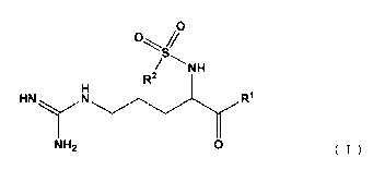
Accordingly, the present invention provides a medical material comprising a
hydrophilic polymer compound immobilized on the surface thereof, said
hydrophilic
polymer compound in which a compound represented by the general formula (I)
and
a copolymer of monomers selected from the group consisting of ethylene glycol,
vinyl acetate, vinyl pyrrolidone, propylene glycol, vinyl alcohol and siloxane
are
bound:
0
C)
NH
R2
HNN R1
NH2 0 === (I)
[wherein R1 represents a (2R,4R)-4-alkyl-2-carboxypiperidino group; R2
represents a phenyl group or a fused polycyclic compound group, the fused
polycyclic compound group being optionally substituted with a lower alkyl
group, a
lower alkoxy group or an amino group which is substituted with a lower alkyl
group].
[0011]
The above copolymer is preferably a polyether-modified silicone.
CA 02839202 2013-12-12
[0012]
The above compound represented by the general formula (I) is preferably
(2R,4R)-4-methy1-1-((2S)-2- [(3RS)-3-methy1-1,2,3,4-tetrahydroquinolin-8-
yl]sulfonyl} amino-5-guanidinopentanoyl)piperidine-2-carboxylic acid.
5 [0013]
Examples of materials of the above medical material include cellulose,
cellulose acetate, polycarbonate, polysulfone (hereinafter referred to as
polyether sulfone, polymethacrylate such as poly(methyl methacrylate)
(hereinafter
referred to as "PMMA"), polyacrylate, polyamide, polyvinylidene fluoride,
polyvinyl
chloride, polyacrylonitrile, polyester, polyurethane, polystyrene,
polyethylene,
polypropylene, polymethylpentene, polyimide and polytetrafluoroethylene.
Preferred is polyester, polyurethane, polystyrene, PMMA, polyvinyl chloride,
polytetrafluoroethylene, or PSf.
[0014]
Examples of the above medical material include implantable artificial organs,
synthetic blood vessels, catheters, stents, blood bags, blood circuits,
artificial lungs,
artificial hearts and lungs, organ adhesion preventive films, contact lenses,
intraocular lenses, surgical auxiliary instruments and separation membranes
such as
hollow fiber membranes or adsorbents that are built in modules for biological
component separation or modules for hemocatharsis. Preferred are hollow fiber
membranes.
[0015]
Further, the present invention also provides a module for hemocatharsis that
is filled with a hollow fiber membrane comprising the above hydrophilic
polymer
compound immobilized on the surface thereof.
EFFECT OF THE INVENTION
[0016]
CA 02839202 2013-12-12
6
According to the present invention, a medical material having a hydrophilic
polymer compound firmly immobilized on the surface thereof, which compound
significantly inhibits both of the blood coagulation reactions in the primary
hemostasis stage in which platelets are involved and in the coagulation
thrombus
formation stage in which blood coagulation factors are involved in a state
retaining
such an anticoagulant activity can be provided.
BRIEF DESCRIPTION OF THE DRAWINGS
[0017]
Figure 1 is a schematic view showing a mini-module prepared in the
examples.
Figure 2 is a schematic view showing a closed circuit used in an in vitro
blood
circulation test.
Figure 3 is a schematic view showing a human blood plasma circulation
circuit used in measurement of the amount of hydrophilic polymer compound
dissolved out.
Figure 4 is a figure showing the results of in vitro blood circulation test
using
a PSf and PMMA hollow fiber membrane mini-module in conditions with no
anticoagulants being added.
MODE FOR CARRYING OUT THE INVENTION
[0018]
Unless otherwise specified, the terms used herein have the following
definitions:
[0019]
The term "hydrophilic" in the term "hydrophilic polymer compound"
immobilized on the medical material of the present invention herein means that
a
compound is water-soluble, or even if a compound is not water-soluble, the
compound interacts with water molecules by electrostatic interaction or
hydrogen
CA 02839202 2013-12-12
7
bond.
[0020]
Examples of "copolymer of monomers selected from the group consisting of:
ethylene glycol, vinyl acetate, vinyl pyrrolidone, propylene glycol, vinyl
alcohol, and
siloxane" (hereinafter referred to as "anti-platelet adhesion copolymer")
include
polymeric compounds composed of polyvinyl alcohol, polyvinyl pyrrolidone,
polyethylene glycols, polypropylene glycol, polyether and polysiloxane or
copolymers or graft materials of monomers of the polymeric compounds with
other
monomers. Preferred is a polymeric compound composed of highly hydrophilic
polyether and polysiloxane or copolymer of partially saponified polyvinyl
alcohol or
vinyl pyrrolidone with vinyl acetate.
[0021]
Examples of "polymer compound composed of polyether and polysiloxane"
include copolymers, polymer complexes or polymer blend products of polyether
and
polysiloxane. The copolymer of polyether and polysiloxane is composed of
polyether units and polysiloxane units, and the copolymer form thereof may be
any of
random copolymer, block copolymer or graft copolymer. Among these, polyether-
modified silicone which is highly hydrophilic is preferred.
[0022]
Examples of "polyether" include structures originated from polyethylene
oxide or polypropylene oxide. "Polyether" herein refers to a structure
represented
by the general formula (II) (R3 represents an alkyl group having 6 carbon
atoms or
less), and a "structure originated from polypropylene glycol" which is one of
the
examples of polyether refers to a structure represented by the general formula
(III).
0 /
(II)
CA 02839202 2013-12-12
8
(III)
[0023]
The term "polyether-modified silicone" refers to a silicone in which polyether
units are bound to side chains of the silicone chain, and may be a polyether-
modified
silicone that is further amino-modified or carboxy-modified.
[0024]
In cases where an anti-platelet adhesion copolymer is partially saponified
polyvinyl alcohol, the degree of saponification thereof is preferably 50 to
less than
100 mol% from the viewpoint of attaining suitable ease of handling or
hydrophilicity,
more preferably 74 to 99.9 mol%, and still more preferably 78 to 95 mol%. A
"degree of saponification" herein refers to a numerical value calculated by
the
equation 1.
Degree of saponification = m/(n + m) x 100 ........ Equation 1
m: the number of structures represented by the general formula (IV) in
polyvinyl alcohol
n: the number of structures represented by the general formula (V) in
polyvinyl alcohol
m
(IV)
n
0
_______________________ 0
= (V)
[0025]
CA 02839202 2013-12-12
9
In cases where an anti-platelet adhesion copolymer is a copolymer of vinyl
pyrrolidone and vinyl acetate, vinyl pyrrolidone units preferably account for
50 unit
mol% or more, and more preferably 60 unit mol% or more, from the viewpoint of
attaining suitable ease of handling or hydrophilicity. On the other hand, from
the
viewpoint of attaining a suitable amount of immobilization to a medical
material,
vinyl pyrrolidone units preferably account for less than 100 unit mol%. Note
that
the percentage of the vinyl pyrrolidone units in the copolymer of vinyl
pyrrolidone
and vinyl acetate (unit mol%) can be calculated by subjecting the copolymer to
1H-
.
NMR measurement (solvent: CDC13).
[0026]
The amount of adsorption of an anti-platelet adhesion copolymer on the
surface of a medical material is preferably 0.1 pg/mm2 or more, more
preferably 1
pg/mm2 or more, and still more preferably 10 pg/mm2 or more.
[0027]
The above amount of adsorption is measured by the following method:
First, an untreated sensor chip (Sensor Chip Au; GE Healthcare) is pretreated
(with
distilled water at 25 C, at a flow rate of 20 lx1/min, for 10 minutes) using a
surface
plasmon resonance system (hereinafter referred to as "SPR") (BIACORE 3000; GE
Healthcare), and a signal value thereof (RU: resonance unit) is measured.
[0028]
A material of medical material, that is, a material to be immobilized is
dissolved in a solvent to prepare a 0.5% by weight solution of material to be
immobilized. One drop of the solution of material to be immobilized is dropped
onto the center of a gold film part of a pretreated sensor chip that has been
installed
in a spin coater and is immediately rotated at 3,000 rpm for 1 minute at room
temperature to coat the sensor chip with the material to be immobilized.
[0029]
CA 02839202 2013-12-12
After confirming that no droplets are present on the sensor chip, the sensor
chip is washed with distilled water using SPR (at 25 C, at a flow rate of 20
[11/min,
for 10 minutes), further washed three times with 0.025 wt % Triton-X100
solution (at
25 C, at a flow rate of 20 ill/min, for 1 minute), and then the signal value
at 10
5 minutes after completion of the washing is measured.
[0030]
Among the sensor chips obtained as described above, ones whose signal value
difference before and after spin coat was within a range from 3,000 to 8,000
were
selected, then washed with distilled water (at 25 C, at a flow rate of 20
for 10
10 minutes), and further washed three times with 0.025 wt % Triton-X100
solution (at
25 C, at a flow rate of 20 til/min, for 1 minute).
[0031]
Ten minutes after completion of the washing, an aqueous solution of a
hydrophilic polymer compound to be adsorbed to a medical material
(concentration:
100 vg/m1) is injected (at 25 C, at a flow rate of 20 ttl/min, for 1 minute),
and washed
with distilled water (at 25 C, at a flow rate of 20 ill/min, for 3 minutes).
The
difference between the signal value immediately before the injection
(hereinafter
referred to as "signal value A") and the signal value at 3 minutes after
completion of
the injection (hereinafter referred to as "signal value B") is determined,
which is
converted as 1 RU = 1 pg/mm2.
[0032]
Subsequently, the sensor chip is washed with distilled water (at 25 C, at a
flow rate of 20 111/min, for 2 minutes), further washed three times with 0.025
wt %
Triton-X100 solution (at 25 C, at a flow rate of 20 1.11/min, for 1 minute),
and then the
aqueous solution of the hydrophilic polymer compound to be adsorbed
(concentration: 100 jig/m1) is again injected (at 25 C, at a flow rate of 20
pl/min, for
1 minute). Thereafter, the same operation is repeated to determine a signal
CA 02839202 2013-12-12
11
difference (difference between signal value A and signal value B) for five
times in
total, and the mean value is regarded as the amount of adsorption of anti-
platelet
adhesion copolymer to the medical material.
[0033]
As a "compound represented by the general formula (I)" which is a compound
having a compound having a guanidine structure, (2R,4R)-4-methy1-14(2S)-2-
{ [(3RS)-3-methy1-1,2,3,4-tetrahydroquinolin-8-yl]sulfonyl } amino-5-
guanidinopentanoyl)piperidine-2-carboxylic acid (hereinafter referred to as
"argatroban") is for example preferred. Argatroban synthesized in 1978 is a
medicinal compound having a selective antithrombin activity of arginine
derivatives.
The phrase "having an antithrombin activity" herein means that a binding
affinity to
thrombin is high, and examples of indexes for evaluating the antithrombin
activity of
a compound include an inhibition constant (hereinafter referred to as "Ki")
which is
calculated from Lineweaver-Burk plot based on the absorbance value of solution
to
be tested. Lower Ki indicates higher binding affinity to thrombin and higher
antithrombin activity. Ki is preferably 10 p,M or less, more preferably 1 1iM
or less,
and still more preferably 500 nM or less. The compound having an antithrombin
activity can be used solely or two or more thereof can be combined to use.
[0034]
Examples of a method of immobilizing the above hydrophilic polymer
compound onto the surface of a medical material include a method comprising
bringing a surface treatment agent containing the above hydrophilicity
compound as
a active component into contact with the medical material and irradiating
thereto
with radiation. Note that, as the type of the irradiating radiation, an
electron beam
or y-ray is preferred. In cases where the method comprising irradiating the
radiation
is difficult, a method comprising spraying or spreading the above
hydrophilicity
compound dissolved or dispersed in an organic solvent such as ethanol or
methanol
CA 02839202 2013-12-12
12
and onto the surface of the medical material and drying can be for example
employed.
EXAMPLES
[0035]
The present invention will be now described in detail below by way of the
examples. However, the present invention is by no means limited thereto.
[0036]
(Example 1)
1. Binding between Amino=Polyether-Modified Silicone and Argatroban:
Argatroban in an amount of 5 mmol was placed in a recovery flask, and 10
mL of anhydrous dimethylformamide (hereinafter referred to as "anhydrous DMF")
was added thereto to dissolve argatroban. Thereafter, 10 mL of 4N hydrochloric
acid/1,4-dioxane (Toyo Kasei Co., Ltd.) was added dropwise while cooling the
recovery flask and the resulting mixture was stirred for 1 hour. Next, the
solvent
was evaporated with a rotary evaporator and the resultant was further dried
overnight
in a vacuum dryer, and added with 25 mL of anhydrous DMF to obtain argatroban
hydrochloride/anhydrous DMF solution.
[0037]
Argatroban hydrochloride/anhydrous DMF solution in an amount shown in
Table 1 was placed in a two-necked flask, and dicyclohexylcarbodiimide
(hereinafter
referred to as "DCC")/anhydrous DMF solution and 4-hydroxybenzotriazole
(hereinafter referred to as "HOBt")/anhydrous DMF solution were each added
thereto
with ice cooling and stirring. A polyether-modified silicone (X-22-3939A; Shin-
Etsu Chemical Co., Ltd.) was further added and the resulting mixture was
allowed to
react at room temperature for 3 days. Next, the reaction solution was placed
in a
dialysis tube (Spectra/Por RC, Pore 6, MWC0=1,000), and dialyzed for 3 days
against distilled water with a volume of more than 10 times while
appropriately
replacing the distilled water. The reaction solution after the dialysis was
filtered,
-
CA 02839202 2013-12-12
13
and the solvent in the filtrate was evaporated with a rotary evaporator,
followed by
drying the resultant overnight in a vacuum dryer to obtain a hydrophilic
polymer
compound (hereinafter referred to as "Example 1 compound").
[0038]
2. Measurement of Antithrombin Activity of Example 1 compound
ECA-T kit (HaemoSys) was used for the measurement. To 100 lit of the
Example 1 compound, 900 [IL of distilled water was added to prepare an aqueous
Example 1 compound solution. The aqueous Example 1 compound solution in an
amount of 30 jiL was sampled and mixed with 100 [tL, of ECA prothrombin buffer
and 254 of ECA-T substrate. After incubated at 37 C for 60 seconds, the
mixture
was set in an apparatus (COATRON Ml(code 80 800 000); Production); and 50 fiL
of ECA ecarin reagent was added thereto to then carry out the measurement.
[0039]
A mixture of 20 L of argatroban solution prepared to an arbitrary
concentration using an ethanol/hydrochloric acid (volume ratio: 4/1) mixed
solvent
and 804, of human blood plasma, or a mixture of 20 pt of blank distilled water
and
80 gL of human blood plasma was each subjected to the measurement using the
ECA-T kit in place of the above aqueous Example 1 compound solution, and a
calibration curve was prepared from the results. The concentration of 1494.3
ppm
by weight in terms of argatroban of the aqueous Example 1 compound solution
calculated from the calibration curve was defined as a value indicating the
antithrombin activity of the aqueous Example 1 compound solution.
[0040]
3. Measurement of Thrombin Inhibition Constant of Example 1 Compound
A bovine thrombin solution (Ito Life Sciences, Inc.) 10,000 U was dissolved
in 1 mL of physiological saline to prepare an aqueous bovine thrombin
solution.
[0041]
CA 02839202 2013-12-12
= 14
S-2238 stock solution (Sekisui Medical Co., Ltd.) 25 mg was dissolved in 40
mL of distilled water to prepare an aqueous S-2238 stock solution.
[0042]
The aqueous bovine thrombin solution, aqueous S-2238 stock solution and
the above aqueous Example 1 compound solution were each diluted by using
dilution
buffer solution (0.05 M Tris, 0.1 M NaC1, 1 mg/mL of bovine serum albumin
(BSA),
pH 7.4).
[0043]
To a 96-well plate, 100 lit of diluted solution of the aqueous S-2238 stock
solution and 50 L of diluted solution of the aqueous Example 1 compound
solution
were dispensed, sealed and then warmed in a thermostat dryer set at 37 C for
30
minutes. Next, 50 1., of the diluted solution of the aqueous bovine thrombin
solution which was heated at 37 C for 30 minutes was further dispensed
thereto, and
absorbance of the resulting mixture was immediately measured with a microplate
reader (measurement wavelength: 405 nm, reference wavelength: 595 nm).
[0044]
Immediately after completion of the first absorbance measurement, the second
absorbance measurement was carried out. The third or later absorbance
measurements were carried out at 4, 6, 8, 10, 12, 14, 16, 18, and 20 minutes,
respectively, after the diluted solution of the aqueous bovine thrombin
solution was
dispensed. Ki was calculated from each of the obtained absorbance values by
using
Lineweaver-Burk plot. The Ki of the Example 1 compound was 11 nM.
[0045]
The Ki of the polyether-modified silicone (X-22-3939A) was also calculated
in the same manner, but the Ki of the polyether-modified silicone without an
antithrombin activity was the same as that of the blank, as expected.
[0046]
CA 02839202 2013-12-12
Further, Ki was also calculated for argatroban in the same manner, and the Ki
thereof was 39 nM which was a three times or more higher value, as compared
with
the Ki of the Example 1 compound.
[0047]
5 From these results, it became apparent that the above hydrophilic
polymer=
compound has an extremely high binding affinity to thrombin, and can give a
prominent antithrombin activity that is much higher than argatroban, which is
known
to have an antithrombin activity, to medical materials including hollow fiber
membranes.
10 [0048]
(Examples 2 to 13)
Examples 2 to 13 compounds were obtained, respectively, in the same manner
as described in Example 1 except that the molar ratios of DCC, HOBt and
polyether-
modified silicone (X-22-3939A) to argatroban hydrochloride and the volume
ratio of
15 anhydrous DMF to polyether-modified silicone were altered and the
antithrombin
activity thereof was measured. The molar ratios of DCC, HOBt and polyether-
modified silicone (X-22-3939A) to argatroban hydrochloride and the measurement
results of the antithrombin activity of each of the Examples 2 to 13 compounds
are
shown in Table 1.
[0049]
CA 02839202 2013-12-12
' 16
[Table 1]
Molar ratio to 1.00 mol of Volume ratio of
Concentration
argatroban hydrochloride anhydrous
DMF to in terms of
Compound 22-
1 volume of
argatroban
X-
DCC HOBt 3939A
polyether-modified (ppm by
silicone
weight)
Example 1 1.07 1.06 0.060 -
1494.3
Example 2 1.04 1.04 0.060 -
831.2
Example 3 0.20 0.20 0.060 1.4
6610.7
Example 4 0.20 0.20 0.030 3.9
8393.3
Example 5 , 1.29 1.27 0.493 1.8
505.3
=
Example 6 1.29 1.27 0.203 4.3
771.7
Example 7 _ 1.29 1.27 0.101 8.6
606.7
Example 8 1.29 1.27 0.067 13.0
441.7
Example 9 1.29 1.27 0.049 17.6
436.7
Example10 1.29 1.27 0.020 42.9
738.9
Examplell 1.29 1.27 0.010 88.2
895.0
Example12 1.00 1.00 0.060-
6000.0
Example13 1 1.00 1.00 0.060 40.0
5999.4
[0050]
The antithrombin activity of the polyether-modified silicone (X-22-3939A)
was also measured and the measured value was the same as that of the blank
distilled
water. It was confirmed that the polyether-modified silicone itself does not
have the
antithrombin activity.
[0051]
(Preparation of PMMA Hollow Fiber)
Five parts by weight of isotactic-PMMA and 20 parts by weight of
syndiotactic-PMMA were added to 75 parts by weight of dimethylsulfoxide, and
the
resulting mixture was stirred at 110 C for 8 hours to obtain a membrane-
forming
liquid. This membrane-forming liquid was extruded from an orifice type
CA 02839202 2013-12-12
17
bicylindrical mouthpiece and passed 300 mm in air, and then the extruded
material
was introduced into a solidification bath containing 100% water to obtain PMMA
hollow fibers having an inner diameter of 0.2 mm and a membrane thickness of
0.03
mm. Note that, as the gas injected into the inside of the fiber,
dry nitrogen was used.
[0052]
(Preparation of PSf hollow fiber)
Eighteen parts by weight of PSf (Udel manufactured by Solvay (registered
trademark) P-3500) and 9 parts by weight of PVP (K30 manufactured by BASF)
were added to a mixed solvent of 72 parts by weight of DMAc and 1 part by
weight
= 10 of water, and the resulting mixture was stirred at 90 C for 14
hours to obtain a
membrane-forming liquid. Also, a core liquid composed of 58 parts by weight of
DMAc and 42 parts by weight of water was prepared. Using an orifice type
bicylindrical mouthpiece whose cyclic slit part had an outer diameter of 0.3
mm and
an inner diameter of 0.2 mm, the membrane-forming liquid and core liquid were
extruded from the outside tube and inside tube, respectively, and introduced
into a
solidification bath containing 100% water that was located 350 mm away from
the
mouthpiece to obtain PSf hollow fibers.
[0053]
(Preparation of PMMA Hollow Fiber Membrane Mini-Module)
A module case having an inner diameter of 10 mm and a length of 120 mm
that, similarly to general hollow fiber-type dialyzers, had two ports
communicating to
the inside of the hollow fibers (blood port) and two ports communicating to
the
outside of the hollow fibers (dialysate port) was prepared.
[0054]
Fifty of the above PMMA hollow fibers were bundled to form a PMMA
hollow fiber membrane, and both ends of the PMMA hollow fiber membrane were
fixed to the above module case using an epoxy-based potting agent with
attention not
CA 02839202 2013-12-12
= 18
to clog the hollow portion of the PMMA hollow fiber membrane. Thereafter, the
PMMA hollow fiber membrane and the inside of the module case were washed with
distilled water to obtain the mini-module 6 as shown in Figure 1.
[0055]
Bis-Tris (Dojindo Laboratories) and sodium chloride were dissolved in
ultrapure water so as to attain a final concentration of 0.25M and 0.5M,
respectively,
and the pH of the resulting solution was adjusted to 5 by adding 6N
hydrochloric acid
dropwise to prepare Bis-Tris buffer solution having 5 times concentration.
[0056]
Distilled water remaining at the side contacting the blood (the inner side of
the PMMA hollow fiber membrane) and the side not contacting the blood (the
outer
side of the PMMA hollow fiber membrane) of the prepared mini-module 6 was
removed with compressed air. Next, the aqueous Example 1 compound solution of
a concentration of 10,000 ppm by weight in terms of argatroban, propylene
glycol,
Bis-Tris buffer solution having 5 times concentration, and distilled water
were mixed
at a volume ratio of 4/3/2/1 to obtain a filling solution.
[0057]
The above filling solution (400 tit) was filled only at the side contacting
the
blood of the mini-module 6 using a syringe. Thereafter, the filling solution
was
removed with compressed air, and the mini-module 6 in which all of the blood
ports
la and lb and the dialysate ports 2a and 2b were tightly capped was irradiated
with y-
ray at a absorbed dose of 25 kGy for about 3 hours.
[0058]
The PMMA hollow fiber membrane 4 and the inner side of the mini-module 6
were washed by passing 0.025% by weight aqueous poly(oxyethylene)octyl phenyl
ether solution into the PMMA hollow fiber membranes 4 and the inner side of
the
mini-module 6 at a flow rate of 10 mL/min for 8 hours using the peristaltic
pump 8.
CA 02839202 20.13-12-12
19
Thereafter, distilled water and physiological saline were flown both at a flow
rate of
mL/min for 30 minutes to carry out further washing to obtain a mini-module on
which the Example 1 compound was immobilized (hereinafter referred to as "PMMA
hollow fiber membrane mini-module 1").
5 [0059]
The aqueous Example 1 compound solution of a concentration of 10,000 ppm
by weight in terms of argatroban, propylene glycol, Bis-Tris buffer solution
having 5
times concentration, and distilled water were mixed at a volume ratio shown in
Table
2 to obtain each filling solution. Each of the mini-modules on which the
Example 1
10 compound was immobilized ("PMMA hollow fiber membrane mini-module 2" to
"PMMA hollow fiber membrane mini-module 4") was prepared by carrying out the
same procedure as described above except that each of the prepared filling
solutions
was used.
[0060]
[Table 2]
Aqueous Example
1 compound Bis-Tris
solution buffer
of a concentration solution
Mini-module Propylene glycolDistilled water
of 10,000 ppm by having 5
weight times
in terms of concentration
argatroban
1 4 3 2 1
`t 2 0.1 3 2 4.9
c:k 3 1 3 2 4
4 5 3 2 0
[0061]
(Preparation of PSf Hollow Fiber Membrane Mini-Module)
A mini-modules on which the Example 1 compound was immobilized
(hereinafter referred to as "PSf hollow fiber membrane mini-module 2") was
CA 02839202 2013-12-12
prepared by carrying out the same procedure as described above except that
PMMA
hollow fibers were replaced with PSf hollow fibers.
[0062]
Distilled water remaining at the side contacting the blood (the inner side of
5 the PSf hollow fiber membrane) and the side not contacting the blood (the
outer side
of the PSf hollow fiber membrane) of the separately prepared mini-module 6 was
removed with compressed air. Next, the aqueous Example 1 compound solution of
a concentration of 10,000 ppm by weight in terms of argatroban, propylene
glycol,
Bis-Tris buffer solution having 5 times concentration, and distilled water
were mixed
10 at a volume ratio of 4/3/2/1 to obtain a filling solution.
[0063]
The above filling solution (400 p,L) was filled only at the side contacting
the
blood of the mini-module 6 using a syringe. Thereafter, the filling solution
was
removed with compressed air, and the mini-module 6 in which all of the blood
ports
15 la and lb and the dialysate ports 2a and 2b were tightly capped was
irradiated with y-
ray at a absorbed dose of 25 kGy for about 3 hours.
[0064]
The PSf hollow fiber membrane 4 and the inner side of the mini-module 6
were washed by passing 0.025% by weight aqueous poly(oxyethylene)octyl phenyl
20 ether solution into the PSf hollow fiber membranes 4 and the inner side
of the mini-
module 6 at a flow rate of 10 mL/min for 8 hours using the peristaltic pump 8.
Thereafter, distilled water and physiological saline were flown both at a flow
rate of
10 mL/min for 30 minutes to carry out further washing to obtain a mini-module
on
which the Example 1 compound was immobilized (hereinafter referred to as "PSf
hollow fiber membrane mini-module 1").
[0065]
The aqueous Example 1 compound solution in a concentration of 10,000 ppm
CA 02839202 20.13-12-12
21
by weight in terms of argatroban, propylene glycol, and Bis-Tris buffer
solution
having 5 times concentration were mixed at a volume ratio shown in Table 3 to
obtain each filling solution. Each of the mini-modules on which the Example 1
compound was immobilized ("PSf hollow fiber membrane mini-module 2" to "PSf
hollow fiber membrane mini-module 5") was prepared by carrying out the same
procedure as described above except that each of the prepared filling
solutions was
used.
[0066]
[Table 3]
Aqueous Example
1 compound Bis-Tris
solution of buffer
a concentration of solution
Mini-module Propylene glycolDistilled water
10,000 ppm by having 5
weight times
in terms of concentration
argatroban
1 4 3 2 1
2 0.1 3 2 4.9
PSf 3 1 3 2 4
4 2 3 2 3
5 5 3 2 0
[0067]
A mini-module in which polyether-modified silicone was immobilized
(hereinafter referred to as "Comparative Example 1 mini-module") was obtained
by
carrying out the same procedure as described above except that polyether-
modified
silicone (X-22-3939A) was used in place of the aqueous Example 1 compound
solution of a concentration of 10,000 ppm by weight in terms of argatroban.
[0068]
(In Vitro Blood Circulation Test)
Blood provided by a volunteer and citric acid was mixed at a volume ratio of
CA 02839202 2013-12-12
22
9/1 to obtain blood supplemented with citric acid. Calcicol in an amount of
43.6 p1
was added, as a procoagulant, to 1 mL of the blood supplemented with citric
acid to
obtain a test blood.
[0069]
Silicone tubes 7a and 7b were connected to the PMMA hollow fiber
membrane mini-module 1 and the peristaltic pump 8 was placed in the middle of
the
silicone tube 7b. The test blood was passed at a flow rate of 0.9 mL/min for 5
seconds from the silicone tube 7a connected to the blood port la, and the test
blood
flown out from the blood port lb was discarded from the silicone tube 7b to
remove
bubbles in the inner side of the PMMA hollow fiber membrane. Subsequently, the
silicone tubes 7a and 7b were connected at the inclusion part 9 to form a
closed
circuit shown in Figure 2.
[0070]
The circulation of the test blood was started at a flow rate of 0.9 mL/min to
measure duration time of circulation until the silicone tubes 7a or 7b came
off the
inclusion part 9 due to an increased inner pressure in the circuit caused by
coagulation thrombus formed in the circuit. The duration time of circulation
in the
case of using the PMMA hollow fiber membrane mini-module 1 was 35 minutes.
Further when evaluated under the same condition, the duration time of
circulation in
the case of using the PSf hollow fiber membrane mini-module 1 was 41 minutes.
[0071]
Figure 4 shows the results of the in vitro blood circulation test that was
carried out as described above using, in place of the PMMA hollow fiber
membrane
mini-module 1, each of the PMMA hollow fiber membrane mini-modules 2 to 4 and
PSf hollow fiber membrane mini-modules 2 to 5. In the PMMA hollow fiber
membrane mini-module, the duration time of circulation reached a plateau once
the
volume ratio of the Example 1 compound solution in the filling solution
exceeded a
CA 02839202 2013-12-12
23
certain value. In contrast, this tendency was not observed in the PSf hollow
fiber
membrane mini-module and the duration time of circulation could be
continuously
extended as appropriate.
[0072]
The mini-module 6 in which no compounds were immobilized on the PMMA
hollow fiber membrane (hereinafter referred to as "Comparative Example 2 mini-
module") was prepared to carry out the same blood circulation test as
described
above. The duration time of circulation in this case was 20 minutes which was
half
or less than that in the case of using the PMMA hollow fiber membrane mini-
module
or PSf hollow fiber membrane mini-module. From these results, it became
apparent
that the above hydrophilic polymer compound exhibited an excellent
anticoagulant
activity for the medical material immobilized therewith.
[0073]
Note that, when the same blood circulation test was carried out as described
above using the Comparative Example 1 mini-module, the duration time of
circulation was 20 minutes, which was the same as the duration of circulation
in the
case of using the Comparative Example 2 mini-module in which no compounds were
immobilized on the PMMA hollow fiber membrane.
[0074]
(Measurement of Amount of Example 1 Compound Dissolved Out)
The silicone tube 7b having an inner diameter of 0.8 mm and a length of 520
mm was connected to the blood port lb of the separately prepared Example 1
mini-
module, and the peristaltic pump 8 was placed in the middle of the silicone
tube 7b.
The silicone tube 7a having an inner diameter of 0.8 mm and a length of 160 mm
was
connected to the blood port la. Thereafter, the other end of each of the
silicone
tubes 7a and 7b was inserted in the polystyrene round tube (Code: 352054;
BECTON
DICKINSON) 10 containing 5 mL of human plasma to prepare a circulating circuit
CA 02839202 2013-12-12
24
shown in Figure 3.
[0075]
Human plasma was circulated at a flow rate of 0.5 mL/min for 4 hours using
the peristaltic pump 8, the concentration of the Example 1 compound in human
plasma in the polystyrene round tube 10 was measured using ECA-T kit. However,
the concentration of the Example 1 compound in human plasma after the
circulation
was below the limit of detection of ECA-T kit, and it was not confirmed that
the
Example 1 compound dissolved out from the PMMA hollow fiber membrane mini-
module. This result shows that the above hydrophilic polymer compound can be
firmly immobilized to medical materials including a hollow fiber membrane.
[0076]
(Evaluation of Adsorption Amount of Anti-Platelet Adhesion Copolymer)
As a copolymer between vinylpyrrolidone and vinyl acetate (hereinafter
referred to as "VA copolymer"), which copolymer composes the above hydrophilic
polymer compound and was one of the anti-platelet adhesion copolymers, PVP (K-
90), VA73, VA64, VASS and VA37 (all of which were from BASF) were provided.
Similarly, as a partially saponified polyvinyl alcohol which was one of the
anti-
platelet adhesion copolymers, PVA217, PVA417 and PVA205c (all of which were
from Kuraray Co., Ltd.) were provided. Further, as a polyether-modified
silicone,
F114, F244, F303, F3031, F348, F350s, F502, F506 and X-22-3939A (all of which
were from Shin-Etsu Silicone) were provided. Note that the provided VA
copolymer, partially saponified polyvinyl alcohol and polyether-modified
silicone
were all diluted with distilled water to prepare an aqueous solution of 10,000
ppm by
weight.
[0077]
Meanwhile, as a polymer compound composing the above hydrophilic
polymer compound, which was not included in the anti-platelet adhesion
copolymer,
CA 02839202 2013-12-12
PEG2000, PEG4000, PEG6000 and PEG20000 (all of which were from Nacalai
Tesque, Inc.), and PEG methyl ether (PEG-em) and PEG dimethyl ether (PEG-dm)
(both were from Sigma-Aldrich) were provided for comparison. Note that the
provided polymer compounds were all diluted with distilled water to prepare an
5 aqueous solution of 10,000 ppm by weight.
[0078]
As a 0.5% by weight solution of a material to be immobilized for
immobilizing an anti-platelet adhesion copolymer, PMMA (weight average
molecular weight: 93000, Sigma-Aldrich)/toluene solution,
10 polyurethane/dimethylacetamide solution, PSf (Udel manufactured by
Solvay
(registered trademark) P-3500)/dimethylacetamide solution, polyvinyl chloride
(weight average molecular weight: 80,000; Sigma-Aldrich)/tetrahydrofuran
solution,
polystyrene (Wako)/chloroform solution and polycarbonate (weight average
molecular weight: 20,000; Teijin Limited)/chloroform solution were each
prepared.
15 [0079]
The adsorption amount of various anti-platelet adhesion copolymers for each
of the materials to be immobilized was measured. The results are shown in
Figure 4.
[0080]
CA 02839202 2013-12-12
* 26
[Table 4]
' Signal value B - Signal value A [pg/mm2]
Adsorbent Material
PMMA PSf Poly-
Polyvinyl chloride Poly- Poly-
urethane
styrene carbonate
PVPK
789 - - - - -
VA37 2760 - - - -
-
VASS 472 - - - -
-
VA64 920 - - - -
-
VA73 426 - - - -
-
PVA
2529 2886 1635 2468 2777 2356
217
1 PVA
2475 2742 1911 2330 2662 2346
417
PVA
2223 2130 1411 1796 1989 1819
205c
F114 1003 844 514 739 621 756
F244 1639 1272 1144 1118
1052 1243
F303 1268 1156 1604 1037 -
1374
F3031 947 559 614 418 339 536
F348 875 784 756 608 283 800
F350s 751 657 674 544 275 591
F502 827 657 696 385 197 482
F506 691 308 437 167 43 279
3939A
1182 910 1204 695 924 1424
PEG
2000 2 - - - -
-
g
2., PEG
o 2 - - - -
-
oa. 4000
0
o PEG
5 - - - - -
- 6000
2
u)
o
4 PEG
-o 113 _ _ _ _
_
cl 20000
.1.) PEG
o 5_ _ _ _
_
5 -me
=,T,' PEG
t 67 - - - -
-
< -dm
[0081]
From the results of Table 4, it became apparent that the anti-platelet
adhesion
CA 02839202 2013-12-12
27
copolymer composing the above hydrophilic polymer compound was not limited to
the polyether-modified silicone (X-22-3939A) and could be firmly immobilized
to
medical materials including a hollow fiber membrane.
[0082]
(Evaluation of Anti-Platelet Adhesion Ability)
The separately prepared module case of the PMMA hollow fiber membrane
mini-module was cut with an ultrasonic cutter to take out the PMMA hollow
fiber
membrane (hereinafter referred to as "Example 1 hollow fiber membrane") on
which
the Example 1 compound was immobilized.
[0083]
A double-stick tape was adhered to one surface of a circular film made of
polyethylene terephthalate having a diameter of 18 mm. The Example 1 hollow
fiber membrane was fixed thereto, and the fixed PMMA hollow fiber membrane was
cut into slivers of a semicylindrical shape to expose the inner surface of the
PMMA
hollow fiber membrane. The Example hollow fiber membrane fixed to the circular
film was placed inside of a Falcon (registered trademark) cylindrical tube (18
mm421),
NO. 2051) that had cut into a cylindrical shape, and the gap between the
cylindrical
tube and circular film was sealed with Parafilm. Thereafter, the cylindrical
tube was
filled with physiological saline.
[0084]
Venous blood immediately after collected from volunteers was placed in a
blood collection tube in which heparin had been collected in advance, and the
resulting mixture was mixed by inverting the tube to prepare blood
supplemented
with heparin. The concentration of the blood supplemented with heparin was
adjusted to be 50 U/mL.
[0085]
After discarding the physiological saline in the above cylindrical tube, 1.0
mL
CA 02839202 2013-12-12
28
of the blood supplemented with heparin was placed thereto, and the cylindrical
tube
was shaken at 37 C for 1 hour. Thereafter, the Example 1 hollow fiber membrane
in the above cylindrical tube was washed with 10 mL of physiological saline,
and
then blood components were fixed by adding physiological saline containing
2.5% by
volume glutaraldehyde, followed by further washing the membranes with
distilled
water. Thereafter, the circular film on which the Example 1 hollow fiber
membrane
was fixed was removed from the above cylindrical tube, and the circular film
on
which the Example hollow fiber membrane was fixed was dried under reduced
pressure at normal temperature at an absolute pressure of 0.5 Torr for 12
hours.
[0086]
The circular film dried under reduced pressure on which the Example 1
hollow fiber membrane was fixed was adhered to the stage of a scanning
electron
microscope with a double-stick tape, and a platinum/palladium thin film was
then
formed on the surface of the Example hollow fiber membranes by sputtering. The
inner surface near the center in the longitudinal direction of the Example
hollow fiber
membrane, wherein a platinum/palladium thin film was formed on the surface
thereof, was observed with a field emission scanning electron microscope
(S800;
manufactured by Hitachi, Ltd.) at a magnification of 1500x, and the number of
adhered platelets in one visual field (4.3x10³ m2) was counted.
[0087]
The integer value of the mean of the numbers of adhered platelets counted in
different 5 visual fields was defined as the number of adhered platelets
(platelets/(4.3x10 3 m2)), and the number of the platelets adhered to the
Example 1
hollow fiber membrane was 1.
[0088]
On the other hand, the separately prepared module case of the Comparative
Example 2 mini-module was cut with an ultrasonic cutter, and the hollow fiber
CA 02839202 2013-12-12
29
membrane on which any compounds were not immobilized (hereinafter referred to
as
"Comparative Example 2 hollow fiber membrane") was taken out to confirm the
number of adhered platelets as well. The number of the platelets adhered to
the
Comparative Example 2 hollow fiber membrane was 100 or more.
[0089]
From these results, it became apparent that the above hydrophilic polymer
compounds can impart a significant anti-platelet adhesion ability to medical
materials
including a hollow fiber membrane.
[0090]
(Measurement of Whole Blood Clotting Time)
The blood collected from volunteers and citric acid were mixed at a volume
ratio of 9/1 to prepare blood supplemented with citric acid.
[0091]
To a cuvette (NON-ACTIVATED CLOTTING TEST KIT), 18 tit of
physiological saline was placed, and 14.8 I, of calcicol was added thereto,
followed
by further adding 342A of blood supplemented with citric acid. Thereafter, the
measurement using Sonoclot blood coagulation/platelet function analyzer (IMI
Co.,
Ltd.) was carried out to define the obtained ACT ONSET value as whole blood
clotting time. The whole blood clotting time of the blood collected by the
volunteers was 545 seconds.
[0092]
When the same measurement were carried out using, in place of physiological
saline, each of 2, 10 and 20 ttM of argatroban solutions (solvent was
methanol/hydrochloric acid (volume ratio: 4/1)), the whole blood clotting
times was
531, 746 and 849 seconds, respectively.
[0093]
When the same measurements were carried out using, in place of
CA 02839202 2013-12-12
physiological saline, each of the aqueous Example 1 compound solutions of 0.3,
1.3
and 2.5 1.1M, the whole blood clotting times was 527, 693 and 730 seconds,
respectively.
[0094]
5 (Example 14)
1. Binding between Vinyl Acetate-Vinyl Pyrrolidone Copolymer and Argatroban
To a screw vial, 14.9 g of tetrahydrofuran, 11.5 g of vinyl acetate, 10.8 g of
N-vinylpyrrolidone, 0.028 g of 2-aminoethanethiol and 0.016 g of
azobisisobutyronitrile were placed, and after sealing the screw vial, the
resulting
10 mixture was sonicated for 10 minutes. The screw vial was once unsealed,
and the
mixture was bubbled with argon gas for 10 minutes. The screw vial was again
sealed, and then immersed in a hot water bath at 60 C under stirring for 1
hour and
further in a hot water bath at 70 C for 6 hours to copolymerize vinyl acetate
with
vinyl pyrrolidone. To this reaction solution, 80 mL of methanol was added, and
the
15 resulting mixture was added to about 5 times amount of ether, followed
by removing
the supernatant. The washing procedure of newly adding ether and removing the
supernatant was repeated three times, and the resultant was dried under
reduced
pressure to obtain a vinyl acetate-vinyl pyrrolidone copolymer. When the
obtained
vinyl acetate-vinyl pyrrolidone copolymer was measured by 1H-NMR (solvent:
20 CDC13), a vinylpyrrolidone unit was 60.6 unit % by mole.
[0095]
The obtained vinyl acetate-vinyl pyrrolidone copolymer, 3.58 g was dissolved
in 20 mL of anhydrous DMF to prepare a vinyl acetate-vinyl pyrrolidone
copolymer/anhydrous DMF solution. The entire volume of the prepared vinyl
25 acetate-vinyl pyrrolidone copolymer/anhydrous DMF solution and 0.5 mL of
argatroban hydrochloride/anhydrous DMF solution (0.49 M) were placed in a two-
necked flask, and 0.5 mL of DCC/anhydrous DMF solution (1.04 M) and 0.5 mL of
CA 02839202 2013-12-12
31
HOBt/anhydrous DMF solution (1.02 M) were each added thereto with ice cooling
and stirring; and the resultant was allowed to react under a nitrogen
atmosphere at
room temperature for 3 days. Next, the reaction solution was placed in a
dialysis
tube (Spectra/Por RC, Pore 6, MWC0=1,000), and dialyzed for 3 days against
distilled water with a volume of more than 10 times while appropriately
replacing the
distilled water. The reaction solution after the dialysis was filtered, and
the solvent
in the filtrate was evaporated with a rotary evaporator, followed by drying
the
resultant overnight in a vacuum dryer to obtain a hydrophilic polymer compound
(hereinafter referred to as "Example 14 compound").
[0096]
2. Measurement of Antithrombin Activity of Example 14 Compound
The antithrombin activity of the Example 14 compound/methanol solution
(concentration: 20% by weight) was measured in the same manner as the
measurement of the antithrombin activity of the Example 1 compound, and the
calculated concentration of 104.1 ppm in terms of argatroban of the Example 14
compound/methanol solution was defined as a value indicating the antithrombin
activity of the Example 14 compound/methanol solution.
[0097]
(Example 15)
Argatroban in an amount of 44.8 mmol was placed in a recovery flask and 50
mL of anhydrous dimethylformamide (hereinafter referred to as "anhydrous DMF")
was added thereto to dissolve argatroban under an Ar flow. Thereafter, the
recovery
flask was cooled in ice. Fifty milliliters of 4 N hydroChloric acid/1,4-
dioxane (Toyo
Kasei Co., Ltd.) were added dropwise and the resulting mixture was stirred at
room
temperature for 1 hour. Next, the solvent was evaporated using a rotary
evaporator
and subjected to azeotropic treatment with dehydrated toluene (Wako). The
resultant was further dried in a vacuum dryer overnight; and anhydrous DMF was
CA 02839202 2013-12-12
= 32
added to the obtained compound to make an argatroban hydrochloride/anhydrous
DMF solution (1.0 M).
[0098]
To a three-necked flask, 46 mL of argatroban hydrochloride/anhydrous DMF
solution was added; and 57.8 mmol of HOBt and 20 mL of anhydrous DMF were
added thereto with ice cooling and stirring. After the mixture was dissolved,
51.0
mmol of DCC was added. To 190 g of amino-polyether-modified silicone (X-22-
3939A; Shin-Etsu Chemical Co., Ltd.) that was in advance dried under reduced
pressure at 40 C for 5 hours, 760 g of anhydrous DMF was added and stirred.
The
anhydrous DMF solution in which argatroban hydrochloride, DCC, and HOBt had
been dissolved was added to the amino-polyether-modified silicone/anhydrous
DMF
solution with ice cooling. Degassing and Ar substitution were repeated five
times
and then the mixture was stirred at room temperature for three days and
allowed to
react. Next, the reaction solution was placed in a dialysis tube (Spectra/Por
RC,
Pore 6, MWC0=15,000), and dialyzed for 7 days against distilled water with a
volume of more than 100 times while appropriately replacing the distilled
water.
The reaction solution after the dialysis was filtered, and the solvent in the
filtrate was
evaporated with a rotary evaporator, followed by drying the resultant
overnight in a
vacuum dryer to obtain a bound substance (hereinafter referred to as "Example
15
bound substance").
[0099]
(Measurement of Antithrombin Activity of Example 15 Bound Substance)
ECA-T kit (HaemoSys) was used for the measurement. To 10 mg of
Example 15 bound substance, 1 ml of distilled water was added to prepare an
aqueous Example 15 bound substance solution. The aqueous Example 15 bound
substance solution in an amount of 30 [IL was sampled and mixed with 1004 of
ECA protirombin buffer and 254 of ECA-T substrate. After incubated at 37 C
CA 02839202 2013-12-12
33
for 60 seconds, the mixture was set in an apparatus (COATRON Ml(code 80 800
000); Production); and 50 IA. of ECA ecarin reagent was added thereto to then
carry out the measurement of antithrombin activity.
[0100]
A mixture of 20 [IL of argatroban solution prepared to an arbitrary
concentration using an ethanol/hydrochloric acid (volume ratio: 4/1) mixed
solvent
and 80 tit of human blood plasma, or a mixture of 20 !IL of blank distilled
water and
80 pL of human blood plasma was each subjected to the measurement using the
ECA-T kit in place of the above aqueous Example 15 bound substance solution,
and
a calibration curve was prepared from the results. The concentration of 2.6
ppm by
weight in terms of argatroban of the aqueous Example 15 bound substance
solution
calculated from the calibration curve was defined as a value indicating the
antithrombin activity of the aqueous Example 15 bound substance solution.
[0101]
From these results, it became apparent that the above hydrophilic polymer
compound can prolong the whole blood clotting time as compared with argatroban
which is known to have an antithrombin activity, even if the concentrations of
the
hydrophilic polymer compound are very low; and the hydrophilic polymer
compound
can impart an excellent anticoagulant activity to medical materials including
a hollow
fiber membrane filled in a module for hemocatharsis.
INDUSTRIAL APPLICABILITY
[0102]
The present invention can be used as a medical material having an excellent
anticoagulant activity in the field of medicine.
DESCRIPTION OF SYMBOLS
[0103]
la, lb Blood port; 2a, 2b Dialysate port; 3 Module case; 4 Hollow
CA 02839202 2013-12-12
' 34
fiber membrane; 5 =-= Potting agent; 6 === Mini-module; 7a, 7b === Silicone
tube; 8 --
Peristaltic pump; 9 === Inclusion part; 10 =-= Polystyrene round tube
,