Note: Descriptions are shown in the official language in which they were submitted.
CA 02839674 2013-12-17
1
WO 2012/177020 PCT/KR2012/004734
Description
Title of Invention: COMPOSITION AND FORMULATION
COMPRISING RECOMBINANT HUMAN IDURONATE-
2-SULFATASE AND PREPARATION METHOD THEREOF
Technical Field
[11 The present invention relates to a composition for the treatment of
Hunter syndrome,
comprising recombinant human iduronate-2-sulfatase (hereinafter referred to as
"IDS"), a formulation comprising the same, and a method for preparing the
same.
[2] More particularly, the composition for the treatment of Hunter
syndrome in ac-
cordance with the present invention comprises as an active ingredient IDS
having an
amino acid sequence represented by SEQ ID NO: 1, wherein cysteine residue at
position 59 in the IDS amino acid sequence of SEQ ID NO: 1 is converted to
formylglycine (FGly: 2-amino-3-oxopropionic acid) at a molar ratio of 65% or
higher,
preferably at a molar ratio of 75% or higher, and more preferably at a molar
ratio of
80% or higher. In addition, the IDS contained in the composition for the
treatment of
Hunter syndrome contains mannose-6-phosphate in an amount of 2.0 to 4.0 moles
per
mole of IDS, preferably in an amount of from 2.3 to 3.5 moles, and more
preferably in
an amount of from 2.5 to 3.0 moles.
1131 The method for preparing the composition for the treatment of Hunter
syndrome in
accordance with the present invention comprises:
[4] (1) culturing a recombinant cell strain transformed with a gene
encoding IDS rep-
resented by SEQ ID NO: 1 and obtaining the culture; and
1151 (2) purifying the culture through anion exchange chromatography,
hydrophobic chro-
matography, cation exchange chromatography, and affinity chromatography,
[6] characterized in that the recombinant cell strain is cultured in the
presence of a hy-
drolysate and the cation exchange chromatography is performed using an eluting
buffer with a pH of 4.0 to 6Ø
1171 Having advantages over conventional products in terms of safety and
pharmaceutical
efficacy, the therapeutic composition comprising IDS and the formulation
comprising
the same can be effectively used to treat Hunter syndrome.
Background Art
1181 Hunter syndrome or mucosaccharidosis type II is a lysosomal storage
disease (LSD)
in which mucopolysaccharides, also known as glycosaminoglycans (GAG), are not
broken down correctly and build up in the body due to a deficiency of IDS. As
GAG
continues to buildup throughout the cells of the body, various signs of Hunter
syndrome become more visible. Physical manifestations for some people with
Hunter
2
WO 2012/177020 PCT/KR2012/004734
syndrome include distinct facial features and a large head. Representative
among the
symptoms of Hunter syndrome are an enlarged abdomen due to hepatomegaly or
splenomegaly, deafness, valvular heart disease, obstructive airway disease and
sleep
apnea. Also, major joints may be affected by Hunter syndrome, leading to joint
stiffness and limited motion. In some cases of Hunter syndrome, central
nervous
system involvement leads to developmental delays and nervous system problems.
Hunter syndrome is a known to occur at a rate of 1 in 162,000 and is a genetic
disorder
in the form of chromosome X-linked recessive and so given the great suffering
to the
family as well as the patient.
1191 Various trials have been carried out thus regarding the treatment of
Hunter syndrome,
including bone marrow graft, enzyme replacement, and gene therapy. While bone
marrow graft is able to stop most of the symptoms, it is difficult to find an
HLA
(human leukocyte antigen) match for all patients. Further, a bone marrow graft
is a
major surgical operation accompanied by several adverse effects, including the
patient's life being put under high risk if the HLA is mismatched. Gene
therapy for
Hunter syndrome delivers a normal IDS gene into the body with the aid of a
viral
vector such as adenovirus or retrovirus or a non-viral vector. However, gene
therapy
remains an experimental technique, and has not been clinically applied. As for
the
enzyme replacement treatment for Hunter syndrome, it administers externally
produced IDS and has the advantage of being simple. However, enzyme
replacement
must be continuously carried out, which incurs a high expense. Elaprase
(Shire Phar-
maceuticals Group), produced using recombinant DNA technology, was approved by
the FDA as an enzyme replacement treatment for Hunter syndrome. However, this
drug is very expensive and suffers from the drawbacks of being poor in effect
and
safety.
[10] As described above, although various therapies for Hunter syndrome
have been
developed, there is still a pressing need for a new therapy and agent that
exhibits high
therapeutic efficacy with high safety.
Disclosure of Invention
Technical Problem
[11] It is an object of the present invention to overcome the problems
encountered in the
prior art and to provide a composition for the therapy of Hunter syndrome,
comprising
recombinant IDS as an active ingredient, which guarantees high therapeutic
efficacy
and safety, as produced by improved culturing and purifying processes, and a
for-
mulation comprising the same.
[12] It is another object of the present invention to provide a method for
preparing the
composition for the treatment of Hunter syndrome and the formulation
comprising the
CA 02839674 2013-12-17
3
WO 2012/177020 PCT/KR2012/004734
same.
Solution to Problem
[13] To achieve the above object, the present invention provides a
composition for the
therapy of Hunter syndrome, comprising as an active ingredient a recombinant
IDS
having an amino acid sequence represented by SEQ ID NO: 1, wherein cysteine
residue at position 59 is converted to formylglycine (FGly) at a molar ratio
of 65% or
higher, preferably at a molar ratio of 75% or higher, and more preferably at a
molar
ratio of 80% or higher.
[14] IDS, herein also called iduronate-2-sulfatase or I2S, has a molecular
size of 56 kDa
when isolated and purified from the human liver, kidney or placenta (Bielicki,
J. et al.
(1990) Biochem, J., 271: 75-86). IDS is expressed as a monomeric protein of
550
amino acids and is secreted into the medium as a mature active protein of 525
amino
acids following cleavage of the 25 amino acid signal peptide. The molecular
weight of
IDS varies with glycosylation and was found to range from approximately 60 to
90
kDa upon treatment with endoglycosidase F, as measured by SDS-PAGE.
[15] IDS contains two disulfide bonds and eight N-linked glycosylation
sites and is
produced as a glycoprotein after undergoing post-translation modification in
which the
N-linked glycosylation sites are occupied by complex, hybrid and high mannose
type
oligosaccharide chains in eukaryotes. Once secreted into the culture medium,
IDS may
be used as a drug after going through typical isolation and purification
processes. IDS
may be in the form of glycoproteins with various glycosylation patterns,
depending on
various factors, including, for example, IDS genetic recombination,
transformation
(e.g., used cell lines), culture and purification techniques.
[16] In this invention, it is disclosed that the content of mannose-6-
phosphate (M6P) and
the conversion ratio of Cys-59 to FGly have a great influence on the
therapeutic
efficacy and safety of IDS. The presence of mannose-6-phosphate (M6P) residues
allows specific binding of the enzyme to M6P receptors on the cell surface,
leading to
cellular internalization of the enzyme, targeting of lysosomes and subsequent
catabolism of accumulated GAG. Biological activity of IDS is also dependent on
a
post-modification of the conserved cysteine (position 59) to formylglycine.
[17] Unless stated otherwise, the term "IDS", as used herein, means a
carbohydrate-
attached IDS protein, that is, a glycosylated IDS. The IDS of the present
invention
preferably has an amino acid sequence of SEQ ID NO: 1, but is not limited
thereto. It
should be apparent to those who have ordinary knowledge in the art
(hereinafter
referred to as "ordinary artisan") that so long as it allows the IDS to retain
the desired
activity, any amino acid sequence in which mutations such as insertion,
deletion and
substitution occur on some amino acid residues of the amino acid sequence of
SEQ ID
CA 02839674 2013-12-17
CA 02839674 2013-12-17
4
WO 2012/177020 PCT/KR2012/004734
NO: 1 falls within the scope of the present invention.
[18] As used herein, the term "glycosylation pattern" of IDS refers to the
profile of
oligosaccharides bound to the eight glycosylation sites of the resulting IDS
(e.g., gly-
cosylation sites and kinds of oligosaccharides).
[19] In one embodiment, the IDS contained in the composition for the
therapy of Hunter
syndrome in accordance with the present invention has the same amino acid
sequence
as is known (SEQ ID NO: 1), but has a different glycosylation pattern and a
different
conversion ratio of cysteine at position 59 to formyl glycine, as described
above (refer
to Examples 1-5 and 1-6).
[20] That is, the IDS used in the composition for the therapy of Hunter
syndrome
according to the present invention has an amino acid sequence of SEQ ID NO: 1
with
the conversion of cysteine at position 59 to formyl glycine (FGly) at a molar
ratio of
65% or higher, preferably at a molar ratio of 75% or higher, and more
preferably at a
molar ratio of 80% or higher, whereas the conversion ratio in Elaprase is ap-
proximately 50% (Genet Med 2006:8(8):465-473). Formylglycine is known to be
deeply involved in the ability of IDS to degrade the substrate, that is the
activity of
IDS. Thus, because the composition of the present invention and the
conventional
agent Elaprase are different, the composition and the formulation according to
the
present invention can exhibit higher therapeutic efficacy for Hunter syndrome
than can
the conventional agent Elaprase because of a greater cytosine to formylglycine
conversion ratio at position 59 on the amino acid sequence of IDS.
[21] In addition, the IDS used in the composition or the formulation for
the therapy of
Hunter syndrome in accordance with the present invention contains mannose-
6-phosphate in an amount of from 2.0 to 4.0 moles per mole of IDS, preferably
in an
amount of from 2.3 to 3.5 moles and more preferably in an amount of from 2.5
to 3.0
moles. M6P plays an important role in the cellular internalization of IDS and
subsequent targeting to intracellular lysosomes. Thus, the formulation of the
present
invention comprising IDS with a high content of M6P guarantees the high
performance
of the receptor-mediated uptake mechanism for this enzyme and targeting to
lysosomes, thereby resulting in the effective catabolism of accumulated GAG.
[22] The formulation for the therapy of Hunter syndrome comprising IDS in
accordance
with the present invention can be prepared by formulating the composition of
the
present invention with a pharmaceutically acceptable carrier into a suitable
form.
[23] As used herein, the term "pharmaceutically acceptable" carrier refers
to a non-toxic,
physiologically compatible vehicle for the active ingredient, which is
suitable for
ingestion by animals, without undue toxicity, incompatibility, instability,
irritation,
allergic response and the like.
11241 The composition according to the present invention may be formulated
with a
5
WO 2012/177020 PCT/KR2012/004734
suitable vehicle depending on the administration route taken. The formulation
according to the present invention may be administered orally or parenterally
but this
is not limited to these. For parenteral administration, a route selected from
among
transdermal, intranasal, intraperitoneal, intramuscular, subcutaneous or
intravenous
routes may be taken.
[25] For oral administration, the pharmaceutical composition may be
formulated in com-
bination with a suitable oral vehicle into powders, granules, tablets, pills,
troches,
capsules, liquids, gels, syrups, suspensions and wafers using a method known
in the
art. Examples of the suitable vehicle useful in the formulation include sugars
such as
lactose, dextrose, sucrose, sorbitol, mannitol, xylitol, erythritol and
maltitol, starches
such as corn starch, wheat starch, rice starch, and potato starches,
celluloses such as
cellulose, methyl cellulose, sodium carboxymethyl cellulose, and hydroxypropyl
methyl cellulose, and fillers such as gelatin and polyvinylpyrrolidone.
Optionally, the
formulation may further comprise a disintegrant such as crosslinked
polyvinylpyrrolidone, agar, alginic acid or sodium alginate. In addition, an
anti-
agglomerating agent, a lubricant, a wetting agent, a fragrant, an emulsifier,
and a
preservative may be further employed.
[26] Also, the composition of the present invention may be formulated in
combination
with a parenteral vehicle into a parenteral dosage form such as an injectable
preparation, a transdermal preparation or an intranasal inhalation using a
method well
known in the art. For use in injection, the formulation must be sterilized and
protected
from contamination with microorganisms such as bacteria and fungi. Examples of
the
vehicle suitable for injection may include, but are not limited to, water,
ethanol, polyol
(e.g., glycerol, propylene glycol, liquid polyethylene glycol, etc.),
combinations
thereof, and/or a vegetable oil-containing solvent or dispersion medium. More
preferably, the vehicle may be an isotonic solution such as Hank's solution, a
Ringer's
solution, triethanol amine-containing PBS (phosphate buffered saline) or
injectable
sterile water, 10% ethanol, 40% propylene glycol and 5% dextrose. In order to
protect
the injectable preparation from microbial contamination, it may further
comprise an
antibacterial and antifungal agent such as paraben, chlorobutanol, phenol,
sorbic acid,
thimerosal, etc. Also, the injectable preparations may further comprise, in
most cases,
an isotonic agent such as sugar or sodium chloride. These formulations are
disclosed in
a document well known in the pharmaceutical field (Remington's Pharmaceutical
Science, 15th Edition, 1975, Mack Publishing Company, Easton, Pennsylvania).
As
concerns inhalation, the formulation according to the present invention may be
delivered conveniently in the form of an aerosol spray from a compressed pack
or
sprayer using a suitable propellant, such as dichlorofluoromethane,
trichlorofluo-
romethane, dichlorotetrafluoroethane, carbon dioxide or a suitable gas. In the
case of
CA 02839674 2013-12-17
6
WO 2012/177020 PCT/KR2012/004734
compressed aerosol, the unit size of a dose may be determined by a valve for
de-
livering a metered amount. For example, gelatin capsules and cartridges for
use in an
inhaler or insufflator can be formulated containing a powder mix of the
compound and
a suitable powder base such as lactose or starch for these systems.
[27] Other suitable pharmaceutical vehicles are described in Remington's
Pharmaceutical
Sciences, 19th ed., Mack Publishing Company, Easton, PA, 1995.
[28] Moreover, the formulation according to the present invention may
further comprise
one or more buffers (e.g., saline or PBS), carbohydrates (e.g., glucose,
mannose,
sucrose or dextran), stabilizers (sodium hydrogen sulfite, sodium sulfite or
ascorbic
acid), anti-oxidants, bacteriostatics, chelating agents (e.g., EDTA or
glutathione),
adjuvants (e.g., aluminum hydroxide), suspending agents, thickeners and/or
preservatives (benzalkonium chloride, methyl- or propyl-paraben and
chlorobutanol).
[29] Also, the composition of the present invention may be formulated into
a dosage form
that allows the rapid, sustained or delayed release of the active ingredient
after being
administered into mammals. An effective amount of the formulation thus
prepared may
be administered via a variety of routes including oral, transdermal,
subcutaneous, in-
travenous and intramuscular routes. The term "effective amount" as used herein
refers
to an amount of IDS that allows tracing the diagnostic or therapeutic effect
to take
place when administered into a patient. The dose of the formulation according
to the
present invention may vary depending on various factors including, the route
of admin-
istration, the type of subject to be treated, the type of disease to be
treated, the admin-
istration route, the severity of the illness, and the patient's age, gender,
weight,
condition, and health state. The formulation comprising IDS according to the
present
invention may be used at a dose of from 0.1 to 10 mg/kg and preferably at a
dose of
from 0.5 to 1.0 mg/kg per dosage.
[30] The method for preparing the therapeutic composition in accordance
with the present
invention comprises:
[31] (1) culturing a recombinant cell strain transformed with a gene
encoding IDS rep-
resented by SEQ ID NO: 1 and obtaining the culture; and
[32] (2) purifying the culture through anion exchange chromatography,
hydrophobic chro-
matography, cation exchange chromatography, and affinity chromatography,
[33] wherein, the recombinant cell strain is cultured in the presence of a
hydrolysate and
the cation exchange chromatography is performed using an eluting buffer with a
pH of
4.0 to 6Ø
[34] More particularly, the method for preparing the therapeutic
composition in ac-
cordance with the present invention comprises:
[35] (1) transforming a host cell with an expression vector carrying a IDS
gene to obtain a
recombinant cell strain;
CA 02839674 2013-12-17
7
WO 2012/177020 PCT/KR2012/004734
[36] (2) culturing the recombinant cell strain in the presence of a
hydrolysate in a serum-
free medium and obtaining the culture;
[37] (3) purifying IDS from the culture through anion exchange
chromatography, hy-
drophobic chromatography, cation exchange chromatography and affinity chro-
matography, said cation exchange chromatography being performed using an
eluting
buffer ranging in a pH from 4.0 to 6.0; and
[38] (4) combining the purified IDS with a pharmaceutically acceptable
carrier.
[39] In the method, step (1) is directed to establishing a recombinant cell
strain by in-
troducing an expression vector carrying an IDS gene into a host cell. The
amino acid
sequence of IDS and a gene encoding IDS are known in the art. A gene that
codes for
the IDS having the amino acid sequence of SEQ ID NO: 1 is preferred, but is
not
provided as a limiting example. If an amino acid sequence retains the activity
of IDS
sought to be brought about by the purpose of the present invention, although
mutated
by insertion, deletion and/or substitution of some amino acid residues on the
amino
acid sequence of SEQ ID NO: 1, its gene may be used in the present invention.
The ex-
pression vector carrying the gene may be constructed using a typical method
known in
the art. In addition, the expression vector may contain a marker gene which
allows the
introduction of the gene to be identified. Examples of the marker gene include
a dihy-
drofolate reductase gene (dhfr), but are not limited thereto. Preferable is a
pJK-
dhfr-0r2-IDS vector (FIG. 2).
[40] The host cells available for step (1) may be animal cells and their
examples include,
but are not limited to, Chinese hamster ovary (CHO) cells, human embryonic
kidney
(HEK) cells, baby hamster kidney (BHK) cells, monkey kidney cell 7 (C057), and
NSO cells, with a preference for CHO cells. CHO cell lines are one of the most
widely
used in the production of biomedical products thanks to their high cell growth
rates and
productivity, ease of genetic manipulation, rapid proliferation in large-scale
suspension
cultures and high adaptation to protein-free media. The transformation in step
(1) may
be carried out according to a protocol known in the art.
[41] In the method, step (2) is directed to culturing the recombinant cell
strain anchoring
the IDS expression vector therein in a serum-free medium. The culturing may be
carried out in a medium and under conditions optimized for the kind of host
cell.
Preferred is a serum-free medium. Being free of sera (e.g., bovine sera), such
media
avoid the likelihood of inducing the side effects or risks associated with
sera.
[42] In one embodiment of the present invention, the culturing of the
recombinant cell
strain transformed with an IDS expression vector may be further scaled up. For
example, the recombinant cell strain of the present invention may be cultured
in a
shaker flask and then scaled up to hundreds to thousands of liters in a
bioreactor. The
culturing step is carried out in the presence of a hydrolysate, which has an
important
CA 02839674 2013-12-17
8
WO 2012/177020 PCT/KR2012/004734
influence on the determination of formylglycine content. Preferably, the
hydrolysate is
added in such an amount as to form a final concentration of 0.1 - 10.0 g/L.
The hy-
drolysate useful in the present invention may be those obtained by hydrolyzing
an
animal or plant material. More particularly, the hydrolysate may be obtained
by hy-
drolyzing at least one selected from the group consisting of, but not limited
to,
soybean, potato, wheat germ, and yeast.
[43] In the method, step (3) is directed to the purification of IDS from
the cell culture
through anion exchange chromatography, hydrophobic chromatography, cation
exchange chromatography, and affinity chromatography.
[44] Preferably, the four chromatographic processes may be performed in
that order.
However, it should be obvious to an ordinary artisan that the order may be
changed if
necessary. Together with the order of the chromatographic processes, the
resins and the
pH values of the eluting buffers are important in determining the
glycosylation pattern
and formylglycine content of IDS.
[45] Anion exchange chromatography is intended to remove dyes and various
impurities
from the cell culture and is performed on a column filled with Q Sepharose
resins
using an eluting buffer with a pH of from 5.5 to 7.5.
[46] Hydrophobic chromatography is intended to remove the dyes and
impurities that
remain after anion exchange chromatography. It is performed on a column filled
with
phenyl Sepharose resins, using an eluting buffer at a pH of from 5.0 to 7Ø
[47] Cation exchange chromatography is intended to select high the
formylglycine content
and remove remaining impurities. It is performed on a column filled with
cation
exchange resins, using an eluting buffer with a pH of from 4.0 to 6Ø
Examples of the
cation exchange resins useful in the present invention may include CM
Sepharose Fast
Flow, SP Sepharose Fast Flow, S Sepharose Fast Flow and Capto MMC, all from GE
Healthcare, but are not limited thereto. Preferably, the eluting buffer ranges
in pH from
4.0 to 6Ø
[48] Affinity chromatography is intended to remove the glycerol used in the
cation
exchange chromatography and concentrate the volume of the eluates. It is
performed
on a column filled with Blue SepharoseTM resins, using an eluting buffer with
a pH of
from 6.0 to 8Ø
[49] The conditions of each type of chromatography may be optimally
modified by the
ordinary artisan. With regard to concrete chromatography conditions, reference
may be
made to Example 1-5 described below.
[50] The method for preparing the composition comprising IDS as an active
ingredient in
accordance with the present invention may further comprise inactivating
viruses that
may be incorporated into the composition. The inactivation may be conducted in
various ways, and preferably by holding the culture at pH 3.0 - 4.0 or under a
high pH
CA 02839674 2013-12-17
9
WO 2012/177020 PCT/KR2012/004734
condition for a predetermined time. The inactivating process may be achieved
during
the purification process, preferably during the chromatography, and more
preferably
between the hydrophobic chromatography and the cation exchange chromatography.
[51] After the chromatographic processes, the active fraction thus obtained
may be con-
centrated and filtered to afford IDS which can be used as the active
ingredient of the
pharmaceutical composition.
[52] The composition may be mixed with a pharmaceutically acceptable
carrier and
formulated into a suitable dosage form.
[53] The composition comprising the IDS, prepared by the method according
to the
present invention, has advantages over conventional IDS compositions as
follows 1) it
exerts higher pharmaceutical efficacy thanks to a higher formylglycine
content, 2) it
can more effectively catabolize GAG accumulated within lysosomes, 3) it is
free of
animal-derived serum and thus safe, and 4) it is safe and efficacious thanks
to its purity
of 99.9% or higher.
Advantageous Effects of Invention
[54] The composition comprising the recombinant IDS and the formulation
comprising
the same in accordance with the present invention are superior in
pharmaceutical
efficacy and safety to the conventional agent Elaprase and thus can be
effectively used
for the therapy of Hunter syndrome.
Brief Description of Drawings
[55] FIG. 1 is a view illustrating a scheme for constructing the pJK-dhfr-
IDS-S1 vector
used to construct an IDS expression vector.
[56] FIG. 2 is a view illustrating a scheme for constructing the IDS
expression vector
pJK-dhfr-0r2-IDS from the pJK-dhfr-IDS-S1 of FIG. 1.
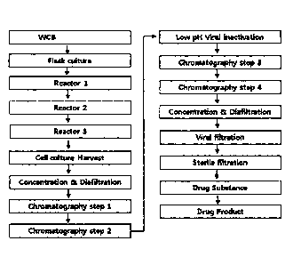
[57] FIG. 3 is a flow chart illustrating the isolation and purification of
IDS from
transformed CHO-DG44.
[58] FIG. 4 is a photograph showing an SDS-PAGE result of IDS for analyzing
the N-
terminal sequence where a marker was run on lane M, glycosylated IDS on lane
1,
PNGase F on lane 2, and deglycosylated IDS on lane 3.
[59] FIG. 5 is a flow chart illustrating the process of analyzing the amino
acid sequence of
IDS.
[60] FIG. 6 is a view showing the amino acid sequence of the IDS of the
present invention
as analyzed by MALDI-MS/MS and LC-ESI-MS/MS.
[61] FIG. 7 is an RP-HPLC chromatogram of non-reduced and reduced IDS
samples
showing the position of disulfide bonds in IDS.
[62] FIG. 8 is a view showing the positions of disulfide bonds in the IDS
of the present
invention as analyzed by MALDI-MS.
CA 02839674 2013-12-17
10
WO 2012/177020 PCT/KR2012/004734
[63] FIG. 9 is a view showing the positions of disulfide bonds in the IDS
of the present
invention as analyzed by MALDI-MS/MS.
[64] FIG. 10 is a view indicating the positions of disulfide bonds in the
IDS of the present
invention, obtained through MALDI-MS/MS.
[65] FIG. 11 is a photograph showing IDS run by SDS-PAGE after treatment
with various
glycoside hydrolase enzymes to examine the glycosylation of the IDS of the
present
invention.
[66] FIG. 12 is of HPAEC-PAD chromatograms showing the content of mannose-
6-phosphate in the IDS of the present invention.
[67] FIG. 13 is a size exclusion chromatogram showing the purity of the IDS
of the
present invention.
[68] FIG. 14 is an ion chromatogram showing the catalytic activity of the
IDS of the
present invention on a natural substrate.
[69] FIG. 15 is Lineweaver-Burk plot showing ratios of cellular uptake
amounts of IDS
relative to amount of IDS added to normal fibroblast cells.
[70] FIG. 16 is a graph showing the amount of the IDS of the present
invention in-
ternalized into normal human fibroblast cells and the cells of patients
suffering from
Hunter syndrome.
[71] FIG. 17 is a view showing measurements of the formylglycine content in
the IDS of
the present invention.
[72] FIG. 18 is a view showing IEF (isoelectric focusing) points of the IDS
of the present
invention before and after cation exchange chromatography wherein M is run on
M
lane, a loaded sample for cation exchange chromatography on lane 1, an eluate
of
cation exchange chromatography on lane 2, and a regeneration solution after
cation
exchange chromatography on lane 3.
Mode for the Invention
[73] A better understanding of the present invention may be obtained
through the
following examples which are set forth to illustrate, but are not to be
construed as
limiting the present invention.
[74] EXAMPLE 1: Preparation of IDS
[75] <1-1> Gene Acquisition
[76] Peripheral blood mononuclear cells (PBMC) were isolated from human
blood as
described previously [S. Beckebaum et al., Immunology, 2003, 109:487-4951.
Total
RNA was extracted from the PBMC according to a protocol described previously
[M.
J. Holland et al., Clin. Exp. Immunol., 1996, 105:429-4351. In order to
construct a
cDNA library from the total RNA, single-stranded cDNA was synthesized using
oligo-
(dT) primer with the aid of a single-strand synthesis kit (Boehringer
mannheim). In this
CA 02839674 2013-12-17
CA 02839674 2015-07-09
11
WO 2012/177020 PCT/KR2012/004734
TM
regard, DEPC-treated distilled water was added to an eppendorf tube containing
I jig
of the total RNA so as to form a final volume of 12.5 fig. Then, 1 jig of a 20
pmol
oligo(dT) primer was added to the tube, followed by incubation at 70 C for 2
min and
cooling. To this reaction mixture were added 4 jig of a reaction buffer, 1 jig
of dNTP,
jig of an RNase inhibitor, and 1 jig of reverse transctiptase which were then
reacted at
42 C for one hour to synthesize single stranded cDNA. PCR was performed on the
cDNA as a template in the presence of primers of SEQ ID NOS: 2 to 4 to amplify
a
human IDS gene. In this context, each primer was designed to contain a
restriction
enzyme recognition site for use in gene cloning.
[77] <1-2> Construction of Expression Vector
[78] A. Construction of pJK-dhfr-1DS-S1 Vector
[79] A light chain signal sequence of an antibody (derived from a part of
the human IgG
light chain) as a non-coding sequence was introduced into the 5'-terminus of
the IDS
gene acquired by Example <1-1> before PCR. After the PCR product obtained
thereby
was run on gel by electrophoresis, the human IDS gene was isolated using a gel
ex-
traction kit. The isolated IDS gene and the pJK-dhfr-0r2 vector (Aprogen) were
digested with EcoRV and ApaI and ligated to each other at 16 C for 20 hours.
The re-
combinant vector thus constructed was transformed into E. coli (DH5a) which
was
then spread over an LB plate containing 50 fig/mL ampicillin and incubated
overnight.
Colonies grown on the plates were selected and cultured so as to isolate the
plasmid
therefrom (FIG. 1).
[80] B. Construction of Recombinant Human IDS Expression Plasmid
[81] In order to change the non-coding sequence of the plasmid constructed
above to a
signal sequence, the recombinant human IDS was subcloned to a pJK-dhfr-or2
vector.
To this end, the pJK-dhfr-IDS-S1 vector was digested with EcoRV and ApaI to
give a
partial IDS gene (1233 bp) which was then inserted into the pJK-dhfr-0r2
vector
previously treated with the same restriction enzymes, to construct a pJK-dhfr-
IDS-S2
vector. In order to introduce non-coding sequence and a signal sequence to the
5'-terminus, an IDS Ni forward primer (SEQ ID NO: 5) and an IDS 4 reverse
primer
(SEQ ID NO: 7) were used for PCR with the pJK-dhfr-IDS-S1 vector serving as a
template. After starting at 94 C for 5 min, PCR was performed with 30 cycles
of 94 C
for I min, 55 C for 30 sec and 72 C for 40 sec and finished by extension at 72
C for 10
mm.
[821 The PCR amplification afforded a partial IDS gene that was 448 bp.
This gene was
used as a template for the PCR which was performed again in the presence of an
IDS
N2 forward primer (SEQ ID NO: 6) and an IDS 4 reverse primer (SEQ ID NO: 7)
under the same conditions as described above. This resulted in the synthesis
of a DNA
fragment 476 bp long.
12
WO 2012/177020 PCT/KR2012/004734
[83] Subsequently, the pJK-dhfr-IDS-52 vector and the recombinant human IDS
gene
fragment (476 bp) were separately digested with EcoRV. The digests were
separated
on gel by electrophoresis to obtain the vector and the 476 bp-long IDS
fragment. These
vector and insert were ligated at 16 C for 12 hours in the presence of T4 DNA
ligase to
construct pJK-dhfr-0r2-IDS plasmid. These procedures are illustrated in FIG.
2.
[84] To confirm the construction of the IDS expression plasmid, DH5 was
transformed
with pJK-dhfr-0r2-IDS and cultured for 24 hours on an LB plate containing
ampicillin
(50 ,ug/mL). From the colonies thus formed, a plasmid was isolated and
digested to
measure the size of the insert. Also, base sequencing was conducted using a T7
primer
(SEQ ID NO: 8).
[85] <1-3> Selection of Recombinant Human IDS Expression Strain
[86] A. Transfection of CHO-DG44
[87] CHO-DG44 was used as a host cell for expressing the IDS of the present
invention.
The mutant Chinese hamster ovary cell CHO-DG44 carries a double deletion for
the
endogenous dhfr (dihydrofolate reductase) gene which encodes DHFR enzyme. The
DHFR enzyme is involved in the conversion of folate through dihydrofolate
(FH2) into
tetrahydrofolate (FH4) which is involved in the de novo synthesis of nucleic
acids. The
level of dhfr in the cells is dependent on the concentration of MTX. MTX,
which is
structurally similar to folic acid, a substrate of DHFR, competes with folic
acid for
binding dihydrofolate reductase, so that most dihydrofolate reductase loses
its activity
in the presence of MTX. Hence, if cells do not amplify a sufficient amount of
dhfr,
they die because they cannot synthesize nucleic acids necessary for their
life. In
contrast, if the amplification is sufficient, the cells can survive under a
high con-
centration of MTX because they are relatively abundant in dhfr. This system
may be
applied to animal cells to select a transfected cell line which can amplify
the dhfr gene
and thus a structural gene of interest.
[88] To this end, a dhfr gene was introduced as an amplifiable marker into
the IDS ex-
pression vector pJK-dhfr-0r2-IDS, constructed in Example 1-2, and gene
amplification
was conducted using MTX and the dhfr gene.
[89] In this regard, the DG44 cell line (obtained from Dr. Chaisin,
Columbia University)
was suspended in 10 mL of DMEM/F12 (supplemented with nucleotides and nu-
cleosides, and 10% fetal bovine serum (FBS)) and harvested by spinning at 1000
rpm
for 5 min. The cells were inoculated into 50 mL of a culture medium in a T-175
flask
and incubated at 37 1 C in a 5 1% CO2 incubator. One day before transfection,
the
culture medium for DG44 cells was removed from the T-175 flask and the cells
were
washed twice with PBS and detached by trypsinization. Then, they were seeded
at a
density of 5x10⁵ cells into a T-25 flask and cultured at 37 1 C for 24 hours
in a 5 1%
CO2 incubator. Bacterial or fungal contamination was examined under an optical
mi-
CA 02839674 2013-12-17
CA 02839674 2015-07-09
13
WO 2012/177020 PCT/KR2012/004734
croscope while PCR-ELISA was performed to examine whether the cells were con-
taminated with mycoplasma.
[90] The germ-free DG-44 cells were transfected with the IDS expression
vector pJK-
dhfr-0r2-I DS, constructed in Example 1-2, using a Lipofectamine kit. In this
regard, 5
fig of the expression vector and 50 gig of Lipofectamine were separately
diluted in 800
TM
fig of Opti-MEM I, rnixed carefully so as not to form bubbles, and left at
room tem-
perature for 15 mm. Meanwhile, DG44 cells were washed once with sterile PBS
and
TM
three times with Opti-MEM i. To the DG44 cells were carefully added the DNA-
TM
lipofectamine mixture and then 6.4 mL of Opti-MEM before incubation at 37 1 C
for
hours in a 5 1% CO2 incubator. Thereafter, the incubation was conducted for an
ad-
ditional 48 hours in the medium supplemented with 8 mL of DMEM/F12 and 1.6 mL
of FBS to promote the recovery of cell membranes and the growth of cells.
TM
[91] B. Selection of Geneticin(G418)-Resistant Cell Strain
[92] The cultured cells were detached with 0.25% trypsin, counted, and
seeded at a
density of 5x10³ cells/well into 96-well plates containing 100 fig of MEM-
alpha
medium (supplemented with 10% dialyzed FBS and 550 yg/mL 0418) per well. Next
day, the same medium was added in an amount of 100 dug/well and the cells were
cultured for 2-3 weeks to form colonies. When the cells grew to 50%
confluency, the
medium was replaced with a fresh one. After maintenance for 3 days, the
culture media
were collected for enzyme analysis.
[93] The medium was replaced with 200 jig of a fresh medium every three
days. On day
3-4 after culturing, non-transfected cells, that is, cells that were not
resistant to
geneticinTM started to detach from the bottom of the 96-well plates when
observed with
an optical microscope. The selected clones were cultured while being
sequentially
transferred from the 96-well plates to 24-well plates, 6-well plates and 100-
mm dishes
in the order. When the cells grew to 80-90% confluency in 100-mm dishes, the
ex-
pression level was measured again. The cells were detached with 0.25% trypsin,
counted and plated at a density of 5x10⁵ cells/well/3 mL into 6-well plates,
maintained
for 3 days and counted. The expression level of the protein was quantitatively
analyzed. According to the analysis results, 15 clones were selected.
[94] C. Selection of IDS Expression Strain with High Expression Capacity
[95] The 15 selected clones were cultured at an increased concentration of
MTX to select
cell strains in which IDS was amplified.
1961 In this context, the cells were inoculated at a density of 1x10⁶
cells/100 mm dish/10
mL of a medium containing MTX and cultured to 80-90% confluency. One tenth of
the volume of the cell culture was inoculated again into 100 mm dish/10 mL.
This sub-
culturing process was repeated twice. The cells were allowed to undergo at
least three
passages so that they were sufficiently adapted to increased MTX
concentrations. The
14
WO 2012/177020 PCT/KR2012/004734
concentration of MTX was increased, from 5 nM for the clones selected after
conducting an analysis for the first three days, to 20 nM. In each step, the
clones
adapted to the increased MTX concentration were cultured for three days to
measure
cell growth rates. IDS expression levels were measured to select cell strains
in which
the amplification of the IDS gene took place, that is, cell strains in which
the re-
combinant IDS was expressed at a high rate. Of the selected cell strains, NI4
was used
in subsequent experiments because it had the highest expression level.
[97] D. Selection of Single Strain by Limiting Dilution
[98] There was the possibility that the strain NI4 might have become mixed
with other
strains. Hence, the strain was separated into a single strain. The N14 clones
which
survived 20 nM MTX were subcloned through limiting dilution so as to select a
desired cell strain.
[99] First, NI4 was inoculated at a density of 0.5 cells/well into IMDM
medium (Gibco
BRL, Cat#12200) in 96-well plates and cultured with the medium replenished
every
three days. On day three, the plates were observed under a microscope to
exclude the
wells in which two or more colonies had been formed per well. The wells in
which
only one colony had formed per well were selected and continued to be
cultured. After
culturing for 15 days, the cells were sub-cultured to 96-well plates and when
cells had
grown to 90% confluency, the medium was freshly replenished.
[100] A total of 263 single strains were identified from the N14 strain. Of
them, strain S46
was found to have the highest IDS activity and named NI4-546.
[101] <1-4> Cell Culture
[102] A. Shaker Flask Culture
[103] The NI4-546 strain was cultured on a large scale to produce the IDS
of the present
invention. The strain was inoculated into an EX-cell 302 serum-free medium
(containing glutamine, dextran sulfate, and pluronic F-68) in 125 mL culture
flasks and
cultured at 37 1 C in a 5 1% CO2 incubator. Subsequently, the cells were
passaged at
a ratio of 1:5-1:8 every two to three days using shaker flasks. Upon the
passage, the
culture volume was gradually increased to approximately 2,400 mL. In many
shaker
flasks, the cells were cultured to a level sufficient to be inoculated into a
fermentor.
[104] B. Culture in 30L Fermentor (working volume 20L)
[105] When the density of the cells in the shaker flasks reached 1.3x10⁶
cells/mL, they
were inoculated into a 30L fermentor. During cell culturing, the culture
conditions
were kept at a dissolved oxygen content of 10% or higher, a culture
temperature of
37 1 C and a pH of 7.0 0.2. If necessary, cell samples were taken and observed
under
a microscope. The cell culture was examined to analyze cell count, cell
viability, pH,
glucose concentration and glutamine concentration. On the basis of the
analysis results,
when it was decided that the cells were sufficiently grown, the cells were
inoculated
CA 02839674 2013-12-17
15
WO 2012/177020 PCT/KR2012/004734
into a 150L fermentor.
[106] C. Culture in 150L Fermentor (working volume 100L)
[107] When the cells in a 30L fermentor reached a density of 0.9x10⁶
cells/mL or higher,
they were inoculated into a 150L fermentor. During cell culturing, the culture
condition was kept at a dissolved oxygen content of 10% or higher, a culture
tem-
perature of 37 1 C and a pH of 7.0 0.2. If necessary, cell samples were taken
and
observed under a microscope. The cell culture was examined to analyze cell
count, cell
viability, pH, glucose concentration and glutamine concentration. On the basis
of the
analysis results, when it was decided that the cells were sufficiently grown,
the cells
were inoculated into a 650L fermentor.
[108] D. Culture in 650L Fermentor (working volume 500L)
[109] When the cells in a 150L fermentor reached a density of 0.9x10⁶
cells/mL or higher,
they were inoculated into a 650L fermentor. During cell culturing, the culture
condition was kept at a dissolved oxygen content of 10% or higher, a culture
tem-
perature of 34 1 C and a pH of 6.9 0.2 for three days and then, at a culture
tem-
perature of 32 1 C and a pH of 6.9 0.2. If necessary, cell samples were taken
and
observed under a microscope to analyze cell counts, cell viability, pH,
glucose concen-
trations and glutamine concentrations. Depending on the analysis result,
glucose and
glutamine concentrations were adjusted to continue cell growth. During the fer-
mentation, a hydrolysate was added to increase the formylglycine conversion.
[110] <1-5> Purification of IDS
[111] IDS was isolated from the cell culture using a series of the
following four chro-
matographic processes.
[112] A. Harvest and Filtration of Culture Medium
[113] When the cell viability remained in the range of 80-85% 10 days after
inoculation
into the 650 L fermentor, culturing was stopped. The cells were harvested from
the
culture using the Millipore POD filter system and DOHC filter (Millipore) at a
pressure
of 0.9 bar or less. After the cells were removed, the supernatant was filtered
through a
pre-filter (Millipore, 0.5 + 0.2 gm) and a 0.45+0.2 gm filter and recovered in
a
disposable sterile vinyl bag. The harvested culture solution was stored at 2-8
C.
[114] B. Concentration and Diafiltration
[115] The filtrate recovered in A was about 10-fold concentrated using an
ultrafiltration
system (Tangential Flow Filtration Membrane System). The membrane (cutoff:
30K,
Pall) installed inside the ultrafiltration system was washed with WFI (water
for
injection) at a flow rate of 20-25 L/min and then equilibrated with a buffer
(pH
7.0-8.0) containing 20 mM sodium phosphate (sodium dihydrogen phosphate
monohydrate and sodium hydrogen phosphate heptahydrate). After equilibration,
the
filtrate was fed into the membrane while recovering the fractions that did not
pass the
CA 02839674 2013-12-17
16
WO 2012/177020 PCT/KR2012/004734
membrane. Once the recovered volume became about 1/10 of the initial volume of
the
filtrate, the concentration procedure was stopped. The buffer was
consecutively
exchanged in a volume three to four times as large as that of the concentrate.
If the
conductivity and the pH fell within the criteria, the process was stopped.
[criteria - con-
ductivity: =5.0 mS/cm, pH 7.0-8.01.
[116] C. Anion exchange chromatography
[117] To remove dyes and various impurities from the concentrate recovered
in B, anion
exchange chromatography was conducted on a column (GE Healthcare) filled with
Q
Sepharose resins (GE Healthcare). The column was equilibrated with equilibrium
buffer (pH 7.0-8.0) containing 20mM sodium phosphate (sodium dihydrogen
phosphate monohydrate and sodium hydrogen phosphate heptahydrate). The con-
centrate obtained in B was filtered through a 0.45+0.2 gm filter (Sartorius)
and loaded
at a flow velocity of 100-120 cm/h into the equilibrated column. After the
loading was
completed, the column was primarily washed with the equilibrium buffer and
then with
washing buffer (pH 5.5-7.5) containing sodium chloride. Subsequently, a target
protein was eluted with an eluting buffer (pH 5.5-7.5) containing sodium
chloride.
[118] D. Hydrophobic chromatography
[119] To remove the dyes and impurities that remained after anion exchange
chro-
matography, hydrophobic chromatography was performed on a column (GE
Healthcare) filled with phenyl Sepharose resins (GE Healthcare). The column
was
equilibrated with equilibrium buffer (pH 5.0-7.0) containing sodium chloride.
The
eluate obtained in C was filtered through a 0.45+0.2 gm filter (Sartorius) and
loaded at
a flow velocity of 70-100 cm/h into the equilibrated column. After the loading
was
completed, the column was washed with the equilibrium buffer. Subsequently, a
target
protein was eluted with an eluting buffer (pH 5.0-7.0) containing glycerol.
[120] E. Inactivation of Virus by Low pH
[121] Viruses that may be derived from host cells or any material used in
the processes
carried out were inactivated by a low pH condition. In this regard, the eluate
obtained
in D was maintained for 2 hours at a pH 3.0 - 4.0 to which its acidity was
adjusted
with 25% acetic acid. Thereafter, the pH of the eluate was increased to 4.0-
5.0 using
0.5 M sodium hydroxide for use in the next process. The inactivation by low pH
was
conducted at 12 2 C.
[122] F. Cation Exchange Chromatography
[123] IDS is glycoprotein with oligosaccharides, and exists as an isomer
that has a different
isoelectric point according to the content of sialic acid at the end of the
Glycan chain.
As oligosaccharides with a negative charge, sialic acid shows a difference in
terms of
the degree of binding to cation exchange resin according to the content of
sialic acid.
Using this characterization, cation exchange chromatography was conducted to
obtain
CA 02839674 2013-12-17
17
WO 2012/177020 PCT/KR2012/004734
IDS showing high activity (a high content of formylglycine) with a high
content of
sialic acid and to remove other impurities [Product impurity (Aggregated IDS,
processed IDS), process impurity (Host Cell protein)]. In detail, a column
filled with
cation exchange CaptoTM MMC resins (GE Healthcare) was equilibrated with
glycerol-
added equilibration buffer (pH 4.0 - 5.0). The inactivated eluate obtained in
E was
filtered through a 0.45+0.2 gm filter (Sartorius) and loaded at a flow
velocity of
100-120 cm/h onto the equilibrated column. Subsequently, the column was washed
with the equilibration buffer, followed by elution with glycerol-added eluting
buffer
(pH 4.0-6.0) to give IDS with a high sialic acid content (isoelectric point
3.5 or less),
high activity (formylglycine content: 80 15%) and high purity (SE-HPLC, 98% or
higher).
[124] G. Affinity chromatography
[125] Affinity chromatography (Blue Sepharose, GE Healthcare) was conducted
to remove
the glycerol used in the cation exchange chromatography and to reduce the
volume of
the eluate. The eluate obtained in F was filtered through a 0.45+0.2 gm filter
(Sartorius)
and loaded at a flow velocity of 100-120 cm/h onto a Blue Sepharose resin-
filled
column (GE Healthcare) that was previously equilibrated with glycerol-added
equi-
libration buffer (pH 4.5-5.5). After completion of the loading, the column was
washed
with washing buffer (pH 4.5-5.5) and the target protein was eluted with
eluting buffer
(pH 6.0 - 8.0).
[126] H. Concentration and Buffer Exchange
[127] An ultrafiltration system (Tangential Flow Filtration Membrane
System) was used to
adjust the protein concentration of the eluate obtained in G and to exchange
the buffer
of the purified protein with formulation buffer. The membrane (cutoff: 10K,
Pall)
installed inside the ultrafiltration system was washed with WFI (water for
injection) at
a flow rate of 450-650 mL/min and then equilibrated with a formulation buffer
(2.25
g/L sodium dihydrogen phosphate monohydrate, 0.99 g/L sodium hydrogen
phosphate
heptahydrate, 8 g/L sodium chloride, pH 6.0 - 7.0) without polysorbate 20,
followed
by concentrating the target protein. The buffer was consecutively exchanged in
a
volume three to four times as large as that of the concentrate. If the
conductivity and
the pH fell within the criteria, the process was stopped. [criteria -
conductivity:
15.0 3.0 mS/cm, pH 6.0-7.01. Adjust the content of the concentrated solution
to 4.0
0.5 mg/mL.
[128] I. Nanofiltration
[129] Using a nano filter (NFP, Millipore), nano filtration was performed
to remove viruses
that might have come from the host cells or any of the materials used.
Integrity test for
filter is performed after washing the nano filter with water for injection.
Once the
integrity test was passed, the nanofilter was equilibrated with 1 L of
formulation buffer
CA 02839674 2013-12-17
18
WO 2012/177020 PCT/KR2012/004734
(2.25 g/L sodium dihydrogen phosphate monohydrate, 0.99 g/L sodium hydrogen
phosphate, 8 g/L sodium chloride, pH 6.0-7.0) without polysorbate 20. After
completion of equilibration, the concentrate obtained in H was passed through
the filter
at a pressure of about 2 bar to produce a nano-filtrate. After filtration was
completed,
the filter was washed with the formulation buffer (post wash solution). After
combining the nano filtration solution and the post wash solution, protein
content is
measured.
[130] J. Drug Substance
111311 The protein concentration of the filtrate obtained in I was adjusted
with formulation
buffer without polysorbate 20. After the addition of polysorbate, the solution
was
filtered through a 0.2 gm filter to produce a drug substance. The drug
substance was
aliquoted and stored in a deep freezer (-70 10 C) until use.
[132] K. Drug Product (Filling, labeling. Packaging)
[133] The stock stored in a deep freezer was thawed in a water bath
maintained at 28 1 C
and diluted to a protein concentration of about 2.05 0.2 mg/mL using
formulation
buffer (2.25 g/L sodium dihydrogen phosphate monohydrate, 0.99 g/L sodium
hydrogen phosphate heptahydrate, 8 g/L sodium chloride, 0.23 g/L polysorbate
20, pH
6.0-7.0). Thereafter, the dilution solution was filtered through a 0.2 gm
filter to
produce a final bulk solution. This final bulk solution was filled in 6 mL
vial with ap-
proximately 3.3 g using auto filling. Once an vial inspection test was passed,
the vials
were packed to produce a drug product.
[134] The procedure from strain culturing to final product production is
illustrated in FIG.
3.
[135] COMPARATIVE EXAMPLE 1: Preparation of Elaprase
[136] Elaprase commercially available recombinant IDS, was used as a
comparative
example.
[137] EXPERIMENTAL EXAMPLE 1: Structural Analysis and Characterization of
Inventive IDS
[138] <1-1> Amino acid sequencing - Internal sequencing
[139] Deglycosylated IDS was separated by SDS-PAGE, followed by gel
slicing. Then,
digests resulting from treatment with various endoprotenases (trypsin,
chymotrypsin,
AspN, chymotrypsin/trypsin, AspN/trypsin, GluC and GluC/trypsin) were analyzed
using MALDI-MS/MS and LC-ESI-MS/MS (FIG. 5). As a result, a total of 525 amino
acid sequences were identified. The amino acid sequences coincided with the
the-
oretical sequence of human IDS (FIG. 6).
[140] <1-2> Disulfide Bond Analysis
[141] In a polypeptide, a disulfide bond is a covalent linkage, usually
derived by the
coupling of two SH groups of cysteine residues, playing an important role in
sta-
CA 02839674 2013-12-17
19
WO 2012/177020 PCT/KR2012/004734
bilizing the higher structure of proteins. Theoretically, the 525 amino acids
of IDS
contain six cysteine residues, four of which form disulfide bonds. In this
example, the
location of cysteine residues responsible for the disulfide bonds of IDS was
identified.
First, IDS was deglycosylated by treatment with PNGase F to exclude the
interference
of sugars. In order to prevent the cysteine residues that do not take part in
the
formation of disulfide bonds from acting as an interfering factor, 4-
vinylpyridine was
used to convert IDS into a non-reduced sample so that the SH groups are
restrained
from randomly forming S-S bonds. Meanwhile, the disulfide bonds were cleaved
by
DTT, followed by blocking with 4-vinylpyridine to give a reduced sample.
Trypsin and
AspN, selected on the result of Experimental Example 1-3, were applied to the
non-
reduced and the reduced sample. The peptide fragments thus obtained were
separated
by RP-HPLC. RP-HPLC chromatograms of the non-reduced and the reduced samples
were compared so as to discriminate the peaks that were found in the non-
reduced
sample, but not in the reduced sample (FIG. 7).
[142] For more exact analysis, fractions at the discriminated peaks were
reduced in size by
additional treatment with endoproteinases, and the peaks containing disulfide
bonds
were analyzed using MALDI-MS (FIG. 8).
[143] Peaks with disulfide bonds were again sequence analyzed using MALDI-
MS/MS
(FIG. 9) to examine the positions of cysteine residues that form disulfide
bonds among
the 525 IDS amino acid residues. As shown in FIG. 10, disulfide bonds were
observed
to form between C146-C159 and between C397-C407.
[144] <1-3> Analysis of Formylglycine Content
[145] IDS degrades heparan sulfate and dermatan sulfate, both of which are
a kind of gly-
cosaminoglycan(GAG). This degradation activity is not acquired until the
cysteine
residue at position 59 in the active site (Cys59) is converted into
formylglycine (FGly)
by post-translational modification. Thus, the degradation activity of IDS was
analyzed
by examining the post-translational modification of Cys59 to FGly. For this
analysis,
AQUA (absolute quantification), a quantitative analysis method based on MS
(Mass
Spectroscopy), was used, in which a radio-labeled synthetic substrate (AQUA
peptide)
was spiked into a sample. To quantitatively analyze formylglycine at Cys59
position, a
serial dilution of AQUA peptide was spiked into a sample and a calibration
curve was
drawn. Ratios of FGly-type peptide to Cys-type peptide were measured by LC-ESI-
MS
analysis, and applied to the AQUA calibration curve to calculate the content
of
formylglycine.
[146] This analysis determined the conversion of Cys59 to FGly at a rate of
80 15%. In
consideration of the Cys50 to FGly conversion rate of about 50% in the
commercially
available agent Elaprase (Elaprase Science Discussion, EMEA, 2007; Genet Med
2006:8(8):465-473), the therapeutic composition comprising the IDS of the
present
CA 02839674 2013-12-17
20
WO 2012/177020
PCT/KR2012/004734
invention and the formulation prepared with the composition is anticipated to
have
much higher therapeutic activity compared to Elaprase.
[147] <1-4> Identification of Glycosylation Pattern
[148] An assay was performed to examine whether the IDS of the present
invention is gly-
cosylated and to identify the glycosylation pattern if any. To this end, IDS
was treated
with various glycoside hydrolase enzymes, the digests were separated on by SDS-
PAGE and their motility patterns were analyzed.
[149] In detail, IDS samples were digested with combinations of the
following four
glycoside hydrolase enzymes and separated by SDS-PAGE.
[150] TABLE 1. Properties of Sugar Cleaving Enzymes
Function /Property
- Cleaves a sugar moiety (N-glycan) from
protein
PNGase F
- Asn at the cleavage site is converted into
Asp
- Cleaves a sugar moiety (N-glycan) from
protein
Endo H - unlike PNGase F, Endo H acts on
oligosaccharides of high-mannose type and
hybrid type
0- -
Cleaves a sugar moiety (0-glycan) from
Glycosidase protein
- Cleaves terminal sialic acid residues of
Sialidase
N-glycan or 0-glycan
[151] As can be seen in FIG. 11, the IDS of the present invention was
cleaved by PNGase
F and Endo H, but not by 0-glycosidase, indicating that the IDS of the present
invention is an N-glycosylated protein. In addition, the IDS was completely
cleaved by
PNGase F, but its size reduction was slight upon treatment with Endo H. PNGase
F
acts on the glycosylation sites of all the three patterns whereas Endo H acts
on the gly-
cosylation sites of high-mannose type and hybrid type. Taken together, these
results
indicate that the IDS contains the three glycosylation patterns complex, high-
mannose
and hybrid.
[152] <1-5> Analysis of Mannose-6-phosphate Content
[153] Binding to a M6P receptor on cells, mannose-6-phosphate (M6P) allows
IDS to be
internalized into cells and thus to hydrolyze heparan sulfate or dermatan
sulfate in
lysosomes. In this Example, IDS was acid hydrolyzed with trifluoroacetic acid
(TFA)
and subjected to HPAEC-PAD (Bio-LC) to quantitatively analyze mannose-
CA 02839674 2013-12-17
21
WO 2012/177020 PCT/KR2012/004734
6-phosphate.
[154] IDS was hydrolyzed with 6.75M TFA and the hydrolysate was analyzed
using liquid
chromatography (High Performance Anion-Exchange Chromatography with Pulsed
Amperometric Detection; HPAEC-PAD). M6P concentration of which was already
known was analyzed under the same condition, and molar ratios of M6P to gly-
coprotein were obtained by comparison of the areas. Analysis was conducted in
triplicate. M6P standard materials and M6P composition chromatograms of the
IDS are
shown in FIG. 12 and the molar ratios of M6P are summarized in Table 2, below.
[155] TABLE 2 Analysis Results for Mannose-6-phosphate Content
[156]
M-6-P Amount Amount Ratio
Run No. Ret.Time pmo1/251fl11 pmo1/25m11 M-6-P/protein
(min) 14-6-P Protein (mol/mol)
13 11.25 1320.59 428 3.09
14 11.23 1241.31 428 2.90
15 11.23 1245.83 428 2.91
AVG 11.24 1269.25 428 2.97
CV , 0.09% 3.51 0.11
[157] As is understood from the data of Table 2, there are approximately 3
moles of M6P
per mole of IDS. From these results, it is inferred that the therapeutic
composition
comprising the IDS of the present invention and the formulation prepared with
the
composition have a high ability to catabolize GAG accumulated in lysosomes.
[158] <1-6> Mass Analysis
[159] Masses of glycosylated IDS and deglycosylated IDS were measured using
MALDI-
TOF-MS. Treatment of glycosylated IDS with PNGase F afforded deglycosylated
IDS.
MALDI-TOF-MS was performed using Voyager-DE PRO Biospectrometry (Applied
Biosystems, USA) coupled with a delayed Extraction laser-desorption mass spec-
trometer. The instrument was normalized with bovine serum albumin and IgGl.
Analysis results are summarized in Table 3, below.
[160] TABLE 3. MALDI-TOF-MSMALDI-TOF-MS Analysis Results of IDS
[161]
CA 02839674 2013-12-17
22
WO 2012/177020 PCT/KR2012/004734
Glyeosylated IDS
m/z charge(z) Protein Mass(Da) remark
25646 3 76935
38708 2 77414
77360 1 77359
154533 1 77266 dimer
Average 77244 210
Deglycosylated IDS
m/z charge(z) Protein Wass(Da) remark
29767 2 59532
34655 PNGase F
59313 1 59312
118706 1 59353 dimer
Average 59399 120
Sample Rolecular Weight
Theoretical 59298 Da
Glycosylated 77244 -1- 210 Da
Deglycasylated 59399 - 120 Da
=
[162] As apparent from the data of Table 3, the molecular size is 77,244 Da
for gly-
cosylated IDS and 59,399 Da for deglycosylated IDS, which is similar to the
molecular
weight calculated on the basis of the amino acid sequence, which is 59,298 Da.
[163] <1-7> Purity Measurement
[164] The purity of IDS was measured using size exclusion chromatography.
Size
exclusion chromatography is a chromatographic method in which molecules in
solution are separated by their relative molecular weight and shape. In size
exclusion
chromatography, proteins larger than the pore size of the column cannot
penetrate the
pore system and pass through the column at once. Subsequently, the analytes
with
CA 02839674 2013-12-17
23
WO 2012/177020 PCT/KR2012/004734
smaller molecular weights or sizes elute later. For this chromatography,
Alliance 2695
HPLC system (Waters, WI, USA) coupled with 2487 UV/VIS detector (Waters, WI,
USA) was employed. Proteins were detected at 214 nm, and analyzed using
Empower
2 Software. The analytes were loaded onto a TSK G30005WXL column linked to a
TSK SWXL guard column (Tosoh, Japan). IDS, after being diluted to a
concentration
of 1.0 mg/mL in a formulation buffer, was loaded in a volume of 10 gm onto the
column. They were allowed to flow with mobile phase (20 mM sodium phosphate
buffer, 200 mM NaC1, pH 7.0) at a flow rate of 0.5 mL/min for 60 mM.
[165] Analysis results are shown in FIG. 13. As can be seen, IDS monomers
had a
retention time of approximately 16.4 mM, and were eluted with 100% purity.
[166] <1-8> Activity Measurement Using Synthetic Substrate
[167] The reaction of IDS with the synthetic substrate
(4-methylumbelliferyl-L-iduronide-2-sulfate Na2 (MU-IdoA-25) for 4 hours
releases
the sulfate moiety (primary reaction). After the primary reaction, the
addition of LEBT
(lysosomal enzymes purified from bovine testes) induces a secondary enzymatic
reaction with the substrate 4-methylumbellifery-L-iduronide (reactant left
after the
release of the sulfate moiety in the primary reaction) to separate the
4-methylumbelliferyl moiety from the L-iduronide moiety. Because the remaining
4-methylumbelliferyl is fluorogenic, the activity of IDS was evaluated by
measuring
the intensity of fluorescence (Ex.355nm/Em.460nm). The IDS of the present
invention
was found to range in specific activity from 19 to 55 nmol/min/gg. This
activity
indicates that formylglycine exists in the active site of the enzyme as a
result of the
post-translational modification of the cysteine residue at position 59 in IDS.
[168] <1-9> Activity Measurement Using Natural Substrate
[169] In order to determine whether the reaction with the IDS and natural
substrate, the
sulfate ions released from the substrate (heparin disaccharide) by reaction
with IDS
were measured. The reaction mixture was loaded onto an ion column (Vydac
302IC)
and allowed to flow with the mobile phase of 0.49 g/L phthalic acid at a flow
rate of 2
ml/min, during which free sulfate ions were detected at 290 nm in negative
mode.
[170] As shown in FIG. 14, the IDS was confirmed to hydrolyze sulfate ion
from heparin
disaccharide, indicating that the IDS is capable of degrading 0-linked sulfate
of
dermatan sulfate and heparan sulfate in vivo.
[171] <1-10> In vivo Cellular Uptake Activity
[172] The cellular internalization activity of the IDS was measured using
the normal fi-
broblast cells and Hunter syndrome patient cells. In this regard, normal
fibroblast cells
and Hunter syndrome patient cells (obtained from Samsung Medical Center,
Seoul,
Korea) were cultured and allowed to be internalized into cells while they were
incubated with various concentrations of IDS at 37 C for 20 hours in a 5% CO2
CA 02839674 2013-12-17
24
WO 2012/177020
PCT/KR2012/004734
incubator. After being harvested, the cells were lyzed, and the level of the
IDS in-
ternalized into the cells was determined in the lysate.
[173] On the basis of the concentration ratio of internalized IDS to IDS
added to the normal
fibroblast cells, a Michaelis-Menten graph and a Lineweaver-Burk plot were con-
structed from which Kuptaiõ (IDS concentration at which the reaction rate is
half of the
maximum rate achieved at saturating substrate concentrations) was calculated.
Kuptaiõ
was calculated to be 18.0 nM or less, indicating that IDS is internalized into
cells by
the binding of the M6P of IDS to M6P receptors on the cell surface (FIG. 15).
[174] Also, the cellular uptake and activity of IDS in Hunter syndrome
patient cells as well
as normal human fibroblast cells were analyzed. The uptake and activity of the
IDS
were increased in both the cells, demonstrating that the IDS of the present
invention is
more efficiently internalized into cells (FIG. 16).
[175] EXPERIMENTAL EXAMPLE 2: Clinical Analysis for Effect of IDS
[176] Thirty one patients with Hunter syndrome were divided into three
groups, ad-
ministered with the IDS of the present invention and analyzed for parameters
as-
sociated with Hunter syndrome. Elaprase a commercially available therapeutic
agent
for Hunter syndrome, was used as a positive control.
[177] <2-1> Change in Urine GAG Level (primary check parameter for validity
test)
[178] The three groups of Hunter syndrome patients were administered for 24
weeks with
Elaprase (0.5 mg/kg) and the IDS of the present invention (0.5 mg/kg and 1.0
mg/kg),
and urine GAG (Glycosaminoglycan) levels were measured as reported previously
(Conn. Tissue Res. Vol.28, pp317-324, 1990.; Ann. Clin. Biochem. Vol.31,
pp147-152, 1994). Measurements are summarized in Table 4, below.
[179] TABLE 4: Change in Urine GAG Level with IDS Administration
Elaprase Inventive IDS Inventive IDS
Group
(0.5 mg/kg) (0.5 mg/kg) (1.0
mg/kg)
Change in
urine GAG - 18.7 - 29.5 - 41.1
level (%)
[180] In Hunter syndrome patients, as shown in Table 4, urine GAG levels
were decreased
by 18.7% upon the injection of Elaprase, but by 29.5% upon the injection of
the IDS of
the present invention at the same dose. In addition, when injected at a dose
of 1.0 mg/
kg, the IDS of the present invention reduced the urine GAG level by as much as
41.1%. These results demonstrate that the IDS of the present invention is
effectively
therapeutic for Hunter syndrome, a disease caused as a result of the
accumulation of
GAG.
[181] <2-2> 6-MWT(6 Minute Walking Test) Change (secondary checking
parameter for
CA 02839674 2013-12-17
25
WO 2012/177020 PCT/KR2012/004734
validity test)
[182] After Hunter syndrome patients were administered with Elaprase and
the IDS of the
present invention for 24 weeks, the distances which they walked for 6 minutes
were
measured according to the method described in AM. J. Respir. Crit. Care. Med.,
Vol
166, pp 111-117, 2002. The results are given in Table 5, below.
[183] TABLE 5: 6-MT Test Results
Elaprase Inventive IDS
Inventive IDS
Group
(0.5mg/kg) (0.5mg/kg) (1.0mg/kg)
6-MT
5.9 67.6 52.8
Distance (m)
6-MWT Change
1.3 18.2 13.4
( )
[184] As shown in Table 5, the 6-WMT change was merely 1.3 % for the
patients ad-
ministered with Elaprase, but increased to 18.2% for the patients administered
with the
same dose of the IDS of the present invention. Hunter syndrome patients have
trouble
walking due to contracture. However, the IDS of the present invention improves
the
symptoms and thus is effective for the treatment of Hunter syndrome.
Sequence Listing Free Text
[185] SEQ ID NO:1 IDS amino acid sequence
[186] SEQ ID NO:2 IDS-1 forward primer sequence
[187] SEQ ID NO:3 IDS-2 forward primer sequence
[188] SEQ ID NO:4 IDS-3 reverse primer sequence
[189] SEQ ID NO:5 IDS-N1 forward primer sequence
[190] SEQ ID NO:6 IDS-N2 forward primer sequence
[191] SEQ ID NO:7 IDS-4 reverse primer sequence
[192] SEQ ID NO:8 T7 forward primer sequence
CA 02839674 2013-12-17