Note: Descriptions are shown in the official language in which they were submitted.
CA 02840043 2013-12219
1
DESCRIPTION
INSTRUMENT FOR CAPTURING FREE THROMBI
TECHNICAL FIELD
[0001]
The present invention relates to an instrument for capturing free thrombi.
BACKGROUND ART
[0002]
In recent years, treatment of aortic diseases employs percutaneous procedures
such as insertion/placement of various instruments including artificial blood
vessels
and stents in the lesion through a catheter introduced into a human body from
a site
of incision of an arterial vessel. However, in such percutaneous procedures,
there is
a risk of releasing of a thrombus from a fragile inner wall of a blood vessel
in the
lesion to cause clogging of a narrow blood vessel in the distal side, leading
to
necrosis of a tissue downstream thereof. In particular, clogging of the
carotid artery
extending to the head with a thrombus may threaten the life.
[0003]
In order to avoid such a risk, an instrument for capturing free thrombi for
capturing thrombi released from a blood vessel, which instrument is
transiently
placed in the distal side rather than the lesion in which an instrument such
as an
artificial blood vessel is to be placed, is being developed (Patent Document
1). The
instrument for capturing free thrombi comprises a filter section composed of a
mesh
material or the like in order to capture the released thrombi.
[0004]
The blood coagulation reaction involved in the formation of a thrombus is a
very complex reaction in which various blood coagulation factors are involved,
and it
has been considered that the stage of primary hemostasis, in which platelets
are
CA 02840043 2013-12-19
2
involved, and the stage of coagulation thrombus formation, in which blood
coagulation factors such as thrombin are involved to stabilize and strengthen
fibrin,
are especially important. No specific compound has been developed that can
inhibit
the blood coagulation reaction in both the stage of primary hemostasis, in
which
platelets are involved, and the stage of coagulation thrombus formation, in
which
blood coagulation factors are involved.
[0005]
Although the blood coagulation reaction is indispensable for achieving
hemostasis upon bleeding caused by injury or the like, there is a danger that
contacting of blood with an instrument such as an artificial blood vessel in a
percutaneous procedure using the instrument may promote the blood coagulation
reaction, causing inhibition of blood flow by formation of a blood clot or
coagulation
thrombus.
PRIOR ART DOCUMENTS
[Patent Document]
[0006]
[Patent Document 1] JP 4073869 B
SUMMARY OF THE INVENTION
PROBLEMS TO BE SOLVED BY THE INVENTION
[0007]
However, at present, since the size of the pores in the filter section of the
instrument for capturing free thrombi needs to be as small as possible in
order to
securely capture free thrombi, blood flow is disturbed to cause congestion,
which
then promotes the blood coagulation reaction and again causes disturbance of
blood
flow, resulting in a vicious circle. Because of such a background, even with
continuous administration of an anticoagulant to the blood of the patient, the
available time of a conventional instrument for capturing free thrombi has
been very
8 CA 02840043 2013-12-19
3
limited. There is an instrument for capturing free thrombi whose surfaces are
coated with an anticoagulant, heparin (Spider FX; ev3), but even this
instrument can
be used for only not more than 1 hour. Therefore, a percutaneous procedure
using
an instrument such as an artificial blood vessel needs to be finished within a
short
period of time, and the burden of the physician who performs the percutaneous
procedure is extremely heavy. Thus, a completely novel method is demanded for
extending the available time of an instrument for capturing free thrombi.
[0008]
In view of this, the present invention aims to provide an instrument for
capturing free thrombi that inhibits the blood coagulation reaction at the
stage of
primary hemostasis, in which platelets are involved, and at the stage of
coagulation
thrombus formation, in which blood coagulation factors are involved, thereby
securely capturing free thrombi and extending the available time of the
instrument.
MEANS FOR SOLVING THE PROBLEMS
[0009]
As a result of intensive study to solve the above-described problems, the
present inventors discovered that an instrument for capturing free thrombi
comprising a compound having an antithrombin activity immobilized on a
surface(s)
thereof shows a remarkable anticoagulant action, and that the compound having
an
antithrombin activity is strongly immobilized on the surface(s) of the
instrument for
capturing free thrombi.
[0010]
That is, the present invention provides an instrument for capturing free
thrombi, comprising a compound having an antithrombin activity immobilized on
a
surface(s) thereof Further, the present invention provides the above-described
instrument of the present invention for use in capturing free thrombi.
Further, the
present invention provides a method for capturing free thrombi, the method
CA 02840043 2013-12-19
4
comprising capturing free thrombi in blood in a living body using the
instrument of
the present invention.
[0011]
The compound having an antithrombin activity is preferably immobilized on
the surface(s) of the instrument for capturing free thrombi as a conjugate
with a
macromolecular compound, preferably a macromolecular compound mainly
constituted by units derived from at least one type of monomers selected from
the
group consisting of ethylene glycol, vinyl acetate, vinyl pyrrolidone,
propylene glycol,
vinyl alcohol and siloxane. That is, the instrument for capturing free thrombi
is
preferably an instrument for capturing free thrombi comprising a conjugate
immobilized on the surface(s), which conjugate is formed between the compound
having an antithrombin activity and a macromolecular compound, preferably a
compound mainly constituted by units derived from at least one type of
monomers
selected from the group consisting of ethylene glycol, vinyl acetate, vinyl
pyrrolidone,
propylene glycol, vinyl alcohol and siloxane.
[0012]
The compound having an antithrombin activity is preferably a compound
represented by the General Formula (I) below:
[0013]
0
/ NH
R2
R1
NH2 0 = = = ( I )
[wherein RI represents a (2R,4R)-4-alkyl-2-carboxypiperidino group, and R2
represents a phenyl group or a fused polycyclic compound residue, which fused
polycyclic compound residue is optionally substituted by a lower alkyl
group(s)
CA 02840043 2013-12-19
and/or lower alkoxy group(s), and/or by an amino group(s) substituted by a
lower
alkyl group(s)].
[0014]
The macromolecular compound is preferably one or more types of
5 compounds selected from the group consisting of polyether-modified
silicones, vinyl
acetate-vinyl pyrrolidone copolymers and partially saponified polyvinyl
alcohols.
The macromolecular compound is especially preferably an amino-polyether-
modified
silicone.
[0015]
The compound represented by General Formula (I) is preferably (2R,4R)-4-
,
methy1-14(2S)-2- [(3R5)-3-methy1-1,2,3,4-tetrahydroquinolin-8-yl] sulfonyl
amino-
- 5-guanidinopentanoyl)piperidine-2-carboxylic acid.
[0016]
The instrument for capturing free thrombi preferably comprises a bag-shaped
filter section, more preferably comprises: a ring-shaped section; a core
section
penetrating the ring-shaped section; a bag-shaped filter section whose open
end is
attached to the ring-shaped section and whose closed end is attached to a part
of the
distal side of the core section; and a support wire section arranged between
the core
section and the ring-shaped section.
[0017]
The material of the filter section is preferably one or more types of
compounds selected from the group consisting of polyesters,
polyalkyl(meth)acrylates, polyurethanes, polyvinyl chloride and polycarbonate,
and
the material is especially preferably polyethylene terephthalate.
EFFECTS OF THE INVENTION
[0018]
The present invention can provide an instrument for capturing free thrombi
CA 02840043 2013-12-19 =
6
comprising a compound that remarkably inhibits the blood coagulation reaction
at the
stage of primary hemo stasis, in which platelets are involved, and at the
stage of
coagulation thrombus formation, in which blood coagulation factors are
involved,
which compound is strongly immobilized on the surface(s) of the instrument
while
maintaining its anticoagulant activity. Further, the instrument for capturing
free
thrombi of the present invention enables secure capturing of free thrombi and
remarkable extension of the available time of the instrument, thereby reducing
the
burden of the physician who performs a percutaneous procedure using an
instrument
such as an artificial blood vessel.
BRIEF DESCRIPTION OF THE DRAWINGS
[0019]
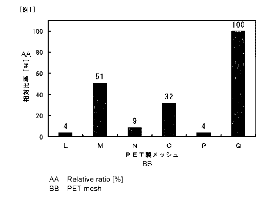
Fig. 1 is a diagram showing the relative ratio of platelets attached to each
prepared polyethylene terephthalate mesh.
Fig. 2 is a schematic view showing the spread state of an embodiment of the
instrument for capturing free thrombi of the present invention.
Fig. 3 is a schematic view showing the folding process of an embodiment of
the instrument for capturing free thrombi of the present invention.
Fig. 4 is a schematic view showing the folded state of an embodiment of the
instrument for capturing free thrombi of the present invention.
BEST MODE FOR CARRYING OUT THE INVENTION
[0020]
The terms used in the present description are as defined below unless
otherwise specified.
[0021]
The term "instrument for capturing free thrombi" means a medical instrument
also called a filter instrument, and comprises a filter section composed of a
mesh
material and/or the like for capturing free thrombi.
CA 02840043 2013-12-19
7
[0022]
Examples of the filter section of the instrument for capturing free thrombi
include a filter section prepared by forming a sheet having a large number of
pores
into a bag shape, and a filter section prepared by forming a sheet,
interweaved with
fibers in the form of a mesh or net, into a bag shape.
[0023]
Examples of the material of the filter section include polymer materials, for
example, polyesters such as polyethylene terephthalate (hereinafter referred
to as
"PET"); polytetrafluoroethylene; cellulose; cellulose acetate; polycarbonate;
polysulfone (hereinafter referred to as "PSI'); polyether sulfone; polyalkyl
(meth)acrylates (the alkyl moiety is preferably C1-C4 lower alkyl, especially
preferably methyl) such as polymethyl methacrylate (hereinafter referred to as
"PMMA"); polyamides; polyvinylidene fluoride; polyvinyl chloride;
polyacrylonitrile; polyurethanes; polystyrene; polyethylene; polypropylene;
polymethylpentene; and polyimides. Among these, polyesters,
polyalkyl(meth)acrylates, polyurethanes, polyvinyl chloride, polycarbonate and
polytetrafluoroethylene are preferred, and, because of its high flexibility
and in vivo
stability, polyesters, especially PET, are more preferred. These materials may
be
used individually, or two or more of these materials may be used in
combination.
[0024]
In cases where the filter section is composed of an organic fiber prepared
with
the above-described polymer materials, the filter section has a fiber diameter
of
preferably not more than 40 tm, more preferably not more than 35 gm, still
more
preferably not more than 30 gm, in order to obtain a thinner filter section
and to
allow easy folding of the filter section. The lower limit of the fiber
diameter is not
limited, and, from the viewpoint of strength, the fiber diameter is usually
not less
than 20 gm.
CA 02840043 2013-12219
8
[0025]
The organic fiber constituting the filter section may be either a monofilament
or a multifilament, and is preferably a monofilament since it is smoother and
less
likely to activate the blood coagulation reaction.
[0026]
The material of the filter section may also be a metal such as stainless steel
or
a nickel-titanium alloy in view of the durability and the shape retention
property. In
cases where a metal is used as the material of the filter section, the
conjugate that
remarkably inhibits the blood coagulation reaction can be strongly immobilized
by,
for example, using a coupling agent that can be adsorbed or bound to the
metal, or
coating the surfaces of the metal with the polymer material.
[0027]
More specific examples of the constitution of the instrument for capturing
free thrombi include:
a constitution in which a flexible wire is bent into a loop shape and both
ends
of the wire is bundled and fixed on a delivery wire, wherein the open end of a
bag-
shaped filter section is attached to the ring-shaped wire;
a constitution in which a plurality of flexible wires are shaped into a
spindle,
and both ends of the wires are fixed at two positions on a delivery wire in
the
longitudinal direction, wherein the open end of a bag-shaped filter section is
attached
in the middle of the wires in the longitudinal direction and the closed distal
end of the
bag-shaped section is attached to the distal ends of the wires; and
a constitution as shown in Figs. 2 to 4, comprising: a ring-shaped section 12
composed of a flexible wire material that can be freely bent and has elastic
recoverability, which ring-shaped section 12 has a nearly circular shape; a
core
section 11 that penetrates the ring-shaped section 12, wherein the shape of
the core
section 11 is linear and can be flexibly changed; a filter section 13 whose
open end is
CA 02840043 2013-1219
9
attached in its entirety to the ring-shaped section 12 and whose closed end is
attached
to a part of the distal end side of the core section 11, which filter section
13 is porous
and bag-shaped; and a plurality of support wire sections 14 composed of linear
members whose shapes can be flexibly changed, which support wire sections 14
are
= 5 arranged between a part of the core section 11 proximal to the
closed end of the filter
section 13 and the ring-shaped section 12; wherein the state of the ring-
shaped
section 12 changes from a folded state where the ring-shaped section 12 is
folded
such that a plurality of mountains facing the distal end side of the core
section 11 and
a plurality of valleys facing the proximal end side of the core section 11
alternately
occur and the mountains and valleys are positioned close to each other, to a
spread
state where the ring-shaped section 12 is spread into a nearly circular shape
due to the
elastic recoverability of the ring-shaped section itself, and the state of the
ring-shaped
section 12 also changes from the spread state to the folded state due to
tensions of the
support wire sections 11 exerted by application of an external force to the
support
wire sections 14 in the direction that causes bundling of the support wire
sections 14
with the core section 11 (Patent Document 1; the instrument 1 for capturing
free
thrombi, having this constitution, is hereinafter referred to as the "filter
instrument
A").
[0028]
With a constitution such as that of the filter instrument A, the spread ring-
shaped section 12 is supported by the support wire sections 14, so that the
direction
of the central axis can be easily adjusted nearly to the direction of blood
flow.
Therefore, even with a simple structure, free thrombi can be stably and
securely
captured into the filter section 13 without missing it to the distal side.
Further,
when the ring-shaped section 12 comes into contact with the inner wall of a
blood
vessel, the elasticity of the ring-shaped section 12 allows appropriate
bending of the
ring-shaped section 12 following contraction of the blood vessel, so that free
thrombi
CA 02840043 2013-12-.19
can be stably and securely captured into the filter section 13 without missing
the free
thrombi to the distal side. Further, when an external force is directly
applied to the
ring-shaped section 12 or when the support wire sections 14 are brought close
to the
core section 11 by an external force while the support wire sections 14 are
kept tense,
5 the open end of the ring-shaped section 12 is made narrower and compactly
folded,
so that the filter instrument A can be appropriately delivered in such a
folded state to
a predetermined site in a blood vessel by insertion of the instrument into a
catheter or
the like, while the filter instrument A in the state where thrombi are
captured in the
filter section 13 can be recovered, without missing the thrombi, by folding
the ring-
10 shaped section 12 and thereby narrowing the open end.
[0029]
Examples of the core section 11 of the filter instrument A include thin,
flexible wires made of stainless steel or a nickel-titanium alloy, which is
excellent in
the elastic recoverability. In such a case, the core section 11 may be
constituted by
a plurality of wires joined together, but the core section 11 is preferably
constituted
by a single wire.
[0030]
Examples of the ring-shaped section 12 of the filter instrument A include a
wire that is composed of the same material as the core section 11 and formed
into a
circular ring shape having a diameter of about 4 to 12 mm.
[0031]
Examples of the filter section 13 of the filter instrument A include those
prepared by forming a flexible, firm, triangular and porous sheet into the
form of a
conical bag. The size of each pore provided in the filter section 13 is
preferably 70
to 200 lam, and, in view of increasing the accuracy of capturing free thrombi
while
suppressing the blood coagulation reaction, the sizes of the pores are more
preferably
uniform. More specifically, the sizes of the pores are preferably within the
range of
CA 02840043 2013-12-19
11
the mean value 20%.
[0032]
The ratio of the area of the pores provided per unit area of the filter
section,
the pore ratio, is preferably not less than 30%, more preferably not less than
40%,
still more preferably not less than 50%, in view of reducing inhibition of
blood flow.
The pore ratio is, of course, less than 100%, and, in view of the strength,
the pore
ratio is usually not more than 90%.
[0033]
Examples of the support wire section 14 of the filter instrument A include
wires made of an appropriate material having high strength, whose tension can
be
increased by the action of an external force, such as metallic wires, and
threads and
wires made of polymer materials, that may be the same as, or different from,
the
material of the core section 11.
[0034]
The term "compound having an antithrombin activity" means a compound
having a high binding affinity to thrombin.
[0035]
The compound having an antithrombin activity is preferably the "compound
represented by General Formula (I)" [wherein RI represents a (2R,4R)-4-alky1-2-
2 0 carboxypiperidino group (the alkyl is preferably lower alkyl), and R2
represents a
phenyl group or a fused polycyclic compound residue, which fused polycyclic
compound residue is optionally substituted by a lower alkyl group(s) and/or
lower
alkoxy group(s), and/or by an amino group(s) substituted by a lower alkyl
group(s);
the lower alkyl is CI-C.4 alkyl] described above that is a compound comprising
a
compound comprising a guanidino structure, more preferably (2R,4R)-4-methy1-1-
((25)-2- { [(3R5)-3-methy1-1,2,3,4-tetrahydroquinolin-8-yl]sulfonyl} amino-5-
guanidinopentanoyl)piperidine-2-carboxylic acid (hereinafter referred to as
CA 02840043 2013-12219
12
"argatroban"). Argatroban is a pharmaceutical compound synthesized in 1978
having the selective antithrombin activity of an arginine derivative. Since
argatroban is commercially available as an antithrombotic drug, a commercially
available product may be used. The term "having the selective antithrombin
activity" herein means to have a high binding affinity to thrombin. Examples
of the
index for evaluating the antithrombin activity of a compound include the
inhibition
constant (hereinafter referred to as "Kr) calculated from the Lineweaver-Burk
plot
based on the absorbance of the test solution. A lower Ki indicates higher
binding
affinity to thrombin, that is, higher antithrombin activity. Ki is preferably
not more
than 10 11M, more preferably not more than 1 tiM, still more preferably not
more than
500 nM. A single compound having an antithrombin activity may be used, or two
or more compounds having an antithrombin activity may be used in combination.
[0036]
The compound having an antithrombin activity is preferably immobilized on
the surface(s) of the instrument for capturing free thrombi as a conjugate
with a
macromolecular compound mainly constituted by units derived from at least one
type
of monomers selected from the group consisting of ethylene glycol, vinyl
acetate,
vinyl pyrrolidone, propylene glycol, vinyl alcohol and siloxane. The
"conjugate"
formed by binding of the compound having an antithrombin activity with the
macromolecular compound is preferably hydrophilic in cases where its aqueous
solution is prepared. The term "mainly constituted" means that not less than
90
mol%, preferably not less than 95 mol%, still more preferably not less than 98
mol%
of the total constitution units (repeating units) constituting the
macromolecular
compound are constituted by the above-described units, and that a unit(s)
other than
the above-described units may be contained at a content of not more than 10
mol%,
preferably not more than 5 mol%, more preferably not more than 2 mol%, as long
as
the unit(s) do/does not adversely affect the effect of the present invention.
The
CA 02840043 2013-12-19
13
macromolecular compound is more preferably a copolymer of monomers selected
from the group consisting of ethylene glycol, vinyl acetate, vinyl
pyrrolidone,
propylene glycol, vinyl alcohol and siloxane, especially preferably a
copolymer of at
least two types of monomers selected from these monomers. The molecular weight
of the macromolecular compound is not limited, and usually about 5,000 to
2,000,000, preferably 10,000 to 1,500,000, in terms of the weight average
molecular
weight.
[0037]
The term "hydrophilicity" means that a compound is soluble in water, or,
even in cases where a compound is insoluble in water, the compound interacts
with
water molecules by electrostatic interactions and/or hydrogen bonds.
[0038]
Preferred examples of the macromolecular compound that is bound to the
compound having an antithrombin activity to constitute the above-described
conjugate, especially the "copolymer of monomers selected from the group
consisting of ethylene glycol, vinyl acetate, vinyl pyrrolidone, propylene
glycol, vinyl
alcohol and siloxane" (hereinafter referred to as an "anti-platelet adhesion
copolymer"), include: polyvinyl alcohol; polyvinyl pyrrolidone; polyethylene
glycol;
polypropylene glycol; and macromolecular compounds composed of polyether and
polysiloxane; and copolymers and graft polymers of monomers of these
macromolecular compounds and other monomers. The macromolecular compound
is preferably a macromolecular compound composed of polyether and
polysiloxane,
partially saponified polyvinyl alcohol, or a copolymer of vinyl pyrrolidone
and vinyl
acetate, having high hydrophilicity. Since these are commercially available, a
commercially available product may be used. The commercially available
products
used in the Examples below are examples of the macromolecular compound that
can
be used in the present invention. These may be used individually, or two or
more of
CA 02840043 2013-12-19
14
these may be used in combination.
[0039]
Examples of the "macromolecular compound composed of polyether and
polysiloxane" include copolymers, polymer complexes and polymer blends of
polyether and polysiloxane. The copolymer of polyether and polysiloxane is
composed of a polyether unit(s) and a polysiloxane unit(s), and the form of
the
copolymer may be any of a random copolymer, block copolymer and graft
copolymer.
A polyether-modified silicone is especially preferred since it has high
hydrophilicity.
[0040]
Example of the "polyether" include structures derived from polyethylene
oxide or polypropylene oxide. The "polyether" herein means a structure
represented
by General Formula (II) (wherein R3 represents an alkyl group having not more
than
6 carbon atoms), and the "structure derived from polypropylene glycol" as an
example of the polyether means a structure represented by General Formula
(III).
[0041]
= = = (II)
[0042]
= = = (III)
[0043]
The "polyether-modified silicone" means a silicone comprising a polyether
unit bound to a side chain of the silicone chain, and may be a polyether-
modified
silicone that is additionally amino-modified or carboxy-modified. The amino
group
or carboxyl group given by the amino modification or carboxy modification may
also
be used for covalent bonding with the compound having an antithrombin
activity.
CA 02840043 2013-12-19
For example, the amino-modified polyether-modified silicone used in the
Examples
below (X-22-3939A; Shin-Etsu Chemical) is a preferred example of commercially
available hydrophilic polyether-modified silicones.
[0044]
5 In cases where the anti-platelet adhesion copolymer is a partially
saponified
polyvinyl alcohol, the degree of saponification is preferably 50 to less than
100 mol%,
more preferably 74 to 99.9 mol%, still more preferably 78 to 95 mol%, in view
of
ease of handling and achievement of preferred hydrophilicity. The "degree of
saponification" herein means the value calculated by Equation 1.
10 [0045]
Degree of saponification = m/(n+m)x100 ... Equation 1
m: number of structures represented by General Formula (IV) in polyvinyl
alcohol
n: number of structures represented by General Formula (V) in polyvinyl
15 alcohol
[0046]
OH j m
= = = (IV)
[0047]
0
________________ 0
= = = ( V )
[0048]
In cases where the anti-platelet adhesion copolymer is a copolymer of vinyl
pyrrolidone and vinyl acetate, the content of vinyl pyrrolidone units is
preferably not
CA 02840043 2013-12219
16
less than 50 unit mol%, more preferably not less than 60 unit mol%, in view of
ease
of handling and achievement of preferred hydrophilicity. On the other hand,
the
content of vinyl pyrrolidone units is preferably less than 100 unit mol% in
view of
achievement of a preferred amount of immobilization to the instrument for
capturing
free thrombi. The ratio of vinyl pyrrolidone units in the copolymer of vinyl
pyrrolidone and vinyl acetate (unit mol%) can be calculated by subjecting the
copolymer to II-I-NMR measurement (solvent: CDC13).
[0049]
The binding between the compound having an antithrombin activity and the
macromolecular compound is preferably achieved by a covalent bond(s) in view
of
preventing loss of the compound having an antithrombin activity. The covalent
bond(s) can be easily formed by performing coupling reaction that forms an
amide
bond(s) and/or ester bond(s) between a functional group(s) such as a free
amino
group(s), carboxyl group(s) and/or hydroxyl group(s) in the compound having an
antithrombin activity and such a functional group(s) in the macromolecular
compound. The coupling reaction can be carried out by, for example, a
conventional method using a commercially available well-known coupling agent
such as dicyclohexylcarbodiimide (DCC), and the reaction is also specifically
described in the Examples below. In cases where the compound having an
antithrombin activity is bound via a covalent bond(s), the antithrombin
activity needs
to be exerted even after the covalent bonding. Whether or not the antithrombin
activity is exerted even after the covalent bonding can be investigated by
measuring
the antithrombin activity of the conjugate after the covalent bonding by the
above-
described method. In cases where the compound having an antithrombin activity
is
a compound represented by the above-described General Formula (I), the
compound
comprises a free amino group at the left end of General Formula (I), and a
free
carboxyl group in R. As described in the Examples below, it has been confirmed
CA 02840043 2013-12-19
17
that the antithi-ombin activity is still exerted even after binding with the
macromolecular compound via the amino group and/or the carboxyl group. In
cases
where the macromolecular compound is a polyether-modified silicone, a
polyether-
modified silicone that is additionally amino-modified or carboxy-modified can
be
used to form an amide bond or ester bond between the amino group or carboxyl
group and the free carboxyl group or amino group of the compound of General
Formula (I) (Examples below). Further, in cases where the macromolecular
compound is a vinyl acetate/polyvinyl pyrrolidone copolymer, an amide bond can
be
formed between the carboxyl group in the vinyl acetate unit and the amino
group in
the compound of General Formula (I) (Examples below). Further, in cases where
the macromolecular compound is a partially saponified polyvinyl alcohol, an
ester
= bond can be formed between the carboxyl group in the compound of General
Formula (I) and the hydroxyl group in the vinyl alcohol unit (Examples below).
[0050]
The amount of the anti-platelet adhesion copolymer adsorbed on the
surface(s) of the filter section of the instrument for capturing free thrombi
is
preferably not less than 0.1 pg/mm2, more preferably not less than 1 pg/mm2,
still
more preferably not less than 10 pg/mm2. The upper limit of the amount of
adsorption is not restricted, and the amount of adsorption is usually not more
than 10
ng/mm2.
[0051]
The amount of adsorption described above is measured by the following
method. First, an untreated sensor chip (Sensor Chip Au; GE Healthcare) is
pretreated (distilled water at 25 C; flow rate, 20 pi/min.; 10 minutes) using
a surface
plasmon resonance apparatus (hereinafter referred to as "SPR") (BIACORE 3000;
GE Healthcare), and the signal value (RU: resonance unit) is measured.
[0052]
CA 02840043 2013-12-19
18
The material for the filter section of the instrument for capturing free
thrombi,
that is, the target material of immobilization, is dissolved in a solvent, to
prepare a
0.5wt% solution of the target material of immobilization. A drop of the
solution of
the target material of immobilization is added to the center of the gold film
area of
the pretreated sensor chip attached to a spin coater, and the sensor chip is
then
immediately coated with the target material of immobilization at room
temperature
by rotation at 3000 rpm for 1 minute.
[0053]
After confirming that no droplet is present on the sensor chip, the sensor
chip
is washed with distilled water using the SPR (25 C; flow rate, 20 .1/min.; 10
minutes), and the sensor chip is then washed 3 times with 0.025 wt% Triton-
X100
solution (25 C; flow rate, 200/min.; 1 minute), followed by measuring the
signal
value 10 minutes after completion of the washing.
[0054]
Among the thus obtained sensor chips, a sensor chip wherein the difference
between the signal values observed before and after the spin coating is within
the
range of 3000 to 8000 is selected, and the selected sensor chip is washed with
distilled water (25 C; flow rate, 20 plimin.; 10 minutes), followed by 3 times
of
washing with 0.025 wt% Triton-X100 solution (25 C; flow rate, 20 ill/min.; 1
minute)
[0055]
Ten minutes after completion of the washing, a solution of the
macromolecular compound to be immobilized on the instrument for capturing free
thrombi (concentration, 100 gimp is injected (25 C; flow rate, 20 ial/min.; 1
minute), followed by washing with distilled water (25 C; flow rate, 20
fal/min.; 3
minutes). The difference between the signal value observed before beginning of
the
injection (hereinafter referred to as the "signal value A") and the signal
value
= CA 02840043 2013-12-19
19
observed 3 minutes after completion of the injection (hereinafter referred to
as the
"signal value B") is calculated, and the resulting value is converted
according to the
following equation: 1 RU=1 pg/mm2.
[0056]
Subsequently, the sensor chip is washed with distilled water (25 C; flow rate,
20 [tl/min.; 2 minutes) and then washed 3 times with 0.025 wt% Triton-X100
solution (25 C; flow rate, 20 ttl/min.; 1 minute), further followed by
injection of the
aqueous solution of the macromolecular compound to be immobilized
(concentration,
100 g/m1) (25 C; flow rate, 20 ill/min.; 1 minute). Thereafter, the same
operation
is repeated to calculate the signal difference a total of 5 times (difference
between the
signal value A and the signal value B), and the average of the obtained values
is
regarded as the amount of adsorption of the anti-platelet adhesion copolymer
to the
instrument for capturing free thrombi.
[0057]
Examples of the method for immobilizing the above-described conjugate on
the surface(s) of the instrument for capturing free thrombi include a method
wherein
a solution comprising the conjugate as an effective component(surface
treatment
agent) is brought into contact with the instrument for capturing free thrombi
and then
a radiation is irradiated thereto, and a method wherein the conjugate
dissolved in an
organic solvent is applied or sprayed onto the instrument for capturing free
thrombi,
followed by drying the instrument. The type of the radiation to be irradiated
is
preferably an electron beam or y-ray. The concentration of the macromolecular
compound solution to be brought into contact with the surface of the
instrument is
determined using as an index the antithrombin activity. Examples of the index
of
the antithrombin activity include the concentration in term of argatroban, and
the
concentration in term of argatroban of the solution of the conjugate to be
brought into
contact with the surface of the instrument for capturing free thrombi is about
10 to
= CA 02840043 2013-12:19
200,000 ppm by weight, more preferably about 50 to 100,000 ppm by weight,
still
more preferably about 1,000 to 100,000 ppm by weight. The conjugate is
preferably
immobilized on at least the filter section of the instrument for capturing
free thrombi.
The instrument for capturing free thrombi of the present invention is placed
in
5 a blood vessel of a living body when it is used. The instrument is
preferably
retained in a catheter, and placed in a blood vessel by insertion of the
catheter into
the blood vessel.
EXAMPLES
[0058]
10 The present invention is described below in detail by way of
Examples, but
the present invention is not limited to these.
[0059]
(Example 1: Binding of Amino-polyether-modified Silicone with Argatroban)
In an eggplant type flask, 5 mmol of argatroban was placed, and 10 mL of
15 anhydrous dimethylformamide (hereinafter referred to as "anhydrous DMF")
was
added thereto to dissolve the argatroban, followed by adding 10 mL of 4 N
hydrochloric acid/1,4-dioxane (Toyo Kasei Co., Ltd.) dropwise to the resulting
solution while cooling the eggplant type flask on ice, and then stirring the
resulting
mixture for 1 hour. Subsequently, the solvent was evaporated with a rotary
20 evaporator, and the resultant was dried overnight in a vacuum drier,
followed by
adding 25 mL of anhydrous DMF thereto to provide an argatroban
hydrochloride/anhydrous DMF solution.
[0060]
The argatroban hydrochloride/anhydrous DMF solution was placed in a two-
necked flask in an amount shown in Table 1, and dicyclohexylcarbodiimide
(hereinafter referred to as "DCC")/anhydrous DMF solution and 4-
hydroxybenzotriazole (hereinafter referred to as "HOBt")/anhydrous DMF
solution
CA 02840043 2013-12-19
21
were added thereto with stirring under ice-cooling, followed by further adding
an
amino-modified polyether-modified silicone (X-22-3939A; Shin-Etsu Chemical) to
the resulting mixture and then allowing the reaction to proceed at room
temperature
for 3 days. Thereafter, the reaction liquid was placed in a dialysis tube
(Spectra/Por
RC Por 6 MWC0=1000), followed by performing dialysis for 3 days against more
than 10 volumes of distilled water while appropriately exchanging the
distilled water.
The reaction liquid after dialysis was filtered, and the solvent of the
obtained filtrate
was evaporated with an rotary evaporator, followed by drying the resultant
overnight
in a vacuum drier, to obtain a conjugate (hereinafter referred to as the
"Example 1
conjugate")
[0061]
(Measurement of Antithrombin activity of Example 1 Conjugate)
The measurement was carried out using ECA-T Kit (HaemoSys). To 100 !IL
of the Example 1 conjugate, 900 1_, of distilled water was added, to prepare
an
aqueous Example 1 conjugate solution. Thereafter, 30 iL of the aqueous Example
1 conjugate solution was collected and mixed with 1001.11, of ECA prothrombin
buffer and 25 tit of ECA-T substrate, and the resulting mixture was incubated
at
37 C for 60 seconds. The mixture was then placed in an apparatus (COATRON
M1 (code 80 800 000); Production) and 50 1.tL of ECA ecarin reagent was
further
added thereto, followed by performing measurement.
[0062]
Measurement using the ECA-T kit was carried out in the same manner as
described above except that a mixture prepared by mixing 20 lit of an
argatroban
solution whose concentration was arbitrarily adjusted using an
ethanol/hydrochloric
acid (volume ratio, 1/4) mixed solvent with 80 t.d., of human blood plasma, or
a
mixture prepared by mixing 20 ttL of distilled water as a blank with 80 1AL of
human
blood plasma, was used instead of the aqueous Example 1 conjugate solution. A
CA 02840043 2013-12219
22
calibration curve was prepared based on the obtained results. The
concentration in
term of argatroban of the aqueous Example 1 conjugate solution calculated
based on
the calibration curve, 1494.3 ppm by weight, was regarded as the value
indicating the
antithrombin activity of the aqueous Example 1 conjugate solution.
[0063]
(Examples 2 to 13)
The compounds of Example 2 to 13 were obtained in the same manner as
Example 1 except that the molar ratios of DCC, HOBt and/or the polyether-
modified
silicone (X-22-3939A) to the argatroban hydrochloride, and/or the volume ratio
of
anhydrous DMF to the polyether-modified silicone, were changed. The
antithrombic activity was measured for each of these compounds. The molar
ratios
of DCC, HOBt and the polyether-modified silicone (X-22-3939A) to argatroban
hydrochloride, and the result of measurement of the antithrombin activity of
each of
the compounds of Examples 2 to 13 are shown in Table 1.
[0064]
[Table 1]
Molar ratio to argatroban Volume ratio of Concentration
hydrochloride (1.00) anhydrous DMF in term of
Compound X-22-
to polyether- argatroban
DCC HOBt 3939A modified (ppm by
silicone (1) weight)
Example 1 1.07 1.06 0.060 1494.3
Example 2 1.04 1.04 0.060 831.2
Example 3 0.20 _ 0.20 0.060 1.4 6610.7
Example 4 0.20 0.20 0.030 3.9 8393.3
Example 5 1.29 1.27 0.493 1.8 505.3
Example 6 1.29 1.27 0.203 4.3 771.7
Example 7 1.29 1.27 0.101 8.6 606.7
Example 8 1.29 1.27 0.067 13.0 441.7
Example 9 1.29 1.27 0.049 17.6 436.7
Example 10 1.29 1.27 0.020 42.9 738.9
Example 11 1.29 1.27 0.010 88.2 895.0
Example 12 1.00 1.00 0.060 - 6000.0
Example 13 1.00 1.00 0.060 40.0 5999.4
CA 02840043 2013-12-19
23
[0065]
Although the polyether-modified silicone (X-22-3939A) was similarly
subjected to measurement of the antithrombin activity, the obtained value was
not
different from the value for distilled water as the blank, so that it was
confirmed that
the polyether-modified silicone itself does not have antithrombin activity.
[0066]
(Measurement of Thrombin Inhibition Constant of Example 1 Conjugate)
In 1 mL of physiological saline, 10,000 U of a bovine thrombin solution (ILS
Inc.) was dissolved, to prepare an aqueous bovine thrombin solution.
[0067]
In 40 mL of distilled water, 25 mg of S-2238 stock solution ( Sekisui Medical
Co., Ltd.) was dissolved, to prepare an aqueous S-2238 stock solution.
[0068]
Using a dilution buffer (0.05 M Tris, 0.1 M NaC1, 1 mg/mL bovine serum
albumin (BSA), pH 7.4), each of the aqueous bovine thrombin solution, the
aqueous
S-2238 stock solution, and the aqueous Example 1 conjugate solution described
above was diluted.
[0069]
Into a 96-well plate, 1001.1L of the diluted aqueous S-2238 stock solution and
501AL of the diluted aqueous Example 1 conjugate solution were aliquoted, and
the
plate was sealed, followed by heating the plate in a constant temperature
dryer at
37 C for 30 minutes. Subsequently, 50 jtL of the diluted aqueous bovine
thrombin
solution heated at 37 C for 30 minutes was further aliquoted, and the
absorbance was
immediately measured using a microplate reader (measurement wavelength, 405
nm,
reference wavelength, 595 nm).
[0070]
After completion of the first measurement of absorbance, the second
CA 02840043 2013-12-19
24
measurement was immediately carried out. The third and later measurements of
absorbance were carried out 4, 6, 8, 10, 12, 14, 16, 18 and 20 minutes after
the
aliquoting of the bovine thrombin dilution, respectively. From the obtained
values
of absorbance, Ki was calculated from the Lineweaver-Burk plot. Ki of the
Example 1 conjugate was 11.2 nM.
[0071]
Ki was also calculated for the polyether-modified silicone (X-22-3939A), but
Ki of the polyether-modified silicone, which has no antithrombin activity, was
the
same as that of the blank, as expected.
[0072]
Further, as a result of similar calculation of Ki for argatroban, Ki was found
to be 39.1 nM, which was not less than 3 times higher than Ki of the Example 1
conjugate.
[0073]
From these results, it is clear that the above-described conjugate has
extremely high binding affinity to thrombin, and hence that the conjugate can
give
remarkable antithrombin activity to the instrument for capturing free thrombi,
which
activity is even higher than that of argatroban, which is known to have
antithrombin
activity.
[0074]
(Example 14: Binding of Vinyl Acetate-Vinyl Pyrrolidone Copolymer to
Argatroban)
In a screw bottle, 14.9 g of tetrahydrofuran, 11.5 g of vinyl acetate, 10.8 g
of
N-vinyl pyrrolidone, 0.028 g of 2-aminoethanethiol and 0.016 g of
azobisisobutyronitrile were placed, and the bottle was sealed, followed by
irradiation
of ultrasonic waves thereto for 10 minutes. The screw bottle was once opened
and
bubbling with argon gas was performed for 10 minutes, followed by sealing the
bottle again. Thereafter, the screw bottle was immersed, with stirring, in a
hot water
CA 02840043 2013-12-19
= 25
= bath at 60 C for 1 hour and then in a hot water bath at 70 C for 6 hours,
to allow
copolymerization reaction of vinyl acetate and vinyl pyrrolidone. To this
reaction
= liquid, 80 mL of methanol was added, and the resulting mixture was added
to about 5
volumes of ether, followed by removal of the supernatant. The operation of
adding
fresh ether and removing the supernatant was repeated 3 times, and the
resultant was
dried under reduced pressure, to obtain a vinyl acetate-vinyl pyrrolidone
copolymer.
The obtained vinyl acetate-vinyl pyrrolidone copolymer was subjected to 11-1-
NMR
measurement (solvent; CDC13), and, as a result, the content of vinyl
pyrrolidone units
was found to be 60.6 unit mol%.
[0075]
In 20 mL of anhydrous DMF, 3.58 g of the obtained vinyl acetate-vinyl
pyrrolidone copolymer was dissolved, to prepare a vinyl acetate-vinyl
pyrrolidone
copolymer/anhydrous DMF solution. In a two-necked flask, the whole vinyl
acetate-vinyl pyrrolidone copolymer/anhydrous DMF solution prepared and 0.5 mL
of argatroban hydrochloride/anhydrous DMF solution (0.49 M) were placed, and
0.5
mL of DCC/anhydrous DMF solution (1.04 M) and 0.5 mL of HOBt/anhydrous DMF
solution (1.02 M) were added thereto with stirring under ice-cooling, followed
by
allowing the reaction to proceed under nitrogen atmosphere at room temperature
for
3 days. Subsequently, the reaction liquid was placed in a dialysis tube
(Spectra/Por
RC Por 6 MWC0-1000), followed by performing dialysis for 3 days against more
than 10 volumes of distilled water while appropriately exchanging the
distilled water.
The reaction liquid after dialysis was filtered, and the solvent of the
obtained filtrate
was evaporated with a rotary evaporator, followed by drying the resultant
overnight
in a vacuum drier, to obtain a conjugate (hereinafter referred to as the
"Example 14
conjugate").
[0076]
(Measurement of Antithrombin activity of Example 14 Conjugate)
= CA 02840043 2013-12-19
26
In the same manner as the measurement of antithrombin activity of the
Example 1 conjugate, measurement was carried out for the Example 14
conjugate/methanol solution (concentration, 20 wt%). The calculated
concentration
in term of argatroban of the Example 14 conjugate/methanol solution, 104.1
ppm,
was regarded as the value indicating the antithrombin activity of the Example
14
conjugate/methanol solution.
[0077]
(Immobilization of Example 1 Conjugate to PET Mesh)
[0078]
Bis-Tris (Dojindo Laboratories) and sodium chloride were dissolved in
ultrapure water such that their final concentrations were 0.25 M and 0.5 M,
respectively, and 6 N hydrochloric acid was added dropwise to the resulting
solution
to adjust the pH to 5, to prepare 5 x Bis-Tris buffer.
[0079]
A PET mesh (fiber diameter, 27 p.m; mesh size, 100 p.m) was formed into a 6-
cm square. The Example 1 conjugate at a concentration in term of argatroban of
50,000 ppm by weight, propylene glycol, 5 x Bis-Tris buffer and distilled
water were
mixed together at a volume ratio of 8/50/20/22, to obtain a treatment liquid.
[0080]
The formed PET mesh was rolled into a cylindrical shape and inserted into a
polypropylene centrifuge tube, followed by adding 5 mL of the treatment liquid
thereto. After irradiation of ultrasonic waves to the tube in a warm bath at
40 C for
1 hour, 'y-ray was further irradiated thereto with an absorbed dose of 25 kGy
for 3
hours.
[0081]
Thereafter, the treatment liquid in the centrifuge tube was removed, and 15
mL of 0.025 wt% aqueous polyoxyethylene octylphenyl ether solution was added
to
= CA 02840043 2013-12-19
27
the centrifuge tube, followed by shaking the centrifuge tube for 10 minutes to
wash
the PET mesh. Thereafter, the aqueous solution in the centrifuge tube was
removed,
and 15 mL of fresh 0.025 wt% aqueous polyoxyethylene octylphenyl ether
solution
was added to the centrifuge tube, followed by 10 minutes of shaking. This
washing
operation was repeated a total of 3 times. Subsequently, the same washing
operation was repeated 10 times using 15 mL each of distilled water and
physiological saline, to prepare a PET mesh to which the Example 1 conjugate
was
immobilized (hereinafter referred to as the "PET mesh L").
[0082]
The PET meshes M to P were prepared by the same operation as in the
preparation of the PET mesh L except that the treatment liquids prepared with
the
volume ratios shown in Table 2 were used instead of the above treatment
liquid.
[0083]
[Table 2]
Mixing volume ratio
Example 1 Compound at
PET mesh concentration in term of
Propylene 5 x Bis- Distilled
argatroban of 50,000 ppm glycol Tris buffer water
by weight
8 50 20 22
0.2 50 20 29.8
2 30 20 48
0 4 30 20 46
16 50 20 14
[0084]
The PET mesh Q was prepared by the same operation as in the preparation of
the PET mesh L except that distilled water was used instead of the above
treatment
liquid.
[0085]
(Measurement of Amount of Example 1 Conjugate Eluted)
The PET mesh L formed into a 6-mm square was placed in a polystyrene
= CA 02840043 2013-12-19
28
round tube (Code: 352054; BECTON DICKINSON), and 5 mL of human blood
plasma was added thereto, followed by shaking the tube for 4 hours. The
concentration of the Example 1 conjugate in the human blood plasma after
shaking
was below the detection limit of the ECA-T kit used for the measurement, and
hence
no elution of the Example 1 conjugate from the PET mesh L was found. This
result
indicates that the above conjugate can be strongly immobilized on the
instrument for
capturing free thrombi.
[0086]
(Evaluation of Amount of Anti-platelet Adhesion Copolymer Immobilized)
As examples of the copolymer of vinyl pyrrolidone and vinyl acetate
(hereinafter referred to as "VA copolymer") to be used as the anti-platelet
adhesion
copolymer constituting the above conjugate, PVP(K-90), VA73, VA64, VASS and
VA37 (all of these were obtained from BASF) were provided. Similarly, as
examples of the partially saponified polyvinyl alcohol to be used as the anti-
platelet
adhesion copolymer, PVA217, PVA417 and PVA205c (all of these were obtained
from Kuraray Co., Ltd.) were provided. Further, as polyether-modified
silicones,
F114, F244, F303, F3031, F348, F350s, F502, F506 and X-22-3939A (all of these
were obtained from Shin-Etsu Silicone, Co., Ltd.) were provided. Each of the
VA
copolymers, partially saponified polyvinyl alcohols and polyether-modified
silicones
provided was diluted with distilled water to prepare its aqueous solution at
10,000
ppm by weight.
[0087]
On the other hand, for comparison, PEG2000, PEG4000, PEG6000 and
PEG20000 (all of these were obtained from Nacalai Tesque); and PEG methyl
ether
(PEG-em) and PEG dimethyl ether (PEG-dm) (both were obtained from Sigma-
Aldrich); were provided as macromolecular compounds that are not included in
the
anti-platelet adhesion copolymer constituting the above conjugate. Each
CA 02840043 2013-12-19
29
macromolecular compound provided was diluted with distilled water to prepare
its
aqueous solution at 10000 ppm by weight.
[0088]
Binding of argatroban to the above VA copolymer or polyether-modified
silicone, binding of argatroban to the partially saponified polyvinyl alcohol,
and
binding of argatroban to PEG2000, PEG4000, PEG6000 or PEG20000 (all of these
were obtained from Nacalai Tesque), or to PEG methyl ether (PEG-em) or PEG
dimethyl ether (PEG-dm), were carried out in the same manner as in Example 14
or
Example 1.
[0089]
As examples of the 0.5 wt% solution of the target material of immobilization,
on which the anti-platelet adhesion copolymer is to be immobilized, a PMMA
(weight average molecular weight, 93000; Sigma-Aldrich)/toluene solution,
polyurethane/dimethylacetamide solution, PSf (UDEL (registered trademark),
manufactured by Solvay; P-3500)/dimethylacetamide solution, polyvinyl chloride
(weight average molecular weight, 80000; Sigma-Aldrich)/tetrahydrofuran
solution,
polystyrene (Wako)/chloroform solution, and polycarbonate (weight average
molecular weight, 20000; TEIJIN Ltd.)/chloroform solution were prepared.
[0090]
The amount of adsorption of each of the various anti-platelet adhesion
copolymers to each target material of immobilization was measured. The results
are
shown in Table 3.
1
.
CA 02840043 2013-12-19
[0091]
[Table 3]
Signal value B - Signal value A [pg/mm2]
Target material of adsorption
-
PMMA
Poly- Poly- Polyvinyl Poly- Poly-
sulfone urethane chloride styrene carbonate
PVPK90 789 - - - - -
_
VA37 2760 - - - - - _
VASS 472 - - - - -
VA64 920 - - - - -
al
-4- VA73 426 - - - - -
c4-
o PVA217 2529 2886 1635
2468 2777 2356
a
.9. PVA417 2475 2742 1911 2330 2662
2346
v)
1) PVA205c 2223 2130 1411 1796 1989
1819
-a
czt F114 1003 844 514 739 621
756_
-+.-1 F244 1639 1272 1144 1118 1052
1243
47
- -..-
F303
a 1268 1156 1604 1037 -
1374_
.-
F3031 947 559 614 418 339
536
.E. F348 875 784 756 608 283
800_
a F350s 751 657 674 544 275
591
a
a F502 827 657 696 385 197
482
g F506 691 308 437 167 43
279
L' X-22-
as 1182 910 1204 695 924
1424
¨ 3939A
a
c.)
0 PEG2000 2 - - - - -
g PEG4000 2 - - - - -
,-, PEG6000
a 5 - - - - -
PEG20000 113- -
- -
PEG-me 5 - - - - -
PEG-dm 67 - - - - -
[0092]
From the results shown in Table 3, it is clear that the anti-platelet adhesion
5 copolymer
constituting the above-described conjugate is not limited to the polyether-
modified silicone (X-22-3939A), and that strong immobilization on the
instrument
for capturing free thrombi is possible.
[0093]
(Evaluation of Number of Platelets Adhered)
10 The PET mesh L formed into a 1-cm square was placed in an arbitrary
well of
a 24-well plate, and 1 mL of phosphate buffered saline (hereinafter referred
to as
was added to the well, followed by incubation of the plate at 37 C for 30
CA 02840043 2013-12-19
31
minutes.
[0094]
Platelet rich plasma (hereinafter referred to as "PRP") was prepared by
mixing 3.2 wt% aqueous sodium citrate solution with human volunteer blood at a
volume ratio of 1/9 and then centrifuging the resulting mixture at 20 C at
1000 rpm
for 15 minutes. The platelet number in PRP was preliminarily measured using
Automated Hematology Analyzer XT-1800i (Sysmex Corporation).
[0095]
The PET mesh L after incubation was transferred to an empty well, and 1 mL
of the prepared PRP was added thereto, followed by additional incubation of
the
mesh at 37 C for 2 hours. This PET mesh L was held with tweezers and placed in
another well containing 1 mL of PBS(-), followed by gently washing the mesh.
This washing operation was repeated a total of 3 times. The whole PBS(-) used
in
the washing was collected in an empty well.
[0096]
To the collected PBS(-), 1 mL of 1 wt% polyoxyethylene octylphenyl
ether/PBS(-) was added, and the resulting mixture was incubated at 37 C for 15
minutes.
[0097]
To an empty well, 200 iL of the incubated solution was collected, and 200 [tL
of PBS(-) was added thereto. To the resulting mixture, 400 [tL of Solution C
(a
mixture prepared by mixing Solution A with Solution B of LDH Cytotoxicity
Detection Kit (manufactured by Takara Bio Inc.) at a volume ratio of 1/45) was
added immediately after its preparation, and the well was covered with
aluminum foil,
followed by leaving the mixture to stand at room temperature for 30 minutes.
The
reaction was then terminated by addition of 200 1AL of IN HC1 (final
concentration,
0.2 N).
CA 02840043 2013-12-19
32
[0098]
The reaction liquid after termination of the reaction was subjected to
measurement of absorbance at a wavelength of 490 nm using a spectrophotometer.
[0099]
The same operation was carried out also for the PET meshes M to Q, and
each reaction liquid after the termination of reaction was subjected to
measurement
of absorbance at a wavelength of 490 nm.
[0100]
The PRP was diluted with PBS(-) to concentrations of 1/10, 1/20, 1/50, 1/100,
1/500 and 1/1000. Each diluted PRP solution was collected into an empty well
of a
24-well plate in an amount of 200 [tL, and 200 1AL of 1% polyoxyethylene
octylphenyl ether /PBS(-) was added thereto, followed by incubating the
resulting
mixture at 37 C for 15 minutes. Each solution after the incubation was
collected
into an empty well in an amount of 200 pL, and 400 1.LL of Solution C
immediately
after preparation was added to the well. The well was covered with aluminum
foil,
and the mixture was left to stand at room temperature for 30 minutes.
Thereafter,
200 L of 1 N HC1 (final concentration, 0.2 N) was added thereto. The reaction
liquids were subjected to measurement of absorbance at a wavelength of 490 nm
using a spectrophotometer, to prepare a calibration curve.
[0101]
From the prepared calibration curve and the absorbance at a wavelength of
490 nm of each of the reaction liquids obtained by the treatment with the PET
meshes L to Q, the platelet number in each reaction liquid was calculated. The
platelet number in the reaction liquid obtained by the treatment with the PET
mesh Q
was defined as 100%, in order to calculate the ratios of the platelet numbers
in the
other reaction liquids (hereinafter referred to as "relative ratios"). The
results are
shown in Fig. 1.
CA 02840043 2013-12-19
33
[0102]
From the results in Fig. 1, it is clear that the above conjugate is capable of
giving remarkable anti-platelet adhesion capacity to the instrument for
capturing free
thrombi.
[0103]
(Measurement of Whole Blood Clotting Time)
Blood collected from a volunteer was mixed with citric acid at a volume ratio
of 9/1, to prepare citrated blood.
[0104]
In a cuvette (NON-ACTIVATED CLOTTING TEST KIT), 18 1.1L of
physiological saline was placed, and 14.8 [IL of Calcicol was added thereto,
followed
by further adding 342 1.1,L of the citrated blood to the resulting mixture.
The mixture
was then subjected to measurement using a Sonoclot coagulation & Platelet
Function
Analyzer (IMI Corporation), and the obtained ACT ONSET value was regarded as
the whole blood clotting time. The whole blood clotting time of the blood
collected
from the volunteer was 545 seconds.
[0105]
The same measurement was carried out using each of 2, 10 and 20 [LM
argatroban solutions (solvent: methanol/hydrochloric acid (volume ratio, 4/1))
instead of physiological saline. As a result, the whole blood clotting time
was 531,
746 and 849 seconds, respectively.
[0106]
The same measurement was carried out using each of 0.3, 1.3 and 2.5 M
aqueous Example 1 conjugate solutions instead of physiological saline. As a
result,
the whole blood clotting time was 527, 693 and 730 seconds, respectively.
[0107]
(Preparation of Instrument for capturing free thrombi)
CA 02840043 2013-12-19
34
A nitinol wire was bent and turned a plurality of times into a ring shape, to
prepare a ring-shaped section 12 that is a circular ring having a diameter of
6 mm.
A polyester mesh sheet having a 100-um mesh size was formed into a conical-bag
shape having a height of 15 mm, to prepare a filter section 13. In this case,
the
bottom of the cone corresponds to the open end 13a, and the top corresponds to
the
closed end 13b.
[0108]
As the core section 11 (including an operation member 15), a stainless-steel
wire was used. At one end of the wire, a guide section llb composed of a
flexible
wire gently curving to form an arc was formed.
[0109]
The core section 11 was made to penetrate the ring-shaped section 12, and the
closed end 13b of the filter section 13 was attached to the core section 11 at
a
position in the proximal side of the guide section at the distal end of the
core section
11. Further, the open end 13a of the filter section 13 was attached to the
ring-
shaped section 12. Four threads made of a synthetic resin or composite fiber,
such
as polyarylate threads, were provided, and each of these was used as a support
wire
section 14. A position on the core section 11 that is proximal to the ring 12
was
used as a support section 14a, and each of the positions defined by dividing
the
circumference of the ring-shaped section 12 into four equal parts was defined
as a
support section 14b. As shown in Fig. 2, each of the four support wire
sections 14
was fixed to the support section 14a and the support section 14b by adhesion
or the
like, to complete a filter instrument A. In the ring-shaped section 12, as
shown in
Fig. 2, a dividing point 121, dividing point 122, dividing point 123 and
dividing point
124 were set at the midpoints of the support sections 14b, and a folding
property was
preliminarily given to the ring-shaped section 12 such that the dividing point
121 and
the dividing point 123, a pair of the dividing points facing each other,
become the
CA 02840043 2013-12-19
bottoms of valleys when these dividing points are bent by an external force in
the
direction of the proximal end 15a of the core section, while the dividing
point 122
and the dividing point 124, the other pair of the dividing points, become the
tops of
mountains when these dividing points are bent by an external force in the
direction of
5 the distal end of the core section, that is, such that the ring-shaped
section 12 shows a
wavy pattern as a whole after folding.
[0110]
The state of the filter instrument A changes from the spread state shown in
Fig. 2 to the folded state by a process in which the support wire sections 14
are
10 bundled with the core section 11 as shown in Fig. 3 by the action of an
external force
to cause bundling of the support sections 14b with the core section, which
makes the
two pairs of dividing points that face each other, that is, the pair of the
dividing point
121 and the dividing point 123, and the pair of the dividing point 122 and the
dividing
point 124, become the bottoms of valleys and the tops of mountains,
respectively,
15 while the filter section 13 is folded as the ring-shaped section 12 is
gradually folded
until the paired dividing points come into contact with each other by the
folding of
the ring-shaped section 12 as shown in Fig. 4. In the folded state, the open
end 13a
of the filter section 13 is almost completely closed.
[0111]
20 (Immobilization of Example 1 Conjugate on Instrument for capturing free
thrombi)
The Example 1 conjugate at a concentration in term of argatroban of 50,000
ppm by weight, propylene glycol, 5 x Bis-Tris buffer and distilled water were
mixed
together at a volume ratio of 8/50/20/22, to obtain a treatment liquid.
[0112]
25 In a container with an appropriate capacity, 2 mL of the treatment
liquid was
placed, and the filter section of the prepared filter instrument A was
completely
immersed therein, followed by irradiation of ultrasonic waves to the filter
section in a
CA 02840043 2013-12-19
36
warm bath at 40 C for 1 hour and then irradiation of 7-ray with an absorbed
dose of 5
kGy for 3 hours.
[0113]
Thereafter, the filter instrument A was transferred to a polypropylene
centrifuge tube containing 15 mL of 0.025 wt% aqueous polyoxyethylene
octylphenyl
ether solution, and the 10-cm portion at the was immersed in the aqueous
solution,
followed by leaving the instrument to stand for 10 minutes. Thereafter, the
aqueous
solution in the centrifuge tube was removed, and 15 mL of fresh 0.025 wt%
aqueous
polyoxyethylene octylphenyl ether solution was added thereto, followed by
leaving
the instrument to stand for 10 minutes. This washing operation was repeated a
total
of 4 times. Subsequently, the same washing operation was repeated 10 times
using
mL each of distilled water and physiological saline, and the instrument was
then
subjected to drying under reduced pressure and EOG sterilization, to prepare a
filter
instrument A on which the Example 1 conjugate was immobilized (hereinafter
15 referred to as the "instrument for capturing free thrombi X").
[0114]
As a control experiment, a filter instrument A separately prepared was
subjected to only EOG sterilization, to provide a filter instrument A to which
the
Example 1 conjugate is not immobilized (hereinafter referred to as the
"instrument
for capturing free thrombi Y").
[0115]
(In Vivo Placement Test)
The instrument for capturing free thrombi X was contracted to a diameter of 3
= Fr, and stored in a catheter. A dog (hybrid beagle) was subjected to
measurement of
the whole blood clotting time (hereinafter referred to as "ACT"), and 1500
units of a
heparin injection was administered to the dog. Thereafter, ACT was measured
again to confirm that ACT was within the range of 200 to 300 s.
=
CA 02840043 2013-12-19
37
[0116]
Into the above dog, a 7-Fr sheath catheter was inserted, and a 6-Fr guide
catheter was further inserted, followed by administration of a contrast medium
to
determine the blood vessel where the instrument for capturing free thrombi X
was to
be placed. The instrument for capturing free thrombi X stored in the catheter
was
then delivered to the carotid artery of the dog (blood vessel diameter, 6 mm).
The
filter section of the instrument for capturing free thrombi X delivered to the
desired
site was opened to begin indwelling of the instrument for capturing free
thrombi X.
Thereafter, a contrast medium was administered to see whether or not blood was
passing through the filter section of the instrument for capturing free
thrombi X.
Administration of an anticoagulant, such as heparin, was not carried out at
all.
[0117]
ACT of the dog became a normal value about 2 hours after the administration
of a heparin injection. However, as a result of the test, inhibition of blood
flow by
the placed instrument for capturing free thrombi X was not observed, and blood
was
capable of stably passing through the instrument for capturing free thrombi X
for not
less than 5 hours after the beginning of indwelling.
[0118]
The same operation as described above was carried out except that the
instrument for capturing free thrombi C was used instead of the instrument for
capturing free thrombi X, to see whether or not blood was passing through the
filter
section of the instrument for capturing free thrombi Y. As a result of the
test,
inhibition of blood flow by the instrument for capturing free thrombi Y was
found,
and the filter section of the instrument for capturing free thrombi Y was
occluded 15
minutes after the beginning of indwelling. On the filter section of the
instrument for
capturing free thrombi Y recovered, formation of thrombi and membranous
deposits
was found.
CA 02840043 2013-12-19
38
[0119]
The same operation as described above was carried out except that the
instrument for capturing free thrombi Y was used instead of the instrument for
capturing free thrombi X, and that heparin was continuously administered
during the
test, to see whether or not blood was passing through the filter section of
the
instrument for capturing free thrombi Y. As a result of the test, inhibition
of blood
flow by the instrument for capturing free thrombi Y was found even with the
continuous administration of heparin, and the filter section of the instrument
for
capturing free thrombi Y was occluded 30 minutes after the beginning of
indwelling.
On the filter section of the instrument for capturing free thrombi Y
recovered,
formation of thrombi and membranous deposits was found.
[0120]
(Example 15)
In an eggplant type flask, 44.8 mmol of argatroban was placed, and 50 mL of
anhydrous dimethylformamide (hereinafter referred to as "anhydrous DMF") was
added thereto under Ar gas flow to dissolve argatroban, followed by cooling
the
eggplant type flask on ice. To the resulting solution, 50 mL of 4 N
hydrochloric
acid/1,4-dioxane (Toyo Kasei Co., Ltd.) was added dropwise, and the resulting
mixture was stirred at room temperature for 1 hour. Subsequently, the solvent
was
evaporated with a rotary evaporator, and the resultant was subjected to
azeotropic
distillation treatment with anhydrous toluene (Wako). The resultant was
further
dried overnight in a vacuum drier, and anhydrous DMF was added to the obtained
compound, to provide an argatroban hydrochloride/anhydrous DMF solution (1.0
M).
[0121]
In a three-necked flask, 46 ml of the argatroban hydrochloride/anhydrous
DMF solution was placed, and 57.8 mmol of HOBt and 20 ml of anhydrous DMF
were added thereto with stirring under ice-cooling. After dissolving the
reagent,
=
CA 02840043 2013-12-19
39
51.0 mmol of DCC was added thereto. To 190 g of amino-polyether-modified
silicone (X-22-3939A; Shin-Etsu Chemical) preliminarily dried under reduced
pressure at 40 C for 5 hours, 760 g of anhydrous DMF was added, and the
resulting
mixture was stirred. The solution of argatroban hydrochloride, DCC and HOBt in
anhydrous DMF was added to the amino-polyether-modified silicone/anhydrous
DMF solution under ice-cooling. After repeating degassing and replacement of
the
atmosphere with Ar 5 times, the mixture was allowed to react at room
temperature
for 3 days with stirring. The reaction liquid was placed in a dialysis tube
(Spectra/Por RC Por 6 MWC0=15,000), followed by performing dialysis for 7 days
against more than 100 volumes of distilled water while appropriately
exchanging the
distilled water. The reaction liquid after dialysis was filtered, and the
solvent of the
obtained filtrate was evaporated with a rotary evaporator, followed by drying
the
resultant overnight in a vacuum drier, to obtain a conjugate (hereinafter
referred to as
the "Example 15 conjugate").
[0122]
(Measurement of Antithrombin activity of Example 15 Conjugate)
The measurement was carried out using ECA-T Kit (HaemoSys). To 10 mg
of the Example 15 conjugate, 1 ml of distilled water was added, to prepare an
aqueous Example 15 conjugate solution. Thereafter, 30 1.11_, of the aqueous
Example
15 conjugate solution was collected and mixed with 1000, of ECA prothrombin
buffer and 25 111, of ECA-T substrate, and the resulting mixture was incubated
at
37 C for 60 seconds. The mixture was then placed in an apparatus (COATRON
M1 (code 80 800 000); Production) and 50 ttL of ECA ecarin reagent was further
added thereto, followed by performing the measurement.
[0123]
Measurement using the ECA-T kit was carried out in the same manner as
described above except that a mixture prepared by mixing 20 g.iL of an
argatroban
=
CA 02840043 2013-12-19
solution whose concentration was arbitrarily adjusted using an
ethanol/hydrochloric
acid (volume ratio, 1/4) mixed solvent with 80 pi, of human blood plasma, or a
mixture prepared by mixing 20 pt of distilled water as a blank with 80 !IL of
human
blood plasma, was used instead of the aqueous Example 15 conjugate solution. A
5 calibration curve was prepared based on the obtained results. The
concentration in
term of argatroban of the aqueous Example 15 conjugate solution calculated
based on
the calibration curve, 2.6 ppm by weight, was regarded as the value indicating
the
antithrombin activity of the aqueous Example 15 conjugate solution.
[0124]
10 (Immobilization of Example 15 Conjugate to Instrument for capturing free
thrombi)
The Example 15 conjugate at a concentration in term of argatroban of 344
ppm by weight, propylene glycol, 5 x Bis-Tris buffer and distilled water were
mixed
together at a volume ratio of 10/50/20/20, to obtain a treatment liquid.
[0125]
15 In a container with an appropriate capacity, 2 mL of the treatment
liquid was
placed, and the filter section of the prepared filter instrument B was
completely
immersed therein, followed by irradiation of ultrasonic waves to the filter
section in a
warm bath at 40 C for 1 hour and then irradiation of y-ray with an absorbed
dose of 5
kGy for 3 hours.
20 [0126]
Thereafter, the filter instrument B was transferred to a polypropylene
centrifuge tube containing 15 mL of 0.025 wt% aqueous polyoxyethylene
octylphenyl
ether solution, and the 10-cm portion at the distal end was immersed in the
aqueous
solution, followed by leaving the instrument to stand for 10 minutes.
Thereafter,
25 the aqueous solution in the centrifuge tube was removed, and 15 mL of
fresh 0.025
wt% aqueous polyoxyethylene octylphenyl ether solution was added thereto,
followed by leaving the instrument to stand for 10 minutes. This washing
operation
CA 02840043 2013-12-19
41
was repeated a total of 4 times. Subsequently, the same washing operation was
repeated 10 times using 15 mL each of distilled water and physiological
saline. The
instrument was then further immersed in 15 ml of distilled water for 30
minutes, and
subjected to drying under reduced pressure and EOG sterilization, to prepare a
filter
instrument B to which the Example 15 conjugate was immobilized (hereinafter
referred to as the "instrument for capturing free thrombi Z").
[0127]
(In Vivo Placement Test)
The instrument for capturing free thrombi X was contracted to a diameter of 3
Fr, and stored in a catheter. A dog (hybrid beagle) was subjected to
measurement of
the whole blood clotting time (hereinafter referred to as "ACT"), and a
heparin
injection was administered to the dog. Thereafter, ACT was measured again to
confirm that ACT was not less than 300 s.
[0128]
To the above dog, a 7-Fr sheath catheter was inserted, and a 6-Fr guide
catheter was further inserted, followed by administration of a contrast medium
to
determine the blood vessel where the instrument for capturing free thrombi X
was to
be placed. The instrument for capturing free thrombi Z stored in the catheter
was
then delivered to the carotid artery of the dog (blood vessel diameter, 6 mm).
The
filter section of the instrument for capturing free thrombi Z delivered to the
desired
site was opened to begin indwelling of the instrument for capturing free
thrombi Z.
Thereafter, a contrast medium was administered to see whether or not blood was
passing through the filter section of the instrument for capturing free
thrombi X.
During the test, an anticoagulant such as heparin was administered to maintain
an
ACT of not less than 300 s.
[0129]
As a result of the test, inhibition of blood flow by the placed instrument for
CA 02840043 2013-12-19
42
capturing free thrombi Z was not observed, and blood was capable of passing
through
the instrument for capturing free thrombi Z for not less than 5 hours after
the
beginning of indwelling.
[0130]
From these results, it is clear that the above conjugate is capable of giving
remarkable anticoagulant action to the instrument for capturing free thrombi.
INDUSTRIAL APPLICABILITY
[0131]
The present invention can be used as an instrument for capturing free thrombi
having excellent anticoagulant action in the field of medicine.
DESCRIPTION OF SYMBOLS
= [0132]
1 Instrument for capturing free thrombi
11 Core section
12 Ring-shaped section
13 Filter section
14 Support wire section