Note: Descriptions are shown in the official language in which they were submitted.
CA 02840400 2013-12-23
WO 2013/009574 PCT/US2012/045584
ORTUOPEDIC 17,1sTRI,TwNT$
CROSS-REFERENCE TO RELATED APPLICATONS
[00011 Ms application claims the benefit of US, Ptovisional Application No,
61/568.157, filed
December 7, 2011, US. Provjsional App1:44:ticii No. 01/05;992, filed July 8,
2011, ILL$.
Provisional Appliotion No; 611501S,000, filed July 8, 2011, US. Provisional
Application No.
611506004, filed July 8:, 2011, all of which are herein incorporated by
referene.
SUBSTITUTE SHEET (RULE 26)
CA 02840400 2013-12-23
WO 2013/009574 PCT/US2012/045584
FIELD OF THE INVENTION
[0002] The invention relates to surgical instruments useful for performing
surgery adjacent a
joint such as for example of the foot or hand.
BACKGROUND
[0003] Various surgical instruments are used in surgical interventions for
conditions affecting a
patient. Such instruments include suture passers, drill guides, and saw
guides. Such instruments
may be used to repair incisions and tears; pass grafts; attach grafts; anchor
implants; cut bone;
form holes for receiving soft tissue, grafts, sutures, pins, and screws;
change the length or
orientation of a bone; provide greater access to a surgical site; and for a
variety of other
purposes.
2
CA 02840400 2013-12-23
WO 2013/009574 PCT/US2012/045584
SUMMARY
[0004] Improved surgical instruments are provided including improved suture
passers, improved
drill guides for forming holes in bones adjacent a joint at locations
referenced to the joint
anatomy, and improved osteotomy guides.
[0005] In one aspect of the invention a suture passer includes a housing
defining a linear motion
axis extending proximally to distally and a needle mounted for translation
along the motion axis
between a first proximal position and a second distal position. The suture
passer may include a
foot mounted to the housing and having an opening in a proximal facing surface
to receive the
needle in the second position.
[0006] In another aspect of the invention a suture passer includes a housing,
a needle, and a foot
and the foot includes a passage from a distal facing surface to an exit
adjacent a needle receiving
opening. A groove may be formed in a proximally facing surface adjacent the
passage and a
notch may be formed in the distal end of the foot adjacent the groove such
that the passage,
groove and notch are able to receive a suture through the distal portion,
across the needle
receiving opening, along the proximally facing surface, and around the distal
end.
[0007] In another aspect of the invention, a suture passer includes a housing,
a needle mounted
for motion between a first position and a second position, and a foot. The
foot may position a
suture in the path of the needle. The needle may have a notch engageable with
the suture in the
second position and impart a proximally directed force on the suture as the
needle moves toward
the first position. The needle may have a shaft with a bevel engageable with
the suture to deflect
the suture away from the needle axis as the needle is moved toward the second
position. The
notch may have a width and a depth. The width and depth may be related to the
diameter of the
suture. The width and depth may be related to the compliance of the suture.
3
CA 02840400 2013-12-23
WO 2013/009574 PCT/US2012/045584
[0008] In another aspect of the invention, a method of passing a suture
through a material
includes positioning a distal portion of a foot of a suture passer behind the
material, extending a
needle through the material; and retracting the needle to retrieve a bight of
suture from the distal
portion proximally through the material. The method may include retrieving
multiple, connected
bights of suture through the material to form a running stitch.
[0009] In another aspect of the invention, a suture passer includes a suture
retriever and a suture.
The suture retriever includes a receiver operable to receive and retain the
suture and the suture
includes a portion receivable and retained by the receiver. The suture may be
received and
maintained by way of a stopper, adhesion, hook and loop engagement, wedging,
grasping, or
other suitable mechanism. For example, the receiver may include an opening and
the suture may
include a stopper insertable into the opening. The opening may include a hole,
slot, groove,
notch, or other opening. The opening may extend through a portion of the
receiver to define a
passage through the portion of the receiver. The stopper may include a hook,
barb, pledget, knot,
plug, toggle, or other stopper. The receiver may receive the stopper by
resilient deformation of
the stopper or receiver, by changing orientation of the stopper from a
receivable orientation to a
retention orientation, or by other reception mechanism. In another example,
the retriever
includes a movable first member mounted for movement relative to a second
member and
movable between a first position in which the suture is receivable between the
members and a
second position in which the suture is grasped by the members.
[0010] The portion of the suture receivable by the receiver may be an end of
the suture, a bight
of the suture, or any other portion of the suture.
4
CA 02840400 2013-12-23
WO 2013/009574 PCT/US2012/045584
[0011] In another aspect of the invention, a suture passer includes a suture
retriever and a suture
and the suture retriever further includes a guide for guiding the suture into
engagement with the
suture receiver.
[0012] In another aspect of the invention, a suture passer includes a suture
retriever and a suture
and the suture retriever further includes a guide for guiding a cutter to form
an opening in
material through which the suture is passed. The guide may include a notch,
groove, eye, tube,
slot, rail, or other suitable guiding member able to guide a cutter. The
cutter may include a wire,
drill, blade, or other suitable cutter. For example, the guide may include a
tube able to receive a
drill and guide it to intersect a receiver. The guide may also be able to
receive the suture and
guide it into engagement with the receiver. The suture passer may further
include a suture
inserter able to engage the suture and the guide and useable to move the
suture into engagement
with the receiver. For example, the suture may have insufficient columnar
rigidity to allow it to
be pushed into engagement with the retriever by itself A suture inserter may
be used to help
advance the suture. A suture inserter may include rods, wires, tubes, or other
suitable members.
[0013] In another aspect of the invention, a guide is registrable with the
joint anatomy and has a
guiding portion aligned to guide the formation of tunnels that intersect the
anatomic insertions
and/or origins of the soft tissues of the joint based on anthropometric data.
[0014] In another aspect of the invention, the guide includes one or more
reference surfaces,
edges, axes, or points that engage or are alignable relative to one or more
anatomical landmarks
of the joint. These landmarks relate to the kinematic operation of the joint.
Anthropometric data
may be utilized to locate a cutter guide so that it aligns with bone features
related to the joint
kinematics.
CA 02840400 2013-12-23
WO 2013/009574 PCT/US2012/045584
[0015] Bone cutters for forming holes may include drills, pins, punches,
broaches, saws, and
other bone cutters. Bone cutting may include drilling, punching, broaching,
slotting, sawing,
slicing, and other cutting operations.
[0016] Guide reference surfaces may be flat, convex, concave, cylindrical,
spherical, or any
other suitable shape to engage or align relative to a landmark.
[0017] Anatomic landmarks may include an articular joint surface, a bone axis,
an
intramedullary canal, a joint plane, a body plane, a bone shaft, a condyle, an
epicondyle, a
ligament attachment, or any other suitable landmark that can be related to a
desired cutter path.
[0018] Bone features may include an articular joint surface, a bone axis, an
intramedullary canal,
a joint plane, a body plane, a bone shaft, a condyle, an epicondyle, a
ligament attachment, or any
other suitable bone feature that is desired to be targeted.
[0019] The guiding portion may include a planar surface, notch, groove, hole,
tube, rail, slot or
other guiding portion able to guide a cutter in predetermined known
relationship to the guide.
[0020] The position and orientation of an object in three dimensional space
may be described
relative to six degrees of freedom relative to three dimensional coordinate
axes including three
translational and three rotational degrees of freedom.
[0021] For example, in a guide configured for a metatarsophalangeal joint of
the human foot, a
concave reference surface may be registered with the convex head of the
metatarsus by engaging
the concave reference surface with the convex metatarsal head. If the
reference surface is
spherical it will engage the spherical metatarsal head to reference the joint
center of rotation and
eliminate all three translational degrees of freedom. The three rotational
degrees of freedom may
be resolved with additional landmarks. For example, by aligning a guide handle
axis parallel to
the axis of the metatarsus two degrees of rotational freedom are eliminated.
The final degree of
6
CA 02840400 2013-12-23
WO 2013/009574 PCT/US2012/045584
rotational freedom may be resolved, for example by aligning a guide surface,
such as the guide
handle top surface, parallel to the transverse plane.
[0022] In another example, the concave reference surface may be cylindrical.
When it is
engaged with the metatarsal head it will resolve two degrees of translational
freedom. The third
translational degree of freedom may be eliminated by aligning a center plane
of the guide with
the axis of the metatarsus. The rotational degrees of freedom may be
eliminated as described
above.
[0023] In another example, a convex reference surface may be registered to a
concave landmark.
For example, a convex reference surface may be registered with the articular
surface of the
proximal phalanx at the MTP joint.
[0024] There are many ways that the guide may be registered to a landmark.
However, the guide
may be designed using anthropometric data so that when it is registered
relative to all six degrees
of freedom, the guiding portion will guide a cutter to intersect a
predetermined joint feature. For
example, the guide may include a hole for guiding a drill to intersect the
anatomic attachment of
a ligament based on the guides relationship to anatomic landmarks. With the
guide registered to
multiple landmarks to fix its orientation relative to the surgical site in
three dimensions, it is
possible to target multiple bone features simultaneously.
[0025] In another aspect of the invention, a guide provides a stable base with
a cutter guide
operable to guide a cutter to separate the bone into two, relatively moveable
portions.
[0026] In another aspect of the invention, a guide includes a cutter guide
operable to guide a
cutter to form two parallel cuts transverse to the bone axis to remove a
predetermined portion of
the bone.
7
CA 02840400 2013-12-23
WO 2013/009574 PCT/US2012/045584
[0027] In another aspect of the invention, a guide provides a reduction
mechanism operable to
reduce a gap between two bone portions with motion along a predefined path.
For example, a
guide may be provided with a mechanism to reduce an osteotomy along the axis
of the bone.
This advantageously preserves joint mechanics of a joint including the cut
bone by maintaining
the instantaneous axis of rotation in the same position relative to the bone
axis. In another
example, a guide may be provided with a mechanism to move portions of a bone
linearly at an
angle transverse to the bone axis. In another example a guide may be provided
with a
mechanism to move portions of a bone along a non-linear path.
[0028] In another aspect of the invention, a guide may include references such
as one or more
reference surfaces, edges, axes, or points that engage or are alignable
relative to one or more
anatomical landmarks of the bone to position a cutter guide and/or a reduction
mechanism in a
predetermined relationship relative to the bone. For example, the cutter guide
may be oriented
relative to the one or more reference surfaces to guide a cutter to cut the
bone so that the cut
surfaces are oriented relative to the dominate loads on the bone to promote
healing. For
example, the cutter guide may cut the bones so that the cut bone surfaces are
normal to the
typical load on the bone to reduce shear forces that may interfere with
healing of the osteotomy.
In another example, the reduction mechanism may be oriented relative to the
one or more
reference surfaces to guide reduction of the bone portions along a mechanical
axis of the bone so
that the healed osteotomy will result in the same kinematic relationships
within an associated
joint.
[0029] In another aspect of the invention, a guide may include a fixation
mechanism to attach the
guide to the bone in a predetermined relationship. The guide may include a
cutter guide portion
operable to guide a cutter to separate the bone into two relatively moveable
portions and the
8
CA 02840400 2013-12-23
WO 2013/009574 PCT/US2012/045584
fixation mechanism may capture the two bone portions so that their relative
positions are
maintained according to a predetermined relationship. The guide may include a
reduction
mechanism operable to guide the two bone portions along a predefined axis as
they are brought
together to be joined. For example, the guide may include a first stage and a
second stage joined
together in linear translating relationship along a single translational
degree of freedom. The
stages may be fixed to a bone. The guide may guide a cutter to separate the
bone into two
relatively moveable portions with one portion being attached to each of the
first and second
stages. The stages may then be operable to move along the single translational
degree of
freedom to move the bone portions together so that the cut surfaces of the
bone portions abut one
another. One or more fasteners may then be used to attach the bone portions to
one another.
[0030] In another aspect of the invention, a guide includes a base member, a
first stage mounted
to the base member in relative translating relationship, and a second stage
mounted to the base
member in relative translating relationship independent of the first stage.
The base member may
include a fixation mechanism operable to attach the base member to a first
portion of an
underlying bone. The second stage may include a fixation mechanism operable to
attach the
second stage to a second portion of the underlying bone. The first stage may
include a cutter
guide operable to guide a cutter to cut the bone. The first stage may be moved
between different
translated positions to guide the cutter to make spaced apart parallel cuts
into the bone to remove
a predetermined amount of bone between the first and second portions of the
bone with parallel
cut surfaces. The second stage may then be translated relative to the base to
move the second
portion of bone into contact with the first portion of bone.
[0031] In another aspect of the invention, the guide may include a fastener
guiding portion
operable to guide placement of the one or more fasteners in a predetermined
orientation relative
9
CA 02840400 2013-12-23
WO 2013/009574 PCT/US2012/045584
to the cut surfaces of the bone. For example, a fastener guide may be operable
to place the one
or more fasteners normal to the cut surfaces.
[0032] In another aspect of the invention, a guide may be configured to cut a
metatarsal bone of
a metatarsophalangeal joint of the human foot. The guide may include a planar
reference surface
engageable with the articular surface of the metatarsus to eliminate one
translational degree of
freedom. The guide may include a second planar reference surface engageable
with the dorsal
aspect of the metatarsal head or another portion of the dorsal surface of the
metatarsus to
eliminate another translational degree of freedom. The guide may include a
center plane
alignable with the axis of the metatarsus to eliminate a third translational
degree of freedom and
one rotational degree of freedom. Positioning the second planar surface
parallel to the transverse
body plane or the dorsal surface of the foot eliminates the remaining two
rotational degrees of
freedom. The guide may include first and second stages mounted to a base
member in translating
relationship along a reduction axis constraining the stages to a single
translational degree of
freedom. The base member and second stage may each receive one or more
fasteners operable
to join each in fixed relationship to an underlying portion of the metatarsus.
For example, the
base and second stage may each have at least two angled holes operable to
receive pins that are
driven into the bone. The angled pins constrain each portion of the metatarsus
in six degrees of
freedom relative to the base and second stage. A cutter guide mounted to the
first stage may be
operable to guide a cutter to cut the metatarsus parallel to the plantar
surface of the foot when the
patient is standing to promote healing of the osteotomy. The first stage may
be repositionable
relative to the underlying bone to guide multiple parallel cuts to remove a
predetermined amount
of bone. The second stage may then be moved relative to the base member to
reduce the
osteotomy along the mechanical axis of the metatarsus. When the bones abut, a
fastener guide
CA 02840400 2013-12-23
WO 2013/009574 PCT/US2012/045584
may be used to guide a fastener into the bone to fix the bone portions
together. The constrained
reduction maintains the relationship of the metatarsal head relative to the
mechanical axis of the
metatarsus to preserve the joint kinematics.
[0033] A fixation mechanism may include one or more pins, screws, straps, and
other suitable
fixation mechanisms.
[0034] Guide reference surfaces may be flat, convex, concave, cylindrical,
spherical, or any
other suitable shape to engage or align relative to a landmark.
[0035] Anatomic landmarks may include one or more articular joint surfaces,
bone axes,
intramedullary canals, joint planes, body planes, bone shafts, condyles,
epicondyles, ligament
attachments, or any other suitable landmark.
[0036] A cutter guide portion may include one or more planar surfaces,
notches, grooves, holes,
tubes, slots or other guiding portion able to guide a cutter in predetermined
known relationship to
the guide.
[0037] A cutter for forming an osteotomy may include an oscillating saw, a
reciprocating saw, a
rotary saw, a band saw, an end mill, an osteotome, a water jet, or any other
suitable cutter.
11
CA 02840400 2013-12-23
WO 2013/009574 PCT/US2012/045584
BRIEF DESCRIPTION OF THE DRAWINGS
[0039] Various examples of the present invention will be discussed with
reference to the
appended drawings. These drawings depict only illustrative examples of the
invention and are
not to be considered limiting of its scope.
[0040] FIG. 1 is side elevation view of the human foot illustrating anatomic
reference planes;
[0041] FIG. 2 is a dorsal view of the metatarsus and phalanx of the right
second
metatarsophalangeal joint of the human foot;
[0042] FIG. 3 is a medial view of the bones of FIG. 2;
[0043] FIG. 4 is a lateral view of the bones of FIG. 2;
[0044] FIG. 5 is a perspective view of an illustrative example of a suture
passer according to the
present invention;
[0045] FIG. 6 is an exploded perspective view of the suture passer of FIG. 5;
[0046] FIG. 7 is a front elevation view of a component of the suture passer of
FIG. 5;
[0047] FIG. 8 is a is a side elevation view of the component of FIG. 7;
[0048] FIG. 9 is a sectional view taken along line 9-9 of FIG. 8;
[0049] FIG. 10 is a side elevation view of the suture passer of FIG. 5;
[0050] FIG. 11 is a top plan view of the suture passer of FIG. 5;
[0051] FIG. 12 is a sectional view taken along line 12-12 of FIG. 11;
[0052] FIG. 13 is a perspective view of a component of the suture passer of
FIG. 5;
[0053] FIG. 14 is a side elevation view of the component of FIG. 13;
[0054] FIG. 15 is a bottom plan view of a component of the suture passer of
FIG. 5;
[0055] FIG. 16 is a side elevation view of the component of FIG. 15;
[0056] FIG. 17 is a sectional view taken along line 17-17 of FIG. 16;
12
CA 02840400 2013-12-23
WO 2013/009574 PCT/US2012/045584
[0057] FIGS. 18A-G are bottom plan views of variations of the component of
FIG. 15;
[0058] FIG. 19 is a partially sectioned side elevation view of the distal end
of the suture passer
of FIG. 5 illustrating a suture being loaded on the suture passer;
[0059] FIG. 20 is a top plan view of the distal end of the suture passer of
FIG. 5 illustrating a
suture being loaded on the suture passer;
[0060] FIG. 21 is a partially sectioned side elevation view of the distal end
of the suture passer
of FIG. 5 illustrating a suture being loaded on the suture passer;
[0061] FIG. 22 is a top plan view of the distal end of the suture passer of
FIG. 5 illustrating a
suture being loaded on the suture passer;
[0062] FIG. 23 is a partially sectioned side elevation view of the distal end
of the suture passer
of FIG. 5 illustrating a suture being loaded on the suture passer;
[0063] FIG. 24 is a top plan view of the distal end of the suture passer of
FIG. 5 illustrating a
suture being loaded on the suture passer;
[0064] FIG. 25 is a perspective view of the suture passer of FIG. 5
illustrating a suture being
loaded on the suture passer;
[0065] FIG. 26 is a partially sectioned side elevation view of the distal end
of the suture passer
of FIG. 5 illustrating the operation of the suture passer;
[0066] FIG. 27 is a top plan view of the distal end of the suture passer of
FIG. 5 illustrating the
operation of the suture passer;
[0067] FIG. 28 is a partially sectioned side elevation view of the distal end
of the suture passer
of FIG. 5 illustrating the operation of the suture passer;
[0068] FIG. 29 is a top plan view of the distal end of the suture passer of
FIG. 5 illustrating the
operation of the suture passer;
13
CA 02840400 2013-12-23
WO 2013/009574 PCT/US2012/045584
[0069] FIG. 30 is a partially sectioned side elevation view of the distal end
of the suture passer
of FIG. 5 illustrating the operation of the suture passer;
[0070] FIG. 31 is a partially sectioned side elevation view of the distal end
of the suture passer
of FIG. 5 illustrating the operation of the suture passer; and
[0071] FIGS. 32-50 are perspective views illustrating the suture passer of
FIG. 5 in use to pass
sutures through a material to create a variety of stitches.
[0072] FIG. 51 is an exploded perspective view of an illustrative example of a
suture passer
according to the present invention;
[0073] FIG. 52 is an exploded perspective view of an illustrative example of a
suture passer
according to the present invention;
[0074] FIG. 53 is a front elevation view of a component of the suture passer
of FIG. 52;
[0075] FIG. 54 is a top plan view of the component of FIG. 53;
[0076] FIG. 55 is a side elevation view of the component of FIG. 53;
[0077] FIG. 56 is a sectional view taken along line 56-56 of FIG. 54;
[0078] FIG. 57 is a perspective view of a component of the suture passer of
FIG. 52;
[0079] FIG. 58 is an enlarged perspective view of the distal end of the
component of FIG. 58;
[0080] FIG. 59 is an enlarged perspective view of the proximal end of the
component of FIG. 58;
[0081] FIG. 60 is a perspective view of a drill assembly useable with the
suture passer of FIG.
52;
[0082] FIGS. 61-70 are side elevation views illustrating the suture passer of
FIG. 2 in use;
[0083] FIG. 71 is a perspective view of an optional component useable with the
suture passers of
FIG. 51 and FIG. 52;
14
CA 02840400 2013-12-23
WO 2013/009574 PCT/US2012/045584
[0084] FIG. 72 is a side elevation view of an alternative suture useable with
the suture passers of
FIG. 51 and FIG. 52;
[0085] FIG. 73 is a side elevation view of an alternative suture useable with
the suture passers of
FIG. 51 and FIG. 52;
[0086] FIG. 74 is a side elevation view of an alternative stopper useable with
the sutures of FIG.
51 and FIG. 52;
[0087] FIG. 75 is a side elevation view of an alternative stopper useable with
the sutures of FIG.
51 and FIG. 52;
[0088] FIG. 76 is a perspective view of an alternative receiver useable with
the suture passers of
FIG. 51 and FIG. 52; and
[0089] FIG. 77 is a perspective view of an alternative receiver useable with
the suture passers of
FIG. 51 and FIG. 52.
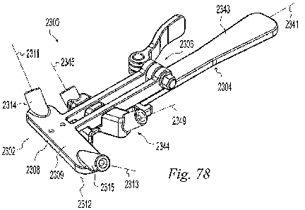
[0090] FIG. 78 is a perspective view of an illustrative example of a guide
according to the
present invention;
[0091] FIG. 79 is a top plan view of the guide of FIG. 78;
[0092] FIG. 80 is an exploded perspective view of the guide of FIG. 78;
[0093] FIG. 81 is an exploded top plan view of the guide of FIG. 78;
[0094] FIG. 82 is a perspective view of the guide of FIG. 78 showing a
position of the guide;
[0095] FIG. 83 is a perspective view of the guide of FIG. 78 showing a
position of the guide;
[0096] FIG. 84 is a perspective view of the guide of FIG. 78 showing a
position of the guide;
[0097] FIG. 85 is a perspective view of the guide of FIG. 78 showing a
position of the guide;
[0098] FIG. 86 is a perspective view of the guide of FIG. 78 showing a
position of the guide;
[0099] FIG. 87 is a perspective view of a tube useable with the guide of FIG.
78;
CA 02840400 2013-12-23
WO 2013/009574 PCT/US2012/045584
[00100] FIG. 88 is a side elevation view of the tube of FIG. 14;
[00101] FIG. 89 is a side elevation view of the guide of FIG. 78 in use
with an MTP joint;
[00102] FIG. 90 is a side elevation view of the guide of FIG. 78 in use
with an MTP joint;
[00103] FIG. 91 is a side elevation view of the guide of FIG. 78 in use
with an MTP joint;
[00104] FIG. 92 is a top plan view of the guide of FIG. 78 in use with an
MTP joint;
[00105] FIG. 93 is a side elevation view of the guide of FIG. 78 in use
with an MTP joint;
[00106] FIG. 94 is a top plan view of the guide of FIG. 78 in use with an
MTP joint;
[00107] FIG. 95 is a side elevation view of the guide of FIG. 78 in use
with an MTP joint;
[00108] FIG. 96 is a top plan view of the guide of FIG. 78 in use with an
MTP joint;
[00109] FIG. 97 is a side elevation view of the guide of FIG. 78 in use
with an MTP joint;
[00110] FIG. 98 is a top plan view of the guide of FIG. 78 in use with an
MTP joint;
[00111] FIG. 99 is a dorsal view of the metatarsus and phalanx of the
right second
metatarsophalangeal joint of the human foot showing tunnels formed utilizing
the guide of FIG.
78;
[00112] FIG. 100 is a medial view of the bones of FIG. 26;
[00113] FIG. 101 is a lateral view of the bones of FIG. 26; and
[00114] FIG. 102 is a perspective view of an illustrative example of a
guide according to
the present invention.
[00115] FIG. 103 is a perspective view of an illustrative example of a
guide according to
the present invention;
[00116] FIG. 104 is an exploded perspective view of the guide of FIG. 103;
[00117] FIG. 105 is a top plan view of the guide of FIG. 103;
16
CA 02840400 2013-12-23
WO 2013/009574 PCT/US2012/045584
[00118] FIG. 106 is a side sectional view of the guide of FIG. 103 taken
along line 106-
106 of FIG. 105;
[00119] FIG. 107 is a bottom plan view of the guide of FIG. 103;
[00120] FIG. 108 is a perspective view of a component pushbutton of the
guide of FIG.
103;
[00121] FIG. 109 is a perspective view of the guide of FIG. 103
highlighting a portion;
[00122] FIG. 110 is a cutaway perspective view of the guide of FIG. 103
detailing the
portion highlighted in FIG. 109 showing the operation of the pushbutton of
FIG. 108;
[00123] FIG. 111 is a cutaway perspective view of the guide of FIG. 103
detailing the
portion highlighted in FIG. 109 showing the operation of the pushbutton of
FIG. 108;
[00124] FIG. 112 is perspective view of an illustrative example of a saw
blade according
to the present invention useable with the guide of FIG. 103 and shown with a
powered
handpiece;
[00125] FIG. 113 is a top plan view of the saw blade of FIG. 112;
[00126] FIG. 114 is a side elevation view of the saw blade of FIG. 112;
[00127] FIG. 115 is a perspective view of the guide of FIG. 103 showing a
position of the
guide;
[00128] FIG. 116 is a top plan view of the guide of FIG. 103 showing the
position of FIG.
115;
[00129] FIG. 117 is a perspective view of the guide of FIG. 103 showing a
position of the
guide;
[00130] FIG. 118 is a top plan view of the guide of FIG. 103 showing the
position of FIG.
117;
17
CA 02840400 2013-12-23
WO 2013/009574 PCT/US2012/045584
[00131] FIG. 119 is a perspective view of the guide of FIG. 103 showing a
position of the
guide;
[00132] FIG. 120 is a top plan view of the guide of FIG. 103 showing the
position of FIG.
119;
[00133] FIG. 121 is a top plan view of the guide of FIG. 103 showing a
position of the
guide on a metatarsus;
[00134] FIG. 122 is a side elevation view of the guide of FIG. 103 showing
the position of
FIG. 121;
[00135] FIG. 123 is a top plan view of the guide of FIG. 103 showing a
position of the
guide on a metatarsus;
[00136] FIG. 124 is a side elevation view of the guide of FIG. 103 showing
the position of
FIG. 123;
[00137] FIG. 125 is a side elevation view of the guide of FIG. 103 showing
a position of
the guide on a metatarsus ready to receive the saw blade of FIG. 112;
[00138] FIG. 126 is a side elevation view of the guide of FIG. 103 showing
the position of
FIG. 125 with the saw blade advanced to cut the metatarsus;
[00139] FIG. 127 is a top plan view of the guide of FIG. 103 showing a
position of the
guide on a metatarsus;
[00140] FIG. 128 is a side elevation view of the guide of FIG. 103 showing
the position of
FIG. 127;
[00141] FIG. 129 is a side elevation view of the guide of FIG. 103 showing
the position of
FIG. 127 with the saw blade advanced to cut the metatarsus;
18
CA 02840400 2013-12-23
WO 2013/009574 PCT/US2012/045584
[00142] FIG. 130 is a top plan view of the guide of FIG. 103 showing the
guide in use to
reduce an osteotomy on a metatarsus;
[00143] FIG. 131 is a side elevation view of the guide of FIG. 103 showing
the position of
FIG. 30 and a pins being inserted to secure the osteotomy;
[00144] FIG. 132 is a perspective view of an osteotomy fixation screw
according to the
present invention;
[00145] FIG. 133 is a perspective view of an osteotomy fixation screw
according to the
present invention; and
[00146] FIG. 134 is a perspective view of an illustrative example of a
guide according to
the present invention.
19
CA 02840400 2013-12-23
WO 2013/009574 PCT/US2012/045584
DESCRIPTION OF THE ILLUSTRATIVE EXAMPLES
[00147] The following illustrative examples illustrate instruments and
techniques for
treating skeletal joints. Instruments and techniques according to the present
invention may be
used in conjunction with any skeletal joint but the illustrative examples are
shown in a size and
form most suitable for the joints of the hand and foot. In particular, the
illustrative examples
depict their use on metatarsophalangeal (MTP) joints of the human foot. The
illustrative
instruments and techniques are also suitable for use on metacarpophalangeal
(MCP) joints of the
human hand.
[00148] FIG. 1 illustrates the anatomic planes of the foot that are used
for reference in this
application. The coronal plane 10 extends from the medial aspect 12 to the
lateral aspect of the
foot and from dorsal 14 to plantar 16 and divides the foot between the toes
and heel. The sagittal
plane 18 extends anterior 20 to posterior 22 and dorsal 14 to plantar 16 and
divides the foot into
medial and lateral halves. The transverse plane 24 extends anterior 20 to
posterior 22 and medial
to lateral parallel to the floor 26.
[00149] FIGS. 2-4 illustrate the metatarsus 30 and proximal phalanx 50 of
the second
MTP joint of the right foot. The medial and lateral epicondyles 32, 34,
located on the medial-
dorsal and lateral-dorsal aspects of the metatarsus 30 respectively, are the
origins of the medial
and lateral proper collateral ligaments (PCLs) 36, 38 and the medial and
lateral accessory
collateral ligaments (ACLs) 40, 42 of the MTP joint. The medial PCL inserts at
the medial-
plantar aspect 52 and the lateral PCL inserts at the lateral-plantar aspect 54
of the proximal
phalanx 50. The ACLs fan out and insert into the plantar plate 44. The
metatarsus includes a
metatarsal head 46 having an articular surface 48 and the proximal phalanx
includes a phalangeal
CA 02840400 2013-12-23
WO 2013/009574 PCT/US2012/045584
head 56 having an articular surface 58. The metatarsus 30 further includes a
longitudinal axis 60
extending lengthwise down the center of the bone.
[00150] The terms "suture" and "suture strand" are used herein to mean any
strand or
flexible member, natural or synthetic, able to be passed through material and
useful in a surgical
procedure. The term "material" is used herein to mean implants, grafts,
fabric, tendon, ligament,
fascia, skin, muscle, bone, and any other material it is desirable to cut or
through which it is
desirable to pass a suture. The term "transverse" is used herein to mean
crossing as in non-
parallel. The term "bight" is used herein to mean a bend or loop formed in the
intermediate
portion of a suture.
[00151] The illustrative examples of FIGS. 5-50 depict instruments and
techniques to pass
a suture through a material. The instruments and techniques may be used to
pass a suture
through any material, at surgical sites anywhere in a patient's body, and for
any purpose. The
instruments and techniques are particularly useful where access to confined
spaces and the
ability to pass a suture through difficult to penetrate materials are needed.
For example, surgery
on the hands and feet often involve working in confined spaces around small
joints and tough
connective tissues through which it may be desirable to pass a suture.
[00152] FIGS. 5-17 depict an illustrative example of a suture passer 100.
The suture
passer 100 includes a housing 200, a needle assembly 300, and a barrel
assembly 400 mounted
together and operable to translate the needle assembly 400 between a first,
retracted position and
a second, extended position to manipulate a suture strand.
[00153] The housing 200 includes a hollow receiver portion 202 having a
hollow through
bore 204 with a longitudinal bore axis 206. An enlarged counter bore 208 is
formed coaxial with
the through bore 204 at a distal end 210 of the receiver 202. An intermediate
portion 212 of the
21
CA 02840400 2013-12-23
WO 2013/009574 PCT/US2012/045584
through bore 204 has flat side walls 214. A handle 220 extends downwardly and
proximally
from the receiver 202 and has a longitudinal handle axis 222. The handle axis
222 forms an
angle 224 with the bore axis 206. The angle 224 is in the range of 90 to 180
degrees; preferably
100 to 140 degrees; more preferably 110 to 130 degrees. In the illustrative
example of FIGS. 5-
17, the angle 224 is 120 degrees. A gusset 226 extends between the handle 220
and the receiver
202 for strength. One or more knobs extend from the housing to provide suture
strand anchor or
routing points. In the illustrative example of FIGS. 5-17, first and second
opposed side knobs
228, 230 and a downwardly projecting bottom knob 232 are mounted to the
receiver 202. Each
knob has a narrow waist 234 and an enlarged head 236 as shown with reference
to the bottom
knob 232. A suture strand may be wrapped or tied around the waist 234 to
secure or route the
suture. 0-rings 238, 240 are provided on the side knobs 228, 230 to grip a
wrapped suture to
facilitate securing and removing a suture strand. As a suture is wrapped
around the side knobs
228, 230, it wedges between the resilient 0-ring 238, 240 and knob compressing
the 0-ring. The
pressure of the 0-ring pressing the suture strand against the knob as well as
the deformation of
the 0-ring around the suture strand temporarily secures the suture.
[00154] The needle assembly 300 includes a piston 310, a stem 330, a
needle 350, and a
button 390. The piston 310 has a generally cylindrical body 312 with a
longitudinal axis 316
extending from a proximal end 318 to a distal end 320. A flange 322 extends
radially outwardly
from the body 312 near the distal end 320. The flange has opposed flattened
sides 324. A bore
326 (FIG. 12) is formed coaxially in the piston 310 at the distal end of the
body 312. The stem
330 includes an elongated hollow cylinder 332 having an outer diameter and an
inner bore 334
defining a longitudinal axis 336 extending from a proximal end 338 to a distal
end 340. The
needle 350 is a generally cylindrical member having a shank 352 with an outer
diameter defining
22
CA 02840400 2013-12-23
WO 2013/009574 PCT/US2012/045584
a longitudinal axis 354 extending from a proximal end 356 to a distal tip 358.
A flange 360
extends radially outwardly from the shank 352 at a position intermediate the
proximal and distal
ends. The needle 350 will be described in greater detail below. The button 390
has a generally
cylindrical body with a longitudinal axis 391 extending from a proximal end
393 to a distal end
395. A bore 398 (FIG. 12) is formed coaxially in the button 390 at the distal
end 395 of the
body. The proximal portion of the needle shank 352 fits within the inner bore
334 of the stem at
its distal end 340. The stem outer diameter, near its proximal end 338, fits
within the bore 326 of
the piston 310. The outer diameter of the piston 310 fits within the bore 204
of the receiver 202
in linear sliding relationship. The flat sides 324 of the piston engage the
flat side walls 214 of
the bore 204 to prevent the needle assembly from rotating relative to the
receiver 202. The
piston flange 322 abuts the proximal end of the intermediate portion 212 of
the bore 204 of the
receiver 202 to provide a stop to needle assembly proximal translation
relative to the receiver
202. The outer diameter of the piston 310, near its proximal end, fits within
the bore 398 of the
button 390 and the button 390 abuts a proximal end 216 of the receiver to
provide a stop to
needle assembly distal translation relative to the receiver 202. The joints
between the button 390
and piston 310, the piston 310 and the stem 330, and stem 330 and needle 350
are secured by
pressing, gluing, pinning, welding, or other suitable securing means.
Alternatively, two or more
of these components or various combinations of them may be made as a single
piece.
[00155] The barrel assembly 400 includes a barrel bushing 410, a barrel
430, and a foot
450. The bushing 410 has a generally cylindrical body 412 having a through
bore 414 with a
longitudinal axis 416 extending from a proximal end 418 to a distal end 420. A
flange 422
extends radially outwardly from the body 412 at a position intermediate the
proximal and distal
ends. An enlarged counter bore 424 (FIG. 12) is formed coaxial with the
through bore 414 at the
23
CA 02840400 2013-12-23
WO 2013/009574 PCT/US2012/045584
distal end 420 of the body 412. The barrel 430 includes an elongated hollow
cylinder 432 having
an outer diameter and an inner bore 434 defining a longitudinal axis 436
extending from a
proximal end 438 to a distal end 440. The foot 450 is a generally hook-shaped
member having a
hollow post 452 having an outer diameter and an inner bore 454 defining a
longitudinal axis 456
extending from a proximal end 458 of the cylinder to a distal end 460 of the
foot 450. The foot
will be described in greater detail below. The foot post 452 outer diameter
fits within the inner
bore 434 of the barrel at its distal end 440. The barrel 430 outer diameter,
near its proximal end
438, fits within the counter bore 424 of the bushing. A coiled compression
spring 250 fits
coaxially over the needle assembly 300 within the bore 204 of the receiver 202
and rests against
the distal end of the piston flange 322. The barrel assembly 400 fits
coaxially over the needle
assembly 300 and the outer diameter of the bushing 410, near its proximal end
418, fits within
the counter bore 208 of the receiver 202 and is pressed proximally until the
flange 422 abuts the
receiver distal end 210. The proximal end of the bushing retains the spring
250 within the bore
204. The joints between the foot 450 and barrel 430, the barrel 430 and
bushing 410, and the
bushing 410 and receiver 202 are secured by pressing, gluing, pinning,
welding, or other suitable
securing means. Alternatively, the bushing, barrel, foot, or any combination
of them may be
made as a single piece. Pressing the button 390 distally translates the needle
assembly from a
first, proximal, retracted position distally along the needle axis 354
compressing the spring 250
and extending the needle 350 through the foot 450 to a second, distal,
extended position.
Releasing the button 390 allows the spring 250 to expand and bias the needle
assembly 300 back
toward the first position. The needle assembly 300 of the illustrative example
of FIGS. 5-17 is a
linear arrangement mounted for linear, coaxial translation in the housing 200
and barrel assembly
400 with the needle projecting straight through the foot to increase rigidity
and power facilitating
24
CA 02840400 2013-12-23
WO 2013/009574 PCT/US2012/045584
driving the needle 350 through difficult to penetrate materials and access
confined spaces. The
barrel 430 may have a circular, polygonal, or any other cross sectional shape.
[00156] FIGS. 13 and 14 illustrate the foot 450 of the illustrative
example of FIGS. 5-17
in greater detail. The hooked portion of the foot 450 includes an elbow 462
having a first,
proximal portion 464 extending distally from the post 452 along a proximal
portion axis 465
diverging from the bore axis 456 at a first angle 466 relative to the bore
axis 456. A second,
distal portion 468 extends distally from the first portion 464 along a distal
portion axis 469
converging toward the bore axis 456 at a second angle 470 relative to the bore
axis 456. The
first and second angles 466, 470 are chosen to allow the foot to extend into a
confined space, for
example behind material such as a portion of soft tissue such as a tendon or
ligament, and
position the receiver 202 so as not to obstruct the users view of the foot and
needle. The first
angle 466 is in the range of 0 to 180 degrees; preferably 0 to 90 degrees;
more preferably 25 to
55 degrees; more preferably 35 to 45 degrees. In the illustrative example of
FIG. 14, the first
angle 466 is approximately 42 degrees. The second angle 470 is in the range of
0 to 90 degrees;
preferably 25 to 55 degrees; more preferably 35 to 45 degrees. In the
illustrative example of
FIGS. 13 and 14, the second angle 470 is also approximately 42 degrees. An eye
472 is formed
through the second portion 468, from a proximal facing surface 474 to a distal
facing surface
476, coaxial with the bore axis 456 for receiving the distal end of the needle
350 when the needle
is in the second position. A hole 478 defining a hole axis 480 extends through
the second
portion 468 from the distal surface 476 and intersecting the eye 472. The hole
478 permits
passing a suture strand from the distal surface 476 of the second portion 468
to the eye 472. The
hole axis 480 forms an angle 482 relative to the bore axis 456. The angle 482
is between parallel
to the proximal facing surface 474 of the second portion 468 and parallel to
the distal facing
CA 02840400 2013-12-23
WO 2013/009574 PCT/US2012/045584
surface of the first portion 464; preferably in the range of 45 to 135
degrees; more preferably 45
to 90 degrees. In the illustrative example of FIGS. 13 and 14, the hole angle
482 is
approximately 90 degrees relative to the bore axis 456. A groove 484 is formed
in the proximal
surface 474 of the second portion 468 communicating from the eye 472 to the
distal end 460. A
notch 486 is formed through the distal end 460 from the proximal surface 474
to the distal
surface 476 and communicating with the groove 484. The groove 484 and notch
486 are sized to
receive a suture strand and retain the strand on the distal end of the foot
450. The proximal
surface 474 of the second portion 468 of the foot 450 provides a supporting
platform for material
through which the needle 350 is passed. The eye 472 allows the needle 350 to
penetrate all the
way through the material and intercept a suture strand extending from the hole
478 to the groove
484.
[00157] FIGS. 15-17 illustrate the needle 350 of the illustrative example
of FIGS. 5-17 in
greater detail. A narrowed shaft 362 extends between the shank 352 and a sharp
tip 364 at the
distal end of the needle. A shoulder 366 defines the transition from the shank
352 to the shaft
362. The shaft 362 is generally rectangular in cross section with a top 368, a
bottom 370, and
opposing sides 372, 374. The corners 376 are rounded. The shaft 362 has a
height 378 between
the top 368 and bottom 370 and a width 380 between the sides 372, 374. Both
the height 378
and width 380 of the shaft are narrower than the shank 352. The width 380 of
the shaft 362 is
greater than its height 378. The ratio of the width 380 to the height 378 is
in the range of 1 to 3;
preferably 2 to 3. In the illustrative example of FIGS. 15-17 the ratio is
approximately 2.3. The
distal end of the shaft is tapered in the width dimension from the full width
to the tip 364. In the
illustrative example of FIGS. 15-17, the shaft is tapered on a single side in
the width dimension
to form a single-sided bevel 382. The distal end of the shaft is tapered in
the height dimension
26
CA 02840400 2013-12-23
WO 2013/009574 PCT/US2012/045584
from the full height to the tip 364. In the illustrative example of FIGS. 15-
17, the shaft is tapered
on opposite sides in the height dimension to form a chisel portion 384. A
notch 386 is formed in
the side of the shaft 362 through the shaft 362 from the top 368 to the bottom
370. The notch
386 has an opening width 388 measured parallel to the needle axis 354, a depth
389 measured
perpendicular to the needle axis 354, and a notch axis 392 forming an angle
394 to the needle
axis 354. In the illustrative example of FIGS. 15-17, the notch has parallel
side walls 396, 398
that are parallel to the axis 392. The notch width 388, depth 389, and angle
394 are selected to
optimize the ability of the needle 350 to capture and retain a suture strand
while avoiding
snagging other material through which the needle 350 passes. FIGS. 18A-18G
illustrate a
variety of needle designs having varying notch width, depth, and angle. The
present inventors
have determined that the balance between capturing and retaining a suture
strand and avoiding
snagging is optimized, in the case of a suture strand with a diameter D, when
the width of the
notch is in the range of 0.9D to 2D. A notch width of 0.9D creates a press fit
depending on the
resilient nature of the suture strand. Preferably, the notch width is in the
range of 1D to 1.5D.
Similarly, the notch depth is optimized when the depth is in the range of
0.75D to 3D. A notch
depth of 0.75D captures the suture but leaves a portion of the suture
projecting from the notch.
Preferably, the depth is in the range of 1D to 2D. The notch angle is in the
range of 30 to 90
degrees; preferably 35 to 55 degrees. In the illustrative example of FIGS. 15-
17, the notch was
optimized for a USP#2-0 suture having a diameter in the range of 0.300-0.339mm
and has a
width of 0.30mm and a depth of 0.46mm and an angle of 45 degrees. The notch
opens toward
the side of the needle 350 and suture passer 100. The bevel 382 leads from the
tip 364 of the
needle along the narrow side of the needle shaft 362 toward the opening of the
notch 386. The
needle may be sized to capture and pass one or more suture strands.
27
CA 02840400 2013-12-23
WO 2013/009574
PCT/US2012/045584
[00158]
FIGS. 19-25 illustrate loading a suture strand 500, having a first end 502 and
a
second end 504 into the suture passer 100 of FIGS. 5-17. A first end 502 of
the suture strand 500
is inserted through the hole 478 in the foot 450 from the distal surface 476
toward the eye 472
and extended past the proximal surface 474 as shown in FIGS. 19 and 20. The
first end 502 of
the suture strand is pulled distally to place the suture strand 500 in the
groove 484 as shown in
FIGS. 21 and 22. The suture strand 500 is wrapped over the distal end 460 in
the notch 486 and
pulled proximally over the distal surface 476 of the second portion of the
foot 450 as shown in
FIGS. 23 and 24. The ends 502, 504 of the suture strand are wrapped around the
side knobs 228
and 230 and retained by the 0-rings 238, 240. In the example of FIG. 25, the
suture strand ends
are routed proximally to the bottom knob 232 wrapped part-way around the
proximal side of the
knob 232 and secured on the side knob opposite the side on which the end was
routed such that
the suture strand is maintained near the center of the suture passer 100 and
better retained on the
foot 450.
[00159]
FIGS. 26-31 illustrate the operation of the suture passer 100. When the button
390 is pressed distally, the needle assembly 300 moves distally relative to
the housing and barrel
assembly along the straight-line motion axis 506 of the suture passer which is
coaxial with the
needle axis 354 and foot bore axis 456. As the needle 350 approaches the
suture strand 500, the
bevel 382 contacts the suture strand 500 and wedges it sideways increasing the
tension in the
suture as shown in FIGS. 26 and 27. Further advancement of the needle 350
moves the notch
386 toward alignment with the suture strand 500 until the tension in the
suture causes the suture
500 to move into the notch 386 as shown in FIGS. 28 and 29. Releasing pressure
on button 390
allows the spring 250 to bias the needle assembly proximally. Depending on the
resilience of the
suture 500 and how tightly it is secured to the knobs 228, 230, the needle may
or may not be able
28
CA 02840400 2013-12-23
WO 2013/009574 PCT/US2012/045584
to retract. By releasing one or both ends 502, 504 of the suture 500, the
suture ends can move
toward the foot 450 and allow the needle to retract and pull a bight 508 of
suture 500 proximally
toward the barrel 430 as shown in FIG. 30. Further retraction of the needle
350 pulls the bight
508 into the barrel 430 (FIG. 31) trapping the bight 508 between the needle
350 and barrel bore
434. To release the bight 508, the button 390 is pressed to advance the needle
350 out of the
barrel 430.
[00160] FIGS. 32-50 depict examples of the illustrative suture passer 100
in use to pass
sutures through a material to create a variety of stitches. Referring to FIG.
32, the suture passer
has been loaded as described relative to FIGS. 19-25. The foot 450 is
positioned adjacent
material 510 through which it is desired to pass the suture 500. The second
portion 468 of the
foot is positioned behind the material 510 with the proximal surface 474
supporting the material
510. Referring to FIG. 33, the button 390 is pressed to advance the needle 350
through the
material 510 and capture the suture 500 in the eye 472 of the foot 450.
Referring to FIG. 34, the
button 390 has been released and the suture ends 502 and 504 have been freed
from the knobs
228, 230 and allowed to move distally so that the needle 350 has retracted and
pulled a bight 508
of suture 500 through the material 510. Referring to FIG. 35, the button 390
has been pressed to
release the bight 508 and the first end 502 has been allowed to drop free from
the passer 100.
Referring to FIGS. 36 and 37, the second end 504 has been removed from the
foot 450 by pulling
the passer 100 proximally away from the bight or by pulling the suture 500
distally away from
the foot 450. The suture ends 502, 504 have been passed through the bight 508
and pulled to
form a stitch in the form of a hitch 512.
29
CA 02840400 2013-12-23
WO 2013/009574 PCT/US2012/045584
[00161] Referring to FIG. 38, instead of pulling the ends 502, 504 through
the bight 508,
the first end 502 has been pulled through the material 510 by pulling on one
side of the bight 508
to form a simple stich 514.
[00162] Referring to FIG. 39, the passer 100 is prepared for making a
running stitch by
pulling suture 500 distally through the foot to create slack 516 between the
foot 450 and material
510. Referring to FIG. 40, the slack 516 and the second end 504 have been
pulled proximally
and secured to the knobs 228, 230. Referring to FIG. 41 a second bight 518 has
been passed
through the material 510 in the same manner as the first bight 508 and the
slack 516 and second
end 504 have been released from the passer 100.
[00163] Referring to FIG. 42, the first and second ends 502, 504 have been
pulled through
to the front side of the material 510 by pulling on one side of each of the
bights 508, 518 to form
a mattress stitch 520 in the material 510.
[00164] Referring to FIG. 43, instead of the ends 502, 504 being pulled
through the
material the first end 502 has been placed through the first bight 508 and the
second end 504 has
been placed through the second bight 518 to form a modified mattress stitch
522 with each end
502, 504 secured by a hitch.
[00165] Referring to FIG. 44, a third bight 524 has been pulled through
the material in the
same manner as the first two bights 508, 518. A stitch may be formed by
placing one or both
ends 502, 504 through the bights 508, 518, 524 to lock the bights as shown in
FIG. 45.
[00166] Referring to FIG. 46, instead of placing the ends through the
bights, the second
bight 518 has been looped through the first bight 508, and the third bight 524
has been looped
through the second bight 518 to form a chain stitch 526.
CA 02840400 2013-12-23
WO 2013/009574 PCT/US2012/045584
[00167] Referring to FIGS. 47 and 48, another alternative to forming
stitches with three
bights is shown. Here, the second bight 518 has been cut to form third and
fourth ends 528, 530.
The third and fourth ends 528, 530 are pulled back through the material 510
and then the first
and third ends 502, 528 are placed through the first bight 508 to form a first
hitch 532 and the
second and fourth ends 504, 530 are placed through the third bight 524 to form
a second hitch
534.
[00168] Alternatively, as shown in FIGS. 49 and 50, the same construct
could be produced
by forming two bights 508, 518, and cutting through the slack 536 on the back
side of the
material 510 to produce third and fourth ends 538, 540 which with the first
and second ends 502,
504 are used to form hitches 542, 544.
[00169] The illustrative examples of FIGS. 5-50 have been shown in use to
pass suture
through material to form illustrative stitches. The invention is not limited
to the specific
instruments and methods depicted. Furthermore, it is to be understood that
instruments and
methods according to the invention may be used to pass any number of bights of
suture through
one or more materials and form any desirable construct.
[00170] The illustrative examples of FIGS. 51-77 depict instruments and
techniques to
pass a suture through a material. Instruments and techniques according to the
illustrative
examples of FIGS. 51-77 may be used to pass a suture through any material, at
surgical sites
anywhere in a patient's body, and for any purpose. Instruments and techniques
according to the
illustrative examples of FIGS. 51-77 are particularly useful to pass a suture
through a bone
tunnel in an orthopedic procedure. For example, it is often desirable to pass
a suture through a
bone tunnel which in turn is used to pass a graft into the tunnel or attach a
graft in the tunnel.
While suture passers in accordance with the illustrative examples of FIGS. 51-
77 may be used
31
CA 02840400 2013-12-23
WO 2013/009574 PCT/US2012/045584
with any material at any location, and in particular with any bone adjacent
any joint within a
patient's body, the illustrative examples are shown in use with a small bone
joint such as in a
hand or foot to form a tunnel in and pass a graft into a metacarpal or
metatarsal bone. In
particular, the illustrative examples are shown in use with a phalanx bone of
the foot.
[00171] FIG. 51 depicts an illustrative example of a suture passer 1100.
The suture passer
1100 includes a suture retriever 1110 and a suture 1150. The retriever 1110
includes a receiver
1112 able to receive and retain a portion of the suture 1150. In the
illustrative example of FIG.
51, the receiver 1112 includes a foot 1114 positionable on one side of a
material through which
the suture is to be passed. The foot 1114 has a proximal end 1116, a distal
end 1118, a front
surface 1115, a back surface 1117 and a longitudinal axis 1120 extending
between the proximal
and distal ends. The foot has an opening 1122 defining a passage through a
portion of the
receiver for receiving the suture 1150 and a sharp tip 1124 able to engage the
material and aid in
maintaining the foot 1114 in a desired location. In the illustrative example
of FIG. 51, the
retriever 1110 further includes a handle 1130 having a proximal end 1132, a
distal end 1134, and
a longitudinal axis 1136 extending between the proximal and distal ends. The
receiver 1112 may
be mounted directly to the distal end 1134 of the handle. In the illustrative
example of FIG. 51,
the receiver 1112 is offset from the handle. An extension 1140 having a
proximal end 1142, a
distal end 1144, and a longitudinal extension axis 1146 extends away from the
distal end 1134 of
the handle transverse to the handle axis 1136. The foot 1114 is mounted to the
distal end 1144 of
the extension 1140 and extends away from the extension 1140 transverse to the
extension axis
1146.
[00172] The suture 1150 includes a proximal end 1152 and a distal end
1154. The distal
end includes a stopper 1156. In the illustrative example of FIG. 51 the
stopper 1156 includes a
32
CA 02840400 2013-12-23
WO 2013/009574 PCT/US2012/045584
hook 1158 formed on the distal end 1154. For example, the distal end may be
bent, molded, heat
set, or otherwise formed into a hook shape. The hook 1158 includes a shank
1160, a bend 1162,
and a barb 1164. The hook 1158 is receivable in the opening 1122. As the hook
1158 is
advanced through the opening 1122, the barb 1164 and shank 1160 engage the
sides of the
opening 1122 and the barb 1164 moves toward the shank 1160. This movement
changes the
orientation of the hook to a receivable orientation in which the barb-shank
maximum dimension
is smaller than the opening 1122 maximum dimension and the hook passes through
the opening.
Once the hook 1158 is through the opening 1122, the barb 1164 springs away
from the shank
1160 and the hook orientation changes to a retention orientation. Pulling the
hook 1158 back
toward the opening causes the barb 1164 to engage the back surface 1117 of the
foot and resist
withdrawal. The bend of the hook 1158 is such that relatively small movement
of the barb 1164
is necessary for insertion of the hook through the opening 1122 but relatively
large movement of
the barb 1164, in the opposite direction, is necessary for removal. The hook
1158 may be
withdrawn by forcing the barb to straighten or by clipping the hook 1158 off
of the suture 1150.
[00173] The proximal end of the suture may be unmodified or it may include
a loop, knot,
hook, barb, or other feature for engaging another material.
[00174] In use, the receiver 1112 is positioned behind material through
which the suture
1150 is to be passed. The distal end 1154 of the suture is advanced through
the material and the
stopper 1156 is engaged with the receiver 1112. The receiver 1112 is then
withdrawn from
behind the material to advance the suture further and retrieve it partially or
fully through the
material. The suture 1150 may be used to connect the material to another
material. For example
the suture 1150 may be used to attach soft tissue to bone. The suture 1150 may
be used to
retrieve something through the material. For example, the suture 1150 may be
used to retrieve a
33
CA 02840400 2013-12-23
WO 2013/009574 PCT/US2012/045584
graft through a bone tunnel. In the illustrative example of FIG. 51, the foot
1114 may be
positioned adjacent a bone with the opening 1122 aligned with a tunnel formed
in the bone and
the tip 1124 engaged with the bone. The distal end 1154 of the suture 1150 may
be advanced
through the bone tunnel and opening 1122 until the hook 1158 engages the foot
1114. The
proximal end 1152 of the suture may be secured to a graft such as by tying,
stitching, looping,
knotting, hooking, or other securing mechanism. The foot may then be withdrawn
away from
the bone tunnel to retrieve the distal 1154 end of the suture and pull the
graft with it. Further
pulling of the suture advances the graft into the bone tunnel.
[00175] FIGS. 52-59 depict an illustrative example of a suture passer 1200
similar to that
of FIG. 51 and including a suture retriever 1300 and a suture 1400. In the
illustrative example of
FIGS. 52-59, the suture retriever 1300 includes a handle 1310, a receiver
1320, and a guide
1380. The handle 1310 includes a proximal end 1312, a distal end 1314, and a
longitudinal axis
1316 extending between the proximal and distal ends. The receiver 1320
includes a foot 1324
positionable on one side of a material through which the suture is to be
passed. The foot 1324
has a proximal end 1326, a distal end 1328, a front surface 1325, a back
surface 1327 and a
longitudinal axis 1330 extending between the proximal and distal ends. The
foot 1324 has an
opening 1332 having an opening axis and able to receiving the suture 1400. The
opening 1332
includes an enlarged counterbore 1333. The foot further includes a sharp tip
1334 able to engage
the material and aid in maintaining the foot 1324 in a desired location. The
receiver 1320 is
offset from the handle 1310. An extension 1340 having a proximal end 1342, a
distal end 1344,
and a longitudinal extension axis 1346 extends away from the distal end 1314
of the handle
transverse to the handle axis 1316. The foot 1324 is mounted to the distal end
1344 of the
extension 1340 and extends away from the extension 1340 transverse to the
extension axis 1346.
34
CA 02840400 2013-12-23
WO 2013/009574 PCT/US2012/045584
[00176] The guide 1380 includes a tube 1382 having an inner surface 1384,
an outer
surface 1386, a proximal end 1388, and a distal end 1390. The inner surface
1384 defines an
inner diameter and a longitudinal axis 1392. The tube 1382 is mounted to the
distal end 1314 of
the handle 1310 with the tube axis 1392 transverse to the handle axis 1316 and
coaxial with the
opening 1332 in the foot 1324. The handle 1310 axis 1316 forms an angle 1317
with the tube
axis 1392. The angle 1317 facilitates manipulating the retriever 1300 while
maintaining a line of
sight for the user and to prevent interference with tissues surrounding the
surgical site. The
angle 1317 may have any suitable value. Preferably the angle 1317 is in the
range of 90 to 270
degrees. The handle 1310 may also be mounted at any location around the
circumference of the
tube 1382. In the illustrative embodiment of FIGS. 52-59, the handle is
coplanar with the foot
1324. The tube 1382 includes a slot 1394 through the sidewall of the tube from
the inner surface
1384 to the outer surface 1386 and extending from the proximal end 1388 to the
distal end 1390.
The guide 1380 and foot 1324 define a space 1396 between them for receiving a
bone.
[00177] The suture 1400 includes a proximal end 1402 and a distal end
1404. The distal
end includes a stopper 1406. In the illustrative example of FIGS. 52-59 the
stopper 1406
includes a pledget 1408. The pledget 1408 is mounted to the suture 1400 such
as by adhering,
welding, crimping, molding or other suitable mounting method. The pledget 1408
may also be
formed as a unitary part of the suture. The pledget is resilient to allow it
to bend or compress to
fit through the opening 1332. It may also be toggled to one side such as for
example by bending
the suture adjacent the pledget 1408 to fit through the opening 1332. In the
illustrative example
of FIGS. 52-59, the pledget 1408 includes radially extending tabs 1410, 1412
that bend from
substantially perpendicular to the suture 1400 to substantially parallel to
the suture 1400 to
reduce the radial dimension of the pledget 1408 and allow it to pass through
the opening in a
CA 02840400 2013-12-23
WO 2013/009574 PCT/US2012/045584
receivable orientation. Once the pledget 1408 is through the opening 1332, the
tabs 1410, 1412
spring back to their initial position and resume a retention orientation. The
proximal end of the
suture 1400 includes a loop 1420. The loop may be formed by tying a knot in a
bight of a single
or multiple strand suture 1400, tying the ends of multiple strands together,
splitting a
monofilament strand, molding, or other suitable loop formation method. In the
illustrative
example of FIGS. 52-59, the loop is formed by molding a loop on a monofilament
strand.
[00178] FIG. 60 illustrates a drill assembly 1500 useable with the suture
passer 200. The
drill assembly 1500 includes a drill tube 1510 and an obturator 1560. The
drill tube 1510
includes a tubular body 1512 having a proximal end 1514, a distal end 1516, an
inner surface
1518, and an outer surface 1520. The inner surface 1518 defines an inner
diameter and a
longitudinal axis 1522 extending between the proximal and distal ends. In the
illustrative
embodiment of FIG. 60, a connector 1524 is mounted to the drill tube 1510 near
the proximal
end 1514. In the illustrative example of FIG. 60, the connector 1524 is a
female Luer-type
fitting. A stop 1528 extends radially outwardly from the body 1512.
[00179] The obturator 1560 includes an elongated body 1562 having a
proximal end 1564,
a distal end 1566, and a longitudinal axis 1568 extending between the proximal
and distal ends.
In the illustrative embodiment of FIG. 60, a connector 1570 is mounted to the
obturator 1560
intermediate the proximal and distal ends. In the illustrative example of FIG.
60, the connector
1570 is a male Luer-type fitting. The obturator 1560 is receivable in the
drill tube 1510 by
inserting the distal end 1566 of the obturator 1560 into the proximal end 1514
of the drill tube
1510 and advancing the obturator until the connectors engage. The obturator
1560 and drill tube
1510 are locked together by rotating the connectors relative to one another.
The drill tube 1510
and obturator 1560 have drilling tips 1526, 1572 that align when the obturator
is inserted into the
36
CA 02840400 2013-12-23
WO 2013/009574 PCT/US2012/045584
drill tube and locked. For example, the drilling tips 1526, 1572 may be formed
by assembling
the obturator 1560 and drill tube 1510, locking them together, and then
grinding the cutting tips
on the drill tube 1510 and obturator 1560 simultaneously. In the illustrative
example of FIG. 60,
when the drill tube 1510 and obturator 1560 are assembled, the drilling tips
1526, 1572 form a
diamond drill tip having primary bevels 1580 formed on opposed first and
second sides and
secondary bevels 1582 to provide relief and improve cutting. The outer
diameter of the drill tube
1510 and the counterbore 1333 of the opening 1332 are sized so that the drill
tube 1510 may be
received in the counterbore 1333.
[00180] FIGS. 61-70 illustrate the illustrative suture passer 200 of FIGS.
52-59 and the
illustrative drill assembly of FIG. 60 in use to form a bone tunnel and load a
graft into the tunnel.
In FIG. 61, the suture retriever 1300 has been positioned adjacent a bone 1600
with the foot 1324
on one side of the bone with the opening 1332 aligned with a desired exit
location for a bone
tunnel and the guide axis 1392 aligned with the desired tunnel axis. By
viewing through the tube
1382 along the axis 1392, the location of the tunnel entrance can be
visualized. The retriever
1300 is shown positioned adjacent a phalanx bone with the extension 1340 in
the joint space and
the guide positioned to form a tunnel from dorsal to plantar through the
proximal phalanx. The
guide may be positioned at any location around the joint to create bone
tunnels at any desired
location in the phalanx or the metatarsus. For example, the guide may be
positioned to create
tunnels for repairing or replacing a proper collateral ligament, accessory
plantar ligament, plantar
plate, or other structure in or around the joint.
[00181] In FIG. 62, the drill assembly 1500 has been guided via the inner
surface 1384 of
the guide tube 1382 to form a tunnel through the bone 1600. Stop 1528 abuts
the proximal end
1388 of the guide 1380 to limit the drilling depth. In the illustrative
examples of FIGS. 52-60,
37
CA 02840400 2013-12-23
WO 2013/009574 PCT/US2012/045584
the stop 1528 abuts the proximal end 1388 when the drill tube 1510 is received
in the
counterbore 1333. Alternatively, the opening in the foot may be sized to
engage the tip of the
drill to limit the depth or a depth stop may be omitted.
[00182] In FIG. 63, the obturator 1560 has been removed leaving the drill
tube 1510 in
place. Optionally, the drill tube 1510 could be removed or a one-piece drill
could be substituted
for the drill assembly 1500. However, by leaving the drill tube 1510 in place,
the drill tube 1510
locks the retriever 1300 in place on the bone, provides guidance for the
suture, and provides a
smooth passage for the suture.
[00183] In FIG. 64, the suture 1400 has been inserted until the stopper
1406 engages the
receiver 1320. In the example of FIG. 64, the pledget 1408 has been forced
through the opening
1332 in the foot 1324.
[00184] In FIG. 65, the drill tube 1510 has been removed leaving the
suture 1400 in place.
[00185] In FIG. 66, the suture 1400 has been pulled through the slot 1394
to free the
proximal end 1402 from the guide tube 1382. The slot 1394 simplifies
withdrawing the retriever
1300 from the surgical site. However, the slot 1394 may be omitted and the
proximal end 1402
of the suture threaded through the guide tube 1382 as the retriever 1300 is
withdrawn.
[00186] In FIG. 67, the retriever 1300 has been withdrawn from the
surgical site taking the
distal end 1404 of the suture 1400 with it and thereby further advancing the
suture 1400 into the
bone tunnel. The suture 1400 may be left attached to the retriever 1300 or it
may be separated
from the retriever by pulling the distal end 1404 back through the foot or
cutting off the distal
end 1404 of the suture.
[00187] In FIG. 68, the distal end 1404 of the suture 1400 has been cut
off to free it from
the retriever 1300 and the retriever 1300 removed.
38
CA 02840400 2013-12-23
WO 2013/009574 PCT/US2012/045584
[00188] In FIG. 69, a graft 1610 has been engaged with the proximal end
1402 of the
suture 1400 by threading it through the loop 1420. Alternatively, a graft or
any other material
may be attached to the distal end for pulling in the opposite direction. In
addition to being used
to retrieve a graft, the suture 1400 may be used as a definitive suture in a
repair or reconstruction.
Also, the suture 1400 may be used to pull another graft retrieval strand such
as, for example, a
larger or more flexible strand.
[00189] In FIG. 70, the suture 1400 has been pulled to advance it through
the bone tunnel
and pull the graft 1610 along with it to position the graft 1610 in the bone
tunnel and the suture
1400 has been removed.
[00190] FIG. 71 illustrates a suture inserter 1710 having an elongated
body 1712 with a
proximal end 1714, a distal end 1716, and a longitudinal axis 1718. The suture
inserter 1710
may be used to advance the suture 1400 into engagement with the receiver 1320
by pushing the
stopper 1406. The suture inserter 1710 or the suture inserter 1710 in
combination with the suture
may have a higher columnar strength than the suture alone and facilitate
advancing the suture
1400. In the illustrative example, the suture inserter includes a longitudinal
passage 1720 for
receiving the suture 1400 with the stopper 1406 adjacent the distal end 1716.
[00191] FIG. 72 illustrates a suture 1730 having two strands 1732 joined
to a stopper 1734
having a proximal end 1736 formed at an angle to the suture strands 1732 so
that the proximal
end 1736 will hook onto the retriever 1320. The suture 1730 is also shown with
the suture
inserter 1710 of FIG. 51 useable to push the stopper 1734. For use in passing
a graft, the suture
strands 1732 may be tied to form a loop, stitched to the graft, wrapped around
the graft, or
otherwise connected to the graft. The suture ends may also be used directly to
attach hard or soft
39
CA 02840400 2013-12-23
WO 2013/009574 PCT/US2012/045584
tissue, implants, or other materials at a surgical site. The suture strands
may also be used directly
as a ligament or tendon replacement.
[00192] FIG. 73 illustrates a suture 1740 having a loop 1742 retained by
swaging a ferrule
1744 to retain the proximal end 1746 of the suture 1740.
[00193] FIG. 74 illustrates a suture 1750 having a stopper 1752 formed of
a block of
resilient material such as, for example, a closed cell foam.
[00194] FIG. 75 illustrates a suture 1760 having a stopper 1762 joined to
a strand 1764 at
a pivot 1766 so that the stopper 1762 can toggle between a receiving position
generally more
parallel to the strand 1764 and a retaining position generally more
perpendicular to the strand
1764.
[00195] FIG. 76 illustrates an alternative foot 1770 to the foot 1324 of
FIG. 52. The foot
1770 has first and second opposable jaws 1772, 1774. The first jaw 1772 is
mounted for rotation
relative to the second jaw about a pivot 1776. The jaws 1772, 1774 are
moveable between a first
closed, position (shown) in which the jaw faces are adjacent one another and a
second, open
position (not shown) in which the first jaw 1772 is pivoted away from the
second jaw 1774 to
create a space between the jaws 1772, 1774 for receiving a suture 1778. The
jaws may be closed
on the suture 1778 to retain the suture and allow it to be retrieved. Any
suitable mechanism may
be used to move the first jaw relative to the second jaw. For example, a
control cable 1779 may
be mounted in the foot and moveable by a remote actuator to move the first jaw
1772 between
the first and second positions.
[00196] FIG. 77 illustrates an alternative foot 1780 to the foot 1324 of
FIG. 52. The foot
1780 has moveable member 1782 mounted for movement relative to an opening 1784
between a
first position in which the opening is not blocked and a suture 1786 may be
received in the
CA 02840400 2013-12-23
WO 2013/009574 PCT/US2012/045584
opening and a second position in which the member 1782 and edge of the opening
1784 grasp
the suture. Any suitable mechanism may be used to move the member 1782. For
example, a
control cable 1788 may be mounted in the foot and moveable by a remote
actuator to move the
member 1782 between the first and second positions.
[00197] The illustrative examples of FIGS. 51-77 have shown the suture
passer 1200 in
use to pass a suture used to pull a graft into a tunnel. However, a suture
passed by the suture
passer may be used in any way that sutures are known to be used. For example a
suture may be
used as a shuttle for pulling another suture, graft, or anything else from
bottom to top rather than
from top to bottom as depicted in the illustrative examples. Single strands,
double strands, or
any number of strands may be passed. Likewise one or more loops may be passed.
Any of these
may be used as a definitive suture in a repair or reconstruction, as a shuttle
for pulling another
material into a desired position, or for any other purpose.
[00198] FIGS. 78-86 illustrate an exemplary guide 2300 for guiding a
cutter to cut a bone.
In this illustrative example, the guide 2300 is configured as a drill guide to
guide a drill, punch,
pin, broach or the like to form holes in the bones adjacent the second MTP
joint of the right
human foot. The drill guide 2300 includes a pair of plate-like members 2302,
2304 joined at a
hinge 2306 allowing a single degree of freedom such that the members may be
pivoted between
a first position and a second position. The members include a plurality of
fixation holes for
receiving fixation devices, e.g. fixation pins or screws, to secure the
members to underlying
bones and guide holes to guide the formation of tunnels in the underlying
bones to facilitate soft
tissue repair, replacement, and/or augmentation around the joint. The first
member 2302 is
configured to overlie the metatarsus and the second member 2304 is configured
to overly the
phalanx.
41
CA 02840400 2013-12-23
WO 2013/009574 PCT/US2012/045584
[00199] The first member 2302 includes a planar top surface2309, a first
end 2308 having
fixation holes 2310, and a metatarsal guide portion 2312. The metatarsal guide
portion 2312 has
a feature for guiding a cutter. In the illustrative example of FIGS. 78-86,
the metatarsal guide
portion 2312 includes medial and lateral spaced apart, hollow, tubular
extensions 2314, 2315
each projecting upwardly and outwardly from the top surface 2309 and
configured as a drill
guide able to guide a drill, punch, broach, pin or the like. The tubular
extensions 2314, 2315 are
oriented so that their axes 2311, 2313 intersect below the metatarsal guide
portion 2312. A
mounting yoke 2320 having opposed spaced apart arms extends from the first end
2308 to a
second end 2322 defining a pair of eyelets 2324 which straddle a block 2326
mounted on the
second member 2304. The block 2326 has a hole 2328 aligned with the eyelets
2324. A bolt
2330 and nut 2325 join the eyelets 2324 and block 2326. A locking cam 2332 is
pinned to the
head 2334 of the bolt 2330 for relative rotation about a pin 2336 and includes
a lever 2337
extending from the cam for rotating the cam 2332 between a locked and unlocked
position. The
bolt 2330 and locking cam 2332 are operable to press the eyelets 2324 together
against the block
2326 to frictionally lock the members 2302, 2304 in relative angular
relationship.
[00200] The second member 2304 includes an elongated handle 2340 having a
longitudinal axis 2341, a planar top surface 2343, fixation holes 2338 and a
phalangeal guide
portion 2344. The phalangeal guide portion 2344 has a feature for guiding a
cutter. In the
illustrative example of FIGS. 78-86, the phalangeal guide portion 2344
includes medial and
lateral spaced apart, hollow, tubular extensions 2346, 2347 each projecting
upwardly and
configured as a drill guide able to guide a drill, punch, broach, pin or the
like along axes 2345,
2349. The phalangeal guide portion 2344 includes a pair of grooves 2348 for
receiving the yoke
2320 of the first member to increase the relative positional accuracy and
stability of the members
42
CA 02840400 2013-12-23
WO 2013/009574 PCT/US2012/045584
relative to one another when the members are locked in the second coaxial
position. The second
member 2304 further includes a head referencing member 2350 having a reference
surface 2351
for engaging an anatomic landmark. In the illustrative example of FIGS. 78-86,
the head
referencing member 2350 has a concave spherical surface able to engage the
articular surface of
the metatarsal head. Opposite the concave surface is a convex back surface
able to engage the
articular surface of the phalangeal head. The second member further includes a
pair of
oppositely, laterally extending bosses 2342 for receiving a band to secure the
guide 2300 to the
phalanx.
[00201] FIG. 82 depicts the guide 2300 locked in the first parallel
position. FIG. 83
depicts the guide 2300 with the cam 2332 unlocked by rotating lever 2337 and
the first member
2302 rotated part-way toward the second position. FIG. 84 depicts the guide
2300 with the first
member 2302 rotated further toward the second position. FIG. 85 depicts the
guide 2300 with
the first member rotated fully into the second position. FIG. 86 depicts the
guide 2300 with the
cam 2332 locked to fix the first and second members 2302, 2304 in the second
position.
[00202] The relative position and orientation of the reference surface
2351 of the head
referencing member 2350, the handle axis 2341, the handle top surface 2343,
the phalangeal
extensions 2346, 2347, and the metatarsal extensions 2314, 2315 are determined
from averaged
anthropometric data relating the metatarsal head articular surface, metatarsal
longitudinal axis,
and transverse plane of the human body to the medial and lateral PCL origins
and insertions
when the guide 2300 is locked in the second position and placed on the bone
with the reference
surface 2350 engaged with the metatarsal head 2106, the handle axis 2341
parallel to the axis110
of the metatarsus, and the handle top surface 2343 parallel to the transverse
plane such that the
43
CA 02840400 2013-12-23
WO 2013/009574 PCT/US2012/045584
metatarsal extension axes 2311, 2313 intersect the PCL origins and the
phalangeal extension
axes 2345, 2349 intersect the PCL origins.
[00203] FIGS. 87 and 88 illustrate an elongated tube 2400 that may be used
with guide
2300 to protect soft tissue, facilitate engaging a cutter with the guide, and
stabilize the cutter.
For example, a long narrow drill, punch, pin, broach, or the like may be
difficult to align with the
extensions 2314, 2315, 2346, 2347 and/or may be so flexible that it tends to
skive off the bone
surface. The tube 2400 includes a tubular shaft 2402 having a proximal end
2404, a distal end
2406, and a longitudinal axis 2408 extending from the proximal end 2404 to the
distal end 2406.
The proximal end is radially enlarged to form a knob 2410. The knob 2410
includes a counter
siffl( 2412 forming a funnel-like lead-in to the inner bore 2414 of the
tubular shaft 2402. The
outside of the shaft 2402 is sized to slide into the extensions of the guide
2300 and extend
through the guide 2300 to contact the underlying bone. The shaft 2402 provides
positive
guidance of the cutter to the bone surface. The knob 2410 provides the user
with a gripping
surface spaced away from the inner bore 2414 to protect the user from being
pricked by the
cutter as the cutter is engaged with the inner bore 2414. The countersink 2412
guides the cutter
into the inner bore 2414.
[00204] FIGS. 89-98 illustrate the guide 2300 in use to guide a cutter to
form holes in the
bones of the second MTP joint to facilitate, e.g., the reconstruction of the
joint. The guide 2300
is brought near the joint with the first and second members folded in the
first position as shown
in FIG. 89. The head referencing member 2350 is inserted into the joint space
between the
metatarsus 2100 and phalanx 2200 as shown in FIG. 90. The concave reference
surface 2351 is
registered with the convex articular surface 2108 of the metatarsal head and
the convex reference
surface opposite the concave reference surface is registered with the concave
articular surface of
44
CA 02840400 2013-12-23
WO 2013/009574 PCT/US2012/045584
the proximal phalanx. The guide handle axis 2341 is oriented parallel to the
axis110 of the
metatarsus and the guide handle top surface 2343 is oriented parallel to the
transverse plane as
shown in FIGS. 91 and 92. The first member is then pivoted into the second
position as shown
in FIGS. 93 and 94. The orientation of the guide 2300 may be checked again.
The cam is
actuated to lock the members relative to one another and fixation devices,
e.g. pins 2360, may be
placed in the guide fixation holes to fix the members to the bones as shown in
FIGS. 95 and 96.
An elastic band 2362 may be wrapped around the phalanx and engaged with the
bosses 2342 to
secure the second member 2304 to the phalanx in addition to, or as an
alternative to, the fixation
pins 2360.
[00205] Once the members are aligned and secured, the guide is used to
guide a cutter to
form one or more tunnels in the bones as shown in FIGS. 97 and 98. The cutter
2364 may be
engaged directly with an extension of a guide portion and advanced into the
bone. Alternatively,
an elongated tube 2400 may first be engaged with the guide portion and
extended to the bone
surface. The cutter may then be engaged with the elongated tube 2400 and
advanced into the
bone.
[00206] FIGS. 99-101 illustrate bone tunnels formed using guide 2300 and
ligaments
reconstructed using the tunnels. The metatarsal guide portion 2312 has guided
a cutter to form a
medial-dorsal tunnel 2500 extending from the medial PCL origin into the
metatarsus and a
lateral-dorsal tunnel 2502 extending from the lateral PCL origin into the
metatarsus. The tubular
extensions 2314, 2315 of the metatarsal guide portion 2312 are oriented so
that their axes
intersect below the dorsal surface of the metatarsus. Thus, the tunnels 2500,
2502 intersect
within the metatarsus and provide a path for fixing grafts 2501, 2503 to
reconstruct one or both
of the PCLs. A graft may be attached to the metatarsus by, e.g., pulling a
traction suture through
CA 02840400 2013-12-23
WO 2013/009574 PCT/US2012/045584
the tunnels and using it to pull the graft into the appropriate tunnel. For
example, to attach a
medial graft 2501, the traction suture may be threaded into the medial tunnel
2500 and out the
lateral tunnel 2502. The suture is then tensioned to draw the graft into the
medial tunnel. The
graft may be fixed by tying the suture, with a suture anchor, with an
interference anchor, or with
other suitable methods of fixation. A lateral graft 2503 may be similarly
positioned and both
medial and lateral grafts may be simultaneously positioned.
[00207] The phalangeal guide portion 2344 has guided a cutter to form a
tunnel 2510
extending from the medial-dorsal surface 2512 of the phalanx to the insertion
2514 of the lateral
PCL on the lateral-plantar surface of the phalanx. The guide has also guided a
cutter to form a
tunnel 2520 extending from the lateral-dorsal surface 2522 of the phalanx to
the insertion 2524
of the medial PCL on the medial-plantar surface of the phalanx. These two
phalangeal tunnels
cross each other without intersecting. Grafts may be pulled into these tunnels
by, e.g., passing a
traction suture through one of the tunnels and drawing the graft into the
tunnel. The holes 2560
are formed by fixation members 2360 used to hold the guide in place.
[00208] The illustrative guide of FIGS. 78-86 includes two separate
members hinged
together. Alternatively, the guide may be provided as two separate guides each
having a joint
reference surface and useable independently to drill tunnels in the metatarsus
and proximal
phalanx. Alternatively, the hinge may be removed and the two members combined
into one non-
movable unitary structure 2600 as shown in FIG. 102.
[00209] The illustrative guide of FIGS. 78-86 is configured to reference
to the anatomy of
the right second MTP joint of the human foot to guide a cutter to form tunnels
in the metatarsus
and phalanx that intersect the medial and lateral PCL origins and insertions
to facilitate routing
and attaching ligaments to reconstruct the PCLs. The guide may be mirrored for
use on the left
46
CA 02840400 2013-12-23
WO 2013/009574 PCT/US2012/045584
foot and the guide may be provided in sizes for different MTP joints and
various sized feet.
However, it has been found that the variation of the PCL origin and insertion
anatomy is
surprisingly small for the second MTP joint across a wide range of foot sizes
and it is possible to
provide a single sized guide for all left second MTP joints and another for
all right MTP joints
for feet from at least a woman's US size 7 to a man's US size 11.
[00210] The medial and lateral ACLs of the MTP joint have origins that are
co-located
with the medial and lateral PCL origins on the metatarsus. The ACLs then fan
out to insert into
the plantar plate close to where the plantar plate transitions into the
intermetatarsal ligament
(IML). Therefore, the same tunnels used to reconstruct the PCL origins may be
used to
reconstruct the ACL origins such that the illustrative guide 2300 configured
for PCL
reconstruction may also be used for ACL reconstruction.
[00211] The drill guide may have any number of cutter guides targeted at
any desired
anatomical feature. While the illustrative examples have depicted a guide
configured for ACL
and PCL reconstruction of the right human MTP joint, the guide may be
similarly configured to
target other ligament reconstructions or other surgical procedures at other
locations throughout
the body.
[00212] FIGS. 103-111 depict an illustrative example of a cut guide for
cutting a material
into two relatively moveable portions and relatively repositioning the
portions. For example, the
guide may be used as an osteotomy guide 3200 and used, e.g., in relatively
repositioning first and
second portions of a bone. For example, the guide 3200 may be used to shorten
a metatarsal
bone by moving proximal and distal portions of the bone closer together. The
illustrative
osteotomy guide 3200 provides a stable base with a cutter guide operable to
guide a cutter to
separate the bone into two, relatively moveable portions. The cutter guide is
further operable to
47
CA 02840400 2013-12-23
WO 2013/009574 PCT/US2012/045584
guide a cutter to form two parallel cuts transverse to the bone axis to remove
a predetermined
portion of the bone. The illustrative osteotomy guide 3200 provides a
reduction mechanism
operable to reduce a gap between the two bone portions with motion along a
predefined axis.
[00213] The guide 3200 includes a proximal base member 3202, an
intermediate inner
stage 3204, and a distal outer stage 3206 all mounted in relative translating
relationship. The
base member 3202 includes an elongated plate-like handle 3208 including a
plurality of fixation
holes 3210. First and second spaced apart, parallel guide arms 3212, 3213 are
rigidly joined to
the base member and extend opposite the elongated handle 3208.
[00214] The arms include annular notches 3214, 3215 for controlled
positioning of the
stages at discrete locations along the arms. The arms define parallel
translation axes 3216, 3217.
Each arm has a radially enlarged stop 3218, 3219 near its distal end against
which the outer stage
3206 may abut to define a distal limit of travel of the outer stage 3206 on
the arms 3212, 3213.
[00215] The inner stage 3204 is a generally box-like member having first
and second
opposed sides 3240, 3241 joined at one end by a first end wall defining a
cutter guide 3242 and
at an opposite end by a second end wall 3244. The second end wall 3244
includes fixation guide
grooves 3245 formed in a plane transverse to the translation axes 3216, 3217.
In the illustrative
example of FIGS. 103-111, the cutter guide 3242 is in the form of a saw guide
having a saw
blade guiding slot 3246. The sides 3240, 3241 include outwardly extending
bosses 3248, 3249
having through holes 3250, 3251 for receiving the guide arms 3212, 3213 in
sliding engagement
so that the inner stage 3204 may be moved axially along the translation axes
3216, 3217.
[00216] The saw blade guiding slot 3246 defines a plane oriented relative
to the
translation axes 3216, 3217 to guide a saw blade in a predetermined
orientation relative to the
axes 3216, 3217. In the illustrative example of FIGS. 103-111, the slot 3246
forms an angle
48
CA 02840400 2013-12-23
WO 2013/009574 PCT/US2012/045584
3252 (FIG. 106) relative to the plane defined by the axes 3216, 3217. This
angle 3252 is
selected to minimize the shear stresses on the healing osteotomy. For example,
in the illustrative
example of FIGS. 103-111, the angle is chosen based on typical metatarsal
anatomy. In a typical
patient standing upright on a level floor, the metatarsal axis 3108 forms an
angle of
approximately 10-40 degrees relative to the floor. It is desirable for the
osteotomy cut surfaces
to be parallel to the floor such that when the patient is standing upright
there is little or no shear
force in the plane of the healing bone surfaces. In use, the guide axes, 3216,
3217 are oriented
parallel to the metatarsal axis 3108. Therefore, the blade guiding slot 3246
is preferably oriented
at an angle 3252 of 10-40 degrees relative to the guide axes 3216, 3217 to
produce the desire cut.
More preferably, the angle 3252 is 15-30 degrees. In the illustrative example
of FIGS. 103-111,
the angle 3252 is 25 degrees.
[00217] A button 3260 is received in a hole 3261 formed in the first side
3240 and biased
by a spring 3263. The button 3260 includes a feature engageable with the
annular notches 3214
of the first arm 3212 to selectively lock the position of the inner stage 3204
relative to the arm as
will be explained more fully below. The feature may be, e.g., a ring, notch,
pin, or other feature
engageable with the annular notches 3214. The first side 3240 includes a
reference mark 3262 to
indicate the position of the inner stage relative to the outer stage to
indicate cut width as will be
explained more fully below. The second side 3241 includes a reference mark
3264 to indicate
the position of the inner stage relative to the outer stage to indicate bone
reduction as will be
explained more fully below.
[00218] The outer stage 3206 is a generally U-shaped member having first
and second
sides 3280, 3281 joined at a first end by a first end wall 3285 and open at
the second end. The
outer stage 3206 includes fixation holes 3288, 3289 for receiving fixation
devices, e.g. pins,
49
CA 02840400 2013-12-23
WO 2013/009574 PCT/US2012/045584
screws or the like, to attach the outer stage to the metatarsal bone near the
head. The outer stage
3206 further includes a first reference member 3220 extending downwardly away
from the stage
as best seen in FIG. 106 and a second reference member 3221 extending
proximally between the
first and second sides. The sides 3280, 3281 include through holes 3282, 3283
for receiving the
guide arms 3212, 3213 in sliding engagement so that the outer stage 3206 may
be moved axially
along the translation axes 3216, 3217. A button 3284, similar to button 3260,
is received in a
hole (not shown) in the second side 3281 and is biased by a spring 3287. The
button 3284 is
engageable with the annular notches 3215 to selectively lock the axial
position of the outer stage
relative to the arms. The first side 3280 includes reference marks, or indicia
3286, indicating the
relative position of the inner stage relative to the outer stage. The indicia
3286, reference mark
3262, and annular notches 3214, 3215 are arranged so that the reference mark
3262 aligns with
indicia 3286 to indicate the relative position of the inner stage, and thus
the saw slot 3246,
relative to the outer stage.
[00219] Further detail of buttons 3260 and 3284 and their operation is
illustrated in FIGS.
108-111 using button 3284 as an example. The button 3284 includes a shaft 3300
extending
from a first end 3302 to a second end 3304 along an axis 3306. The first end
3302 defines an
enlarged head 3308. The shaft includes a notch 3310 transverse to the axis
3306 and able to
receive a portion of the second guide arm 3215 in transverse sliding
relationship. A key 3312
projects into and blocks a portion of the notch 3310. The button 3284 and
spring 3287 are
placed into the hole in the second side 3281 of the outer stage 3206. The
second arm 3213 is
inserted through the hole 3283 in the second side 3281 of the outer stage 3206
and through the
notch 3310 in the button shaft 3300. The arm 3213 abuts a portion of the
button 3284 and retains
the button in the outer stage 3206. The spring 3287 biases the button 3284
outwardly causing the
CA 02840400 2013-12-23
WO 2013/009574 PCT/US2012/045584
key 3312 to press against the arm 3213. When the key 3312 is aligned with an
annular notch
3215, the spring biases the key 3312 into the notch 3215 and prevents the arm
3213 from sliding
in the hole 3283 relative to the stage 3206 as shown in FIG. 110. Pressing the
button 3284
inwardly compresses the spring 3287 and moves the key 3312 out of the groove
allowing the
stage 3206 to translate along the arm 3213 as shown in FIG. 111.
[00220] FIGS. 112-114 illustrate a saw blade 3400 useable with the guide
3200. The
blade may, e.g., be gripped in a chuck 3402 of a powered handpiece 3404 to
drive the blade to
cut a bone. In the illustrative example of FIGS. 112-114, the blade 3400 is an
oscillating blade
used with a powered oscillating saw. The blade 3400 is a generally plate-like
member having a
first end 3410, a second end 3412, and a longitudinal axis 3414 extending from
the first end 3410
to the second end 3412. The first end 3410 defines a hub 3416 adapted for
engagement with
the powered handpiece 3404. The second end 3412 defines cutting teeth 3418
adapted to cut
bone. An intermediate portion 3420 having parallel, planar top and bottom
surfaces 3422, 3424
connects the hub 3416 and teeth 3418. The teeth have a thickness 3426 defined
perpendicular to
the top and bottom surfaces 3422, 3424. In use, the blade produces a cut
having a width, or kerf,
equal to the thickness 3426. In order for the blade to produce a cut that
results in an osteotomy
reduction that is in whole units, the thickness 3426 is related to the angle
of the cut relative to the
direction of reduction of the osteotomy which is parallel to the axes 3216,
3217 of the guide
arms. To produce an osteotomy and resulting reduction of one unit length, the
thickness 3426 is
made equal to one unit length times the sine of angle 3252. In the
illustrative example of FIGS.
103-111, the minimum osteotomy is two millimeters which corresponds to a
single cut with the
saw blade 3400. Therefore, the thickness 3426 is equal to two millimeters
times the sine of 25
degrees or 0.845 mm. In the illustrative saw blade of FIGS. 112-114, the
intermediate portion
51
CA 02840400 2013-12-23
WO 2013/009574 PCT/US2012/045584
3420 has a thickness 3428 equal to the thickness 3426 of the teeth and the saw
slot 3246 in the
inner stage 3204 is sized to receive the intermediate portion 3420 in close
fitting relationship to
provide support to the blade 3400. In the illustrative embodiment of FIGS. 112-
114, the hub
3416 has a thickness 3430 less than the thickness 3428.
[00221] FIGS. 115-120 illustrate various positions of the illustrative
guide 3200 of FIGS.
3-11. FIGS. 115 and 116 illustrate the guide 3200 in an initial position in
which the outer stage
3206 abuts the stops 3218, 3219 and the inner stage 3204 is positioned for a
minimal cut. Indicia
may be provided to indicate the initial position. In the illustrative example
shown, engraved
arrows 3450, 3451 indicate the initial position of the outer stage 3206. An
arrow 3450 on the top
surface of the second side 3281 of the outer stage 3206 points distally at the
distal edge of the
surface. An arrow 3451 on the top of the stop 3219 points proximally at the
proximal edge of the
stop 3219. The outer stage 3206 is in the first, initial, or start position
when the arrows are
brought together until the stage 3206 abuts the stop 3219. The inner stage
3204 is in the initial
position when the reference mark 3262 is aligned with the first of the indicia
3286 labeled "2"
indicating that a single cut will remove bone resulting in 2mm of reduction.
Note that from this
position, the outer stage 3206 can be moved 2mm proximally relative to the
inner stage 3204 to
reduce the osteotomy, or in other words close the gap, created by a single cut
with the illustrative
saw blade of FIGS. 112-114. This is indicated by an arrow 3452 labeled
"REDUCE" on the top
surface of the outer stage 3206 and aligned 2mm distal of the reference mark
3264 on the inner
stage 3204. In the illustrative example, the stages 3204, 3206 and annular
notches 3214, 3215 of
the guide arms are arranged to allow positive relative positioning of the
stages 3204, 3206 in
2mm increments.
52
CA 02840400 2013-12-23
WO 2013/009574 PCT/US2012/045584
[00222] FIGS. 117 and 118 illustrate the guide in a second position in
which the inner
stage 3204 has been translated relative to the outer stage 3206 and base 3202
until the reference
mark 3262 is aligned with one of the indicia 3286 indicating the desired
osteotomy reduction if
more than 2mm is desired. In FIGS. 117 and 118, the reference mark 3262 is
aligned with the
indicia labeled "6" indicating that a second cut made with the guide in this
position will, in
combination with the first cut, remove a section of bone 6mm long as measured
parallel to the
guide axes 3216, 3217 and will thus result in a 6mm reduction parallel to the
guide axes 3216,
3217.
[00223] FIGS. 119 and 120 illustrate the guide in a third position in
which the outer stage
3206 has been translated relative to the inner stage 3204 and base 3202 until
the arrow 3452 is
aligned with the reference mark 3264 indicating that the osteotomy has been
reduced.
[00224] FIGS. 121-131 illustrate the illustrative guide 3200 of FIGS. 103-
111 in use to
form and reduce an osteotomy on a metatarsus 3100. In FIGS. 121 and 122, the
guide has been
placed in the initial position as shown in FIGS. 115 and 116 and placed over
the metatarsus
3100. The first reference member 3220 is inserted into the joint space until
the second reference
surface 3221 abuts the dorsal surface of the metatarsal head 3104. The guide
is pressed
proximally until the reference member 3220 abuts the metatarsal head 3104. The
handle
longitudinal axis 3209 is aligned with the metatarsal axis 3108. The top
surfaces of the stages are
leveled side-to-side and the cut plane is positioned parallel to the
transverse axis. In this
position, the guide 3200 is registered axially relative to the distal end of
the metatarsal head 3104
and all six degrees of freedom of the guide 3200 have been constrained.
[00225] Referring to FIGS. 123 and 124, the outer stage 3206 is affixed to
a first portion
of the bone, for example at or near the metatarsal head, with fixation members
3460, e.g. pins,
53
CA 02840400 2013-12-23
WO 2013/009574 PCT/US2012/045584
screws, bands, or the like. The handle 3208 is affixed at a second portion of
the bone proximal to
the first portion.
[00226] A cutter is engaged with the cutter guide and advanced to cut the
bone. In the
illustrative example of FIGS. 125 and 126 a saw blade 3400 is engaged with the
saw slot 3246
and reciprocated via a powered handpiece 3404 to separate the metatarsus into
a distal portion
affixed to the outer stage 3206 and a proximal portion affixed to the handle
3208.
[00227] If more than a 2mm osteotomy reduction is desired, the inner stage
3204 is
adjusted to the desired amount of reduction by pressing the button 3260 to
unlock the stage and
sliding it to reposition the cutter guide for a second cut. In the
illustrative example of FIGS. 127
and 128, the inner stage 3204 is adjusted to remove bone corresponding to a
6mm osteotomy as
indicated by the alignment of the reference mark 3262 with the indicia 3286
labeled "6".
[00228] The saw blade is advanced a second time to cut the bone and any
loose pieces of
bone are removed to yield the desired osteotomy as shown in FIG. 129.
[00229] The osteotomy is reduced by moving the outer stage with the
attached metatarsal
head until the osteotomy is closed. In the illustrative example of FIGS. 130
and 131, the button
3284 is pressed to unlock the outer stage 3206 and the outer stage 3206 and
affixed metatarsal
head 3110 are translated proximally parallel to the guide axes 3216, 3217
until the distal portion
of bone abuts the proximal portion of bone and the osteotomy is closed.
Complete reduction is
also indicated by the arrow 3452 aligning with the reference mark 3264.
[00230] One or more fixation devices, e.g. a screw, pin, wire, cable, or
the like, may be
used to affix the proximal and distal portions of bone. For example pins may
be inserted through
the metatarsus to join the portions of bone. Pins may be inserted freehand or
guided. In the
illustrative example of FIGS. 130 and 131, pins 3454 are inserted by guiding
them in grooves
54
CA 02840400 2013-12-23
WO 2013/009574 PCT/US2012/045584
3245 to orient them at a desired angle relative to the osteotomy; for example
perpendicular to the
osteotomy.
[00231] FIG. 132 depicts an illustrative example of a screw assembly 3461
useable as an
alternative to pins 3454 to join the bone portions. The screw assembly 3461
includes a screw
3462 having a threaded shaft 3464 with a distal self-drilling, self-tapping
tip 3466 and a proximal
head 3468. A driver 3470 is joined to the head 3468 of the screw in torque
transmitting
relationship. In the illustrative embodiment of FIG. 132 the screw 3462 and
driver 3470 are
formed as a unitary member with a tapered connecting portion 3472 able to
transmit torque but
able to be broken by bending the assembly at the portion 3472.
[00232] FIG. 133 depicts an illustrative example of a screw assembly 3480
similar to
assembly 3461 but having three screws 3462 stacked to form the assembly. In
use, after the
distal screw is driven, it is separated from the others by bending at tapered
connection 3482.
Each subsequent screw may be driven and separate in like manner. In this way,
three screws
may be driven in quick succession without the need to take time to load each
individual screw on
a driver or change the driver and without risk of dropping a screw.
[00233] FIG. 134 depicts an illustrative example of a guide 3500 similar
to the illustrative
guide 3200 of FIGS. 103-111. However, guide 3500 includes slots 3502 in the
sides 3240, 3241
of the inner stage 3204 near the second end wall 3244 to receive a modular
drill/fastener guide
3504 having a frame 3506 with outwardly extending tabs 3508 engageable with
the slots 3502.
A tubular guide member 3510 is mounted on the frame 3506. The tabs and slots
allow the
drill/fastener guide 3504 to be engaged with the inner stage 3204 in
predetermined known
relationship to the guide axes 3216, 3217 and thus the osteotomy when the axes
3216, 3217 are
aligned with the bone axis 3108. In use, after the osteotomy is reduced, the
drill/fastener guide
CA 02840400 2013-12-23
WO 2013/009574
PCT/US2012/045584
3504 is mounted on the inner stage 3204 and used to guide drills, pins, screws
or any other
suitable member into the underlying bone in a predetermined orientation. In an
alternative
example, the drill/fastener guide 3504 is permanent part of the guide 3500.
[00234] The
illustrative instruments and methods have been shown in use to create and
reduce an osteotomy on a metatarsal bone adjacent the second MTP joint of the
human foot.
However, guides and methods within the scope of the invention may be used with
any bone in
the foot, hand, or other part of a patient's body. Likewise, the illustrative
instruments and
methods have been shown in use to carry out a constrained, axial, linear
reduction of the
osteotomy. However, other reductions also fall within the scope of the
invention. For example,
the guide can be configured to move the cut portions of bone linearly at some
angle transverse to
the bone axis to produce an offset reduction. In another example, the guide
can be configured to
move the cut portions of bone relatively along a curved path such as for
example for reducing a
wedge osteotomy. For example, the guide may include hinges, curved tracks, or
the like to
produce any desired reduction motion.
[00235] The
illustrative instrument has been shown with a base, inner stage, and outer
stage. However, the first and second stages may be mounted for relative motion
without the
need for a separate base member. For example, the first stage may be connected
to a first bone
portion and the second stage may be connected to a second bone portion. The
first stage may
include a cutter guide to guide a cutter to cut the bone. The first stage may
include additional
cutter guides to guide additional cuts. For example the first stage may
include a series of spaced
apart saw blade slots to guide a saw blade to remove a desired thickness of
bone. The stages
may then be moved relative to one another to reduce the osteotomy.
56