Note: Descriptions are shown in the official language in which they were submitted.
CA 02841538 2014-01-10
WO 2013/009966
PCT/US2012/046430
METHODS AND DEVICES FOR KNEE JOINT REPLACEMENT WITH ANTERIOR
CR.UCTATE LIGAMENT SUBSTITUTION
CROSS REFERENCES
[0001] The present application claims priority to U.S. Provisional Patent
Application No.
61/507,434 entitled "Methods and Devices for Knee Joint Replacement with
Anterior
Cruciate Ligament Substitution" filed July 13, 2011, which is hereby
incorporated by
reference in its entirety.
FIELD OF THE INVENTION
[0002] The present invention relates to methods and devices for knee joint
replacement with
anterior cruciate ligament (ACL) substitution, and in particular to methods
and devices for
substituting a prosthesis for an ACL.
BACKGROUND OF THE INVENTION
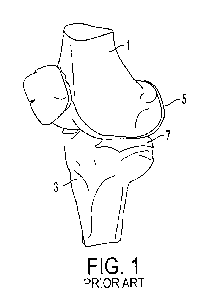
[0003] FIG. 1 illustrates a typical knee joint including a femur 1 and a tibia
3, shown with
healthy femur cartilage 5 and healthy tibia cartilage 7. The knee joint
includes three primary
elements: a medial tibiofemoral joint, a lateral tibiofemoral joint, and a
central
patellofemoral joint. Joint trauma or diseases such as osteoarthritis and
rheumatoid arthritis
can cause severe damage to one or more of these elements. In a case where one
or more of
the knee elements are traumatized or diseased, while the other one or two knee
elements are
healthy, the traumatized or diseased element(s) can be replaced in a partial
knee replacement
surgical procedure. In a case where all three primary elements are traumatized
or diseased,
all three elements can be replaced in a total knee replacement surgical
procedure.
[0004] In both partial and total knee replacement surgical procedures, the
traumatized or
diseased ones of the knee's bony surfaces, e.g., femur, tibia, and patella,
can be replaced by
prosthetic components. The knee's soft-tissue structures, particularly
ligaments surrounding
the knee joint, can be largely left intact. The knee's major ligament
structures include medial
and lateral collateral structures, and anterior and posterior cruciate
ligaments. These
ligamentous structures play a significant role in controlling the motion and
stability of a knee
joint. With regards to the cruciate ligaments, the posterior cruciate ligament
(PCL) is
generally present and well-functioning in patients undergoing partial or total
knee
replacement surgery. However, in at least some patients, the anterior cruciate
ligament
(ACL) can be absent or non-functional at surgery due to prior trauma or
gradual degradation.
CA 02841538 2014-01-10
WO 2013/009966
PCT/US2012/046430
[0005] Traditional partial knee replacement prostheses have no mechanism for
substitution of
ACL function. Consequently, patients with an absent or non-functional ACL may
end up
receiving total joint replacement, which is a generally more invasive
procedure than partial
knee replacement and which replaces the healthy element(s) of the patient's
knee.
Alternatively, instead of total knee replacement, patients with an absent or
non-functional
ACL may undergo additional surgery prior to a partial knee replacement
surgical procedure
to reconstruct the ACL, such as with a soft tissue graft.
[0006] In traditional total knee replacement surgical procedures, patients
receive a type of
prosthesis, e.g., a cruciate retaining (CR) type implant, that allows the
present and
well-functioning PCL to be retained. However, even for patients who have a
functional
ACL, the ACL is traditionally resected during surgery prior to implantation of
a CR type
implant because of difficulty in achieving optimal soft-tissue balancing and
component
placement with both the ACL and PCL present. However, traditional CR
prostheses have no
mechanism for substitution of the ACL function. Consequently, following CR
prosthesis
implantation, the knee shows abnormal motion patterns characterized by
features such as
reduced tibial internal rotation and paradoxical anterior femoral translation.
[0007] Accordingly, there remains a need for improved knee prostheses and
methods for
treating disease and trauma affecting the knee.
SUMMARY OF THE INVENTION
[0008] The present invention generally provides methods and devices for knee
joint
replacement with anterior cruciate ligament (ACL) substitution. In one aspect,
a medical
device is provided that includes a tibial implant, a femoral implant, and a
post. The tibial
implant has an inferior surface and an opposite, superior surface. The
inferior surface is
configured to be fixed to a tibia of a patient. The femoral implant is
mateable to the tibial
implant and has an inferior surface and an opposite, superior surface. The
superior surface of
the femoral implant is configured to be fixed to a femur of the patient, and
the tibial implant
is configured to articulate relative to the femoral implant when the tibial
implant is fixed to
the tibia and the femoral implant is fixed to the femur. The post extends from
the superior
surface of the tibial implant near an edge thereof. The post is configured to
be substantially
centered on the tibia when the tibial implant is fixed thereto such that the
post simulates an
ACL when the tibial implant is fixed to the tibia and the femoral implant is
fixed to the
femur.
- 2 -
CA 02841538 2014-01-10
WO 2013/009966
PCT/US2012/046430
[0009] The tibial implant can have a variety of configurations. The tibial
implant can have a
medial compartment configured to be seated on a medial surface of the tibia
with a first
portion of the tibial implant being seated on or over the tibia's medial
surface and a second,
substantially smaller portion of the tibial implant being seated on or over
the tibia's lateral
surface. The tibial implant can have a lateral compartment configured to be
seated on a
lateral surface of the tibia with a first portion of the tibial implant being
seated on or over the
tibia's lateral surface and a second, substantially smaller portion of the
tibial implant being
seated on or over the tibia's medial surface. The tibial implant can have
medial and lateral
compartments. The lateral compartment can be configured to be seated on a
lateral surface of
the tibia such that the lateral surface is substantially covered by the
lateral compartment. The
medial compartment can be configured to be seated on a medial surface of the
tibia such that
the medial surface is substantially covered by the medial compartment.
[0010] The post can have a variety of configurations. The post can be
asymmetric in sagittal,
coronal, and transverse planes. The post can be integrally formed with the
tibial implant, or
the post can be a discrete element configured to couple to the tibial implant.
[0011] In some embodiments, the device can include a femoral notch structure
coupled to the
femoral implant. The femoral notch structure can be configured to prevent the
post from
impinging on a lateral surface of the femur through a full range of knee
flexion when the
tibial implant is fixed to the tibia and the femoral implant is fixed to the
femur. The post can
be configured to articulate relative to the femoral notch structure.
[0012] In another aspect, a medical method is provided that includes
implanting a partial
knee prosthesis in a patient to replace one of a medial tibiofemoral joint of
a knee and a
lateral tibiofemoral joint of the knee such that an inferior surface of a
tibial implant of the
knee prosthesis faces a tibia of the knee, a superior surface of the tibial
implant faces an
inferior surface of a femoral implant of the knee prosthesis, a superior
surface of the femoral
implant faces a femur of the knee, and a post extending from the superior
surface of the tibial
implant functions as a substitute for an ACI, of the knee. The tibial implant
and the post are
configured to articulate relative to the femoral implant, and the post does
not impinge on a
lateral surface of the femur when the post articulates relative to the femoral
implant through a
full range of knee flexion.
[0013] In another embodiment, a medical method is provided that includes
implanting a total
- 3 -
CA 02841538 2014-01-10
WO 2013/009966
PCT/US2012/046430
knee prosthesis in a patient to replace both of a medial tibiofemoral joint of
a knee and a
lateral tibiofemoral joint of the knee such that an inferior surface of a
tibial implant of the
knee prosthesis faces a tibia of the knee, a superior surface of the tibial
implant faces an
inferior surface of a femoral implant of the knee prosthesis, a superior
surface of the femoral
implant faces a femur of the knee, and a post extending from the superior
surface of the tibial
implant functions as a substitute for an ACL of the knee. The tibial implant
and the post are
configured to articulate relative to the femoral implant, and the post does
not impinge on a
lateral surface of the femur when the post articulates relative to the femoral
implant through a
full range of knee flexion.
BRIEF DESCRIPTION OF THE DRAWINGS
[0014] The invention will be more fully understood from the following detailed
description
taken in conjunction with the accompanying drawings, in which:
[0015] FIG. 1 (PRIOR ART) is a perspective view of a typical normal human
knee;
[0016] FIG. IA is view of one embodiment of a knee prosthesis having an ACL-
substitution
member including a plurality of discrete pieces;
[0017] FIG. I B is a top view of one embodiment of a knee prosthesis;
[0018] FIG. IC is a sagittal cross-sectional view of the knee prosthesis of
FIG. I B;
[0019] FIG. ID is a coronal cross-sectional view of the knee prosthesis of
FIG. I B;
[0020] FIG. I E is a side view of one embodiment of a knee prosthesis
including an
ACL-substitution member and a femoral notch structure configured to engage
through a full
range of knee motion;
[0021] FIG. IF is top, partial view of one embodiment of a tibial insert;
[0022] FIG. 1G is coronal section view B-B of the tibial insert of FIG. IF and
a femoral
implant;
[0023] FIG. 2 is a posterior perspective view of one embodiment of a medial
knee prosthesis
attached to a tibia and a femur;
[0024] FIG. 3 is a posterior perspective view of one embodiment of a lateral
knee prosthesis
-4-
CA 02841538 2014-01-10
WO 2013/009966
PCT/US2012/046430
attached to a tibia and a femur;
[0025] FIG. 4 is a top view of a tibial insert of the medial knee prosthesis
of FIG. 2 seated on
the tibia;
[0026] FIG. 5 is a side perspective view of the tibial insert of FIG. 4;
[0027] FIG. 6 is a top view of a tibial insert of the lateral knee prosthesis
of FIG. 3;
[0028] FIG. 7 is a side view of the tibial insert of FIG. 6;
[0029] FIG. 8 is a perspective view of the tibial insert of FIG. 6;
[0030] FIG. 8A is a side view of one embodiment of a knee prosthesis including
a tibial post
located substantially anterior to tibial center;
[0031] FIG. 8B is top view of the femoral component of the prosthesis of FIG.
8A;
[0032] FIG. 8C is a top view of the prosthesis of FIG. 8A;
[0033] FIG. 9 is a top view of the tibial insert of the lateral knee
prosthesis of FIG. 3 seated
on the tibia;
[0034] FIG. 10 is another top view of the tibial insert of FIG. 9;
[0035] FIG. 11 is a perspective view of one embodiment of a medial knee
prosthesis attached
to a tibia, the medial knee prosthesis including a post gradually blending
into a tibial insert of
the prosthesis;
[0036] FIG. 12 is a schematic view of one embodiment of a lateral knee
prosthesis including
a tibial post and a tibial insert, the tibial post having a lateral edge
extending back to a
posterior edge of the tibial insert;
[0037] FIG. 13 is a top view of the medial knee prosthesis of FIG. 2 attached
to the tibia;
[0038] FIG. l 3A is a top view of an embodiment of a lateral knee prosthesis
attached to a
tibia;
[0039] FIG. 14 is a side perspective view of the medial knee prosthesis of
FIG. 13;
- 5 -
CA 02841538 2014-01-10
WO 2013/009966
PCT/US2012/046430
[0040] FIG. 14A is a side perspective view of the lateral knee prosthesis of
FIG. 13A;
[0041] FIG. 15 is a top view of one embodiment of a medial knee prosthesis
attached to a
tibia, the prosthesis including a discrete femoral notch structure and a
discrete femoral
implant;
[0042] FIG. 16 is a side perspective view of the medial knee prosthesis of
FIG. 15;
[0043] FIG. 17 is a perspective view of one embodiment of a lateral knee
prosthesis in an
extended or closed position;
[0044] FIG. 18 is a perspective view of the prosthesis of FIG. 17 in a flexed
or open position;
[0045] FIG. 19 is a top view of one embodiment of a lateral knee prosthesis
attached to a
tibia, the prosthesis including a post having a rounded top;
[0046] FIG. 20A is a side schematic view of the prosthesis of FIG. 19;
[0047] FIG. 20B is a side schematic view of one embodiment of a lateral knee
prosthesis
attached to a tibia, the prosthesis including a post having an angled or
chamfered top;
[0048] FIG. 20C is a side view of an embodiment of a bone shaping tool;
[0049] FIG. 20D is a top view of another embodiment of a bone shaping tool
adjacent to an
embodiment of a femoral component;
0050] FIG. 20E is a top view of an embodiment of a femoral trial component
that includes
one or more guiding slots;
[0051] FIG. 20F is a side view of an embodiment of a trial tibial insert that
has a larger size
than an embodiment of a tibial insert;
[0052] FIG. 20G is a side view of the trial tibial insert of FIG. 20F
positioned adjacent a
femoral bone and an embodiment of a femoral component;
[0053] FIG. 21 are top schematic views of one embodiment of a lateral knee
prosthesis
including a tibial post and a femoral intercondylar structure, the post, an
anterior surface, and
a lateral surface of the femoral intercondylar structure having concentric
circular profiles;
- 6 -
CA 02841538 2014-01-10
WO 2013/009966
PCT/US2012/046430
[0054] FIG. 21A is top, partial view of one embodiment of a prosthesis
including a tibial post
including angled cuts;
[0055] FIG. 21B is a perspective, partial view of the prosthesis of FIG. 21A;
[0056] FIG. 21C is a sagittal section view A-A of the tibial insert of FIG.
21A;
[0057] FIG. 21D is a coronal section view B-B of the tibial insert of FIG.
21A;
[0058] FIG. 22 is a top view of one embodiment of a medial knee prosthesis
attached to a
tibia, the prosthesis having a convex tibial post and a convex femorai
intercondylar notch;
[0059] FIG. 23 is a schematic, sagittal plane cross-sectional view of the
prosthesis of FIG.
22;
[0060] FIG. 24 is a schematic, sagittal plane cross-sectional view of one
embodiment of a
medial knee prosthesis, the prosthesis having a concave tibial post and a
convex femoral
intercondylar notch;
[0061] FIG. 25 is a schematic, sagittal plane cross-sectional view of one
embodiment of a
medial knee prosthesis, the prosthesis having a convex tibial post and a
concave femoral
intercondylar notch;
[0062] FIG. 26 is a schematic, sagittal plane cross-sectional view of one
embodiment of a
medial knee prosthesis, the prosthesis having a flat tibial post and a convex
femoral
intercondylar notch;
[0063] FIG. 27 is a schematic, sagittal plane cross-sectional view of one
embodiment of a
medial knee prosthesis, the prosthesis having a convex tibial post and a flat
femoral
intercondylar notch;
[0064] FIG. 28 is a top view of one embodiment of a lateral knee prosthesis
attached to a
tibia, the prosthesis having a concave tibial post and a convex femoral
intercondylar notch;
[0065] FIG. 29 is a schematic, corona] plane cross-sectional view of the
prosthesis of FIG.
28;
[0066] FIG. 30 is a schematic, coronal plane cross-sectional view of one
embodiment of a
lateral knee prosthesis attached to a tibia, the prosthesis having a flat
tibial post and a flat
- 7 -
CA 02841538 2014-01-10
WO 2013/009966
PCT/US2012/046430
femoral intercondylar notch;
[0067] FIG. 31 is a schematic, coronal plane cross-sectional view of one
embodiment of a
lateral knee prosthesis attached to a tibia, the prosthesis having a convex
tibial post and a
convex femoral intercondylar notch;
[0068] FIG. 32 is a schematic, corona' plane cross-sectional view of one
embodiment of a
lateral knee prosthesis attached to a tibia, the prosthesis having a flat
tibial post and a convex
femoral intercondylar notch;
[0069] FIG. 33 is a schematic, coronal plane cross-sectional view of one
embodiment of a
lateral knee prosthesis attached to a tibia, the prosthesis having a convex
tibial post and a flat
femoral intercondylar notch;
[0070] FIG. 34 is a top perspective view of one embodiment of a total knee
replacement
prosthesis;
[0071] FIG. 35 is a perspective view of one embodiment of a total knee
replacement
prosthesis attached to a femur, the prosthesis being in an extended or closed
position and
including a femoral notch structure;
[0072] FIG. 36 is a perspective view of the prosthesis of FIG. 35 not attached
to bone and in
a flexed or open position;
[0073] FIG. 37 is a perspective view of the prosthesis of FIG. 34 attached to
a femur and
showing a representation of a PCL ligament;
[0074] FIG. 38 is a perspective view of the prosthesis of FIG. 37 not attached
to bone and
with the representation of the PCL ligament in positions corresponding to
different knee
flexion angles;
[0075] FIG. 39 is a top view of a tibial implant of the prosthesis of FIG. 34;
[0076] FIG. 40 is a side view of the tibial implant of FIG. 39;
[0077] FIG. 41 is a perspective view of the tibial implant of FIG. 39;
[0078] FIG. 42 is a top view of the tibial implant of FIG. 39;
- 8 -
CA 02841538 2014-01-10
WO 2013/009966
PCT/US2012/046430
[0079] FIG. 43 is another top view of the tibial implant of FIG. 39;
[0080] FIG. 44 is a perspective view of one embodiment of a total knee
replacement
prosthesis including a post gradually blending into a tibial insert of the
prosthesis;
[0081] FIG. 45 is a schematic view of one embodiment of a total knee
replacement prosthesis
including a tibial post and a tibial insert, the tibial post having a lateral
edge extending back
to a posterior edge of the tibial insert;
[0082] FIG. 46 is a perspective view of one embodiment of a total knee
replacement
prosthesis attached to a femur and having a post with a height configured to
avoid
impingement with the lateral femoral condyle;
[0083] FIG. 47 is a top view of one embodiment of a total knee replacement
prosthesis
including a post having a rounded top;
[0084] FIG. 48 is a side schematic view of the prosthesis of FIG. 47;
[0085] FIG. 49 is a perspective view of one embodiment of a total knee
replacement
prosthesis in an extended or closed position;
[0086] FIG. 50 is a perspective view of the prosthesis of FIG. 49 in a flexed
or open position;
[0087] FIG. 51 is a top view of one embodiment of a total knee replacement
prosthesis
having a convex tibial post and a convex femoral intercondylar notch;
[0088] FIG. 52 is a top view of one embodiment of a total knee replacement
prosthesis
having a concave tibial post and a convex femoral intercondylar notch;
[0089] FIG. 53 is a schematic, coronal plane cross-sectional view of the
prosthesis of FIG. 52
attached to a tibia;
[0090] FIG. 54 is a schematic, coronal plane cross-sectional view of one
embodiment of a
total knee replacement prosthesis attached to a tibia, the prosthesis having a
flat tibial post
and a flat femoral intercondylar notch;
[0091] FIG. 55 is a schematic, coronal plane cross-sectional view of one
embodiment of a
total knee replacement prosthesis attached to a tibia, the prosthesis having a
convex tibial
post and a convex femoral intercondylar notch;
- 9 -
CA 02841538 2014-01-10
WO 2013/009966
PCT/US2012/046430
[0092] FIG. 56 is a schematic, coronal plane cross-sectional view of one
embodiment of a
total knee replacement prosthesis attached to a tibia, the prosthesis having a
flat tibial post
and a convex femoral intercondylar notch;
[0093] FIG. 57 is a schematic, coronal plane cross-sectional view of one
embodiment of a
total knee replacement prosthesis attached to a tibia, the prosthesis having a
convex tibial
post and a flat femoral intercondylar notch;
[0094] FIG. 58 is a graph showing motion of a medial flexion facet center
(FFC) of a total
knee replacement prosthesis as a function of knee flexion during a simulated
lunge activity
for a ACL-substituted CR. implant of the prosthesis and for a conventional CR.
implant;
[0095] FIG. 59 is a graph showing motion of a lateral FFC of the prosthesis of
FIG. 58 as a
function of knee flexion during a simulated lunge activity for the ACL-
substituted CR
implant and for a conventional CR implant;
[0096] FIG. 60 is a graph showing motion of the medial FFC of the prosthesis
of FIG. 58 as a
function of knee flexion during a simulated deep knee bending activity for the
ACL-
substituted CR implant and for a conventional CR implant;
[0097] FIG. 61 is a graph showing motion of the lateral FFC of the prosthesis
of FIG. 58 as a
function of knee flexion during a simulated deep knee bending activity for the
ACL-
substituted CR. implant and for a conventional CR implant;
[0098] FIG. 62 is a graph showing motion of the medial FFC of the prosthesis
of FIG. 58 as a
function of knee flexion during a simulated chair rise/sit activity for the
ACL-substituted CR
implant and for a conventional CR implant;
[0099] FIG. 63 is a graph showing motion of the lateral FFC of the prosthesis
of FIG. 58 as a
function of knee flexion during a simulated chair rise/sit activity for the
ACL-substituted CR.
implant and for a conventional CR implant;
[00100] FIG. 64
is a graph showing motion of the medial FFC of the prosthesis of FIG.
58 as a function of knee flexion during a simulated stair ascent activity for
the ACL-
substituted CR. implant and for a conventional CR implant;
[00101] FIG. 65 is a graph showing motion of the lateral FFC of the prosthesis
of FIG. 58 as
-10-
CA 02841538 2014-01-10
WO 2013/009966
PCT/US2012/046430
a function of knee flexion during a simulated stair ascent activity for the
ACL-substituted CR
implant and for a conventional CR implant;
[00102] FIG. 66 is a graph showing motion of the medial FFC of the prosthesis
of FIG. 58 as
a function of knee flexion during simulated walking for the ACL-substituted CR
implant and
for a conventional CR implant;
[00103] FIG. 67 is a graph showing motion of the lateral FFC of the prosthesis
of FIG. 58 as
a function of knee flexion during simulated walking for the ACL-substituted CR
implant and
for a conventional CR implant;
[00104] FIG. 68 is a medial/lateral cross-sectional view of one embodiment of
a tibial insert
of a knee prosthesis having a reduced articular surface;
[00105] FIG. 69A is a medial/lateral cross-sectional view of another
embodiment of a tibial
insert of a knee prosthesis having a reduced articular surface;
[00106] FIG. 69B is a side view of the tibial insert of FIG. 69A adjacent a
femur;
[00107] FIG. 70 is a medial/lateral cross-sectional view of yet another
embodiment of a
tibial insert of a knee prosthesis having a reduced articular surface;
[00108] FIG. 71 is a medial/lateral cross-sectional view of an embodiment of a
tibial insert
having a concave medial profile and a convex lateral profile;
[00109] FIG. 72 is a medial/lateral cross-sectional view of an embodiment of a
tibial insert
having an angled anterior edge;
[00110] .FIG. 73 is a medial/lateral cross-sectional view of an embodiment of
a tibial insert
of a knee prosthesis having a reduced distal femoral condyle radius, the
tibial insert shown
adjacent a femur;
[00111] FIG. 74A is a side, partially transparent view of an ACL and PCL
substituting
prosthesis including a femoral component and a tibial insert including a
tibial post;
[00112] FIG. 74B is another view of the prosthesis of FIG. 74A;
[00113] FIG. 75 is a partial side cross-sectional view of an embodiment of a
femoral
component mated to an anterior and posterior tibial post, the femoral
component having an
- 11 -
CA 02841538 2014-01-10
WO 2013/009966
PCT/US2012/046430
increased thickness and radius;
[00114] FIG. 76 is a partial side cross-sectional view of an embodiment of a
femoral
component mated to an anterior and posterior tibial post, the femoral
component having an
increased radius;
[00115] FIG. 77 is a top view of one embodiment of a tibial post having a
convex profile,
the tibial post engaged with a femoral notch having a rounded profile;
[00116] FIG. 78 is top view and a perspective view of one embodiment of a
tibial post
having a convex profile engaged with a femoral notch having a concave profile;
[00117] FIG. 79 is a side view of one embodiment of a tibial post engaging a
femoral cam;
[00118] FIG. 80A is a sagiftal view of an embodiment of a tibial post that is
angled
posteriorly;
[00119] FIG. SOB is a sagittal view of an embodiment of a tibial post that has
an anteriorly
angled anterior surface and a posteriorly angled posterior surface;
00120] FIG. 81A is a top view of one embodiment of a tibial implant including
a movable
lateral tibial insert;
[00121] FIG. 81B is a side cross-sectional view of a portion of the tibial
implant of FIG.
81A;
[00122] FIG. 81C is a side view of an embodiment of a tibial baseplate having
a
substantially flat top surface profile;
[00123] FIG. 81D is a coronal cross-sectional view of a portion of the tibial
implant of FIG.
81A;
[00124] FIG. 81E is a coronal cross-sectional view of an embodiment of a
tibial implant
having a baseplate with a substantially flat profile;
[00125] FIG. 81F is a coronal cross-sectional view of an embodiment of a
tibial implant
having a baseplate with a substantially convex profile;
[00126] FIG. 82A is a top view of an embodiment of a tibial implant including
a movable
-12-
CA 02841538 2014-01-10
WO 2013/009966
PCT/US2012/046430
medial tibial insert and a movable lateral insert;
[00127] FIG. 82B is a side view of an embodiment of a tibial baseplate having
a
substantially flat top surface profile and opposed side rails for a medial
tibial insert and
opposed side rails for a lateral tibial insert;
[00128] FIG. 82C is a side view of an embodiment of a tibial baseplate having
a relatively
small radius convex structure on a top surface thereof configured to movably
mate a tibial
insert thereto; and
[00129] FIG. 82D is a side view of an embodiment of a tibial baseplate having
a relatively
large radius convex top surface thereof configured to movably mate a tibial
insert thereto.
DETAILED DESCRIPTION OF THE 'INVENTION
[00130] Certain exemplary embodiments will now be described to provide an
overall
understanding of the principles of the structure, function, manufacture, and
use of the devices
and methods disclosed herein. One or more examples of these embodiments are
illustrated in
the accompanying drawings. Those skilled in the art will understand that the
devices and
methods specifically described herein and illustrated in the accompanying
drawings are non-
limiting exemplary embodiments and that the scope of the present invention is
defined solely
by the claims. The features illustrated or described in connection with one
exemplary
embodiment may be combined with the features of other embodiments. Such
modifications
and variations are intended to be included within the scope of the present
invention.
[00131] Various exemplary methods and devices are provided for knee joint
replacement
with anterior cruciate ligament (ACL) substitution. In general, the methods
and devices can
allow a knee joint to be partially or totally replaced in conjunction with
substitution of the
knee joint's ACL. In other words, when an ACL is absent, non-functional, or
otherwise
needs repair during a partial or total knee replacement surgical procedure, a
partial or total
knee replacement prosthesis can be implanted in the same surgical procedure as
an ACL
substitute. Providing a substitute for an ACL with a knee replacement
prosthesis can help
reduce a number of surgical procedures needed to repair the knee and/or can
help the knee's
functionality approach 100% after surgery.
[00132] The prostheses described herein can be funned of one or more
materials, such as
polyolefins, polyethylene, ultra-high molecular weight polyethylene, medium-
density
-13-
CA 02841538 2014-01-10
WO 2013/009966
PCT/US2012/046430
polyethylene, high-density polyethylene, medium-density polyethylene, highly
crosslinlced
ultra-high molecular weight polyethylene (UHMWPE), etc. Exemplary embodiments
of
UHMWPE prosthesis materials and manufacturing processes are described in US.
App. No.
08/600,744 (now US Pat. No. 5,879,400) filed February 13, 1996, entitled "Melt-
Irradiated
Ultra High Molecular Weight Polyethylene Prosthetic Devices;" US. App. No.
12/333,572
filed December 12, 2008, entitled "Radiation And Melt Treated Ultra High
Molecular Weight
Polyethylene Prosthetic Devices;" US. App. No. 11/564,594 (now US Pat. No.
7,906,064)
filed November 29, 2006, entitled "Methods For Making Oxidation Resistant
Polymeric
Material;" US App. No. 12/522,728 filed April 5, 2010, entitled "Methods For
Making
Oxidation-Resistant Cross-Linked Polymeric Materials;" US App. No. 11/030,115
(now US
Pat. No. 7,166,650) filed January 7, 2005, entitled "High Modulus Crosslinked
Polyethylene
With Reduced Residual Free Radical Concentration Prepared Below The Melt;" US
App. No.
12/041,249 filed March 3, 2008, entitled "Cross-Linking Of Antioxidant-
Containing
Polymers;" which are hereby incorporated by reference in their entireties.
[00133] Generally, a knee replacement prosthesis, also referred to herein as a
"knee
replacement prosthesis," a "prosthesis," and an "implant," can include a
medial or lateral
femoral component, also referred to herein as a "femoral implant," a femoral
intercondylar
notch structure, a medial or lateral tibial insert, also referred to herein as
a "tibial implant,"
and an ACL-substitution member, also referred to herein as an "ACL-
substitution member,"
"ACL-substituting post," a "tibial post," and a "post." The femoral
intercondylar notch
structure can be formed integrally with the femoral component, or the femoral
intercondylar
notch structure can be a discrete element from the femoral component. The ACL-
substitution
member can be configured to engage with the femoral intercondylar notch
structure, also
referred to herein as a "femoral intercondylar notch structure" and a "femoral
notch
structure." The ACL-substitution member can extend from a surface of the
tibial insert, such
as by being an integral part thereof, by being integrally formed with another
portion of the
prosthesis, or by being a discrete element configured to couple to the tibial
insert. In an
exemplary embodiment, the ACL-substitution member can be integrally formed
with a tibial
baseplate of the prosthesis. In other exemplary embodiments, the A.CL-
substitution member
can be integrally formed with the tibial insert and extend from a tibial
articular surface
thereof. The ACL-substitution member can be a unitary or singular element, or
it can include
a plurality of discrete pieces. FIG. 1A illustrates an exemplary embodiment of
a prosthesis 8
having an ACL-substitution member 10 including multiple pieces, e.g., an
anterior piece 10a
-14-
CA 02841538 2014-01-10
WO 2013/009966
PCT/US2012/046430
and a lateral piece 10b, configured to engage with corresponding regions of
the femoral
notch. For reference, a top side of FIG. IA is an anterior side of the
prosthesis 8, and a right
side of FIG. IA is a lateral side of the prosthesis 8. Thus, an anterior part
12 of the anterior
piece 10a is on a left side of FIG. IA, and a posterior lateral part 14 of the
lateral piece 10b is
on a bottom side of FIG. 1A. Exemplary embodiments of articular surface
geometry are
described in Intl. App. No. PCMJS2010/059387 filed December 8, 2010, entitled
"Implant
For Restoring Normal Range Of Flexion And Kinematics Of The Knee," which is
hereby
incorporated by reference in its entirety.
[00134] Embodiments of prostheses described herein can generally be configured
to
substitute the function of an ACL via engagement of the femoral intercondylar
notch with the
prosthesis, e.g., with the ACL-substitution member of the prosthesis, during a
full range of
knee motion, e.g., in a range of about -20 to 160 knee flexion, and/or
during only early knee
flexion, e.g., in a range of about -20 to 40 . In an exemplary embodiment,
the ACL-
substitution member configured to engage the femoral intercondylar notch can
have a low
profile, e.g., be a short post. In another embodiment of a prosthesis 16,
shown in FIGS. I B,
1C, and ID, an ACL-substitution member configured to engage the femoral
intercondylar
notch can include a two-step eminence between the medial and lateral tibial
plateau that
blends smoothly with the medial and lateral articular surfaces in the corona]
and sagittal
planes. Radii R1, R2, R3, R4 of the prosthesis 16 can be in a range of about 2
to 100 mm,
e.g., about 2 to 30 mm, about 5 to 25 mm, about 12 to 20 mm, about 25 to 50
mm, about 55
to 95mm, etc. The radii RI and R3 are at a tibial eminence of the prosthesis
16, e.g., at an
ACL-substitution member 18 of the prosthesis 16. In an exemplary embodiment,
the radii R I
and R3 can each be about 10 mm, and the radii R2 and R4 can each be about 5
mm.
[00135] In another embodiment, a prosthesis can be configured to restrict
mediolateral
motion of the prosthesis's femoral component, which can prevent impinging a
PCL between
the femoral component and the prosthesis's tibial post and can prevent
impinging the tibial
post against femoral bone. An exemplary embodiment of such a prosthesis is
illustrated in
FIGS. IF and 1G in which a central eminence portion 216 of a tibial articular
surface
adjacent a post 222 of a tibial insert 218 substantially conforms to a surface
of a femoral
implant 220 mateable to the tibial insert 218. This substantial conformity can
restrict
mediolateral motion of the femoral implant 220 and thereby prevent impingement
of a PCL
and/or femoral bone against the post 222.
-15-
CA 02841538 2014-01-10
WO 2013/009966
PCT/US2012/046430
[00136] Embodiments of prostheses described herein can be configured to be
fixed to a
patient's tibia, which can facilitate healing and/or functionality of the
prosthesis. In one
embodiment, the prosthesis can be configured to be directly fixed to a tibia
using bone
cement. As will be appreciated by a person skilled in the art, any bone cement
can be used to
so affix the prosthesis. In another embodiment, the prosthesis can be
nonremovably coupled
to a base, e.g., a biocompatible metallic base. The metal base can be
configured to be fixed
to a tibia by using bone cement and/or by bone ingrowth or ongrowth at the
bone/base
interface. In yet another embodiment, the prosthesis can be molded into a
base, e.g., a
biocompatible metallic base, by forming a monoblock implant. In still another
embodiment,
the prosthesis can be removably coupled to a base, e.g., a biocompatible
metallic base using a
locking mechanism. The locking mechanism can be configured to be actuated to
affix the
prosthesis to the base either during manufacture or intraoperatively during
surgery.
[00137] In use, with the prosthesis implanted in a patient, during knee
flexion from an
extended position, the ACL-substitution member can be configured to engage
with the
femoral notch structure, which can prevent the patient's femur from displacing
posteriorly,
and can gradually guide the femur's external rotation. In an exemplary
embodiment, during
knee flexion from an extended position, anterior and lateral edges of the ACL-
substitution
member can be configured to engage with anterior and lateral edges of the
femoral notch
structure. With the prosthesis implanted in the patient, during terminal
extension from a
flexed position, the ACL-substitution member can be configured to engage with
the femoral
notch structure, which can pull the patient's femur forward, and can gradually
guide the
femur's internal rotation. Generally, as illustrated in an embodiment shown in
FIG. 1E, an
ACL-substitution member 20 and a femoral notch structure 22 can be configured
to engage
through the full range of knee motion,. The femoral notch structure 22 is
shown in cross-
section in FIG. 1E at different flexion angles. In an exemplary embodiment,
this engagement
can occur during only early knee flexion, e.g., in a range of about -20 to 40
. In this way, the
ACL-substitution member and the femoral notch engagement can be configured to
substitute
for an absent, non-functional, or otherwise damaged ACL ligament. The knee
replacement
prosthesis can also be configured to accommodate a patient's PCL. Because a
patient's PCL
can be generally present and well-functioning in patients undergoing partial
or total knee
replacement surgery, the prosthesis can be implanted in the patient while
allowing the
patient's PCL to remain and be functional in the patient's body.
-16-
CA 02841538 2014-01-10
WO 2013/009966
PCT/US2012/046430
[00138] Knee replacement prostheses described herein can be configured to be
used in
partial knee replacement surgical procedures and in total knee replacement
surgical
procedures. Exemplary embodiments of prostheses for both types of procedures
are
discussed in turn below.
[00139] FIG. 2 illustrates an exemplary embodiment of a knee replacement
prosthesis
configured to provide substitution of an ACL in partial knee replacement
surgery. The
prosthesis of FIG. 2 is a medial femoral prosthesis configured to resurface a
medial tibial
compartment. In FIG. 2 showing the prosthesis implanted in a patient, the
patient's PCL
ligament 24 is represented as a cylinder joining the tibial insertion of the
ligament 24 to its
insertion on the medial femoral condyle within the intercondylar region. As in
the illustrated
embodiment, the prosthesis can include a femoral implant 26, a tibial implant
28, an
ACL-substituting post 30, and a femoral notch structure 32. The prosthesis
shown in FIG. 2
is a medial prosthesis, but a lateral prosthesis can be configured similarly
to the prosthesis of
FIG. 2. Further, any medial prosthesis described herein can be similarly
configured as a
lateral prosthesis, and vice versa. FIG. 3 illustrates an exemplary embodiment
of a lateral
knee replacement prosthesis including a femoral implant 34, a tibial implant
36, an ACL-
substituting post 38, and a femoral notch structure 40 configured to provide
substitution of an
ACL in partial knee replacement surgery and to resurface a lateral tibial
compartment. FIG.
3 also represents the patient's PCL ligament 42 as a cylinder.
[00140] The tibial implant 36 can have a variety of configurations. Although
in the
illustrated embodiment the post 38 is integrally formed with the tibial
implant 36, in some
embodiments, the post 38 and the tibial implant 36 can be discrete elements.
If the post and
the tibial implant are discrete elements, in any of the embodiments described
herein, the post
can be configured to removably and replaceably couple to the tibial implant.
In this way, a
kit can be provided including a plurality of different posts, e.g., posts
having different sizes,
being formed from different materials, etc., and a tibial implant configured
to couple to each
of the different posts. Similarly, a kit can be provided including a plurality
of different tibial
implants and one post, or a plurality of different posts, the one post or each
of the plurality of
posts being configured to couple to any one of the tibial implants.
[00141] Generally, a medial tibial implant can be configured as a substitute
for a medial
tibiofemoral joint. As in the embodiment illustrated in FIGS. 2, 4, and 5, the
tibial implant 28
can have a shape substantially conforming to a shape of a medial tibial
compartment, e.g., a
-17-
CA 02841538 2014-01-10
WO 2013/009966
PCT/US2012/046430
medial surface of a tibia 29. The tibial implant 28 can have a size, e.g., a
surface area
configured to face the medial tibial compartment, substantially similar to the
medial tibial
compartment such that the tibial implant 28 can be seated on the medial tibial
compartment
without extending beyond outside edges of the tibia 29 except for a portion
extending over a
portion of a lateral tibial compartment, e.g., a lateral surface of the tibia
29. In other words,
the tibial implant 28 can have a size and shape such that the tibial implant
28 can be seated on
the tibia 29 with a first portion of the tibial implant 28 being seated on or
over the tibia's
medial surface and a second, substantially smaller portion of the tibial
implant being seated
on or over the tibia's lateral surface.
[00142] The tibial implant 28 can have the post 30 coupled thereto near an
edge thereof such
that the post 30 can be positioned at a region near a center of the proximal
tibial bone, as also
illustrated in FIGS. 4 and 5, such that the post 30 can occupy a lateral
portion of the
intercondylar region. The post 30 coupled to the tibial implant 28 can have a
variety of
configurations. As in the illustrated embodiment, the post 30 can be
asymmetric in sagittal,
coronal, and transverse planes. For non-limiting example, with reference to
the embodiment
of the lateral prosthesis illustrated in FIG. 3, the tibial implant 36 of
which having the post 38
integrally formed therewith is also illustrated in FIGS. 6-8, 9, and 10, the
post 38 can have an
anteroposterior length a in a range of about 5 to 35 mm, e.g., in a range of
about 10 to 20 mm,
about 15 mm, etc. The prosthesis's post can have a mediolateral width b of in
a range of
about 5 to 25 mm, e.g., in a range of about 5 to 20 mm, in a range of about 5
to 15 mm, in a
range of about 8 to 15 mm, about 9 mm, etc. The post 38 can have a posterior
height c in a
range of about 1 to 25 mm, e.g., in a range of about 5 to 20 mm, in a range of
about 5 to 15
mm, about 8 mm, etc. The post 38 can have an anterior height d in a range of
about 3 to 25
mm, e.g., in a range of about 5 to 20 mm, in a range of about 8 to 15 mm,
about 10 mm, etc.
In some embodiments, the post's anterior post height can be less than or equal
to the post's
posterior post height. The post 38 can have a posterior slope in the sagiftal
view such that its
height anteriorly, e.g., in a range of about 8 to 15 mm, can be higher than
its height
posteriorly, e.g., in a range of about 5 to 10 mm.
[00143] The location of the post 38 relative to the tibial insert 36 can vary.
For non-limiting
example, with reference to the embodiment of the lateral prosthesis
illustrated in FIGS. 9 and
10, a distance e from an anterior edge of the post to an anterior edge of the
tibial base can be
in a range of about 5 to 40 mm, e.g., in a range of about 10 to 30 mm, in a
range of about 15
-18-
CA 02841538 2014-01-10
WO 2013/009966
PCT/US2012/046430
to 25 mm, about 22 mm, etc. A distance f from a posterior edge of the post 38
to the anterior
edge of the tibial base can be in a range of about 5 to 60 mm, e.g., in a
range of about 15 to
45 mm, in a range of about 30 to 40 mm, about 37 mm, etc. A distance g from a
lateral edge
of the post 38 to the lateral edge of the tibial base can be in a range of
about 10 to 50 mm,
e.g., in a range of about 15 to 45 mm, in a range of about 25 to 35 mm, about
30 mm, etc. A
distance h from a medial edge of the post 38 to the lateral edge of the tibial
base can be in a
range of about 15 to 60 mm, e.g., in a range of about 25 to 50mm, in a range
of about 35 to
45 mm, about 43 mm, etc.
[00144] In another exemplary embodiment, as illustrated in FIGS. 8A, 8B, and
8C, a tibial
post 44 of a tibial insert 43 can be located substantially anterior to the
tibial center, which can
avoid potential impingement of the post 44 with a PCL ligament 46, which is
illustrated as a
cylinder in FIGS. 8A and 8C. Optionally, as illustrated in FIG. 8B, which
shows a femoral
component 45 of the prosthesis 43, a femoral intercondylar notch 48 can be
extended
anteriorly to enable engagement of the femoral notch 48 with the anteriorly
located tibial post
44. A dotted line in FIG. 8B illustrates a conventional femoral intercondylar
notch 48'.
[00145] In another exemplary embodiment, a tibial post can gradually blend
into a tibial
insert, which can improve strength of the post. FIG. 11 illustrates an
exemplary embodiment
of a prosthesis including a gradually blending tibial post 50 adjacent a space
52 for a PCL.
[00146] In yet another exemplary embodiment, a lateral edge of a post can be
extended back
to a posterior edge of a tibial insert, which can increase tibial post
strength. This embodiment
can allow gradual tibial post-femoral notch engagement from full flexion to
extension, e.g.,
155' to -20 , e.g., about 160 , and gradual disengagement from extension to
flexion, e.g., -20
to 155', e.g., about 160 . FIG. 12 illustrates an exemplary embodiment of a
prosthesis
including a tibial insert having such an extending post 54. In an exemplary
embodiment, an
anterior width i of the post 54 can be in a range of about 3 to 25mm, e.g., in
a range of about
to 20 mm, about 15 mm, etc.; a central width j of the post 54 can be in a
range of about 3
to 25mm, e.g., in a range of about 5 to 15mm, about 8 mm, etc.; and a length k
of the post 54
can be in a range of about 5 to 35mm, e.g., in a range of about 15 to 30 mm,
about 28 mm,
etc. FIG. 12 shows a base profile 56 of the tibial insert by dotted outline,
with a space 58 for
a PCL (not shown) being located adjacent the post 54.
[00147] Referring again to the embodiment of FIG. 2, the femoral implant 26
and the
-19-
CA 02841538 2014-01-10
WO 2013/009966
PCT/US2012/046430
femoral notch structure 32, also shown in FIGS. 13 and 14, can also have a
variety of
configurations. The tibial implant 28 can articulate against, e.g., relative
to, the femoral
implant 26, and the ACL-substituting tibial post 30 can articulate against the
femoral notch
structure 32. The prosthesis shown in FIGS. 2, 13, and 14 is a medial
prosthesis, but similar
to that mentioned above, a lateral prosthesis, such as an embodiment shown in
FIGS. 13A
and I4A, can be configured similarly to the prosthesis of FIGS. 2, 13, and 14.
FIGS. 13A
and I4A illustrate an exemplary embodiment of a lateral knee replacement
prosthesis
including a femoral implant 33, a tibial implant 35 attached to a tibia bone
41, an ACL-
substituting post 37, and a femoral notch structure 39.
[00148] Although in the illustrated embodiment of FIGS. 2, 13, and 14 the
femoral notch
structure 32 is integrally formed with the femoral implant 26, in some
embodiments, the
femoral notch structure and the femoral implant can be discrete elements.
FIGS. 15 and 16
illustrate an exemplary embodiment of a prosthesis including a discrete
femoral notch
structure 56 and a discrete femoral implant 58. Such a discrete femoral notch
structure 56
can be independently mounted on the femoral bone. A tibial implant 60 in the
embodiment
of FIGS. 15 and 16 can articulate against the femoral implant 58, and an ACL-
substituting
tibial post 62 coupled to a tibia bone 64 can articulate against the femoral
notch structure 56
that is independently mounted on the femoral bone.
[00149] In addition to articulating against a tibial post, a femoral notch
structure can be
configured to prevent the post from impinging on the lateral femoral bone
through the full
range of knee flexion, e.g., between extended and flexed positions of the
knee. In an
exemplary embodiment, a height of the femoral notch structure can be
configured to prevent
such impingement, such as by being in a range of about Ito 30 mm, e.g., in a
range of about
2 to 15 mm, in a range of about Ito 20 mm, in a range of about 5 to15 mm,
about 10 mm,
etc. FIGS. 17 and 18 illustrate an exemplary embodiment of a lateral
prosthesis in which a
height L of a femoral notch structure 66 of a prosthesis is configured to
prevent a tibial post
from impinging on the lateral femoral bone between an extended position (FIG.
18) and a
flexed position (FIG. 17). The notch structure's height L can be in a range of
about I to 30
mm, e.g., in a range of about 5 to 15 mm, in a range of about 1 to 20 mm,
about 10 mm, etc.
In the embodiment shown in FIGS. 17 and 18, the notch structure 66 is separate
from the
femoral implant such that it is configured to be independently mounted to a
femoral bone, but
as mentioned above, a notch structure can be integrally formed with a femoral
implant.
- 20 -
CA 02841538 2014-01-10
WO 2013/009966
PCT/US2012/046430
Alternatively or in addition to a height of a femoral notch structure, an edge
of a tibial post
can be configured to prevent the post from impinging on the lateral femoral
bone through the
full range of knee flexion. As in an exemplary embodiment illustrated in FIGS.
19 and 20A,
a lateral edge of an ACL-substituting tibial post 68 of a tibial insert of a
lateral prosthesis can
be rounded at a tip 68a thereof at a radius r, and a height of the post 68 can
be configured to
avoid impingement with lateral femoral bone 70. Being rounded at the tip 68a
can allow the
tibial post 68 to avoid impingement with the lateral femoral bone 70. The
radius r can be,
e.g., in a range of about 2 to 25 mm. FIGS. 19 and 20A also show the tibial
insert coupled to
a femoral component 72. FIG. 20B illustrates another embodiment of a lateral
edge of an
ACL-substituting tibial post 76 of a tibial insert of a lateral prosthesis
that can be chamfered
or cut at an angle 7. At a tip 76a thereof The angle 7 can be in a range of
about 5 to 70 .
Being chamfered or angled at the tip 76a can allow the tibial post 76 to avoid
impingement
with the lateral femoral bone. FIG. 20B also shows the tibial insert coupled
to a femoral
component 74.
[001501 Alternatively or in addition to a height of a femoral notch structure
and/or an edge
of a tibial post, the lateral femoral condyle bone can be contoured during
surgery to prevent
the post from impinging on the lateral femoral bone, e.g., bone overhanging
into the femoral
notch, through the full range of knee flexion. As will be appreciated by a
person skilled in
the art, the lateral femoral condyle bone can be contoured in a variety of
ways, such as by
using a bone shaping tool, e.g. a burr, a reciprocating saw, etc. In an
exemplary embodiment,
the bone shaping tool has a geometry configured to match the femoral
component's
intercondylar notch, which can help ensure clearance of bone in the
intercondylar region.
FIGS. 20C and 20D illustrate embodiments of such bone shaping tools 297, 298,
with the
bone shaping tool 299 of FIG. 20D being shown adjacent to a femoral component
299 having
an intercondylar notch with matching geometry to the tool 299.
[00151] A prosthesis can include one or more guiding slots configured to
facilitate the bone
contouring, e.g., by providing adequate clearance for tool(s) used to contour
the bone and/or
by providing adequate bony under hang (e.g., under hang in a range of about 1
to 5 mm).
The one or more guiding slots can be formed in a femoral component of a
prosthesis or in a
femoral trial component inserted into a patient prior to implantation of a
femoral component
and, in an exemplary embodiment, can include at least one guiding slot in a
lateral portion of
the femoral component. FIG. 20E illustrates an embodiment of a femoral trial
component
-21-
CA 02841538 2014-01-10
WO 2013/009966
PCT/US2012/046430
292 including two guiding slots 293a, 293b in a lateral portion of the femoral
trial component
292, although any number of slots can be provided. If multiple guiding slots
are provided,
the guiding slots 293a, 293b can intersect one another, which can allow a tool
to smoothly
transition between slots oriented at different angles in the femoral trial
component 292. In
some embodiments, a trial tibial insert can include a tibial post having a
larger size than a
tibial post coupled to a tibial insert to be implanted after the "trial"
insertion of the trial tibial
insert, which can help ensure that enough bone has been cleared so as to not
impinge bone
against the tibial post coupled to the tibial insert to be implanted. FIGS.
20F and 20G
illustrate an embodiment of a trial tibial post 294 of a tibial insert that
has a larger size than a
tibial post 294a of a tibial insert to be implanted. FIG. 20G shows the trial
tibial post 294
adjacent a femoral component 295 and a femoral bone 296.
[00152] The femoral intercondylar notch can have a profile substantially
matching that of a
tibial post. Substantially matching the profiles of the femoral intercondylar
notch and the
post can allow the post to guide femoral rotation and can maintain continuous
contact with
the femoral notch even if the femoral component is rotationally mal-aligied
with respect to
the tibia. As discussed above, the medial edge of an ACL substituting post can
be contoured
to avoid impingement with the PCL and can have a generally curved or straight
profile. As in
an exemplary embodiment illustrated in FIG. 21, a tibial post 78 of a lateral
prosthesis and a
lateral femoral intercondylar edge 80 can have substantially matching
concentric circular
profiles. In an exemplary embodiment, a radius r5 of the circular profiles can
be in a range of
about 3 to 50 mm, e.g., in a range of about 5 to 30 mm, in a range of about 8
to 15 mm, about
mm, etc. In another exemplary embodiment illustrated in FIGS. 21A, 21B, 21C,
and 21D,
a contour of a medial edge 77a and a posterior edge 77b of a tibial post 77
can be configured
to prevent impingement of a PCL in the form of angled cuts. In an exemplary
embodiment,
an angle 9 of the posterior edge 77b can be in a range of about 3 to 80 , and
an angle lp of
the medial edge 77a can be in a range of about 3' to 80 .
[00153] FIGS. 22-27 illustrate various embodiments of prostheses having posts
and femoral
intercondylar notches with substantially matching profiles. Generally, in
these embodiments,
an anterior edge of a tibial post has a convex, concave, or flat profile and
can engage with an
anterior edge of a femoral notch, which also has a convex, concave or flat
profile. In an
exemplary embodiment, a radius of the convex profile or the concave profile
can be in a
range of about 3 to 50 mm, e.g., in a range of about 5 to 30 mm, in a range of
about 8 to 15
- 22 -
CA 02841538 2014-01-10
WO 2013/009966
PCT/US2012/046430
mm, about 10mm, etc. FIGS. 22 and 23 illustrate a convex femoral notch 82 of a
femoral
component 86 engaging with a tibial insert 88 with a tibial post 84 having a
convex profile.
FIG. 24 illustrates an embodiment of a convex femoral notch 90 engaging with a
tibial post
92 having a concave profile. FIG. 25 illustrates an embodiment of a concave
femoral notch
94 engaging with a tibial post 96 having a convex profile. FIG. 26 illustrates
an embodiment
of a convex femoral notch 98 engaging with a tibial post 100 having a flat
profile. FIG. 27
illustrates an embodiment of a flat femoral notch 102 engaging with a tibial
post 104 having a
convex profile.
[00154] FIGS. 28-33 illustrate various embodiments of prostheses having posts
and femoral
intercondylar notches with substantially matching profiles. Generally, in
these embodiments,
a tibial post occupies a lateral portion of the intercondylar region, and both
a lateral edge of a
tibial post and a mating femoral notch can have a convex, concave, or flat
profile. In an
exemplary embodiment, a radius of the convex profile or the concave profile
can be in a
range of about 3 to 50 mm, e.g., in a range of about 5 to 30 mm, in a range of
about 8 to 15
mm, about 10 mm, etc. FIGS. 28 and 29 illustrate an embodiment of a convex
femoral notch
108 of a femoral component 106 engaging with a tibial insert 110 with a tibial
post 112
having a concave profile. FIG. 30 illustrates an embodiment of a flat femoral
notch 114
engaging with a tibial post 116 having a flat profile. FIG. 31 illustrates an
embodiment of a
convex femoral notch 118 engaging with a tibial post 120 having a convex
profile. FIG. 32
illustrates an embodiment of a convex femoral notch 122 engaging with a tibial
post 124
having a flat profile. FIG. 33 illustrates an embodiment of a flat femoral
notch 126 engaging
with a tibial post 128 having a convex profile.
[00155] As mentioned above, embodiments of prostheses described herein can be
configured
to substitute function of an ACL at least during early knee flexion, such as
by a tibial insert of
the prosthesis including a tibial post configured to eliminate abnormal
posterior subluxation
of the femur in early knee flexion. Conventional tibial insert articular
surfaces can, however,
have a relatively high anterior lip height, e.g., in a range from about 6 to
11 mm, which may
hinder effectiveness of the tibial post in substituting ACL function. Thus,
tibial insert
articular surfaces of prostheses described herein can have a lower anterior
lip height, e.g., in a
range of about 0 to 6 mm, e.g., less than 6 mm, than an anterior lip height in
conventional
tibial inserts. FIG. 68 illustrates an embodiment of a tibial insert 224
having an anterior lip
height 224h that is less than an anterior lip height 224h' of a conventional
tibial insert 224',
-23-
CA 02841538 2014-01-10
WO 2013/009966
PCT/US2012/046430
shown by dotted line in FIG. 68. FIGS. 69A and 69B illustrate an embodiment of
a tibial
insert 226 having an anterior radius 226r, e.g., in a range of about 70 to 150
mm, that is
higher than an anterior radius, e.g., in a range of about 30 to 60 mm, of a
conventional tibial
insert, thereby allowing an anterior lip height 226h of the tibial insert 226
to be lower than an
anterior lip height of the convention tibial insert. The anterior radius 226r
of the tibial insert
226 can be two or more times larger, e.g., over four times larger, than that
of a conventional
tibial insert. To allow for the lower anterior lip height 226h, a low point
226p of the tibial
insert 226 can be located more anteriorly than a low point of a conventional
tibial insert such
that a distance 226D between the low point 226p and a lateral edge of the
tibial insert 226 can
be greater than a distance between a low point and a lateral edge of the
conventional tibial
insert. FIG. 70 illustrates an embodiment of a tibial insert 228 having a
lower anterior lip
height than a conventional tibial insert by having an intermediate radius
228r, located
between an anterior radius 228e and a posterior radius 228e' of the tibial
insert 228, that can
be substantially larger than the anterior radius 228e. The intermediate radius
228 can be, e.g.,
in a range of about 70 to 300 mm, and the anterior radius 228? can be, e.g.,
in a range of
about 30 to 60 mm. The intermediate radius 228 of the tibial insert 228 can
therefore be two
or more times larger, e.g., at about five times larger, than that of a
conventional tibial insert.
In some embodiments, the intermediate radius 228r can be substantially flat.
[00156] Medial and lateral anterior lip heights of a tibial insert can have
different heights to
allow for ACL substitution at least during early knee flexion. In a normal
knee, the ACL
attaches to the lateral femoral condyle and pulls the ACL more anteriorly on
the tibia than the
medial femoral condyle. Thus, generally, an anterior medial lip height of a
tibial insert can
be gxeater than an anterior lateral lip height of the tibial insert. Medial
and lateral tibial insert
profiles can be different from one another to reflect this normal ACL
function. FIG. 71
illustrates an embodiment of a tibial insert 230 having a convex lateral
profile 230L and a
concave medial profile 230M. These profile geometries can result in an
anterior medial lip
height that is greater than an anterior lateral lip height by an amount 230D,
e.g., greater by at
least 1 mm, e.g., in a range of about 1 to 10 mm. These profile geometries can
allow the
lateral femoral condyle to be located more anteriorly than the medial femoral
condyle.
[00157] In some embodiments, an anterior edge of a tibial insert can extend at
an angle
relative to a base of the tibial insert, which can allow for ACL substitution
at least during
early knee flexion by increasing a tibiofemroal contact area during knee
extension. The
- 24 -
CA 02841538 2014-01-10
WO 2013/009966
PCT/US2012/046430
anterior location of a femoral component on a tibia due to engagement of the
femoral
component against the tibial insert's post can pull the femur forward on the
tibia. Thus, in
extension and particularly in hyperextenstion, the femoral component contacts
the tibial insert
at its anterior edge. The angled anterior edge can therefore increase
tibiofemoral contact.
FIG. 72 illustrates an embodiment of a tibial insert 232 having an anterior
edge 232A
extending at a non-zero angle a relative to a base 232B of the tibial insert
232 such that the
anterior edge 232A extends anteriorly at the non-zero angle a. The angle a can
be up to
about 30 , e.g., about 15 , up to about 5 , in a range of about 5 to 10 , in
a range of about
to 20 , in a range of about 20 to 30 , etc.
[00158] Instead of reducing an anterior lip height of a tibial insert as
compared to a
conventional tibial insert, a distal femoral condyle radius of a femoral
implant can be reduced
as compared to a conventional tibial insert, thereby allowing a prosthesis
including the
femoral implant to substitute function of an ACL at least during early knee
flexion. FIG. 73
illustrates an embodiment of a tibial insert 234 having a reduced distal
femoral condyle radius
234r. The distal femoral condyle radius 234r is medial in the illustrated
example, but similar
to that mentioned above regarding medial/lateral prostheses, a distal femoral
condyle radius
of a tibial insert can be lateral, thereby allowing an anterior lip height
234h of the tibial insert
234 to be greater than or equal to an anterior lip height of the convention
tibial insert, and still
allow for ACL substitution function without impediment. To allow for the
greater anterior
lip height 234h, a low point 234p of the tibial insert 234 can be located more
posterior than a
low point of a conventional tibial insert such that a distance 234D between
the low point
234p and a lateral edge of the tibial insert 234 can be greater than a
distance between a low
point and a lateral edge of the conventional tibial insert.
[00159] As mentioned above, prostheses described herein can be configured for
use in total
knee replacement surgical procedures. Generally, total knee replacement
prostheses can be
configured similarly to the partial knee replacement prostheses discussed
above and variously
illustrated in FIGS. 2-33 and 68-73 except that the total knee replacement
prostheses can be
configured to resurface both a medial tibial compartment and a lateral tibial
compartment. In
other words, a total knee replacement prosthesis can be configured to be
seated on the medial
and lateral tibial compartment to provide total knee replacement and an ACL
substitution.
Like-named elements of partial knee replacement prostheses and total knee
replacement
prostheses discussed herein can generally be similarly configured.
- 25 -
CA 02841538 2014-01-10
WO 2013/009966
PCT/US2012/046430
[00160] FIG. 34 illustrates an exemplary embodiment of a knee replacement
prosthesis
configured to provide substitution of an ACL in total knee replacement
surgery. As in the
illustrated embodiment, the prosthesis can include a femoral implant 130, a
tibial implant
132, and an ACL-substituting post 134. The tibial implant 132 can include a
space 136
adjacent the post 134 configured to accommodate a PCL (not shown). A
prosthesis
configured for total knee replacement surgery can also include a femoral
component 140
including a femoral notch structure 138, such as in an exemplary embodiment
illustrated in
FIGS. 35 and 36. FIGS. 35 and 36 show the femoral component 140 coupled to a
tibial insert
142 including a tibial post 144, and FIG. 35 shows a posterior view of the
femoral component
140 coupled to a femoral bone 146 and the prosthesis seating a PCL 148, which
is illustrated
as a cylinder.
[00161] FIG. 37 illustrates a posterior view of the prosthesis of FIG. 34 in
use with the
patient's PCL ligament 131 being represented as a cylinder joining the tibial
insertion of the
ligament 131 to its insertion on the medial femoral condyle within the
intercondylar region.
FIG. 38 shows the prosthesis and PCL ligament 131 of FIG. 37 with the PCL
ligament 131 in
different positions corresponding to different knee flexion angles between
about 0 to 70 .
FIGS. 39-43 illustrate the tibial implant 132 of the prosthesis of FIG. 34 and
variously
include reference characters al, bl, cl, dl, el, fl, gl , and h I respectively
corresponding to
length, width, posterior height, anterior height, and distances of the post
134 similar to that
discussed above with reference to FIGS. 6-10. As shown, for example, in FIGS.
42 and 43,
the tibial implant 132 in a total knee replacement prosthesis can be generally
kidney-shaped
to substantially match the tibial surfaces to which it can be affixed.
[00162] FIGS. 44 and 45 illustrate exemplary embodiments of total knee
replacement
prostheses that are respectively similar to the embodiments of FIGS. 11 and 12
discussed
above. FIG. 44 illustrates an exemplary embodiment of a prosthesis including a
gradually
blending tibial post 150 adjacent a space 152 for a PCL. FIG. 45 illustrates
an exemplary
embodiment of a prosthesis including a tibial insert having an extending post
154, the post
154 having an anterior width il, a central width j I, and a length k I. FIG.
45 shows a base
profile 156 of the tibial insert by dotted outline, with a space 158 for a PCL
(not shown)
being located adjacent the post 154.
[00163] As discussed above, a notch structure and/or a post can be configured
to prevent the
post from impinging on the lateral femoral bone through the full range of knee
flexion. FIG.
-26-
CA 02841538 2014-01-10
WO 2013/009966
PCT/US2012/046430
46 illustrates an exemplary embodiment of a prosthesis having a tibial insert
162 having a
post 160 with a height configured to avoid impingement with the lateral
femoral condyle.
The prosthesis can also include a femoral component 164 adjacent a femoral
bone 166. The
patient's PCL ligament 168 is represented as a cylinder. FIGS. 47 and 48,
similar to FIGS.
19 and 20A, illustrate an exemplary embodiment of a prosthesis having a
lateral edge of an
ACL-substituting tibial post 170 of a lateral prosthesis rounded on top. FIGS.
47 and 48 also
illustrate a femoral component 172 mated to a tibial insert 174 that includes
the post 170.
FIGS. 49 and 50, similar to FIGS. 17 and 18, illustrate an exemplary
embodiment of a
prosthesis in which a height L I of the prosthesis's notch structure 176 can
be configured to
prevent a post 178 of a tibial implant 180 from impinging on the lateral
femoral bone
between an extended position (FIG. 49) and a flexed position (FIG. 50). The
height Li of the
notch structure 176 can be in a range of about 1 to 20 mm, in a range of about
5 to 15 mm,
about 10 mm, etc.
[00164] Similar to that discussed above, a femoral intercondylar notch of a
total knee
replacement prosthesis can have a profile substantially matching that of the
prosthesis's post.
FIG. 21 also illustrates an exemplary embodiment of a tibial post of a total
knee replacement
prosthesis having a concentric circular profile substantially matching
concentric circular
profile of anterior and lateral surfaces of the femoral intercondylar notch
structure. FIGS.
23-27 discussed above also illustrate exemplary embodiments of prostheses
having posts and
femoral intercondylar notches with substantially matching profile, where the
embodiment of
FIG. 23 shows a sagittal cross section of an embodiment of a total knee
replacement
prosthesis 182 illustrated in FIG. 51 that includes a femoral component 184
and a tibial insert
186. The prosthesis 182 of FIG. 51 includes a convex tibial post 188 and a
convex femoral
intercondylar notch 190. Similar to FIGS. 28-33 discussed above, respectively,
FIGS. 52-57
illustrate various embodiments of total knee replacement prostheses having
posts and femoral
intercondylar notches with substantially matching profiles. FIG. 52
illustrates one
embodiment of a total knee replacement prosthesis having a tibial insert 192
with a concave
tibial post 194 and a femoral component 196 with a convex femoral
intercondylar notch 198.
FIG. 53 is a coronal plane cross-sectional view of the prosthesis of FIG. 52
attached to a tibia.
FIG. 54 illustrates one embodiment of a total knee replacement prosthesis
attached to a tibia
and having a flat tibial post 200 and a flat femoral intercondylar notch 202.
FIG. 55
illustrates one embodiment of a total knee replacement prosthesis attached to
a tibia and
having a convex tibial post 204 and a convex femoral intercondylar notch 206.
FIG. 56
- 27 -
CA 02841538 2014-01-10
WO 2013/009966
PCT/US2012/046430
illustrates one embodiment of a total knee replacement prosthesis attached to
a tibia and
having a flat tibial post 208 and a convex femoral intercondylar notch 210.
FIG. 57
illustrates one embodiment of a total knee replacement prosthesis attached to
a tibia and
having a convex tibial post 212 and a flat femoral intercondylar notch 214.
[00165] In addition to a prosthesis for total knee replacement being
configured for ACL
substitution, the prosthesis can be configured for PCL substitution. Providing
a substitute for
a PCL with a knee replacement prosthesis can help reduce a number of surgical
procedures
needed to repair the knee and/or can help the knee's functionality approach
100% after
surgery. Generally, PCL and ACL substituting total knee replacement prostheses
can be
configured similarly to ACL-only substituting knee replacement prostheses
discussed herein
except that the PCL and ACL substituting total knee replacement prostheses can
be
configured for ACL substitution via engagement of an anterior surface of the
prosthesis's
tibial post with the anterior femoral intercondylar notch. In contrast, a
conventional
prosthesis substitutes PCL function via the engagement of a posterior femoral
cam and a
posterior surface of a tibial post. FIGS. 74A and 74B illustrate an embodiment
of an ACL
and PCL substituting total knee replacement prosthesis including a femoral
component 236
including a PCL substituting cam 236p and an ACL substituting cam 236a, and a
tibial insert
including a tibial post 238. Because of the absence of the PCL, an
intercondylar notch of the
femoral component 236 can have a relatively large surface area configured to
mate with the
tibial post's geometry, as shown in FIGS. 74A and 74B, thereby allowing
contact stresses at
the mating interface to be reduced. In some embodiments, such as in an
embodiment
illustrated in FIG. 75, this relatively large surface area can be achieved by
a thickness 240t,
e.g., a thickness in a range of about 4 to 10 mm (e.g., greater than 5 mm), of
a femoral notch
240 of a femoral component being greater than a thickness, e.g., in a range of
about 2 to 5
mm, of a femoral notch in a conventional femoral component, and by a radius
240r, e.g., in a
range of about 5 to 30 mm, of the femoral notch 240 being greater than a
radius, e.g., in a
range of about 2 to 5 mm, of a femoral notch in a conventional femoral
component. In other
embodiments, such as in an embodiment illustrated in FIG. 76A, this relatively
large surface
area can be achieved without increasing thickness 242t but by a radius 242r,
e.g., in a range
of about 5 to 30 mm, of a femoral notch 242 being greater than a radius, e.g.,
in a range of
about 2 to 5 mm, of a femoral notch in a conventional femoral component.
[00166] Tibial posts of prostheses configured to substitute ACL and PCL
function can have a
-28-
CA 02841538 2014-01-10
WO 2013/009966
PCT/US2012/046430
variety of profiles. As in one embodiment shown in FIG. 77, a tibial post 244
can have a
convex profile in a top-down view, which can be configured to engage a rounded
geometry of
a femoral notch 246 of a femoral component. The convex profile of the post 244
can have a
radius 244r, e.g., in a range of about 5 to 60 mm. A posterior surface 244p of
the post 244
can have a flat profile configured to engage with a flat posterior femoral
cam. In another
embodiment, shown in FIG. 78, anterior and posterior surfaces of a tibial post
248 can have a
convex profile configured to engage with a concave profile of a femoral
intercondylar notch
250 and a posterior femoral cam 252. The convex profile of the post 248 can
have a radius
248R, e.g., in a range of about 5 to 60 mm. In yet another embodiment, shown
in FIG. 79, a
tibial post 250 can have an angled posterior surface 250p configured to engage
a posterior
femoral cam 252 and configured to allow asymmetric posterior motions of the
medial and
lateral condyles. In a sagittal view, anterior and posterior surfaces of an
ACL and PCL
substituting post can be angled. The angles of the surfaces can both be
anterior, both be
posterior, or one of each. The angle degree of the surfaces can vary, such as
being a positive
angle up to about 15 . FIG. 80A illustrates one embodiment of a post 254 that
is posteriorly
sloped relative to a base 254b of a tibial insert including the post 254,
which includes
posteriorly sloped posterior and anterior surfaces 254a, 254p. FIG. 80B
illustrates one
embodiment of a post 256 that is anteriorly and posteriorly sloped relative to
a base 256b of a
tibial insert including the post 256, which includes a posteriorly sloped
posterior muface 256p
and an anteriorly sloped anterior surface 256a.
[00167] In any of the prosthesis embodiments disclosed herein, a tibial insert
can be in a
fixed, non-variable position relative to a tibial base such that a post
coupled to the tibial
insert, whether the post is integral with the tibial insert or is a discrete
element from the tibial
insert, can be in a fixed, non-variable position relative to the tibial base.
Alternatively, in any
of the prosthesis embodiments disclosed herein, particularly in total knee
replacement
prostheses, the a tibial insert can be in non-fixed, non-variable positions
relative to a tibial
baseplate. In other words, a prosthesis can be a mobile bearing implant in
which the tibial
insert is not in a fixed, non-variable position relative to the prosthesis's
tibial base.
[00168] As in an embodiment illustrated in FIG. 81A, 81B, and 81D, a mobile
bearing tibial
insert 258 of a total knee replacement prosthesis can include a base, e.g., a
baseplate 260, a
medial tibial insert 262 fixedly coupled to the baseplate 260, and a lateral
tibial insert 264
movably coupled to the baseplate 260 such that the lateral tibial insert 264
can move relative
- 29 -
CA 02841538 2014-01-10
WO 2013/009966
PCT/US2012/046430
to the baseplate 260 and to the medial tibial insert 262. A tibial post 266
can be coupled,
either integrally or as a discrete element, to the medial tibial insert 262.
The lateral tibial
insert 264 can therefore be movable relative to the post 266. The lateral
tibial insert 264 can
be movably coupled to the baseplate 260 in a variety of ways, such as by a
railJtrack system.
The baseplate 260 includes an anterior-posterior rail 268, as shown in FIGS.
81A and 81D,
and the lateral tibial insert 264 includes a rail 270, but the baseplate 260
could include a rail
with the lateral tibial insert including a track. The railJtrack in the
illustrated embodiment has
a T-shaped cross-section, but a rail/track system can have any cross-sectional
shape. The
lateral tibial insert 264 can be configured to be substantially conforming to
a mating lateral
femoral condyle, as shown in FIG. 81B. FIG. 81B also shows movable motion of
the lateral
tibial insert 264 relative to the baseplate 260 and the post 266 with the
lateral tibial insert 264
in solid line in a first position and in dotted line in a second, different
position. Only two
different positions of the lateral tibial insert 264 is shown in FIG. 81B, but
the lateral tibial
insert 264 can be movable between any number of positions relative to the
baseplate 260 and
the post 266. A surface 260s of the baseplate 260, e.g., a top surface, to
which the inserts
262, 264 can be coupled can have a convex profile in a sagittal view, as shown
in FIG. 81B.
The ba.seplate's convex profile can have a radius 260r, e.g., in a range of
about 20 to 200 mm,
in a range of about 60 to 200 mm, in a range of about 20 to 100 mm, etc.
Alternatively, as
shown in FIG. 81C, a surface 260s' of a baseplate 260' to which medial and
lateral tibial
inserts can be coupled can be substantially flat in a sagittal view. A tibial
baseplate 275
including a substantially flat baseplate surface can, as shown in one
embodiment in FIG. 81E,
be movably coupled to a tibial insert by including opposed side rails 272 that
define a channel
274 in which the tibial insert 276 can move. Similarly, a tibial baseplate
275a including a
convex baseplate surface 275b can, as shown in one embodiment in FIG. 81F, be
movably
coupled to a tibial insert by including one side rail 272a that defines an
interior guide surface
along which a tibial insert 276a can move.
[00169] Although a tibial post can be coupled to a tibial insert coupled to a
baseplate in a
mobile bearing implant as discussed above, in another embodiment, a tibial
post can be
coupled to a baseplate, either integrally or as a separate element, while
medial and/or lateral
tibial inserts coupled to the baseplate can be movably coupled to the
baseplate. In an
exemplary embodiment, both the medial and lateral tibial inserts can be
movably coupled to
the baseplate. FIG. 82A illustrates one embodiment of a tibial baseplate 278
having a medial
tibial insert 279a movably coupled thereto, a lateral tibial insert 279b
movably coupled
-30-
CA 02841538 2014-01-10
WO 2013/009966
PCT/US2012/046430
thereto, and a tibial post 280 non-movably coupled thereto either integrally
or as a separate
element. The medial and lateral tibial inserts 279a, 279b can therefore each
be movable
relative to the post 280 and relative to each other. The medial and lateral
tibial inserts 279a,
279b can each be coupled to the baseplate 278 in any way, same or different
from one
another, such as by being movable within respective tracks 281a, 281b formed
in the
baseplate 278.
[00170] In another embodiment, similar to that discussed above regarding a
prosthesis
including a tibial post coupled to a tibial insert coupled to a baseplate in a
mobile bearing
implant, a movable medial or lateral tibial insert can be movably coupled to a
baseplate
having a substantially flat top surface including opposed side rails defining
a channel in
which a tibial insert can move. The side rails for a lateral tibial insert can
be a farther
distance apart from one another than side rails for a medial tibial insert
such that the medial
tibial insert can be configured to undergo less anteroposterior translation
compared to the
lateral tibial insert. This movement can allow normal kinematics characterized
by greater
anteroposterior tibiofemoral motion in the lateral compartment of the knee.
FIG. 82B
illustrates an embodiment of a baseplate 281 including a substantially flat
baseplate surface to
which a medial tibial insert 282a and a lateral tibial insert 282b can be
coupled. The surface
can include anterior-poster opposed side rails 283a spaced a distance 284a
apart from one
another between which the medial tibial insert 282a can move and anterior-
poster opposed
side rails 283b spaced a farther distance 284b apart from one another between
which the
lateral tibial insert 282b. The side rails 283a, 283b can therefore be
configured as anterior-
posterior stops, e.g., one 283a located posteriorly and the other 283a located
anteriorly and
one 283b located posteriorly and the other 283b located anteriorly, so as to
allow their
associated tibial insert to move within a define anterior-posterior area.
[00171] In another embodiment, a baseplate can include a protruding convex
member on a
top surface thereof configured to allow a tibial insert coupled to the
baseplate to pivot
thereabout. In one embodiment illustrated in FIG. 82C, a baseplate 285 can
include a
protruding convex member 286 about which a tibial insert 287, e.g., a medial
tibial insert, can
pivot. The protruding convex member 286 can have a relatively small radius
286r, e.g., in a
range of about 3 to 30 mm. By having a relatively small radius 286r, the
protruding convex
member 286 can allow the tibial insert 287 to have relatively limited
anteroposterior motion.
FIG. 82C shows the tibial insert 287 in a solid line in a first position at
one end of the insert's
-31-
CA 02841538 2014-01-10
WO 2013/009966
PCT/US2012/046430
range of pivotal motion and in a dotted line in a second position at the other
end of the
insert's range of pivotal motion. FIG. 82D illustrates a baseplate 288
including a convex
surface 289 to which a tibial insert 290, e.g., a lateral tibial insert, can
mate and be movable
relative thereto. The convex surface 289 can have a relatively large radius
289r, e.g., in a
range of about 50 to 200 mm, which can allow for greater anterior-posterior
motion to occur
than with a smaller radius. FIG. 82D shows the tibial insert 290 in a solid
line in a first
position at one end of the insert's range of pivotal motion and in a dotted
line in a second
position at the other end of the insert's range of pivotal motion. A baseplate
including the
relatively small radius protruding convex member 286 of FIG. 82C for a medial
tibial insert
and the relatively large radius convex surface 289 of FIG. 82D for a lateral
tibial insert can
allow for greater mobility of medial relative to a lateral side of the tibia,
which can allow for
natural medial pivot kinetics.
[00172] EXAMPLES
[00173] The performance of an ACL-substituted CR prosthesis configured for
total knee
replacement surgery was compared with that of a conventional CR. implant. Five
different
activities of a knee including the prosthesis were simulated, namely lunge,
deep knee bend,
chair rise/sit, stair ascent, and walking. These simulations were carried out
using a Virtual
Knee Simulator, available from LifeModeler Inc. of San Clemente, CA, and the
motion of
the medial and lateral flexion facet centers (FFC) were measured during each
activity. In all
simulations the A.CL ligament was absent, while the PCL ligament was present.
During all
simulated activities, the ACL-substituted prosthesis showed kinematics close
to that of
healthy knees. In contrast, the conventional CR prosthesis showed abnormal
posterior
location of the femur at full extension and abnormal anterior sliding during
early to mid-
flexion for all the simulated activities.
00174] FIGS. 58-67 illustrate the prosthesis along with various graphical
results of the
comparisons. Generally, FIGS. 58-65 illustrate results of simulations for the
lunge, deep
knee bend, and chair rise/sit activities with reference to in vivo knee motion
data for healthy
subjects variously extracted from Johal et al., "'Fibio-Femoral Movement In
The Living
Knee: A Study Of Weight Bearing And Non-Weight Bearing Knee," J Biomech. 2005
Feb,
38(2):269-76; Komistek et al., "In Vivo Fluoroscopic Analysis Of The Normal
Human
Knee," Clin Orthop Relat Res. 2003 May, (410):69-81; and Moro-oka, et al.,
"Dynamic
Activity Dependence Of In Vivo Normal Knee Kinematics," j Orthop Res. 2008
Apr,
-32-
CA 02841538 2014-01-10
WO 2013/009966
PCT/US2012/046430
26(4):428-34. Generally, FIGS. 66 and 67 illustrate graphs showing results of
simulated
walking with reference to in vivo knee motion data for patients who received
bi-unicondylar
implants that preserve both the ACL and PCL ligaments extracted from Banks et
al.,
"Comparing In Vivo Kinematics Of Unicondylar And Bi-Unicondylar Knee
Replacements,"
Knee Surg Sports Traumatol Arthrosc. 2005 Oct, 13(7):551-6.
[00175] FIGS. 58 and 59 illustrate graphical results of motion during
simulated lunge
activity, one cycle of flexion from 0 to 120 and one cycle of extension from
120 to 0 , with
reference to healthy subject data from Johal et al., referenced above. FIG. 58
shows the
motion of the medial FFC 300 as a function of knee flexion angle during a
lunge activity.
The medial FFC in the conventional CR implant was shifted posteriorly at full
extension and
showed abnormal anterior sliding in early to mid-flexion, e.g., from 0 to 50
. In contrast, the
ACL-substituted CR prosthesis showed more normal medial FFC motion, with
minimal
anterior-posterior translation until 90' flexion followed by posterior
translation at higher
flexion angles. FIG. 59 shows the motion of the lateral FFC 302 as a function
of knee flexion
angle during a simulated lunge activity. The lateral FFC in the conventional
CR implant was
again shifted posteriorly at full extension and showed abnormal anterior
sliding during early
to mid-flexion. In contrast, the ACL-substituted CR prosthesis showed
kinematics very close
to the in vivo kinematics of healthy knees.
[00176] FIGS. 60 and 61 illustrate graphical results of motion during
simulated deep knee
bend activity, one cycle of flexion from 0 to 155 and one cycle of extension
from 155 to 0 ,
with reference to healthy subject data from Johal et al., referenced above.
FIG. 60 shows the
motion of the medial FTC 304 as a function of knee flexion angle during a deep
knee bending
activity. The medial FFC in the conventional CR implant was shifted
posteriorly at full
extension and showed paradoxical anterior sliding in a mid-flexion range,
e.g., from about 0'
to 550. In contrast, the ACL-substituted CR prosthesis showed more normal
medial FFC
motion, with minimal anterior-posterior motion until 85 flexion followed by
posterior
translation at higher flexion angles. FIG. 61 shows the motion of the lateral
FFC 306 as a
function of knee flexion angle during a simulated deep knee bending activity.
The lateral
FFC in the conventional CR implant was again dislocated posteriorly at full
extension and
showed paradoxical anterior sliding in the mid-flexion range. On the other
hand, the ACL-
substituted CR prosthesis showed kinematics closely mimicking the in vivo
kinematics of
healthy knees.
-33-
CA 02841538 2014-01-10
WO 2013/009966
PCT/US2012/046430
[00177] FIGS. 62 and 63 illustrate graphical results of motion during
simulated rising from
and sitting into a chair, one full cycle from 100 to 1050 flexion and from 10
to 105' flexion,
with reference to healthy subject data from Komistek et al., referenced above.
Similar to the
lunge and deep knee bending activities, the medial FFC of the conventional CR
prosthesis
again showed abnormal posterior location and anterior sliding during a
simulated chair
rise/sit activity, as shown in FIG. 62. The motion of the medial FFC 308 for
the ACL-
substituted CR. prosthesis was much more consistent with the in vivo data.
Like the medial
FFC, the lateral FFC for the conventional CR prosthesis showed abnormal
posterior location
at full extension followed by anterior sliding, as shown in FIG. 63. In
contrast, the lateral
FFC 310 of the ACL-substituted prosthesis showed posterior rollback of the
lateral FFC
consistent with in vivo data.
[00178] FIGS. 64 and 65 illustrate graphical results of motion during
simulated stair ascent,
one full cycle from 00 to 90 flexion and from 90 to 00 flexion, with
reference to healthy
subject data from Moro-oka et al., referenced above. FIG. 64 shows that during
the simulated
stair ascent, the medial FFC of the conventional CR. prosthesis showed
abnormal posterior
location at full extension, followed by anterior sliding. The motion of the
medial FFC 312
motion for the ACL-substituted CR prosthesis was much more stable, although it
did not
show the posterior rollback seen in the in vivo data. Like the medial FTC, the
lateral FTC for
the conventional CR prosthesis also showed abnormal posterior location at full
extension
followed by anterior sliding, as shown in FIG. 65. In contrast, the lateral
FFC 314 of the
ACL-substituted prosthesis showed posterior rollback consistent with in vivo
data.
[00179] FIGS. 66 and 67 illustrate graphical results of motion during
simulated walking, one
full gait cycle going from. 00 to 65 flexion and from 65 to 00 flexion, with
reference to data
from Banks et al., referenced above. FIG. 66 shows the motion of the medial
FFC as a
function of knee flexion angle during simulated walking. The medial FFC in the
conventional CR implant was located posteriorly at full extension and showed
significant
anterior sliding during flexion. In contrast, the ACL-substituted CR
prosthesis showed more
stable medial FFC 316 motion, similar to that seen in vivo for patients with
ACL and PCL
preserving implants. FIG. 67 shows the motion of the lateral flexion facet
center as a
function of knee flexion angle during simulated walking. The lateral FFC in
the conventional
CR implant was again located posteriorly at full extension and showed abnormal
anterior
sliding with flexion. In contrast, the ACL-substituted CR prosthesis showed
lateral FFC 318
-34-
CA 02841538 2014-01-10
WO 2013/009966
PCT/US2012/046430
motion similar to that seen in vivo for patients with ACL and PCL preserving
implants.
[00180] The devices disclosed herein can be designed to be disposed of after a
single use, or
they can be designed to be used multiple times. In either case, however, the
device can be
reconditioned for reuse after at least one use. Reconditioning can include any
combination of
the steps of disassembly of the device, followed by cleaning or replacement of
particular
pieces, and subsequent reassembly. In particular, the device can be
disassembled, and any
number of the particular pieces or parts of the device can be selectively
replaced or removed
in any combination. Upon cleaning and/or replacement of particular parts, the
device can be
reassembled for subsequent use either at a reconditioning facility, or by a
surgical team
immediately prior to a surgical procedure. Those skilled in the art will
appreciate that
reconditioning of a device can utilize a variety of techniques for
disassembly,
cleaning/replacement, and reassembly. Use of such techniques, and the
resulting
reconditioned device, are all within the scope of the present application.
[00181] One skilled in the art will appreciate further features and advantages
of the invention
based on the above-described embodiments. Accordingly, the invention is not to
be limited
by what has been particularly shown and described, except as indicated by the
appended
claims. All publications and references cited herein are expressly
incorporated herein by
reference in their entirety.
[00182] What is claimed is:
-35-