Note: Descriptions are shown in the official language in which they were submitted.
CA 02841573 2014-01-13
WO 2013/009851 PCT/US2012/046226
OXIDATION RESISTANT BIOPROSTHETIC TISSUES
AND PREPARATION THEREOF
FIELD OF THE INVENTION
The invention relates generally to oxidation resistant bioprosthetic tissues
and
preparation thereof. In particular, the invention relates to bioprosthetic
tissues treated
with an anti-oxidant to prevent oxidative degeneration of the bioprosthetic
tissues.
BACKGROUND OF THE INVENTION
Bioprosthetic heart valves are now used in an estimated 200,000 patients
annually world wide. They generally fail over time due to heterograft leaflet
malfunction.
In about 80% of cases, bioprosthetic leaflet calcification is the cause of
failure, and there
has been an extensive amount of research related to this failure mechanism.
The
remaining 20% of these bioprostheses fail without evidence of calcification.
Efforts have
been made to prevent degeneration of bioprosthetic heart valves by mitigating
calcification.
Oxidative stress occurs in living organisms due to an inflammatory cell
response
that gives rise to reactive oxygen species (ROS), and these can include nitric
oxide,
peroxynitrite, hydrogen peroxide, and superoxides. ROS can modify proteins and
other
macromolecules with either loss of function or structural damage. Living cells
maintain
an anti-oxidant system involving chiefly intracellular glutathione, and a
variety of
enzymes that can break down ROS. These ROS-enzymes include superoxide
dismutase,
catalase, glutathione reductase and others.
It has been reported that hypertension and metabolic disorders are two common
risk factors for degenerative aortic valve disease (DAVD) (Yehgiazaryan et al.
Infectious
Disorders - Drug Targets 2008; 8:88-99). Metabolic syndrome (MS) has been
found
associated independently with accelerated degeneration of bioprosthetic valves
(Briand
et al. Circulation 2006; 114:1-512-7). However, whether oxidative stress is a
contributing cause of the material failure of bioprosthetic heart valves has
not been
studied. Nor has the possibility of using anti-oxidants for preventing
bioprosthetic heart
valve leaflet deterioration been considered or reported.
Therefore, there remains a need for an effective treatment for preventing
degeneration of bioprosthetic tissues in a subject.
SUMMARY OF THE INVENTION
The present invention is based on the realization that certain bioprosthetic
tissue,
such as a glutaraldehyde fixed heterograft tissue, contains neither viable
cells nor ROS-
scavenging enzymes, and thus is uniquely susceptible to oxidative attack. The
applicant
CA 02841573 2014-01-13
WO 2013/009851
PCT/US2012/046226
proposes to treat bioprosthetic tissue with an anti-oxidant to prevent
oxidative
degeneration of the bioprosthetic tissues.
In a first aspect of the invention, a bioprosthetic tissue treated with an
effective
amount of an antioxidant to prevent oxidative degeneration of the
bioprosthetic tissue in
a subject is provided. The bioprosthetic tissue may for example be in the form
of a
bioprosthetic heart valve leaflet. The antioxidant is covalently immobilized
to the
bioprosthetic tissue. The bioprosthetic tissue is treated with an effective
amount of an
antioxidant to prevent oxidative degeneration of the bioprosthetic tissue in a
subject,
such that the antioxidant is covalently immobilized to the bioprosthetic
tissue.
The bioprosthetic tissue may be a heterog raft or a homog raft. The
bioprosthetic
tissue may be a bovine, ovine, porcine, equine, human tissue, or other
vertebrate
derived tissue. The bioprosthetic tissue may also be a heart, heart valve,
pericardium,
vascular graft, urinary tract, bladder component, tendon, bowel, or soft
tissues. For
example, the bioprosthetic tissue may be a heart valve tissue, preferably a
porcine aortic
valve or bovine pericardium. The bioprosthetic tissue may be fixed with
glutaraldehyde,
with epoxy-compounds (e.g., triglycidylamine), or other uncommonly used
crosslinkers
such as photo-fixation, or microwave fixation. Alternatively, the
bioprosthetic tissue
may be cryopreserved with liquid nitrogen.
The antioxidant may be derived from a natural antioxidant, for example
glutathione, ascorbic acid (vitamin C), lipoic acid, uric acid, carotenes, a-
tocopherol
(vitamin E), ubiquinol (coenzyme Q) and melatonin. Suitable coupling to the
bioprosthetic tissue may be effected by linkers that react with carboxyls
(e.g.,
carbodiimide), thiol reactive crosslinkers (such as described in Fishbein, I.,
Alferiev, I.,
Bakay, M., Stachelek, S.J., Sobolewski, P., Lai, M., Choi, H., Chen, LW., and
Levy, R.J.,
Local delivery of gene vectors from bare-metal stents by use of a
biodegradable
synthetic complex inhibits in-stent restenosis in rat carotid arteries,
Circulation (2008)
117, 2096-2103), or via amino-reactive linking using succinimide-related
agents, for
example as shown in Scheme 4. Additional methods of attaching natural
antioxidants
such as those listed above to the bioprosthetic tissue may include use of
substituted
benzophenone-based photochemical linking techniques, for example as described
in U.S.
Pat. 7,635,734. Additionally, ascorbic acid may be attached using "click
chemistry" or
Staudinger ligation, using methods known in the art. Melatonin may for example
be
attached by appropriate substitution of the methyl group on the acetyl
residue.
Additional details regarding suitable techniques (reactions with carboxy and
amino
groups, photo-binding, chemistry of thiols and thiol-reactive groups, "click
chemistry",
Staudinger ligation, avidin-biotin binding, etc.) are described by Greg T.
Hermanson,
Bioconjugate Techniques, 2nd Edition, Elsevier Inc., 2008. Binding of
glutathione via the
CA 02841573 2014-01-13
WO 2013/009851 PCT/US2012/046226
thiol group to the bioprosthetic tissue may also be used, resulting in
formation of a
dialkyl sulfide that may provide activity as a thiosynergist (discussed
below). Suitable
techniques for such binding are described, for example, by Fishbein et al.,
mentioned
above.
The antioxidant may also be a synthetic antioxidant, for example, a phenol-
based
antioxidant. Preferably, the antioxidant is 4-substituted 2,6-di-tert-
butylphenol (DBP) or
a derivative thereof. More preferably, the antioxidant is 4-hydroxy-3,5-di-
tert-
butylphenolpropylamine hydrochloride (DBP-amine.1-1CI). The antioxidant may
also be a
combination of two or more antioxidant compounds.
Also provided is a method for preparing a bioprosthetic tissue of the present
invention. The method comprises immobilizing covalently an effective amount of
an
antioxidant to the bioprosthetic tissue to prevent oxidative degeneration of
the
bioprosthetic tissue in a subject.
Also provided are compositions for preventing oxidative degeneration of a
bioprosthetic tissue in a subject. The compositions comprise an antioxidant or
combination of two or more antioxidants capable of being covalently
immobilized to the
bioprosthetic tissue. Suitable exemplary compositions according to the
invention include
tissue-reactive antioxidant constructs 7, 8 and 9 as shown in Scheme 3.
Also provided is a method for preventing oxidative degeneration of a
bioprosthetic
tissue in a subject is provided. The method comprises immobilizing covalently
an
effective amount of one or more antioxidant(s) to the bioprosthetic tissue.
Also provided is a method of treating a subject in need of a bioprosthesis, in
which the bioprosthesis has immobilized to it one or more antioxidants as
described
above and elsewhere herein.
The subject may be a human, female or male. The subject may have suffered
from the metabolic syndrome, hormonal deregulation, hypertension, extreme
stress, or
weight loss. The subject may have suffered from increased reactive oxygen
species
(ROS), or hyperglycemia-induced oxidative stress.
BRIEF DESCRIPTION OF THE DRAWINGS
Figure 1 shows wet weight loss in a glutaraldehyde pretreated bovine
pericardium
sample following peroxide incubation (Oxidized) as compared with a control
sample
(Control). An asterisk (*) means significant difference between the Control
and Oxidized
samples at p<0.02.
Figure 2 shows (A) dry weight loss in a glutaraldehyde pretreated bovine
pericardium sample following collagenase treatment on the remaining residual
material
after peroxide incubation (Oxidized) as compared with a control sample
(Control), and (B)
Fourier transform infrared (FTIR) spectroscopy of glutaraldehyde fixed bovine
CA 02841573 2014-01-13
WO 2013/009851 PCT/US2012/046226
4 ¨
pericardium before (Control) and after oxidation (Oxidized) demonstrating an
increased
peak at 1550 cm-1. An asterisk (*) in Figure 2(A) means significant difference
between
the Control and Oxidized samples at p<0.02.
Figure 3 shows nitrotyrosine immunostaining on (A) glutaraldehyde-pretreated
and calcified 90-day rat subdermal explants, (B) glutaraldehyde-ethanol-
pretreated and
non-calcified 90-day rat subdermal explants, and (C) calcified 59-day sheep
mitral valve
explants, using rabbit anti-nitrotyrosine IgG (left panels) or nonspecific
rabbit IgG (right
panels) at the same concentration of 5 pg/ml.
Figure 4 shows Fourier transform infrared (FTIR) spectroscopy of
glutaraldehyde
fixed bovine pericardium samples before (Control) and after the covalent
attachment of
DBP (DBP-Amine Modified) demonstrating novel peaks at 2900 cm-1.
Figure 5 shows reduction of oxidation signal caused by increasing
concentrations
of DBP in solution.
Figure 6 shows oxidation signals obtained from glutaraldehyde fixed bovine
pericardium samples before (Control) and after the covalent attachment of DBP
(DBP-
Amine Modified). An asterisk (*) means significant difference between the
Control and
DBP-Amine Modified samples at p<0.02.
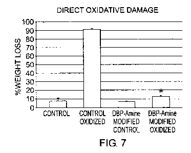
Figure 7 shows wet weight loss due to direct oxidative damage in
glutaraldehyde
fixed bovine pericardium samples before (Control) and after the covalent
attachment of
DBP (DBP-Amine Modified), either following peroxide incubation (Oxidized) or
not. An
asterisk (*) means significant difference between the Control and DBP-Amine
Modified
samples at p<0.02.
Figure 8 shows dry weight loss in glutaraldehyde pretreated bovine pericardium
samples before (Control) and after the covalent attachment of DBP (DBP-Amine
Modified)
after collagenase treatment of the remaining residual material following
peroxide
incubation (Oxidized) or not. An asterisk (*) means significant difference
between the
Control and DBP-Amine Modified samples at p<0.02.
DETAILED DESCRIPTION OF THE INVENTION
The present invention is based on the discovery that immobilizing an anti-
oxidant
or a combination of two or more antioxidants to a bioprosthetic heart valve
leaflet tissue
may reduce oxidative stress-based damage to the tissue. In one example,
covalent
attachment of an anti-oxidant, for example, 4-hydroxy-3,5-di-tert-
butylphenylpropylamine hydrochloride (DBP-amine=HCI), to a bovine pericardium
provided significant protection to the pericardium against oxidative damage.
Bioprosthetic Tissue
The present invention provides a bioprosthetic tissue incorporating an
effective
amount of an antioxidant to prevent oxidative degeneration of the
bioprosthetic tissue in
CA 02841573 2014-01-13
WO 2013/009851 PCT/US2012/046226
a subject, in which the antioxidant is covalently immobilized or attached to
the
bioprosthetic tissue.
A bioprosthetic tissue is a biological tissue obtained from an animal (e.g., a
human) for use in a bioprosthesis, either in a fresh state or after being
preserved from
decay by, for example, chemical fixation or freezing. The bioprosthetic tissue
may be a
heterograft or a homograft. It may be a tissue obtained from any mammal (e.g.,
bovine,
ovine, porcine and human tissues). Examples of the bioprosthetic tissue
include heart,
heart valve, pericardium, vascular graft, urinary tract, bladder component,
tendon,
bowel, and soft tissues. Preferably, the bioprosthetic tissue is a heart valve
tissue. Any
animal derived membranous material may be suitable for use, for example,
equine
pericardium, kangaroo pericardium, porcine pericardium, and bovine
pericardium.
Preferably, the bioprosthetic tissue is a porcine aortic valve or a bovine
pericardium.
The bioprosthetic tissue may be fixed by any method suitable for subsequent
implantation of the bioprosthetic tissue into a subject. Preferably, the fixed
bioprosthetic
tissue retains free groups for covalent attachment of an antioxidant. The free
groups
may be carboxylic groups, for example, from residues of aspartic and glutamic
acids.
The bioprosthetic tissue may be fixed chemically. Preferably, the
bioprosthetic tissue is
fixed with glutaraldehyde or epoxy-compounds (e.g., triglycidylannine). The
bioprosthetic tissue may also be cryopreserved with liquid nitrogen.
Antioxidants
The antioxidant may be any molecule capable of inhibiting oxidation of other
molecules, and suitable for covalent attachment to a bioprosthetic tissue.
Techniques for
covalent attachment of two molecules, including small molecules and/or
biological
molecules, are well known in the art. For example, chemical strategies used to
attach
phenolic antioxidants covalently to polyurethane to prevent polyurethane
oxidative
degradation may be adapted for covalent immobilization of the phenolic
antioxidants to a
bioprosthetic tissue.
The antioxidant may be derived from a natural antioxidant and modified for
covalent attachment to a bioprosthetic tissue. Examples of natural
antioxidants include
glutathione, ascorbic acid (vitamin C), lipoic acid, uric acid, carotenes, a-
tocopherol
(vitamin E), ubiquinol (coenzyme Q) and melatonin.
The antioxidant may also be a synthetic antioxidant. Many commonly used
antioxidants may be used, directly or with some modifications known in the
art, for
covalent attachment or immobilization to a bioprosthetic tissue. Antioxidants
from any
of several major classes may be employed. These include hindered amines (e.g.,
derivatives of 2,2,6,6-tetramethylpiperidine, Scheme 2, structure 4), aromatic
amines
(e.g., N, N'-disubstituted-p-phenylenediamines (e.g. Scheme 2 structure 1),
alkylated
CA 02841573 2014-01-13
WO 2013/009851 PCT/US2012/046226
"6"
diphenylamines (e.g., Scheme 2, structure 2), and derivatives of
dihydroquinoline (e.g.,
Scheme 2, structure 3), high temperature stabilization agents such alpha-
tocopherol,
hydroxylamines (e.g., Scheme 2, structure 5), and lactones (e.g.,
benzofuranones,
especially arylbenzofuranones such as in Scheme 2, structure 6 and Scheme 3,
structure
8), and hindered phenols (e.g., 2,6-di-tert-butyl-p-cresol). Another useful
class includes
sulfur-based hydroperoxide decomposers known as thiosynergists, one example of
which
is shown in Scheme 3 as structure 7. Others are well known in the art. A
thiosynergist
may also be introduced by binding N-acetylcysteine by the thiol group to the
bioprosthetic tissue, thereby forming a dialkyl sulfide. Suitable techniques
for such
binding are described, for example, by Fishbein et al., mentioned above.
Combinations of two or more antioxidants may also be used, including
combinations from the same class and/or combinations employing antioxidants
from
different classes. Such combinations may result in longer antioxidant activity
and/or
synergy. For example, combinations of thiosynergists with antioxidants from
other
classesõ for example hindered phenols, may afford pronounced synergistic
action.
In some embodiments, the antioxidant is a phenol-based antioxidant. For
example, the antioxidant may be 4-substituted 2,6-di-tert-butylphenol (DBP) or
a
derivative thereof. Preferably, the antioxidant is 4-hydroxy-3,5-di-tert-
butylphenolpropylamine hydrochloride (DBP-amineliC1). Covalent binding of the
antioxidant molecule to the tissue should be performed in a way not
compromising the
effectiveness of the antioxidant action (for example, covalent immobilization
of hindered
phenols should not affect the phenolic OH, critical for the activity).
The subject is a mammal, for example, dog, cat, racehorse, bull, or human, in
need of a bioprosthesis. Preferably, the subject is a human. The subject may
be a
female or male. The subject may be a child or an adult.
The subject may have suffered from a condition, disorder or disease, for
example,
the metabolic syndrome, hormonal deregulation, hypertension, extreme stress
and
weight loss. A subject having the metabolic syndrome is defined as set forth
in the US
National Cholesterol Education Program Adult Treatment Panel III (2001).
(JAMA.
2002;287(3):356-359.PDF attached). The subject may have suffered from a renal
disease or calcium/phosphorus imbalance. The subject may have suffered from
increased reactive oxygen species (ROS). The subject may also have suffered
from
hyperglycemia-induced oxidative stress.
For each of the bioprosthetic tissues described herein, a preparation method
is
provided. The method comprises immobilizing covalently an effective amount of
an
antioxidant to the bioprosthetic tissue to prevent oxidative degeneration of
the
bioprosthetic tissue in a subject.
CA 02841573 2014-01-13
WO 2013/009851 PCT/US2012/046226
rv 7 rv
A bioprosthetic heart valve leaflet comprising a bioprosthetic tissue of the
present
invention is provided. The bioprosthetic tissue is treated with an effective
amount of an
antioxidant to prevent oxidative degeneration of the bioprosthetic tissue in a
subject,
and the antioxidant is covalently immobilized to the bioprosthetic tissue.
The present invention provides a method for preventing oxidative degeneration
of
a bioprosthetic tissue in a subject. The method comprises immobilizing
covalently an
effective amount of an antioxidant to the bioprosthetic tissue.
The term "preventing" as used herein means reducing or mitigating. Prevention
may be assessed by quantitating a number of different parameters. Material
studies
such as described in the Examples can assess in vitro statistically
significant differences
in prevention of mass loss of weight due to oxidative stress, resistance to
collagenase
digestion¨versus quantitation of increased susceptibility to this enzyme
following
oxidative stress. In vivo mitigation in an actual heart valve implant may be
measured
by cardiovascular testing such as echo-cardiograms, angiograms and
computerized
tomography, all of which may document improved bioprosthetic function over
time due
to prevention of oxidative stress. Thus, the reduction of oxidative
degeneration of a
bioprosthetic tissue may be measured by a conventional method, for example,
reduced
weight loss or an extension of functional life of the bioprosthetic tissue.
Therefore, in
animals, including human subjects, the extension of functional life of a
bioprosthesis
(e.g., a medical device) comprising the bioprosthetic tissue may be determined
by
quantitative testing such as ultrasound or x-ray/angiogram imaging.
Microscopic or
material analyses may be used to determine the success of the methods and
compositions of this invention experimentally in retrieved in vivo specimens.
In the prevention method according to the present invention, the bioprosthetic
tissue may be a heterog raft or a homograft. It may be a tissue obtained from
any
mammal (e.g., bovine, ovine, porcine and human tissues). Examples of the
bioprosthetic tissue include heart, heart valve, pericardium, vascular graft,
urinary tract,
bladder component, tendon, bowel, and soft tissues. Preferably, the
bioprosthetic tissue
is a heart valve tissue. More preferably, the bioprosthetic tissue a porcine
aortic valve or
a bovine pericardium.
In a prevention method according to the present invention, the bioprosthetic
tissue may be fixed by any method suitable for subsequent implantation of the
bioprosthetic tissue into a subject. Preferably, the fixed bioprosthetic
tissue retains free
groups for covalent attachment of an antioxidant or combination of
antioxidants. The
free groups may be carboxylic groups, for example, from residues of aspartic
and
glutamic acids in the tissue proteins. The bioprosthetic tissue may be fixed
chemically.
Preferably, the bioprosthetic tissue is fixed with glutaraldehyde or epoxy-
compounds
CA 02841573 2014-01-13
WO 2013/009851
PCT/US2012/046226
(e.g., triglycidylamine). The bioprosthetic tissue may also be cryopreserved
with liquid
nitrogen.
In a prevention method according to the present invention, the antioxidant may
be any molecule capable of inhibiting oxidation of other molecules, and
suitable for
covalent attachment to a bioprosthetic tissue. Techniques for covalent
attachment of
two molecules, including small molecules and/or biological molecules, are well
known in
the art. For example, chemical strategies used to attach phenolic antioxidants
covalently
to polyurethane to prevent polyurethane oxidative degradation may be adapted
for
covalent immobilization of the phenolic antioxidants to a bioprosthetic
tissue. The
antioxidant may be derived from a natural antioxidant and modified for
covalent
attachment to a bioprosthetic tissue. Examples of natural antioxidants include
glutathione, ascorbic acid (vitamin C), lipoic acid, uric acid, carotenes, a-
tocopherol
(vitamin E), ubiquinol (coenzyme Q) and melatonin.
The antioxidant may also be a synthetic antioxidant. Many commonly used
antioxidants may be used, directly or with some modifications known in the
art, for
covalent attachment or immobilization to a bioprosthetic tissue. Antioxidants
from any
of several major classes may be employed. These include hindered amines (e.g.,
derivatives of 2,2,6,6-tetramethylpiperidine), aromatic amines (e.g., N, N'-
disubstituted-
p-phenylenediamine, alkylated diphenylamines, and derivatives of
dihydroquinoline),
high temperature stabilization agents such alpha-tocopherol, hydroxylamines
(e.g.,
Scheme 2, structure 5), and lactones (e.g., benzofuranones, such as in Scheme
2,
structure 6 and Scheme 3, structure 8), and hindered phenols (e.g., 2,6-di-
tert-butyl-p-
cresol). Another useful class includes sulfur-based hydroperoxide decomposers
known
as thiosynergists, one example of which is shown in Scheme 3 as structure 7.
Others
are well known in the art.
Combinations of two or more antioxidants may also be used, including
combinations from the same class and/or combinations employing antioxidants
from
different classes. Such combinations may result in longer antioxidant activity
and/or
synergy. For example, combinations of thiosynergists with antioxidants from
other
classes, for example hindered phenols, may afford pronounced synergistic
action.
In some embodiments, the antioxidant is a phenol-based antioxidant. For
example, the antioxidant may be 4-substituted 2,6-di-tert-butylphenol (DBP) or
a
derivative thereof. Preferably, the antioxidant is 4-hydroxy-3,5-di-tert-
butylphenolpropylamine hydrochloride (DBP-amine=FIC1).
In a prevention method according to the present invention, the subject may be
a
mammal, for example, a pet (e.g., dogs and cats) or human. Preferably, the
subject is a
human. The subject may be a female or male. The subject may be a child or an
adult.
CA 02841573 2014-01-13
WO 2013/009851
PCT/US2012/046226
9
The subject may have suffered from a condition, disorder or disease, for
example, the
metabolic syndrome, hormonal deregulation, hypertension, extreme stress and
weight
loss. A subject having the metabolic syndrome is defined as set forth in the
US National
Cholesterol Education Program Adult Treatment Panel III (2001). The subject
may have
suffered from a renal disease or calcium/phosphorus imbalance. The subject may
have
suffered from increased reactive oxygen species (ROS). The subject may also
have
suffered from hyperglycemia-induced oxidative stress.
A composition for preparing a bioprosthetic tissue, fabricating a
bioprosthetic
heart valve leaflet comprising a bioprosthetic tissue, or preventing oxidative
degeneration of a bioprosthetic tissue in a subject is provided. The
compositions
comprise an antioxidant or combination of two or more antioxidants capable of
being
covalently immobilized to the bioprosthetic tissue.
For a composition of the present invention, the bloprosthetic tissue may be a
heterograft or a homograft. It may be a tissue obtained from any mammal (e.g.,
bovine,
ovine, porcine and human tissues). Examples of the bioprosthetic tissue
include heart,
heart valve, pericardium, vascular graft, urinary tract, bladder component,
tendon,
bowel, and soft tissues. Preferably, the bioprosthetic tissue is a heart valve
tissue. More
preferably, the bioprosthetic tissue is a porcine aortic valve or a bovine
pericardium.
For a composition of the present invention, the bioprosthetic tissue may be
fixed
by any method suitable for subsequent implantation of the bioprosthetic tissue
into a
subject. Preferably, the fixed bioprosthetic tissue retains free groups for
covalent
attachment of an antioxidant. The free groups may be carboxylic groups, for
example,
from residues of aspartic and glutamic acids. The bioprosthetic tissue may be
fixed
chemically. Preferably, the bioprosthetic tissue is fixed with glutaraldehyde
or epoxy-
compounds (e.g., triglycidylamine). The bioprosthetic tissue may also be
cryopreserved
with liquid nitrogen.
In a composition of the present invention, the antioxidant may be any molecule
capable of inhibiting oxidation of other molecules, and suitable for covalent
attachment
to a bioprosthetic tissue. Techniques for covalent attachment of two or more
molecules,
including small molecules and/or biological molecules, are well known in the
art. For
example, chemical strategies used to attach phenolic antioxidants covalently
to
polyurethane to prevent polyurethane oxidative degradation may be adapted for
covalent immobilization of the phenolic antioxidants to a bioprosthetic
tissue. The
antioxidant may be derived from a natural antioxidant and modified for
covalent
attachment to a bioprosthetic tissue. Examples of natural antioxidants include
glutathione, ascorbic acid (vitamin C), lipoic acid, uric acid, carotenes, a-
tocopherol
(vitamin E), ubiquinol (coenzyme Q) and melatonin.
CA 02841573 2014-01-13
WO 2013/009851 PCT/US2012/046226
¨ 10 ¨
The antioxidant may also be a synthetic antioxidant. Many commonly used
antioxidants may be used, directly or with some modifications known in the
art, for
covalent attachment or immobilization to a bioprosthetic tissue. Antioxidants
from any
of several major classes may be employed. These include hindered amines (e.g.,
derivatives of 2,2,6,6-tetramethylpiperidine), aromatic amines (e.g., N, N'-
disubstituted-
p-phenylenediamine, alkylated diphenylamines, and derivatives of
dihydroquinoline),
high temperature stabilization agents such alpha-tocopherol, hydroxylamines
(e.g.,
Scheme 2, structure 5), and lactones (e.g., benzofuranones, such as in Scheme
2,
structure 6 and Scheme 3, structure 8), and hindered phenols (e.g., 2,6-di-
tert-butyl-p-
cresol). Another useful class includes sulfur-based hydroperoxide decomposers
known
as thiosynergists, one example of which is shown in Scheme 3 as structure 7.
Others
are well known in the art.
Combinations of two or more antioxidants may also be used, including
combinations from the same class and/or combinations employing antioxidants
from
different classes. Such combinations may result in longer antioxidant activity
and/or
synergy. For example, combinations of thiosynergists with antioxidants from
other
classes, for example hindered phenols, may afford pronounced synergistic
action.
In some embodiments, the antioxidant is a phenol-based antioxidant. For
example, the antioxidant may be 4-substituted 2,6-di-tert-butylphenol (DBP) or
a
derivative thereof. Preferably, the antioxidant is 4-hydroxy-3,5-di-tert-
butylphenolpropylamine hydrochloride (DBP-amine=FICI).
For a composition of the present invention, the subject may be a mammal, for
example, a pet (e.g., dogs and cats) or human. Preferably, the subject is a
human. The
subject may be a female or male. The subject may be a child or an adult. The
subject
may have suffered from a condition, disorder or disease, for example, the
metabolic
syndrome, hormonal deregulation, hypertension, extreme stress and weight loss.
A
subject having the metabolic syndrome is defined as set forth in the US
National
Cholesterol Education Program Adult Treatment Panel III (2001). The subject
may have
suffered from a renal disease or calcium/phosphorus imbalance. The subject may
have
suffered from increased reactive oxygen species (ROS). The subject may also
have
suffered from hyperglycemia-induced oxidative stress.
Tissue-Reactive Antioxidant Constructs
Antioxidant constructs capable of covalently binding to the bioprosthetic
tissue can be
considered to have the essential structure shown in Scheme 1, where A is an
antioxidant,
n is an integer from 1 to 10, L is a linker and X is a tissue-reactive
functionality.
Scheme 1
(A)n-L-X
CA 02841573 2014-01-13
WO 2013/009851 PCT/US2012/046226
^, 11 ¨
In addition to hindered phenols, following are non-limiting examples of
antioxidant functionalities (A) that may be tethered to the bioprosthetic
tissue: (1)
aromatic amines: N,N'-disubstituted-p-phenylenediamines (Scheme 2, 1),
alkylated
diphenylamines (Scheme 2, 2), derivatives of dihydroquinoline (Scheme 2, 3);
(2)
hindered amines, for example derivatives of 2,2,6,6-tetramethylpiperidine
(Scheme 2,
4); (3) hydroxylamines (Scheme 2, 5), and (4) arylbenzofuranones (Scheme 2,
6).
Scheme 2
R¨HN 40. NH¨R R .1 it
11101
CH3
N
1 2 H
3
0
CH3
CH3 0
1110R
N¨OH (CH3)3C io
E¨CH3
cH3
4 5 (cH3)3c
6
The linker L may contain one or more carbon atoms. It may also include
carbonyls and
heteroatoms (e.g., 0, S, N and P) and arylene spacers. The linker should be
attached to
the antioxidant in a way not affecting the antioxidant action, preferably to a
hydrocarbon
radical R shown in Scheme 2. The tissue-reactive functionality X may for
example be an
amino group (e.g., for reaction with the residual carboxy groups of the
tissue), a carboxy
group (or its active ester, e.g., for reaction with the amino groups of the
tissue), a
photo-reactive group (e.g., aromatic azide, residue of benzophenone, and
anthraquinone,
which are capable of binding to the tissue under UV-irradiation), or any other
group
capable of participating in any known method of bioconjugation (e.g.,
chemistry of thiols
and thiol-reactive groups, click chemistry, and Staudinger ligation).
Examples of suitable tissue-reactive antioxidant constructs are shown in
Scheme
3.
CA 02841573 2014-01-13
WO 2013/009851 PCT/US2012/046226
¨ 12 ¨
Scheme 3
OH 0
(CH3)3C C(CH3)3 0
(CH3)3C
NH2
C(CH3)3
8
(CH3)3C
HOF 0
C(CH3)3
F N =
7 CH3
N3
H CH3
9
Tissue-reactive antioxidant construct 7 has two hindered phenolic antioxidant
moieties, a
linker containing heteroatoms S and N, and a tissue-reactive group of N1-12.
The sulfur
atoms in the linker serve also as synergists, enhancing the antioxidant action
of the
hindered phenolic moieties. Tissue-reactive antioxidant construct 8 is an
example of a
tissue-modifier containing an arylbenzofuranone antioxidant moiety. Constructs
7 and 8
may be covalently bonded to the tissue with the aid of a coupling agent (e.g.,
1-(3-
dimethylaminopropy1)-3-ethylcarbodiimide hydrochloride (EDC)), or other agents
suitable for coupling of carboxy and amino groups (e.g., 1-cyclohexy1-3-
morpholinoethylcarbodiimide metho p-toluene sulfonate (CMC), and benzotriazol-
1-
yloxy-tris-dimethylamino-phosphonium hexafluorophosphate). An intermediate-
stabilizing agent (e.g., N-hydroxysuccinimide (SuOH), 1-hydroxy 7-
azabenzotriazole
(HOAt), pentafluorophenol, etc.) may additionally be used with certain
coupling agents
to increase efficacy by stabilizing the active intermediate, as known in the
art. Other
coupling agents, for example benzotriazol-1-yloxy-tris-dimethylamino-
phosphonium
hexafluorophosphate (BOP), do not require such stabilization.
Construct 9 contains a dihydroquinoline-type of antioxidant having a linker
containing a
carbonyl and a N-heteroatom, wherein the, tissue-reactive group is a photo-
activatable
fluoroaromatic azide. Other techniques for photoactive coupling may also be
used, for
example as disclosed in U.S. Pat. 7,635,734. In addition thiol based chemistry
may be
used, as well as affinity methods such as avidin-biotin etc. Suitable thiol-
based
CA 02841573 2014-01-13
WO 2013/009851 PCT/US2012/046226
13 ¨
chemistry is described for example in Fishbein, I., Alferiev, I., Bakay, M.,
Stachelek, S.J.,
Sobolewski, P,, Lai, M., Choi, H., Chen, I.W., and Levy, R.J., Local delivery
of gene
vectors from bare-metal stents by use of a biodegradable synthetic complex
inhibits in-
stent restenosis in rat carotid arteries, Circulation (2008) 117, 2096-2103,
and is based
on well known chemical methodologies. Suitable coupling using avidin-biotin
affinity is
described for example in Wilchek, M., and Bayer, E.A., Introduction to avidin-
biotin
technology, Methods Enzymol (1990) 184, 5-13. The methods of the invention may
further comprise similarly tethering another type of agent, for example an
agent suitable
for preventing calcification, to the tissue.
Exemplary Embodiments of the Invention
In some embodiments, the invention provides a bioprosthetic tissue treated
with an
effective amount of an antioxidant or combination of antioxidants to prevent
oxidative
degeneration of the bioprosthetic tissue in a subject, wherein the antioxidant
Is
covalently immobilized to the bioprosthetic tissue.
In some embodiments, the bioprosthetic tissue is a heterograft.
In some embodiments, the bioprosthetic tissue is a homograft.
In some embodiments, the bioprosthetic tissue is a tissue selected from the
group
consisting of bovine, ovine, porcine, equine, other non-human vertebrate
tissues and
human tissues.
In some embodiments, the bioprosthetic tissue is a tissue selected from the
group
consisting of heart, heart valve, pericardium, vascular graft, urinary tract,
bladder
component, tendon, bowel, and soft tissues.
In some embodiments, the bioprosthetic tissue is a heart valve tissue.
In some embodiments, the bioprosthetic tissue is a porcine aortic valve.
In some embodiments, the bioprosthetic tissue is a bovine pericardium.
In some embodiments, the bioprosthetic tissue is fixed with glutaraldehyde.
In some embodiments, the bioprosthetic tissue is preserved with liquid
nitrogen.
In some embodiments, the bioprosthetic tissue is one in which the antioxidant
is derived
from a natural antioxidant selected from the group consisting of glutathione,
ascorbic
acid (vitamin C), lipoic acid, uric acid, carotenes, a-tocopherol (vitamin E),
ubiquinol
(coenzyme Q) and melatonin.
In some embodiments, the bioprosthetic tissue is one in which the antioxidant
is a
synthetic antioxidant.
In some embodiments, the bioprosthetic tissue is one in which the antioxidant
is a
phenol-based antioxidant.
In some embodiments, the bioprosthetic tissue is one in which the antioxidant
is 4-
substituted 2,6-di-tert-butylphenol (DBP) or a derivative thereof.
CA 02841573 2014-01-13
WO 2013/009851 PCT/US2012/046226
¨ 14 -
In some embodiments, the bioprosthetic tissue is one in which the antioxidant
is 4-
hydroxy-3,5-di-tert-butylphenolpropylamine hydrochloride (DBP-aminetIC1).
In some embodiments, the bioprosthetic tissue is one in which the antioxidant
comprises
a hindered phenol and at least one additional antioxidant selected from the
group
consisting of aromatic amines, alkylated diphenylamines, derivatives of
dihydroquinoline,
hindered amines, hydroxylamines and arylbenzofuranones.
In some embodiments, the bioprosthetic tissue is one in which the antioxidant
comprises
a hindered phenol and a thiosynergist.
In some embodiments, the bioprosthetic tissue is one in which the antioxidant
comprises
a compound according to structure 7
OH
(CH3)3C C(CH3)3
H2
(CH3)3C
HO
C(CH3)3
7
In some embodiments, the bioprosthetic tissue is one in which the antioxidant
comprises
a compound according to structure 8
0
0
(CH3)3C 401
NH2
C(CH3)3
8
CA 02841573 2014-01-13
WO 2013/009851 PCT/US2012/046226
In some embodiments, the bioprosthetic tissue is one in which the antioxidant
comprises
a compound according to structure 9
0
=
F (40
Cr-13
N3
H CH3
9
In some embodiments, the bioprosthetic tissue is a heart valve leaflet.
5 The invention provides a method for preparing a bioprosthetic tissue
according to any of
the aforementioned embodiments, comprising immobilizing the antioxidant to the
bioprosthetic tissue.
The Invention provides a method for treating a subject in need of a
bioprosthetic tissue,
comprising treating the subject with the bioprosthetic tissue according to any
of the
10 aforementioned embodiments.
In some embodiments, the method of treating a subject is one in which the
subject is a
human.
In some embodiments, the method of treating a subject is one in which the
subject is a
female.
15 In some embodiments, the method of treating a subject is one in which
the subject is a
male.
In some embodiments, the method of treating a subject is one in which the
subject has
suffered from the metabolic syndrome, hormonal deregulation, hypertension,
extreme
stress or weight loss.
In some embodiments, the method of treating a subject is one in which the
subject has
suffered from increased reactive oxygen species (ROS).
In some embodiments, the method of treating a subject is one in which the
subject has
suffered from hyperglycemia-induced oxidative stress.
The invention also provides a compound according to structure 7
CA 02841573 2014-01-13
WO 2013/009851
PCT/US2012/046226
¨ 16 ¨
OH
(CH3)3C las C(CH3)3
H2
(CH3)3C 401
HO
C(CH3)3
7
The invention also provides a compound according to structure 8
0
0
(CH3)3C
NH2
C(CH3)3
8
The invention also provides a compound according to structure 9
0
CH3
N3
H CH3
9
EXAMPLES
Example 1. Oxidative stress in bioprosthetic heart valve deterioration
In vitro model system studies were carried out using a modification of a well
established oxidative stress system involving incubating synthetic polymers in
hydrogen
peroxide solutions (Stachelek Si, et al. 1 Blamed Mater Res A 2006;78:653-61).
This
experimental system was originally developed and validated for investigating
polyurethane oxidative degradation, and was adapted for studies of oxidative
stress in
bioprosthetic heart valve leaflet samples.
CA 02841573 2014-01-13
WO 2013/009851 PCT/US2012/046226
^, 17
In brief, bovine pericardium was fixed in 0.6% glutaraldehyde using
established
conditions for preparing this material for use in bioprosthetic heart valves.
These
glutaraldehyde cross-linked bovine pericardium samples (typically 1cm x 1cm)
were
incubated in 20% H202 for 14 days at 37 C. Solutions were changed every three
days.
At the end of the incubation period, the samples were washed with water or
PBS, and
stored at 4 C until later analysis. Samples designated as controls were
incubated in
water only. The wet weight of each sample was recorded prior to the start of
the
incubation, and after the wash steps at the end of the study. The percent
weight loss
from the original wet weight is presented in Figure 1 as a direct index of
oxidative
damage. The control and oxidized samples from the in vitro model of oxidative
degradation were lyophilized for 48 hours. The dry weight of each sample was
recorded
prior to treatment with collagenase. Samples were digested for 24 hours at 37
C in a
solution containing 600 units/micollagenase from Clostridium histolyticum
(Sigma 9001-
12-1), phosphate buffered saline and 0.1% bovine serum albumin. Following
digestion,
samples were pelleted by centrifugation at 10,000 rpm at 4 C. Sample were
washed
with saline and lyophilized for 48 hours. The weight of each sample was
recorded after
the lyophilization period. Data were reported as a percent weight loss from
the original
dry weight and is presented in Figure 2A as % weight loss after collagenase
treatment
(P<0.001). As indicated, oxidative stress due to hydrogen peroxide exposure
causes
substantial material loss (Figure 1). In addition, the material remaining
after peroxide
exposure is significantly more susceptible to collagenase digestion (Figure
2A). Thus,
glutaraldehyde pretreated bovine pericardium is strongly affected by oxidative
stress
with primary material degradation and increased susceptibility to enzymatic
digestion.
In addition, oxidation gave rise to an increased 1550 cm"" peak per FTIR
(Figure 2B) that
is likely due oxidation reaction products.
Prior in vivo model studies have established that bioprosthetic heart valve
leaflet
calcification occurs in either rat subdermal implants or sheep mitral valve
replacements,
which display a pathology that is comparable to calcific failure of human
implants of
bioprosthetic heart valves (Schoen and Levy, Ann Thorac Surg 2005;79:1072-80).
In
the studies shown below, an immunostaining marker for ROS, nitrotyrosine, was
used to
demonstrate the strong presence of ROS, per nitrotyrosine positive staining in
calcified
bioprosthetic heart valve leaflets retrieved from rat subdermal implants and
sheep mitrel
valve replacements. The presence of nitrotyrosine indicates that nitric oxide-
peroxynitrite oxidative stress has modified the proteins of these heart valve
leaflets.
Since calcification was also present in the positively staining retrievals,
the results imply
that oxidative stress can occur in the presence of calcification, and may even
contribute
to the calcification mechanism.
CA 02841573 2014-01-13
WO 2013/009851 PCT/US2012/046226
¨ 18 r-
Nitrotyrosine immunostaining was carried out on formalin-fixed explants from
' either 90-day rat subdermal experiments (Figure 3A: glutaraldehyde-
pretreated and
calcified, or Figure 3B: glutaraldehyde-Et0H-pretreated and non-calcified) or
from a
sheep mitral valve replacement (Figure 3C: 59 days duration and calcified).
Sections of
12pm thickness cut from formalin-fixed explants were stained either using
rabbit anti-
nitrotyrosine (provided by Dr. H. Ischiropoulos; The Children's Hospital of
Philadelphia),
or as a negative control, nonspecific rabbit IgG, in both cases at an
immunoglobulin
concentration of 5pg/ml. Sections were blocked for non-specific peroxidase and
reaction
with secondary antibody (biotinylated goat anti-rabbit) before color
development,
sequentially using a VectaStain ABC kit and ImmPACT DAB chromogen (Vector
Labs, Inc,
Burlingame, CA) per standard procedures well known in the art. Positive
staining is
indicated by intense red-brown color, which is absent in non-calcified rat
subdermal
implants or when non-immune IgG was used. Representative results are shown
indicating the presence of strong nitrotyrosine staining co-incident with
calcification.
Additional negative control studies included anti-nitrotyrosine which was pre-
absorbed
with blocking peptide (gift of Dr. H Ischiropoulos); this demonstrated the
specificity of
positive sample stains for nitrotyrosine. Thus, these in vivo results
demonstrate the
presence of oxidative stress in calcified explants from both rat subdermal
studies and
sheep circulatory explants. Nitrotyrosine is a specific by-product of
oxidative stress, in
which peroxynitrite reacts with protein-based tyrosine residues. Thus, it can
be seen
that oxidative stress was clearly present in calcifying bioprosthetic heart
valves.
Example 2. Prevention of oxidative stress-induced damage in bioprosthetic
heart valve
leaflets
In these studies, it was investigated whether the use of an anti-oxidant could
prevent the extensive breakdown of bioprosthetic leaflets due to ROS. A local
therapy
strategy was investigated involving the covalent attachment of an antioxidant
compound
to bioprosthetic leaflets that were already crosslinked with glutaraldehyde.
A chemical immobilization strategy was adopted to improve the oxidative
stability
of a bioprosthetic tissue via covalent attachment of antioxidant functions
based on
hindered phenolic residues covalently immobilized on collagenous biomaterials.
The
modification employed direct coupling of the collagen's carboxylic groups
(from residues
of aspartic and glutamic acids) with the amine groups of a hindered phenolic
antioxidant
using water-soluble 1-(3-dimethylaminopropyI)-3-ethylcarbodiimide
hydrochloride (EDC).
Among many possibilities, 4-hydroxy-3,5-di-tert-butylphenylpropylamine
hydrochloride
(DBP-amine=HCI) was used as a suitable antioxidant possessing an amino group,
which
is water-soluble and can be used in aqueous media (preparation described in
Dyubchenko et al., Pharm. Chem. J. 2006, 40(5), 243-247, translated from
Khimiko-
CA 02841573 2014-01-13
WO 2013/009851 PCT/US2012/046226
Farmatsevticheskii Zhurnal 2006, 40(5), 10 - 13). To capture the unstable
intermediate
formed in the reaction of carboxylic groups with EDC, N-hydroxysuccinimide
(SuOH) was
added into the reaction mixture. As a result, more stable amine-reactive Su-
esters were
formed, and the antioxidant was then bound to the tissue via stable amide
bonds, as
shown in Scheme 4.
Scheme 4
C(CH3)3
OH
OH
(H3C)3C C(CH3)3 1111 r rqj
13/3
0 NH
COOH COOSu
EDC, SuOH H2N
Collagen fiber pHw catera. 5.5 Collagen fiber Collagen fiber
Thus, to carry out these reactions a solution was prepared from water (9.5
ml),
4-hydroxy-3,5-di-tert-butylphenylpropylamine hydrochloride (DBP-amine.HCI, 110
mg)
and N-hydroxysuccinimide (SuOH, 50 mg). To accelerate the dissolution of DBP-
amine=HCI, the mixture may be warmed to ca. 40 - 50 C, but should be cooled
before
the addition of SuOH. The solution was adjusted to pH ca. 5.5 with 0.05 M
aqueous
KHCO3 solution, and 1-ethy1-3-(3-dimethylaminopropyl)carbodlimide
hydrochloride (EDC,
0.12 g) was added immediately before use. The tissue was allowed to react in
the above
solution for 24 - 36 h at room temperature with gentle stirring. Finally, the
tissue was
thoroughly rinsed with copious amounts of water. After rinsing with water, the
tissue
could be repeatedly incubated as above with a fresh portion of the solution,
if necessary.
FTIR analyses (Figure 4) demonstrated the presence of two novel peaks at ca.
2900 cm-1,
suggesting the covalent attachment of DBP.
The ability of DBP to inhibit ROS activity was verified both in solution and
when
bound to glut-pretreated pericardium, as described above, by monitoring the
fluorescence of dihydrorhodamine-123 (DHR123; Molecular Probes Inc., Eugene
OR),
which is a commonly used measure of oxidation due to ROS. Briefly, the
capacity of DBP
to reduce ROS activity was proven in this system by titrating DBP in solution
against the
oxidant hydrogen peroxide (H202) in the presence of DHR123. This resulted in a
dose-
dependent inhibition of oxidation signal (Figure 5). Under the same
conditions, samples
of pericardium to which DBP had been covalently attached likewise reduced
oxidation
(Figure 6).
CA 02841573 2014-01-13
WO 2013/009851
PCT/US2012/046226
¨ 20
DBP-modified glutaraldehyde-fixed bovine pericardium was investigated in the
same hydrogen peroxide system described above (Figures 1 and 2) to assess ROS-
damage to this biomaterial. Accordingly, Figure 7 shows weight loss due to
direct
oxidative damage, and Figure 8 shows weight loss after subsequent collagenase
digestion. Both indicate a significant (P*<0.001) protective effect of DBP
modification
against oxidative damage, re. weight loss and subsequent collagenase
digestibility
(Figures 7 and 8).
All documents, books, manuals, papers, patents, published patent applications,
guides, abstracts, and other references cited herein are incorporated by
reference in
their entirety. Other embodiments of the invention will be apparent to those
skilled in
the art from consideration of the specification and practice of the invention
disclosed
herein. It is intended that the specification and examples be considered as
exemplary
only, with the true scope and spirit of the invention being indicated by the
following
claims.