Note: Descriptions are shown in the official language in which they were submitted.
CA 02841704 2013-12-27
WO 2013/008106
PCT/1B2012/052077
1
ENDOBRONCHIAL TUBE
FIELD OF THE INVENTION
The present invention relates to upper airway tubes and in particular,
to an endobronchial tube with an integrated image sensor and light source.
BACKGROUND OF THE INVENTION
Respiratory tubes for example endobronchial tubes, endotracheal
tubes, tracheostomy tubes are used to ventilate at least a portion of the
io respiratory system or lungs of a subject. Such respiratory tubes may be
inserted in a number of ways via a non-invasive approach through an orifice
or cavity such as the oral or nasal cavity. Alternatively such tubes may be
introduced to a body via a minimally invasive external incision creating a
port for tube insertion for example through the trachea in a tracheotomy
is procedure.
Such respiratory tubes may be provided as double lumen tubes, or
single lumen tubes for selectively ventilating a portion of the respiratory
system. For example endobronchial tubes, whether, double lumen tubes or a
single lumen tube may be utilized for one- lung ventilation procedures or for
20 selective lung ventilation of the left or right bronchi, during one-
lung
ventilation procedures.
In order to perform one- lung ventilation procedures without
25 complications, the position of the respiratory tube placed within either
the left
or right bronchi and the trachea must be closely monitored or at least
confirmed prior to initiating a procedure. Various technologies are available
to confirm the tube's placement, for example capnograph, auscultation,
bronchoscope and x-ray.
CA 02841704 2013-12-27
WO 2013/008106
PCT/1B2012/052077
2
However these procedures take time, technique and skill to perform
and therefore it is not feasible to continuously monitor the tube's placement.
In particularly when the subject is moved during a procedure the tube's
location may change leading to potentially dangerous displacement of the
tube possibly suffocating the subject or inappropriate ventilation of the
patient, for example not ventilating the correct portion of the respiratory
system.
Verification by means of a bronchoscope is currently the gold
standard, but none of the mentioned confirmation techniques provide
io continuous monitoring of the carina or provide for correct tube
positioning.
Furthermore, drawbacks with respect to the design and sensitivity of the
bronchoscope, render its cleaning process elaborate and often inefficient and
costly process, that may lead to cross infection between subjects.
is SUMMARY OF THE INVENTION
There is an unmet need for, and it would be highly useful to have an
endobronchial tube capable of continuously and seamlessly inspect the
location and implantation of the endobronchial tube relative to the Tracheal
Carina.
20 The present
invention overcomes the deficiencies of the background
by providing an endobronchial tube having an integrated image sensor and
corresponding light source.
A preferred embodiment of the present invention provides for a
respiratory tube, and an endobronchial tube, designed for oral or nasal
25 insertion via the trachea and into a lung to maintain airway patency
and/or
deliver anesthetic, inhalation agent or other medical gases, and secure
ventilation.
Most preferably the endobronchial tube of the present invention may
be made of medical grade materials for example including but not limited to
30 plastic, rubber, polymers or silicone or the like materials as is known
in the
art.
CA 02841704 2013-12-27
WO 2013/008106
PCT/1B2012/052077
3
Most preferably the endobronchial tube of the present invention
provides for continuous monitoring of the Tracheal Carina (herein "TC"),
allowing a user, physician, nurse, or caregiver to verify the correct
placement
of the endobronchial tube
Most preferably the endobronchial tube includes an integrated image
sensor, optionally and preferably in the form of CCD or CMOS camera
provided for visualizing the carina to confirm the correct placement of the
tube within the trachea and bronchi, assuring correct ventilation during
procedures for example including but not limited to one lung ventilation.
io Most preferably the integrated camera and light source provide
continuous verification of the correct placement of the endobronchial tube.
The continuous placement verification allows a caregiver the opportunity to
detect any dangerous situation, for example cuff dislodgement, providing
sufficient time to react to the situation as is necessary. Moreover blood and
secretion accumulation or any other unexpected incidents during surgery,
which might cause risk to the patient, may be observed.
A preferred embodiment of the present invention provides for an
endobronchial tube with an integrated image sensor, for example including
but not limited to CCD or CMOS camera, with a corresponding light source,
for example including but not limited to a Light Emitting Diode ('LED')
while optimizing the lumen patency for both adequate airflow performance
through the tube. Most preferably the image sensor and corresponding light
source are provided in a dedicated lumen along the lengh of the
endobronchial tube. Most preferably the image sensor is further provided
with a cleaning nozzle to ensure an open field of view distal to the image
sensor. Most preferably the length of the dedicated image sensor lumen is
provided paralleled with the length of the tracheal lumen, therein both
tracheal lumen and image sensor lumen are of essentially the same length.
Optionally the length of the dedicated image sensor lumen may be provided
according to the length of the bronchial lumen.
CA 02841704 2013-12-27
WO 2013/008106
PCT/1B2012/052077
4
Optionally the endobronchial tube may be provided with two
dedicated image sensor lumen. Optionally a first dedicated image sensor
lumen is provided according to the length of the tracheal lumen and a second
dedicated image sensor lumen is provided according to the length of the
bronchial lumen.
A preferred embodiment of the present invention provides for an
endobronchial tube with an integrated image sensor and light source provide
a continuously and unobstructed view and illumination of the carina, left
bronchi, right bronchi, bronchial cuff and bronchial bifurcations, within a
io single field of view.
An optional embodiment of the present invention provides for
utilizing at least one or more bronchial cuff. Optionally at least two or more
bronchial cuffs may be utilized to provide adequate sealing of the bronchi.
Optionally the bronchial cuff may be provided in varying shapes so as
to better fit the bronchi for example include but is not limited to spherical,
elliptical, helical, hourglass, trapezoidal, or the like.
Optionally different bronchial cuff configured and shaped according to
anatomy and placement location, for example anatomy based on
configuration of a cuff for left bronchi placement and for right bronchi
placement.
Within the context of this application the term endobronchial tube may
be used interchangeably with any one of Tracheobronchial tube, double
lumen tube, double lumen endobronchial tube, double lumen endotracheal
tube, to collectively refer to a tube and/or catheter utilized for selectively
ventilating a subject via both lungs, one of the lungs or a portion of one or
both of the lungs.
An endobronchial tube comprising at least two lumen of different
lengths for selectively associating with a patient about at least two
locations
relative to the Tracheal Carina, the tube comprising:
a. a first lumen having an open distal end that associates proximally to the
Carina within the Trachea, with a first inflatable cuff;
CA 02841704 2013-12-27
WO 2013/008106
PCT/1B2012/052077
b. a second lumen having an open distal end that extends distally, past the
Carina and associates within one of the Left Bronchial branch and Right
Bronchial branch with a second inflatable cuff;
c. a dedicated image sensor lumen spanning the length of said first lumen,
5 the dedicated image sensor lumen comprising an image sensor and
illumination source disposed adjacent to the distal end of said first lumen,
and configured to provide an image of the Tracheal bifurcation of the
Tracheal Carina, the openings of the Left Bronchial branch, and the opening
Right Bronchial branch; and
io d. at least one dedicated cleaning lumen disposed parallel with said
dedicated
image sensor lumen along the length of said endobronchial tube and wherein
said cleaning lumen is configured to forms a cleaning nozzle at the distal
end, wherein said cleaning nozzle is directed toward said image sensor
lumen at its distal end.
Optionally, said cleaning nozzle is provided with a diameter from
0.1mm to 2mm.
Optionally, said cleaning nozzle is provided with a diameter of
0.6mm.
Optionally, said cleaning lumen is provided with two or more
cleaning nozzles about either side of said image sensor.
Optionally, said two or more cleaning nozzles cooperate with one
another.
Optionally, said cleaning lumen provides for suctioning or flushing
the image sensor field of view.
Optionally, said dedicated image sensor lumen is disposed within the
wall of said tube about an anterior or posterior portion between said first
and
second lumen.
Optionally, the second lumen comprises a second image sensor
providing an image of the Right bronchi or Left bronchi.
CA 02841704 2013-12-27
WO 2013/008106
PCT/1B2012/052077
6
Optionally the image sensor may be a CCD image sensor or CMOS
image sensor.
Optionally, the first lumen further comprises a light source disposed
proximal to the distal end of said first lumen and adjacent to the image
sensor.
Optionally, the light source may be selected from the group consisting
of a LED, optical fiber, waveguide, light guide, and any combination thereof.
Optionally the image sensor may be disposed within a dedicated
channel embedded within a wall of the first lumen.
io Most preferably the image sensor may be associated with an auxiliary
device for example including but not limited to a display and power supply
at the proximal end of the tube most preferably about the first lumen,
through a single dedicated connector for example including but not limited
to a USB connector.
Optionally the endotracheal tube may be adapted for non-invasive
insertion through the oral cavity or nasal cavity.
Optionally the endotracheal tube may be adapted for insertion through
an external port or incision.
Optionally the endotracheal tube may be adapted for insertion through
a surgical procedure or other invasive procedure.
Unless otherwise defined, all technical and scientific terms used herein
have the same meaning as commonly understood by one of ordinary skill in
the art to which this invention belongs. The materials, methods, and
examples provided herein are illustrative only and not intended to be
limiting.
BRIEF DESCRIPTION OF THE DRAWINGS
The invention is herein described, by way of example only, with
reference to the accompanying drawings. With specific reference now to the
drawings in detail, it is stressed that the particulars shown are by way of
example and for purposes of illustrative discussion of the preferred
CA 02841704 2013-12-27
WO 2013/008106
PCT/1B2012/052077
7
embodiments of the present invention only, and are presented in order to
provide what is believed to be the most useful and readily understood
description of the principles and conceptual aspects of the invention. In this
regard, no attempt is made to show structural details of the invention in more
detail than is necessary for a fundamental understanding of the invention, the
description taken with the drawings making apparent to those skilled in the
art how the several forms of the invention may be embodied in practice.
In the drawings:
Figures 1A-B show schematic illustrations of an exemplary
io endobronchial tube according to an optional embodiment of the present
invention; Figure 1 A shows the endobronchial tube within the right bronchi;
Figure 1B shows the endobronchial tube within the left bronchi;
Figure 2 shows a schematic sectional view of the Tracheal Carina as
seen from the endobronchial tube according to an optional embodiment of
the present invention;
Figure 3 shows a perspective views of an exemplary endobronchial
tube according to an optional embodiment of the present invention;
Figure 4A shows a perspective view of an exemplary endobronchial
tube according to an optional embodiment of the present invention;
Figure 4B shows a close up view of notch exit point for the image
sensor connector according to the present invention;
Figure 5 shows a perspective view of exemplary endobronchial tube
according to an optional embodiment of the present invention;
Figure 6 shows a perspective view of exemplary endobronchial tube
according to an optional embodiment of the present invention, depicting the
curvature of the tube;
Figures 7A-F shows varying close up views of the distal end of the
endobronchial tube according to optional embodiments of the present
invention;
CA 02841704 2013-12-27
WO 2013/008106
PCT/1B2012/052077
8
Figures 8A-B show cross-sectional views about different portions of
the endobronchial tube according to optional embodiments of the present
invention; and
Figure 9 shows a close up view of the image sensor with integrated
light source within a dedicated lumen disposed within the wall of the
endobronchial tube according to an optional embodiment of the present
invention.
DESCRIPTION OF THE PREFERRED EMBODIMENTS
The principles and operation of the present invention may be better
understood with reference to the drawings and the accompanying
description. The following reference labels listed below are used throughout
the drawings to refer to objects having similar function, meaning, role, or
objective.
10 Stylet;
12 Y-connector;
14 Air Balance Cap;
Endobronchial Tube connector assembly;
22 Endobronchial Tube connector proximal end;
20 24 Tracheal lumen connector portion;
26 Bronchial lumen connector portion;
28 Endobronchial Tube connector distal end;
50 endobronchial tube system;
100 endobronchial tube;
101 sectional view;
102 tube proximal end;
104 tube distal end;
104a distal curvature;
106 tube medial portion;
106a medial curvature;
108 midline partition;
CA 02841704 2013-12-27
WO 2013/008106
PCT/1B2012/052077
9
110 tracheal lumen;
111 tracheal lumen connector;
112 tracheal cuff;
112n tracheal cuff notch;
114 tracheal lumen distal end;
116 tracheal lumen proximal end;
118 tracheal cuff connector;
120 bronchial lumen;
122 bronchial cuff;
124 bronchial lumen distal end;
126 bronchial lumen proximal end;
128 bronchial cuff connector;
130 injection tube connector;
150 image sensor with integrated illumination;
150c image sensor;
1501 illumination source;
150L image sensor lumen
152 image sensor notch;
154 image sensor conductor;
156 image sensor cleaning nozzle;
158 image sensor connector;
160 cleaning lumen;
TR Trachea;
TC Tracheal Carina;
BR Right Bronchi;
BL Left Bronchi.
Figure 1A shows a schematic illustration of an exemplary
endobronchial tube 100 according to an optional embodiment of the present
invention placed within the right bronchi (BR). Figure 1B shows a
CA 02841704 2013-12-27
WO 2013/008106
PCT/1B2012/052077
schematic illustration of an endobronchial tube 100 within the left bronchi
(LB).
Endobronchial tube 100 is a dual lumen tube comprising a first
tracheal lumen 110 and a second bronchial lumen 120. Most preferably a
5 midline partition 108 defines the individual lumen into tracheal lumen
110
and bronchial lumen 120. Tracheal lumen 110, most preferably, ends within
the trachea while the bronchial lumen 120 ends within the bronchi, left or
right. Therein tracheal lumen 110 and bronchial lumen 120 are configured
to have different lengths, wherein the bronchial lumen 120 extends past
io and/or distally to tracheal lumen 110.
Each lumen comprises an inflatable cuff respectfully, tracheal cuff
112 and bronchial cuff 122. Tube 100 is placed such that the tracheal lumen
110 is placed within the Trachea by way of cuff 112 proximally, above, the
tracheal carina. Most preferably the tracheal carina may be continually
visualized with an image sensor and light source 150. Optionally image
sensor and light source 150 may be integrated within tracheal lumen 110
about its distal end 114. Optionally and most preferably image sensor and
light source 150 may be integrated within a dedicated channel or peripheral
lumen 150L within a wall of the tracheal lumen 110. Most preferably image
sensor 150 provides a cross sectional view 101, shown in Figure 2.
Most preferably image sensor and light source 150 are provided in the
form of at least one or more light emitting diode ('LED') 1501 and image
sensor 150c for example including but not limited to a CCD or CMOS,
(Figure 9) providing a view 101 showing the status of the bronchi, Figure 2.
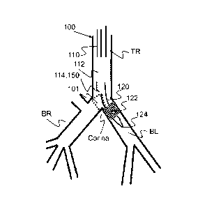
Figure 2 shows a schematic sectional view of the Tracheal Carina as
seen from endobronchial tube 100, provided by image sensor and light
source 150, allowing the visualization of bronchial cuff 122 disposed within
the left bronchi BL, the patency of the left bronchi, the patency of the right
bronchi, the tracheal carina, bronchial bifurcation, in a single field of view
101. Optionally a similar view may be provided with image sensor 150
when tube 100 is disposed with the right Bronchi BR as shown in FIG. 1A.
CA 02841704 2013-12-27
WO 2013/008106
PCT/1B2012/052077
11
Figure 3 shows endobronchial double lumen tube system 50
comprising endobronchial tube 100 and optional various auxiliary devices
that may be used in conjunction with and/or facilitate the use of tube 100.
Optionally auxiliary devices may for example include but is not
limited to stylet 10, Y-connector 12, air balance caps 14, and an
endobronchial tube connector assembly 20, or the like adjunct device
utilized facilitating the use of tube 100 as is known in the art.
Stylet 10 most preferably is utilized to facilitate placement of tube
100, as is known and accepted in the art.
Y-connector 12 most preferably provides for simultaneously
connecting both lumens of double lumen tube 100 to a single ventilation
source.
Endobronchial Tube connector assembly 20 provides for individually
connecting to tracheal lumen 110 and bronchial lumen 120. Connector
assembly 20 comprises a proximal end 22, distal end 28, and respective
tracheal lumen connector portion 24 and Bronchial connector portion 26.
Most preferably proximal end 22 provides for connecting and/or
otherwise associating the tube 100 at proximal end 102 at about the
individual lumen tracheal lumen 110 and bronchial lumen 120 to auxiliary
devices for example including but not limited to ventilation sources.
Most preferably distal end 24 provides for coupling and/or otherwise
associating with tube 100.
Figure 3 further provides a perspective view of a preferred double
lumen endobronchial tube 100 comprising tracheal lumen 110 having a
tracheal lumen distal end 114 and bronchial lumen 120 having a bronchial
lumen distal end 124.
Tube 100 further comprises tracheal cuff 112, shown in its expanded
state, provided for securely placing and/or anchoring tube 100 within the
trachea while ventilating the lungs through tracheal lumen 110.
Tube 100 further comprises bronchial cuff 122, shown in its expanded
and/or inflated state, provided for securely placing and/or anchoring tube
CA 02841704 2013-12-27
WO 2013/008106
PCT/1B2012/052077
12
100 within the bronchi, left or right. Most preferably cuff 122 provides for
selectively controlling the ventilation to the bronchial arch wherein it is
placed (left or right). For example ventilation to either the left or right
bronchi may be completely blocked so as to allow a procedure on the
respective lung (for example right) while allowing the ventilation of the
other lung (for example left) via tracheal lumen 110.
Most preferably tracheal cuff 112 may be inflated and/or deflated via
cuff tracheal connector 118.
Most preferably bronchial cuff 122 may be inflated and/or deflated
io via cuff bronchial connector 128.
Most preferably injection tube connector 130 provides an access point
to a dedicated lumen about each of the tracheal tube 110 and bronchial tube
120, preferably for delivering drugs, suctioning liquids about tracheal distal
114 and/or bronchial lumen distal end 124.
Figures 4A provide a further perspective view of endobronchial tube
100, showing image sensor connector 158. Most preferably image sensor
connector 158 is provided in the form of a USB connector that provides both
for image and power supply to image sensor 150 disposed in a dedicated
lumen near distal end 114. Optionally and preferably image sensor and
illumination 150 may be rendered function when connected to a display and
power source (not shown) via connector 158.
Figure 4B provides a close up view showing the image sensor notch
152 disposed about the proximal end of image sensor lumen 150L providing
an exit point for image sensor conducting wires 154, most preferably
provided for both image transfer and power supply to image sensor and
illumination source 150.
Figure 5 provides a further perspective view of tube 100 provided
from a face on view showing the separation of tracheal lumen 110 and
bronchial lumen 120 at distal end 104 of tube 100.
Figure 6 provides a further schematic illustrative depiction of tube
100 showing a perspective view of tube 100 with the bronchial cuff 122 and
CA 02841704 2013-12-27
WO 2013/008106
PCT/1B2012/052077
13
tracheal cuff 112 removed. Figure 6A shows the curvature provided at both
the medial section 106 and distal end 104 therein defining a medial curvature
106a and a distal curvature 104a. Curvatures 104a and 106a are provided to
so that tube 100 fits within the upper airway tract's anatomy.
Most preferably medial curvature 106a is provided for the ease of
accessing and introducing tube 100 within the trachea through the oral cavity
and pharynx. Most preferably, curvature 106a, is provided with an angle
from about 100 degrees to about 160 degrees.
Most preferably distal curvature 104a is proved for ease of accessing
and introducing distal end 104 into one of the bronchi, left or right.
Optionally and preferably distal curvature 104a may be specific for
individual left or right endobronchial tubes. Optionally distal curvature may
be configured to be from about 25 degrees to about 70 degrees. Optionally
and preferably about 35 degrees as shown.
Optionally the length of tube 100 may be provided with a length from
about 200mm to about 550mm. Optionally and preferably the length of tube
100 may be selected in accordance with a user's anatomy.
Optionally endobronchial tube 100 may be provided with different
sizes, length, diameters as known and accepted in the art. Optionally tube
100 may be provided with a gauge from about 26 Fr to about 44Fr, or from
about For example the external diameter of tube 100 may be provided in
varying gauges and/or sizes for example including but not limited to 28 Fr,
32 Fr, 35 Fr, 37 Fr, 39 Fr and 41Fr, within the context of this application
the
units 'Fr' refer to the gauge of the tube 100 in the units French as is a
common term of the art. Alternatively the gauge and or size of tube 100 may
be provided in the SI units of millimeters `mm'. The tube 100 according to
the present invention may be provided with an external diameter of about
9.3mm, 10.7mm, 11.7mm, 13mm and 13.7mm.
Optionally and preferably the length and diameter (also referred to as
gauge) of tube 100 may be correlated with one another.
CA 02841704 2013-12-27
WO 2013/008106
PCT/1B2012/052077
14
Figure 7A shows a close up view of distal end 104 of tube 100 shown
in Figure 6 providing a close up view. Figure 7A further shows a close up
view of curvature 104a showing the flaring of distal end 104 from the
tracheal lumen into the side portion of bronchial lumen 120.
Figures 7A-E show various close up view of distal end 104 specific to
curvature 104a showing the flaring and tapering of distal end 104 from the
tracheal lumen into the side portion of bronchial lumen 120.
Figures 7D-E provide further close up views of the distal end of
image sensor lumen 150L and cleaning nozzle 156, most preferably provided
io for
cleaning image sensor. Optionally and preferably cleaning nozzle 156 is
provided with an opening having a diameter from about 0.1mm to about
2mm. Optionally and preferably cleaning nozzle 156 may be provided with
a diameter of about 0.6mm.
Image sensor 150 is most preferably provided in a dedicated lumen
150L that spans the length of tube 100. Most preferably lumen 150 is
disposed between tracheal lumen 110 and bronchial lumen 120.
Most preferably distal end of lumen 150L provides for visualizing the
carina and the bronchial cuff 122, for example as shown in Figure 2.
Most preferably the diameter of image sensor lumen 150L is variable
along the length of tube 100. Most preferably image sensor lumen 150 is
smallest at the proximal end 102 and largest at the distal end 104.
Optionally and preferably at proximal end 102 sensor lumen 150L is
configured to have an elliptical cross-section. Optionally and preferably at
distal end of sensor lumen 150L is configured to have a circular cross-
section.
Most preferably alongside image sensor lumen 150L is a dedicated
cleaning lumen 160 that has a distal end defining a cleaning nozzle 156, as
shown, providing for cleaning image sensor 150 about its distal end.
Optionally and preferably cleaning nozzle 156 is provided with a curvature
and/or angle so as to direct cleaning solution, fluid, gas or the like flowing
fluid toward and/or away from integrated image sensor 150 and more
CA 02841704 2013-12-27
WO 2013/008106
PCT/1B2012/052077
preferably image sensor 150c. For example cleaning lumen 160 may be
utilized to clear mucus or the like biological obstruction from in front of
integrated image sensor 150 by flushing with a flowing fluid, for example a
liquid or gas, from the proximal end of lumen 160 through to its distal end at
5 forming cleaning nozzle 156. Optionally cleaning lumen 160 may be used to
clear the viewing field of integrated image sensor 150 by applying
suctioning therein suctioning in front of the field of view to keep it clean.
Figure 7F shows a close up view of cleaning nozzle 156 as directed
toward image sensor 150 about the distal end of lumen 150L. Optionally and
io preferably cleaning nozzle 156 is configured such that it provides for
maintaining an open field of view of the Tracheal Carina for integrated
images sensor 150.
Optionally and preferably the distal end of cleaning lumen 160 may
be curved such that the distal end is directed toward the distal end of image
15 sensor lumen 150L therein providing for forming at least one or more
cleaning nozzle 156 that is optionally and preferably directed toward image
sensor 150, for example as shown in Figure 7E.
Optionally tube 100 may be provided with at least two or more
cleaning lumen 160. Optionally a first cleaning lumen may be provided for
flushing biological obstruction while a second cleaning lumen may be
provided for suctioning biological obstructions away from the distal end 114.
Optionally a plurality of cleaning lumen 160 may be disposed on opposite
sides of integrated image sensor 150. Optionally a plurality of cleaning
lumen 160 may be configured to cooperate with one another, for example a
first lumen would flush biological obstructions toward a dsecond cleaning
lumen where the obstruction is carried away by suctioning. Optionally at
least two or more cleaning lumen may be utilized concertedly to either
suction or flush obstructions distal to integrated image sensors 150, therein
most preferably ensuring an open viewing field. Optionally a plurality of
cleaning lumen may be provided with different diameters and or sizes.
CA 02841704 2013-12-27
WO 2013/008106
PCT/1B2012/052077
16
Figure 8A shows a cross sectional view of tube 100 about its proximal
end 102 having tracheal lumen 110 and a bronchial lumen 120 defined on
either side of a midline partition 108. Most preferably tube 100 comprises a
plurality of peripheral lumen disposed internally and/or within the walls of
tube 100. Most preferably a plurality of peripheral lumen may be disposed
about the circumference of tube 100 and span essentially the length of tube
100, about the tracheal lumen 110 and/or bronchial lumen 120. Optionally
and preferably the peripheral lumen may for example include but is not
limited to a suctioning lumen, cuff inflating lumen, electronic lumen, image
io sensor lumen, cleaning lumen, injection tube lumen, or the like.
Most preferably tube 100 includes a dedicated lumen 150L provided
for image sensor and integrated illumination source 150. Most preferably
lumen 150L provides for housing the image sensor 150 at its distal end
(Figure 7E-F) and housing image sensor conducts for example in the form of
a wire 154, disposed along the length of lumen 150L, and a image sensor
notch 152 disposed near the proximal end of lumen 150L allowing image
sensor conductor 154 and connector 158 to be disposed external to tube 100.
Optionally and preferably lumen 150L is disposed about the anterior
portion of tube 100 about the middle of the cross-section of tube 100. Most
preferably lumen 150L is disposed anterior to partition 108. Optionally
lumen150L may be disposed about the posterior portion of tube 108 therein
posterior to partition 108.
Most preferably on both sides of lumen 150L are dedicated lumen
running along the length of tube 100 and most preferably running parallel
with lumen 150L. Optionally and preferably at least one or more of lumen
are provided as a dedicated cleaning lumen 160. Optionallly both lumen
flanking lumen 150L may be dedicated cleaning lumen 160.
Most preferably tube wall further comprises lumen 112L and 122L
respectively corresponding to tracheal lumen 110 and bronchial lumen 120.
Optionally and preferably lumen 112L and 122L are provided for inflating
and/or deflating cuffs 112 and 122 respectively.
CA 02841704 2013-12-27
WO 2013/008106
PCT/1B2012/052077
17
Figure 8B shows the same image as in Figure 8A however showing
the cross-section near tracheal lumen distal end 114 of tube 100. Most
preferably at tracheal lumen distal end 114 image sensor lumen 150L is
provided with a lumen having a larger radius than that provided at the
proximal end 102 as shown in Figure 8A. Most preferably tube 100 is
expanded about distal end 104 and lumen 150L to accommodate integrated
image sensor 150. Optionally image sensor lumen 150 about the external
surface of tube 110 is widened and/or expanded 1.5mm to 5mm from distal
end 114 of tracheal lumen 110.
Optionally the image sensor dedicated lumen 150L is provided with
an notch 150n disposed 22.5mm from the proximal end 102 of tube 100 and
a exit notch having a diameter of about 1.5mm.
Figure 9 shows a close up bottom-up view of the integrated image
sensor 150 within dedicated electronics lumen 150L disposed within the wall
of the endobronchial tube 100, showing image sensor 150c optionally and
preferably provided in the form of a CCD or CMOS or the like, and
illumination source 1501 most preferably provided in the form of at least
one and more preferably at least two or more LED, as shown.
While the invention has been illustrated primarily with reference to a
left bronchi endobronchial tube, it will be appreciated that the present
invention is not limited to a left bronchi endobronchial tube where the
inventive and novel aspects equally covers a right bronchi endobronchial
tube.
While the invention has been described with respect to a limited
number of embodiments, it will be appreciated that many variations,
modifications and other applications of the invention may be made.