Note: Descriptions are shown in the official language in which they were submitted.
WO 2013/001363 PCT/1B2012/001546
ESOPHAGEAL STIMULATION DEVICES AND METHODS
CROSS-REFERENCE TO RELATED APPLICATIONS
10001] This application claims priority to U.S. Provisional Patent
Application
No. 61/501,338, filed June 27, 2011, and U.S. Provisional Patent Application
No.
61/61.2,072, filed March 16, 2012, both entitled "ESOPFIAGEAL STIMULATION
DEVICE"
BACKGROUND
Field of the invention
[09021 .. The present invention, in some embodiments thereof, relates to
devices
and methods for generating motility in GI organs, and in particular to devices
and
methods for generating esophageal motility for diminishing retrograde flow of
gastric
contents.
Desctiption of the Related Art
[00031 The esophagus is a tubular muscular organ having a length of
approximately 25 cm, located, between the upper esophageal sphincter (UES) and
the
lower esophageal sphincter (LES). The esophagus functions solely to deliver
food from
the mouth to the stomach using peristaltic muscle motion. Peristalsis is a
sequential,
coordinated contraction wave that travels the entire length of the esophagus,
propelling
intraluminal contents distally to the stomach. Primary peristalsis is the
peristaltic wave
triggered by the swallowing center. The peristaltic contraction wave travels
at a speed of
approximately 2 crrils and correlates with manometry-recorded contractions.
The
secondary peristaltic wave is induced by esophageal distension from the
retained bolus,
refluxed material, or swallowed air, with the primary role to clear the
esophagus of
retained food or any gastroosophageal relluxatc. Tertiary contractions are
simultaneous,
isolated, dysfunctional contractions. Anesthetization or sedation are
suspected of causing
suspension of esophageal peristaltic motility and lowers LES pressure, hence
gastric
content are more prone to infiltrate and travel proximally in the esophagus.
CA 2842001 2018-07-04
CA 02842001 2014-01-15
WO 2013/001363 PCT/IB2012/001546
100041 Gastric contents refluxing through the esophagus are known to affect
conditions which may increase morbidity and mortality rates. Gastroesophageal
Reflux
(GER) is a condition, in which the LES opens spontaneously, for varying
periods of time,
or does not close properly and stomach contents rise up into the esophagus. In
Laryrtgophary-ngeal Reflux (LPR), the retrograde flow of gastric contents
reaches the
upper aero-digestive tract. In order to diminish and treat such conditions,
efforts have
been made to develop medical and surgical means for improving LES
functionality and
for creating a substitute sphincter proximally adjacent the stomach. in some
occasions it
may be advantageous to develop a second "line of defense" provided proximally
to the
LES along the esophagus, especially to push back any gastric contents or chyme
that
infiltrated the LES or any substitute or supplement thereof. Such a need may
arise, for
example, in cases of intubation and/or ventilation, usually in anesthetized
ICU patients,
CVA patients, or others, in which esophageal motility is muted or less
dominant.
[00051 Tubefeeding (e.g., "gastric feeding" or "enteral feeding") is a
common
and life preserving procedure, however complications can arise. GER is
commonly
associated with tubefeeding, including in usage of nasogastric tubing (NOT)
and other
gastric feeding practices. Research in past years has discussed the emergence
of GER as
an effect of the use of NGT (see for example in Ibanez et al.,
"Gastroesophageal reflux in
intubated patients receiving enteral nutrition: effect of supine and
semirecumbent
positions", WEN J Parenter Enteral Nutt 1992 Sep-Oct;16(5):419-22; in Manning
et al.,
"Nasogastric intubation causes gastroesophageal reflux in patients undergoing
elective
laparotomy", Surgery. 2001 Nov;130(5):788-91; and in Lee et al., "Changes in
gastroesophageal reflux in patients with nasogastric tube followed by
percutaneous
endoscopic gasirostomy", J Fomios Med Assoc. 2011 Feb; 110(2) :115-9).
100061 Pulmonary aspiration is the entry of material from the oropharynx or
gastrointestinal tract into the larynx and lower respiratory tract.
Consequences of
pulmonary aspiration range from no injury at all, to chemical pneumonitis or
pneumonia,
to death within minutes from asphyxiation. One common cause of pulmonary
aspiration
is aspiration of gastric contents, as suggested in relevant literature (see
for example
Pellegrini et al., "Gastroesophageal reflux and pulmonary aspiration:
incidence,
functional abnormality, and results of surgical therapy". Surgery. 1979
Jul;86(1):110-9,
indicating that incidence of aspiration is due to a motor disorder that
interferes with the
2
CA 02842001 2014-01-15
WO 2013/001363 PCT/1B2012/001546
ability of the esophagus to clear refluxed acid, and that abnormal pulmonary
symptoms
can induce or result from gastroesophageal reflux).
100071 Ventilator-associated pneumonia (VAP) is pneumonia that develops 48
hours or longer after mechanical ventilation is given by means of an
endotracheal tube or
tracheostomy. VAP results from the invasion of microorganisms into the lower
respiratory tract and lung parenchyma. Intubation compromises the integrity of
the
oropharynx and trachea and allows oral and gastric secretions to enter the
lower airways.
The aetiopathogenesis of VAP requires abnormal oropharyngeal and gastric
colonization
and the further aspiration of their contents to the lower airways. Known risk
factors for
gastric colonization include: alterations in gastric juice secretion;
alkalinization of gastric
contents; administration of enteral nutrition; administration of antacids; and
the presence
of bilirubin. According to Torres et al. (in "Stomach as a source of
colonization of the
respiratory tract during mechanical ventilation: association with ventilator-
associated
pneumonia", Eur Respir J. 1996 Aug; 9(8):1729-35), although the role of the
colonized
gastric reservoir in the development of VAP remains debatable, there is major
evidence in
the literature in favor of the gastric origin of part of these pulmonary
infections.
[0008] US Patent Application No. 2011/0130650 relates to an enteral feeding
device comprising "expandable means which prevents or significantly reduces
aspirations
from the alimentary tract to the respiratory system. In further aspects, the
invention
relates to systems comprising said enteral feeding device, methods and uses
thereof."
100091 US Patent Application No. 2010/0160996 " relates to methods and
apparatuses for treating ailments by "inserting a balloon-electrode device
into an
esophagus of a mammal, the balloon-electrode device including: (i) a
nasogastral (NO)
tube having an internal passageway and an external surface, (ii) at least one
electrode
coupled to the external surface of the NO tube, (iii) a conductor extending
through the
internal passageway of the NO tube and electrically connecting to the
electrode, and (iv)
a balloon surrounding the electrode and a portion of the NO tube; inflating
the balloon
with fluid such that the electrode is substantially centrally located within
an interior
volume of the balloon; and applying at least one electrical signal to the
electrode via the
conductor such that an electromagnetic field emanates from the electrode to at
least one
of nerves and muscles of the mammal."
[00101 US Patent Application No. 2008/0249507 relates to a "food
administering apparatus including a feeding tube, having a distal outlet and
proximal
3
CA 02842001 2014-01-15
WO 2013/001363 PCT/1B2012/001546
inlet, adapted for insertion of the distal outlet into the stomach of an adult
patient while
the proximal inlet is outside the patient, the tube being suitable for
administering food or
medicine from a proximal port to the distal outlet and at least one electrode
mounted on
the tube."
SUMMARY
[0011] .. According to an aspect of some embodiments of the present invention,
there is provided a system for evoking esophageal motion. In some embodiments,
the
esophageal motion includes at least one local contraction. In some such
embodiments, at
least one local contraction decreases a local segment of the esophagus lumen,
optionally
to at least 50% its initial diameter. In another embodiment, the at least one
local
contraction fully closes a local segment of the esophagus. In some
embodiments, at least
one local contraction develops a local esophageal pressure of at least 15
mmHg, and
optionally at least 25 mmHg, or higher, or lower or intermediate to said
values.
[00121 In some embodiments, the esophageal motion is a patterned motion
including at least two evoked contractions at different esophageal portions.
Optionally,
the different esophageal portions include adjacent esophageal portions and/or
remote
esophageal portions. In some embodiments, the at least two evoked contractions
are
sequentially and/or timely generated according to a preset sequence. In some
embodiments, the esophageal motion includes a distally advancing contraction
wave,
optionally though not necessarily including peristalsis. In some embodiments,
use of
such a system and/or method of esophageal stimulation diminishes retrograde
flow of
stomach contents. In some cases, such a method accomplishes this result by
stimulating
the esophagus to produce a distally travelling wave of contractions that
simulate natural
peristalsis.
100131 In some embodiments, the system for evoking esophageal motion
includes an elongated member sized and configured for nasal or oral placement
in a
patient's esophagus. In some embodiments, the elongated member is a medical
intubation
device, and optionally, a gastric feeding tube.
[0014] In some embodiments, the system further includes at least one
stimulator mounted or mountable on the elongated member, adapted to stimulate
a chosen
portion of the esophagus to evoke a local shaped contractive reaction.
Optionally, the at
least one stimulator is fixed to the elongated member. In some embodiments,
alternatively
or additionally, the at least one stimulator is provided with a fixator
configured for
4
CA 02842001 2014-01-15
WO 2013/001363 PCT/1B2012/001546
mounting the at least one stimulator on a chosen external portion of the
elongated
member. The fixator may be slidably movable along a length of the elongated
member,
optionally restrainedly securable around the chosen external portion of the
elongated
member, and/or optionally fixedly lockable to the chosen external portion of
the
elongated member thereby preventing sliding therealong.
[0015] In some embodiments, the at least one stimulator includes at least
two
stimulators sequentially positioned along an esophageal length; each
stimulator is
configured to stimulate a different esophageal portion. Optionally, a
plurality of
stimulators is provided along the effective length of the medical intubation
device. In
some embodiments, a distance of less than 5 cm exists between at least two of
the
stimulators, and a distance of greater than 10 cm exists between a proximal
most
stimulator and a distal most stimulator.
[00161 In some embodiments, the at least one stimulator includes an
electrode,
or a plurality of electrodes, for allowing local electrical stimulation(s) of
muscle tissue
and/or neural tissue, adjacent and/or in direct contact. The electrode(s) may
be shaped as
chosen or needed, as known in the relevant art, and may be, for example,
circular,
rectangular, or ring shaped.
100171 in some embodiments, the at least one stimulator includes an
expandable member, which is optionally a mechanical stimulator, optionally
inflatable,
and sized and/or shaped when expanded to radially stretch out an esophageal
portion in a
manner that evokes a shaped contractive reaction distal to the esophageal
portion.
(0018) in some embodiments, the system further includes a generator
connected to the at least one stimulator. The generator may be provided
outside the
patient body or alternatively be sized and configured for prolonged intra-oral
or intra-
esophageal placement. The generator may be an electrical signal generator
adapted to
generate electrical stimulations via at least one electrode or at least two
electrodes
electrically connected thereto. Alternatively, the generator may include a
pump for cases
of inflatable stimulators. The generator may be a pulse generator and/or may
be able to
generate different shaped signals, for example a step wave, a sine wave, a saw-
tooth
wave, a variable width pulse or any combination thereof. The generator may
include or be
connectable to a power source, which may or may not comprise an element of the
system.
In some embodiments, the power source may be sized and configured for
prolonged intra-
oral or intra-esophageal placement.
CA 02842001 2014-01-15
WO 2013/001363 PCT/1B2012/001546
100191 In some embodiments of the invention, the system further includes at
least one sensor mounted or mountable on the elongated member. The at least
one sensor
may be mounted distally to a distal-most stimulator. Optionally, a proximal-
most sensor
is positioned at least 5 cm distally to the distal-most stimulator, optionally
at least 10 cm,
optionally approximately 20 cm, or higher, or lower, or intermediate to said
values. In
some embodiments, the at least one sensor comprises at least one of: a pH
sensor, a
pressure sensor, a manometer, an impedance sensor, a motion sensor, a
capacitance
sensor and a mechanical sensor.
[0020] .. In some embodiments, the system for evoking esophageal motion
includes a catheter and a controller, wherein the catheter and controller are
configured for
wired or wireless communication with each other. The catheter includes a
plurality of
electrodes and at least one pH sensor. In some embodiments, the controller is
configured
and programmed to initiate an electrical stimulation via at least one of the
plurality of
electrodes in response to at least one pH sensor sensing a local pH less than
3. In use, the
at least one pH sensor of various embodiments senses local pH in real-time,
and at least
one of the plurality of electrodes is stimulated upon the at least one pH
sensor sensing a.
local pH below 3 in real-time. In some embodiments, the plurality of
electrodes and the
one or more pH sensors are arranged such that upon a pH sensor sensing a local
pH less
than 3, one or more electrodes positioned proximally to the pH sensor are
stimulated.
100211 .. in an aspect of some embodiments, there is provided a method for
generating esophageal motion. In some embodiments, the method comprises a step
of
positioning at least two electrodes, including a proximally positioned
electrode and a
distally positioned electrode, at distant portions along the esophagus.
Optionally, the
method includes also a step of electrically connecting the at least two
electrodes to a
generator. Optionally, the method further includes a step of generating a
signal sequence
including a first signal at the proximally positioned electrode thereby
stimulating a
proximal esophageal tissue and a second signal at the distally positioned
electrode
thereby stimulating a distal esophageal tissue. In some embodiments, the
signal sequence
produces a contraction wave that travels a length of the esophagus.
100221 Optionally, additionally or alternatively, a method for generating
esophageal motion with the system will include a step of placing in an
esophagus the
elongated member and at least one electrode mountable thereon, and generating
at least
one stimulating signal to evoke a local shaped contractive reaction. The local
shaped
6
CA 02842001 2014-01-15
WO 2013/001363 PCT/1B2012/001546
contractive reaction may be a spasm, a full contraction, a partial
contraction, a peristalsis
or any combination thereof.
10023] A method for connecting at least one electrode to a gastric tube pre-
positioned in a patient's esophagus may include a step of locating a target
portion on the
gastric tube at a chosen distance from a proximal end thereof. Optionally, the
method also
includes a step of providing an electrode fixator configured for fixedly
covering a portion
of the gastric tube. Optionally, the electrode fixator comprises at least one
electrode
electrically connectable with a signal generator and locking means.
Optionally, the
method also includes a step of positioning the electrode fixator over the
target portion.
Optionally, the positioning includes sleeving the electrode fixator over and
along the
gastric tube. (Hereinafter, sleeving is defined as sliding a sleeve, sock, or
other tubular-
shaped element, rigid or nonrigid, over, around, and along an object, so as to
at least
partially encase said object.) Optionally, the method also includes a step of
applying the
locking means to fixedly lock the electrode fixator in place. In some
embodiments, the
gastric tube may be partially withdrawn to expose the target portion.
[0024] .. Unless otherwise defined, all technical and/or scientific terms used
herein have the same meaning as commonly understood by one of ordinary skill
in the art
to which the invention pertains. Although methods and materials similar or
equivalent to
those described herein can be used in the practice or testing of embodiments
of the
invention, exemplary methods and/or materials are described below. in case of
conflict,
the patent specification, including definitions, will control. In addition,
the materials,
methods, and examples are illustrative only and are not intended to be
necessarily
limiting.
BRIEF DESCRIPTION OF THE DRAWINGS
[0025] Some embodiments of the invention are herein described, by way of
example only, with reference to the accompanying drawings. With specific
reference
now to the drawings in detail, it is stressed that the particulars shown are
by way of
example and for purposes of illustrative discussion of various embodiments. In
this
regard, the description taken with the drawings makes apparent to those
skilled in the art
how embodiments of the invention may be practiced. In the drawings:
7
CA 02842001 2014-01-15
WO 2013/001363 PCT/1B2012/001546
100261 Fig. 1A. schematically illustrates an exemplary nasogastric tube
positioned in a patient's esophagus and including a plurality of stimulators,
in accordance
with an embodiment of the present invention;
10027] Fig. 113 schematically illustrates an exemplary oral feeding tube
positioned in a patient's esophagus and including a mono-polar stimulator, in
accordance
with an embodiment of the present invention;
10028] Fig. 1C schematically illustrates an exemplary feeding tube
positioned
in a patient's esophagus and including a plurality of stimulators and a
sensor, in
accordance with an embodiment of the present invention;
10029] Figs. 2A-C schematically illustrate a partial cut view of a
contraction
wave stimulating system provided in an esophagus, shown at different operation
stages,
in accordance with some embodiments of the present invention;
[0030] .. Figs. 3A-D schematically illustrate a first exemplary stimulation
sequence and a correspondingly generated patterned esophageal motion, in
accordance
with some embodiments of the present invention;
[00311 Figs. 4A-D schematically illustrate a second exemplary stimulation
sequence and a correspondingly generated patterned esophageal motion, in
accordance
with some embodiments of the present invention;
[0032] .. Fig. 5A schematically illustrates a top view of an exemplary
esophageal intubation tube provided with a plurality of terminals comprising
two
electrodes each; an exemplary signal sequence from each terminal is also
illustrated, in
accordance with some embodiments of the present invention;
[00331 .. Fig. 5B schematically illustrates a top view of an exemplary
esophageal intubation tube provided with a plurality of terminals comprising
two
electrodes each; an exemplary signal sequence from each terminal is also
illustrated, in
accordance with some embodiments;
[0034] .. Fig. 6 schematically illustrates a top view of an exemplary
esophageal
intubation tube provided with a plurality of terminals comprising three
electrodes each, in
accordance with some embodiments of the present invention;
[0035] Fig. 7 schematically illustrates a top view of an exemplary
esophageal
intubation tube that is provided with a plurality of terminals comprising two
electrodes
each and is coupled to an array of switches, in accordance with some
embodiments of the
present invention;
8
CA 02842001 2014-01-15
WO 2013/001363 PCT/1B2012/001546
100361 Fig. 8 schematically illustrates a top view of an exemplary
esophageal
intubation tube having a plurality of electrodes with polarities modulating
over time to
create a stimulation sequence, in accordance with some embodiments of the
present
invention;
100371 Fig. 9 schematically illustrates a top view of an exemplary
esophageal
intubation tube having a plurality of electrodes with polarities modulating
over time to
create another stimulation sequence, in accordance with some embodiments of
the present
invention;
[0038] Figs. I0A-B schematically illustrate a partial isometric view and a
partial top view of an exemplary NO tube provided with a plurality of
electrodes, in
accordance with some embodiments of the present invention;
[0039] Figs. 11A-B schematically illustrate a partial top view of an
exemplary
NO tube provided with a plurality of expandable stimulators, before and after
actuation,
in accordance with some embodiments of the present invention;
[0040] Fig. 12 schematically illustrates an exemplary NO tube positioned in
a
patient's esophagus and provided with a fixedly positioned stimulator fixator,
in
accordance with some embodiments of the present invention;
[0041] Figs. 13A-1) schematically illustrate different exemplary fixators,
in
accordance with some embodiments of the present invention;
[00421 Figs. 14A-B schematically illustrate an exemplary stretchable sleeve-
type fixator, in accordance with some embodiments of the present invention;
[00431 Fig. 15 schematically illustrates an exemplary delivery device for
delivering fixators to a feeding tube, in accordance with some embodiments of
the present
invention; and
[0044] Fig. 16 schematically illustrates a partial cut view of an exemplary
self-expandable electrode fixator partially emerging from a delivery catheter,
in
accordance with some embodiments of the present invention.
DETAILED DESCRIPTION OF CERTAIN EMBODIMENTS
[0045] The following preferred embodiments may be described in the context
of exemplary esophageal stimulation procedures for ease of description and
understanding. However, the invention is not limited to the specifically
described devices
and methods, and may be adapted to various clinical applications without
departing from
9
CA 02842001 2014-01-15
WO 2013/001363 PCT/1B2012/001546
the overall scope of the invention. For example, devices and related methods
including
concepts described herein may be used for stimulating other GI organs such as
but not
limited to the: stomach wall, duodenum, jejunum, ileum, caecum, small
intestine, colon,
large intestine, throat and gullet.
[0046] The present invention, in some embodiments thereof, relates to
devices
and methods for generating motility in GI organs, and in particular to devices
and
methods for generating, at least, esophageal motility for diminishing
retrograde flow of
gastric contents.
[0047] An aspect of some embodiments relates to a system for generating a
patterned esophageal motion. A patterned esophageal motion may be any local or
cross-
esophageal muscular expansion or contraction, or any combination thereof,
evoked and/or
orchestrated following generated stimulation. The pattern may be a chosen
shape and/or
magnitude of a local esophagus contraction and/or a distally progressive
contraction wave
having chosen characteristics, including but not limited to contraction force,
wave travel
velocity and wave occurrence frequency. In some embodiments, the patterned
esophageal
motion includes peristalsis, optionally simulating a naturally occurring
esophageal
peristalsis or creating a synthetic peristalsis based on. an algorithmic
sequence of
stimulations, and/or any combination of local contractions, distally
progressive
contraction wave and/or selectively evoked naturally occurring peristalsis at
a patient's
esophagus.
100481 .. In some embodiments, the system includes at least one stimulator
adapted to stimulate a portion of the esophagus to evoke a shaped contractive
reaction. In
some embodiments, the at least one stimulator includes an expandable,
optionally
inflatable, member, sized and/or shaped when expanded to radially stretch out
an
esophageal portion in a manner that evokes a shaped contractive reaction
distal to the
esophageal portion. A.n inflatable stimulator may be connected to a pump,
optionally
hydraulic or pneumatic, and may be selectively inflated or deflated according
to a chosen
scheme, such as, for example, a predetermined and/or programmed scheme, and
optionally a scheme including pulsatory actuation.
[0049] Optionally, alternatively or additionally, the at least one
stimulator
includes an electrode configured for electrical stimulation of
adjacent/contacting
esophagus muscle tissue. A stimulating electrode may be connectable or
provided readily
connected with a generator, optionally a pulse generator, configured to
generate a chosen
CA 02842001 2014-01-15
WO 2013/001363 PCT/1B2012/001546
sequence of stimulations. Optionally, alternatively or additionally, an
internal power
and/or signal source may be provided with the system that is sized and
configured for
intra-body (e.g., intra-orally) placement, optionally in or adjacent the
esophagus. In some
other optional embodiments, a power and/or signal source may be provided
(e.g., worn)
on the patient. In some exemplary embodiments, at least one electrode and/or
sensor is
connected with such an internal power source sized and configured for
placement on a
medical intubation device (e.g., a feeding tube).
[0050] In some embodiments, the system includes a plurality of stimulators
provided at different relative locations within the esophagus.
[0051] A. local contraction of the esophagus, or any combination or pattern
of
esophageal contractions may increase local and/or average esophageal pressure.
Optionally, alternatively or additionally, stimulation is used to decrease
local and/or
average volume entrapped along the esophagus lumen between the LES and the UES
thereby increasing local and/or average pressure. By increasing the pressure
at a local
segment of the esophagus lumen, a retrograded material or chyme may be forced
to travel
backward to a distal lumen segment being less pressured, whereas by increasing
the
average or overall pressure in the esophagus, a possible reflux causing
positive pressure
difference between the stomach and the esophagus may be diminished and even
reversed,
thereby diminishing the possibility or volume of refluxed material or even
preventing
reflux. In some embodiments, a local and/or average pressure caused by a
single evoked
contraction or a series of evoked contractions may be equal or higher than 15
mmHg,
optionally equal or higher than 25 mmHg, optionally equal or higher than 50
mmHg, and
optionally equal or higher than 100 mmHg, or lower, higher, or intermediate to
any of
said values.
[0052] In some embodiments, the system further includes, is provided with,
or
is connected to a medical intubation device that is sized and configured for
nasal or oral
placement in a patient's esophagus. In som.e embodiments, the medical
intubation device
is a gastric feeding tube.
[0053] In some embodiments, at least one stimulator is fixed to the medical
intubation device. Optionally, alternatively or additionally, at least one
stimulator is
provided with a fixator configured for fixedly covering a portion of the
medical
intubation device. The fixator may be slidably movable along a length of the
medical
intubation device and/or may be restrainedly securable around the portion of
the medical
11
CA 02842001 2014-01-15
WO 2013/001363 PCT/1B2012/001546
intubation device. In some embodiments, the fixator is fixedly lockable to the
portion of
the medical intubation device thereby preventing sliding therealong.
10054] A fixator may be sleeved and/or otherwise coupled to the medical
intubation device after the latter has been partially or fully withdrawn from
a patient's
esophagus or trachea. Alternatively, a fixator may be mounted on to a medical
intubation
device prior to initial placement in the patient. A proper location of a
fixator and/or
stimulator may be achieved under imagery guidance (e.g., x-ray). Optionally,
alternatively or additionally, means (e.g., recesses, indentations, etc.) are
provided or
created on portions of the medical intubation device to allow controlled
positioning by
engaging the fixator/stimulator thereto. In cases in which the medical
intubation device is
kept in place within the patient, means may be applied to distally advance a
fixator/stimulator along and over the medical intubation tube's outer
periphery to a
chosen location, optionally under x-ray monitoring.
[00551 In some embodiments, the at least one stimulator includes at least
two
stimulators sequentially positioned along an esophageal length, each
stimulator being
configured to stimulate a different esophageal portion. Optionally, a
plurality of
stimulators is provided along the effective length of the medical intubation
device.
100561 in some embodiments wherein the at least one stimulator comprises a
plurality of electrodes, the electrodes are arranged in groups refired to
herein as
terminals. In some embodiments, two electrodes form a terminal. in some such
embodiments, one electrode is a positive electrode, which receives current
from a signal
generator, and the other electrode is a negative electrode, which is grounded.
The
distance between each terminal may be fixed or variable, and the terminals are
spaced
such that the distance between each terminal is greater than the distance
between each
electrode within any given terminal. For example, the width of the terminal
(i.e., the
distance between the electrodes of a terminal) may be 5-10 mm, and optionally
8 mm.
The distance between each terminal may be 15-30 mm, optionally 20 mm, or
optionally,
below, above, or intermediate to said values. In other embodiments having two
electrodes per terminal, the system also comprises an array of controlled
relays coupled to
the electrodes. The array of controlled relays may be configured to
selectively transition
each electrode between a positively connected state, a grounded state, and a
disconnected
state. In still other embodiments, three electrodes form a terminal. In such
embodiments,
two of the electrodes may be grounded, and the third electrode, which is
positioned
12
CA 02842001 2014-01-15
WO 2013/001363 PCT/1B2012/001546
between the two grounded electrodes, may be a positive electrode connected to
a signal
generator. The electrodes are positioned such that the positive electrode will
close a
circuit with the two negative (grounded) electrodes of the same terminal. Such
a design
may position the center of stimulation at the location of the positive
electrode.
100571 In some embodiments, the system includes at least one sensor.
Optionally, the sensor is provided on the medical intubation device distally
to the at least
one stimulator. Optionally, the sensor is a pH sensor, optionally adapted to
sense a
change (e.g., decrease) of local pH, for example due to the presence of
gastric contents
proximally to the LES. Optionally, alternatively or additionally, an impedance
sensor
may be used, configured for sensing a change in impedance of tissues provided
between
stimulators and/or electrodes, optionally correlative to a reaction to gastric
contents or
other substances. Optionally, alternatively or additionally, other sensor
types may be
used, including but not limited to a pressure sensor, a. manometer, a moisture
sensor, a.
temperature sensor, a motion sensor, a capacitance sensor and a mechanical
sensor.
[00581 In an aspect of some other embodiments, there is provided a method
for generating esophageal peristalsis in a patient intubated with a gastric
tube. In some
embodiments, the method comprises at least one of the following steps,
optionally with
no particular order:
1. positioning at least two electrodes, including one or more proximally
positioned electrodes and one or more distally positioned electrodes, at
spaced
positions along the gastric tube, where the positions are selected such that
after installation of the gastric tube, the at least two electrodes will be
between
the upper esophageal sphincter (UES) and the lower esophageal sphincter
(LES);
2. electrically connecting the at least two electrodes to a generator; and/or
3. generating a signal sequence including a first signal at the proximally
positioned electrode thereby stimulating a proximal esophageal tissue and a
second signal at the distally positioned electrode thereby stimulating a
distal
esophageal tissue.
[00591 .. In some embodiments, the electrodes apply electrical current in a
series
of one or more electrical trains (also referred to herein as pulse groups),
wherein each
train is composed of a series of cycles, and each cycle includes one pulse.
Pulses within a.
train or pulse group are characterized by an intetpulse spacing, and different
pulse groups
13
CA 02842001 2014-01-15
WO 2013/001363 PCT/1B2012/001546
are separated by an intergroup spacing. Generally, the interpulse spacing
between pulses
within a group or train is less than the intergroup spacing between at least
some groups.
Each electrical pulse has an amplitude; in preferred embodiments, the
amplitude is higher
than a stimulating threshold, wherein the stimulating threshold is the minimum
voltage at
which a local contraction occurs when applied to a portion of the esophagus.
In some
embodiments, the stimulating threshold is between 5V and 20V, optionally
between 8V
and by or between 10V and 15V; in other embodiments, the stimulating threshold
may
be higher or lower than said values. Each pulse is provided for a duration of
time. In
some embodiments, the pulse width (i.e., the duration) is equal to or greater
than 5
milliseconds, and optionally, equal to or greater than 10 milliseconds. The
applied pulse
is followed by a duration of lower current and/or no current. Together, one
pulse and one
duration of low current compose a cycle. In some embodiments, one cycle lasts
20 ms; in
other embodiments, one cycle lasts 15 ms, or optionally 30 ms, or less than,
greater than,
or intermediate to said values. In some embodiments, multiple cycles are
provided
successively such that together the cycles form. a train having a duration of
one to two
seconds. in other embodiments, trains are provided that are longer or shorter
in duration.
The train is then followed by a duration of no current or low current produced
by below-
threshold voltages.
100601 In some embodiments, the sequence of trains or other signal sequence
produces a contraction wave that travels a length of the esophagus. In some
embodiments, the contractions generate or simulate natural peristalsis.
100611 In some embodiments, before each train or pulse, one or more below-
threshold pulses are applied to the tissue to prime the tissue and induce it
to contract more
firmly and efficiently and to begin contracting at lower voltage stimulation
levels.
Optionally, a preliminary, below-threshold train is applied before each
stimulating train
or pulse. In some embodiments, a continuous below-threshold train is applied
to specific
portions of the esophagus to desensitize, and thereby avoid unneeded
contractions within,
said portions. For example, the LES must be open in order for material to pass
from the
esophagus into the stomach. in one embodiment therefore, one or more
electrodes may
also be positioned on the gastric tube such that after installation they are
adjacent the LES
to provide a continuous below-threshold train which will be applied to the LES
to
desensitize it so that it does not contract when material arrives. Such
electrode(s) may
also be used to close the 1,ES if that is a desired response under some
circumstances.
14
CA 02842001 2014-01-15
WO 2013/001363 PCT/1B2012/001546
100621 In an aspect of some embodiments, there is provided a method for
connecting at least one electrode to a gastric tube readily positioned in a
patient's
esophagus. In some embodiments, the method comprises at least one of the
following
steps, optionally with no particular order:
1. locating a target portion on the gastric tube at a chosen distance from a
proximal end thereof;
2. providing an electrode fixator configured for fixedly covering a portion of
the
gastric tube, the electrode fixator comprising at least one electrode
electrically
connectable with a signal generator and locking means;
3. positioning the electrode fixator over the target portion; and/or
4. applying the locking means to fixedly lock the electrode fixator in place.
[0063.1 In some embodiments, at least one of the steps includes the use of
internal and/or external imagery. Optionally, additionally or alternatively,
imaging
guidance, optionally including x-ray and/or RF sources, may be applied, for
example, to
locate the electrode and change its position on the feeding tube while in the
patient. This
may allow the clinician to keep the feeding tube tip in appropriate position
while
adjusting the location of the electrode.
100641 .. in some embodiments of the invention, the fixator positioning
includes
a step of: sleeving the electrode fixator over and along the gastric tube.
Optionally, the
method further comprises a step of: partially withdrawing the gastric tube to
expose the
target portion.
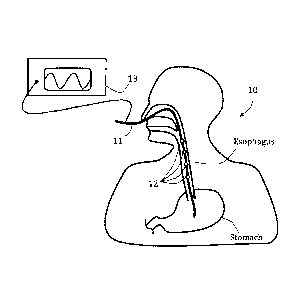
(0065) Referring now to the drawings, Fig. IA schematically illustrates an
exemplary system 10 comprising an. elongated member 11 positioned in a
patient's
esophagus and including a plurality of stimulators 12, in accordance with an
embodiment.
Elongated member 11 may be any plastic or elastic rod or tube sized to enter
and be
pushed through the esophagus, preferably with no injury to adjacent tissues.
Elongated
member may be a probe, a catheter and/or a nasoga.stric tube (NGT), the latter
is
optionally used for injecting thod directly to a patient's stomach and/or
pumping out
chyme to relieve excessive gastric pressure. Stimulators 12 may be any
mechanical,
electrical or chemical local muscle or neural stimulators. Four stimulators 12
are shown
for illustrative purposes, although any other number of stimulators may be
provided. In
some exemplary embodiments, stimulators 12 are or include at least one
electrode. In
some embodiments, each shown stimulator 12 represents a number of electrodes
provided
CA 02842001 2014-01-15
WO 2013/001363 PCT/1B2012/001546
around a local periphery of elongated member 11.. In some embodiments,
stimulators 12
are provided in a sequential order, optionally having a constant or
selectively changeable
distance therebetween. Optionally, stimulators 12 comprise bi-polar electrodes
so that
pairs of adjacent non-short-circuited electrodes can be used for closing an
electrical
circuit and thereby stimulate an esophageal muscle tissue in-contact and in-
between the
two electrodes. A generator 13, optionally an electrical signal generator, is
shown
connected to stimulators 12 via elongated member 11, optionally over and along
its outer
periphery or via a lumen thereof. To produce a series of esophageal
contractions in
accordance with a chosen scheme or logic, such as optionally simulating a
naturally
occurring esophageal peristalsis, separate generator outputs may be provided
to separate
electrodes or electrode groups 12. In some advantageous embodiments, the
spacing
between electrodes or electrode groups 12 is less than 5 cm, and the distance
between the
most proximal electrode or electrode group 12 and most distal electrode or
electrode
group 12 is at least 10 cm. This allows sequential stimulation of the
electrodes or
electrode groups 12 along a significant portion of the esophagus between the
UES and the
LES.
100661 in Fig. 113, an exemplary system 20 is schematically illustrated
comprising an oral feeding tube 21 positioned in a patient's esophagus and
including a
mono-polar stimulator 22, in accordance with an embodiment. Although it is
commonly
more safe and convenient to place an esophageal intubation via a nasal cavity,
there might
be circumstances (e.g., with infant patients) where a tube is inserted via the
oral cavity as
suggested in this figure. Mono-polar stimulator 22 is electrically connected
to an outside
source or ground (shown. in the figure as "(-)") and is selectively capable of
closing an
electrical circuit with an external electrode 23, optionally positioned on the
patient's neck
skin. A single electrode may be used to stimulate a neutrally sensitive region
thereby
evoking an esophageal contraction wave from the stimulated region and
downward,
optionally to the LES or the stomach interim. Optionally, alternatively or
additionally, a
single electrode may be used for local muscle contraction in order to serve as
a barrier for
refluxed gastric contents and/or for decreasing overall esophagus volume and
increasing
esophageal pressure.
[00671 In Fig. IC, an exemplary system 30 is schematically illustrated
comprising a feeding tube 31 positioned in a patient's esophagus and including
a plurality
of stimulators 32 and a sensor 33, in accordance with an embodiment. Feeding
tube 31
16
CA 02842001 2014-01-15
WO 2013/001363 PCT/1B2012/001546
may be used to introduce partly digested food or fluids directly to the small
intestine (e.g.,
opened at the duodenum or at the jejunum). Sensor 33 may be a pH sensor,
optionally
positioned adjacent or proximal to the LES or stomach entry. In the case of a
substantially
low pH, such as in the presence of acid refluxed chyme, sensor 33
automatically signals
and/or initiates the stimulations protocol for electrodes 32 to force the
gastric content to
flow back towards the stomach. In cases where no sensor is present, different
stimulation
protocols may apply, for example continuous stimulation regimes in which
different
electrodes are used sequentially to stimulate local tissues at specific
frequencies and
magnitudes. Optionally, alternatively or additionally, a local esophageal
contraction or
spasm is evoked, for any chosen duration, to act as a local physical barrier,
thereby
preventing or diminishing refluxed substance from passing therethrough. Such a
local
contraction/spasm may be singular or generated at different occasions and/or
portions of
the esophagus.
[0068] Reference is now made to Figs. 2A-C which schematically illustrate
a.
partial cut view of a contraction wave stimulating system 35 provided in an
esophagus,
shown at different operation stages, in accordance with some embodiments. As
shown in
Fig. 2A, in one embodiment, a gastric content or chyme travels proximally away
from the
stomach adjacent to a pH sensor 36 previously deployed in the esophagus. Once
a pH
change is sensed, proximally positioned stimulators 38 initiate a stimulation
having a
magnitude and/or frequency adapted to evoke a distally advancing esophageal
contraction
wave capable of pushing back the chyme. As shown in Figs. 2B and 2C, a
contraction
wave CW is created by adjacent stimulators 38 and moves distally while pushing
the
chyme back towards the stomach. Optionally, CW simulates a naturally occurring
esophageal peristalsis, although the motion may be difkrent from natural
perista.lsis in at
least one factor, for example, in magnitude, speed and/or frequency.
[0069] Reference is now made to Figs. 3.A-D which schematically illustrate
a
first exemplary stimulation sequence 40 and a correspondingly generated
patterned
esophageal motion, using a stimulation system 60, in accordance with some
embodiments. As shown, system 60 includes a catheter 61 extending across a
length of
the esophagus and a plurality of bi-polar stimulation electrode pairs,
including a
proximal-most electrode 62, then electrode 63, electrode 64 and electrode 65.
In this
embodiment, each electrode encircles the catheter. Stimulation sequence or
protocol 40
generates a combination, of signals through different channels, including a
channel 42
17
CA 02842001 2014-01-15
WO 2013/001363 PCT/1B2012/001546
adapted to stimulate an esophageal muscle tissue provided between electrodes
62 and 63,
a channel 44 adapted to stimulate an esophageal muscle tissue provided between
electrodes 63 and 64, and a channel 46 adapted to stimulate an esophageal
muscle tissue
provided between electrodes 64 and 65. As shown, channel 42 stimulates the
esophagus
with voltage V at duration ATI thus evoking a local contraction CNTRII at the
same
duration. Immediately following, channel 44 stimulates the esophagus with
voltage V at
duration AT12 thus evoking a second local contraction CNTR11 at the same
duration.
This is followed by stimulation through channel 46 with voltage V at duration
AT13,
which evokes a third local contraction CN1R13 at the same duration.
[00701 Figs. 4A-D schematically illustrate a second exemplary stimulation
sequence 50 and a correspondingly generated patterned esophageal motion, still
using
stimulation system 60, in accordance with some embodiments. This time two
channels,
52 and 54, are shown with corresponding stimulation durations AT21 and AT23
that are
overlapping at partial duration A122. This way, a traveling contraction wave
simulates a.
general peristaltic motion in which a first local contraction CNTR21 extends
distally to
become CNTR22 and only afterwards diminishes to leave a distal local
contraction
CNTR23.
10071.1 Fig. 5A. schematically illustrates an exemplary esophageal
intubation
tube 200 provided with a plurality of terminals 210 comprising two electrodes
each: a
positive electrode 212 and a negative (grounded) electrode 214, in accordance
with some
embodiments. The electrodes are spaced such that the distance 218 between each
terminal is greater than the distance 216 between each electrode within any
given
terminal. As used in this application, whenever a distance between electrodes
is
mentioned, the center to center distance is being referred to. The electrodes
212 and 214
of each terminal 210 are connected to a remote electrical signal generator via
electrical
circuitry (not shown). A current or voltage, optionally a pulsed current or
voltage, is
provided to the positive electrode 212. An exemplary signal sequence 220 is
also
illustrated in Fig. 5A. As shown, a train 222 of pulses 224 is provided to
each terminal
210. In some embodiments, the signal sequence 220 is staggered in time such
that
distally-located terminals receive stimulating trains 222 after more
proximally-located
terminals. By providing a plurality of terminals 210 receiving staggered
signal
sequences, a wave of contractions, optionally simulating peristalsis, may be
generated. In
this example there are three "waves" of stimulations that progress down the
esophagus
18
CA 02842001 2014-01-15
WO 2013/001363 PCT/1132012/001546
and a second wave starts only after the first wave is finished (with no
overlapping). A
different approach is seen in Fig. 5B, where a second wave 228 starting at the
upper
portion of the esophagus begins before a first wave 226 of stimulations down
the
esophagus is completed. In this implementation, there may be two distant
esophagus
portions which contract at the same time. This may increase overall
peristalsis efficacy,
while better overcoming still retrograding material that managed to
"infiltrate" through
distal contractions/waves.
[00721 .. Another exemplary esophageal intubation tube 250, illustrated
schematically in accordance with some embodiments, is provided in Fig. 6. The
esophageal intubation tube 250 is provided with a plurality of terminals 260
comprising
three electrodes each. In some embodiments, each terminal includes one
positive
electrode 263 and two negative electrodes 261 and 262 on either side of the
positive
electrode 263. With such a configuration, the positive electrode 263 of a
terminal is
positioned far closer to the negative electrodes 261 and 262 of the same
terminal, at both
directions, than to any other negative electrodes (e.g., 264). Such a
configuration allows
for a more controlled discharge of current and a more controlled area of
stimulation. In
some embodiments, the positive electrode 263 is located equidistant to both
negative
electrodes 261 and 262 within a terminal 260, thereby centering stimulations
at the
location of the positive electrode 263. The same stimulation protocol of Fig.
5 can be
used with the electrodes of Figure 6 where each terminal 260 has two grounded
(or other
low potential) electrodes rather than one.
100731 In Fig. 7õ an exemplary esophageal intubation tube 230 is
schematically illustrated having a plurality of ter inals 232 comprising two
electrodes
234 each. In accordance with some embodiments, the esophageal intubation tube
230 of
Fig. 7 is coupled to an array 240 of switches 242. In one embodiment, the
array 240 of
switches 242 electrically connects each electrode 234 to a signal generator or
a grounding
source or leaves the electrode 234 disconnected. Each electrode 234 is
configured to
selectively transition between each of the three states (connected to the
signal generator,
connected to ground, and disconnected), as directed by the array 240. By
selectively
transitioning the electrodes between the various connected states, the area of
stimulation
can be changed.
[00741 Fig. 8 schematically illustrates the polarity of various electrodes
274
modulated over time, wherein the electrodes 274 are positioned on an exemplary
19
CA 02842001 2014-01-15
WO 2013/001363 PCT/1B2012/001546
esophageal intubation tube 270, in accordance with some embodiments. in the
embodiment of Fig. 8, the electrodes arc arranged into terminals 272 at spaced
positions
along the length of the esophageal intubation tube 270 between the UES and the
LES.
Each electrode 274 on the esophageal intubation tube 270 may be coupled to an
array of
switches (such as shown in Fig. 7). With such an arrangement, the polarity of
the
electrodes 274 can be modulated over time, as directed by the array of
switches, to
generate a sequence of voltage applications. One potential sequence of voltage
applications is provided in Fig. 8; however, any sequence may be applied, and
all such
sequences are contemplated herein. As depicted, all electrodes with "(+)"
located beside
them are receiving a voltage from a signal generator; the electrode having "(-
)" beside it
is grounded (or at another low potential); and all electrodes without a symbol
are
disconnected from the signal generator. The general area of stimulation at
each depicted
time is represented by the drawn ellipses. As shown, the area of stimulation
may be
controlled and changed over time. This is one way to produce a distally
traveling wave
while controlling the "length" of the stimulated portions. Here, the length is
chosen
between proximal-most "+" and distal
100751 Similarly, Fig. 9
schematically illustrates the polarity of various
electrodes 284 modulating over time, wherein the electrodes 284 are positioned
on an
exemplary esophageal intubation tube 280, in accordance with some embodiments.
Fig. 9
illustrates another potential sequence of voltage applications provided to
produce an
exemplary wave of distally-progressing contractions within the esophagus. The
degree of
spatial overlapping between stimulations need not be coherent. For example, in
first
change of polarity there is substantial overlap, then small overlap, then
substantial
overlap, etc.
[0076] 100741 Reference
is now made to Figs. 10A-B which schematically
illustrate a partial isometric view and a partial top view of an exemplary
stimulating
system 70 comprising an NO tube 71 and a plurality of electrodes 73 and 74, in
accordance with some embodiments. Electrodes 73 and 74 are connected to a
remote
electrical signal generator (not shown) via electrical circuitry 75 provided
over NO tube
71 or embedded in its wall. Electrodes 73 and 74 may fully or partially
encircle the
circumference of the tube 71. Opening 72 is provided at the lower end to
deliver food
and other nutrients to the stomach.
CA 02842001 2014-01-15
WO 2013/001363 PCT/IB2012/001546
100771 An alternative stimulator system 80 is shown in Figs. 11.A-B, which
schematically illustrate a partial top view of system 80 comprising an NO tube
81 and a
plurality of expandable stimulators 82 and 84, before and after actuation, in
accordance
with some embodiments. In some exemplary embodiments, stimulators 82 and/or 84
are
inflatable, and optionally toroidal shaped balloons, which encircle portions
of the NO
tube 80. The distal expandable stimulator 82 is optionally connectable to a
remote pump
(not shown.) via line 83, whereas the proximal stimulator 84 is optionally
connectable to
the pump via line 85. Lines 83 and/or 85 may be hydraulic or pneumatic lines
configured
to provide pressurized media from the pump into stimulators 82 and/or 84,
correspondingly. Optionally, the pumped medium is provided in a pulsatory
fashion. In
Fig. 11B, stimulator 82 is shown in a maximally expanded form. In some
embodiments,
stimulator 82 may expand to a predetermined and/or limited shape and/or size,
which
causes an esophageal tissue in contact to radially stretch open in order to
evoke a natural
downward peristalsis, and optionally, to simulate a spontaneous naturally
occurring
peristalsis.
[00781 .. In some instances it may be advantageous to add a stimulating device
over an existing intubation tube nested in a patient's esophagus, as for
example in a
patient entering ICU with an NOT in place. Fig. 12 schematically illustrates
an exemplary
system 100 which comprises an NOT 110, positioned in a patient's esophagus and
provided with a fixedly positioned stimulator fixator 120, in accordance with
some
embodiments. The fixator 120 includes at least one stimulator (e.g., a balloon
type or
electrode-type) and is shown connected to a remote generator 130. The fixator
120 may
be pushed along a length of the NOT 110 to a. chosen distance or esophagus
portion.
Optionally, alternatively or additionally, the NOT 110 is partially withdrawn,
optionally
until a target NOT portion is expelled from the body and/or is conveniently
reachable to
place the fixator 120 thereto. The fixator 120 may be sleeved along the NOT
110, or it
may be a cuff-type fixator, deployable to restrictively compress the at least
one stimulator
in place along the NOT 110.
[00791 Figs. I 3A.-D schematically illustrate different exemplary fixators,
in
accordance with some embodiments. In Fig. I3A, an elongated slitted sleeve 131
is
shown, partially covering a proximal portion of an NOT, including a plurality
of
electrodes 133 electrically connectable to a remote source (e.g., an
electrical signal
generator) via a cord 134. The slitted sleeve 131 includes a slit 132 across
its entire
21
CA 02842001 2014-01-15
WO 2013/001363 PCT/1B2012/001546
length, thereby facilitating its fixation to the NOT without a need to
substantially widen it
before. In some embodiments, the slitted sleeve 131 is self-contractible in a
way that
totally avoids movement along the NOT once fixated thereto. Fig. 13B shows a
different
exemplary embodiment in which electrodes are fixated to an NGT using distinct
cuff-like
fixators: a distal electrode 142 is fixed to the NOT using a fixator 141, and
a proximal
electrode 144 is fixed to the NOT with a fixator 143. A cord 145 connects the
electrodes
to a remote signal generator (not shown). Figs. I3C and 13D show transverse
cross-
sections of different cuff-like stimulator fixators 150 and 155,
correspondingly. The cuff:.
like fixator 150 includes a body 151, two opposing electrodes 152 connectable
to a
remote generator by a cord 153. A locking means 154 is provided in body 151 in
the form
of a snap-lock. When the locking means 154 is opened, the fixator 150 allows
slippage
over a standard sized NOT, and when locked it is restricted in place, and may
optionally
slightly constrict the NOT portion it is confined to. The cable-tie type
fixator 155
similarly includes a body 156 housing two opposing electrodes 157 connectable
to a.
remote generator with a cord 158. Unlike the fixator 150, the fixator 155
includes a cable-
tie type fastener 159 (comprising a gear-rack member and a ratchet member) as
the
locking means, allowing an operator to adjust the tightness of the fixator to
adequately
fixate the electrodes in place. In some exemplary embodiments, the deformation
of the
NOT as a result of the cuffing ensures a substantial grip and/or friction to
disable any
movement of the cuff along the tube, while preferably not restricting the
NGT's inner
lumen to a smaller diameter. In some embodiments, the cuffing narrows the
diameter of
the NOT's inner lumen by no more than 10% of its cross-section.
100801 Figs. 14A-B schematically illustrate an exemplary stretchable sleeve-
type .fixator 160, in accordance with some embodiments. The fixator 160
includes a
stretchable tubular body 161 and a plurality of electrodes 162. In Fig. 14A,
the fixator
160 is shown compressed and having an optional radially expanded form which
allows it
to be easily sleeved about an NOT portion, whereas in Fig. 14B, it is
stretched open over
most of the NOT portion and confined from stretching further by the NG'rs
diameter. In
some embodiments, the fixator body 161 is braided from elastic fibers, either
polymeric
and/or metallic. Optionally, the body 161 is self-elongating. In some
embodiments of the
invention, an operator (e.g., a medical staff member) pushes the compressed
fixator 160
over the NOT until reaching a chosen position and then releases it to stretch
open in
22
CA 02842001 2014-01-15
WO 2013/001363 PCT/1B2012/001546
place. Optionally, the operator further stretches the fixator 160 to
plastically deform a
portion thereof and thereby further fixate it in place.
10081] Reference is now made to Fig. 15 which schematically illustrates an
exemplary delivery device 170 for delivering fixators, such as the cuff-like
fixator 150, to
a feeding tube (not shown), in accordance with some embodiments of the present
invention. The delivery device 170 includes a handheld body 171 and two
opposing jaws
172 and 173, axially movable relative to each other. A trigger 174 is manually
operable to
decrease a distance between jaws 172 and 173 from a first wider distance, in
which the
fixator 150 is maintained in an open state, to a second narrower distance, in
which the
fixator 150 is forced to compress and lock. Optionally, the first wider
distance and/or the
second narrower distance are predetermined and/or programmable. In some
embodiments, the delivery device is configured to grab and fixate a fixator in
a sequential
manner, whereas in other embodiments, the delivery device may be housing a
cartridge
filled with fixators and is applicable for stapling fixators in sequence until
the cartridge is
emptied. The delivery device 170 may be reusable and may be configured to
allow for
replacing singular fixators or fixator cartridges. Alternatively, the delivery
device 170
may be configured for disposable single usage. The delivery device 170 may
include a
mechanical, electrical and/or electromechanical mechanism (not shown) to
operate the
stapling following triggering. Optionally, the delivery device 170 includes a
safety
mechanism (not shown).
100821 A stimulator fixator may be deployed to radially expand against the
esophagus inner walls instead of compressing onto a tube or being provided as
a radially
non-compliant member (e.g., a probe or a catheter). Fig. 16 schematically
illustrates a
partial cut view of an exemplary self-expandable electrode fixator 180
partially emerging
from a delivery catheter 182, in accordance with sonic embodiments of the
present
invention. As shown, .fixator 180 includes a radially elastic body 181,
self:expandable
from a smaller confined diameter to a final fully expanded diameter. A
plurality of
electrodes 183 are fixated to body 181 in a manner that does not damage its
ability to
expand as needed. Fixator body 181 is delivered in a confined smaller diameter
in
delivery catheter 182 thereby allowing an easier advancing in the esophagus.
Once in
place, catheter 182 may be withdrawn, leaving in place fixator 180, and
allowing it to
gradually expand until complete removal. In some embodiments, fixator body 181
is
configured to freely expand up to a diameter that is greater than the inner
diameter of the
23
WO 2013/001363 PCT/IB2012/001546
esophagus, therefore it is lept securely in place by continuously applying
expansive
forces towards the surrounding esophagus walls.
[00831 Although
the invention has been described in conjunction with specific
embodiments thereof, it is evident that many alternatives, modifications and
variations
will be apparent to those skilled in the art, Accordingly, it is intended to
embrace all such
alternatives, modifications and variations that fall within the broad
scope of the
appended claims.
100841
In
addition, citation or identification of any reference in this application
shall not be
construed as an admission that such reference constitutes prior art. To the
extent that
section headings are used, they should not be construed as necessarily
limiting.
2.4
CA 2842001 2018-07-04