Note: Descriptions are shown in the official language in which they were submitted.
CA 02842611 2015-08-12
- 1 -
Method and apparatus for detecting clots in a liquid and laboratory
automation system
The invention relates to a method and an apparatus for detecting clots in a
liquid, the liquid being comprised in a sample container, and a laboratory
automation system comprising the apparatus.
In the technical field of laboratory automation sample containers comprising
centrifuged blood samples may have to be processed. The blood samples
may be separated into serum and cruor (blood cells) by a separating medium.
If e.g. an aliquot of the serum has to be generated, part of the serum has to
be transferred to another sample container, e.g. by means of a pipette
device. If clots are present in the serum, the pipette device may not function
properly, since clots may block or close an opening of the pipette device.
US 5,540,081 B discloses a pipetting apparatus with clot detection. Clot
detection is based on measuring pressure difference using pressure sensors.
In one aspect, the invention provides a method for detecting clots in serum,
the serum being comprised in a sample container, the sample container
comprising a centrifuged blood sample, the blood sample being separated
into the serum and at least one other component, the method comprising the
steps: a) irradiating light having a first wavelength to the sample container
by
means of a first light source at a vertical irradiating position (P_0 to P_n),
such that the light irradiated by the first light source passes through the
sample container along a first measurement path (R_1), b) measuring an
intensity of light having the first wavelength passing along the first
measurement path (R1) and exiting the sample container, c) moving the
CA 02842611 2015-08-12
- 2 ¨
sample container relative to the first light source without changing the
vertical
irradiating position (P_O to P_n), such that the light irradiated by the first
light
source passes through the sample container along a second measurement
path (R_2) being different from the first measurement path (R_1), d)
measuring an intensity of light having the first wavelength passing along the
second measurement path (R_2) and exiting the sample container, and e)
detecting clots in response to the measured intensity corresponding to the
first measurement path (R_1) and the measured intensity corresponding to
the second measurement path (R_2)
In one aspect, the invention provides an apparatus adapted to perform a
method as described herein, the apparatus comprising: the first light source,
a first measuring unit adapted to measure an intensity of light having the
first
wavelength and exiting the sample container, and a computing unit, adapted
to detect clots in response to the measured intensities.
In one aspect, the invention provides a laboratory automation system for
processing components comprised in a sample container, the system
comprising: the apparatus as described herein, and at least one aliquoter unit
functionally coupled to the apparatus, wherein the aliquoter unit is adapted
to
perform aliquoting in response to the clot detection.
The method is intended for detecting clots in a liquid, the liquid being
comprised in a conventional sample container. A clot typically consists of
afibrinogenaemia fibers, coagulum, fat/protein agglutination or the like.
The sample container comprises a centrifuged blood sample. The blood
sample is separated into serum (or plasma) and other components, e.g. cruor
(blood cells) and a separating medium (gel). The serum or plasma is the
CA 02842611 2015-08-12
- 3 ¨
liquid. The serum or plasma, the separating medium and the cruor may be
comprised in the sample container as horizontally separated layers. The
content of sample container may be reagent free. In other words, during and
before clot detection no reagent, especially no reagent causing coagulation,
is added to the content of the sample container.
Light having a first wavelength is irradiated or projected to the sample
container from a first light source. The light source may e.g. be a laser
diode,
wherein the light emitted by the laser diode may be collimated by a
conventional collimator, such the light is emitted in form of a beam having a
defined diameter and direction in space.
The sample container may be a conventional cylindrical sample tube as used
in laboratory automation. The sample container or tube may have a
substantially round cross section (view from top).
The light may be emitted perpendicular to a vertical axis of the sample
container at a changeable vertical irradiating position or vertical projecting
position, such that the light passes through the sample container along a
first
measurement path, the first measurement path also being perpendicular to
the vertical axis. Perpendicular means an angle ranging between 85 degrees
and 95 degrees, such as between 89 degrees and 91 degrees. Further, the
first measurement path may intersect the vertical axis of the sample
container, i.e. go through the center of the sample container.
Next, an intensity of light originating from the first light source, passing
along
the first measurement path and exiting the sample container is measured. In
other words, the transmission of light having the first wavelength is measured
along the first measurement path.
CA 02842611 2015-08-12
- 4 ¨
Light having the first wavelength is substantially transmitted by serum,
plasma, a separating medium and a material of the sample container, but
substantially blocked or absorbed by the clot, so that if a clot is located on
the
first measurement path the corresponding measured intensity decreases
significantly or may be even close to zero.
The sample container is moved relative to the first light source without
changing the vertical irradiating position, such that the light irradiated by
the
first light source passes through the sample container along a second
measurement path being different from the first measurement path. The
second measurement path may also be perpendicular to the vertical axis of
the sample container. Further, the second measurement path may intersect
the vertical axis of the sample container, i.e. go through the center of the
sample container.
Light having the first wavelength generated by the first light source is
irradiated to the sample container perpendicular to the vertical axis of the
sample container, such that the light passes along the second measurement
path.
An intensity of light passing through the second measurement path and
exiting the sample container is measured. In other words, the transmission of
light having the first wavelength is measured along the second measurement
path.
A clot, if any, is detected in response to the measured intensity
corresponding
to the first measurement path and the measured intensity corresponding to
the second measurement path.
CA 02842611 2015-08-12
,
,
- 5 ¨
The clot, if any, may be detected for a given vertical irradiating position,
if the
measured intensity of the first measurement path differs from the measured
intensity of the second measurement path by more than a given quantity.
In order to move the sample container relative to the first light source the
sample container may be rotated around the vertical axis of the sample
container. Alternatively or additionally, the first light source may be
rotated
around the vertical axis of the sample container.
The vertical irradiating position may be changed, e.g. to gather further
measured intensities corresponding to different vertical irradiating
positions.
This may e.g. be done to detect vertical clot boundaries.
Light having a second wavelength may be irradiated to the sample container
at different vertical irradiating positions. Vertical irradiating positions
corresponding to the first and the second wavelengths may be identical.
An intensity of light having the second wavelength exiting the sample
container may be measured at the different vertical irradiating positions, and
positions of the components or layers, e.g. the separating medium, the serum
and the cruor, may be calculated in response to the measured intensities
corresponding to the second wavelength and the measured intensities
corresponding to the first wavelength. The method regarding calculating the
positions of the components may be performed as disclosed in US
2012/0013889 Al.
CA 02842611 2015-08-12
- 6 ¨
The calculation of the vertical positions of the components may be done
before clot detection. The clot detection may be performed only for a given
component, e.g. only for serum or plasma.
At least a part of the hardware used for calculating the positions of the
separating medium, the serum and the cruor may also be used for clot
detection, thereby generating synergies reducing cost, complexity, etc.
The first wavelength may range from 400 nm to 1200 nm. The first
wavelength may be chosen such that the light having the first wavelength
may pass through the liquid and the separating medium basically without
damping. In other words, light having the first wavelength is substantially
transmitted by serum, plasma, a separating medium and a material of the
sample container, but substantially blocked or absorbed by the clot, so that
if
a clot is located on the first measurement path the corresponding measured
intensity decreases significantly or may be even close to zero.
The second wavelength may range from 1300 nm to 1700 nm. The second
wavelength may be chosen such that the light having the second wavelength
is basically absorbed by the liquid but may pass through the separating
medium basically without damping. In other words, the second wavelength is
substantially blocked or absorbed by the clot, serum, plasma, and cruor, but
is substantially transmitted by the separating medium and the material of the
sample container.
By changing the vertical irradiating position the sample container may be
inserted into a sample container rack or carrier, wherein the clot detection
is
simultaneously performed. By performing two tasks, namely clot detection
and rack insertion, in parallel, the overall processing time may be reduced.
CA 02842611 2015-08-12
- 7 ¨
The apparatus is adapted for detecting clots in a liquid, the liquid being
comprised in a sample container. The apparatus may be adapted to perform
the method described above.
The apparatus comprises a first light source, e.g. a laser diode including
corresponding collimation optics, adapted to irradiate light to the sample
container having a first wavelength, e.g. perpendicular to a vertical axis of
the
sample container, at a changeable vertical irradiating position.
The apparatus further comprises a first measuring unit, e.g. a photo diode or
photo transistor, adapted to measure an intensity of light having the first
wavelength passing along a first measurement path and exiting the sample
container.
A computing unit, e.g. a microprocessor, is adapted to detect a clot in
response to the measured intensities.
The apparatus may comprise a driving unit adapted to grip and move the
sample container relative to the first light source. The driving unit may e.g.
rotate the sample container around the vertical axis of the sample container.
The apparatus may comprise a second light source adapted to irradiate light
having a second wavelength to the sample container at different vertical
irradiating positions, and a corresponding second measuring unit adapted to
measure an intensity of light having the second wavelength and exiting the
sample container.
CA 02842611 2015-08-12
,
,
- 8 ¨
The computing unit may be adapted to calculate vertical positions of
components comprised in the sample container, e.g. the separating medium,
the serum or plasma and the cruor, in response to the measured intensities
corresponding to the second wavelength and the measured intensities
corresponding to the first wavelength.
A laboratory automation system is adapted to process components
comprised in a sample container.
The system includes the apparatus as described above.
The system further includes at least one laboratory station functionally
coupled to the apparatus. The system may include different laboratory
stations, such as pre analytical stations, analytical stations and post
analytical
stations.
The apparatus and the laboratory station(s) may be functionally coupled be
means of a data bus enabling data exchange between the apparatus and the
laboratory station(s).
The laboratory station is adapted to operate in response to the clot
detection.
The laboratory stations may be an aliquoter unit having a pipetting unit, the
pipetting unit having a tip, wherein during aliquoting the aliquoter unit is
adapted to control a vertical position of the tip in response to a detected
vertical position of at least one interface between different components, such
that only a desired component is transmitted into secondary tubes. Further,
the aliquoter unit controls aliquoting in response to the clot detection
result
provided by the apparatus for detecting clots. If a clot is detected, the
CA 02842611 2015-08-12
- 9 ¨
aliquoter unit may e.g. control the vertical and/or horizontal position of the
tip
such that the tip is not blocked or absorbed by the clot. Alternatively, a
sample container including clot (or a given number of clots and/or a clot
having a dimension bigger than a threshold) may be omitted from further
processing.
The system may further include a sample container transport unit adapted to
transport sample containers between different laboratory stations. The
sample container transport unit comprises a number, e.g. 10 to 200, of
sample container carriers. The driving unit is adapted to insert a sample
container into a sample container carrier parallel to clot detection, thus
increasing the overall processing performance.
The sample container transport unit may include a conveyor (belt), wherein
the sample container carriers are attached to the conveyor.
The invention will now be described with respect to the attached drawings,
wherein
Fig. 1 schematically depicts an apparatus for detecting clots in a liquid,
the liquid being comprised in a sample container,
Fig. 2 schematically illustrates a method for detecting clots in a liquid,
the
method using the apparatus depicted in Fig. 1,
Fig. 3 schematically illustrates a laboratory automation system comprising
the apparatus depicted in Fig. 1, and
CA 02842611 2015-08-12
- 10 ¨
Fig. 4 schematically illustrates aspects of the laboratory automation
system depicted in Fig. 3 in more detail.
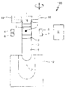
Fig. 1 schematically depicts an apparatus 100 for detecting clots 1 in a
liquid
in form of (blood) serum 2.
A conventional transparent sample container 3 comprises a centrifuged blood
sample. The blood sample is separated into serum 2 and cruor 8 by a
separating medium 9. The serum 2, the separating medium 9 and the cruor 8
are comprised in the sample container 3 as different horizontally separated
layers. The content of the sample container 3 is reagent free, i.e. during and
before clot detection no reagent, especially no reagent causing coagulation,
is added to the content of the sample container 3. The sample container 3 is
closed by means of a removable cap 16.
The apparatus 100 comprises a first light source in form of a laser diode 4
emitting light having a first wavelength of 800 nm and corresponding
conventional collimation optics (not shown). Opposite to the laser diode 4 at
an identical vertical level a first measuring unit in form of a photo diode 5
(and
corresponding analog and digital circuitry, not shown) is arranged, the photo
diode 5 being adapted to measure an intensity of light being emitted by the
laser diode 4 and travelling along a measurement path through the sample
container 3.
The apparatus 100 further comprises a second light source in form of a laser
diode 6 emitting light having a second wavelength of 1550 nm and
corresponding conventional collimation optics (not shown). Opposite to the
laser diode 6 at an identical vertical level a second measuring unit in form
of
a photo diode 7 is arranged, the photo diode 7 being adapted to measure an
CA 02842611 2015-08-12
- 11 ¨
intensity of light being emitted by the laser diode 6 and travelling along a
measurement path through the sample container 3.
The apparatus 100 further comprises a driving unit in form of a pick-and-
place unit 10 for vertically moving the sample container 3 relative to the
laser
diodes 4 and 6 and the photo diodes 5 and 7. The pick-and-place unit 10 is
further adapted to rotate the sample container 3 around a vertical axis Z of
the cylindrical sample container 3.
A computing unit in form of a microprocessor 11 is functionally coupled to the
laser diodes 4 and 6, the photo diodes 5 and 7 and the pick-and-place unit
10.
The microprocessor 11 may control the laser diodes 4 and 6 to continuously
emit light or to emit light only at discrete vertical positions. The
microprocessor 11 may further control the laser diodes 4 and 6 to generate
light pulses.
The microprocessor 11 further reads out the photo diodes 5 and 7 to gather
measured intensities at the different vertical positions.
The microprocessor 11 further controls the pick-and-place unit 10 to cause a
vertical movement and a rotation.
The microprocessor 11 further conventionally calculates vertical positions of
the separating medium 9 and of the serum 2 in response to read measured
intensities. This may e.g. be done as disclosed in US 2012/0013889 Al.
CA 02842611 2015-08-12
- 12 ¨
The microprocessor 11 is further adapted to detect the depicted clot 1, as
will
be described with reference to fig. 2.
Fig. 2 schematically illustrates a method for detecting the clot 1.
Fig. 2 depicts a number of different vertical (irradiating) positions P_O to
P_n.
Starting with vertical irradiating position P_O, light generated by the laser
diode 4 is irradiated to the sample container 3 perpendicular to the vertical
axis Z of the sample container 3, such that the light passes through the
sample container 3 along a first measurement path R_1 having the vertical
irradiating position P_O.
A resulting intensity of light passing along the first measurement path R_1 is
measured.
Now the sample container 3 is rotated around the vertical axis Z for e.g. 45
degrees without changing the vertical irradiating position P_O, such that the
light irradiated by the first laser diode 4 passes through the sample
container
3 along a second measurement path R_2 being different from the first
measurement path R_1.
Now, a resulting intensity of light passing along the second measurement
path R_2 is measured.
Optionally, the sample container 3 may be further rotated around the vertical
axis Z for e.g. - 45 degrees with respect to the start angle, again without
changing the vertical irradiating position P_O, such that the light irradiated
by
the first laser diode 4 passes through the sample container 3 along a third
CA 02842611 2015-08-12
- 13 ¨
measurement path R_3 being different from the measurement paths R_1 and
R_2. Accordingly, a resulting intensity of light passing along the third
measurement path R_3 is measured.
Self-evidently, even more than three different measurement paths may be
evaluated.
After the intensities corresponding to the measurement paths R1 to R3 have
been measured, the microprocessor 11 compares the measured intensities. If
the intensities differ by more than a given quantity, a clot would be
detected.
Since at the vertical irradiating position P_O no clot is present, the
measured
intensities are basically identical and consequently no clot is detected.
Now the vertical irradiating position is changed to vertical irradiating
position
P 1 and the above described steps are repeated using the resulting
measurement paths R1 to R3. The measurement paths R1 to R3 of the
vertical irradiating position P_1 differ from the measurement paths R1 to R3
of the vertical irradiating position P_O only in their vertical position.
Since at
the vertical irradiating position P_1 no clot is present, the measured
intensities are again basically identical and consequently no clot is
detected.
Now the vertical irradiating position is changed to vertical irradiating
position
P_2 and the above described steps are repeated using the resulting
measurement paths R1 to R3.
As shown in the diagram, depicting the measured intensity of measurement
path R2 over the vertical irradiating position, the measured intensity of
measurement path R2 is lowered, since the clot 1 is located within the
measurement path R2. Since the clot 1 is not located within the measurement
CA 02842611 2015-08-12
- 14 ¨
paths R1 and R3, the corresponding measured intensities are significantly
higher than the measured intensity corresponding to measurement path R2.
Thus, by comparing the measured intensities the clot 1 is detected.
By changing the vertical irradiating positions the sample container 3 is at
least partially inserted into a sample container carrier 12. By performing two
tasks, namely clot detection and carrier insertion, in parallel, the overall
processing time may be reduced.
Clots not being exactly symmetric to the vertical axis Z may safely be
detected by this method, since such clots cause inhomogeneous measured
intensities.
The vertical irradiating position is changed to final vertical irradiating
position
P_n which denotes the end vertical position of the serum 2. The end vertical
position of the serum 2 may have been determined before in a conventional
manner.
Under certain circumstances a clot may also be determined without rotating
the sample container 3. If e.g. the vertical interface between the serum 2 and
the separating medium 9 has been determined conventionally before clot
detection, it may be monitored, if for a certain vertical irradiating position
within the serum 2 the measured intensity is below a given threshold and/or is
smaller than measured intensities corresponding to other vertical irradiating
position within the serum 2. If this would be the case, a clot could be
determined.
CA 02842611 2015-08-12
- 15 ¨
Using this method, even clots being basically symmetric to the vertical axis Z
may safely be detected. Further, a number of clots and/or a vertical and
horizontal circumference of a clot may be determined.
Fig. 3 schematically illustrates a laboratory automation system comprising the
apparatus 100, a centrifuge 15, and an exemplary laboratory station in form
of an aliquoter unit 14. The apparatus 100 and the aliquoter unit 14 are
functionally coupled by means of a conventional data or field bus. Self-
evidently, the system may include further laboratory stations, such as pre
analytical stations, analytical stations and post analytical stations.
The sample containers 3 are supplied after being centrifuged by means of the
centrifuge 15 or already centrifuged within racks.
The aliquoter unit 14 transfers part of the serum 2 to one or more secondary
tubes (not shown). The aliquoter unit 14 conventionally includes a pipetting
unit (not shown), the pipetting unit having a tip (not shown), wherein during
aliquoting the aliquoter unit 14 is adapted to control a vertical position of
the
tip in response to a detected vertical position of an interface between the
serum 2 and the separating medium 9, such that the tip remains within the
serum 2 above the separating medium 9.
Further, the aliquoter unit 14 controls aliquoting in response to the clot
detection result provided by the apparatus 100 for detecting clots. If a clot
1 is
detected, the aliquoter unit 14 may e.g. control the vertical and/or
horizontal
position of the tip such that the tip is not blocked or absorbed by the clot
1.
Alternatively, a sample container 3 including a clot 1 (or a given number of
clots and/or a clot having a dimension bigger than a threshold) may be
omitted from further processing.
CA 02842611 2015-08-12
- 16 ¨
The system further includes a sample container transport unit adapted to
transport sample containers 3 between the apparatus 100, the aliquoter unit
14 and further laboratory stations (not shown). The sample container
transport unit includes a number of sample container carriers 12 and a
conveyor 13, wherein the sample container carriers 12 are attached to the
conveyor 13.
Fig. 4 schematically illustrates the driving unit or pick-and-place unit 10
and
the sample container transport unit in more detail.
The driving unit or pick-and-place unit 10 includes a gripper to grip the
sample container 3. The driving unit or pick-and-place unit 10 further
includes
means to provide a relative motion between the light sources 4 and 6 as well
as the measuring units 5 and 7 and the sample container 3 in both a
substantially vertical direction aligned with the central axis Z of the
cylindrical
sample container 3 and in a rotational direction about the central axis Z of
the
sample container 3.
The driving unit or pick-and-place unit 10 inserts a sample container 3 into a
corresponding sample container carrier 12, wherein the apparatus 100
simultaneously detects the vertical position of an interface and performs clot
detection. During insertion the conveyor 13 is stopped. After insertion the
conveyor 13 is moved such that an empty sample container carrier 12 is
placed under the pick-and-place unit 10, such that a further sample container
3 may be inserted into the empty sample container carrier 12.