Note: Descriptions are shown in the official language in which they were submitted.
CA 02843098 2014-01-24
PCT/EP2012/064470 - 1 -
2011P15310W0US
Description
Fluidic Cell Guidance for Flow Cytometry
The present invention relates to flow cytometry, and in
particular to cell guidance.
In the field of cell measurement and cell detection, optical
measurement methods, such as scattered-light or fluorescence
measurement, and magnetic detection methods are known.
Particularly in magnetic detection methods, for cell sorting,
cell guidance or cell enrichment, magnetophoresis is known, in
which a magnetic force is exerted on the marked cells by means
of magnetic guide strips, in such a way that these cells can be
separated or also aligned with a cell measuring device
following these guide strips. To date, with the aid of a
gradient magnetic field, enrichment of marked cells or
particles has been carried out on a substrate surface on which
the cells, or particles to be detected, are in turn aligned by
means of magnetophoresis.
In order to produce such a magnetophoretic enrichment and
alignment section, it is known to apply magnetic strips onto
the substrate, for example by lithography. Such production
methods, however, are very elaborate and therefore
disadvantageous for the production of a component which is
intended for large production numbers owing to its use. A
further disadvantage of the magnetophoretic enrichment section
is the silicon footprint thereby increased. For the integration
of an enrichment section and cell measuring device on a silicon
chip, the size of the latter exceeds reasonable costs for the
use of such components.
CA 02843098 2014-01-24
PCT/EP2012/064470 - 2 -
2011P15310W0US
It is an object of the present invention to provide a more
simply producible apparatus for flow cytometry.
The object is achieved by an apparatus as claimed in patent
claim 1. A method for cell guidance is specified in patent
claim 11. A production method for an apparatus according to the
invention is specified in patent claim 13. Advantageous
configurations of the invention are the subject-matter of the
dependent claims.
The apparatus according to the invention for flow cytometry
comprises a flow channel, a magnetic unit, which is arranged
below the channel bottom of the flow channel and is configured
in order to generate a gradient magnetic field which permeates
the volume enclosed by the flow channel, at least one cell
measuring device, and at least one guide step, which is
arranged in the flow channel in such a way that cells that can
flow through the flow channel can be deflected toward the cell
measuring device by the guide step. This has the advantage that
the cells to be detected in a microfluidic system can be
enriched in two dimensions by the flow conditions and the
gradient magnetic field. The apparatus furthermore has the
advantage of being able to obviate magnetophoretic enrichment
and therefore of being structurally much less elaborate than
previously known enrichment sections.
In one advantageous embodiment of the invention, the apparatus
comprises a flow channel which is configured with respect to
channel diameter and surface condition of the inner wall of the
channel in such a way that a flow of a complex cell suspension
in the flow channel can be generated with a laminar flow
profile. In particular, the flow channel is a microfluidic
channel. Operation is preferably carried out with relatively
large channel diameters, which ensure laminar flow of a complex
solution without obstructions occurring, for example due to
CA 02843098 2014-01-24
PCT/EP2012/064470 - 3 - '
2011P15310WOUS
deposits. The configuration of the channel with the guide step
furthermore ensures enrichment in the direction of the cell
measuring device, which makes it possible to obviate a Y-shaped
microfluidic system such as is used for example for the
separation of marked cells in the prior art.
In another advantageous embodiment of the invention, the
apparatus comprises a magnetic unit which is configured in
order to generate a gradient magnetic field by which
magnetically marked cells can be enriched on the channel
bottom. The marking of the cells is, in particular,
superparamagnetic marking, for example by means of
superparamagnetic beads. This has the advantage that all
magnetically marked cells can be enriched on the channel
bottom, where they are brought in the laminar flow to the at
least one guide step, so that they can be deflected by the
latter. In this case, the guide step has a height of about the
cell diameter of the cell type to be detected.
The guide step in the apparatus is, in particular, an elevation
relative to the channel bottom or is formed from a depression
relative to the channel bottom. That is to say, the guide step
forms for example a narrowing of the channel by extending as an
elevation into the channel volume, or it forms a widening of
the channel by being formed as the edge of a depression, so to
speak a trough, in the channel bottom. By virtue of these guide
step embodiments, different fluid-mechanical influences can be
exerted on the cell sample.
In the case of elevations relative to the channel bottom, the
step height is the height of the elevation, and in the case of
a depression relative to the channel bottom the step height is
so to speak the depth of the trough in the channel bottom. In
this case, the trough outer wall, onto which the flow runs, so
to speak forms the guide step.
CA 02843098 2014-01-24
PCT/EP2012/064470 - 4 -
2011P15310WOUS
In particular, the apparatus comprises a multiplicity of guide
steps. These are arranged in the flow channel in such a way
that cells that can flow through the flow channel can be
enriched by the guide steps in a subvolume of the flow channel
over a subsurface of the channel bottom. This has the advantage
that the apparatus does not involve a Y-shaped microfluidic
system in which marked cells are sorted, but instead enrichment
of the cells to be detected is possible within the sample
volume. Expediently, the subvolume or the subsurface of the
channel bottom lies in the middle of the flow channel, toward
which the cells can be enriched from both sides. In particular,
the cell measuring device is then also arranged within the
subsurface of the channel bottom. The cell measuring device is,
for example, arranged on or in the channel bottom. In
particular, the detection region of the cell measuring device
extends beyond the subvolume above the cell measuring device.
The elevations of the guide steps are, in particular,
configured in such a way that the cells cannot become stuck in
the intermediate spaces between the guide steps, and cannot
obstruct these intermediate spaces. The structure height, that
is to say the height of the steps relative to the channel
bottom, is therefore preferably of the order of the cell
diameter, preferably slightly less than the cell diameter. The
arrangement of a plurality of guide steps must leave free a
sufficiently large subregion of the channel bottom, on which
the enriched cells can continue on their way through the flow
channel. Either a sufficiently wide channel is kept free
between the guide steps or, as an alternative, a sufficient
offset is ensured in the case of finger structures.
In one exemplary embodiment of the apparatus, the guide steps
may be formed by means of photoresist strips, for example on a
silicon wafer. To this end, the photoresist steps are generated
in particular by means of photolithography.
CA 02843098 2014-01-24
PCT/EP2012/064470 - 5 -
2011P15310W0US
Advantageously, the enrichment section is formed as a unitary
plastic part with the guide steps, in particular by means of
injection molding, so that the enrichment section does not
occupy any silicon substrate. This has the advantage of
reducing the silicon footprint and therefore the production and
component costs of the flow cytometry apparatus.
In one advantageous configuration of the invention, the guide
steps of the apparatus extend over the channel bottom in such a
way that magnetically marked cells, which experience a magnetic
force in the direction of the channel bottom and a fluidic shear
force in the flow direction, can cross the guide steps only on a
path over a predeterminable subsurface of the channel bottom.
That is to say, the guide steps meet in particular with the
channel walls on both sides of the channel bottom in such a way
that magnetically marked cells enriched on the channel bottom
cannot flow along the channel walls. In particular, the guide
steps extend over both longitudinal halves of the channel
bottom, respectively from one channel wall approximately as far
as the middle of the channel, where a passage for cells enriched
on the channel bottom is ensured in the flow direction. In this
case, the guide steps may, in particular, be arranged in such a
way that the subsurface over which the marked cells are enriched
is a narrow rectangular subsurface which extends along the
middle of the channel in the flow direction. As an alternative,
the guide steps may also engage in one another in the manner of
fingers, so that the subsurface over which the cells are
enriched represents a subsection extending in a zigzag or in the
shape of a wave. In particular, the subsurface in the direction
of which the cells are enriched may also taper in the course of
the flow channel in the flow direction.
In one advantageous embodiment of the invention, the guide
steps are configured integrally with the channel bottom. In
particular, the guide steps may be configured with the channel
bottom as an injection-molded part. The embodiment as
CA 02843098 2014-01-24
PCT/EP2012/064470 - 6 -
2011P15310WOUS
an injection-molded part has the advantage that, for the cell
measurement, the enrichment section does not additionally have
to be arranged on the substrate, which is expediently a silicon
wafer in most cases. In this way, the so-called footprint, that
is to say the size of the silicon substrate, for the flow
cytometry component can be reduced considerably, which also
reduces the cost of such a flow cytometry apparatus.
Furthermore, the configuration of the fluid-mechanical
enrichment section is substantially simpler to produce, above
all compared with lithographic methods such as are used, for
example, in the production of magnetophoretic enrichment
sections.
In particular, the guide steps are straight linear elevations
relative to the channel bottom. With the straight linear shape,
the cells enriched on the bottom are transported by the laminar
flow along the steps, without perturbing turbulence occurring
in the flow at the channel bottom. As an alternative, the guide
steps may extend in a curve in the direction of the middle of
the channel. For the orientation of the straight linear steps,
these are in particular arranged at an acute angle with respect
to the flow direction. That is to say that the enriched cells
which are to be transported along the guide steps are not held
back by the latter, but rather the transport of the cells
continues in the flow direction.
Advantageously, the apparatus with the enrichment section
comprises a combination of guide steps on a separate plastic
channel segment, these being for example configured integrally
with the channel bottom, and a small part of the enrichment
section by means of photoresist steps on the silicon wafer, on
which the cell measuring device is also arranged. By means of
such a combination, the silicon footprint can be reduced. The
cells are enriched on an enrichment section of any desired length
by the geometry of the guide steps and the fluid-mechanical
conditions and, as soon as they reach the silicon wafer,
CA 02843098 2014-01-24
=
PCT/EP2012/064470 - 7 -
2011P15310W0US
they are also enriched there before the cell measuring device,
which is preferably also preceded by a few guide steps, in
order to maintain the enrichment and alignment of the cells
when passing over the new channel bottom substrate.
The hybrid form of the enrichment section thus forms an
advantageous variant for reducing the silicon footprint. The
structure of the fluid-mechanical enrichment section by means
of the guide steps on a plastic substrate then, for example,
precedes the silicon die. In particular, the magnetoresistive
components for detection of the magnetically marked individual
cells lie on the silicon die.
A hybrid enrichment section of this type may, for example, also
comprise a part in which the guide steps contain a proportion
of nickel, or are produced as nickel strips. With a proportion
of nickel in the guide steps, excess unbound magnetic markers
can be retained by magnetic holding forces on the nickel strips
or nickel guide steps, and so to speak filtered out of the
complex suspension. In particular, nickel guide steps are
structured by means of laser ablation. In particular, the guide
steps with a proportion of nickel precede the enrichment
section with the guide steps not containing nickel, that is to
say they are arranged before the guide steps in the flow
direction in the channel. As an alternative, however, the guide
and filter strips containing nickel may also be arranged on the
silicon substrate immediately before reaching the cell
measuring device, and fulfill there the double function of
enrichment and guidance as well as filtering of excess markers.
The dynamic enrichment and cell guidance in the flow ensures
the advantage of the apparatus that enrichment and measurement
can be carried out in one channel. The apparatus is not
intended for sorting by means of Y-shaped separation of marked
cells from the surrounding
CA 02843098 2014-01-24
PCT/EP2012/064470 - 8 -
2011P15310WOUS
complex suspension, and furthermore excess markers do not have
to be separated elaborately from the suspension, but can be
retained by the guide steps. In particular, the guide steps are
arranged in terms of their height and their angle with respect
to the flow direction in such a way that unbound magnetic
markers, which are very much smaller in terms of their
hydrodynamic diameter than marked cells, remain on the guide
steps and are held back, i.e. they cannot cross the steps. Only
the larger fractions or particles, such as marked cells in the
complex suspension, are entrained in the laminar flow and are
thus transported along the steps. That is to say nonmagnetic,
or nonmagnetophoretic, enrichment as in this case, by means of
the guide steps, can also exert a filter effect on excess and
therefore undesired markers in the measurement region of the
cell measuring device.
In order to reinforce the fluid-mechanical filter effect at the
guide steps, which in particular are nonmagnetic, i.e. do not
contain a proportion of nickel, these may be varied in terms of
height, i.e. in particular adapted to the size of the cell type
to be detected and the size of the unbound magnetic particles,
or markers, to be filtered. In one advantageous embodiment, the
step height increases in the course of the flow channel in the
flow direction. The first step is still very low and can be
crossed by most particles and cells. In the course of the
channel, the step height then rises increasingly and thus
retains larger and larger particles. Only the magnetically
marked cells which are intended to be detected are not stopped
by the steps, but are transported along the steps and
concentrated in a subvolume of the channel. To this end, all
steps point in particular toward this subvolume, which lies
particularly in the middle of the channel on the channel
bottom.
The channel diameter, or the channel height and width, are in
particular a few hundred pm, for example 200 pm. The
CA 02843098 2014-01-24
=
PCT/EP2012/064470 - 9 -
2011P15310WOUS
step height is dependent on the cell type to be detected and
the cell diameter thereof, and is in particular a few
micrometers, for example 10 pm or up to 30 pm. The flow channel
may thus, in particular, guide a sufficiently large volume of a
complex suspension without thereby being obstructed.
In the method according to the invention for magnetic flow
cytometry, a laminar flow of a cell sample is generated and the
cells are enriched by means of guide steps in a predeterminable
subvolume of the flow channel. In this case, the cells to be
detected are magnetically marked and are dynamically enriched
on the channel bottom in a gradient magnetic field. This method
has the advantage that fluid-mechanical and magnetic forces
interact in such a way that magnetically marked cells can be
enriched in a controlled manner in a predeterminable volume,
without their needing to be separated from the cell suspension.
In one advantageous embodiment of the method, the subvolume
extends in the flow direction along the channel bottom, so that
the cells are guided along an axis individually over a cell
measuring device.
For the flow cytometry method, for example, a blood sample is
transported in a laminar fashion through the microfluidic
system. In the flow, the cells are partially aligned close to
the channel bottom by the structuring of the substrate, i.e.
the guide steps. In the gradient field, for example,
superparamagnetic analytes are drawn onto the structured
channel bottom and detected there magnetoresistively.
In the method according to the invention for magnetic flow
cytometry, three forces thus act on the magnetically marked
cells or on magnetic beads, or generally on magnetic particles
to be detected: The magnetic force of the gradient magnetic
field, which is generated by the magnetic unit below the
channel bottom. Magnetic field strengths of this gradient field
are, for example,
CA 02843098 2014-01-24
PCT/EP2012/064470 - 10 -
2011P15310WOUS
between 1 and 300 mT. This magnetic force thus attracts the
cells perpendicularly toward the channel bottom surface.
Furthermore, the shear force of the flowing cell sample acts on
the individual cells. The flow is, in particular, a laminar
flow. This force thus acts in the direction of the cell sample
flow through the channel. A third force is exerted by the guide
steps on the channel bottom, which represent a fluid-mechanical
obstacle for the magnetically marked cells enriched on the
bottom. The effect of this is that, in order to proceed further
in the flow direction, the cells move along the guide steps
toward the middle of the channel or in general, depending on
the orientation of the guide steps, toward a subregion of the
channel. The magnetic marking is preferably carried out using
superparamagnetic particles.
When the flow cytometry method is carried out, the flow rate,
the surface property and the magnetic field strength also play
a role. The flow rate, for instance, is adapted in particular
to the cell sample and above all to the channel diameter, in
order to ensure a laminar flow. By means of surface
functionalization, the surface properties of the channel inner
walls and of the channel bottom can be optimized. By means of
the field strength of the gradient magnetic field, further
influence can be exerted on the cells to be deflected and
enriched. The cells to be detected also have mechanical
properties, which can be influenced by the values of the flow
rate, surface condition and magnetic field strength.
In the production method according to the invention for an
apparatus for flow cytometry, guide steps are configured
integrally with the channel bottom, particularly as an
injection-molded part.
The apparatus according to the invention thus has the advantage
of offering a solution for flow cytometry without
magnetophoresis. For example, a substrate structured in this
CA 02843098 2014-01-24
PCT/EP2012/064470 - 10a -
2011P15310WOUS
way,
CA 02843098 2014-01-24
PCT/EP2012/064470 - 11 -
2011P15310WOUS
which correspondingly guides and enriches the magnetically
marked cells by the guide steps, i.e. the structure of the
substrate bottom, can be produced by various techniques, inter
alia injection molding or embossing. Accordingly, no
lithographic outlay such as in magnetophoretic enrichment is
necessary. So to speak, the magnetic guide lines of a
magnetophoretic enrichment section are replaced by a three-
dimensional structure of the substrate bottom. In particular,
the preferred herringbone shape is adopted in this case. The
structuring has, in particular, linear elevations which are
referred to as guide steps. These are arranged in particular at
a steep angle to the flow direction through the channel. The
steps typically measure heights of between 0.1 and 20 pm
relative to the channel bottom. In their width, the guide steps
measure between 1 and 100 pm, for example. The length of the
guide steps is selected, as a function of the channel width, in
such a way that they end at the channel edge with the channel
wall, and reach approximately to the middle of the channel.
Either they reach only almost as far as the middle, so that a
passage remains between the guide steps which extend from both
sides in the direction of the middle of the channel, or as an
alternative they extend in terms of their length beyond the
middle of the channel and are then arranged engaging in one
another in the manner of fingers. The angle with respect to the
flow direction is for example less than 45 , in particular less
than 20 .
In the flow cytometry method, in particular, a blood sample is
transported in a laminar fashion through the microfluidic
system. Cells within this blood sample are partially aligned
close to the substrate surface by the substrate structuring.
Magnetically marked analytes, in
particular
superparamagnetically marked analytes, are attracted in the
gradient field onto the substrate surface, i.e. onto the
channel bottom, and are guided close to the substrate, i.e. on
the channel bottom, where the substrate structuring can
CA 02843098 2014-01-24
'
PCT/EP2012/064470 - ha -
2011P15310WOUS
influence them. The cells enriched and aligned in this way can
then be detected magnetoresistively.
CA 02843098 2014-01-24
=
PCT/EP2012/064470 - 12 -
2011P15310W0US
Embodiments of the present invention will be described by way
of example with reference to Figures 1 to 7 of the appended
drawing:
Figure 1 shows a perspective representation of the channel
bottom with the cell-guiding elevations,
Figure 2 shows a longitudinal section, or a side view, of the
channel bottom with underlying permanent magnets,
Figure 3 shows a cross section, or a front view, of the flow
channel,
Figure 4 shows a plan view of the arrangement with the guide
troughs, or steps, arranged in a herringbone fashion,
Figure 5 shows another plan view of the arrangement with the
guide troughs, or steps, arranged in a herringbone
fashion,
Figure 6 shows an enlarged detail of Figure 4, and
Figure 7 shows a hybrid cell enrichment section.
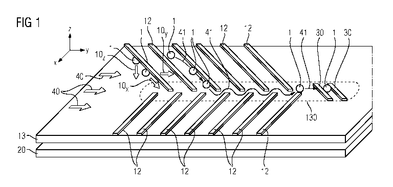
Figure 1 shows a perspective view of the channel bottom 13,
which is represented as a flat substrate. At a distance
thereunder, a further flat cuboid 20 is shown, which represents
the magnetic unit 20. The magnetic unit is, in particular, a
permanent magnet. The magnetic unit 20 may also extend over an
area larger than that of the channel bottom 13, in order to
ensure a homogeneous magnetic field in the region of the flow
channel 100. In particular, the magnetic unit 20 generates in
the flow channel 100 a gradient field in which magnetic
particles, for instance the magnetically marked cells 1 or
unbound magnetic markers, are enriched in the negative z
,
CA 02843098 2014-01-24
'
PCT/EP2012/064470 - 12a -
2011P15310W0US
direction toward the channel bottom 13. The x, y and z
directions are respectively indicated by small coordinate
systems at the edge in the figures. In Figure 1, a multiplicity
of guide steps 12 which are represented as narrow cuboids are
arranged on the channel bottom substrate 13. These elevations
12 meet, in particular,
CA 02843098 2014-01-24
=
PCT/EP2012/064470 - 13 -
2011P15310WOUS
the edge of the channel bottom, or the channel walls 14. The
channel walls 14 are not shown in the representation of Figure
1. The guide steps 12 project into the middle of the channel,
although they do not join there with the opposite guide steps
but either leave a straight passage in the middle or engage in
one another in the manner of fingers, in such a way that a
zigzag or serpentine line can extend through the guide steps.
Possible flow paths of magnetically marked cells 1 are indicated
by arrows 41 in Figure 1. The magnetically marked cells 1 are
shown as circles or ovals. The forces 10õz acting on the cells
are indicated by double arrows. In turn, wide double arrows
indicate the flow direction 40, which extends from left to right
in Figure 1. In the flow channel 100, the magnetically marked
cells 1 are thus introduced at one end within a complex cell
suspension and flow in the flow direction 40 through the
enrichment section with the guide steps 12. Owing to the
magnetic force 10, which points in the direction of the channel
bottom 13, the shear force 10 of the liquid in laminar flow,
which points in the flow direction 40, and owing to the guide
steps 12 which represent a barrier, which in turn exert a
mechanical force 10x in the x-y plane of Figure 1 on the cells 1,
the cells 1 are displaced along the guide steps in the direction
of the subregion 130 of the channel bottom 13. At the end of
this subregion 130, in which the cells 1 are concentrated, there
is furthermore the cell measuring device 30 which, in
particular, comprises at least one magnetoresistive element.
Figure 2 shows a longitudinal section, or side view, of an
apparatus similar to that in Figure 1. In this case, two flat
rectangles which represent the substrate, or the channel bottom
13, and at a distance thereunder the magnetic unit 20, are
arranged above one another. As an alternative to the embodiment
shown, the permanent magnet may also be arranged directly below
the channel bottom 13 without a separation. Above the channel
bottom 13, the flow direction 40, in Figure 2 from left to
right,
CA 02843098 2014-01-24
=
PCT/EP2012/064470 - 14 -
2011P15310WOUS
is in turn indicated by a double arrow, and a cross section
through three of the guide steps 12 as well as through the cell
measuring device 30 at the right-hand end of Figure 2, and
therefore at the end of the enrichment section. Owing to the
permanent magnet 20, the magnetically marked cells 1 experience
a magnetic force 10 perpendicularly in the direction of the
channel bottom 13. The height of the guide steps 12 is in
particular adapted to the extent, i.e. the hydrodynamic
diameter, of the magnetically marked cells 1, and is in
particular slightly less than the cell diameter. With a height
which is too low, however, the magnetically marked cells would
not experience any guide force 10 due to the steps 12, but
would be carried away over them in the laminar flow. With
excessively high barriers 12, the magnetically marked cells 1
would no longer experience any shear force lOy due to the flow,
and would remain behind the steps 12.
Figure 3 shows a cross section, or the front view, of the flow
channel 100. In Figure 3, the magnetic unit 20 and, at a
distance thereover, the substrate 13 for the channel bottom are
in turn shown as narrow rectangles, the channel wall 14 which
encloses a cuboid channel volume being arranged thereover. In
the flow channel 100, the subvolume 110 in which the
magnetically marked cells 1 are enriched is also represented by
dashes.
Figure 4 in turn shows a plan view of the channel bottom 13, on
which the flow direction from left to right in Figure 4 is
again indicated by double arrows 40. Respectively at the side
of the channel bottom 13, the channel walls 14 are represented
in section by shading. A dashed line, which denotes the end of
the magnetic region, respectively extends inside the channel
walls 14. That is to say, the distance between the dashed lines
200 shows the width of the region permeated by the magnetic
field. It is, in particular, wider than the flow channel 100.
This ensures that the magnetic field in the channel volume is
CA 02843098 2014-01-24
PCT/EP2012/064470 - 14a -
2011P15310W0US
as homogeneous as possible. The region 200 permeated by the
magnetic field is generated by the magnetic
CA 02843098 2014-01-24
PCT/EP2012/064470 - 15 -
2011P15310WOUS
unit 20, which is arranged below the channel bottom 13, as can
be seen in Figures 1 to 3. In the channel 100, guide steps 12
are in turn arranged at an angle 6 with respect to the channel
wall 14, so that the guide steps 12 point from the channel wall
14 in the direction of the middle of the channel in the flow
direction 40. The magnetically marked cells 1, as indicated by
the flow paths 41, can thus be deflected at the guide steps 12
in the direction of the subregion 130, which extends as far as
or beyond the cell measuring device 30.
Figure 5 shows a possible arrangement of guide steps 12, which
are arranged at a very acute angle 6 with respect to one
another. The channel width 100 is again indicated. Figure 6
shows an enlarged detail of Figure 5 with guide steps 12,
converging at an acute angle 6, which have a step thickness or
width d and a distance D between the steps. The angle 6 at
which the steps 12 are arranged with respect to the flow
direction 40 may, for example, be measured relative to the
midline of the channel as shown in Figure 6, or relative to the
channel wall 14. Again, magnetically marked cells 1 are
indicated as small circles in Figure 6. It is illustrated here
that a sufficiently wide flow path through between the steps 12
is ensured for the cells 1, so that they do not obstruct the
guide step intermediate spaces.
Lastly, Figure 7 shows another possible configuration of the
apparatus, with a hybrid enrichment section. In the left-hand
region of Figure 7, the enrichment section is shown on a
plastic substrate 13 with plastic guide steps 12, with which
lead to the described fluid-mechanical enrichment of the cells
1. This is followed in the right-hand region of the drawing by
the substrate 13 of the silicon chip 15, on which the cell
measuring device 30 is arranged. This may, as shown in the
example of Figure 7, also have further guide steps 125 which,
in particular, continue the enrichment section onto the
subregion 130.
CA 02843098 2014-01-24
=
PCT/EP2012/064470 - 16 -
2011P15310WOUS
The flow direction 40 is again represented by a double arrow
from left to right in the drawing. The magnetically marked
cells I are represented as ovals, and their flow paths are
denoted by arrows 41. In the example shown in Figure 7, the
guide steps 12, which meet the channel walls 14 on both sides,
do not engage in one another in the manner of fingers, but
leave a straight flow region open in the region of the middle
of the channel which lies in the enrichment subregion 130. In
order still to guide the cells 1 straight over the cell
measurement region 30 after the enrichment section through the
guide steps 12, the silicon chip 15 also has a small portion of
an enrichment section with guide steps 125. These may, for
example, also contain a proportion of nickel in the material of
the guide steps 125 and therefore filter out still unbound
markers by magnetic retaining forces before the cell measuring
device 30. As an alternative, the guide steps 125 may be
produced on the silicon chip 15, for instance by photoresist
structures. Furthermore, Figure 7 again shows by double arrows
the deflecting force 10x which guides the cells 1 along the
guide steps 12 to the middle, as well as the shear force lOy of
the fluid flow, which points in the flow direction 40.