Note: Descriptions are shown in the official language in which they were submitted.
CA 02843183 2014-01-24
APPARATUS AND METHODS TO MODULATE PELVIC
NERVOUS TISSUE
CLAIM OF PRIORITY
This application claims the benefit of priority of Sachs et al., U.S.
Provisional Patent Application Serial No. 61/511,776, entitled "SYSTEMS AND
METHODS TO MODULATE BLADDER FUNCTION OR TREAT PELVIC
PAIN," filed on July 26, 2011 (Attorney Docket No. 3426.001PRV), and the
benefit of priority of Hlavka et al., U.S. Provisional Patent Application
Serial
No. 61/565,460, entitled "SYSTEMS AND METHODS TO MODULATE
BLADDER FUNCTION OR TREAT PELVIC PAIN," filed on November 30,
2011 (Attorney Docket No. 3426.002PRV), each of which is hereby
incorporated by reference herein in its entirety.
BACKGROUND
Urinary incontinence (UI) is the involuntary leakage of urine. There are
several types of urinary incontinence, including urge urinary incontinence
(UUI)
and stress urinary incontinence (SUI). Urge urinary incontinence is the
involuntary loss of urine while suddenly feeling the need or urge to urinate.
Stress urinary incontinence, typically affecting females, is the involuntary
loss of
urine resulting from increased abdominal pressure, such as generated by
physical
activity, exercising, coughing, sneezing, laughing, lifting, etc. Mixed
incontinence combines attributes of SUI and UUI.
Overactive bladder (OAB) is the strong, sudden urge to urinate, with or
without urinary incontinence, usually with frequency and nocturia. The urge
associated with overactive bladder can be assessed using the subjective
experience of the patient, with or without any objectively verifiable metric,
condition, behavior, or phenomena.
Historically, attempts have been made to translate the subjective patient
experience of overactive bladder into a verifiable clinical test. Based upon
work
in spinal cord injury patients, it was hypothesized that the sensation of
urgency
and the result of urine leakage was due to non-volitional urinary bladder
detrusor
muscle contractions. Consequently, there was a push to implement urodynamic
testing to observe and quantify the presumed detrusor contractions. However,
1
CA 02843183 2014-01-24
the results found a poor correlation (e.g., 60%) between observed detrusor
overactivity and the experience of urgency, and also found that asymptomatic
individuals may exhibit detrusor contractions during urodynamic testing.
Given the limitations of urodynamic testing, the diagnosis and treatment
decisions for overactive bladder transitioned to being assessed wholly by the
patient's subjective experience. However, the detrusor muscle and its
contractions are still considered to have a major role in overactive bladder.
Bladder control is a complex combination of voluntary and involuntary
neurologic control, which responds to a highly distributed set of afferent
(sensory) nerves associated with the bladder. Also, there is evidence of a
myogenic origin for at least a portion of bladder wall contractile activity.
While
there are some descriptive hallmarks of idiopathic overactive bladder (e.g.,
thickened wall, characteristic "patchy" denervation, changes in smooth muscle
and collagen morphology, increased electrical connectivity), there is no
specific
anatomic cause of OAB (e.g., a lesion, defect, injury, etc.), and also it is
believed
that there is no commensurate remedy for the cause. Neurogenic injury (e.g.,
spinal cord injury) and bladder outlet obstruction (BOO) can both lead to
overactive bladder due to a chronic state of bladder inflation and a "high
pressure" bladder. However, resolution of an outlet obstruction fails to
rectify
overactive bladder symptoms in a significant fraction (e.g., 25%) of these
patients.
Overactive bladder affects at least 33 million patients in the United States
alone, representing 16% of the adult United States population and roughly $12
billion dollars in healthcare cost. Overactive bladder and urinary
incontinence
significantly affect the quality of life and the ability of patients to
maintain their
lifestyle, including socializing, mobility, or independence. Further, urinary
incontinence is one of the most common reasons for entering long-term care
facilities, such as nursing homes, and is also a significant risk factor for
injury
due to falls resulting from hurrying to the toilet in response to urge.
Referring to FIGS. 1-3, the anatomy of the female bladder is described to
provide context for discussion of previously-known treatment modalities, and
is
illustrative of why a significant unmet need for improved treatment modalities
remains. In particular, FIG. 1 depicts a lateral sectional of the anatomical
structures of a bladder (B) and a urethra (U), while FIG. 2 depicts an
anterior
2
CA 02843183 2014-01-24
sectional view of the bladder and urethra. FIGS. 1-2 further illustrate a
trigone
(T), ureteral ostium (0) (also referred to as a ureteral orifice), detrusor
muscle
(D), a neck (N), an interureteric crest (C), a fundus (F), and a body (BB).
FIG. 3 depicts a cross sectional view of a wall of the bladder, including
an intravesical region (IR) (also referred to as the cavity), mucous membrane
(also referred to as the mucosa), lamina propria (LP), muscularis propria
(MP),
adventitia (A), and perivesical fat (PF). The mucous membrane lines the
intravesical region (IR) of the bladder and includes a three-layered
epithelium,
collectively referred to as transitional cell epithelium (TCE) or urothelium,
and
basement membrane (BM). The three layers of the transitional cell epithelium
include the basal cell layer, the intermediate cell layer, and the surface
cell layer.
The basal cell layer can renew the transitional cell epithelium by cell
division.
New cells can migrate from the basal layer to the surface cell layer, and the
surface cell layer can be covered by glycosaminoglycan (GAG) layer (GL). The
function of GAG layer is controversial, possibly serving as an osmotic barrier
or
even an antibacterial coating for transitional cell epithelium. The basement
membrane is a single layer of cells that separates transitional cell
epithelium
from the lamina propria.
Lamina propria (also referred to as the submucosa or suburothelium) is a
sheet of extracellular material that may serve as a filtration barrier or
supporting
structure for the mucous membrane and includes areolar connective tissue and
contains blood vessels, nerves, and in some regions, glands. Muscularis
propria
(also referred to as the detrusor muscle or the muscle layer) may be
interlaced
with lamina propria and may have three layers of smooth muscle, the inner
longitudinal, middle circular, and outer longitudinal muscle.
When the bladder is empty, the mucosa has numerous folds called rugae.
The elasticity of rugae and transitional cell epithelium allow the bladder to
expand as the bladder fills with fluid. The thickness of the mucosa and
muscularis propria can range between approximately 2 to 5 mm when the
bladder is full and between approximately 8 to 15 mm when the bladder is
empty.
The outer surface of muscularis propria may be lined by adventitia A
about the posterior and anterior surface of the bladder or by the serosa about
the
superior and upper lateral surfaces of the bladder. Perivesical fat (PF) can
3
CA 02843183 2014-01-24
surround the bladder outside of the serosa or adventitia. In some cases, a
variety
of fascia layers may surround or support the organs of the pelvis.
Collectively,
the fascias near the urinary bladder can be referred to as perivesical fascia.
A number of therapies have been developed for treating overactive
bladder, including delivery of anticholinergic drugs, bladder retraining,
sacral
nerve stimulation (SNS), intravesical drug infusions, surgical denervation
procedures, surgeries to increase bladder volume (e.g., detrusor myomectomy,
augmentation cystoplasty) and botulinum toxin (e.g., Botox , Dysport , etc.)
injections into the bladder wall. Each of these therapies has drawbacks, as
described below.
Anticholinergic drugs, used alone or in combination with traditional
nonsurgical approaches, such as bladder retraining, Kegel exercises,
biofeedback, etc., often is used as first-line therapy for overactive bladder;
however, the mode of action is uncertain. Anticholinergic drug use was
initially
thought to decrease contractions of the detrusor muscle during the filling
stage
(e.g., detrusor muscle overactivity, unstable detrusor muscle, etc.). However,
it
is now believed that anticholinergic drugs may not change detrusor muscle
contractility, but instead modulate afferent (e.g., cholinergic) nervous
traffic to
the central nervous system.
Efficacy of anticholinergic drugs is generally quite modest, as
approximately 50% of patients find such therapy subjectively inadequate. A
reduction of 10% to 20% in the number of micturations per day (e.g., from 11
micturations to 9 micturations) and a reduction of 50% in urinary incontinence
episodes (e.g., from 2 per day to 1 per day) is typical. However, these
effects are
frequently inadequate to significantly improve patient quality of life (QOL).
Many patients would not even notice a change of 2 micturations per day unless
they are keeping a log for a formal study. The remaining urinary incontinence
episodes, although slightly less in number, continue to maintain the stigma
and
lifestyle limitations of the disease, such as the inability to travel or to be
active,
social withdrawal, etc. In addition, anticholinergic drugs can have side
effects,
including dry mouth, constipation, altered mental status, blurred vision,
etc.,
which may be intolerable, and in many instances outweigh the modest benefits
attained. Approximately 50% of patients abandon anticholinergic therapy within
6 months.
4
CA 02843183 2014-01-24
Sacral nerve stimulation (SNS) has a higher level of efficacy (e.g., up to
80% in well-selected and screened patients), but here too the mode of action
is
not well understood. The clinical benefit of SNS for urinary incontinence was
a
serendipitous finding during clinical trials of SNS for other conditions. The
SNS
procedure has a number of drawbacks: it is expensive and invasive, and
requires
surgery for temporary lead placement to test for patient response, followed by
permanent lead placement and surgical implantation of a pulse generator in
patients who responded favorably to the temporary lead. Regular follow-ups
also are required to titrate SNS stimulation parameters, and battery
replacements
are necessary at regular intervals.
A variety of surgical denervation or disruption procedures have been
described in the literature, but most have showed poor efficacy or durability.
The Ingelman-Sundberg procedure, first developed in the 1950s and described in
Ingelman-Sundberg, A., "Partial denervation of the bladder: a new operation
for
the treatment of urge incontinence and similar conditions in women," Acta
Obstet Gynecol Scand, 38:487, 1959, involves blunt surgical dissection of the
nerves feeding the lateral aspects of the bladder near its base. The nerves
are
accessed from the anterior vaginal vault, with the dissection extending
bilaterally
to the lateral aspect of the bladder. The denervation process is accomplished
somewhat blindly, using blunt dissection of the space and targeting the
terminal
pelvic nerve branches. Although capable of producing promising results, the
procedure as originally proposed entails all of the drawbacks and expense
normally associated with surgical procedures.
McGuire modified the Ingelman-Sundberg procedure in the 1990s, as
described in Wan, J., et al., "Ingelman-Sundberg bladder denervation for
detrusor instability," J. Urol., suppl., 145: 358A, abstract 581, 1991, to
employ a
more limited and central dissection within the serosal layer of the bladder,
staying medial to the vaginal fornices. Surgical candidates for the Modified
Ingelman-Sundberg procedure can be screened to isolate likely "responders"
using sub-trigonal anesthetic injections. As reported in 1996 by Cespedes in
Cespedes, R.D., et al., "Modified Ingelman-Sundberg Bladder Denervation
Procedure For Intractable Urge Incontinence," J. Urol., 156:1744-1747 (1996),
64% efficacy was observed at mean 15 month follow-up following the
procedure. In 2002, Westney reported in Westney, 01., et al., "Long-Term
5
CA 02843183 2014-01-24
Results Of Ingelman-Sundberg Denervation Procedure For Urge Incontinence
Refractory To Medical Therapy," J. Urol., 168:1044-1047 (2002), achieving
similar efficacy at mean 44 month follow-up after the procedure. More
recently,
in 2007, Juang reported in Juang, C., et at., "Efficacy Analysis of Trans-
obturator Tension-free Vaginal Tape (TVT-0) Plus Modified Ingelman-
Sundberg Procedure versus TVT-0 Alone in the Treatment of Mixed Urinary
Incontinence: A Randomized Study," E. Urol., 51:1671-1679 (2007), using a
combination of a transvaginal tape (TVT) sling (the "gold standard" surgical
therapy for stress incontinence) and the Modified Ingelman-Sundberg procedure
for mixed incontinence patients and showed a significant benefit for including
the Modified Ingelman-Sundberg procedure, over the TVT sling alone, out to 12
months follow-up following the procedure.
Despite its clinical success, however, the Modified Ingelman-Sundberg
procedure has not been widely adopted, as it is highly invasive and requires
general anesthesia. Further, the terminal nerve branches are not visible to a
surgeon, and thus, the dissection must be performed using approximate
anatomical landmarks rather than using direct visualization of target nerve
branches. Possible complications of the Modified Ingelman-Sundberg procedure
include the risks associated with anesthesia, blood loss, vaginal numbness or
fibrosis, adhesions, fistulas, vaginal stenosis, wound infection, or
dyspareunia
(pain with intercourse). Perhaps most importantly, efficacy of the Modified
Ingelman-Sundberg procedure may be dependent upon surgical skill and
technique.
More recently, another therapy involving injection of botulinum toxin
(e.g., Botox ) into the bladder wall has been developed to address the
symptoms
of overactive bladder by blocking nerve traffic and causing temporary muscle
paralysis following injection. During the injection procedure, which may be
performed in a physician's office under local anesthesia, a cystoscope is
introduced into the bladder through the urethra and a number of separate
needle
injections (e.g., 20-30) are made into the bladder wall. Initially the
trigone, the
area of the bladder defined by the ostia of the two ureters and the urethra,
was
avoided due to concerns about procedural pain due to dense afferent
innervation
of the trigone region and the potential for vesicoureteral reflux. However,
the
trigone region has more recently been included, and sometimes specifically
6
CA 02843183 2014-01-24
targeted to the exclusion of the dome of the bladder. Initially, botulinum
toxin
was assumed to act only on the efferent motor nerves (e.g., causing partial
paralysis of the detrusor muscle). More recent research indicates that
botulinum
toxin may have an effect on afferent sensory nerves as well. U.S. Patent
8,029,496 to Versi provides an example of a system for delivering such a
therapeutic agent to the trigone of the bladder through the vaginal wall.
Typically, botulinum toxin injections achieve a fairly high level of
efficacy (e.g., resolution of symptoms), with maximum changes in cystometric
capacity peaking at 4 weeks and complete continence being achieved in about
half of patients. However, botulinum toxin does carry with it the risks of
systemic effects, such as flu-like symptoms, nausea, weakening of respiratory
muscles, transient muscle weakness, allergic reaction, or developed
sensitivity.
Other adverse events associated with botulinum toxin injections include acute
urinary retention (AUR), large postvoid residual volume (PVR), difficulty in
urination ("straining"), and urinary tract infection (UTI). Challenges with
botulinum toxin therapy include procedural skill (e.g., dexterity with
cystoscope
and needle), uncontrolled drug diffusion, variable needle penetration depth,
and
reproducibility of technique. In addition, the effects of botulinum toxin wear
off
with time, typically after 6-9 months, requiring repeat injections for the
lifetime
of the patient.
Stress urinary incontinence, typically affecting females, is an anatomic
issue where the pelvic floor has been damaged and weakened, such as during
childbirth. Here, front line therapies are conservative (e.g., Kegel exercises
or
biofeedback), and a variety of minimally invasive surgical therapies are
available
as second line therapies. Examples of these second line therapies include
sling
procedures, bladder neck suspension, transvaginal tape (TVT), etc. In each,
the
procedure is a day surgery performed on an outpatient basis. Success rates are
high, and the procedures have been embraced by the medical community.
In addition, new therapies have been developed to treat stress urinary
incontinence, such as the Renessa system offered by Novasys Medical, Inc.,
which is used in an office-based procedure. U.S. Patent No. 6,692,490 to
Edwards, assigned to Novasys Medical, discloses the treatment of urinary
incontinence and other disorders by the application of energy and drugs.
7
CA 02843183 2014-01-24
Finally, a majority of males will develop some degree of urinary
obstruction from benign prostate hyperplasia (BPH), or "enlarged prostate",
over
their lifetime. Since urinary obstruction is known to be a cause of overactive
bladder, bladder symptoms in males are generally presumed to be secondary to
the enlarged prostate. However, resolution of the urinary obstruction (e.g.,
by
one of the many variants of transurethral treatments of the prostate) does not
resolve the bladder symptoms in about a quarter of the patients. Thus, it
would
be desirable to offer a minimally invasive therapeutic procedure targeting
these
remaining patients whose symptoms remain after prostate therapy.
Further, there is a growing preference for "watchful waiting" for prostate
disease, even for cases of actual prostate cancer, and many of these patients
will
develop symptoms of overactive bladder due to the urinary obstruction from
their growing prostate. Thus, there is the potential to provide a therapy that
targets the bladder symptoms prior to or instead of providing therapy
targeting
the prostate itself.
Males also may experience idiopathic OAB, that is OAB not secondary
to an enlarged prostate or other urinary obstruction, and require a primary
therapy for the OAB symptoms.
In view of the foregoing, it would be desirable to provide a minimally
invasive procedure for modulating bladder function to treat or resolve
overactive
bladder and provides durable relief for patients suffering from these
debilitating
conditions.
OVERVIEW
The present inventors have recognized, among other things, apparatus
and methods configured to provide therapy to non-mucosal target tissue (or a
target volume of tissue) to modulate bladder function. In an example, energy
can be delivered to denervate selected portions of the bladder, such as
afferent
nerves located within or proximate to the trigone region of the bladder wall,
to
modulate bladder function and thereby provide relief for at least one of a
sense
of urge, incontinence, frequency, nocturia, bladder capacity, or pain.
In some examples, denervation may be accomplished by delivering
thermal energy (e.g., using RF energy, microwaves, or high intensity focused
ultrasound) to layers of the bladder wall beneath the mucosal layer, such as
8
CA 02843183 2014-01-24
within or proximate to the trigone region. In the context of this disclosure,
tissue
of the female anatomy targeted for energy delivery may include one or more
tissue layers of the bladder wall beneath the mucosa and extending to (but not
including) the anterior vaginal wall, and are collectively referred to herein
as
"non-superficial tissue." Further, in the context of this disclosure, tissue
of the
male anatomy targeted for energy delivery may include one or more layers of
the
bladder wall beneath the mucosa and extending to and including the perivesical
fat layer, and in the context also is referred to as "non-superficial tissue".
In still
other examples, thermal energy may be delivered to neural tissue, such as a
pelvic nerve or its branches, within or proximate to the bladder wall to
modulate
nerve traffic to or from at least a portion of the bladder, thereby modulating
bladder function. In accordance with some examples, suction may be used to
grasp and conform a mucosal surface of the bladder wall to a first surface of
a
device, and energy can be delivered to non-superficial target tissue at a
substantially uniform depth from the mucosal surface. Cooling also may be
provided to reduce heat buildup in the mucosa. However, in some examples, a
mucosal surface of the bladder wall superficial to the non-superficial target
tissue can be retained substantially intact without cooling, such as by
inserting an
energy delivery element in the non-superficial target tissue at a
substantially
uniform distance from the first surface of the device and delivering energy to
the
non-superficial target tissue from that substantially uniform depth beneath
the
mucosal surface. The systems and methods described herein may be configured
to deliver energy, such as thermal energy, to target tissue either from within
a
lumen or cavity of a body organ, for example, the bladder, or from a lumen or
cavity of an adjacent organ, such as the vagina.
In the alternative, or optionally in addition, the systems and methods
described herein may provide that one or more areas of the bladder be isolated
or
supported such as to suppress the sense of urgency. For example, surgical
barriers or treatments may be used to reduce stretch of a selected region of
the
bladder, such as the trigone, or alternatively used as an adjunct to energy
delivery to prevent nerve regrowth in a treated portion of the bladder.
This overview is intended to provide an overview of subject matter of the
present patent application. It is not intended to provide an exclusive or
9
CA 02843183 2014-01-24
exhaustive explanation of the invention. The detailed description is included
to
provide further information about the present patent application.
BRIEF DESCRIPTION OF THE DRAWINGS
In the drawings, which are not necessarily drawn to scale, like numerals
or letters may describe similar components in different views. Like numerals
having different letter suffixes may represent different instances of similar
components. The drawings illustrate generally, by way of example, but not by
way of limitation, various examples discussed in the present document.
FIGS. 1-3 depict views of anatomical structures to which the apparatus
and methods of the present subject matter may be applied to treat overactive
bladder.
FIG. 4 is a plan view of an exemplary embodiment of a system
constructed in accordance with the principles of the present subject matter.
FIGS. 5A and 5B are, respectively, a lateral view of a female bladder
within which is inserted the distal region of a device of the present subject
matter
and a magnified view of the energy delivery element of that device.
FIG. 6 is a lateral view of a female bladder within which is inserted the
distal region of an alternative embodiment of a device of the present subject
matter.
FIG. 7 is a plan view of a further alternative embodiment of a device of
the present subject matter that employs suction to grasp and conform a target
tissue.
FIGS. 8A to 8C are plan, bottom and sectional views, respectively of the
distal region of the device of FIG. 7.
FIGS. 9A and 9B are plan and side section views of the distal region of
FIGS. 8A through 8C receiving a portion of bladder tissue, while FIG. 9C is
schematic diagram illustrating the ablation zone formed by the device of FIGS.
7
9.
FIGS. 10A and 10B are, respectively, a plan view of the distal region of
an alternative embodiment of the device of FIG. 7 that employs retractable
needle electrodes, and a magnified view of a portion of such needle
electrodes.
FIGS. 11A and 11B are, respectively, bottom and side sectional views of
the distal region of the device of FIGS. 10A and 10B.
CA 02843183 2014-01-24
FIG. 12 is a sectional view of an alternative embodiment of the device of
FIGS. 10 and 11 showing an optional cooling channel configuration.
FIG. 13 is an interior view of the posterior of a female bladder showing
associated vessels, the location of the trigone, and alternative possible
ablation
regions.
FIGS. 14-17 are interior views of the posterior of a female bladder
showing various ablation patterns.
FIG. 18 is a side sectional view of the anatomy of a female abdomen and
pelvis depicting abdominal access to the anterior of the bladder.
FIG. 19 is a plan view of the anatomy of a female pelvis depicting access
to the trigone of the bladder via an opening formed in the anterior vaginal
wall.
FIG. 20 is a lateral sectional view of a female bladder and urethra
depicting an embodiment of apparatus of the present subject matter including
an
inflatable balloon for positioning an energy delivery element within the
bladder.
FIG. 21 is a lateral sectional view of a female bladder and urethra
depicting a laser embodiment of apparatus of the present subject matter.
FIG. 22 is a lateral sectional view of a female bladder and urethra
depicting a microwave embodiment of apparatus of the present subject matter.
FIG. 23 is a plan view of the distal region of an embodiment of apparatus
of the present subject matter wherein the energy delivery element is embedded
on an inflatable balloon.
FIG. 24 is a plan view of the distal region of a further alternative
embodiment of apparatus of the present subject matter wherein the energy
delivery element includes a plurality of electrodes disposed on an expandable
wire structure.
FIGS. 25 and 26 are lateral sectional views of a female pelvis including
cooling devices used in conjunction with the energy delivery apparatus of the
present subject matter.
DETAILED DESCRIPTION
The present inventors have recognized, among other things, apparatus
and methods configured to provide denervation of selected tissue within or
proximate to a bladder wall at a substantially uniform distance from a mucosal
surface of the bladder wall to reduce afferent nerve traffic of the pelvic
region
11
CA 02843183 2014-01-24
from reaching the sacral spinal cord and via ascending spinal cord pathways,
the
brain. In particular, the apparatus and methods of the present subject matter
can
be directed to denervating non-superficial tissue, such as non-superficial
tissue
corresponding to the trigone region of the bladder. By reducing afferent nerve
traffic from the pelvic region, subsequent efferent nerve traffic via spinal
cord
reflexes or subsequent central efferent sympathetic, parasympathetic, or
somatic
nerve traffic from the brain, will be suppressed. The apparatus and methods
described herein may be configured to provide permanent or semi-permanent
therapy to modulate bladder function, for example, to improve bladder function
relating to urine storage or evacuation, reflux prevention, afferent input to
the
central nervous system, and phenomena associated with bladder dysfunction,
such as incontinence, nocturia, excessive frequency or sensations generated by
the bladder, such as urgency, fullness, pressure, pain, etc. Although
described
throughout this disclosure in the context of female anatomy, it should be
understood that the present subject matter also may be advantageously employed
in treating pelvic nervous tissue of the male anatomy.
The subject matter disclosed herein can encompass a variety of methods
that may be beneficially employed to modulate, ablate, scar, destroy,
vaporize,
isolate, shrink, stun, paralyze, kill, remove, debulk or disrupt, etc.,
portions of
the non-superficial tissue of the bladder, so as to preserve the mucosal
layer, and
optionally, deeper bladder tissues (e.g. deep detrusor, adventitial,
perivesical fat,
etc.) or adjacent organs (e.g., anterior vaginal wall, rectum, etc.). In an
example,
preserving the mucosal layer or the mucosal surface can include maintaining
cellular viability of a substantial portion of either the mucosal layer or the
mucosal surface. The apparatus and methods described herein therefore include
modalities that mimic or replicate the paralytic or nerve blocking effects of
botulinum toxin injections to treat overactive bladder, but provide a more
durable and precise procedure ¨ with a shorter onset of action and without the
risk of associated systemic and urologic side effects or adverse events.
In one example, the effects of botulinum toxin injections (e.g., 20-30
injections, etc.) into the detrusor muscle or trigone may be replicated by
providing energy delivery via electrical (RF) or thermal (high intensity
ultrasound, microwave or cryogenically) application of energy to substantially
ablate or disrupt afferent nerve traffic in non-superficial tissue of the
trigone
12
CA 02843183 2014-01-24
region of the bladder, while avoiding damage to the inner (mucosal) and
optionally, outer layers of the bladder (e.g., deep detrusor, adventitia,
perivesical
fat or) or adjacent organs (e.g., anterior vaginal wall, rectum, etc.), and
without
impacting functioning of the urethra, urethral os, ureters, or urethral ostia.
Accordingly energy may be applied in a number of patterns, including linear
lesions (e.g., crossing or non-crossing), closed loop lesions (e.g., a circle
isolating the trigone or portions of the trigone), curved lesions, foot-print
lesions,
or others are possible.
The foregoing apparatus and methods may be employed not only to treat
bladder dysfunction, such as urge incontinency and overactive bladder, but
also
to reduce symptoms associated with generalized pelvic pain transmitted via the
meshwork of afferent nervous tissue located in the trigone of the bladder.
The present subject matter further contemplates the apparatus and
methods that may be used alone, or as an adjunct to nerve denervation, to
treat
bladder dysfunction, such as urgency, frequency, urge incontinence, overactive
bladder, nocturia, etc., by stiffening or remodeling the trigone of the
bladder. In
accordance with this aspect of the present subject matter, mechanical
structures
may be embedded or formed in situ within the trigone, such as in the non-
superficial tissue, that stiffen the trigone region and reduce activation of
sense
receptors within that region. Such apparatus may include, for example,
insertion
of biocompatible support bars within the non-superficial tissue of the
trigone,
injection of drugs or polymers that effect the tissue layer or polymerize in
situ to
make the tissue layer more rigid, and less susceptible to stretching such as
would
activate sense receptors to generate nervous traffic corresponding to a sense
of
bladder fullness. Although described herein as optional, such apparatus and
methods can, in certain cases, be used on a stand-alone basis to treat the
symptoms of bladder dysfunction. Although denervation of the trigone by
energy delivery can provide durable relief of bladder symptoms, mechanical
barrier structures implanted adjacent and deep to the denervated region may be
used to reduce the likelihood that the nerve meshwork will regenerate by
ingrowth from surrounding regions of the bladder.
A. Energy Delivery Modalities
Wide-spread disruption of a mucosa of a trigone of the bladder can result
in an increase in overactive bladder (OAB) symptoms (e.g., urgency, frequency,
13
CA 02843183 2014-01-24
pain, etc.) during the healing phase of the mucosa. For example, when a
mueosal layer of the bladder is traumatized (e.g., in the presence of a
lesion,
such as a Hunner's lesion, in an interstitial cystitis (IC) or painful bladder
syndrome (PBS) patient, as the result of fulguration of a small lesion in the
bladder, or in the presence or as the result of one or more other traumas),
overactive bladder type symptoms can subsist until the mucosal surface can
regenerate.
In accordance with one aspect of the present subject matter, non-
superficial tissue of the bladder and further tissue extending as far as the
anterior
vaginal wall, (e.g., corresponding to the trigone region of the bladder) can
be
treated via energy delivery in amounts sufficient to ablate and denervate
nerve
pathways disposed within the treated tissue. Preserving the mucosa, or also
the
external muscular or adventitial layers of the bladder, is effective in
preventing
fistula or cystocele formation. Energy delivery may be accomplished by any of
a variety of modalities, so long as denervation of the non-superficial tissue
can
be controlled depth-wise to ensure that the treatment does not damage the
bladder mucosa or penetrate to a depth that could damage adjacent organs
(e.g.,
the vaginal wall), laterally to ensure that the ureters and ureteral ostia are
not
damaged, and caudally to ensure that the urethra or the urethral os are not
damaged. Energy delivery apparatus of the present subject matter may include
systems that induce hyperthermia, such as monopolar or bipolar electrocautery
systems, radio frequency (RF), pulsed radiofrequeney (PRF), microwave, high
intensity ultrasound, contact laser, visual laser, plasma, phase change (e.g.,
steam
to water), hypothermia, such as cryosurgical systems, or mechanical
disruptions,
such as extracorporeal shockwaves, cavitation or vibration in an amount
sufficient to induce tissue necrosis.
In accordance with one aspect of the subject matter, energy may be
applied topically (e.g., to an exposed surface, such as exposed by an invasive
procedure, to an endoluminal or intravesical surface or to the surface of any
other natural body cavity), or by a form configured to penetrate into the
tissue
layer to be treated (e.g., needle electrode). In accordance with one aspect of
the
subject matter disclosed herein, tissue treatment can be conducted by
delivering
the energy directly into the non-superficial tissue to avoid damaging the
superficial layers using suction apparatus that draws tissue within an offset
14
CA 02843183 2014-01-24
region of the apparatus to a precise, predetermined depth. Where topical
application of energy is employed, such treatment can be performed while
simultaneously or intermittently cooling the mucosal surface, such as
described
herein below.
Referring to FIG. 4, exemplary device 10 of the present subject matter is
described for delivering energy to the bladder. Device 10 includes handle 11,
elongated shaft 12, flexible end region 13 and energy delivery element 14 (or
a
therapy delivery element). Energy delivery element 14 can be configured to
provide therapy to a target volume or to a non-superficial target tissue, and,
in
certain examples, can include a monopolar or bipolar needle electrode for
applying RF energy, a resistive heating element, microwave element, ultrasound
or high intensity focused ultrasound, laser, cryotherapy, plasma or phase
change
or other energy delivery element as are known in the art, so long as the
energy
delivery element is capable of delivering controlled quantities of energy to
specific, targeted, tissue regions. Handle 11 may be coupled via cable 15 to
an
external power supply (not shown) appropriate for the selected energy delivery
element 14. Handle 11 may include button 16 for activating energy delivery via
energy delivery element 14, and further include actuator 17 for selectively
bending flexible end region 13 from its unflexed position (indicated in dotted
line in FIG. 4) to direct energy delivery element 14 into contact with a
desired
target, for example, under visual guidance provided by a cystoscope. In an
example, the elongated shaft 12 can include a length selected to facilitate
non-
invasive insertion of energy delivery element 14 and flexible end region 13
into
the bladder via the urethra. Alternatively, elongated shaft may be configured
for
passage via a surgical or minimally invasive opening. Device 10 can include
durable components suitable for repeated sterilization and reuse with multiple
patients, or may be disposable after a single use. In some examples, cable 15
may be omitted, and the external power supply may be incorporated into handle
16.
With respect to FIGS. 5A and 5B, is a lateral view of female bladder B
showing device 20 having shaft 21 inserted into the bladder with the energy
delivery element located between and above ureteral ostium 0 in contact with
the dome of the bladder. Device 20 is similar in construction to device 10
illustrated in FIG. 4, and includes similar components arranged as described
for
CA 02843183 2014-01-24
device 10. In this example, the energy delivery element of device 20 includes
needle electrodes 22, 23 that extend from distal end 24 of flexible region 25
of
elongated shaft 21. In accordance with one aspect of the present subject
matter,
needle electrodes 22, 23 are configured to extend a total length 14 beyond
distal
end 24 of flexible region 25 such that the distal ends of the needle
electrodes do
not extend into the adventitia when fully deployed. In addition, needle
electrodes 22, 23 include an electrically insulative coating extending over
length
L2, such that RF current does not flow between the proximal portions of needle
electrodes during energy delivery and thus does not cause hyperthermia in the
mucosal layer. In one embodiment, depths L1 and L2 may be approximately 4 to
2 mm, respectively, ensuring that energy delivered by needle electrodes 22, 23
stays predominantly within the non-superficial tissue and avoiding damage to
the
mucosa and nearby organs and structures.
As will be understood by those of ordinary skill in the art, RF energy
delivery has practical advantages of being relatively inexpensive, with low
cost
generators readily commercially available. In addition, materials used to
manufacture RF electrodes are relatively low cost and suitable for disposable
devices. RF electrodes also tend to generate the most intense energy density,
and therefore heat, immediately near the electrode tips, with energy density
falling off quickly with distance. Accordingly, needle electrodes 22, 23 may
be
used to ablate a readily definable zone within a tissue. Alternatively, RF may
be
used to deliver energy at lower levels to generate temperatures appropriate to
remodel collagen located within the targeted tissue, without causing necrosis.
At
lower power densities and with careful control to limit tissue temperatures,
heat
may be used to denature collagen without frank ablation. Denatured collagen
will tend to shrink, thicken, and stiffen, although strength is initially
diminished
until healing takes place.
Bi-polar radio frequency (RF) needle ablation is particularly useful for
targeted ablation, as bi-polar RF needle electrodes may be used to achieve
highly
localized ablation in the region between the needles, with little or no
current
spreading elsewhere in the body. Bi-polar RF needle ablation can also obviate
the need for a separate grounding plate and risks from inadequately placed or
missing grounding plates, such as skin burns, etc. In an example, the target
area
16
CA 02843183 2014-01-24
can include a target volume of tissue between and along at least a portion of
the
needles.
Using a bipolar configuration, with substantially parallel RF ablation
needles, an ablation region shaped like a figure-eight in cross section can be
created in a target tissue. In particular, narrow needle placements (e.g., 3
to 5
mm distance between the needles), can be employed with conventional
electrosurgical generators to yield thin (e.g., less than 5 mm) ablation
zones,
thereby protecting both the superficial mucosal layer and deeper layers beyond
the desired ablation zone. In an example, needle spacing on the order of 3 to
5
mm and needle depths on the order of 3 to 5 mm can result in ablation regions
that can protect both the superficial mucosal layer, such as the mucosal
surface,
and in certain examples, deeper layers beyond the desired ablation zone.
FIG. 6 is a lateral view depicting device 30 having shaft 31 inserted into
a female bladder (B) with the energy delivery element disposed in contact with
trigone (T) of the bladder in an area between and below ureteral ostium (0).
Device 30 is similar in construction to device 10 illustrated in FIG. 4, and
includes similar components arranged as described for device 10. In this
embodiment, energy delivery element 32 can include a microwave or high
intensity focused ultrasound elements, which are known in the art as being
capable of causing ablation at a specified tissue depth without causing
necrosis
of intervening tissue. Energy delivery element 32 is disposed on flexible end
region 33, which may be articulated or bent using actuator located on the
handle
of device 30 to cause energy delivery element 32 to contact the tissue surface
substantially perpendicularly, as may be confirmed visually using a
cystoscope.
In this manner, the physician can confirm that energy delivery from energy
delivery element 32 is normal to the tissue surface, and thus will denervate
or
ablate the desired tissue layers without damaging intervening tissue layers.
Microwave technology as may be employed in device 30 is moderately more
expensive and complex than the RF technology employed in device 20 of FIGS
5A and 5B, with a higher cost of disposables associated with manufacture of
the
microwave antenna. However, as noted above, microwave offers the advantage
of being able to design a "field" effect, with more uniform energy density and
greater depth of penetration than for RF. While in FIG. 6 the device 30 is
depicted in contact with the trigone, in other examples, the device 30 can be
17
CA 02843183 2014-01-24
configured target other locations within the bladder, such as detrusor muscle
(D),
etc.
It should be understood that other energy delivery modalities may be
beneficially employed in apparatus constructed in accordance with the
principles
of the present subject matter, including RF energy, low frequency AC energy,
DC pulse energy, plasma, etc., to cause tissue necrosis and denervation,
provided
that such energy modalities be configured to provide energy at selected depths
and with sufficient precision to avoid damage to the ureters, ureteral ostia,
urethra, and urethral os. For example, laser technology, of any wavelength, is
relatively expensive due to the cost of the laser itself. However, laser
technology
has the advantage of being able to choose a specific wavelength of light that
offers the optimal penetration and absorption characteristics for the desired
therapy. Laser therapy can include interstitial laser coagulation (ILC), laser
interstitial thermal therapy (LITT), laser-induced interstitial thermotherapy,
laser-induced thermotherapy, interstitial laser therapy, or the like.
A variant of laser therapy is photodynamic therapy, where a
photosensitizer is used in combination with a light source (e.g., laser,
etc.). The
combination of the photosensitizer, light, and tissue oxygen leads to the
destruction of tissue exposed to the light. The photosensitizer can be
delivered
systemically via intravenous therapy (IV), but can also be delivered
intravesically, such as to reduce or eliminate systemic effects (e.g., several
weeks of sunlight sensitivity, etc.). Energy delivery may also be accomplished
via other modalities, including heat therapy, such as using hot water
balloons,
free-flowing hot water, steam, etc. or cryotherapy, which may be used to
freeze
selected tissues, as described in later examples.
B. Tissue Suction-Enabled Embodiments
Referring now to FIGS. 7 through 9, an exemplary embodiment of the
present subject matter is described in which a device including an elongated
shaft having a distal region is configured to receive (word choice??) a tissue
surface (e.g., a mucosal surface of a bladder wall), superficial to a target
volume,
at a first surface of the distal region. A therapy delivery element having a
longitudinal portion (e.g., one or more needles, etc.) can be configured to be
inserted or disposed in a target volume at a substantially uniform distance
from
the first surface to provide therapy to the target volume. In an example,
suction
18
CA 02843183 2014-01-24
or one or more other forces can be employed to bring the tissue surface in
contact with the first surface (e.g., to grasp and conform, etc.), so that,
when the
therapy delivery element is disposed in the target volume, ablation or other
therapy can be obtained at a predetermined depth below the mucosal surface of
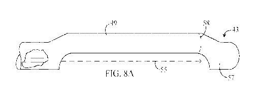
In FIG. 7, device 40 includes handle 41 coupled to elongated shaft 42 and
FIGS. 8A and 8B depict plan and bottom views of distal region 43 of
19
CA 02843183 2014-01-24
suitable linkage. When actuator 45 of handle 41 is depressed, needle
electrodes
51, 52 extend across offset region 54 of distal region 43 (as indicated by
dotted
lines 55 in FIGS. 8A and 8B. Optionally, distal-most portion 57 of distal
region
43 may include channels 56 that capture the distal ends of needle electrodes
51,
In the embodiment depicted in FIGS. 8A and 8B, needle electrodes 51,
52 are relatively long (e.g., 15 to 20 mm) and oriented parallel to one
another to
While the embodiment of FIGS. 8A and 8B employs straight needles and
In an example, the needles can be configured to be inserted into and
disposed in a target volume of tissue at a substantially uniform distance from
the
first surface of the offset portion. In an example, when fully inserted, the
length
of the needles can approach or exceed 15 mm. In certain examples, over the
CA 02843183 2014-01-24
span of 15 mm, the substantially uniform distance between the needle and the
first surface of the offset portion can range from 0 to 6 mm, with the lower
range
associated with a tighter zone of ablation. If the substantially uniform
distance is
too great, the layered ablation becomes harder to control. In an example, the
range can include 0 to 3 mm, 0 to 2 mm, and 0 to 1 mm, etc.
Other means for ensuring good tissue approximation within offset region
54 may be readily envisioned. The bladder trigone and its underlying tissues,
such as the anterior vaginal wall in the case of the female patient, are quite
mobile and easily deformed. Thus, only modest forces can be required to urge
the bladder trigone to conform to the offset region of the device. For
example, a
counter-pressure from the opposite side can be used to cause the distal end of
the
bladder device to receive the tissue at a first surface, in certain examples,
conforming the tissue to the distal end of the bladder device. In the female
patient, the counter-pressure may be applied from the vagina; in the male
patient,
the counter-pressure may be applied from the rectum. Such counter-pressure
may be provided by the fingers of a physician or may be applied using a rigid
or
semi-rigid probe as illustrated for later examples. In addition, the profile
of the
probe may be configured to mate with the profile of the bladder device. In
this
manner, the two mating profiles may be used to clamp the bladder and
associated tissue (and either vaginal or rectal tissue, depending on the sex
of the
patient) therebetween. The probe and the bladder device also may include
orienting features or linking mechanisms to enable simple and repeatable
clamping of the tissue, such as magnets (e.g., of opposite polarity, inserted
into
an opposing body cavity, such as the rectum, the vagina, etc.).
In the examples of FIGS. 8A and 8B, suction can be used to firmly, yet
reversibly conform the tissue to offset region 54 of distal region 43, by
atraumatically grasping and holding the tissue to the distal region 43.
Apertures
50 are connected, such as using sealed channels or tubing, to an external
vacuum
pump that supplies the suction. A variety of configurations, such as holes,
slots,
meshes, etc., may be used to provide conforming pressure. The apertures may
be connected using substantially leak-tight channels located within or
proximate
to the shaft of the device to an external vacuum pump. Suction fixation of
tissue
to device 40 is simple, quick, and easily maintained while passing the needle
electrodes into the tissue. Needle electrodes 51, 52 may be, for example, 22
21
CA 02843183 2014-01-24
gauge needles (or other gauge needles) that pass easily through the tissue,
tracking along a straight line and exiting the tissue in the same geometric
plane
as they entered the tissue. The use of suction beneficially distributes the
holding
force over a large surface area without causing harm to the tissue.
The design of the embodiment of FIGS. 8A and 8B further lends itself to
the inclusion of a number of safety features. First, direct vision capability
may
be used to locate or position device 40. For example, a channel may be
incorporated into the shaft of device 40 that allows insertion of a
traditional
urethroscope or other visual device for visual confirmation of the location or
placement of the active portion (e.g., suction zone, needle electrodes, etc.)
of
device 40. Second, the order of operation (e.g., initiation of suction,
capture of
tissue, needle electrode advancement, RF power application, needle electrode
retraction, release of tissue, termination of suction, etc.) of device 40 may
include a variety of safety interlocks, mechanical/hardware or software, to
ensure the correct order of operations. For example, one or more of the
following features may be incorporated to ensure safe operation of device 40:
= needle electrode advancement may be prevented until a pressure
gauge records that tissue has been firmly captured within offset
region 54 by suction through apertures 50;
= power to the needle electrodes may be prevented until the needle
electrodes are extended;
= complete and correct needle electrode advancement may be
confirmed by electric or mechanical contacts, and RF power
prevented until this confirmation; and
= suction may be applied until needle electrodes are retracted.
The needle electrodes also may be automatically retracted (e.g., by a spring
that
has been stretched as they were inserted, by an electromechanical actuator,
etc.)
if suction tissue capture was lost (e.g., as registered by a pressure gauge).
Device
40 can include secondary mechanism (e.g., a failsafe mechanism) for retracting
the needle electrodes if the device or mechanism otherwise jams or fails to
perform as intended.
As illustrated schematically in FIGS. 9A and 9B, when distal region 43 is
disposed in contact with tissue, for example trigone, and suction is coupled
to
device 40 via suction line 48, a portion of the tissue is drawn into offset
region
22
CA 02843183 2014-01-24
54, such as using one or more suction ports (e.g., denoted by the arrows in
FIGS.
9A and 9B) to a depth L3. Depressing actuator 45 can cause needles 51, 52 to
penetrate and extend across the portion of the tissue captured in offset
region 54
until the distal ends of the needles engage channels 56 disposed in the distal-
most portion for distal region 43. In an example, having needle electrodes
engage channels 56 locks device 40 on to the tissue during the ablation
process.
Actuation of button 46 on handle 41 causes RF current to flow between needle
electrodes 51, 52, thereby causing uniform ablation of tissue captured between
the needles.
In accordance with one aspect of the present subject matter, depth L3 is
selected to so that only tissue located wherein a predetermined non-
superficial
layer is ablated during energy delivery. The width of the ablation zone is
determined by the energy delivered into the tissue, as well as the spacing L4
(see
FIG. 9B). Optionally, needle electrodes 51, 52 may include an electrically
insulative coating disposed over a length L5 of the needle electrodes where
they
exit channels 53 and enter channels 56 (when fully extended across offset
region
54), to reduce energy deposition into the mucosa where the needle electrodes
penetrate the tissue. Illustratively, offset region 54 in distal region 43 has
a
length of about 15 to 20 mm, depth L3 can be about 4mm, and width L4 between
the needles is about 1 to 7 mm.
As illustrated in FIG. 9C, the configuration of device 40 ensures that a
highly repeatable and well-defined ablation zone 57 of length L6 that is
created
at a predetermined depth in the non-superficial layer, while also providing
protection zones 58, 59 that mitigate damage to mucosal layers and tissue
regions outside of the bladder, such as the vaginal wall. In certain examples,
offset region 54 of L4 is selected to create ablation zone 57 having a depth
of
about 2 to 3 mm. It should be understood, however, that the width and depth of
the ablation zone may be tailored to a specific patient's anatomy by adjusting
the
energy delivery parameters. For example, the bladder thickness for a patient
may be determined using ultrasound imaging, and the RF energy parameters
adjusted accordingly based on the observed thickness (e.g., using a look-up
table
available in the instruction manual accompanying device 40). In addition,
device 40 may be manufactured in a number of sizes, each having a different
23
CA 02843183 2014-01-24
offset region 49 that provides a specified length and depth L3 for offset
region 54
of distal region 43, and width L4 between the needle electrodes.
Referring now to FIGS. 10A and 10B, the distal region of an alternative
embodiment of a bipolar RF, suction-enabled device 60 of the present subject
matter is described. Device 60 is similar in construction to that depicted in
FIG.
7, except that device 60 includes a differently configured distal region 61.
In
particular, instead of a single pair of needle electrodes that are deployed
axially
as described for the preceding embodiment, device 60 includes a plurality of
needle electrodes 62 that are selectively extended from distal region 61 by
depressing the actuator on the device handle. As shown in FIG. 10B, needle
electrodes 62 can include an electrically insulative coating 63 that extends
over a
proximal length L7 of the electrodes, to reduce energy delivery into the
mucosal
layer. Like device 20 depicted in FIGS. 5A and 5B, needle electrodes 62 extend
a maximum distance L8 when fully deployed that ensures that the tips of needle
electrodes do not extend into or through the adventitia. Illustratively,
depths L7
and L8 are about 2 mm and 4 to 5 mm, respectively.
Referring to FIGS. 11A and 11B, distal region 61 includes plurality of
slots 64 through which suction is drawn to secure distal region 61 to tissue
to be
treated. As depicted by the sectional view of FIG. 11B, needle electrodes 62
are
joined to member 65, which positioned in suction manifold 66 and is configured
to be advanced and retracted by operation of the actuator on the handle of the
device, thereby selectively extending or retracting plurality of needle
electrodes
62 through apertures 67.
FIG. 12 depicts a further alternative of device 60 of FIGS. 10 and 11, in
which distal region 61 includes a heat sink for cooling the superficial layers
of
bladder tissue during operation of needle electrodes 62, including a cooling
channel 68 disposed in a separate plane of distal region 61 above slots 64
through which suction can be drawn. In this manner, a coolant, such as chilled
saline, may be circulated through coolant channel 68 during the ablation
procedure to act as a heat sink that draws heat away from the mucosal layer,
and
reduces the risk of superficial damage. Alternatively, the heat sink for
cooling
can include separate channels in distal region 61 that permit a chilled
biocompatible fluid, such as chilled saline, to be infused between distal
region
24
CA 02843183 2014-01-24
61 and the bladder surface to reduce excess heat buildup that could damage the
mucosa.
As will be apparent from the preceding description, device 60 is used to
cause ablation zones of predetermined size within the non-superficial tissue
of
the bladder. In operation, distal region 61 of device 60 is inserted into the
bladder (e.g., through the urethra or a minimally invasive opening through the
bladder wall), such that distal region 61 is disposed in contact with tissue,
for
example, the trigone. Then, suction is coupled to device 60 via a suction line
so
that suction is drawn through slots 64 and apertures 67, thereby engaging
distal
region 61 into contact with the tissue. While suction continues to retain the
tissue in contact with distal region 61, the actuator on device 60 is
depressed to
advance member 65 and fully extend needles 62 to penetrate the bladder wall.
RF energy is then supplied to needles, which causes RF current to flow between
needle electrodes 62 (or between needle electrodes and a grounding pad if a
monopolar configuration is used), thereby causing a substantially uniform
ablation zone for tissue received at the distal region 61. As discussed above,
insulative coating 63 can ensure that the delivered energy does not damage the
mucosa, while the overall deployed length of needle electrodes 62 can ensure
that ablation does not penetrate to the anterior vaginal wall, and is confined
within the bladder wall.
C. Exemplary Methods
With respect to FIGS. 13, illustrative methods of treating bladder
dysfunction in accordance with the present subject matter are described. FIG.
13
is an exemplary interior view of a female bladder (B) looking toward a
posterior
trigone (T), and further illustrating the relative locations of ureteral ostia
(0),
ureters (UR), urethral os (UO), and urethra (U), and a dashed line extending
between the ureteral ostia representing an imaginary interureteric bar (IB).
The
distance between ureteral ostia may vary between approximately 2 to 5 cm,
depending upon body size and the volume of fluid in bladder. The distance
between urethra and interureteric bar is approximately 3 cm, depending upon
body size and the volume of fluid in the bladder. Area proximate or including
the ureters, ureteral ostia, urethral os, or urethra should be avoided during
therapy, so as to avoid inadvertent damage to these structures, and thus
maintain
normal function of the urethra, urethral os, ureters, or ureteral ostia.
CA 02843183 2014-01-24
FIG. 13 further depicts examples of different ablation regions AR1, AR2,
constituting regions of the bladder in which it may be desirable to ablate or
denervate all or substantially all of the non-superficial tissue in those
regions.
Ablation region AR1 illustratively is located at least one of below (e.g.,
caudal
to) or between ureteral ostia, and may approach ureteral ostia, but leaving a
safety region between ureteral ostia and ablation region AR1 of at least one
of 1
to 25 mm, 1 to 20 mm, 2 to 10 mm, or 2.5 to 7.5 mm. For example, an upper
border of ablation region AR1 may extend above interureteric bar, towards the
dome of the bladder by at least one of 0 to 30 mm, 0 to 20 mm, or 0 to 10 mm.
In other cases, the upper border of ablation region AR1 may extend below
interureteric bar towards the base of the bladder by at least one of 0 to 20
mm or
0 to 10 mm. The lower border of ablation region AR1 may extend above urethra
or the neck of the bladder by at least one of 2 to 25 mm, 2 to 20 mm, 2 to 10
mm, or 2 to 5 mm, so as to avoid inadvertent damage to urethra or the internal
urethral sphincter.
At least a portion of ablation region AR1 may be beneficially targeted for
therapy. The bladder may be emptied prior to the ablation procedure to provide
for a thicker wall (e.g., a mucosa plus muscle layer thickness between 8 to 15
mm), or filled prior to the ablation procedure to provide for a thinner wall
(e.g., a
mucosa plus muscle layer thickness between 2 to 5 mm), or partially filled to
provide a thickness between that of an empty and full bladder (e.g., mucosa
plus
muscle layer thickness between 3 to 14 mm). The selected depth of penetration
of the therapy from an inner wall of the bladder may be between at least one
of 0
to 3 mm, 0.5 to 5 mm, or 5 to 15 mm.
Still referring to FIG. 13, alternative exemplary ablation region AR2 is
substantially trapezoidal in shape and roughly approximates the shape of
trigone.
In this case, ablation region AR2 approaches ureteral ostia, but again leaving
a
safety region between ureteral ostia and the outer margin of ablation region
AR1
of at least one of 1 to 25 mm, 1 to 20 mm, 2 to 10 mm, or 2.5 to 7.5 mm.
Although ablation region AR2 illustratively is substantially trapezoidal,
other
shapes or sizes may be used, such as substantially rectangular, triangular,
arcuate, ovoid, etc. A single portion of ablation region AR2 may be targeted
by
providing energy delivery to create one or more lesions. In an example,
multiple
portions of ablation region AR2 may be treated, within a single treatment or
26
CA 02843183 2014-01-24
multiple treatments. Treated portions of an ablation region may overlap, as
described below.
It should be understood that each of ablation regions AR1, AR2 should
be selected so as avoid damage to the ureters, ureteral ostia, urethra and
urethral
For example, device 40 of FIG. 7 may include a guide wire lumen
disposed on upper surface of distal region that accepts a conventional guide
wire
in an over-the-wire or rapid exchange manner. In use, a distal end of a guide
wire can first be inserted through a ureteral os and extended a distance into
the
27
CA 02843183 2014-01-24
during use and ensuring that the ablation zone does not encompass sensitive
areas, such as the ureters and ureteral ostia.
Additionally, because the distance between the ureteral ostia varies
depending upon body size or the volume of fluid in the bladder, a measuring
device, coupled to a visualization device, the treatment device, or other
device
configured to be inserted into the bladder, may be used to measure the
distance
between the ureteral ostia. As a further example, a measuring device may
include an expandable member, such as a balloon, having calibration marks that
may be compared to the distance between the ureteral ostia. In this case, the
treatment device may be selected or adjusted in response to the measured
distance or the volume of the bladder may be adjusted, such as by introducing
or
removing fluid to provide a desired distance between the ureteral ostia.
Referring now to FIGS. 14 through 17, illustrative ablation patterns that
may be generated within bladder B are described. Each of FIGS. 14 through 17
illustrates a posterior view of a view of the interior of bladder (B), with
ureteral
ostia (0), trigone (T), and urethra (U) identified. More specifically, FIG. 14
depicts a pointillist ablation pattern such as may be created using the single
contact point devices of FIGS. 4-6. FIG. 14 depicts the contact area of the
energy delivery element as shaded circles 70, with the concentric dotted lines
illustratively indicating the ablation zone corresponding to each contact
area. As
will be observed in FIG. 14, most of the dotted concentric circles overlap,
including a substantially total ablation of the non-superficial tissue in the
targeted treatment area. Illustratively, an expanded or reduced subset of
target
areas similar to those used for botulinum toxin injections may be employed to
define the treatment area.
FIG. 15 depicts a generally circular ablation pattern targeting the edges
of trigone (T), by ablating substantially circular ablation patterns 80 and 81
around ureteral ostia (0) and ablation arc 81 located about the urethral
ostium.
The circular patterns depicted in FIG. 15 may be generated using, for example,
energy delivery elements as described above, such as microwave, high intensity
ultrasound, laser, etc. Such devices may likewise be used to create ablation
pattern 90 depicted in FIG. 16, which substantially circumscribes trigone.
Other
ablation patterns also may be used, such as non-crossing or crossing linear
ablation patterns, concentric ablation patterns that target an atypical region
of the
28
CA 02843183 2014-01-24
bladder wall. For example, an atypical region of the bladder may include one
or
more areas of unusual morphology or activity, such as areas of denervation or
increased local contractile activity or electrical foci. In such cases,
targeting
therapy at the atypical regions of the bladder may provide advantages similar
to
those provided by electrophysiology treatments in the heart, such as ablation
or
isolation of an ectopic focus or ectopic foci for cardiac arrhythmias (e.g.,
to treat
atrial fibrillation, tachycardia, etc.).
FIG. 17 depicts ablation pattern 100 that encompasses substantially the
entire trigone, and advantageously may be generated using devices 40, 60
described above with respect to FIGS. 7 through 12. Such ablation patterns can
be used when treating a bladder wall that exhibits larger than typical or more
frequent local contractile activity, areas of dense afferent innervation, or
areas of
target efferent innervation. As will be apparent, combinations of the
foregoing
described ablation patterns may be beneficially employed. For example, one or
more areas of the bladder may be targeted for treatment, including one or more
of the following areas, among others: (1) the trigone; (2) the detrusor
muscle; (3)
the fundus; (4) an apex; (5) the body; (6) the neck; (7) the urethral ostia;
(8) the
ureteral ostia (one or both); (9) areas of the bladder having unusual
morphology
or activity, such as areas of denervation or increased local contractile
activity or
electrical foci; (10) functional areas of the bladder, such as functional
muscular
units; or (11) areas dense with nervous tissue or where nerves in the bladder
wall
concentrate to enter/exit the bladder.
In some examples, partial denervation can be beneficial to substantially
total ablation. For example, in cases where the patient is observed or
measured
to have a relatively thin bladder wall, it may be desirable to use a linear
crossing
pattern for ablation, or to circumscribe the trigone, while retaining areas of
intact
non-ablated tissue to ensure that an entirely ablate region does not present a
risk
of rupture immediately post treatment, or that scar tissue does not cause the
bladder wall to become unduly rigid after once the ablated region fully heals.
Accordingly, ablation therapy performed in accordance with the present
subject matter may be calibrated or controlled to provide partial or specified
therapy at a desired position, such as to avoid undesired conditions (e.g.,
acute
urinary retention, post void residual, straining, etc.). For example, in the
particular case of nerve ablation proximate the bladder wall, it may be
29
CA 02843183 2014-01-24
advantageous to achieve only a portion of denervation in a particular region.
Partial denervation can include substantially 100% denervation of a particular
area or any desired subrange, such as 70-90%, 60-80%, 50-70%, 40-60%, etc.
Therapy also may be limited to a particular area, including specific
dimensions
or surface areas of treatment (e.g., 4-5 square centimeters, extending not
more
than 1 cm beyond the border of the trigone, etc.). Alternatively or in
addition,
the extent of treatment may be defined relative to particular patient anatomy
(e.g., 80% of the area of the trigone).
As will be readily appreciated by those familiar with ablation
technologies, the degree of therapy may be controlled by controlling the
density
of a pattern of energy delivery. For example, a pattern of lesions that
include
both ablated zones and non-ablated zones (e.g., 75% ablated and 25% non-
ablated, etc.) may be selected or defined to produce a desired degree of
therapy.
Further, the degree of therapy may be controlled by a limited time and
duration
of therapy. The amount of damage (e.g., damage to nerves, muscle, etc.) can be
correlated to specified therapy parameters, such as time, temperature,
frequency,
amplitude, etc.
As noted above, the degree of therapy also may be controlled by limiting
the layers of tissue affected. For example, the therapy can be limited to not
extending beyond certain specific depths or specific anatomic layers of the
bladder wall. For example, therapy may be targeted to treat deep layers of the
bladder wall, such as the muscle and serosa OR ADVENTITIA, while protecting
one or more layers proximate the body of the bladder, such as the
glycosaminoglycan layer, mucosa, urothelium, the surface cell layer of the
epithelium, intermediate cell layer of the epithelium, etc. Other combinations
of
layers may be targeted for therapy or protected, for example, by protecting
non-
targeted tissue using a cooling balloon or other device or method to remove
heat.
More specifically, it may be desirable to include a capability to cool the
bladder wall tissue directly in contact with the energy delivery element, so
as to
avoid damaging the mucosa. Devices constructed in accordance with the present
subject matter therefore may include features designed to protect selected
structures from inadvertent damage, such as the ureteral and urethral ostia.
While this may be accomplished by a variety of algorithms and controls (e.g.,
measuring electrode temperatures, measuring tissue temperatures or impedances,
CA 02843183 2014-01-24
timers, visual feedback, etc), it may in addition be advantageous to use a
large
thermal mass to moderate temperatures except at the desired location. Examples
include the use of a heat sink, such as fluids (e.g., water, saline, etc.),
which may
be heated or cooled to a temperature distinct from room temperature. Such a
heat sink may be either static (e.g., an inflated balloon) or dynamic (e.g.,
fluid
flowing in an open or continuous loop).
For example, a balloon may be filled with a continuously circulating flow
of chilled (e.g., using ice-water bath) saline mixed with contrast media and
used
to cool tissues in direct contact with the balloon, while allowing an internal
microwave antenna or other energy delivery element to therapeutically heat
underlying tissues. In this manner, at least a portion of the mucosa of the
bladder can be protected while treating one or more portions of the underlying
or
adjacent bladder tissue, such as the basement membrane, suburothelium,
submucosa, lamina propria, muscle, adventitia, or serosa.
As a further example, a ureter may be protected, such as by inserting a
catheter, a cooling balloon, or other cooling device proximate to or into the
ureter prior to or concurrently to treating proximate bladder or nerve
tissues. For
example, the device may be configured so that its distal tip is positioned
within
the ureter, and delivers thermal energy to target tissue (e.g., nervous tissue
innervating the trigone proximate the ureter, etc.) from the energy delivery
element while the distal tip of the device concurrently cools at least a
portion of
the ureter (e.g., an interior of the ureter) to prevent damage to the ureter
from the
thermal energy.
It should be understood that in the case of cryotherapy, a heat sink may
be used to heat non-target tissues rather than remove heat from the non-target
tissue. For example, an ablation device constructed in accordance with the
principles of the present subject matter and using a cryogenic probe may
include
a flow of warmed saline through channels along the shaft of the device to
prevent cold damage to the urethra and to localize the cold to the cryogenic
probe.
In still other examples, a thermal mass, such as sterile saline, may be
introduced into adjacent body spaces, such as the peritoneum, to inhibit or
prevent the inadvertent spread of energy delivered during therapy. For
example,
sterile normal saline may be introduced into the peritoneum to expand the
31
CA 02843183 2014-01-24
abdomen and create a buffer between the bladder and the intestines. A thermal
mass also may be introduced into the rectum, vagina, or uterus either free-
flowing or encapsulated in a balloon. Further, a cooling device may be
inserted
into the bladder if the energy delivery element is in a different location
(e.g., in
In addition to treating bladder dysfunction, pelvic nervous tissue or
nerves also may be targeted for therapy or ablation to treat generalized
pelvic
pain, including nervous tissue on, within, or proximate the bladder wall,
including on or within bladder tissue, such as the lamina propria,
suburothelium,
32
CA 02843183 2014-01-24
urethra, and somatic nerves S2-S3 and their branches, which lead to the
pudendal nerve supplying the external sphincter.
Further in accordance with the principles of the present subject matter, a
variety of access routes may be utilized to perform the therapies described
herein. Examples include open surgical access (e.g., laparotomy) or minimally
invasive access (e.g., laparoscopic) to the abdominal cavity, the retropubic
extraperitoneal space (the "Retzius space"), and different portions of the
bladder
(e.g., the dome of the bladder covered by the peritoneum, anterior aspects of
the
bladder, lateral aspects of the bladder, etc.). Transvaginal, transcervical,
transuteral, transurethral, or transrectal access is also possible. FIGS. 18-
22
illustrate exemplary views of gaining access to the bladder of a female
patient.
FIG. 18 is a lateral view of a female pelvis depicting the relative
locations of vagina (V), pubic symphysis (PS), rectum (R), bladder (B), and
urethra (U), and includes peritoneal or pre-peritoneal dissection through
abdominal wall (AB). Incision 110 is be made in abdominal wall, exposing the
space of Retzius proximate bladder. The bladder may be directly accessed
through incision 110. FIG. 19 depicts a female perineum including vaginal
speculum 116 and stay sutures 116, 117 configured to retract and hold open
labia
minor. The bladder may be accessed through opening 118 formed in anterior
vaginal wall (VW).
More specifically, the urinary bladder lies immediately outside of the
peritoneum, and is loosely attached to the peritoneum from the apex, across
the
dome, and down to the vesicouterine pouch. Intraabdominal access allows direct
vision of the superior and posterior aspects of the bladder. Dissection of the
peritoneum off the abdominal wall develops the Retzius space, which provides
direct access to the anterior portion of the bladder lying immediately
adjacent to
the pubic symphasis. Once the bladder is reached (e.g., from either
intraabdominal or extraperitoneal routes), the bladder can be dissected from
its
lateral and posterior adherents to expose the trigone region or the bladder
wall
can be incised and later repaired to provide access to the interior of the
bladder.
The anterior wall of the vault of the vagina, which lies immediately
posterior to the bladder, may be incised to expose the urethra, which is the
typical surgical exposure for a transvaginal tape procedure to treat stress
incontinence. The transvaginal route is a commonly performed, widely accepted
33
CA 02843183 2014-01-24
procedure having minimal morbidity. Access to the bladder neck, the trigone of
the bladder, or areas off of the midline of the bladder may be provided using
a
similar, somewhat deeper dissection of the vault of the vagina.
Advantageously, injections into the nerves supplying the trigone may be
made both from the interior of the bladder and from the vagina (e.g.,
lidocaine or
other anesthesia injections) to elicit a temporary effect as a screening
method
prior to denervation.
As will be apparent to a physician of ordinary skill in the art, the female
bladder also may be accessed via a conventional laparoscopic procedure or
percutaneous suprapubic access via a cannula. Alternatively, percutaneous
access to a female pelvis may be established through the perineum, for
example,
using a cannula to access to the space between the posterior bladder wall and
the
anterior vaginal wall. Access to the bladder also may be obtained using a
conventional cystoscope or other visualization device inserted into the
bladder
through the urethra, providing viewing access to one or more structures in the
bladder, such as the trigone or ureteral ostia. The treatment devices
described
above may be inserted through a working channel in the cystoscope or
alternatively the treatment device may include an imaging system, for example
as described as an optional feature of the embodiment of FIG. 7. As described
above, the visualization device may be used to position the energy delivery
element at a desired position in the bladder and ensure that a safety margin
exists
between a possible ablation region and one or more features of the bladder,
such
as the ureteral ostia, ureter, urethra, urethral os, the urethral sphincter,
etc. In
addition, the visualization device may be configured to provide a cooling
function (e.g., chilled saline irrigation, a cooling balloon, etc.).
Use of a combination of access routes may be advantageous. For
example, being able to access both sides of a desired tissue target may allow
for
improved energy density or temperature control at one or more locations. For
example, a combination of intravesical and vaginal access may be used to place
one or more auxiliary cooling devices (e.g., a balloon inflated with saline)
on
one or both sides of the desired tissue target to isolate increased
temperatures to
deeper tissues layers. Further, combining energy delivery from both sides of
the
desired tissue target may serve to increase temperatures or energy densities
in
the region of overlap (e.g., deep within the tissue), while minimizing
34
CA 02843183 2014-01-24
temperatures or densities in the superficial regions (e.g., the bladder
glycosaminoglycan layer, urothelium, mucosa, or the vaginal wall). In this
manner, therapy advantageously may be directed to desired regions or layers of
tissue, while minimizing undesired trauma or injury to other tissue.
Furthermore, where it is intended to use the apparatus and methods of the
present subject matter to relieve pelvic pain, pelvic nerves or nervous tissue
may
be accessed without puncture or incision via navigation through the uterus,
laterally through the fallopian tubes, and exiting via the abdominal ostium.
D. Additional Embodiments
In accordance with another aspect of the present subject matter, lesion-
creating elements may be combined in structures that aid in creation of
desired
pattern, such as expanding mesh cages, wire loops, expanding balloons, etc.
This portion of the disclosure describes additional examples constructed in
accordance with the principles of the present subject matter.
Inflated balloons (e.g., inflated with gas or liquid, such as saline or
contrast agent) may be used to provide an integrated heating element (e.g., a
microwave antenna, RF electrode, PRF electrode, laser fiber, ultrasound, etc.)
at
a specific location. In an example, a balloon may be used to centralize an
internal microwave antenna (e.g., along an axis of the balloon), or to locate
a
heating element a specific, known, or desired distance from the desired tissue
target (e.g., ranging from zero distance, or contact, to the full diameter of
the
balloon). One or more balloons may have therapy delivery elements mounted or
placed on the exterior of the balloon, such as RF electrodes, RF needles, etc.
For example, an elongated shaft, such as a catheter, configured to be
passed through the urethra into the bladder, such as a Foley or one or more
other
catheters, may include a balloon and an energy delivery device, such as
internal
microwave antenna, RF electrodes embedded in the wall of the balloon, an
inlet/outlet configured to receive or output a heated or cooled gas or fluid,
or
ultrasound transducer. The energy delivery element may be integrated with the
balloon. The balloon may be inflated after urethral insertion and counter-
traction applied to bring the balloon into intimate contact with the bladder
trigone or bladder neck. In this manner, the energy delivery element will be
automatically and reproducibly positioned correctly relative to targeted
tissues.
In addition, the balloon can be configured to inflate in a desired shape
(e.g.,
CA 02843183 2014-01-24
cylindrical, ovoid, pancake, arching, triangular, pyramidal, conformable to
the
surrounding bladder, etc.) so as to facilitate targeting of the desired
tissues.
Referring now./ to FIG. 20, a lateral view of bladder (B) is shown
including device 120 having expandable element 121, such as a balloon,
mounted to the distal end of elongated shaft 122. Device 120 illustratively is
configured to provide access to bladder through urethra (U). FIG. 20 shows the
relative positions of ureteral ostium (0) and ureter (UR).
Expandable element 121 is shown in its expanded deployed state (e.g.,
for a balloon, an inflated position) and has a reduced diameter state (e.g.,
for a
balloon, a deflated position) that enables it to be advanced through urethra
into
bladder. Expandable element 121 is coupled to an energy delivery element, as
described below. Expandable member 121, in the contracted state may be
inserted into bladder through urethra, and then transitioned to the expanded
deployed state (as shown in FIG. 20) to position the energy delivery element
at a
desired position within the bladder, such as between the ureteral ostia. In
FIG.
20, the position of the energy delivery element with respect to expandable
element 121 is illustrated using by energy delivery zone 123. Expandable
element 121 may be configured so that it can be wedged into a specific
position
in bladder, such as against the trigone, for example, to set the distance
between
the energy delivery element and the wall of the bladder. Expandable element
121 can include a substantially compliant element configured to conform to the
geometry of bladder, a substantially rigid element configured to take a
desired
shape or to conform at least a portion of the bladder wall to a desired shape,
or
combinations of both.
If expandable element 121 does not fully fill the bladder, a tension force
may be used to seat the expandable element 121 in the neck of the bladder to
position expandable element 121 or the energy delivery device at the desired
location. The tension force may be applied by a user (e.g., as specified, for
example, as directed by the Instructions-for-Use accompanying device 120), or,
in certain examples, by an auxiliary device. For example, device 120 may
include a slidable anchor configured to traction expandable device 121 by
bearing against the perineum (e.g., where urethra exits the body). The
slidable
anchor may include a calibration device (e.g., a spring element, etc.)
configured
to adjust the amount of force applied to the perineum or the bladder.
36
CA 02843183 2014-01-24
Illustratively, device 120 includes handle 124 configured to allow a user
to position expandable element 121 at the desired position in bladder. The
handle may have a fixed position with respect to expandable element 121 or the
energy delivery device, and may include indicator 125, such as a marking or a
feature (e.g., an arrow), configured to provide information regarding the
orientation of expandable element 121 or the energy delivery device to the
user.
Indicator 125 may include a tactile feature, an accelerometer, audio
notification,
or one or more other notifications of device orientation.
FIGS. 21 and 22 depict lateral sectional views of female bladder (B)
including energy delivery devices 130, and 140, respectively, configured to
access bladder through urethra (U). FIG. 21 depicts device 130 having a laser
energy delivery element including light fiber 131 configured to transmit laser
energy 132 to a bladder wall and inflatable balloon 133 proximate to or
surrounding light fiber 131. Laser energy 132 may be transmitted to or through
at least a portion of the bladder wall or surrounding structure.
In the embodiment of FIG. 21, inflatable balloon 133 is configured to
position the energy delivery element a specified distance from the bladder
wall,
as may be required for proper operation of the energy delivery element or to
provide local cooling (e.g., proximate the laser energy 132, such as by
removing
heat from the target area) to protect tissue proximate inflatable balloon 133.
For
example, inflatable balloon 133 may be sized to protect specific layers of the
bladder wall from damage (e.g., glycosaminoglycan layer, mucosa, urothelium,
suburothelium, submucosa, lamina propria, muscularis propria, etc.) or to
control
tissue damage until a certain depth. One or more characteristics of the laser
energy (e.g., frequency, amplitude, etc.) may be modulated to control the
maximum depth of tissue damage. Although FIG. 21 depicts device 130
positioned near trigone T proximate the posterior bladder wall, distal region
134
of device 130 may be articulitable so that the energy delivery element may be
positioned at other locations within or outside the bladder.
Referring now to FIG. 22, device 140 is depicted that includes a
microwave energy delivery element, including microwave antenna 141
configured to deliver microwave energy 142 to the bladder wall. Device 140
further includes inflatable balloon 143 proximate to or surrounding microwave
antenna 141. Microwave energy 142 is delivered to or through at least a
portion
37
CA 02843183 2014-01-24
of the bladder wall or surrounding structure. Device 140 may be configured to
be disposed in other locations within the bladder, and further, inflatable
balloon
143 may be configured to position microwave antenna 141 in any desired
position (e.g., at a specified location) or to provide local cooling, thereby
protecting tissue proximate balloon 143.
Referring now to FIGS. 23 and 24, further alternative examples of
apparatus constructed in accordance with the principles of the present subject
matter are described. FIG. 23 depicts a distal end of device 150 including
elongated shaft 151 having inflatable balloon 152 with energy delivery
electrode
153 (e.g., RF electrode, microwave antenna, etc.) embedded in the exterior of
balloon 152. FIG. 24 depicts a distal end of device 160 including elongated
shaft 161 having plurality of energy delivery elements 162 disposed on
expandable wire structure 163. Device 160 is configured so that after
expandable wire structure 163 is inserted into the bladder (e.g., such as
through
the urethra), expandable wire structure 163 may be deployed to urge plurality
of
energy delivery elements 162 against a desired portion of the interior of the
bladder.
Further alternative examples of devices configured to be inserted into the
bladder may include other expandable structures, such as linkages, deflectable
catheters, mesh cages, shape memory structures, with or without a balloon,
that
may be inserted into the bladder (e.g., via the urethra or a suprapubic
catheter) in
a contracted state, and then expanded to form two or three dimensional shapes
within the bladder. Such expandable structures may be configured to conform to
desired target tissues or areas. Expanding structures also may be positioned
proximate an outside bladder wall and used to form two or three dimensional
shapes configured to conform to desired target tissues or areas on or
proximate
to the outside bladder wall. For example, a device penetrated into the
perineum
or inserted through the vagina, along a course substantially parallel to the
posterior bladder wall, may include a variety of deployable needles, for
example,
to form a desired pattern. In addition to locating the energy delivery element
with respect to a target tissue, expandable structures or balloons also may be
used to mount multiple energy delivery elements and to control the relative
positions of these elements. For example, patterns of therapy delivery may be
38
CA 02843183 2014-01-24
created either through the simultaneous or sequential activation of the
multiple
therapy delivery elements.
As discussed above, devices constructed in accordance with the
principles of the present subject matter advantageously may incorporate some
degree of steerability to position the energy delivery elements relative to
desired
treatment target locations. Steering may be passive (e.g., a pre-curved device
that can be straightened to pass it up the urethra), active (e.g., a catheter
that
curls, such as based upon tensioning of an integral pull-wire), via a separate
auxiliary device (e.g., an external delivery sheath), external magnetic field
(Stereotaxis), or employ the shape-memory aspects of certain alloys (e.g.,
nickel
titanium (Nitinol), etc.).
FIGS. 25 and 26 provide lateral section views of a female pelvis
including device 160, which illustratively may consist of devices 150 or 160,
inserted into bladder (B) through urethra (U). The relative locations of
bladder
(B), vagina (V), pubic symphysis (PS), and urethra also are shown. In each of
FIGS. 25 and 26, device 160 includes inflatable balloon 161 (or an expandable
wire structure) configured to be inserted into bladder through urethra in a
contracted state and then inflated or otherwise deployed in bladder. For
example, device 160 may be configured to provide either heat or cold to tissue
of
or proximate to bladder, vagina, or other location. Inflatable balloon 161
(e.g., a
heat sink) may be configured to remove heat from or to provide heat to at
least a
portion of the bladder, such as at least a portion of trigone (T) of the
bladder.
Alternatively, cooling device 161 may include one or more other energy
delivery
components, such as electrodes, ultrasound transducer, microwave antenna, PRF
antenna, or RF antennas, etc., configured to provide thermal energy to tissue
within or proximate to a bladder wall.
FIG. 25 depicts auxiliary cooling device 170 including balloon 171 that
is configured to remove heat from or to provide heat to at least a portion of
vagina. FIG. 26 includes cannula 180 having plurality of needle electrodes 181
that may be selectively deployed within or proximate to the bladder wall (and
similar in construction to the needle electrodes described with respect to the
embodiment of FIGS. 11 and 12). Needle electrodes 181 may be configured in a
bipolar or monopolar arrangement. Needle electrodes 181 also may be
configured to be inserted within or through at least a portion of the bladder
wall.
39
CA 02843183 2014-01-24
The ability for the physician to directly visualize the bladder or other
structures during therapy may be beneficial. As discussed for preceding
examples, the energy delivery element may be configured to be used within a
working channel of a cystoscope or other visualization element, either rigid
or
flexible. Alternatively, device carrying the energy delivery element may
incorporate a visualization element (e.g., lens and fiber optic, CCD chip,
light
source, etc.). Such a visualization element may either be disposable or a re-
usable element that is coupled to a disposable energy delivery device prior to
use. Such devices also include fiducial markings or measurements, auxiliary
measuring tools, engage anatomic landmarks, or provide tactile feedback to
assist the physician during positioning and use of the device.
E. Adjunctive Therapies
Several procedures can serve as useful adjuncts to improve the
permanence of the relief sought to be provided by the apparatus and methods of
the present subject matter. For example, a prior successful round of botulinum
toxin therapy or anesthetic injection (e.g., lidocaine, etc.) may be used to
isolate
or screen a likely "responder" to the proposed therapy. For example, if a
patient's symptoms improve with a transient intervention, then that patient
may
be a good candidate for an ablation procedure of the tissue using the
apparatus
and methods described in the present disclosure. If the patient's symptoms do
not improve or worsen with the transient intervention, then that patient may
not
be a good candidate for an ablation procedure that targets the anesthetized
tissue.
As a convenient screening tool, lidocaine and other anesthetics have the
advantage that their affects wear off after a period of hours instead of a
period of
6 to 9 months observed for botulinum toxin injections. Examples of local
anesthetics include lidocaine, prilocaine, tetracaine, and benzocaine. The
local
anesthetic may be applied in the form of a liquid, viscous liquid, spray, or
gel.
Alternatively, application of cold (cryoanesthesia) may be used to
temporarily numb a desired area, transiently disabling nerve conduction or
muscle contractility, and allowing assessment of a patient's symptoms after
the
intervention until the target tissue rewarms. Examples of cryoanesthesia
include
liquid nitrogen spray, argon, refrigerant (e.g., Freon), or chilled saline.
The
cryoanesthesia may be applied either directly to target tissue or contained in
a
structure, such as a needle, probe, lumen, catheter, balloon, sac, etc.
CA 02843183 2014-01-24
Dyes or other markers may be used in conjunction with injections to aid
in providing permanent or more permanent therapy to the same locations as
previous, and successful, injections of a pharmacologic agent. For example, a
dye or other marker may include commonly used medical dyes, such as indigo
carmine, methylene blue, etc. In addition, more permanent dyes, such as are
used in tattoos, may be used when a more durable mark is desired, such as when
botulinum toxin is injected into the bladder wall as a screening test.
As a further adjunct to, or in lieu of, the energy delivery modalities
disclosed above, it is contemplated that denervation of the bladder may be
accomplished or rendered more permanent by mechanically disrupting afferent
nervous tissue within the bladder wall. First, nerves of the bladder may be
accessed, such as using balloon dissection of the space lateral to the bladder
to
dissect the peritoneum from the abdominal wall along natural tissue planes in
variations of laparoscopic surgery. For example, a dissection balloon may be
mounted on an atraumatic probe and tunneled along the lateral border of the
bladder. An auxiliary light source within the bladder (e.g., inserted via the
urethra) may facilitate this procedure by providing transillumination that
aids the
tunneling process. The probe also may include technology to visualize and aid
in following of the natural tissue planes.
Second, the durability of the denervation may be improved by delaying
or preventing nerve regrowth. This may be accomplished by surgically
implanting a physical barrier into the space dissected, ablated, destroyed, or
otherwise damaged to denervate the bladder (e.g., the lateral aspect of the
bladder). Suitable materials for such barriers may include polypropylene mesh,
polytetrafluoroethylene (PTFE) or expanded PTFE films, adhesion barriers,
hyaluronic acid membranes (e.g., Seprafilm by Genzyme , indicated as an
adhesion barrier for the pelvis), polyethylene glycol (PEG), injections or
deposits of liquid hyaluronic acid, and other polymers or hydrogels. The
barrier
also may include biologic materials, such as collagen, pericardium, mucosa
(intestine), fibrin, etc., such as after suitable processing for sterility and
to render
these materials non-immunogenic.
Chemical treatments, such as corrosive or cytotoxic chemicals, also may
be used to coat or inject the dissected or otherwise damaged areas so as to
kill or
deactivate exposed nerves. Suitable chemicals may include caustic chemicals
41
CA 02843183 2014-01-24
(e.g., sodium hydroxide, potassium hydroxide, caustic pencils, etc.), alkalis,
strong acids (e.g., sulfuric acid, nitric acid, hydrochloric acid, etc.),
concentrated
solutions of weaker acids or bases (e.g., formic acid, acetic acid, etc.),
Lewis
acids (e.g., anhydrous aluminum chloride, boron trifluoride, zinc chloride,
etc.),
strong oxidizing agents (e.g., hydrogen peroxide), other corrosive chemicals,
and
neurotoxins. ..
Additionally, fixative agents, such as glutaraldehyde,
fonnaldehyde/formalin, alcohols, mercuric chloride, potassium dichromate,
sodium sulfate, concentrated sugars, etc., may be used to stabilize and
strengthen
tissues. Capsaicin or other members of the vanilloid family may be used to
affect the nerves. Additionally, fixatives (e.g., zinc chloride paste) may be
used
during or prior to denervation, and applied topically (e.g., to the mucosa
from
inside the bladder) or injected into the bladder wall.
Chemical agents also may be used such as in conjunction with surgical or
energy delivery denervation described above, either to extend the durability
of
the denervation or to block pain. To prevent the inadvertent dispersion of the
chemical agent, it may be delivered as a gel, foam, paste, solid, or other non-
liquid form.
As a still further adjunct to the ablation apparatus and methods described
above, or as an alternative to such approaches, it is hypothesized that
bladder
dysfunction may be treated by reducing the elasticity of selected portions of
the
bladder, such as the trigone, to reduce activation of stretch receptors
located in
that region. In particular, the bladder trigone is a smooth triangular region
of the
internal urinary bladder formed by the two ureteral orifices and the internal
urethral orifice. The trigone is densely innervated, including terminal
branches
of the pelvic nerve, and is sensitive to expansion, pressure or change in
pressure,
signaling the brain that the bladder needs to be emptied. The trigone is of
different embryologic origin than the rest of the bladder, as it is derived
from the
caudal end of mesonephric ducts of mesodermal origin, as the rest of the
bladder
is of endodermal origin. In females, the mesonephric ducts regress, causing
the
trigone to be less prominent, but still present.
More specifically, it can be hypothesized that stretch receptors in the
trigone are responsible for the sensation of urgency that is the hallmark of
overactive bladder. It is further hypothesized that bladder distension during
42
CA 02843183 2014-01-24
filling is largely confined to the dome of the bladder, which is free of
attachments, while the trigone region's local stretch is restricted by the
natural
attachments of the three lumens and the attachments of the trigone to the
underlying vagina (e.g., in the female) or prostate (e.g., in the male). Upon
substantial filling, stretch of the dome begins to be transmitted to the
relatively
stiff trigone, giving the sensation of fullness in a patient, which eventually
sharpens to urgency. In the symptomatic overactive bladder patient,
uncontrolled detrusor muscle contractions may cause stretch in the trigone
region, causing a sensation of urgency even at low bladder volumes.
To address the foregoing phenomenon, it is hypothesized that isolating
the trigone region, or other region sensitive to filling, from stretch (e.g.,
due to
non-volitional detrusor muscle contractions) may suppress abnormal sensations
of urgency. Similarly, providing additional support to the trigone region may
aid
its natural contraction during the filling phase and delay funneling of the
bladder
neck, which can initiate progressive continued micturation. Conceptually, this
theory of urge incontinence and overactive bladder may be liken to a circle
can
be drawn on the side of a partially inflated balloon. Further inflating the
balloon
results in an increase in the diameter of the drawn circle, which illustrates
the
additional stretch applied to the balloon. If instead a physical ring of
specified
diameter were glued to the side of the balloon, further inflation of the
balloon
would not result in further expansion of the encircled portion of the balloon.
Thus, the area of the balloon within and immediately adjacent to the ring
effectively is isolated from the increasing volume or stretch of the balloon.
Likewise, if a physical bar were attached to the side of a partially inflated
balloon, increased inflation of the balloon would result in some increased
stretch
of the balloon material, especially in a direction orthogonal to the axis of
the
physical bar. However, the stretch of the balloon would be limited in the
region
immediately adjacent to the physical bar, especially in a direction parallel
to the
axis of the physical bar.
Analogously, the inventors hypothesize that implanting a device within
the bladder wall will stiffen tissues, either by the implant itself or by the
buildup
of scar tissue (e.g., a healing response, etc.) that encapsulates the implant.
Accordingly, bladder dysfunction may be treatable by modifying the mechanical
43
CA 02843183 2014-01-24
properties (e.g., stiffness or strengthen) of tissues that reduce stretch in
adjacent
or circumscribed regions.
In accordance with this aspect of the present subject matter, an implant,
such as a suture, may be used to isolate the trigone region from stretch
resulting
from filling of the bladder. The suture may include a running "purse string"
suture that encircles the trigone region to limit the stretch of the trigone
by
_
having the suture bear at least a portion or the entire load from bladder
expansion. Such a "purse string" suture need not be continuous or even form a
complete circle to reduce the stretch of the trigone during bladder filling.
For
example, several linear suture lines could substantially surround the trigone,
while gaps could be left at certain locations (e.g., in areas near delicate
structures
to be avoided, such as the ureters, the urethra, etc.) to prevent inadvertent
damage. In some cases, it may be desirable to leave at least one gap or space
to
allow for some stretch of the trigone for normal urinary function.
As a further embodiment, reinforcing the trigone itself is possible. For
example, one or more lines of suture placed across the trigone, either or both
parallel or transverse to the axis of the urethra, may serve to stiffen it.
Off axis
(e.g., neither parallel nor perpendicular to the urethra) orientations or
combinations of orientations (e.g., an 'X' shape) also may offer other
advantages. Other patterns, including weaves or combinations of elements, may
be useful.
In addition, other support structures may be used to support or isolate the
trigone, or to reinforce the trigone from the exterior of the bladder. For
example,
a polypropylene or other artificial mesh or patch may serve as a mechanical
buttress for weakened tissues or as a scaffold that elicits ingrowth of new
tissue
that locks the mesh in position and adds to the strength of the healed
structure.
A prosthetic mesh or patch may be implanted at the location of the trigone,
either
within the detrusor muscle or just external to the bladder in the
vesicovaginal
potential space. Furthermore, such support structure may be affixed to the
interior of the bladder wall where, in addition to preventing stretch, it
prevents
activation of pressure sensory nerves. Flexible, substantially rigid, or rigid
structures (e.g., a rigid bar, etc.), or combinations thereof, may be
implanted in
intra-detrusor muscle or vesicovaginal locations to reinforce the detrusor
muscle.
As with the suture lines described above, such bars may either encircle the
44
CA 02843183 2014-01-24
trigone or cross it in various combinations or permutations. Woven polyester
mesh also may be used to stiffen the trigone.
As a further embodiment, an injectable agent may be inserted within a
desired plane of tissue. This injectable agent can be configured to "cure" or
harden in situ to yield a stable element. Examples of cures include chemical
reactions (e.g., RTV silicone, epoxy, etc.), light activations (e.g., commonly
blue
light or UV, etc.), thermoset, etc. Chemical agents that cause fibrosis or
tissue
stiffening via other mechanisms, such as via body response to the agent, also
may be injected or otherwise applied to the target region of the bladder wall
or
adjacent tissue. Such agents may include sclerotherapy agents used to scar
blood
vessels, such as sodium tetradecyl sulfate, polidocanol, etc. The sclerosant
may
be applied in liquid, foam, gel, or paste form. Other suitable agents include
dextrose solution, similar to that used in prolotherapy of ligaments and
tendons
or platelet rich plasma (PRP) in a gel or graft matrix preparation to
stimulate
collagen ingrowth.
The support structures described in the preceding paragraphs may be
used in combination to provide relief. For example, an injectable agent may be
combined with a mesh or other support structure for tissue ingrowth and
anchoring. The combined support structure may range from mesh coverings of
discrete bars to structures analogous to battens used in sails, where the
support
structure is mostly a membrane (e.g., mesh, etc.) having relatively small
stiffeners (e.g., rigid bars, etc.) interspersed. For example, two or more
substantially parallel bars may be arranged across or from borders of a mesh
panel.
Support structures described above may be configured as a full or partial
ring, including as arcuate segments, semicircles, or rings with a gap, such as
like
the capital Greek letter omega (n). Such full or partial rings may be mated
with
a mesh panel, such as using the ring to form a border for the mesh that
provides
stiffness and ease of insertion, while the mesh serves to hold the ring in
place,
both acutely and after tissue ingrowth.
Surgical access from the vaginal anterior wall provides direct access to
the urethra and bladder neck, such as typically done during a transvaginal
tape
procedure for stress urinary incontinence. In an example, deeper dissection
posteriolaterally provides access to both sides of the trigone. Accordingly,
using
CA 02843183 2014-01-24
this exposure, a partial ring (e.g., a horseshoe, a capital Greek omega, etc.)
may
be implanted that substantially encircles the entire trigone. This partial
ring may
be covered by a polypropylene mesh sleeve that serves to prevent erosion or to
provide a matrix for tissue ingrowth.
Suitable support structure also may have an inwardly directed bias (e.g.,
pinching the trigone inwards to further reduce wall stress, etc.). For
example, an
undersized ring may be implanted that compresses tissue within its
circumference. Such a ring may first be placed and then constricted into a
smaller opening. While this constriction could be performed intraoperatively,
it
can also be done some delay after initial implantation (e.g., after the ring
has
adhered to the surrounding tissue). In addition, the ring could be held in its
expanded state by a resorbable material (e.g., a resorbable suture), such that
after
some period of implantation (e.g., one to four weeks), the resorbable material
can degrade and release the ring to collapse to its smaller shape.
It is advantageous to keep foreign bodies outside of the bladder due to the
problems with encrustation and stone formation that have been observed when a
foreign body is exposed to the urine environment in the bladder. However, to
avoid such problems, the foreign body need only to be covered by intact
mucosa,
and thus, may reside within or outside of the bladder wall. Furthermore, all
of
the foregoing therapies, when applied to the bladder, should result in either
an
intact mucosa or a mucosa that can heal without complication.
While the above discussion has been directed toward the reducing stretch
of the trigone, it should be understood that the apparatus and methods
described
herein are not limited to use in the trigone region, but may be beneficially
used
in other areas of the bladder, including the fundus, the apex, the body, the
neck,
the dome, the detrusor muscle, etc.
It is contemplated that in addition to implantation of surgical barriers or
restrains within the bladder wall, such as described above, it may be possible
to
induce thermal denaturation of collagen contained within the bladder wall and
thereby modify both the physical and mechanical properties of the bladder. In
particular, when heated to a temperature in the range of 65-70 deg C, collagen
fibrils within tissue begin to break their chemical bonds and transition from
a
native highly ordered state to a more random orientation which causes the
collagen-containing tissue to shrink by up to 50%. The amount of shrinkage
46
CA 02843183 2014-01-24
experienced by the tissue depends upon the parameters of the heat applied and
to
the original orientation of the collagen within the tissue. This effect is
used
therapeutically in orthopedic applications (e.g., shoulder instability),
aesthetics
(skin tightening, facelifts, etc.), and urology (e.g., stress incontinence).
While in
Accordingly, as an adjunct to the energy delivery apparatus and methods
In certain applications of thermal denaturation, especially those where
the affected tissue experiences large physiologic loads, initial degradation
of
tissue mechanical properties in the interval before the body's healing
response
strengthens the tissue may be of concern, requiring external support (e.g., a
While collagen denaturation may be used to shrink tissues (e.g., due to
the natural tendency of the collagen fibers to shorted when not constrained),
47
CA 02843183 2014-01-24
gas, or by using an inflated balloon with heating device. In some cases, a
combination of heat and distention may be employed to ablate afferent nervous
tissue within the bladder wall, so as to reduce pain or urge sensation.
Expanding the cystometric volume of the bladder may serve several
functions. First, some conditions, especially interstitial cystitis (IC) are
primarily associated with a constricted bladder volume. Further, increased
bladder volume may decrease frequency, one of the hallmark symptoms of
overactive bladder. Finally, increasing the bladder volume should serve to
decrease wall stress at a given volume, reducing afferent nerve traffic and
potentially decreasing overactive bladder. Expanding the volume of the bladder
using the collagen remodeling technique described above may be advantageous
when compared to previously-known surgical techniques that are used to
increase bladder capacity (e.g., detrusor myomectomy, enterocystoplasty,
etc.),
which are highly invasive procedures involving significant complications and
morbidity.
F. Treatment of Chronic Genitourinary Pelvic Pain Syndromes
Pelvic pain disorders involve functional abnormalities of muscle
tensioning and relaxation having inflammatory and immunologic components.
Pelvic pain disorders are difficult to treat because the pathophysiology is
poorly
understood. There is no single universally effective therapy available for
treatment of this malady. Bidirectional neural cross talk and cross
sensitization
between the colon, pelvis, and lower urinary tract due to convergence of
pelvic
pain afferents result in overlap of clinical pain syndromes. These cross organ
reflexes help integrate sexual, bowel, and bladder function. However,
sensitization of afferent pathways of one viscera by irritation in another
viscera
may play a role in pelvic pain syndromes.
Chronic female pelvic pain syndromes include pain related to conditions
that affect the reproductive tract (e.g., endometriosis, pelvic inflammatory
disease, vulvodynia, vaginismus, dyspareunia, etc.), levator ani pain, or
irritable
bowel syndrome. Previously known treatments typically include
antidepressants, anxiolytics, gabapentin, local anesthetic injections,
steroids,
pelvic floor exercises, dietary changes, or soft tissue mobilization.
Chronic male pelvic pain syndromes include perineal, lower abdominal,
testicular, penile, scrotal, or testicular pain, and is the most common form
of
48
CA 02843183 2014-01-24
prostatitis. In addition to pain, urinary symptoms and pain with ejaculation
may
accompany prostatitis. Previously known therapies typically include
symptomatic treatment with a variety of anti-inflammatory medications,
anesthetics, analgesics, or muscle relaxants, as well as therapies aimed at
treating
presumed etiologies, such as infection (e.g., antibiotics) or obstruction
(e.g.,
alpha adrenergic blockade). Also, botolinum toxin may improve voiding
dysfunction and pain in some of these patients.
Painful bladder syndrome, or interstitial cystitis, involves chronic lower
urinary tract irritative symptoms (e.g., urinary urgency or frequency) and
pelvic
pain in the absence of other pathology. Painful bladder syndrome affects both
men and women and is often associated with increased pain with bladder
filling,
which is often extreme in severity during flare-ups. Neuroplasty due to
enhanced activation of nociceptive afferent pathways can result in prolonged
pain responses and spread of the pain to previously uninvolved neurons.
Previously-known drug therapy for painful bladder syndrome generally
aims at a variety of potential etiologies, including bladder irrigation with
hyaluronic acid, heparin, or vallinoids (e.g., resiniferatoxin), suppressing
mast
cell histamine release using antihistamines, or modulation of neurosensory
activity with oral drugs, such as amitryptiline, duloxetine, gabapentin, or
topiramate. Although botulinum toxin delivered by injection into the bladder
wall or instillation into the bladder has been observed to transiently improve
symptoms, there typically is a 7 to 30 day delay prior to maximal effect, with
waning efficacy. In spite of these therapies targeting a variety of
mechanisms, a
large number of patients with painful bladder syndrome still require narcotic
use
to help them tolerate the pain.
In spite of the long list of therapies used to treat patients suffering from
chronic genitourinary pelvic pain syndromes, these therapies are often not
optimally effective, have side effects or risks, or lack good prospective data
to
support their use. The present inventors believe that the apparatus and
methods
described in this disclosure advantageously may be used to reduce discomfort
and improve function in certain patients with a chronic genitourinary pelvic
pain,
without requiring drugs, which can have systemic side effects, and without the
need to perform an invasive injection or surgical procedure. More
specifically,
the present inventors hypothesize that certain of the examples described above
49
CA 02843183 2014-01-24
for treating bladder dysfunction beneficially may be used in the treatment of
three general categories of chronic genitourinary pelvic pain syndromes: (1)
chronic female pelvic pain syndromes; (2) chronic male pelvic pain syndromes;
(3) and painful bladder syndrome. It is believed that the energy delivery
In accordance with this aspect of the subject matter, the energy delivery
Afferent or efferent peripheral nerves that can be targeted for therapy
CA 02843183 2014-01-24
rectum, colon, or any of the other access routes described elsewhere in this
disclosure.
Additional Notes and Examples
Example 1 can include or use subject matter (such as an apparatus, a
method, a means for performing acts, or a device readable medium including
instructions that, when performed by the device, can cause the device to
perform
acts), such as an apparatus, such as can include or use: an elongated shaft
having
a distal region; and an energy delivery element sized and shaped to be
positioned
at a desired position within a bladder and configured to deliver energy to non-
superficial target tissue within or proximate to a bladder wall to modulate
bladder function while retaining a mucosal surface of the bladder wall
superficial
to the non-superficial target tissue substantially intact, wherein the distal
region
of the elongated shaft is configured to position the energy delivery element
at the
desired position within the bladder.
Example 2 can include or use, or can optionally be combined with the
subject matter of claim 1, to optionally include or use the bladder function
including at least one of a sense of urge, sense of pressure, incontinence,
frequency, nocturia, bladder capacity, or pain.
Example 3 can include or use, or can optionally be combined with the
subject matter of one or any combination of Examples 1 or 2 to optionally
include or use the desired position within the bladder including a trigone
region
of the bladder.
Example 4 can include or use, or can optionally be combined with the
subject matter of one or any combination of Examples 1 through 3 to optionally
include or use the desired position within the bladder including between or
below the ureteral orifices.
Example 5 can include or use, or can optionally be combined with the
subject matter of one or any combination of Examples 1 through 4 to optionally
include or use the energy delivery element being configured to deliver energy
to
non-superficial target tissue at a substantially uniform distance from the
mucosal
surface of the bladder to modulate bladder function.
Example 6 can include or use, or can optionally be combined with the
subject matter of one or any combination of Examples 1 through 5 to optionally
51
CA 02843183 2014-01-24
include or use the elongated shaft being configured to remove heat from non-
target tissue including the mucosal surface of the bladder wall superficial to
the
non-superficial target tissue.
Example 7 can include or use, or can optionally be combined with the
subject matter of one or any combination of Examples 1 through 6 to optionally
include or use the elongated shaft being configured to receive a liquid and to
remove heat from non-target tissue using the liquid.
Example 8 can include or use, or can optionally be combined with the
subject matter of one or any combination of Examples 1 through 7 to optionally
include or use the energy delivery element being configured to modulate nerve
traffic to or from at least a portion of the bladder.
Example 9 can include or use, or can optionally be combined with the
subject matter of one or any combination of Examples 1 through 8 to optionally
include or use the non-superficial target tissue including a pelvic nerve
within or
proximate to the bladder wall.
Example 10 can include or use, or can optionally be combined with the
subject matter of one or any combination of Examples 1 through 9 to optionally
include or use the proximate to the bladder wall in a female patient including
at
least one of the space between a posterior bladder wall and an anterior wall
of
the vagina, or the space between the anterior bladder wall and a transversalis
fascia.
Example 11 can include or use, or can optionally be combined with the
subject matter of one or any combination of Examples 1 through 10 to
optionally
include or use the proximate to the bladder wall in a male patient including
at
least one of the space between the posterior bladder wall and the anterior
wall of
the rectum, the space between the base of the bladder wall and the
retroprostatic
fascia, or the space between the anterior bladder wall and the transversalis
fascia.
Example 12 can include or use, or can optionally be combined with the
subject matter of one or any combination of Examples 1 through 11 to
optionally
include or use the energy delivery element including a thermal energy delivery
element configured to deliver thermal energy to the non-superficial target
tissue
within or proximate to the bladder wall to modulate bladder function.
Example 13 can include or use, or can optionally be combined with the
subject matter of one or any combination of Examples 1 through 12 to
optionally
52
CA 02843183 2014-01-24
include or use the thermal energy delivery element being configured to deliver
thermal energy to ablate the non-superficial target tissue within or proximate
to
the bladder wall to modulate nerve traffic to or from at least a portion of a
bladder.
Example 14 can include or use, or can optionally be combined with the
subject matter of one or any combination of Examples 1 through 13 to
optionally
include or use the energy delivery element including a radio frequency (RF)
energy source.
Example 15 can include or use, or can optionally be combined with the
subject matter of one or any combination of Examples 1 through 14 to
optionally
include or use the energy delivery element including at least one of a
microwave
energy source, a laser energy source, a cryo energy source, an ultrasound
energy
source, or a mono or bipolar electrocautery energy source.
Example 16 can include or use, or can optionally be combined with the
subject matter of one or any combination of Examples 1 through 15 to
optionally
include or use a heat sink coupled to the distal region of the elongated
shaft, the
heat sink configured to protect non-target tissue.
Example 17 can include or use, or can optionally be combined with the
subject matter of one or any combination of Examples 1 through 16 to
optionally
include or use the distal region of the elongated shaft including an
expandable
member configured to position the energy delivery element at the desired
position within the bladder.
Example 18 can include or use, or can optionally be combined with the
subject matter of one or any combination of Examples 1 through 17 to
optionally
include or use an indicator configured to provide information indicative of
the
orientation of the energy delivery device in the bladder.
Example 19 can include or use, or can optionally be combined with the
subject matter of one or any combination of Examples 1 through 18 to
optionally
include or use the expandable member including a balloon configured to remove
heat from non-target tissue.
Example 20 can include or use, or can optionally be combined with the
subject matter of one or any combination of Examples 1 through 19 to
optionally
include or use a first surface in the distal region of the elongated shaft,
the first
surface configured to receive the mucosal surface, wherein the energy delivery
53
CA 02843183 2014-01-24
element includes a longitudinal portion configured to be disposed in the non-
superficial target tissue at a substantially uniform distance from the first
surface
of the apparatus to provide therapy to the non-superficial target tissue.
Example 21 can include or use, or can optionally be combined with the
subject matter of one or any combination of Examples 1 through 20 to
optionally
include or use the energy delivery element is configured to provide therapy to
the non-superficial target tissue at a substantially uniform distance from and
along the first surface of the apparatus.
Example 22 can include or use, or can optionally be combined with the
subject matter of one or any combination of Examples 1 through 21 to
optionally
include or use the first surface of the apparatus being configured to grasp
and
conform the mucosal surface to at least a portion of the first surface of the
apparatus.
Example 23 can include or use, or can optionally be combined with the
subject matter of one or any combination of Examples 1 through 22 to
optionally
include or use the first surface of the apparatus including a suction port
configured to apply suction to and grasp and conform the mucosal surface to at
least a portion of the first surface of the apparatus.
Example 24 can include or use, or can optionally be combined with the
subject matter of one or any combination of Examples 1 through 13 to
optionally
include or use the energy delivery element including a first needle electrode
configured to be disposed in the non-superficial target tissue at a
substantially
uniform distance from the first surface of the apparatus and to deliver energy
to
the non-superficial target tissue.
Example 25 can include or use subject matter (such as an apparatus, a
method, a means for performing acts, or a device readable medium including
instructions that, when performed by the device, can cause the device to
perform
acts), such as an method, such as can include or use delivering energy to non-
superficial target tissue within or proximate to a bladder wall to modulate
bladder function while retaining a mucosal surface of the bladder wall
superficial
to the non-superficial target tissue substantially intact.
Example 26 can include or use, or can optionally be combined with the
subject matter of one or any combination of Examples 1 through 25 to
optionally
include or use the delivering energy to modulate bladder function including
54
CA 02843183 2014-01-24
delivering energy to modulate at least one of a sense of urge, sense of
pressure,
incontinence, frequency, nocturia, bladder capacity, or pain.
Example 27 can include or use, or can optionally be combined with the
subject matter of one or any combination of Examples 1 through 26 to
optionally
include or use the delivering energy to non-superficial target tissue within
or
proximate to the bladder wall including delivering energy to non-superficial
target tissue within a trigone region of a bladder.
Example 28 can include or use, or can optionally be combined with the
subject matter of one or any combination of Examples 1 through 27 to
optionally
include or use the delivering energy to non-superficial target tissue within
or
proximate to the bladder wall including delivering energy to non-superficial
target tissue within the bladder wall between or below the ureteral orifices.
Example 29 can include or use, or can optionally be combined with the
subject matter of one or any combination of Examples 1 through 28 to
optionally
include or use the delivering energy to non-superficial target tissue to
modulate
bladder function including delivering energy to non-superficial target tissue
at a
substantially uniform distance from the mucosal surface of a bladder to
modulate
bladder function.
Example 30 can include or use, or can optionally be combined with the
subject matter of one or any combination of Examples 1 through 29 to
optionally
include or use removing heat from non-target tissue including the mucosal
surface of the bladder wall superficial to the non-superficial target tissue.
Example 31 can include or use, or can optionally be combined with the
subject matter of one or any combination of Examples 1 through 30 to
optionally
include or use the removing heat including receiving a liquid at an elongated
shaft to remove heat from non-target tissue.
Example 32 can include or use, or can optionally be combined with the
subject matter of one or any combination of Examples 1 through 20 to
optionally
include or use the delivering energy to modulate bladder function including
delivering energy to modulate nerve traffic to or from at least a portion of a
bladder.
Example 33 can include or use, or can optionally be combined with the
subject matter of one or any combination of Examples 1 through 32 to
optionally
CA 02843183 2014-01-24
include or use the delivering energy to non-superficial target tissue
including
delivering energy to a pelvic nerve within or proximate to the bladder wall.
Example 34 can include or use, or can optionally be combined with the
subject matter of one or any combination of Examples 1 through 33 to
optionally
include or use the delivering energy to non-superficial target tissue within
or
proximate to the bladder wall in a female patient including at least one of
bladder tissue, the space between a posterior bladder wall and an anterior
wall of
the vagina, or the space between the anterior bladder wall and a transversalis
fascia.
Example 35 can include or use, or can optionally be combined with the
subject matter of one or any combination of Examples 1 through 34 to
optionally
include or use the delivering energy to non-superficial target tissue within
or
proximate to the bladder wall in a male patient including at least one of
bladder
tissue, the space between a posterior bladder wall and an anterior wall of the
rectum, the space between a base of the bladder wall and a retroprostatic
fascia,
or the space between an anterior bladder wall and a transversalis fascia.
Example 36 can include or use, or can optionally be combined with the
subject matter of one or any combination of Examples 1 through 35 to
optionally
include or use the delivering energy including delivering thermal energy to
the
non-superficial target tissue within or proximate to the bladder wall to
modulate
bladder function.
Example 37 can include or use, or can optionally be combined with the
subject matter of one or any combination of Examples 1 through 36 to
optionally
include or use the delivering energy including delivering thermal energy to
ablate the non-superficial target tissue within or proximate to the bladder
wall to
modulate nerve traffic to or from at least a portion of a bladder.
Example 38 can include or use, or can optionally be combined with the
subject matter of one or any combination of Examples 1 through 37 to
optionally
include or use the delivering energy including using a radio frequency (RF)
energy source.
Example 39 can include or use, or can optionally be combined with the
subject matter of one or any combination of Examples 1 through 38 to
optionally
include or use the delivering energy including using at least one of a
microwave
56
CA 02843183 2014-01-24
energy source, a laser energy source, a cryo energy source, an ultrasound
energy
source, or a mono or bipolar electrocautery energy source.
Example 40 can include or use, or can optionally be combined with the
subject matter of one or any combination of Examples 1 through 20 to
optionally
include or use protecting non-target tissue using a heat sink, wherein the
protecting includes retaining a mucosal surface of the bladder wall
superficial to
the non-superficial target tissue substantially intact.
Example 41 can include or use, or can optionally be combined with the
subject matter of one or any combination of Examples 1 through 40 to
optionally
include or use positioning an energy delivery element at a desired position
within the bladder using an expandable member.
Example 42 can include or use, or can optionally be combined with the
subject matter of one or any combination of Examples 1 through 41 to
optionally
include or use the expandable member including a balloon configured to remove
heat from non-target tissue.
Example 43 can include or use, or can optionally be combined with the
subject matter of one or any combination of Examples 1 through 42 to
optionally
include or use receiving the mucosal surface at a first surface of an
apparatus,
and positioning a longitudinal portion of an energy delivery element in the
non-
superficial target tissue at a substantially uniform distance from the first
surface
of the apparatus to provide therapy to the non-superficial target tissue.
Example 44 can include or use, or can optionally be combined with the
subject matter of one or any combination of Examples 1 through 43 to
optionally
include or use the delivering energy including delivering energy to provide
therapy to the non-superficial target tissue at a substantially uniform
distance
from and along the first surface of the apparatus.
Example 45 can include or use, or can optionally be combined with the
subject matter of one or any combination of Examples 1 through 44 to
optionally
include or use grasping a portion of the mucosal surface, and conforming the
mucosal surface to at least a portion of the first surface of the apparatus.
Example 46 can include or use, or can optionally be combined with the
subject matter of one or any combination of Examples 1 through 45 to
optionally
include or use the grasping and conforming includes using a suction port
57
CA 02843183 2014-01-24
configured to apply suction to and grasp and conform the mucosal surface to at
least a portion of the first surface of the apparatus.
Example 47 can include or use, or can optionally be combined with the
subject matter of one or any combination of Examples 1 through 46 to
optionally
include or use the delivering energy to non-superficial target tissue
including:
positioning a first needle electrode in the non-superficial target tissue at a
substantially uniform distance from the first surface of the apparatus; and
delivering energy to the non-superficial target tissue using the first needle
electrode.
Example 1A can include or use subject matter (such as an apparatus, a
method, a means for performing acts, or a device readable medium including
instructions that, when performed by the device, can cause the device to
perform
acts), such as an apparatus, such as can include or use an apparatus
configured to
modulate bladder function, comprising: a device sized and shaped to be
inserted
into a bladder through a urethra, the device including: an elongated shaft
having
a distal region; a first surface in the distal region of the elongated shaft,
the first
surface configured to receive a mucosal surface of a bladder wall superficial
to a
target volume; and a therapy delivery element having a longitudinal portion
configured to be disposed in the target volume at a substantially uniform
distance from the first surface of the device to provide therapy to the target
volume.
Example 2A can include or use, or can optionally be combined with the
subject matter Examples lA to optionally include or use the therapy delivery
element being configured to provide therapy to the target volume at a
substantial
uniform distance from and along the first surface of the device.
Example 3A can include or use, or can optionally be combined with the
subject matter of one or any combination of Examples lA through 2A to
optionally include or use the therapy delivery element being configured to
provide therapy to the target volume while retaining the mucosal surface of
the
bladder wall superficial to the target volume substantially intact.
Example 4A can include or use, or can optionally be combined with the
subject matter of one or any combination of Examples lA through 3A to
optionally include or use the therapy delivery element being configured to
58
CA 02843183 2014-01-24
provide therapy to the target volume at least 2mm from the first surface of
the
device.
Example 5A can include or use, or can optionally be combined with the
subject matter of one or any combination of Examples lA through 4A to
optionally include or use the first surface of the device being configured to
grasp
and conform the mucosal surface to at least a portion of the first surface.
Example 6A can include or use, or can optionally be combined with the
subject matter of one or any combination of Examples lA through 5A to
optionally include or use the first surface of the device includes a suction
port
configured to apply suction to and grasp and conform the mucosal surface to at
least a portion of the first surface of the device.
Example 7A can include or use, or can optionally be combined with the
subject matter of one or any combination of Examples lA through 6A to
optionally include or use a suction source configured to provide suction to
the
suction port.
Example 8A can include or use, or can optionally be combined with the
subject matter of one or any combination of Examples 1A through 7A to
optionally include or use the first surface of the device including a
plurality of
suction ports configured to apply suction to and grasp and conform the mucosal
surface to at least a portion of the first surface of the device.
Example 9A can include or use, or can optionally be combined with the
subject matter of one or any combination of Examples lA through 8A to
optionally include or use the first surface of the device being configured to
receive an external force, the external force configured to conform the
mucosal
surface to at least a portion of the first surface of the device.
Example 10A can include or use, or can optionally be combined with the
subject matter of one or any combination of Examples lA through 9A to
optionally include or use the first surface of the device defining a first
plane.
Example 11A can include or use, or can optionally be combined with the
subject matter of one or any combination of Examples lA through 10A to
optionally include or use the distal region of the elongated shaft including a
first
lumen and a first opening at a distal end of the first elongated shaft, at
least a
portion of the first lumen and the first opening defining a second surface,
wherein the device includes a second portion distal to the first longitudinal
59
CA 02843183 2014-01-24
portion, the second portion including the first surface of the device, the
first
surface of the device being a substantially uniform distance from the second
surface.
Example 12A can include or use, or can optionally be combined with the
subject matter of one or any combination of Examples lA through 11A to
optionally include or use the therapy delivery element including a first
needle
electrode moveable in the first lumen and extendable out of the first opening,
the
first needle electrode configured to be disposed in the target volume at the
substantially uniform distance from the first surface of the device and to
deliver
energy to the target volume.
Example 13A can include or use, or can optionally be combined with the
subject matter of one or any combination of Examples 1A through 12A to
optionally include or use the first opening being at the distal end of the
first
lumen.
Example 14A can include or use, or can optionally be combined with the
subject matter of one or any combination of Examples lA through 13A to
optionally include or use the first needle electrode being extendable out of
the
first opening and into the target volume.
Example 15A can include or use, or can optionally be combined with the
subject matter of one or any combination of Examples 1A through 14A to
optionally include or use the distal region of the elongated shaft including
first
and second lumens and first and second openings at a distal end of the first
longitudinal portion, at least a portion of the first and second lumens and
the first
and second openings defining a second surface, wherein the device includes a
second portion distal to the first longitudinal portion, the second portion
including the first surface of the device, the first surface of the device
being
substantially parallel to the second surface.
Example 16A can include or use, or can optionally be combined with the
subject matter of one or any combination of Examples 1A through 15A to
optionally include or use: a first needle electrode moveable in the first
lumen
and extendable out of the first opening; and a second needle electrode
moveable
in the second lumen and extendable out of the second opening, wherein the
first
and second needle electrodes are configured to be disposed in the target
volume
CA 02843183 2014-01-24
at the substantially uniform distance from the first surface of the device and
to
deliver bipolar radio frequency (RF) energy to the target volume.
Example 17A can include or use, or can optionally be combined with the
subject matter of one or any combination of Examples 1 A through 16A to
optionally include or use the therapy delivery element being configured to
provide thermal energy to the target volume at the substantially uniform
distance
from and along the first surface of the device.
Example 18A can include or use, or can optionally be combined with the
subject matter of one or any combination of Examples lA through 17A to
optionally include or use the therapy delivery element is configured to
modulate
at least one of a sense of urge, a sense of pressure, incontinence, frequency,
nocturia, bladder capacity, or pain.
Example 19A can include or use, or can optionally be combined with the
subject matter of one or any combination of Examples lA through 18A to
optionally include or use the therapy delivery element being configured to
modulate nerve traffic to or from at least a portion of the bladder.
Example 20A can include or use, or can optionally be combined with the
subject matter of one or any combination of Examples lA through 19A to
optionally include or use the therapy delivery element being configured to
provide energy to ablate at least a portion of the target volume.
Example 21A can include or use, or can optionally be combined with the
subject matter of one or any combination of Examples 1A through 20A to
optionally include or use the device being sized and shaped to be positioned
at a
desired position within the bladder.
Example 22A can include or use, or can optionally be combined with the
subject matter of one or any combination of Examples lA through 21A to
optionally include or use the desired position within the bladder includes at
least
a portion of a trigone region of the bladder.
Example 23A can include or use, or can optionally be combined with the
subject matter of one or any combination of Examples lA through 22A to
optionally include or use the desired position within the bladder including
between or below the ureteral orifices.
Example 24A can include or use, or can optionally be combined with the
subject matter of one or any combination of Examples lA through 23A to
61
CA 02843183 2014-01-24
optionally include or use the first surface including a suction lumen
configured
to provide suction to the suction port.
Example 25A can include or use subject matter (such as an apparatus, a
method, a means for performing acts, or a device readable medium including
instructions that, when performed by the device, can cause the device to
perform
acts), such as a method, such as can include or use: receiving a mucosal
surface
of a bladder, superficial to a target volume within a bladder wall, at a first
surface of a distal region of a device; and inserting a longitudinal portion
of a
therapy delivery element into the target volume at a substantially uniform
distance from the first surface of the device; and providing therapy to the
target
volume using the therapy delivery element.
Example 26A can include or use, or can optionally be combined with the
subject matter of one or any combination of Examples lA through 25A to
optionally include or use the providing therapy to the target volume including
providing therapy to the target volume at a substantially uniform distance
from
and along the first surface of the device.
Example 27A can include or use, or can optionally be combined with the
subject matter of one or any combination of Examples 1A through 26A to
optionally include or use the providing therapy to the target volume including
providing therapy to the target volume while retaining the mucosal surface of
the
bladder wall superficial to the target volume substantially intact.
Example 28A can include or use, or can optionally be combined with the
subject matter of one or any combination of Examples lA through 27A to
optionally include or use the inserting the therapy delivery element into the
target volume a substantially uniform distance from the first surface of the
device includes at least 2mm from the first surface of the device.
Example 29A can include or use, or can optionally be combined with the
subject matter of one or any combination of Examples lA through 28A to
optionally include or use: grasping a portion of the mucosal surface; and
conforming the mucosal surface to at least a portion of the first surface of
the
device.
Example 30A can include or use, or can optionally be combined with the
subject matter of one or any combination of Examples lA through 29A to
62
CA 02843183 2014-01-24
optionally include or use the grasping the portion of the mucosal surface
including using a suction port on or proximate to the first surface of the
device.
Example 31A can include or use, or can optionally be combined with the
subject matter of one or any combination of Examples 1 A through 30A to
optionally include or use the first surface of the device defining a first
plane.
Example 32A can include or use, or can optionally be combined with the
subject matter of one or any combination of Examples 1A through 31A to
optionally include or use the inserting the longitudinal portion of the
therapy
delivery element includes inserting a first needle into the target volume at a
substantially uniform distance from the first surface of the device.
Example 33A can include or use, or can optionally be combined with the
subject matter of one or any combination of Examples 1A through 32A to
optionally include or use the inserting the longitudinal portion of the
therapy
delivery element includes inserting first and second needles into the target
volume at a substantially uniform distance from the first surface of the
device,
wherein the providing therapy to the target volume using the therapy delivery
element includes delivering bipolar radio frequency (RF) energy to the target
volume.
Example 34A can include or use, or can optionally be combined with the
subject matter of one or any combination of Examples lA through 33A to
optionally include or use the providing therapy to the target volume including
provide thermal energy to the target volume at the substantially uniform
distance
from and along the first surface of the device.
Example 35A can include or use, or can optionally be combined with the
subject matter of one or any combination of Examples 1A through 34A to
optionally include or use the providing therapy to the target volume includes
to
modulate at least one of a sense of urge, a sense of pressure, incontinence,
frequency, nocturia, bladder capacity, or pain.
Example 36A can include or use, or can optionally be combined with the
subject matter of one or any combination of Examples lA through 35A to
optionally include or use the providing therapy to the target volume including
to
modulate nerve traffic to or from at least a portion of the bladder.
Example 37A can include or use, or can optionally be combined with the
subject matter of one or any combination of Examples 1A through 23A to
63
CA 02843183 2014-01-24
optionally include or use the providing therapy to the target volume including
ablating at least a portion of the target volume.
Example 38A can include or use, or can optionally be combined with the
subject matter of one or any combination of Examples 1A through 37A to
optionally include or use inserting a
device into a bladder through a urethra;
and positioning the device at a desired position within the bladder.
Example 39A can include or use, or can optionally be combined with the
subject matter of one or any combination of Examples lA through 38A to
optionally include or use the positioning the device at the desired position
within
the bladder including positioning the device proximate to at least a portion
of a
trigone region of the bladder.
Example 40A can include or use, or can optionally be combined with the
subject matter of one or any combination of Examples lA through 39A to
optionally include or use the positioning the device at the desired position
within
the bladder includes positioning the device between or below the ureteral
orifices.
The above detailed description includes references to the accompanying
drawings, which form a part of the detailed description. The drawings show, by
way of illustration, specific embodiments in which the invention can be
practiced. These embodiments are also referred to herein as "examples." In the
event of inconsistent usages between this document and any documents
incorporated by reference, the usage in this document controls.
While one or more of the figures described herein reference the female
anatomy, the systems and methods disclosed herein are equally applicable to
the
male anatomy, and the use of female anatomy should not be construed as
limiting the invention in any way.
In this document, the terms "a" or "an" are used, as is common in patent
documents, to include one or more than one, independent of any other instances
or usages of "at least one" or "one or more." In this document, the term "or"
is
used to refer to a nonexclusive or, such that "A or B" includes "A but not B,"
"B
but not A," and "A and B," unless otherwise indicated. In the appended claims,
the terms "including" and "in which" are used as the plain-English equivalents
of the respective terms "comprising" and "wherein." Also, in the following
claims, the terms "including" and "comprising" are open-ended, that is, a
64
CA 02843183 2014-01-24
system, device, article, or process that includes elements in addition to
those
listed after such a term in a claim are still deemed to fall within the scope
of that
claim. Moreover, in the following claims, the terms "first," "second," and
"third," etc. are used merely as labels, and are not intended to impose
numerical
requirements on their objects.
The above description is intended to be illustrative, and not restrictive.
In other examples, the above-described examples (or one or more aspects
thereof) may be used in combination with each other. Other embodiments can
be used, such as by one of ordinary skill in the art upon reviewing the above
description. The Abstract is provided to comply with 37 C.F.R. 1.72(b), to
allow the reader to quickly ascertain the nature of the technical disclosure.
It is
submitted with the understanding that it will not be used to interpret or
limit the
scope or meaning of the claims. Also, in the above Detailed Description,
various
features may be grouped together to streamline the disclosure. This should not
be interpreted as intending that an unclaimed disclosed feature is essential
to any
claim. Rather, inventive subject matter may lie in less than all features of a
particular disclosed embodiment. Thus, the following claims are hereby
incorporated into the Detailed Description, with each claim standing on its
own
as a separate embodiment. The scope of the invention should be determined
with reference to the appended claims, along with the full scope of
equivalents to
which such claims are entitled.