Note: Descriptions are shown in the official language in which they were submitted.
SYSTEMS AND METHODS FOR TREATING A TISSUE SITE WITH REDUCED
PRESSURE INVOLVING A REDUCED-PRESSURE INTERFACE HAVING A
CUTTING ELEMENT
[0001]
BACKGROUND
[0002] The present disclosure relates generally to medical treatment systems
and, more
particularly, but not by way of limitation, to systems, methods, and
apparatuses for treating a
tissue site with reduced pressure involving a reduced-pressure interface
having a cutting
element.
[0003] Clinical studies and practice have shown that providing a reduced
pressure in
proximity to a tissue site augments and accelerates the growth of new tissue
at the tissue site.
The applications of this phenomenon are numerous, but application of reduced
pressure has
been particularly successful in treating wounds. This treatment (frequently
referred to in the
medical community as "negative pressure wound therapy," "reduced pressure
therapy," or
"vacuum therapy") provides a number of benefits, which may include faster
healing and
increased formulation of granulation tissue. Typically, reduced pressure is
applied to tissue
through a manifold device. The porous pad contains cells or pores distributes
reduced
pressure to the tissue and channel fluids that are drawn from the tissue.
1
CA 2843317 2018-11-06
CA 02843317 2014-01-27
WO 2013/016240
PCT/US2012/047736
SUMMARY
[0004] According to an illustrative embodiment a reduced-pressure interface
for
providing reduced pressure through a sealing member to a distribution manifold
includes a
housing having a flange portion and a cavity wall portion such that the cavity
wall portion
forms a cavity having a tissue-facing cavity opening. A conduit port is
coupled to the cavity
wall and has a conduit aperture, such that the conduit port is adapted to
receive a reduced-
pressure delivery conduit. An attachment device is coupled to a tissue-facing
side of the
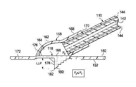
flange portion of the housing such that the attachment device couples the
housing to the
sealing member. Additionally, a cutting element is at least temporarily
coupled to the housing
proximate to the tissue-facing cavity opening such that the cutting element is
adapted to form
an aperture in the sealing member when the cutting element is driven into the
sealing member
with a driving force.
[0005] According to another illustrative embodiment a system for treating a
tissue site
on a patient with reduced pressure includes a distribution manifold for
placing proximate to
the tissue site, a sealing member for covering the distribution manifold and a
portion of intact
epideimis of the patient to form a sealed space, a reduced-pressure interface
for providing
reduced pressure through the sealing member to the distribution manifold, a
reduced-pressure
source, and a reduced-pressure delivery conduit for fluidly coupling the
reduced-pressure
source to the reduced-pressure interface. The reduced-pressure interface
includes a housing
having a flange portion and a cavity wall portion such that the cavity wall
portion forms a
cavity having a tissue-facing cavity opening, a conduit port coupled to the
cavity wall and
having a conduit aperture such that the conduit port is adapted to receive the
reduced-pressure
delivery conduit, an attachment device coupled to a tissue-facing side of the
flange portion of
the housing such that the attachment device couples the housing to the sealing
member, and a
cutting element at least temporarily coupled to the housing proximate to the
tissue-facing
cavity opening. The cutting element is adapted to form an aperture in the
sealing member
when the cutting element is driven into the sealing member with a driving
force.
[0006] According to another illustrative embodiment a method for treating a
tissue site
on a patient with reduced pressure includes disposing a distribution manifold
proximate to the
tissue site and covering the distribution manifold and a portion of intact
epidermis of the
patient with a sealing member to folin a sealed space in which the
distribution manifold is
disposed. The sealing member has a first side and a second, tissue-facing
side. The method
further includes providing a reduced-pressure source, coupling a reduced-
pressure interface
CA 02843317 2014-01-27
WO 2013/016240
PCT/US2012/047736
proximate to the first side of the sealing member, and fluidly coupling a
reduced-pressure
delivery conduit between the reduced pressure source and the reduced-pressure
interface. The
reduced-pressure interface includes a housing having a wall portion such that
the wall portion
forms a cavity having a tissue-facing cavity opening, a conduit port coupled
to the cavity wall
for receiving the reduced-pressure delivery conduit, an attachment device for
coupling the
reduced-pressure interface to the sealing member, and a cutting element at
least temporarily
coupled to the housing proximate to the tissue-facing cavity opening such that
the cutting
element is adapted to perforate the sealing member when the cutting element is
driven into the
sealing member with a driving force. The method also includes applying a
driving force to the
reduced-pressure interface of sufficient strength to cause the cutting element
to perforate the
sealing member.
[0007] According to yet another illustrative embodiment, an interface for
providing
reduced pressure through a drape to a manifold includes a housing having a
flange portion and
a cavity wall portion. The cavity wall portion forms a cavity and a cavity
wall aperture is
formed within the cavity wall portion for receiving a tube. The interface
further includes a
coupler positioned on a tissue-facing side of the flange portion of the
housing for attaching the
housing to the drape and a protrusion coupled to the housing proximate to the
flange portion.
The protrusion extends beyond the tissue-facing side of the flange portion of
the housing and
is configured to form an aperture in the drape when the protrusion is driven
into the drape with
the reduced pressure.
[0008] According to another illustrative embodiment, a system for treating a
wound
with reduced pressure includes a manifold for positioning adjacent the wound,
a drape for
covering the manifold and a portion of intact epideimis of the patient to form
a sealed space, a
reduced-pressure interface for providing reduced pressure through the drape to
the manifold, a
reduced-pressure source, and a conduit for fluidly coupling the reduced-
pressure source to the
reduced-pressure interface. The reduced-pressure interface includes a housing
having a flange
portion and a cavity wall portion. The cavity wall portion forms a cavity and
a cavity wall
aperture is formed within the cavity wall portion for receiving a tube. The
reduced-pressure
interface further includes a coupler positioned on a tissue-facing side of the
flange portion of
the housing for attaching the housing to the drape and a protrusion coupled to
the housing
proximate to the flange portion. The protrusion extends beyond the tissue-
facing side of the
flange portion of the housing and is configured to form an aperture in the
drape when the
protrusion is driven into the drape with the reduced pressure.
3
CA 02843317 2014-01-27
WO 2013/016240
PCT/US2012/047736
[0009] In another illustrative embodiment, a method for treating a wound on a
patient
with reduced pressure includes disposing a manifold proximate to the wound,
covering the
manifold and a portion of intact epidermis of the patient with a drape to form
a sealed space in
which the manifold is disposed. The drape has a first side and a second,
tissue-facing side.
The method further includes providing a reduced-pressure source, coupling a
reduced-pressure
interface proximate to the first side of the drape, and fluidly coupling a
tube between the
reduced-pressure source and the reduced-pressure interface. The reduced-
pressure interface
includes a housing having a flange portion and a cavity wall portion. The
cavity wall portion
forms a cavity and a cavity wall aperture is formed within the cavity wall
portion for receiving
a tube. The reduced-pressure interface further includes a coupler positioned
on a tissue-facing
side of the flange portion of the housing for attaching the housing to the
drape and a protrusion
coupled to the housing proximate to the flange portion. The protrusion extends
beyond the
tissue-facing side of the flange portion of the housing and is configured to
form an aperture in
the drape when the protrusion is driven into the drape with a driving force.
The method
further includes applying the driving force to the reduced-pressure interface
of sufficient
strength to cause the protrusion to perforate the drape.
[0010] Other features and advantages of the illustrative embodiments will
become
apparent with reference to the drawings and detailed description that follow.
4
CA 02843317 2014-01-27
WO 2013/016240
PCT/US2012/047736
BRIEF DESCRIPTION OF THE DRAWINGS
[0011] FIGURE 1 is a schematic perspective view of an illustrative embodiment
of a
system for treating a tissue site with reduced pressure;
[0012] FIGURE 2 is a schematic, cross-sectional view of an illustrative
embodiment of
a multi-lumen conduit of the system shown in FIGURE 1 taken along line 2-2;
[0013] FIGURE 3 is a schematic, cross-sectional view of one illustrative
embodiment
of a reduced-pressure interface having a cutting element for use as part of a
system for treating
a tissue site with reduced pressure;
[0014] FIGURE 4 is a schematic, bottom view of the reduced-pressure interface
of
.. FIGURE 3;
[0015] FIGIJRE 5A is a schematic, cross-sectional view of the reduced-pressure
interface of FIGURE 3 under reduced pressure prior to the cutting element
perforating a
sealing member;
[0016] FIGURE 5B is another schematic, cross-sectional view of the reduced-
pressure
.. interface of FIGURE 3 under reduced pressure after the cutting member has
perforated the
sealing member;
[0017] FIGURE 5C is another schematic, cross-sectional view of the reduced-
pressure
interface of FIGURE 3 under reduced pressure after the cutting member has
perforated the
sealing member and the cutting element has been removed;
[0018] FIGIJRE 6 is a schematic, top perspective view of another illustrative
embodiment of a reduced-pressure interface having a cutting element for use as
part of a
system for treating a tissue site with reduced pressure;
[0019] FIGURE 7 is a schematic, cross-sectional view of the reduced-pressure
interface of FIGURE 6;
[0020] FIGURE 8 is a schematic, bottom perspective view of a portion of the
reduced-
pressure interface of FIGURE 6;
[0021] FIGURE 9A is a schematic, cross-sectional view of the reduced-pressure
interface of FIGURES 6-8 being applied and prior to reduced pressure being
supplied;
[0022] FIGIJRE 9B is a schematic, cross-sectional view of the reduced-pressure
interface of FIGURES 6-8 under reduced pressure prior to the cutting element
perforating a
sealing member;
5
CA 02843317 2014-01-27
WO 2013/016240
PCT/US2012/047736
[0023] FIGURE 9C is a schematic, cross-sectional view of the reduced-pressure
interface of FIGURES 6-8 under reduced pressure after the cutting member has
perforated the
sealing member and the cutting element has been removed: and
[0024] FIGURE 10 is a schematic diagram of a representative pressure set-up
pattern.
6
CA 02843317 2014-01-27
WO 2013/016240
PCT/US2012/047736
DETAILED DESCRIPTION OF ILLUSTRATIVE EMBODIMENTS
[0025] In the following detailed description of the illustrative embodiments,
reference
is made to the accompanying drawings that form a part hereof. These
embodiments are
described in sufficient detail to enable those skilled in the art to practice
the invention, and it is
understood that other embodiments may be utilized and that logical structural,
mechanical,
electrical, and chemical changes may be made without departing from the spirit
or scope of the
invention. To avoid detail not necessary to enable those skilled in the art to
practice the
embodiments described herein, the description may omit certain information
known to those
skilled in the art. The following detailed description is, therefore, not to
be taken in a limiting
sense, and the scope of the illustrative embodiments are defined only by the
appended claims.
Unless otherwise indicated, as used herein, "or" does not require mutual
exclusivity.
[0026] The term "reduced pressure" as used herein generally refers to a
pressure less
than the ambient pressure at a tissue site that is being subjected to
treatment. In most cases,
this reduced pressure will be less than the atmospheric pressure at which the
patient is located.
Alternatively, the reduced pressure may be less than a hydrostatic pressure
associated with
tissue at the tissue site. Unless otherwise indicated, values of pressure
stated herein are gauge
pressures. References to increases in reduced pressure typically refer to a
decrease in absolute
pressure, and decreases in reduced pressure typically refer to an increase in
absolute pressure.
[0027] Referring now to the drawings and initially to FIGURES 1-5C, and
specifically
to FIGURES 1 and 3, a system 100 for treating a tissue site 102 on a patient
104 with reduced
pressure is presented. The system 100 includes a reduced-pressure dressing 106
for disposing
proximate the tissue site 102. The system 100 also includes a reduced-pressure
treatment unit
108 fluidly connected to the reduced-pressure dressing 106 through a reduced-
pressure
delivery conduit 110 for applying reduced pressure to the tissue site 102. The
reduced-
pressure dressing 106 may further include a distribution manifold 112, a
sealing member 114,
and a reduced-pressure interface 116. The reduced-pressure interface 116
includes a cutting
element 118 adapted to form an aperture 120 (see Fig. 5B) in the sealing
member 114.
Including the cutting element 118 on the reduced-pressure interface 116
provides a number of
potential benefits. The benefits may include ease of application and the
reduction of error
when foliating the aperture 120. In a non-limiting example, errors in (1)
positioning the
aperture 120 on the dressing, (2) sizing of the aperture 120, and (3) the
formation of the
aperture 120 may be reduced. Incorrectly forming the aperture 120 may leave
portions of the
7
CA 02843317 2014-01-27
WO 2013/016240
PCT/US2012/047736
sealing member 114 in a position that can block the aperture 120 when reduced
pressure is
applied.
[0028] The system 100 may be used with various different types of tissue sites
102.
The tissue site 102 may be a wound 122 or wound cavity. As shown in at least
FIGURES 5A-
5C, the tissue site 102 or wound 122, may be through an epidermis 124 and into
a
subcutaneous tissue or any other tissue. The tissue site 102 may be the bodily
tissue of any
human, animal, or other organism, including bone tissue, adipose tissue,
muscle tissue, dermal
tissue, vascular tissue, connective tissue, cartilage, tendons, ligaments,
body cavity or any
other tissue. Treatment of the tissue site 102 may include removal of fluids,
e.g., exudate or
ascites.
[0029] Referring still to FIGURES 1-5C, the distribution manifold 112 is
proximate
the tissue site 102 and has a first side 128 and a second, tissue-facing side
130. The term
"distribution manifold" as used herein generally refers to a substance or
structure that is
provided to assist in applying reduced pressure to, delivering fluids to, or
removing fluids
from the tissue site 102. The distribution manifold 112 typically includes a
plurality of flow
channels or pathways that distribute fluids provided to and removed from the
tissue site 102
around the distribution manifold 112. In one illustrative embodiment, the flow
channels or
pathways are interconnected to improve distribution of fluids provided or
removed from the
tissue site 102. The distribution manifold 112 may be a biocompatible material
that is capable
of being placed in contact with the tissue site 102 and distributing reduced
pressure to the
tissue site 102. Examples of the distribution manifold 112 may include,
without limitation,
devices that have structural elements arranged to form flow channels, such as,
for example,
cellular foam, open-cell foam, porous tissue collections, liquids, gels, and
foams that include,
or cure to include, flow channels. The distribution manifold 112 may be porous
and may be
made from foam, gauze, felted mat, or any other material suited to a
particular biological
application. In one embodiment, the distribution manifold 112 is a porous foam
and includes a
plurality of interconnected cells or pores that act as flow channels. The
porous foam may be a
polyurethane, open-cell, reticulated foam such as GranuFoam0 material
manufactured by
Kinetic Concepts, Incorporated of San Antonio, Texas. In sonic situations, the
distribution
manifold 112 may also be used to distribute fluids such as medications,
antibacterials, growth
factors, and various solutions to the tissue site 102. Other layers may be
included in or on the
distribution manifold 112, such as absorptive materials, wicking materials,
hydrophobic
materials, and hydrophilic materials.
8
CA 02843317 2014-01-27
WO 2013/016240
PCT/US2012/047736
[0030] In one illustrative the distribution manifold 112 may be constructed
from
bioresorbable materials that do not have to be removed from a patient's body
following use of
the system 100. Suitable bioresorbable materials may include, without
limitation, a polymeric
blend of polylactic acid (PLA) and polyglycolic acid (PGA). The polymeric
blend may also
include without limitation polycarbonates, polyfumarates, and capralactones.
The distribution
manifold 112 may further serve as a scaffold for new cell-growth, or a
scaffold material may
be used in conjunction with the distribution manifold 112 to promote cell-
growth. A scaffold
is a substance or structure used to enhance or promote the growth of cells or
fonnation of
tissue, such as a three-dimensional porous structure that provides a template
for cell growth.
Illustrative examples of scaffold materials include calcium phosphate,
collagen, PLAJPGA,
coral hyclroxy apatites, carbonates, or processed allograft materials.
[0031] The distribution manifold 112 may be covered by the sealing member 114,
which may also be referred to as a drape. The sealing member 114 forms a
sealed space 132
over the tissue site 102. The sealing member 114 has a first side 134, and a
second, tissue-
facing side 136. The sealing member 114 may be any material that provides a
fluid seal.
"Fluid seal," or "seal," means a seal adequate to maintain reduced pressure at
a desired site
given the particular reduced-pressure source or subsystem involved. The
sealing member 114
may, for example, be an impermeable or semi-peimeable, elastomeric material.
"Elastomeric"
means having the properties of an elastomer. Elastomer generally refers to a
polymeric
material that has rubber-like properties. More specifically, most elastomers
have ultimate
elongations greater than 100% and a significant amount of resilience. The
resilience of a
material refers to the material's ability to recover from an elastic
deformation. Elastomers that
are relatively less resilient may also be used as these elastomers are more
likely to tear when
faced with the cutting element 118. Examples of elastomers may include, but
are not limited
to, natural rubbers, polyisoprene, styrene butadiene rubber, chloroprene
rubber, polybutadiene,
nitrile rubber, butyl rubber, ethylene propylene rubber, ethylene propylene
diene monomer,
chlorosulfonated polyethylene, polysulfide rubber, polyurethane (PU), EVA
film, co-
polyester, and silicones. Additional, specific examples of dressing sealing
member materials
include a silicone drape, 3M Tegaderm0 drape, polyurethane (PU) drape such as
one available
from Avery Dennison Corporation of Pasadena, California. An additional,
specific non-
limiting example of a dressing sealing member material includes a 30 m matt
polyurethane
film such as the InspireTM 2317 manufactured by ExopackTM Advanced Coatings of
Matthews,
North Carolina.
9
CA 02843317 2014-01-27
WO 2013/016240
PCT/US2012/047736
[0032] An attachment device 138 may be used to hold the sealing member 114
against
a portion of the patient's intact epidermis 124 or another layer, such as a
gasket or additional
sealing member. The attachment device 138 may take numerous forms. For
example, the
attachment device 138 may be a medically acceptable adhesive, such as a
pressure-sensitive
adhesive, that extends about a periphery or all of the sealing member 114. The
attachment
device 138 may also be a sealing ring or other device. The attachment device
138 is disposed
on the second, tissue-facing side 136 of the sealing member 114. Before use,
the attachment
device 138 may be covered by a release liner (not shown).
[0033] The reduced-pressure interface 116 may be positioned adjacent to or
coupled to
the sealing member 114 to provide fluid access to the distribution manifold
112. Another
attachment device 152 similar to the attachment device 138 may be used to hold
the reduced-
pressure interface 116 against the sealing member 114. The reduced-pressure
delivery conduit
110 fluidly couples the reduced-pressure treatment unit 108 and the reduced-
pressure interface
116. The reduced-pressure interface 116 allows the reduced pressure to be
delivered to the
tissue site 102. While the amount and nature of reduced pressure applied to a
tissue site will
typically vary according to the application, the reduced pressure will
typically be between -5
mm Hg (-667 Pa) and -500 mm Hg (-66.7 kPa) and more typically between -75 mm
Hg (-9.9
kPa) and -300 mm Hg (-39.9 kPa). For example, and not by way of limitation,
the pressure
may be -12, -12.5, -13, -14, -14.5, -15, -15.5, -16, -16.5, -17, -17.5, -18, -
18.5, -19, -19.5, -20,
-20.5, -21, -21.5, -22, -22.5, -23, -23.5, -24, -24.5, -25, -25.5, -26, -26.5
kPa or another
pressure.
[0034] As shown, the reduced-pressure delivery conduit 110 is a multi-lumen
conduit.
It should be understood, however, that the reduced-pressure delivery conduit
110 may be in
many forms and may comprise a single lumen. The reduced-pressure delivery
conduit 110
may include a primary lumen 142 and at least one sensing lumen 144. In one
illustrative the
primary lumen 142 is a central lumen 146 and the at least one sensing lumen
144 is one or
more peripheral lumens 148. The primary lumen 142 and the at least one sensing
lumen 144
are adapted to maintain fluid isolation between the primary lumen 142 and the
at least one
sensing lumen 144 as the reduced-pressure delivery conduit 110 transports
fluids from the
reduced-pressure interface 116 to the reduced-pressure treatment unit 108.
Liquids or
exudates communicated from the distribution manifold 112 through the primary
lumen 142 are
removed from the reduced-pressure delivery conduit 110 and retained within a
liquid-
collection chamber (not explicitly shown) in fluid communication with the
reduced-pressure
CA 02843317 2014-01-27
WO 2013/016240
PCT/US2012/047736
treatment unit 108. The at least one sensing lumen 144 fluidly communicates
reduced
pressure representative of the tissue site 102 to an instrumentation unit 150.
[0035] The reduced-pressure treatment unit 108 may include a liquid-collection
chamber, or a collection canister, and the instrumentation unit 150 in fluid
communication
with a reduced-pressure source 140. The instrumentation unit 150 may include a
microprocessor 154 adapted to process pressure signals received by the reduced-
pressure
delivery conduit 110, monitor the pressure signals, and issue alerts according
to a pre-
determined pressure configuration. The pre-determined pressure configuration
may include a
pressure set-up pattern of sustained decrease, increase, and relative
stability within an
application time period as will be described in more detail with respect to
FIGURE 10 below.
[0036] In an illustrative embodiment, the reduced-pressure source 140 is an
electrically-driven vacuum pump. In another implementation, the reduced-
pressure source 140
may instead be a manually-actuated or manually-charged pump that does not
require electrical
power. The reduced-pressure source 140 instead may be any other type of
reduced pressure
pump, or alternatively a wall suction port such as those available in
hospitals and other
medical facilities. The reduced-pressure source 140 may be housed within or
used in
conjunction with the reduced-pressure treatment unit 108, which may also
include the
instrumentation unit 150. The instrumentation unit 150 may include sensors,
processing units,
alaim indicators, memory, databases, software, display units, and user
interfaces that further
facilitate the application of reduced pressure treatment to the tissue site
102.
[0037] In one example, pressure-detection sensors (not shown) located in the
instrumentation unit 150 may be disposed at or near the reduced-pressure
source 140. The
pressure-detection sensors may receive pressure data, or a pressure signal,
from the reduced-
pressure interface 116 via the at least one sensing lumen 144 that is
dedicated to delivering
reduced pressure data to the pressure-detection sensors. The pressure signal
or data may be
representative of a pressure at a distal end 156 of the at least one sensing
lumen 144. The
pressure-detection sensors may communicate with a processing unit that
monitors and controls
the reduced pressure that is delivered by the reduced-pressure source 140. In
one
embodiment, the pressure-detection sensors communicate with the processing
unit to monitor
whether the pressure signal follows the pressure set-up pattern. In the event
the pressure
signal does not follow the pressure set-up pattern within an application time
period that may
be predetermined, the instrumentation unit 150 provides an indication to a
caregiver. The
indication may be in the form of a visual or audible alert or alarm. Other
indications may be
used. In an alternative, but not mutually exclusive, embodiment, the pressure-
detection
11
CA 02843317 2014-01-27
WO 2013/016240
PCT/US2012/047736
sensors may communicate with the processing unit to monitor whether the
pressure signal
does follow the pressure set-up pattern within an application time period. In
the event the
pressure signal does follow the pressure set-up pattern, the instrumentation
unit 150 provides
an indication to the caregiver. The indication that the pressure set-up
pattern has been
followed may be different than the indication that the pressure set-up pattern
has not been
followed.
[0038] Referring now primarily to FIGURES 3-5C, an illustrative embodiment of
the
reduced-pressure interface 116 is presented in more detail. The reduced-
pressure interface 116
includes a housing 158, a conduit port 168 coupled to the housing 158, and the
attachment
device 152 for coupling the reduced-pressure interface 116 to the sealing
member 114. The
reduced-pressure interface 116 further includes the cutting element 118.
[0039] The housing 158 may have a flange portion 160 and a cavity wall portion
162.
The cavity wall portion 162 forms a cavity 164 having a tissue-facing cavity
opening 166.
The conduit port 168 is coupled to or formed as part of the cavity wall
portion 162 of the
housing 158. The conduit port 168 includes a conduit aperture 170 whereby the
conduit port
168 is adapted to receive the reduced-pressure delivery conduit 110. The
attachment device
152 may be coupled to a tissue-facing side 172 of the flange portion 160 for
coupling the
housing 158 to the first side 134 of the sealing member 114. The housing 158
is made of a
semi-rigid material that is capable of collapsing under a force such as a
driving force 174. In a
non-limiting example, the reduced-pressure interface 116, and thus the housing
158, may he
made from a plasticized polyvinyl chloride (PVC), polyurethane, cyclic olefin
copolymer
elastomer, thermoplastic elastomer, poly acrylic, silicone polymer, and
polyether block amide
copolymer.
[0040] The cutting element 118 may be at least temporarily coupled to the
housing 158
_______________________________________________________________ proximate to
the tissue-facing cavity opening 166. The cutting element 118 is adapted to
foi in
the aperture 120 in the sealing member 114 when the cutting element 118 is
driven into the
sealing member 114 with the driving force 174. The driving force 174 may also
cause the
cutting element 118 to penetrate or cut a portion of the distribution manifold
112. The driving
force 174 may be manually applied to an exterior 184 of the reduced-pressure
interface 116
causing the housing 158 to collapse and thereby driving or pushing the cutting
element 118
into the sealing member 114. In another embodiment, the driving force 174 is
applied by
applying reduced pressure to the cavity 164 such that a cavity pressure (Pe)
in the cavity 164 is
less than a threshold pressure (Pr). When the cavity pressure (Pe) is less
than the threshold
pressure (Pr), the cavity wall portion 162 collapses and with continued
reduced pressure
CA 02843317 2014-01-27
WO 2013/016240
PCT/US2012/047736
impacts the cutting element 118 thereby driving a portion of the cutting
element 118 through
the sealing member 114. The threshold pressure (Pr) is at least in part
dependent on the type
and thickness of the material used for the housing 158. In the event reduced
pressure is
applied to the cavity 164, a tensile force may be applied to the sealing
member 114 causing the
sealing member 114 to pull into the cavity 164. This movement further assists
with the cutting
element 118 moving into the sealing member 114.
[0041] In one embodiment, the cutting element 118 includes a base member 176
and a
stylus member 178 coupled to the base member 176. The stylus member 178 has a
leading
edge 180 and is configured to perforate the sealing member 114 to foun the
aperture 120 in the
sealing member 114. In one embodiment, the leading edge 180 is serrated. In
another
embodiment, the leading edge 180 is serrated or configured to perforate the
sealing member
114 orthogonally. The sealing member 114 may be perforated orthogonally to
inhibit the cut
sealing member 114 from blocking the reduced-pressure delivery conduit 110
during reduced
pressure therapy. The base member 176 may be sized and configured to form an
interference
fit with the tissue-facing cavity opening 166, whereby the cutting element 118
is releasably
coupled to the housing 158. Thus, in one embodiment, the cutting element 118
may be
removed prior to use if not desired or after perforating the sealing member
114.
[0042] The cutting element 118 may have a piercing length (Lp) extending the
length
(L) of the stylus member 178. The length (L) of the stylus member 178 extends
from the base
member 176 to a tip 182 of the stylus member 178. In one embodiment, the
piercing length
(Lp) is less than 3 centimeters. In another embodiment, the piercing length
(Lp) is less than 2
centimeters. The distribution manifold 112 may have a thickness greater than T
when subject
to reduced pressure such that the piercing length (Lp) of the cutting element
118 is less than
the thickness T, i.e., Lp < T. One benefit of the piercing length (Lp) being
less than the
thickness, T, of the distribution manifold 112 under reduced pressure is that
the cutting
element 118 cannot completely cut through the distribution manifold 112 and
reach the tissue
site 102.
[0043] As previously mentioned, the cutting element 118 may be only
temporarily
coupled to the housing 158. In one embodiment, the cutting element 118 may be
removed by
a care giver. In another embodiment, the cutting element 118 may be formed
from a liquid
soluble material such as a water soluble material adapted to allow the cutting
element 118 to
dissolve. For example, the water soluble material may include at least one of
the following:
Polyvinyl alcohol (PVOH), polyvinyl pyrrolidone, hydroxyl and carboxyl
modified cellulose,
hydroxyl and carboxyl modified acrylics, starch, sugars (sucrose, glucose,
fructose), weak
13
acids (tartaric, citric, malic), salts (sodium chloride, sodium carbonate,
sodium bicarbonate),
polyethylene oxide (PLO), polyethylene glycol (PEG). The cutting element 118
may dissolve
as liquids are removed from the tissue site 102. Reduced pressure is applied
to the reduced-
pressure interface 116 after perforating the sealing member 114 typically
causing liquids to be
removed from the tissue site 102. After a sufficient amount of time, liquids
removed from the
tissue site 102 cause the cutting element 118 to substantially dissolve. The
cutting element
118 may dissolve within 2 minutes, 5 minutes, 10 minutes, or another time
period. As the
cutting element 1(8 is dissolved it is removed by the reduced-pressure
delivery conduit 110
with liquids from the tissue site 102. A liquid, e.g., saline solution, may
also he introduced
through the reduced-pressure delivery conduit 110 or otherwise to dissolve the
cutting element
118.
[0044] As shown in FIGURE SC, once the cutting element 118 has substantially
dissolved, reduced pressure applied through the reduced-pressure interface 116
creates
sufficient reduced pressure in the cavity 164 to pull a portion of the
distribution manifold 112
into the cavity 164 such that the distribution manifold 112 abuts a distal end
186 of the
reduced-pressure delivery conduit 110 to include a distal aperture of the
at least one
sensing lumen 144. Allowing the distribution manifold 112 to completely abut
the distal end
186 of the reduced-pressure delivery conduit 110 may help ensure fluid
isolation between each
of the lumens in the reduced-pressure delivery conduit 110. The distribution
manifold 112
may provide a barrier between the primary lumen 142 and the at least one
sensing lumen 144.
Additionally, having the reduced-pressure delivery conduit 110 in direct
contact with the
distribution manifold 112 may help ensure that there is a constant low
velocity liquid flow into
the reduced-pressure delivery conduit 110 which may minimize the instance of
aerosolized
particles being deposited around the at least one sensing lumen 144 and may
also provide a
filter to liquids entering the at least one sensing lumen 144.
[0045] In operation, a caregiver may treat the tissue site 102 on the patient
104 with a
method that includes disposing the distribution manifold 112 proximate to the
tissue site 102.
The distribution manifold 112 and a portion of intact epidermis 124 of the
patient 104 is
covered with the sealing member 114 to form the sealed space 132 in which the
distribution
manifold 112 is disposed. The reduced-pressure interface 116 is coupled to the
sealing
member 114. The reduced-pressure delivery conduit 110 is fluidly coupled on
one end to the
reduced-pressure source 140 and on the opposing end to the reduced-pressure
interface 116.
The driving force 174 is then applied to the reduced-pressure interface 116
with sufficient
14
CA 2843317 2018-11-06
strength to cause the cutting element 118 to perforate (e.g., pierce, tear,
cut or otherwise create
the aperture 120) the sealing member 114.
[0046] In one embodiment, the reduced-pressure interface 116 includes the
housing
158 having the wall portion, wherein the wall portion forms the cavity 164
having the tissue-
facing cavity opening 166. The housing 158 is formed of a semi-rigid material
that collapses
when under reduced pressure less than the threshold pressure (Pr). The conduit
port 168 is
coupled to the wall portion of the housing 158. The conduit port 168 is
further coupled to the
reduced-pressure delivery conduit 110. Reduced pressure is supplied to the
reduced-pressure
interface 116 through the reduced-pressure delivery conduit 110 and We conduit
port 168.
When reduced pressure levels in the cavity 164 are less than the threshold
pressure (Pr), the
wall portion collapses under the driving force 174 and impacts the cuttinp.
element 118,
driving a portion of the cutting element 118 through the sealing member 114 to
perforate the
sealing member 114..
[0047] In one embodiment, in response to the sealing member 114 being
perforated,
liquid is removed from the tissue site 102 through the reduced-pressure
delivery conduit 110.
Liquid is removed from the tissue site 102 by virtue of reduced pressure. The
liquid causes
the cutting element 118 to dissolve. Once the cutting element 118 has
substantially dissolved,
reduced pressure within the cavity 164 of the reduced-pressure interface 116
causes a portion
of the distribution manifold 112 to be pulled into the cavity 164 and abut the
reduced-pressure
delivery conduit 110. Fluid may then be directly transferred from the
distribution manifold
112 to the reduced-pressure delivery conduit 110 without going through an
additional medium
or open space.
[0048] Referring now primarily to FIGURES 6-9C, another illustrative
embodiment of
a reduced-pressure interface 216 is presented. The reduced-pressure interface
216 is
analogous in many respects to the reduced-pressure interface 116 of FIGURES 3-
5C. The
reduced-pressure interface 216 includes a housing 258 and a cutting element
218. The
housing 258 may have a flange portion 260 and a cavity wall portion 262. The
flange portion
260 may be coupled to the sealing member 114 by the attachment device 152. The
cavity wall
portion 262 is collapsible under reduced pressure. The cavity wall portion 262
may include a
bellows configuration 290 for permitting the cavity wall portion 262 to
collapse when a cavity
164 pressure (Pt) inside a cavity 264 is less than a threshold pressure (131)
on an absolute
pressure side.
[0049] The cutting element 218 may include a conduit adapter 292, an adapter
flange
294, a tube extension 296, a base member 276, and a stylus member 278. The
conduit adapter
CA 2843317 2018-11-06
292 is configured to receive the reduced-pressure delivery conduit 110 to
provide fluid
communication between the reduced-pressure treatment unit 108 and the tissue
site 102. "Ilie
conduit adapter 292 includes a protrusion 293 for engaging the primary lumen
142 of the
reduced-pressure delivery conduit 110. The protrusion 293 may be sized and
configured to
extend into the primary lumen 142 and to form an interference fit. The
protrusion 293 may
help maintain fluid isolation between the primary lumen 142 and the at least
one sensing
lumen 144. The adapter flange 294 is positioned on an exterior 284 of the
cavity wall portion
262. The tube extension 296 is connected to the adapter flange 294 and is
sized and
configured to mate with a conduit aperture 298. The tube extension 296 is
further configured
to extend through the conduit aperture 298. In a specific, non-limiting
example, the conduit
adapter 292, the adapter flange 294, and the tube extension 296 may be formed
from materials
to include plasticized polyvinyl chloride (PVC), polyurethane, cyclic olefin
copolymer
elastomer, thermoplastic elastomer, poly acrylic, silicone polymer, and
polyether block amide
copolymer.
[0050] The base member 276 may be at least temporarily coupled to the tube
extension
296. The stylus member 278 is directly coupled to the base member 276 and may
include a
first blade 297 and a second blade 299 configured to make orthogonal cuts in
the sealing
member 114 when the housing 258 is compressed with a driving force 274 thereby
impacting
the cutting element 218. The stylus member 278 is thus driven into the sealing
member 114.
The driving force 274 may be manually applied to the exterior 284 of the
reduced-pressure
interface 216 causing the housing 258 to collapse and thereby driving or
pushing the cutting
element 218 into the sealing member 114. In another embodiment, the driving
force 274 is
applied by applying reduced pressure to the cavity 264 such that the cavity
pressure (Pa) in the
cavity 264 is less than a threshold pressure (PO. When the cavity pressure
(Pa) in the cavity
264 is less than the threshold pressure (Pt), the cavity wall portion 262
collapses and impacts
the cutting element 218. With continued reduced pressure, a portion of the
cutting element
218 is driven through the sealing member 114. The threshold pressure (P1) is
at least in part
dependent on the type and thickness of material used for the housing 258. In
the event
reduced pressure is applied to the cavity 264, a tensile force 273 may be
applied to the sealing
member 114 causing the sealing member 114 to he pulled into the cavity 264.
This movement
helps the cutting element 218 to be driven into the sealing member 114.
[0051] As previously mentioned, the base member 276 may be only temporarily
coupled to the housing 258. In one embodiment, the base member 276 and the
stylus member
278 may be formed from a liquid soluble material such as a water soluble
material adapted to
16
CA 2843317 2018-11-06
allow the cutting element 118 to dissolve. The water soluble material may
include at least one
of the following: Polyvinyl alcohol (PVOH), polyvinyl pyrrolidone, hydroxyl
and carboxyl
modified cellulose, hydroxyl and carboxyl modified acrylics, starch, sugars
(sucrose, glucose,
fructose), weak acids (tartaric, citric, malic), salts (sodium chloride,
sodium carbonate, sodium
bicarbonate), polyethylene oxide (PEO), polyethylene glycol (PEG). The base
member 276
and the stylus member 278 may dissolve as liquids are removed from the tissue
site 102.
Reduced pressure is applied to the reduced-pressure interface 216 typically
causing liquids to
be removed from the tissue site 102. After a sufficient amount of time,
liquids removed from
the tissue site 102 may cause the base member 276 and the stylus member 278 to
substantially
dissolve. The base member 276 and the stylus member 278 may dissolve within 2
minutes, 5
minutes, 8 minutes, 10 minutes, or another time period. As the base member 276
and the
stylus member 278 are dissolved, the base member 276 and the stylus member 278
are
removed by the reduced-pressure delivery conduit 110 with the liquids from the
tissue site
102. While the base member 276 and the stylus member 278 may be dissolvable,
it is worth
noting that the conduit adapter 292, the adapter flange 294, and the tube
extension 296 do not
dissolve.
[0052] Once the base member 276 and the stylus member 278 have substantially
dissolved,
reduced pressure applied through the reduced-pressure interface 216 may create
sufficient
reduced pressure in the cavity 264 to pull a portion of the distribution
manifold 112 into the
cavity 264 and the primary lumen 142 of the reduced-pressure delivery conduit
110. The
distribution manifold 112 abuts the distal end 186 of the reduced-pressure
delivery conduit
110 including the distal aperture of the at least one sensing lumen 144.
Allowing the
distribution manifold 112 to completely abut the distal end 186 of the reduced-
pressure
delivery conduit 110 may help ensure fluid isolation between each of the
lumens in the
reduced-pressure delivery conduit 110.
[00531 Referring now primarily to FIGURE 10, a schematic diagram of a pressure
set-
up pattern is presented. The pressure set-up pattern may be a pre-determined
pressure
configuration. Pressure-detection sensors may communicate with a processing
unit to monitor
whether pressure signals received from a reduced-pressure interface follow or
is consistent
with the pressure set-up pattern. The pressure set-up pattern may be
representative of whether
a cutting element of the reduced-pressure interface has pierced a sealing
member. The
pressure set-up pattern may represent four main events. First, a period of
sustained pressure
decrease (reduced pressure increase) may be indicative of a period of time
prior to the cutting
element piercing the sealing member. This segment is shown generally by
reference numeral
17
CA 2843317 2018-11-06
CA 02843317 2014-01-27
WO 2013/016240
PCT/US2012/047736
302. During this period of time, pressure is decreasing in a cavity of the
reduced-pressure
interface causing the cavity to collapse. Second, a threshold pressure (Pr) is
reached and
pressure increases (reduced pressure decreases) indicating that the cutting
element has pierced
the sealing member. 'Phis segment is generally shown by numeral 304. The
pressure should
increase as the pressure in the sealed space beneath the sealing member
stabilizes. And third,
a period of pressure stability is reached as shown generally as reference
numeral 306.
[0054] Although the present invention and its advantages have been disclosed
in the
context of certain illustrative, non-limiting embodiments, it should be
understood that various
changes, substitutions, peimutations, and alterations can be made without
departing from the
scope of the invention as defined by the appended claims. It will be
appreciated that any
feature that is described in connection to any one embodiment may also be
applicable to any
other embodiment.
[0055] It will be understood that the benefits and advantages described above
may
relate to one embodiment or may relate to several embodiments. It will further
be understood
that reference to 'an' item refers to one or more of those items.
[0056] The steps of the methods described herein may be carried out in any
suitable
order, or simultaneously where appropriate.
[0057] Where appropriate, aspects of any of the examples described above may
be
combined with aspects of any of the other examples described to form further
examples
having comparable or different properties and addressing the same or different
problems.
[0058] It will be understood that the above description of preferred
embodiments is
given by way of example only and that various modifications may be made by
those skilled in
the art. The above specification, examples and data provide a complete
description of the
structure and use of exemplary embodiments of the invention. Although various
embodiments
of the invention have been described above with a certain degree of
particularity, or with
reference to one or more individual embodiments, those skilled in the art
could make
numerous alterations to the disclosed embodiments without departing from the
scope of the
claims.
18