Note: Descriptions are shown in the official language in which they were submitted.
CA 02843959 2014-02-26
VARIABLE POROSITY INTRAVASCULAR IMPLANT AND MANUFACTURING
METHOD
FIELD
[0001] The present disclosure relates generally to intravascular implants and
more particularly to
occlusive devices such as vascular stents.
BACKGROUND
[0002] Vascular disorders and defects such as aneurysms and other
arteriovenous malformations
often occur near the junction of large arteries, for instance at the base of
the brain in the Circle of
Willis. As aneurysms develop they typically form as a saccular aneurysm
protruding from a wall
of a vessel and have a neck and a dome portion. Alternatively, aneurysms can
form as fusiform
malformations that balloon a cross-section of the affected vessel.
[0003] As an aneurysm develops, the arterial internal elastic lamina
disappears at the base of the
neck portion, the media thins, and connective tissue replaces smooth-muscle
cells. As the
aneurysm is continually subjected to vascular blood pressure and blood flow,
the aneurysm will
grow outwardly from the wall of the vessel, which can cause pressure on the
surrounding tissue
as the sac or fusiform contacts the surrounding tissue. When the malformation
occurs in the
brain, this pressure can lead to serious mass effects, such as cognitive
impairment, loss of vision,
and nerve palsies. Additionally, as the aneurysm is subject to vascular blood
pressure and blood
flow, the walls of the aneurysm weaken, usually in the dome portion, which can
eventually cause
the aneurysm to tear or rupture. Ruptured aneurysms are the most common cause
of
subarachnoid hemorrhages, which have a mortality rate of approximately 50%.
[0004] Aneurysms and other malformations are especially difficult to treat
when located near
critical tissue or where ready access to the malformation is not available.
Both difficulty factors
apply especially to cranial aneurysms. Surgical methods have developed to
treat cranial
aneurysms and generally include eliminating blood flow to the aneurysm by
placing a clip
around the neck of a saccular aneurysm or by blocking off a fusiform aneurysm
by cliping both
ends of the fusiform and detouring blood flow around the secluded fusiform
through an
implanted vessel graft. Due to the sensitive brain tissue surrounding cranial
blood vessels and
1
I I
,
CA 02843959 2014-02-26
the restricted access, it is challenging and risky to surgically treat defects
of the cranial
vasculature.
[0005] Alternatives to such surgical procedures include endovascular delivery
of an implantable
device, such as a stent-like device or embolic coil, through a microcatheter
delivery device. In
one such procedure to treat a saccular-form cranial aneurysm, the distal end
of an embolic coil
delivery catheter is initially inserted into non-cranial vasculature of a
patient, typically a femoral
artery in the groin, and guided to the aneurysm. The aneurysm sac is then
filled with embolic
material, such as platinum coils, that forms a solid, thrombotic mass that
protects the vessel walls
from blood pressure and flow. This treatment method is advantageous in that it
only occludes
blood flow to the aneurysm leaving the surrounding portions of the vessel
unobstructed.
However, it cannot treat fusiform aneurysms, and the aneurysm volume is
permanently
maintained.
[0006] Another technique involving the use of an intravascular implant
delivers, by a
microcatheter, an occlusive device in the form of a tubular, stent structure.
Stents can be
braided, woven, or wound from various filaments, such as a wire or wires,
laser-cut from metal,
or made in various other ways. They can either be self-expanding or can be
expanded by another
device such as a balloon. What most have in common is radial symmetry, i.e., a
uniform
porosity, meaning that they do not cover one portion, side, or radial sector
of the vessel more or
less porously than other sectors. Their symmetric construction, and therefore
coverage of vessel
walls, is relatively homogeneous around any given transverse slice or cross-
section.
[0007] This homogenous structure can be disadvantageous in that such stents
not only occlude or
block blood flow to the aneurysm, but they also block blood pressure and flow
along the entire
length of the stent, which often impedes flow into surrounding joined vessels,
such as perforator-
type vessels branching off of the parent vessel. The use of a non-
discriminatory occlusive device
in this type of vessel can cause unintended harm to the patient if the
openings, or ostia, of the
perforator vessels are blocked.
[0008] Some have developed selectively-occlusive devices that discriminately
block flow to an
aneurysm while simultaneously allowing flow to surrounding vessels. These
attempts to create
discriminate occlusion devices have used multilayered devices, varied the
amount of filaments
2
CA 02843959 2014-02-26
along the length of the intravascular implant, or changed the picks per inch
along the length of
the intravascular implant. But, generally, these devices face difficulties in
manufacturing and
increased costs due to difficulties in creating the multiple layers or
variations in the number of
filaments to create the varied porosity regions.
[0009] Accordingly, there remains a need for a device that effectively
occludes a neck or
fusiform of an aneurysm or other arteriovenous malformation in a parent vessel
without blocking
flow into perforator vessels communicating with the parent vessel that is
structurally sound and
easily manufactured.
SUMMARY
[0010] A vascular occlusion device for effectively occluding blood flow and
pressure to a
vascular defect while simultaneously not occluding blood flow and pressure to
adjacent
vasculature is provided. The vascular occlusion device can include a tubular
member that has
variable porosity regions along its length. The tubular member can be formed
of a plurality of
filaments that have different cross-sectional shapes along their length that
are indexed to the
variable porosity regions along the length of the tubular member.
[0011] In some embodiments, the vascular occlusion device includes a tubular
member formed
from a plurality of braided filaments. The braided filaments can define an
outer surface having a
mesh pattern with mesh openings defined by the braided filaments. The tubular
member can
have a first porosity region along a first length portion of the tubular
member and a second
porosity region along a second length portion of the tubular member. The
porosity of the first
porosity region can be less than the porosity of the second porosity region.
The first porosity
region can include filaments having a different shape than the filaments in
the second porosity
region and the tubular member can have a constant pick count throughout its
length. In another
embodiment the tubular member can have a braid angle that is substantially
similar throughout
the tubular member.
[0012] In some embodiments, the tubular member is an intravascular stent,
which can be radially
compressible. The first length portion is at an intermediate portion of the
tubular member
proximal to a distal end of the tubular member and distal to a proximal end of
the tubular
3
CA 02843959 2014-02-26
member. The second length portion can be adjacent to the distal end of the
tubular member
and/or the proximal end of the tubular member. The first length portion of the
tubular member
can extend over a distance in the range of about 5 mm to about 25 mm. The
first porosity region
can include filaments having a flattened cross-sectional shape having a
length, a width, and a
thickness. The width can be greater than the thickness and less than the
length of the filaments in
the first porosity region having a flattened cross-sectional shape. The width
of the filaments
having a flattened cross-sectional shape is in the range of about 0.001 inches
to about 0.05
inches. The thickness of the filaments having a flattened cross-sectional
shape is in the range of
about 0.0003 inches to about 0.010 inches. The filaments having a round cross-
sectional shape
can have a diameter in the range of about 0.0005 inches to about 0.0100
inches.
[0013] The filaments in the first porosity region can be exclusively of a
flattened cross-sectional
shape, or can be a mixture of filaments with a flattened cross-sectional shape
and/or round cross-
sectional shape. The filaments in the second porosity region can have a round
cross-sectional
shape. The mesh openings formed from the braided filaments can have a
polygonal shape and
the mesh openings of the first porosity region can be smaller than the mesh
openings of the
second porosity region. The mesh openings of the first porosity region can
have an inscribed
circle diameter in the range of about 10 gm to about 500 gm and the mesh
openings of the
second porosity region have an inscribed circle diameter in the range of about
400 gm to about
1000 gm. The number of filaments forming the tubular member can be in the
range of about 8 to
about 288. For example, the number of filaments forming the tubular stent can
be selected from
the group consisting of 8, 16, 32, 48, 64, 72, 96, 120, 144, 192, and 288.
[0014] In another aspect, a method of manufacturing a tubular intravascular
implant includes
providing a plurality of supply spools, each having a supply of a filament
having a round cross-
sectional shape. The method further includes advancing the filaments on each
supply spool to a
corresponding collection spool and deforming a selected number of the
filaments in a selected
region thereof at selected intervals between the supply spools and the
collection spools. The
filaments can be deformed such that at least some of the collection spools
have filaments with a
round cross-sectional shape and a flattened cross-sectional shape. According
to the method, the
filaments in the collection spools are utilized in a filament braiding device
to form a tubular
member with an outer surface defined by the braided filaments. All of the
collection spools used
4
CA 02843959 2014-02-26
in the braiding device can have filaments with a flattened cross-sectional
shape, or alternatively
only a portion of the collection spools used in the braiding device can have
filaments with a
flattened cross-sectional shape.
[0015] The tubular member formed by the method can have a length with regions
of a first,
lower porosity and regions of a second, higher porosity. The method can also
include the step of
cutting the tubular member to form a plurality of intravascular stents; each
sent having a first
length region of a first, lower porosity characterized by the presence of
filaments having a
flattened cross-sectional shape. The intravascular stents can each have at
least one second length
region of a second, higher porosity characterized by the presence of filaments
having a rounded
cross-sectional shape.
BRIEF DESCRIPTION OF DRAWINGS
[0016] This invention will be more fully understood from the following
detailed description
taken in conjunction with the accompanying drawings, in which:
[0017] FIG. 1 is a cross-sectional view of an exemplary vascular occlusive
device implanted
within a vessel having a saccular aneurysm;
[0018] FIG. 2 is a cross-sectional view of an exemplary vascular occlusive
device implanted
within a vessel having a fusiform aneurysm;
[0019] FIG. 3 is a partial cross-sectional view of an exemplary vascular
occlusive device;
[0020] FIG. 4 is a magnified view of a portion of the device of FIG. 3;
[0021] FIG. 5 is a partial cross-sectional view of another embodiment of an
exemplary vascular
occlusive device;
[0022] FIG. 6 is a top view of an exemplary filament for use in forming a
vascular occlusive
device;
[0023] FIG. 7 is a cross-section view of the exemplary filament of FIG. 6 at
Section A-A;
[0024] FIG. 8 is a schematic view of an exemplary system for forming exemplary
filaments;
CA 02843959 2014-02-26
[0025] FIG. 9 is a schematic view of an exemplary braiding system.
DETAILED DESCRIPTION
[0026] Certain exemplary embodiments will now be described to provide an
overall
understanding of the principles of the structure, function, manufacture, and
use of the devices
and methods disclosed herein. One or more examples of these embodiments are
illustrated in the
accompanying drawings. Those skilled in the art will understand that the
devices and methods
specifically described herein and illustrated in the accompanying drawings are
non-limiting
exemplary embodiments and that the scope of the present invention is defined
solely by the
claims. The features illustrated or described in connection with one exemplary
embodiment may
be combined with the features of other embodiments. Such modifications and
variations are
intended to be included within the scope of the present invention
[0027] Further, in the present disclosure, like-numbered components of the
embodiments
generally have similar features, and thus within a particular embodiment each
feature of each
like-numbered component is not necessarily fully elaborated upon.
Additionally, to the extent
that linear or circular dimensions are used in the description of the
disclosed systems, devices,
and methods, such dimensions are not intended to limit the types of shapes
that can be used in
conjunction with such systems, devices, and methods. A person skilled in the
art will recognize
that an equivalent to such linear and circular dimensions can easily be
determined for any
geometric shape. Sizes and shapes of the systems and devices, and the
components thereof, can
depend at least on the anatomy of the subject in which the systems and devices
will be used, the
size and shape of components with which the systems and devices will be used,
and the methods
and procedures in which the systems and devices will be used.
[0028] To treat vascular disorders and defects, such as aneurysms and other
arteriovenous
malformations, intravascular implants, such as stents, can be implanted to
span a length of vessel
containing the defect to occlude blood pressure and flow to the defect. For
instance, a stent can
be delivered to the site of an aneurysm and positioned in such a manner as to
occlude blood
pressure and flow to the aneurysm walls. By occluding, i.e., blocking or
obstructing, blood flow
to the aneurysm, the risk of the aneurysm rupturing is reduced. But, in
treating the vascular
6
CA 02843959 2014-02-26
defect, it is important to avoid unnecessary occlusion of blood flow and
pressure to adjacent
vascular tissue, such as perforator vessels.
[0029] The present disclosure relates to a vascular occlusion device, such as
a variable porosity
stent, that is configured to occlude flow to a vascular defect while allowing
flow to adjacent
vessel tissue. The device utilizes a tubular member formed from a plurality of
braided filaments.
As explained below, the tubular member can include an outer surface having a
mesh pattern with
mesh openings defined by the braided filaments. The tubular member is
constructed such that
the porosity varies at different regions along the length of the member. For
example, the tubular
member can have a first porosity region along a first length portion of the
tubular member and a
second porosity region along a second length portion of the tubular member. In
some
embodiments, the first porosity region is a center portion of the tubular
member. The first
porosity region can include filaments having a different shape than the
filaments in the second
porosity region. By changing the shape of the filaments at selected regions
along the length of
the tubular member, the porosity of a given region can be altered while
maintaining a constant
pick count throughout the length of the stent. For example, the cross-
sectional shape of the
filament in the first porosity region can be selected to be different than the
cross-sectional shape
of the filament in the second porosity region so as to have a lower porosity
in the first porosity
region than the second porosity region. In this manner it is possible to vary
the porosity from the
first region to the second region by changing only the shape of the filaments,
holding the other
structural characteristics of the tubular member substantially constant along
the length of the
tubular member. That is, the number of filaments, pick count, braid angle, or
braid pattern is the
same in the first porosity region as in the second porosity region.
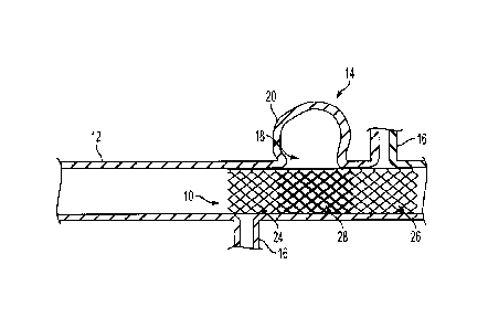
[0030] FIGS. 1 and 2 illustrate embodiments wherein a variable porosity stent
10 is placed
within a vessel 12 so as to occlude or obstruct blood flow and pressure to a
vascular defect 14
while simultaneously allowing substantially unimpeded blood flow and pressure
to adjacent
vessel tissue, such as perforator vessels 16. The vessel 12 can be any
vasculature, for example a
cranial blood vessel such as those found in the Circle of Willis. As shown in
FIG. 1, the vascular
defect 14 can be a saccular form aneurysm having a neck 18 and a dome portion
20. As shown
in FIG. 2, the vascular defect 14 can be a fusiform aneurysm wherein a cross-
sectional portion 22
of the vessel 12 is ballooned in a radial direction. In treating either the
saccular aneurysm of
7
CA 02843959 2014-02-26
FIG. 1 or the fusiform aneurysm of FIG. 2, the vascular occlusion device is
placed along the
length of the defective vessel 12 to occlude blood flow and pressure to the
aneurysm walls 20,
22.
[0031] FIG. 3 illustrates one embodiment of a tubular stent 10 used in
treating the vascular
defects 14 of FIGS. 1 and 2 according to the present invention. The stent 10
can have a proximal
region 24, distal region 26, and center region 28, wherein the center region
28 is intermediate the
proximal and distal regions 24, 26. In the embodiment shown in FIG. 3, region
28 of stent 10
represents a first porosity region having a porosity that is different (i.e.,
lower) than that of
regions 24 and 26, which represent a second porosity region. The difference in
porosity is
achieved by changing the cross-sectional shape of the filaments 30 in region
28, as explained
below. The stent 10 can be a braided stent having one or more filaments 30 of
stent material
woven, braided, or otherwise formed into a desired tubular shape and pattern.
[0032] FIG. 4 illustrates the braided, mesh structure of the stent 10. As
mentioned above, the
stent can be formed of braided filaments 30 that cross at junctions referred
to as picks 32 to form
a mesh. The mesh density is a function of the degree of spacing between the
filaments 30 in the
braid. Structures with more closely spaced filaments have a higher mesh
density than structures
with filaments that are less closely spaced. One measure of mesh density can
be determined
based on the number of picks 32 per inch of the material. A pick, as
understood by a person
skilled in the art, is a point where filaments intersect.
[0033] Porosity is a measure of the tendency of a material or structure to
allow passage of a fluid
therethrough. A material or structure with higher porosity has a higher fluid
flow across the
material than another material with lower porosity. The porosity of a braided
structure, such as a
stent, can be a function of the mesh density as well as the surface area of
the filaments that form
the structure as well as the number of filaments, the number of picks per
inch, and the interstitial
surface area between filaments as discussed below.
[0034] As mentioned previously, according to the present invention the cross-
sectional shape of
the filaments 30 can be selectively altered in certain regions, before
braiding, to produce a stent
having a region of lower porosity. By altering the cross-sectional shape of
the filaments 30,
the interstitial surface area between filaments 30 can be controlled.
8
CA 02843959 2014-02-26
=
[0035] The interstitial surface area between filaments can be determined by
measuring an
inscribed circle diameter 36 (FIG. 4) in the open spaces between the filaments
30. For a non-
circular shape, such as a triangle, square, or diamond, the inscribed circle
diameter 36 is the
diameter of the largest circle that fits entirely within the shape, i.e., the
diameter of a circle that is
tangent to the sides of the shape. The lower porosity regions of the stent 10
can have an
inscribed circle diameter 36 in the range of about 1 gm to about 400 gm, and
more particularly
about 100 gm. For example, the inscribed circle diameter 36 of the first
porosity region 28 of
the stent 10 shown in FIGS. 1-4 can be about 100 gm. The higher porosity
regions, i.e., second
porosity regions 24, 26, of the stent 10 can have an inscribed circle diameter
36 that is greater
than about 400 gm. For example, second porosity regions 26, 24 of the stent 10
shown in FIGS.
1-4 can be in the range of about 400 p.m to about 1000 pm.
[0036] To decrease the inscribed circle diameter 36 and thus decrease
porosity, the cross-
sectional shape of the filament 30 can be changed to increase the surface area
of the filament 30
along selected portions of the filament 30 length that will correspond to the
lowered porosity
region(s) along the length of the stent 10. For example, a substantially round
filament 30 can be
flattened along a portion of the filament 30 that corresponds to the first
porosity region 28 (e.g.,
the center region) of the stent 10. As shown in FIGS. 1-4 and 6, the first
porosity region 28 is
formed of filaments 30 that have a substantially flattened cross-sectional
shape, sometimes
referred to as a ribbon shape. Further, higher porosity regions of the
filaments used in forming
the stent (i.e., regions 24 and 26) can have a substantially round cross-
sectional shape, which for
example is the unaltered or natural shape of the filament. It is understood
that any initial or
unaltered cross-sectional shape can be utilized, so long as the shape allows
for alteration of the
filament cross-sectional shape such that the inscribed circle diameter 36 in
regions of a stent
formed with shape-altered filaments can be smaller than the inscribed circle
diameter 36 in the
regions formed of filaments that are not shape-altered. By way of example
substantially
rectangular, triangular, and round cross-sectional shapes can be used.
[0037] In some embodiments, the number of filaments 30 braided to form the
stent 10 is uniform
along the entire length of the stent 10. Additionally, the filaments 30
forming the stent 10 are
continuous along the entire length of the stent 10, i.e., the filaments 30
found in the first porosity
region 28 of the stent 10 are the same filaments 30 found in the second
porosity 24, 26. As
9
CA 02843959 2014-02-26
explained above, the only difference between the filaments in the first
porosity region 28 and the
second porosity regions 24, 26 is the cross-sectional shape of the filament
30.
[0038] In the embodiments of FIGS. 1-4 the first or lower porosity region 28
is formed using
filaments that are exclusively of an altered, i.e., substantially flattened
cross-sectional shape.
One skilled in the art will appreciate that the first of reduced porosity
region can alternatively be
formed using some filaments having an altered (such as flattened) cross-
sectional shape together
with other filaments having an unaltered shape, such as a rounded shape. FIG.
5 illustrates an
example of such a stent where only some of the filaments used in forming the
first or lower
porosity region 28' of the stent 10' have an altered (e.g., flattened) cross-
sectional shape. As is
shown, the stent 10' has a first filament type 38 that has an unaltered and
substantially constant
cross-sectional shape along its length and a second filament type 40 that has
at least two cross-
sectional shapes along its length, an altered cross-sectional shape and an
unaltered cross-
sectional shape. The proportion of filaments altered to unaltered filaments in
the first porosity
region 28' can vary depending upon porosity characteristics desired for the
stent. Generally,
region 28' of stent 10' will have at least as many and typically more
filaments with an altered
cross-sectional shape in region 28'. For example, the filaments with an
altered shape typically
comprise about 50 percent to about 99 percent of the fibers in region 28'.
More typically about
60 percent, about 70 percent, about 80 percent, or about 90 percent of the
fibers in region 28' are
those having an altered cross-sectional shape. Despite the stent 10' having
filaments of different
cross-sectional shapes within first porosity region 28', as in other
embodiments, the number of
filaments 38, 40 is uniform along the entire length of the stent 10' and the
filaments 38, 40
themselves are continuous along the entire length of the stent 10', i.e., the
filaments found in the
center portion 28' of the stent are the same filaments 38, 40 found in the end
portions 24', 26'.
[0039] FIG. 6 illustrates an exemplary filament 30 used to form the braided
stent 10. The
filament has a first portion 42 and a second portion 44 having a rounded cross-
sectional shape,
which is the unaltered filament shape. Another region of filament 30, shown as
middle portion
46 in FIG. 6, has an altered cross-sectional shape, i.e., a flattened or
somewhat oval cross-
sectional shape. The flattened portion 46 has a width 48 across the center of
the flattened portion
46 that is wider than the diameter 52 of the adjacent round cross-section
portions 42, 44. When a
stent is formed using filament 30, the region braided with portion 46 will
have a smaller
CA 02843959 2014-02-26
inscribed diameter than regions braided with portions 42 and 44. By way of
example, the width
48 can be in the range of about 0.001 inches to about 0.05 inches. FIG. 7
illustrates a cross-
section of the filament 30 as viewed along line A-A of FIG. 6. As is shown,
the flattened portion
46 will have a thickness 50 that is less than the diameter 52 of the round
cross-section. The
thickness 50 can be any desired thickness, for example the thickness 50 can be
in the range of
about 0.0003 inches to about 0.010 inches. The diameter 52 of the round cross-
sectional portion
of the filament can have any desired diameter, for example the diameter 52 can
be in the range of
about 0.0005 inches to about 0.0100 inches. The flattened middle portion 46
can have feathered
ends 54 yielding a somewhat an oval shape when viewed from the top as is shown
in FIG. 6.
When braided, the flattened middle portion 46 of the filament 30 can be
indexed about the region
of the stent 10 that is to form the first or lower porosity region. For
example, in the stent 10
shown in FIG. 3, the flattened portion 46 of the filaments 30 form the center
region of the stent,
which is the lower porosity region 28. The flattened middle portion 46 can
have a length that
will yield a center, lower porosity region of the stent that is large enough
to cover the defect 14
to be treated but not so large as to occlude blood flow unnecessarily to
adjacent vascular tissue.
[0040] One skilled in the art can readily determine the dimensions of a stent
as deemed
appropriate for a given application. The stent 10 can have a length that is so
dimensioned as to
stretch across a vascular defect 14. For example, the stent 10 length can be
in the range of about
mm to about 100 mm.
[0041] The stent 10 can be self-expanding and radially compressible such that
the stent 10 has a
first, constrained diameter that is smaller than a second, unconstrained
diameter that the stent
assumes in its natural state. The unconstrained diameter should be so
dimensioned as to be
sufficiently larger than the vessel within which it is to be implanted to be
safe and to maintain
proper position. Generally, vessel 12 diameters will range from about 2 mm to
about 5 mm and
thus the stent 10 unconstrained outer diameter can be in the range of about
2.5 mm to about 5.5
mm, but the stent can have any desired diameter. The constrained diameter can
be dimensioned
for endovascular delivery, for example the constrained diameter can be in the
range of about 0.01
inches to about 0.100 inches. Additionally, the stent 10 can be configured to
provide structural
support to the vessel 12 once placed in the vasculature in its expanded form.
To aid in placement
and blood flow, the ends 24, 26 of the stent 10 can be flared.
11
CA 02843959 2014-02-26
[0042] Self-expanding stents can be constructed from a variety of filament
materials known to
those skilled in the art. These materials include stainless steel, cobalt-
chromium alloys, nickel,
titanium, nitinol, and polymeric materials. Polymeric materials known to those
skilled in the art
can include, without limitation, shape memory polymers, silicone,
polyethylenes, polyurethanes,
polyethylene terephthalate (PET) polyesters, polyorthoesters, polyolefins,
polyvinyls,
polymethylacetates, polyamides, napthalane dicarboxylene derivatives, silks,
polytetraflyouroethylenes, and polyanhydrides. The filament material can also
be bioabsorbable
or radio-opaque, for instance by having an inner core formed of gold,
platinum, iridium, or any
other known radio-opaque material.
[0043] To effectively treat a defect, such as the aneurysms 14 shown in FIGS.
1 and 2, the stent
can have a variable porosity along the length of the tubular stent 10. For
example, first
porosity region 28 of the stent can be of a lower porosity than other regions
of the stent, such as
second porosity regions 24, 26. Although region 28 is shown to be disposed
between regions 24
and 26, other arrangements of lower and higher porosity regions are possible.
Additionally, the
stent 10 can have multiple regions of lower porosity. For example, the stent
10 can have a distal
region, proximal region, first center region, second center region, and third
center region,
wherein each region has a different porosity than the others (not shown). In
any event, the lower
porosity region can have a length that is sufficient to occlude flow to the
defect, for example the
length of the lower or first porosity region 28 can be in the range of about 5
mm to about 25 mm.
In the embodiments illustrated in FIGS. 1-3, the center region 28 is
configured to have a lower
porosity and thus occlude blood flow to the neck 18 or walls 20, 22 of the
aneurysm 14 and the
proximal and distal regions 24, 26 are configured to allow blood flow and
pressure without any
substantial occlusion thereof to the adjacent perforator vessels 16.
[0044] The stent 10 can have a substantially constant number of picks-per-inch
count along the
length of the stent 10. For example, the picks-per-inch count in the region 24
can be the same as
the picks-per-inch count in the region 26, which is the same as the picks-per-
inch count in the
region 28. For example, the picks-per-inch can be in the range of about 20
picks-per-inch to
about 250 picks-per-inch.
12
CA 02843959 2014-02-26
[0045] As mentioned above, when braided, the filaments 30 forming the stent 10
can intersect to
create polygonal mesh openings. The size of the polygonal mesh opening can
then be measured
by the inscribed circle diameter as described herein. The stent 10 can be
formed so as to yield a
first region having a first inscribed circle diameter (i.e., a first or lower
porosity region) and a
second region having a second inscribed circle diameter that is larger than
the first inscribed
circle diameter (i.e., a higher porosity region). Generally, the mesh openings
of the first porosity
region can have an inscribed circle diameter in the range of about 10 gm to
about 500 gm and
the mesh openings of the second porosity region can have an inscribed circle
diameter in the
range of about 400 1AM to about 1000 gm.
[0046] FIG. 8 illustrates an exemplary manufacturing system 56 to produce a
filament 30 having
alternating round and flat cross-sectional shapes. A supply spool 58 is first
provided. The
supply spool 58 should be wound with a supply filament 60 having a round cross-
sectional
shape. This can be formed of a typical stent filament material as described
above and as is
known in the art. The supply filament 60 from the supply spool 58 is then fed
to a collection
spool 62 configured to receive processed filament 30. Intermediate the supply
spool 58 and
collection spool 60, the supply filament 60 is fed through a press or stamping
device 64, such as
a pneumatic press. The press 64 can have a die set 66 that provides the means
for altering (e.g.,
flattening) the filament 60. The die set 64 can be adjusted to control the
thickness and length of
the flattened section of filament 46 created by stamping the round supply
filament 60 as it moves
through the press 64. The press 64 can be configured to press any diameter of
filament 60 and
the die length, die pressure, die shims that control the thickness, and spool
speed can be
independently controlled and calibrated to produce the desired dimensions of
the processed
filament 30. Using this press 64, the supply filament 60 is pressed at set
intervals to produce a
filament 30 having alternating round 42, 44 and flat 46 cross-sectional
shapes. The processed
filament 30 is stored on the collection spool 62 once the filament is
processed and ready to be
braided.
[0047] Braiding of filaments 30 includes the interlacing of at least two
sections of filament 30
such that the paths of the filament 30 sections are substantially diagonal to
the stent 10 delivery
direction, forming a tubular structure. Generally, braided stents can have a
polygonal interstitial
surface shape and can include a diamond braid having a 1/1 intersection
repeat, a regular
13
CA 02843959 2014-02-26
polygonal braid having a 2/2 intersection repeat, and a Hercules braid having
a 3/3 intersection
repeat. Moreover, a triaxial braid may also be used. A triaxial braid has at
least one filament
section that typically runs in the longitudinal direction or axial direction
of the stent to limit
filament movement. Moreover, an interlocking three-dimensional braided
structure or a multi-
layered braided structure can also be used. A multi-layered braided structure
is defined as a
structure formed by braiding wherein the structure has a plurality of distinct
and discrete layers.
[0048] FIG. 9 illustrates an exemplary braiding device 68. The braiding device
68 can have a
spool loading mechanism 70 and a braiding mandrel 72 is first loaded with the
desired filaments
wound on spools 74 disposed in the spool loading mechanism 70. For example,
the collection
spools 62 of processed filaments 30 can be loaded into the braiding machine
68. The collection
spools 62 used in the braiding machine 68 can have filaments 30 with flattened
cross-sectional
shapes as described above, filaments 60 with round cross-sectional shapes, or
combinations of
both. If only collection spools 62 having flattened cross-sectional shapes are
utilized, the
resulting stent 10 can be of the form shown in FIGS. 1-4. If a combination of
collection spools
62 having flattened cross-sectional shapes and spools having a round cross-
sectional shape are
used, then the resulting stent 10' can be of the form shown in FIG. 5. The
collection spools 62
should be indexed in the braid machine 68 so that any flattened portions 46 of
the filaments on
the collection spools corresponds to a desired region of lower porosity in the
resulting stent 28.
For example, the collection spools 62 can be indexed so that the flattened
portion is indexed to
an indexing line 76 such that the flat portion 46 of the filament 30
corresponds to the center
region 28 of the stent intermediate the end portions 24, 26 of the stent. The
braided stent 10 can
be cut to length distally of the braiding mandrel 72.
[0049] Alternatively, the region of lower porosity can have more filaments or
more picks per
inch than the region of higher porosity. But, by changing only the cross-
sectional shape of the
filaments and keeping the number of filaments and picks per inch uniform along
the length of the
stent, manufacturing can be simplified as the braiding process is
uncomplicated by changing the
number of filaments or braiding pattern during the braiding process. Thus, a
preferred
embodiment is one that has a uniform filament count and picks per inch along
the entire length
of the stent.
14
CA 02843959 2014-02-26
[0050] As mentioned, the mesh density, and therefore the porosity, can also
depend on the braid
angle. Generally, the braid angle is defined as the angle between crossing
filaments at a braid
pick. Typically three braid angles are relevant: the braid angle during
construction on a braiding
machine, the braid angle when the stent is unconstrained, and the braid angle
when the stent is
constrained. The braid angle during construction is generally larger than the
unconstrained and
constrained braid angle. The braided structure is formed having a braid angle
from about 30 to
about 150 with respect to the longitudinal axis of the braided structure.
[0051] When deploying the stent 10 into a vessel 12, the braid angle is
reduced as the stent 10 is
compressed radially to fit into the vessel 12. The braid angle then expands
when the stent 10
moves from the constrained position to its unconstrained position. Preferably,
the stent 10 will be
formed such that the braid angle is uniform along the length of the tubular
member 10 when the
tubular member 10 is either entirely constrained or unconstrained, such that
the braid angle in the
first length is the same as the braid angle in the second length.
[0052] A person skilled in the art will appreciate that the present invention
has application in
conventional minimally-invasive and open surgical instrumentation as well
application in
robotic-assisted surgery. While in many cases the description uses cranial
vasculature,
aneurysms, and stents configured for the treatment thereof as an exemplary
delivery location and
implant, this is by way of illustration only. The methods and devices
described herein can be
applied to virtually any vasculature, defect, and intravascular implant.
[0053] The devices disclosed herein can also be designed to be disposed of
after a single use, or
they can be designed to be used multiple times. In either case, however, the
device can be
reconditioned for reuse after at least one use. Reconditioning can include any
combination of the
steps of disassembly of the device, followed by cleaning or replacement of
particular pieces and
subsequent reassembly. In particular, the device can be disassembled, and any
number of the
particular pieces or parts of the device can be selectively replaced or
removed in any
combination. Upon cleaning and/or replacement of particular parts, the device
can be
reassembled for subsequent use either at a reconditioning facility, or by a
surgical team
immediately prior to a surgical procedure. Those skilled in the art will
appreciate that
reconditioning of a device can utilize a variety of techniques for
disassembly,
cleaning/replacement, and reassembly. Use of such techniques, and the
resulting reconditioned
device, are all within the scope of the present application.
[0054] One skilled in the art will appreciate further features and advantages
of the invention
based on the above-described embodiments. Accordingly, the invention is not to
be limited by
what has been particularly shown and described, except as indicated by the
appended claims.
- 16 -
O&M FkIlVdetilidelleargOed 2020-08-14