Note: Descriptions are shown in the official language in which they were submitted.
CA 02844779 2014-02-10
WO 2013/028625
PCT/US2012/051596
NEUROGENIC AND GLIOGENIC FACTORS
AND ASSAYS THEREFOR
CROSS REFERENCE TO RELATED APPLICATIONS
[0001] This application claims the benefit of United States provisional
patent
application number 61/575,378, filed August 19, 2011; and United States
provisional
patent application number 61/580,991, filed December 28, 2011; the
specifications
and drawings of which are incorporated herein by reference in their entireties
for all
purposes.
STATEMENT REGARDING FEDERAL SUPPORT
[0002] Not applicable.
FIELD
[0003] This application is in the field of substances that promote
neurogenesis
and gliogenesis, and assays for such substances.
BACKGROUND
[0004] Mesenchymal stromal cells contain a population of multipotent
cells,
known as mesenchymal stem cells (reviewed in [1]). A major source of
mesenchymal
stem cells (MSC) in adult mammals is the bone marrow; multipotent cells
obtained
from bone marrow are known variously as mesenchymal stem cells (MSC), marrow
adherent stromal cells (MASC), marrow adherent stem cells, and bone marrow
stromal cells (BMSC). Mesenchymal stem cells have been studied as a potential
cellular therapy for the repair of neural tissue (reviewed in [2]).
Transplantation of
MSC or MSC derivatives into the nervous system has been shown to be beneficial
in
many models of neurodegenerative diseases including stroke, Parkinson's
disease,
spinal cord injury, multiple sclerosis, and neonatal hypoxic-ischemic brain
injury [3-
9].
[0005] Current evidence suggests that the transplantation of MSC or their
derivatives activates endogenous regeneration mechanisms both in injured
neural
tissue [9-13] and in normal brain tissue [14]. These regenerative processes
include
enhanced proliferation of endogenous neural stem cells, increased survival of
newborn neurons [10-11], gliogenesis [7], and modulation of inflammatory
cytokine
1
CA 02844779 2014-02-10
WO 2013/028625
PCT/US2012/051596
production [15]. It is thought that the neuroprotection and enhancement of
neural
proliferation are mediated, at least in part, by diffusible neurotrophic
factors and
cytokines secreted by the transplanted cells. Indeed, MSC have been shown to
secrete
a number of growth factors in culture [16, 17]; and the identity of the growth
factors
secreted can be modulated by transplantation in a neurodegenerative
environment [18,
19].
[0006] It is thus important to identify the factors produced by MSC,
and their
derivatives, that are responsible for the neuropoietic and gliogenic
activities of
mesenchymal cells. The study of interactions between MSC and neural cells in
vitro
poses the challenge of creating culture conditions that are suitable for
several different
cell types (e.g., neurons, glial cells, neural stem cells, mesenchymal cells),
each
having different requirements for substrate and growth media. Indeed, in most
systems, co-culture conditions (for example, the presence or absence of serum,
or the
use of MSC monolayers as substrate for small numbers of neural cells)
selectively
favor certain cells at the expense of others, which leads to inconsistent
results [23-26]
and prevents the adequate quantification of MSC effects. For example, certain
culture
systems are favorable to the growth of neurons, but not of glial cells; and no
system
has been found that supports the growth of neural precursor cells and the
three major
types of neural cell (neuron, oligodendrocyte and astrocyte) simultaneously.
[0007] The effects of MSC and other substances on proliferation and
differentiation of neural stem cells into various neural lineages (i.e.,
neuropoiesis) are
commonly studied in vitro using mitogen-driven neurospheres as a source of
neural
stem/early precursor cells; subsequently, their differentiation is induced by
plating
neurospheres on an adhesive substrate and withdrawing the mitogenic growth
factors
[23-26]. However, cells in neurospheres may not reflect a natural pool of
neural
precursors because their growth conditions select for responders to non-
physiologically high concentrations of growth factors and unattached growth
[27, 28].
It is thus possible that by the beginning of co-culturing, cells derived from
neurospheres may have been reprogrammed by the culture conditions.
Furthermore,
in neuro sphere co-culture experiments the state of growth of neural stem cell
progenitors is difficult to observe because it occurs in a "blind spot" within
the
neurospheres themselves. Finally, induction of neural differentiation through
the
change of cell attachment status may obscure the effects of test substances,
in the
neuro sphere system.
2
CA 02844779 2014-02-10
WO 2013/028625
PCT/US2012/051596
[0008] For the reasons stated above, systems capable of quantifying
the
effects of neurogenic and gliogenic factors on neural precursor cells,
neurons,
astrocytes and oligodendrocytes under the same conditions have not been
available.
[0009] SB623 cells are derived from MSC by transfecting MSC with a
vector
encoding a Notchl intracellular domain. See, e.g., U.S. Patent No. 7,682,825.
Previous work has shown that ECM produced by human MSC, and SB623 cells
derived therefrom, effectively supports the growth and differentiation of rat
embryonic cortical cells without added factors or serum (29, see also US
2010/0310529, the disclosure of which is incorporated by reference in its
entirety for
the purpose of describing certain properties of the ECM produced by MSC and
SB623
cells).
[0010] As set forth above, there remains a need for a simple and
accurate in
vitro system that models the interactions of substances possessing neurogenic
and/or
gliogenic activity (e.g., MSC and their derivatives, e.g., SB623 cells) with
complex
populations of neural cells, and quantifies the potency of such substances.
SUMMARY
[0011] Provided herein are in vitro systems for co-culture of MSC,
and/or
their derivatives (e.g., SB623 cells), with neural cell populations under
conditions that
optimize the ability to quantitate the effects of factors that influence the
growth and
differentiation of the different neural cells in the culture.
[0012] These culture systems can be used to provide quantitative
functional
assays for measuring the effects of substances (e.g., MSC and their
derivatives, e.g.,
SB623 cells, conditioned medium, polypeptides, organic compounds) on various
types of neural cells (e.g. neurons, astrocytes, oligodendrocytes). In
particular,
neurotrophic, neurogenic, gliotrophic and gliogenic factors, and sources of
such
factors, can be identified and quantitated.
[0013] Using the assays described herein, a number of substances
having
neurogenic and gliogenic activity have been identified.
[0014] Accordingly, the present disclosure provides, inter alia, the
following
embodiments.
1. A method for testing for a substance that promotes
neurogenesis, the
method comprising:
(a) culturing mesenchymal stem cells (MSC) on a solid substrate;
3
CA 02844779 2014-02-10
WO 2013/028625
PCT/US2012/051596
(b) removing the MSC from the substrate, such that an extracellular
matrix produced by the MSC remains on the substrate;
(c) culturing embryonic cortical cells on the substrate of step (b);
(d) adding a substance to the culture of step (c); and
(e) measuring growth of neurons;
wherein growth of neurons indicates that the substance promotes
neurogenesis.
2. The method of embodiment 1, wherein the MSC are obtained from
a
human.
3. The method of embodiment 1, wherein the solid substrate is selected
from the group consisting of plastic, nitrocellulose and glass.
4. The method of embodiment 1, wherein the embryonic cortical cells are
obtained from a mouse or a rat.
5. The method of embodiment 1, wherein the substance is a chemical
compound or a polypeptide.
6. The method of embodiment 1, wherein the substance is a cell or a cell
culture. In certain embodiments, the cell is a mesenchymal stem cell. In
additional
embodiments, the cell is a descendant of a mesenchymal stem cell that has been
transfected with a nucleic acid encoding a Notch intracellular domain (a SB623
cell).
7. The method of embodiment 6, wherein the neurogenesis is promoted
by a protein expressed on the surface of the cell.
8. The method of embodiment 1, wherein the substance is a conditioned
medium from a cell culture.
9. The method of embodiment 1, wherein growth of neurons is measured
by neurite outgrowth or by expression of a marker selected from the group
consisting
of microtubule-associated protein 2 (MAP2), doublecortin (DCX), beta-tubulin
type
III (Tun), synaptophysin and neuron-specific enolase.
10. The method of embodiment I, wherein growth of neurons is compared
to growth of neurons in the absence of the substance.
11. A method for testing for a substance that promotes gliogenesis, the
method comprising:
(a) culturing mesenchymal stem cells (MSC) on a solid substrate;
(b) removing the MSC from the substrate, such that an extracellular
matrix produced by the MSC remains on the substrate;
4
CA 02844779 2014-02-10
WO 2013/028625
PCT/US2012/051596
(c) culturing embryonic cortical cells on the substrate of step (b);
(d) adding a substance to the culture of step (c); and
(e) measuring growth of glial cells;
wherein growth of glial cells indicates that the substance promotes
gliogenesis.
12. The method of embodiment 11, wherein the MSC are obtained from a
human.
13. The method of embodiment 11, wherein the solid substrate is selected
from the group consisting of plastic, nitrocellulose and glass.
14. The method of embodiment 11, wherein the embryonic cortical cells
are obtained from a mouse or a rat.
15. The method of embodiment 11, wherein the substance is a chemical
compound or a polypeptide.
16. The method of embodiment 11, wherein the substance is a cell or a cell
culture. In certain embodiments, the cell is a mesenchymal stem cell. In
additional
embodiments, the cell is a descendant of a mesenchymal stem cell that has been
transfected with a nucleic acid encoding a Notch intracellular domain (a SB623
cell).
17. The method of embodiment 16, wherein the gliogenesis is promoted by
a protein expressed on the surface of the cell.
18. The method of embodiment 11, wherein the substance is a conditioned
medium from a cell culture.
19. The method of embodiment 11, wherein the growth of glial cells is
compared to growth of glial cells in the absence of the substance.
20. The method of embodiment 11, wherein the glial cells are astrocytes.
21. The method of embodiment 20, wherein growth of the astrocytes is
measured by expression of glial fibrillary acidic protein (GFAP), Glast, or
glutamine
synthetase.
22. The method of embodiment 11,wherein the glial cells are
oligodendrocytes.
23. The method of embodiment 22, wherein growth of the
oligodendrocytes is measured by expression a marker selected from the group
consisting of 2', 3'-cyclic nucleotide 3' phosphodiesterase (CNPase), the 01
antigen,
the 04 antigen, myelin basic protein, oligodendrocyte transcription factor 1,
5
CA 02844779 2014-02-10
WO 2013/028625
PCT/US2012/051596
oligodendrocyte transcription factor 2, oligodendrocyte transcription factor
3, NG2,
and myelin-associated glycoprotein.
24. A method for testing for a substance that promotes neurogenesis, the
method comprising:
(a) culturing cells on a solid substrate, wherein the cells are
descendants of mesenchymal stem cells that have been transfected with a
nucleic acid
encoding a Notch intracellular domain;
(b) removing the cells from the substrate,
(c) culturing embryonic cortical cells on the substrate of step (b);
(d) adding a substance to the culture of step (c); and
(e) measuring growth of neurons;
wherein growth of neurons indicates that the substance promotes
neurogenesis.
25. The method of embodiment 24, wherein the MSC are obtained from a
human.
26. The method of embodiment 24, wherein the solid substrate is selected
from the group consisting of plastic, nitrocellulose and glass.
27. The method of embodiment 24, wherein the embryonic cortical cells
are obtained from a mouse or a rat.
28. The method of embodiment 24, wherein the substance is a chemical
compound or a polypeptide.
29. The method of embodiment 24, wherein the substance is a cell or a cell
culture. In certain embodiments, the cell is a mesenchymal stem cell. In
additional
embodiments, the cell is a descendant of a mesenchymal stem cell that has been
transfected with a nucleic acid encoding a Notch intracellular domain (a SB623
cell).
30. The method of embodiment 29, wherein the neurogenesis is promoted
by a protein expressed on the surface of the cell.
31. The method of embodiment 24, wherein the substance is a conditioned
medium from a cell culture.
32. The method of embodiment 24, wherein growth of neurons is
measured by neurite outgrowth or by expression of a marker selected from the
group
consisting of microtubule-associated protein 2 (MAP2), doublecortin (DCX),
beta-
tubulin type III (TuJ1), synaptophysin and neuron-specific enolase.
6
CA 02844779 2014-02-10
WO 2013/028625
PCT/US2012/051596
33. The method of embodiment 24, wherein growth of neurons is
compared to growth of neurons in the absence of the substance.
34. A method for testing for a substance that promotes gliogenesis, the
method comprising:
(a) culturing cells on a solid substrate, wherein the cells are
descendants of mesenchymal stem cells that have been transfected with a
nucleic acid
encoding a Notch intracellular domain;
(b) removing the cells from the substrate, such that an extracellular
matrix produced by the cells remains on the substrate;
(c) culturing embryonic cortical cells on the substrate of step (b);
(d) adding a substance to the culture of step (c); and
(e) measuring growth of glial cells;
wherein growth of glial cells indicates that the substance promotes
gliogenesis.
35. The method of embodiment 34, wherein the MSC are obtained from a
human.
36. The method of embodiment 34, wherein the solid substrate is selected
from the group consisting of plastic, nitrocellulose and glass.
37. The method of embodiment 34, wherein the embryonic cortical cells
are obtained from a mouse or a rat.
38. The method of embodiment 34, wherein the substance is a chemical
compound or a polypeptide.
39. The method of embodiment 34, wherein the substance is a cell or a cell
culture. In certain embodiments, the cell is a mesenchymal stem cell. In
additional
embodiments, the cell is a descendant of a mesenchymal stem cell that has been
transfected with a nucleic acid encoding a Notch intracellular domain (a SB623
cell).
40. The method of embodiment 34, wherein the gliogenesis is promoted by
a protein expressed on the surface of the cell.
41. The method of embodiment 34, wherein the substance is a conditioned
medium from a cell culture.
42. The method of embodiment 34, wherein the growth of glial cells is
compared to growth of glial cells in the absence of the substance.
43. The method of embodiment 34, wherein the glial cells are astrocytes.
7
CA 02844779 2014-02-10
WO 2013/028625
PCT/US2012/051596
44. The method of embodiment 43, wherein growth of the astrocytes
is
measured by expression of glial fibrillary acidic protein (GFAP), Glast, or
glutamine
synthetase.
45. The method of embodiment 34,wherein the glial cells are
oligodendrocytes.
46. The method of embodiment 45, wherein growth of the
oligodendrocytes is measured by expression a marker selected from the group
consisting of 2', 3'-cyclic nucleotide 3' phosphodiesterase (CNPase), the 01
antigen,
the 04 antigen, myelin basic protein, oligodendrocyte transcription factor 1,
oligodendrocyte transcription factor 2, oligodendrocyte transcription factor
3, NG2,
and myelin-associated glycoprotein.
47. A method for testing for a substance that promotes the growth
of
neural precursor cells (NPC), the method comprising:
(a) culturing mesenchymal stem cells (MSC) on a solid substrate;
(b) removing the MSC from the substrate, such that an extracellular
matrix produced by the MSC remains on the substrate;
(c) culturing embryonic cortical cells on the substrate of step (b);
(d) adding a substance to the culture of step (c); and
(e) measuring growth of neural precursor cells;
wherein growth of NPC indicates that the substance promotes the growth of
NPC.
48. The method of embodiment 47, wherein the MSC are obtained from
a
human.
49. The method of embodiment 47, wherein the solid substrate is
selected
from the group consisting of plastic, nitrocellulose and glass.
50. The method of embodiment 47, wherein the embryonic cortical
cells
are obtained from a mouse or a rat.
51. The method of embodiment 47, wherein the substance is a
chemical
compound or a polypeptide.
52. The method of embodiment 47, wherein the substance is a cell or a cell
culture. In certain embodiments, the cell is a mesenchymal stem cell. In
additional
embodiments, the cell is a descendant of a mesenchymal stem cell that has been
transfected with a nucleic acid encoding a Notch intracellular domain (a SB623
cell).
8
CA 02844779 2014-02-10
WO 2013/028625
PCT/US2012/051596
53. The method of embodiment 52, wherein the growth of neural precursor
cells is promoted by a protein expressed on the surface of the cell.
54. The method of embodiment 47, wherein the substance is a conditioned
medium from a cell culture.
55. The method of embodiment 47, wherein growth of NPC is measured
by expression of nestin or SOX2.
56. The method of embodiment 47, wherein growth of NPC is compared to
growth of NPC in the absence of the substance.
57. A method for testing for a substance that promotes the growth of
neural precursor cells (NPC), the method comprising:
(a) culturing cells on a solid substrate, wherein the cells are
descendants of mesenchymal stem cells (MSC) that have been transfected with a
nucleic acid encoding a Notch intracellular domain;
(b) removing the cells from the substrate, such that an extracellular
matrix produced by the cells remains on the substrate;
(c) culturing embryonic cortical cells on the substrate of step (b);
(d) adding a substance to the culture of step (c); and
(e) measuring growth of NPC;
wherein growth of NPC indicates that the substance promotes growth of NPC.
58. The method of embodiment 57, wherein the MSC are obtained from a
human.
59. The method of embodiment 57, wherein the solid substrate is selected
from the group consisting of plastic, nitrocellulose and glass.
60. The method of embodiment 57, wherein the embryonic cortical cells
are obtained from a mouse or a rat.
61. The method of embodiment 57, wherein the substance is a chemical
compound or a polypeptide.
62. The method of embodiment 57, wherein the substance is a cell or a cell
culture. In certain embodiments, the cell is a mesenchymal stem cell. In
additional
embodiments, the cell is a descendant of a mesenchymal stem cell that has been
transfected with a nucleic acid encoding a Notch intracellular domain (a SB623
cell).
63. The method of embodiment 62, wherein the growth of neural precursor
cells is promoted by a protein expressed on the surface of the cell.
9
CA 02844779 2014-02-10
WO 2013/028625
PCT/US2012/051596
64. The method of embodiment 57, wherein the substance is a conditioned
medium from a cell culture.
65. The method of embodiment 57, wherein growth of NPC is measured
by expression of nestin, Glast or SOX2.
66. The method of embodiment 57, wherein growth of NPC is compared to
growth of NPC in the absence of the substance.
67. A method for testing for a substance that promotes the
differentiation
of neural precursor cells (NPC), the method comprising:
(a) culturing mesenchymal stem cells (MSC) on a solid substrate;
(b) removing the MSC from the substrate, such that an extracellular
matrix produced by the MSC remains on the substrate;
(c) culturing embryonic cortical cells on the substrate of step (b);
(d) adding a substance to the culture of step (c); and
(e) measuring differentiation of NPC;
wherein differentiation of NPC indicates that the substance promotes the
differentiation of NPC.
68. A method for testing for a substance that promotes the
differentiation
of neural precursor cells (NPC), the method comprising:
(a) culturing cells on a solid substrate, wherein the cells are
descendants of mesenchymal stem cells (MSC) that have been transfected with a
nucleic acid encoding a Notch intracellular domain;
(b) removing the cells from the substrate, such that an extracellular
matrix produced by the cells remains on the substrate;
(c) culturing embryonic cortical cells on the substrate of step (b);
(d) adding a substance to the culture of step (c); and
(e) measuring differentiation of NPC;
wherein differentiation of NPC indicates that the substance promotes the
differentiation of NPC.
69. The method of either of embodiments 67 or 68, wherein the MSC
are
obtained from a human.
70. The method of either of embodiments 67 or 68, wherein the
solid
substrate is selected from the group consisting of plastic, nitrocellulose and
glass.
71. The method of either of embodiments 67 or 68, wherein the
embryonic
cortical cells are obtained from a mouse or a rat.
CA 02844779 2014-02-10
WO 2013/028625
PCT/US2012/051596
72. The method of either of embodiments 67 or 68, wherein the substance
is a chemical compound or a polypeptide.
73. The method of either of embodiments 67 or 68, wherein the substance
is a cell or a cell culture. In certain embodiments, the cell is a mesenchymal
stem cell.
In additional embodiments, the cell is a descendant of a mesenchymal stem cell
that
has been transfected with a nucleic acid encoding a Notch intracellular domain
(a
SB623 cell).
74. The method of embodiment 73, wherein the neurogenesis is promoted
by a protein expressed on the surface of the cell.
75. The method of either of embodiments 67 or 68, wherein the substance
is a conditioned medium from a cell culture.
76. The method of either of embodiments 67 or 68, wherein differentiation
of NPC is compared to differentiation of NPC in the absence of the substance.
77. The method of either of embodiments 67 or 68, wherein differentiation
of NPC is evidenced by neurite outgrowth, or by expression of a marker
selected from
the group consisting of microtubule-associated protein 2 (MAP2), doublecortin
(DCX), beta-tubulin type III (TuJ1), synaptophysin, neuron-specific enolase,
glial
fibrillary acidic protein (GFAP), glutamine synthetase, the GLAST glutamate
transporter, 2', 3'-cyclic nucleotide 3' phosphodiesterase (CNPase), the 01
antigen, the
04 antigen, myelin basic protein, oligodendrocyte transcription factor 1,
oligodendrocyte transcription factor 2, oligodendrocyte transcription factor
3, NG2,
and myelin-associated glycoprotein.
78. A composition comprising a solid substrate with a biological layer
deposited thereon, wherein the biological layer is an extracellular matrix
deposited by:
(a) a mesenchymal stem cell (MSC), or
(b) a MSC that has been transfected with a nucleic acid, wherein the
nucleic acid encodes a Notch intracellular domain but does not encode full-
length
Notch protein.
79. The composition of embodiment 78, wherein the MSC are obtained
from a human.
80. The composition of embodiment 78, wherein the solid substrate is
selected from the group consisting of plastic, nitrocellulose and glass.
81. The composition of embodiment 78, further comprising embryonic
cortical cells.
11
CA 02844779 2014-02-10
WO 2013/028625
PCT/US2012/051596
82. The composition of embodiment 81, wherein the embryonic cortical
cells are obtained from a mouse or a rat.
83. The composition of embodiment 81, further comprising a test
substance.
84. The composition of embodiment 83, wherein the test substance is a
chemical compound or a polypeptide.
85. The composition of embodiment 83, wherein the test substance is a cell
or a cell culture. In certain embodiments, the cell is a mesenchymal stem
cell. In
additional embodiments, the cell is a descendant of a mesenchymal stem cell
that has
been transfected with a nucleic acid encoding a Notch intracellular domain (a
SB623
cell).
86. The composition of embodiment 83, wherein the test substance is a
conditioned medium from a cell culture.
87. A kit for determining the effect of a substance on neuropoiesis,
neurogenesis, astrocytogenesis, or oligodendrocytogenesis; the kit comprising
the
composition of any of embodiments 78-86.
88. The kit of embodiment 87, further comprising one or more reagents for
detection of a neuronal or glial marker molecule.
89. The kit of embodiment 88, wherein the detection is by
immunohistochemistry.
90. The kit of embodiment 89, wherein the reagent comprises one or more
antibodies.
91. The kit of embodiment 90, wherein the one or more antibodies are
specific to one or more antigens selected from the group consisting of
microtubule-
associated protein 2 (MAP2), doublecortin (DCX), beta-tubulin type III (Tull),
synaptophysin, neuron-specific enolase, glial fibrillary acidic protein
(GFAP), Glast,
glutamine synthetase, 2', 3'-cyclic nucleotide 3' phosphodiesterase (CNPase),
the 01
antigen, the 04 antigen, myelin basic protein, oligodendrocyte transcription
factor 1,
oligodendrocyte transcription factor 2, oligodendrocyte transcription factor
3, NG2,
and myelin-associated glycoprotein.
92. The kit of embodiment 88, wherein the detection is by quantitative
reverse transcription/polymerase chain reaction (qRT-PCR).
93. The kit of embodiment 92, wherein the reagent comprises one or more
oligonucleotide primers or oligonucleotide probes.
12
CA 02844779 2014-02-10
WO 2013/028625
PCT/US2012/051596
94. The kit of embodiment 93, wherein the one or more
oligonucleotide
primers or oligonucleotide probes specifically detect a nucleic acid encoding
a protein
selected from the group consisting of microtubule-associated protein 2 (MAP2),
doublecortin (DCX), beta-tubulin type III (Tun), synaptophysin, neuron-
specific
enolase, glial fibrillary acidic protein (GFAP), Glast, glutamine synthetase,
2', 3'-
cyclic nucleotide 3' phosphodiesterase (CNPase), the 01 antigen, the 04
antigen,
myelin basic protein, oligodendrocyte transcription factor 1, oligodendrocyte
transcription factor 2, oligodendrocyte transcription factor 3, NG2, and
myelin-
associated glycoprotein.
BRIEF DESCRIPTION OF THE DRAWINGS
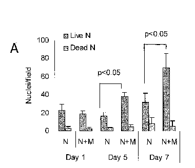
[0015] Figures 1A and 1B show results of measurements of
proliferation of
rat neural cells (denoted "N") alone or in co-culture with MSC (denoted "M")
on
ECM-coated plates. Figure 1A shows measurements of the number of DAPI-stained
neural cell (non-MSC) nuclei in co-cultures at three different time points
(Day I, Day
5 and Day 7 after beginning of co-culture). For each pair of bars, the left-
most bar
represents the number of live neural cells, and the right-most bar represents
the
number of dead neural cells, as assessed by nuclear morphology. Figure 1B
shows
cell number, as assayed by relative levels of the rat noggin gene, in neural
cells
(denoted "N") cultured alone or co-cultured with MSC (denoted "M"), and in MSC
cultured in the absence of rat neural cells. Data for two time points (Day 1
and Day
7) are shown.
[0016] Figure 2, panels A to E, show the time-course of expression of
mRNAs for doublecortin (DCX) (FIG. 2A), microtubule-associated protein-2
(MAP2)
(FIG. 2B), nestin (Nes) (FIG. 2C), glial fibrillary acidic protein (GFAP)
(FIG. 2D)
and 2', 3'-cyclic nucleotide 3' phosphodiesterase (CNP) (FIG. 2E) in cultures
of rat
neural cells (N) and co-cultures of rat neural cells and MSC (N+M) on ECM-
coated
plates. Co-cultures contained 200 MSC per well. "Days" refers to days after
initiation of co-culture.
[0017] Figure 3 shows results of quantitative PCR studies indicating that
expression of RNAs encoding various neural markers in rat E18 cortical cells
is MSC
dose-dependent in co-cultures grown on extracellular matrix (ECM). MSC-dose
responses of rat nestin (rNes), MAP2 (rMAP2), and CNPase (rCNPase) gene
expression were assessed on day 5, rat GFAP (rGFAP) and human GAP (huGAP)
13
CA 02844779 2014-02-10
WO 2013/028625
PCT/US2012/051596
expression were assessed on day 7. No signal from human MCS or SB623 cells
alone
was detected in any rat expression assays, and no signal from rat cells was
detected in
the human GAP expression assay.
[0018] Figure 4 shows relative expression levels of various markers,
determiner by qRT-PCR, in co-cultures of rat neural cells and MSC on ECM-
coated
plates. The rat markers are nestin (Nes), CNPase (CNP), doublecortin (DCX),
microtubule-associated protein-2 (MAP2), glial fibrillary acidic protein
(GFAP), and
glyceraldehyde-3-phosphate dehydrogenase (ratGAP). The human marker, used to
identify and quantitate MSC in the cultures, is glyceraldehyde-3-phosphate
dehydrogenase (huGAP). Expression of Nestin and CNPase was assayed after 5
days
of co-culture; all other markers were assayed after 7 days of co-culture. An
expression level of 1 was arbitrarily assigned to be the level at the lowest
MSC dose
(32 cells per well).
[0019] Figure 5 shows the effect of MSC concentration on levels of
expression of CNPase mRNA at two different stages of co-culture on ECM-coated
plates. CNPase mRNA levels were quantitated by qRT-PCR. A relative expression
level of 1 was arbitrarily set as the highest level observed on the particular
day of
assay (day 5 or day 7).
[0020] Figure 6 shows levels of marker expression in co-cultures of
rat neural
cells and MSC conducted under non-adherent conditions. Neural cells were also
cultured without MSC in the presence of bFGF and EGF as a control. For each
set of
conditions the bars represent, from left to right, expression levels of rat
nestin (rNes),
rat microtubule-associated protein-2 (rMAP2), rat glial fibrillary acidic
protein
(rGFAP), rat doublecortin (rDCX), rat 2', 3'-cyclic nucleotide 3'
phosphodiesterase
(rCNPase), rat glyceraldehyde-3-phosphate dehydrogenase (rGAP) and human
glyceraldehyde-3-phosphate dehydrogenase (huGAP). Neural marker gene
expression level in the presence of bFGF/EGF was assigned a value of 1 and
other
values were expressed correspondingly for all markers except GFAP, which was
assigned a value of 0.1 in the bFGF/EGF sample.
[0021] Figure 7 shows levels of marker expression in co-cultures of rat
neural
cells and MSC conducted under different attachment conditions. "ECM" indicates
co-culture on plates coated with SB623 cell-derived extracellular matrix.
"Orn/FN"
indicates co-culture on plates coated with omithine and fibronectin. "ULA"
indicates
culture on Ultra Low Attachment plates. On ECM and Orn/FN plates, neural cells
14
CA 02844779 2014-02-10
WO 2013/028625
PCT/US2012/051596
were co-cultured with MSC at a 10:1 ratio (1.5 x 10⁴ cells/cm2). On ULA
plates,
neural cells were cultured either with MSC at a 2:1 ratio ("+MSC, 5X") or in
the
absence of MSC in medium supplemented with growth factors ("FGF2/EGF"). For
each set of conditions, the bars represent, from left to right, expression
levels of rat
nestin (Nes), rat 2', 3'-cyclic nucleotide 3' phosphodiesterase (CNPase), rat
glial
fibrillary acidic protein (GFAP), rat doublecortin (DCX), and human
glyceraldehyde-
3-phosphate dehydrogenase (huGAP). Nestin and CNPase levels were assayed after
5
days of culture or co-culture ("5d"); all other markers were assayed at 7 days
("7d").
[0022] Figure 8 shows effect of heparinase on expression of nestin
mRNA by
neural cells. Rat cortical cells were cultured on ECM-coated plates. Prior to
plating
of the cortical cells, the ECM-coated plates had been treated with two
concentrations
of heparinase 1(0.5 Units/ml and 1.5 Units/nil), or with heparinase buffer ("H-
Buffer") or were untreated ("No add"). Nestin mRNA expression was measured by
qRT-PCR 5 days after initiation of culture. The amount of nestin mRNA detected
in
cells cultured on untreated ECM-coated plates was arbitrarily assigned a
relative
expression level of 1.
[0023] Figures 9A-9D show the effects of purified growth factors
(EGF,
BMP6, HB-EGF) and MSC conditioned medium (CM) on relative expression levels
of mRNAs encoding various neural markers, by neural cells cultured on ECM-
coated
plates, determined by qRT-PCR. Figure 9A shows effects on expression of
Nestin, a
marker for neural precursor cells. Figure 9B shows effects on expression of
doublecortin (DCX), a marker for nascent neurons. Figure 9C shows effects on
expression of CNPase, an oligodendrocyte marker. Figure 9D shows effects on
expression of GFAP, a marker for astrocytes.
[0024] Figure 10 shows effects of an anti-FGF2 neutralizing antibody on
nestin expression by neural cells in neural cell/MSC co-cultures on ECM-coated
plates. Rat cortical cells (5,000 cells) were cultured by themselves ("No
MSC") or
co-cultured with 200 MSC ("+MSC"). Additional co-culture samples also
contained
either a neutralizing anti-FGF2 antibody ("+MSC+bFM1") or a non-neutralizing
anti-
FGF2 antibody ("+MSC+bFM2"). Nestin expression was assayed 5days after
beginning of culture or co-culture. Levels of nestin expression in cortical
cells
cultured in the absence of MSC were arbitrarily assigned a relative expression
value
of 1.
CA 02844779 2014-02-10
WO 2013/028625
PCT/US2012/051596
[0025] Figure 11 shows the effects of MSC conditioned medium, and of
FGF2-depleted MSC conditioned medium, on nestin expression in cultured rat
neural
cells. Rat cortical cells were cultured on ECM-coated plates without further
additions
("No add"), with MSC conditioned medium ("CM"), with MSC conditioned medium
that had been depleted of FGF2 by immunoprecipitation ("FGF2-depleted CM"),
and
with MSC conditioned medium treated with a control antibody that did not react
with
FGF2 ("IP-Control-CM"). Nestin expression was assayed 5days after beginning of
culture. Levels of nestin expression in cortical cells cultured in the absence
of
conditioned medium were arbitrarily assigned a relative expression value of 1.
[0026] Figure 12 shows levels of mRNAs encoding nestin (Nes) and glial
fibrillary acidic protein (GFAP), expressed by neural cells cultured on ECM-
coated
plates in the presence of 200 mesenchymal stem cells ("+MSC, 200 cells") or a
1:10
dilution of conditioned medium from mesenchymal stem cells ("+CM, 10%").
Control cells were cultured in the absence of MSC or conditioned medium ("No
add"). Assay for nestin was conducted 5 days after beginning of culture; assay
for
GFAP was conducted 7 days after beginning of culture. The level of each marker
expressed in co-culture with MSC was arbitrarily assigned a relative
expression value
of 1.
[0027] Figure 13 shows levels of GFAP mRNA, assayed 7 days after
beginning of culture or co-culture, in rat cortical cells cultured on ECM-
coated plates.
Cortical cells were co-cultured with MSC ("MSC"), co-cultured with MSC in the
presence of 30 ng/ml recombinant noggin protein ("MSC+noggin"), or co-cultured
with MSC in the presence of an anti-BMP4 antibody ("MSC+anti-BMP4"). The level
of GFAP mRNA expressed in co-culture with MSC was arbitrarily assigned a
relative
expression value of 1.
[0028] Figure 14 shows expression levels of mRNAs for human bone
morphogenetic protein-4 ("huBMP4"), human glyceraldehyde-3-phosphate
dehydrogenase ("huGAP"), human fibroblast growth factor-2 ("huFGF2") and rat
glial fibrillary acidic protein ("rGFAP") in co-cultures of rat neural cells
and MSC on
ECM-coated plates. Prior to co-culture, MSC were transfected with siRNA pools
targeted to human BMP-4 sequences ("N+siBMP4-MSC") or a control non-BMP4-
targeted siRNA ("N+siContr-MSC"). Neural cells were also cultured separately
in
the absence of MSC ("N alone").
16
CA 02844779 2014-02-10
WO 2013/028625
PCT/US2012/051596
DETAILED DESCRIPTION
[00291 It has proven difficult to establish in vitro culture
conditions that will
support the growth and differentiation of the various different types of
neural cells.
The present inventors have devised an in vitro culture system in which neural
precursor cells, neurons, astrocytes and oligodendrocytes are all able to grow
and
differentiate. The culture system disclosed herein thus allows, for the first
time,
quantitative evaluation of the effect of a test substance on the growth and
differentiation of neural cells. The system comprises a culture of neural
cells (e.g.
embryonic rodent cortical cells) on an extracellular matrix in the presence of
a test
substance, followed by analysis of the neural cell culture for the expression
of one or
more marker molecules. The extracellular matrix used in these assays is
produced by
(a) a mesenchymal stem cell, or (b) a mesenchymal stem cell that has been
transfected
with a nucleic acid, wherein the nucleic acid encodes a Notch intracellular
domain but
does not encode full-length Notch protein (e.g., a SB623 cell).
[0030] Various aspects of this system contribute to its ability to provide
quantitative information on the potency of various neurogenic and gliogenic
factors.
In one aspect, the neural cells are cultured on an extracellular matrix
produced by
MSC or 5B623 cells (cells that have been derived from MSC by transfecting MSC
with a vector containing sequences encoding an Notch intracellular domain). In
another aspect, the amount of time that the neural cells are co-cultured with
a test
substance is chosen to optimize detection and quantitation of the marker that
is being
assayed. The duration of co-culture prior to assay is unique to each marker.
For
example, co-culture is conducted for five days for measurement of nestin and
CNPase; and for seven days for measurement of GFAP, DCX and MAP2. In yet
another aspect, the concentration of cells in the culture is optimized. For
example,
neural cells are used at a concentration of 1.5 x 10⁴ cells/ml; MSC and SB623
cells
are used at a concentration of 0.5-1.5 x 10³ cells/ml.
[0031] The quantitative assay system disclosed herein utilizes ECM
from
mesenchymal cells such as MSC and their derivatives (e.g., 5B623 cells) as a
biological substrate for co-cultures of test substances (e.g., MSC or their
derivative
5B623 cells, conditioned medium, growth factors, cytokines) and neural cell
populations, and provides a culture system that is favorable to the growth of
both
mesenchymal cells and neural cells. Such a system, in turn, allows
quantitation of the
17
CA 02844779 2014-02-10
WO 2013/028625
PCT/US2012/051596
effects of mesenchymal cells, as well as effects of other cells and
substances, on the
growth and differentiation of various types of neural cells.
[0032] The advantages of the assays described herein include that
fact that
developmental transitions occur under physiological conditions and over a
physiological time-course, rather than in response to abnormal physical
conditions,
such as attachment or aggregation (cf neurosphere cultures). In addition, the
stage of
development of the cells being assayed can be easily detelinined, as
development
does not occur in the interior of a neurosphere. Finally, the assays disclosed
herein do
not require external growth factors; thus allowing the effects of such factors
to be
quantitated in this system.
[0033] Using this system, the inventors have determined that not only
does
mesenchymal cell ECM support the growth of neural cell populations (such as,
for
example, embryonic cortical cells), but that addition of MSC or SB623 cells to
neural
cell populations growing on mesenchymal cell ECM substantially enhances growth
and differentiation of all neural lineages (e.g., neurons, astrocytes and
oligodendrocytes).
[0034] Compared to existing co-culture systems, much lower ratios of
mesenchymal cells to neural cells are capable of inducing significant growth
and
differentiation of neural cells in the ECM-based co-cultures described herein.
For
example, the assay systems described herein are sensitive enough to detect the
effect
of approximately 50 mesenchymal cell on 5,000 neural cells.
[0035] Provided herein are quantitative assays for neurogenic and
gliogenic
factors, as well as factors that promote the growth and differentiation of
neural
precursor cells. The assays can also be used to identify and quantitate
sources of such
factors, such as cell cultures or conditioned media.
[0036] To conduct the assays, MSC or SB623 cells (referred to
collectively as
"mesenchymal cells") are grown in a vessel, such as a tissue culture dish, for
a period
of time sufficient for the cells to lay down an extracellular matrix on the
surface of the
vessel. Any solid substrate can be used as a surface on which the cells are
grown, as
long as it supports the growth of the cells and the elaboration of an
extracellular
matrix by the cells. Suitable substrates include plastic, glass or
nitrocellulose.
Further, the substrate may be coated with a substance such as, for example,
fibronectin or collagen, or a reconstituted basement membrane such as, for
example,
MatrigelTM.
18
CA 02844779 2014-02-10
WO 2013/028625
PCT/US2012/051596
[0037] An example of a suitable substrate is a plastic tissue culture
dish or
flask. The cells can be grown for one day, two days, three days, one week, two
weeks, one month, or any time interval therebetween as desired. For additional
details on ECM elaborated by MSC and SB623 cells, see U.S. Patent Application
Publication No. 2010/0310529, the disclosure of which is incorporated by
reference
for the purpose of describing ECM elaborated by MSC and SB623 cells (denoted
"differentiation-restricted descendants of MASCs" in that publication) and its
properties.
[0038] MSC can be obtained by selecting adherent cells from bone
marrow
samples. Bone marrow can be obtained commercially (e.g., from Lonza,
Walkersville, MD) or from bone marrow biopsies. Other sources of mesenchymal
stem cells include, for example, adipose tissue, dental pulp, cord blood,
placenta and
the decidua. MSC can be obtained from any animal, including mammals, and
including humans.
[0039] Exemplary disclosures of MSC are provided in U.S. patent application
publication No. 2003/0003090; Prockop (1997) Science 276:71-74 and Jiang
(2002)
Nature 418:41-49. Methods for the isolation and purification of MSC can be
found,
for example, in U.S. Patent No. 5,486,359; Pittenger et al. (1999) Science
284:143-
147 and Dezawa et al. (2001) Eur. I Neurosci. 14:1771-1776. Human MSC are
commercially available (e.g., BioWhittaker, Walkersville, MD) or can be
obtained
from donors by, e.g., bone marrow aspiration, followed by selection for
adherent bone
marrow cells. See, e.g., WO 2005/100552.
[0040] SB623 cells are derived from MSC by transfecting MSC with a
vector
containing sequences that encode a Notch intracellular domain (NICD) but do
not
encode the full-length Notch protein, such that the transfected cells express
exogenous
NICD but do not express exogenous full-length Notch protein. Methods for
obtaining
MSC, and for deriving SB623 cells from MSC populations, are described, for
example, in US Patent No. 7,682,825 and in US Patent Application Publication
No.
2010/0266554, the disclosures of which are incorporated by reference for the
purposes of describing MSC and SB623 cells, and methods of obtaining these
cells.
[0041] Subsequent to growth on the substrate for a predeteunined
amount of
time, the MSC or SB623 cells are removed from the substrate, leaving behind an
extracellular matrix deposited on the substrate. Methods of removing cells
from a
substrate are well known in the art. In the practice of the methods disclosed
herein,
19
CA 02844779 2014-02-10
WO 2013/028625
PCT/US2012/051596
removal of cells from the substrate must be sufficiently gentle that the ECM
that has
been elaborated by the cells remains on the substrate. Such methods include,
for
example, treatment with non-ionic detergent (e.g., Triton X-100, NP40) and
alkali
(e.g. NH4OH). See the "Examples" section infra for additional details.
[0042] The ECM-containing substrate is then used as a substrate for co-
culture
of neural cells and one or more test substance(s), and the effect of the test
substance(s)
on the neural cells is determined and quantitated. Introduction of the neural
cells and
the test substance to the culture can be simultaneous, or in either order.
[0043] Any type of neural cell or neural cell population can be used;
such
cells are known in the art. A convenient source of neural cell populations are
rodent
embryonic cortical cells (e.g., from rat or mouse), which can be obtained
commercially (BrainBits, Springfield, IL). In certain embodiments, the neural
cell
population is enriched in neural precursor cells.
[0044] A test substance can be any chemical compound, macromolecule
(e.g.,
nucleic acid or polypeptide), cell, cell culture, cell fraction or tissue, or
combination
thereof. For example, growth factors and cytokines, low molecular weight
organic
compounds, mRNA molecules, siRNA molecules, shRNA molecules, antisense RNA
molecules, ribozymes, DNA molecules, DNA or RNA analogues, proteins (e.g.,
transcriptional regulatory proteins), antibodies (e.g., neutralizing
antibodies), enzymes
(e.g., nucleases), glycoproteins, glycans, proteoglycans, cells, cell membrane
preparations, cell cultures, conditioned medium from cell cultures,
subcellular
fractions and tissue slices or tissue fractions are all suitable test
substances. Test
substances can also include electromagnetic radiation such as, for example, X-
rays,
light (e.g., ultraviolet, infrared) or sound (e.g., subsonic or ultrasonic
radiation). In
certain embodiments, the combination of a protein and a neutralizing antibody
to the
protein is used as a test substance.
[0045] Naturally-occurring test substances can include soluble
molecules
(e.g., proteins) synthesized and secreted by cells, as well as molecules
(e.g., proteins)
that are synthesized by a cell, transported to the cell surface, and remain
embedded in
the cell surface, with all or a portion of the molecule exposed to the
exterior of the cell
(i.e., surface molecules, surface proteins or surface glycoproteins).
[0046] Neural cells and test substances are co-cultured for an
appropriate
amount of time, as determined by the practitioner of the method. For example,
co-
culture can be conducted for 1 hour, two hours, three hours, four hours, six
hours, 12
CA 02844779 2014-02-10
WO 2013/028625
PCT/US2012/051596
hours, one day, two days, three days, four days, five days, six days, one
week, two
weeks, one month, or any time interval therebetween.
[0047] The effect(s) of the test substance(s) on the neural cells is
determined
by measuring the expression of one or more markers in the neural cells.
Depending
on the marker or markers chosen, it is possible to assay for formation of
neural
precursor cells, neurons, astrocytes, or oligodendrocytes.
[0048] In one embodiment, the effect of a particular protein, either
native or
recombinant, on neurogenesis or gliogenesis can be determined by adding the
protein
to a culture of neural cells growing on a MSC or SB623 ECM and assaying for
the
appropriate neuronal or glial marker. Optionally, a low concentration (1%, 2%,
5%,
10%, 20%, 30%, 40%, 50% or any value therebetween) of conditioned medium from
MSC or SB623 cells can also be included in the culture. For example, inclusion
of
conditioned medium can provide additional factors required for the process
under
study, other than the one being tested, thereby allowing the effect of one
component
of a multi-factor signaling system to be assessed.
[0049] Molecular and morphogenetic markers for neural precursor
cells,
neurons, astrocytes and oligodendrocytes are well-known in the art; the
following are
provided as examples.
[0050] Markers for neural precursor cells include, for example,
nestin,
glutamate transporter (GLAST), 3-phosphoglycerate dehydrogenase (3-PGDH,
astrocyte precursors), ephrin B2 (EfnB2), Sox2, Pax6, and musashi. In certain
embodiments, proliferative capacity can also be used as a marker for neural
precursor
cells. Proliferative capacity can be measured, for example, by incorporation
of
bromodeoxyuridine, carboxyfluorescein diacetate succinimidyl ester (CFSE)
labeling,
expression of Ki-67 or expression of proliferating cell nuclear antigen
(PCNA).
[0051] Markers for neurons include, for example, microtubule-
associated
protein 2 (MAP2), P-tubulin isotype III (also known as 13-111 tubulin and TuJ-
1),
doublecortin (DCX), neurofilament proteins (e.g., neurofilament-M),
synaptophysin,
and neuron-specific enolase (also known as enolase-2 and gamma enolase).
Neurite
outgrowth can also be used as a marker for neuronal development.
[0052] Additional neuronal markers are listed in the following table:
21
CA 02844779 2014-02-10
WO 2013/028625
PCT/US2012/051596
Early Neuronal Markers
ATH1 [MATH1] Nuclear
ASH1 [MASH1] Nuclear
Hes5 Nuclear
HuC (Hu, Rodent) Very early
marker, Nuclear
HuD Nuclear
lnternexin a I Cytoplasmic,
soma, early
neurites
L1 neural adhesion Plasma
molecule membrane
MAP1B [MAP5] Cytoplasmic,
soma, dendritic
MAP2A, 2B Cytoplasmic,
soma, dendritic
Nerve Growth Plasma
Factor Rec (NGFR) membrane
p75
Nestin Cytoplasmic
NeuroD Nuclear
Neurofilannent L 68 Cytoplasmic
22
CA 02844779 2014-02-10
WO 2013/028625
PCT/US2012/051596
kDa
Neuron Specific Cytoplasmic
Enolase (NSE)
NeuN Nuclear,
Nloc-2.2 [NK-2] Nuclear
Noggin Secreted
Pax-6 Nuclear, eye
development
PSA-NCAM, clone Plasma
2-2B membrane
Tbrl Nucleus
Tbr2 Nucleus
Tubulin, 3W Cytoplasmic,
neuritis
TUC-4 Axonal growth
cones
Tyrosine Cytoplasmic,
Hydroxylase (TH) adrenergic
neuron lineage
,
Immature Neuron & Growth Cone
_ Markers:-
_-
Collapsin Response Growth cone
Mediated Protein 1
[CRMP1]
23
CA 02844779 2014-02-10
WO 2013/028625
PCT/US2012/051596
Collapsin Response Growth cone
Mediated Protein 2
[CRMP2]
Collapsin Response Growth cone
Mediated Protein 5
[CRMP5]
Contactin-1 Cytoplasmic
Contactin-1 Cytoplasmic
Cysteine-rich motor Cytoplasmic,
neuron 1 [CRIM1] motor neurons
c-Ret phosphor Cytoplasmic
Serine 696
Doublecortin [DCX] Cytoplasmic,
migrating
neurons
Ephrin A2 Plasma
membrane
Ephrin A4 Plasma
membrane
Ephrin A5 Plasma
membrane
Ephrin B1 Plasma
membrane
Ephrin B2 Plasma
membrane
Ephrin B Plasma
phosphoTyr298 membrane
Ephrin B Plasma
phosphoTyr317
1 membrane
Ephrin B Plasma
phosphoTyr331 membrane
GAP-43 , Plasma
membrane
24
CA 02844779 2014-02-10
WO 2013/028625
PCT/US2012/051596
GAP-43, Plasma
phosphoSer 41 membrane
HuC/D
Internexin alpha
Laminin-1 Plasma
membrane
LINGO-1 Cytoplasmic
MAP1B [MAP5]
Mical-3 Growth cones
NAP-22 Plasma
membrane,
growth cones
NGFR
Nestin
Netrin-1 Plasma
membrane
Neurite Outgrowth
Quantification Assay
kit
Neuropilin Plasma
membrane
Plexin-Al Plasma
membrane,
growth cone
RanBPM Cytoplasmic,
growth cone
Semaphorin 3A Plasma
membrane,
growth cone
Semaphorin 3F Plasma
membrane
CA 02844779 2014-02-10
WO 2013/028625
PCT/US2012/051596
Semaphorin 4D Plasma
membrane
Slit2 Secreted
Slit3 Secreted
Staufen Cytoplasmic
Tbr 1 &2
Trk A Plasma
membrane
Tubulin, 13111
TUC-4
Neuronal Markers ¨ Nuclear ,
HuD Postmitotic
neurons
NeuN Nuclei of most
neurons
Peripherin Peripheral
neurons
Neuronal Markers ¨ Cytoplasmic
MAP2A, B, C. I All neurons,
soma, dendrites
Tubulin, 13111 All neurons,
soma, axons
CDK5 [NCLK], Soma
perikarya
MacMARCKS Soma
MARCKS Soma
Neurofilaments All neurons,
soma, axons,
26
CA 02844779 2014-02-10
WO 2013/028625
PCT/US2012/051596
proximal
dendrites
Neuron Specific Cytoplasmic
Enolase (NSE)
Parvalbumin Neurons,
muscle
Protein Gene All neurons,
Product 9.5 neuroendocrine
[PGP9.5] cells
STEP NMDAR
expressing
neurons
STOP [N-STOP, Soma, dendrites
Stable tubule-only
polypeptide]
Tau Axons
Tau phospho Axons
specific
CD90 [Thy-1] Neurons,
thymocytes,
connective
tissue
CDw90 [Thy-1.1] Neurons,
thymocytes,
connective
tissue
Encephalopsin PO, PVN,
Purkinje cells,
other select
regions
GAD65 [Glutamate Glutamatergic
Decarboxylase] neurons
GAP-43 [Growth Differentiating
Associated Protein and
43] regenerating
neurons
LINGO-1 Differentiating
27
CA 02844779 2014-02-10
WO 2013/028625
PCT/US2012/051596
and
regenerating
neurons
Na+/K+ ATPase All neurons
subunits
Neuron Cell Surface Neurons, glia
Antigen [A2B5]
Post-synaptic
receptors
4.1G Neuron specific
Acetylcholinesterase Cholinergic
Ack1 Clathrin-
mediated
endocytosis
AMPA Receptor Postsynaptic
Binding Protein
[ABP]
ARG3.1 Presynaptic,
plasticity related
Arp2 Most neurons
E-Cadherin Cell junctions
N-Cadherin Cell junctions
Calcyon Postsynaptic,
Dopaminergic
Catenin, alpha and Cell junctions
beta
Caveolin Presynaptic
CHAPSYN-110 Postsynaptic
[PS D93]
Chromogranin A
Peripheral,
Neuroendocrine,
presynaptic
28
CA 02844779 2014-02-10
WO 2013/028625
PCT/US2012/051596
Clathrin light chain Presynaptic
Cofilin Postsynaptic
Complexin 1 Presynaptic
[CPLX1, Synaphin
2]
Contactin-1 Cell junctions
CRI PT Postsynaptic
Cysteine String Presynaptic
Protein [CSP]
Dynamin 1 and 2 Presynaptic
Flotillin-1 Presynaptic
Fodrin Perisynaptic
GRASP Postsynaptic
GRIP1 Postsynaptic
Homer Postsynaptic
Mint-1 Presynaptic
Munc-18 Presynaptic
NSF Presynaptic
PICK1 Postsynaptic
PSD-95 Postsynaptic
RAB4 Presynaptic
Rabphillin 3A Presynaptic
SAD A & B Presynaptic
SAP-102 Postsynaptic
29
CA 02844779 2014-02-10
WO 2013/028625
PCT/US2012/051596
SHANKla Postsynaptic
SNAP-25 Presynaptic
Snapin Presynaptic
Spinophilin Postsynaptic,
[Neurabin-1] dendritic spines
Stargazin Postsynaptic,
AMPAR
Striatin Postsynaptic,
dendritic
SYG-1 Perisynaptic
Synaptic Vesicle Presynaptic
Protein 2A & 2B
Synapsin 1 Presynaptic
Synapsin 1 phospho Presynaptic
specific
Synaptobrevin Presynaptic
[VAMP]
Synaptojanin 1 Presynaptic
Synaptophysin Presynaptic
Synaptotagmin Presynaptic
Synaptotagmin Presynaptic
phospho specific
synGAP Postsynaptic
Synphilin-1 Perisynaptic,
synuclein
related
Syntaxin 1, 2, 3, 4 Presynaptic
Synuclein alpha Presynaptic
CA 02844779 2014-02-10
WO 2013/028625
PCT/US2012/051596
VAMP-2 I Presynaptic
Vesicular Presynaptic
Acetylcholine
Transporter
[VAChT]
Vesicular GABA Presynaptic
transporter [VGAT;
VIAAT]
Vesicular Glutamate Presynaptic
Transporter 1, 2, 3
[VGLUT]
Vesicu lar Presynaptic
Monoamine
Transporter 1, 2
[VMAT]
Neuronal Markers ¨ Cholinergic
Acetylcholine (ACh) Presynaptic
Acetylcholinesterase Perisynaptic
Choline Cytoplasmic
Acetyltransferase
[ChAT]
Choline transporter Plasma
Membrane
Vesicular Presynaptic
Acetylcholine
Transporter
[VAC hT]
Neuronal Markers ¨ Dopaminergic
Adrenaline Presynaptic
Dopamine Presynaptic
Dopamine Beta Cytoplasmic
Hydroxylase [DBH]
Dopamine Plasma
31
CA 02844779 2014-02-10
WO 2013/028625
PCT/US2012/051596
Transporter [DAT] Membrane
L-DOPA Cytoplasmic
Nitric Oxide- Presynaptic
Dopamine
Norepinephrine Presynaptic
Norepinephrine Plasma
Transporter [NET] Membrane
Parkin Cytoplasmic
Tyrosine
Hydroxylase [TH]
TorsinA Cytoplasmic, ER
Neuronal Markers ¨ Serotonergic ,
DL-5- Presynaptic
Hydroxytryptophan
Serotonin Presynaptic
Serotonin Plasma
Transporter [SERT] Membrane
Tryptophan Cytoplasmic
Hydroxylase
Neuronal Markers ¨ GABAergic
DARPP-32 GABAergic
neurons in CNS;
Medium spiny
neurons
GABA Presynaptic
GABA Transporters Plasma
1, 2, 3 Membrane
Glutamate Cytoplasmic
Decarboxylase
[GAD]
32
CA 02844779 2014-02-10
WO 2013/028625
PCT/US2012/051596
Vesicular GABA Presynaptic
transporter [VGAT;
VIAAT]
Neuronal Markers ¨ Glutannatergic
Glutamate Presynaptic
Glutamate Plasma
Transporter, Glial Membrane
Glutamate Plasma
Transporter, Membrane
Neuronal
Glutamine Cytoplasmic
Glutamine Cytoplasmic
Synthetase, clone
Gs-6
Vesicular Glutamate Presynaptic
Transporter 1, 2, 3
[VGLUT]
[0053] Glial fibrillary acidic protein (GFAP), glutamate transporter
(GLAST),
3-PGDH and glutamine synthetase can be used as markers for astrocytes.
[0054] Markers for oligodendrocytes include, for example, the A2B5
antigen,
[0055] Expression of markers can be measured by techniques that are
well-
[0056] Expression of mRNA can be measured and quantitated by methods
including, for example, blotting, nuclease protection and reverse
transcription-
[0057] Depending on the test substance and marker being assayed, it
may be
necessary to ensure that the assay is specific for the molecule produced by
the neural
33
CA 02844779 2014-02-10
WO 2013/028625
PCT/US2012/051596
cell and does not cross-react with the same or a similar molecule produced by
the test
substance, especially if the test substance is a cell, such as a MSC or a
SB623 cell.
For example, MSC express nestin; therefore, if nestin is being assayed as a
marker for
a neural precursor cell in a co-culture of rat cortical cells and human MSC,
an
antibody specific for rat nestin is used in the assay. Similarly, if nucleic
acid
expression is assayed, such as by quantitative reverse
transcription/polymerase chain
reaction (qRT-PCR) or TaqMan, species-specific primers (and probes, if
applicable)
are used.
[0058] In certain embodiments, the expression of a marker by a neural
cell in
the assay described above (i.e. co-culture of neural cells and test substance)
is
compared to the expression of the same marker in neural cells in the absence
of the
test substance(s).
[0059] The assays described herein can also be used to quantitate the
differentiation of neural precursor cells (NPCs) by measuring expression of
markers
characteristic of the progeny of NPCs, which include neurons, astrocytes and
oligodendrocytes. Such markers are well-known in the art and exemplary markers
have been described herein.
[0060] In certain embodiments, a kit for assaying the neurogenic or
gliogenic
potential of a test substance, or for assaying the ability of a substance to
promote the
growth and/or differentiation of neuronal precursor cells, is provided. The
kit
contains one or both of MSC and 5B623 cells (optionally in a cryopreserved
state),
along with one or more culture vessels, and optionally culture medium, to
allow the
user to grow the MSC or SB623 cells on the culture vessel. The kit may also
contain
reagents (e.g., nonionic detergents such as Triton X-100 or Nonidet P-40;
ammonium
hydroxide) for removing the MSC or SB623 cells from the culture vessel so as
to
leave an extracellular matrix deposited on the surface of the culture vessel.
The kit
can also contain a sample of neural cells (e.g., rat El 8 cortical cells).
Labeled
antibodies to various neuronal and glial markers may also be included in the
kit; and
oligonucleotide probes and/or primers specific for mRNAs encoding neuronal and
glial markers can also be included. Any type of reagent that will detect a
neuronal or
glial marker (e.g., a protein) or its encoding mRNA can be included in the
kit.
Reagents and/or buffers and/or apparatus suitable for immunohistochemistry,
FACS,
RT-PCR, electrophysiology and pharmacology can also be included in the kit.
34
CA 02844779 2014-02-10
WO 2013/028625
PCT/US2012/051596
[0061] In additional embodiments, a kit as disclosed herein can
contain one or
more culture vessels with an ECM from MSC or SB623 cells deposited thereon.
Such
a kit can optionally include neural cells and reagents (e.g., antibodies,
probes,
primers) to detect neuronal and/or glial markers. Such a kit can also
optionally
include reagents and/or buffers suitable for immunohistochemistry, FACS, RT-
PCR,
electrophysiology and pharmacology.
[0062] In additional embodiments, a kit can contain purified
extracellular
matrix from MSC or SB623 cells (or a mixture thereof), for application to a
culture
vessel.
[0063] In further embodiments, a kit comprises a solid substrate (e.g., a
culture vessel) with a biological layer deposited thereon, wherein the
biological layer
is an extracellular matrix deposited by:
(a) a mesenchymal stem cell, or
(b) a mesenchymal stem cell that has been transfected with a nucleic
acid, wherein the nucleic acid encodes a Notch intracellular domain but does
not
encode full-length Notch protein.
[0064] In the operation of the kits, neural cells are grown in
contact with an
extracellular matrix from MSC or SB623 cells, in the presence of a test
substance, and
the neural cells are analyzed for the expression of a chosen neuronal or glial
marker.
With certain of the aforementioned kits, deposition of the ECM on a culture
vessel
(by MSC and/or SB623 cells) and removal of the cells that elaborated the ECM,
is
conducted by the user prior to adding the neural cells and the test substance
to the
culture vessel.
EXAMPLES
General Methods
MSC and SB623 cell preparation
[0065] MSC and SB623 cell preparation has been described [29].
Briefly,
human adult bone marrow aspirates (Lonza, Walkersville, MD) were grown in
aMEM (Mediatech, Herndon, VA) supplemented with 10% fetal bovine serum (FBS)
(Hyclone, Logan, UT), 2mM L-glutamine, and penicillin/streptomycin (both from
Invitrogen, Carlsbad, CA). On the second passage, some cells were
cryopreserved
(MSC preparation) and some cells were plated for the preparation of SB623
cells. For
CA 02844779 2014-02-10
WO 2013/028625
PCT/US2012/051596
SB623 cell preparation, MSC were transfected with a pCI-neo expression plasmid
encoding the human Notchl intracellular domain (NICD). After one day of
culture,
transfected cells were placed under selection with G418 (Invitrogen) for 7
days, after
which selection was removed and the cultures were grown and expanded by
passaging
twice. SB623 cells were then harvested and cryopreserved using Cryostor CS5
(BioLife Solutions, Bothell, WA). Cells from 3 different donors were used in
the
studies described herein. MSC and SB623 cells were thawed and washed once with
aMEM before use. For co-culture experiments, cells were then resuspended in a
neural growth medium consisting of Neurobasal medium supplemented with 2% B27
and 0.5 mM GlutaMAX (all from Invitrogen). For the production of ECM coating
or
the production of conditioned medium (CM), cells were plated in aMEM
supplemented with 10% FBS and penicillin/streptomycin.
Plate coating
[0066] For the preparation of wells coated with ECM, SB623 cells were
plated
at 3x10⁴cells/cm2 in 96-well plates or on glass cover slips (Fisher
Scientific,
Pittsburgh, PA) which were placed into 12-well plates (all plates were
purchased from
Corning Inc, Coming, NY) and grown for 5 days. Subsequently the medium was
changed to serum-free, and the cells were cultured for an additional 2 days.
Cells
were then removed from the ECM using a protocol described previously [29] with
some modifications. Briefly, cells were treated with 0.2% Triton X-100 (Sigma-
Aldrich, St. Louis, MO) in water at room temperature for 40 mm; then cell
lysates
were carefully aspirated, and a 1:100 (v/v) solution of concentrated NH4OH
(Sigma-
Aldrich) in water was slowly added for 5-7 mm, then removed. For washing, the
wells were filled completely with PBS and incubated for at least 3 hours.
Wells were
either used immediately or stored at 4 C.
Conditioned medium (CM) preparation
[0067] MSC or SB623 cells were plated at 3x10⁴cells/cm2 and grown in
ocMEM supplemented with 10% FBS and penicillin/streptomycin for 3-4 days until
confluence. Then the medium was replaced with Neurobasal medium (Invitrogen),
and the cultures were incubated for 1-2 hours. This medium was discarded and
replaced with fresh Neurobasal medium, using half of the volume typically used
for
36
CA 02844779 2014-02-10
WO 2013/028625
PCT/US2012/051596
cell growth. The cells were incubated for 24 hours, after which the medium was
collected, and particulate matter was removed by centrifugation. The medium
was
dispensed in aliquots and stored at -70C . MSC-CM preparations were
supplemented
with 2% B27 and 0.5 mM GlutaMAX before use.
Preparation of rat embryonic brain cortical cells
[0068] Rat embryonic (E18) brain cortex pairs were purchased from
BrainBits
(Springfield, IL); and a cell suspension was prepared as described [29].
Briefly,
cortices were incubated with 0.25% Trypsin/ EDTA at 37 C for 5-7 min, and
trypsin
was removed. The tissue was washed with aMEM containing 10% FBS, then with
PBS. DNase (MP Biomedicals, Solon, OH) at 0.25 mg/ml was then added, and the
contents of the tube were mixed by vortexing for 30 sec. The resulting cell
suspension was triturated, diluted with PBS, pelleted and then resuspended in
neural
growth medium (described above).
Co-culture experiments
[0069] Plates, coated as described above, were pre-warmed with a
portion of
neural growth medium, and then varying numbers of mesenchymal cells were
added.
Subsequently, neural cells were added at a density of 1.5x10⁴ cells/cm2 to all
but
control wells, and cultures were incubated for the indicated time periods. For
each
time point, a separate plate was used that included quadruplicate samples. For
quantitation, cells were plated in 96-well plates and MSC or SB623 cells were
added
at decreasing densities, starting at 1.5x10³ cells/cm2 (i.e., 500 cells per
well) For
immunostaining, cultures were plated on ECM-coated cover slips in 12-well
plates.
MSC or SB623 cells were added at a constant cell density of 1.5x10³ cells/cm2
unless
indicated otherwise.
[0070] In a subset of experiments, in which the effects of cells (MSC
or
SB623) were compared to the effects of their conditioned medium, a cryopre
served
aliquot of cells was used to generate the conditioned medium prior to an
experiment,
and an aliquot of cells from the same donor was thawed on the day of
experiment to
generate a corresponding cell suspension. Cells were applied at decreasing
concentrations as described above and conditioned medium was used at
decreasing
concentrations starting from 50% of total medium. For quantitation of gene
37
CA 02844779 2014-02-10
WO 2013/028625
PCT/US2012/051596
expression, all culture conditions were tested on the same PCR plate using the
same
standard curve for each neural marker.
[0071] Medium was not changed during co-culture experiments (which
lasted
for 7- 8 days in 96-well format and for up to 14 days with the cells on cover
slips).
No signs of culture decline were noticed and the viability of neural cells was
above
95% when assessed on the last day of culturing using Trypan Blue exclusion.
[0072] In another set of experiments, when cells were co-cultured
under non-
adherent conditions, Ultra-Low Adhesion Costar 12- or 24-well plates were
used.
Mesenchymal and neural cells were mixed in the indicated quantities and plated
in
neural growth medium. As a control, neural cells were grown alone or in the
presence
of 20 ng/ml or 50 ng/ml each of EGF and FGF2 purchased from either R&D Systems
(Minneapolis, MN) or Peprotech (Rocky Hill, NJ). Medium was not changed over
the
course of the experiment (2 weeks).
Immunocytochemistrv
[0073] Cultures that were grown on glass cover slips were fixed with
4%
paraformaldehyde (Electron Microscopy Science, Hatfield, PA) for 20 min,
washed
once with PBS and incubated for 30 min in blocking solution containing 10%
normal
donkey serum (Jackson Immunoresearch, West Grove, PA), 1% bovine serum
albumin (Sigma-Aldrich), 0.1% Triton X-100. Then a goat polyclonal antibody
against rat Nestin (R&D Systems, Cat #AF2736) was added into the blocking
solution
at 1:1000 and incubated overnight at 4C . Cover slips were washed with PBS and
then either rabbit polyclonal anti-glial fibrillary acidic protein (GFAP)
(Dako,
Denmark) (1:2000), mouse monoclonal anti-microtubule-associated protein 2
(MAP2)
(Sigma-Aldrich) (1:1000), or mouse monoclonal anti- 2', 3'-cyclic nucleotide
3'-
phosphodiesterase (CNPase) (Millipore, Billerica, MA) (1:200) was added and
the
cover slips were incubated for 1 hour at room temperature. After washing,
cover slips
were incubated for 1 hour with secondary antibodies: DyLight 488-conjugated
AffiniPure donkey anti-goat F(ab')2 fragments of IgG (1:1000) in combination
with
either DyLight 549 488-conjugated AffiniPure anti-rabbit F(ab')2 fragments of
IgG
(1:2000) or Cy3-conjugated AffiniPure donkey anti-mouse IgG (1:1000), all from
Jackson Immunoresearch, and all selected for use in multiple labeling by the
manufacturer. After washing with PBS and water, the slips were mounted with
38
CA 02844779 2014-02-10
WO 2013/028625
PCT/US2012/051596
ProLong Gold antifade reagent containing 4',6-diamidino-2-phenylindole (DAPI)
(Invitrogen).
[0074] In some experiments, prior to fixation, cells were incubated
for 7-8
hours with 10 uM 5-bromo-2'-deoxyuridine (BRDU, from Sigma-Aldrich) with or
without mitomycin at a concentration of 50 ug/ml. Cultures then were fixed
with 2%
PFA, permeabilized with 0.5% Triton and treated with deoxyribonuclease (MP
Biomedicals, Solon, OH) in a buffer containing 0.15 M NaCl and 4.2 mM MgC12
for
1 h at 37 C. The cultures were then post-fixed with cold methanol (Fisher
Scientific,
Fair Lawn, NJ) for 10 min. After blocking as described above, the cultures
were
incubated with anti-BRDU monoclonal antibody (BD Pharmingen), then with the
anti-mouse secondary antibody described above, then with Alexa Fluor-
conjugated
TUJ1, a Neuronal Class III fl-Tubulin-specific antibody (Covance, Princeton,
NJ).
[0075] Fluorescence microscopy was conducted using a Nikon Eclipse50i
(Nikon Instruments, Melville, NY) and a Nikon Digital Camera DXM1200C.
[0076] Under the conditions described herein, none of antibodies reacted
with
the mesenchymal cells.
Gene expression quantification
[0077] Growth and culture of cells, for quantitation of expression of
mRNAs
encoding various neural markers, was conducted in 96-well plates. After
culturing for
the indicated time, the culture medium was carefully and completely aspirated
using a
Nunc ImmunoWasher equipped with 10 ul pipette tips; and cells were lysed with
20
ul/well of lysis buffer, either Cell-to-SignalTM (Applied Biosystems/Ambion,
Austin,
TX) or SideStep (Agilent Technologies, Santa Clara, CA) for 3 mm. Then the
lysates were carefully pipetted up and down and samples (in quadruplicate)
were
combined pair-wise (thus making biological duplicates), transferred to a
storage plate
and frozen at -70C .
[0078] For gene expression testing, samples were thawed and aliquots
were
diluted 1:10 with PCR-grade water. A sample with a high expected expression
level
was also used to prepare serial dilutions in 10% lysis buffer to serve as a
series of
standards for the quantification. The diluted samples were used as templates
in one-
step qRT-PCR reactions, in combination with QuantiTect Probe RT-PCR Master
Mix from Qiagen (Valencia, CA) and TaqMan gene expression assays purchased
from Applied Biosystems (Foster City, CA). The absence of cross-reaction
between
39
CA 02844779 2014-02-10
WO 2013/028625
PCT/US2012/051596
corresponding mRNAs from rat and human cells was established in experiments
involving rat neural cells, as well as human mesenchymal and neural cells. The
following pre-optimized assays, all designed across exon-exon boundaries, were
used:
rat-specific ¨ nestin (Rn005643942n1), MAP2 (Rn00565046 ml ), GFAP
(Rn00566603_m1), CNPase (Rn01399463_m1), Doublecortin
(Dcx)(Rn00584505_m1), glyceraldehyde 3-phosphate dehydrogenase (rGAP)(Rn-
1462661_g1); and human-specific ¨ glyceraldehyde 3-phosphate dehydrogenase
(huGAP) (4333764F), bone morphogenetic protein 4 (BMP4) Hs00370078_ml, and
fibroblast growth factor 2 Hs00266645_ml. Numbers in parentheses refer to the
manufacturer's (Applied Biosystems) assay ID numbers. For amplification
reactions, a
LightCycler 480 (Roche, Mannheim, Germany) was programmed according to the
Master
Mix manufacturer's protocol, with 40-60 amplification cycles. Analysis was
done
using a Second Derivative Maximum method.
Assessment of neurito genesis
[0079] Neural cells were plated at 1.5x10⁴ cells/cm2 alone or
together with
either MSC or SB623 cells at 1.5x10³ cells/cm2 on ECM-covered glass cover
slips in
neural growth medium containing 10-fold less B27 than normally used (0.2%),
and
allowed to grow for 18-24 hrs. Then the cultures were fixed and stained for
MAP2
expression and mounted with DAPI-containing medium, as described above.
Approximately 12-15 fields were photographed using the same exposure time, and
a
total of 20 neurons per condition were analyzed. The length of the longest
neurite
was measured for each cell and the number of neurites per cell was counted.
Example 1: Effect of mesenchymal cells on proliferation and neuronal
differentiation
[0080] The composition of ECM-based neural cultures grown with or
without
mesenchymal cells was first analyzed using immunocytochemistry for Nestin, a
marker of neural stem cells/early progenitors, and MAP2, a neuronal marker. At
various times cultures were fixed and incubated with rat-specific anti-Nestin
antibody
and anti- MAP2 antibody. All cultures were also counterstained with the
nucleus-
specific dye DAPI. Fixed and stained cultures were then examined by
fluorescence
microscopy. On day 1, single positive Nest and MAP2+ cells were present in all
CA 02844779 2014-02-10
WO 2013/028625
PCT/US2012/051596
cultures tested, along with some MAP2+Nes+ double-positive cells, in which
MAP2
staining and Nestin staining were co-localized. There was also a small
fraction of
double-negative cells. By day 3, no double-positive cells were detected, while
single-
positive cells extended their processes. On day 5, MAP2+ neurons continued to
extend neurites, while Nest cells were significantly increased in numbers. At
this
time point, Nest cells formed colonies. A greater number of colonies, and
larger
colony size, were observed in the presence of mesenchymal cells. Double-
positive
cells were not detected.
[00811 By day 9, a large number of MAP2+Nes+ double-positive cells
were
observed, as well as MAP2+Nes- neurons and MAP2-Nes+ cells. The double-
positive
cells were found in greater numbers in co-cultures with SB623, compared to co-
cultures with MSC, while cultures without mesenchymal cells had the smallest
number of MAP2+Nes+ cells. These double-positive cells had nuclei of a
characteristic bilobular shape, thin MAP2-positive processes, and a strong
Nestin
reactivity localized to a perinuclear area - most frequently, to a cleft
between two
nuclear lobes situated adjacent to the most prominent outgrowth. This
morphology
resembled that of the MAP2 Nes+ double-positive cells present on the first day
of
culturing.
[00821 When neural cells and mesenchymal cells were co-cultured on
PDL,
approximately the same frequency of double-positive and single-positive cells
were
observed on Day lof culture as were observed when cells were co-cultured on
ECM.
At later time points, no developed single-positive Nest cells were detected
(very rare
Nest cells were round, with dense nuclei). By Day 5 of culture, only a small
number
of double-positive cell colonies, each consisting of very few cells, were
observed.
These results indicate that, on PDL, most or all Nest cells were committed to
neuronal
differentiation. At day 9, double-positive colonies were more prominent in the
presence of mesenchymal cells than in their absence.
[00831 The status of mesenchymal cells in co-cultures was also
examined
using phase contrast microscopy and staining for a-smooth muscle actin, a
mesenchymal marker. On PDL, mesenchymal cells were barely spread and usually
disappeared around day 5-7. On ECM, mesenchymal cells were easily detected
throughout the duration of co-culturing; they appeared well-spread, moving,
and
appeared to be slowly proliferating.
41
CA 02844779 2014-02-10
WO 2013/028625
PCT/US2012/051596
[0084] Neuritogenesis was very active on both ECM and PDL in co-
cultures,
but was further enhanced in the presence of mesenchymal cells during first 18-
24 hrs
in culture. To increase this differential response, cultures were plated in
neural
growth medium with a low concentration of B27 supplement. Under these
conditions,
longer neurites were observed in the presence of MSC or SB623 cells after 24
hours
of co-culturing, than in cultures of neural cells alone (Table 1). However, no
significant difference in numbers of neurites was noticed (Table 1).
Table 1: Length and Numbers of Neurites on Day 1
ECM (n=19) ECM + MSC (n=23)
ECM + SB623 cells (n=22)
length number length number length number
Median 15 2 18 3 25 3
Average 15.3+/- 6.3 2.6 22+1-13.9 2.6 31.6+/- 19.7 3.1
Maximum 25 70 85
Neurite length is expressed in pin
Example 2: Effect of mesenchymal cells on astrogenesis
[0085] Astrocyte development was assayed by immunohistochemical
analysis
for expression of glial fibrillary acidic protein (GFAP). Double-staining of
ECM-
based cultures for glial fibrillary acidic protein (GFAP, an astrocyte marker)
and
Nestin revealed the absence of GFAP reactivity before day 3 in all cultures.
Around
day 5, GFAP-expressing cells began to be observed in co-cultures within Nest
colonies as single- or double-positive cells. GFAP-expressing cells were not
observed
in cultures not containing mesenchymal cells. Different colonies had variable
proportions of GFAP Nes+ cells and the more fully differentiated GFAP+Nes-
cells.
No GFAP cells were detected at this time in cultures lacking mesenchymal
cells. On
day 9, all three phenotypes (GFAP+Nes+, GFAP-Nes+, and GFAP+Nes") were present
in all cultures, with GFAP Nes- cells predominating.
[0086] With respect to its intracellular localization, GFAP
immunoreactivity
appeared initially (at day 5) as intercalating filaments beside or within
Nestin
filaments in some Nestin-positive filamentous cells. Later, Nestin was
practically
displaced by GFAP in certain cells, as evidenced by a patchy distribution of
Nestin
staining, while other cells continued to express Nestin only. In co-cultures,
GFAP Nes+ cells had generally the same morphology as GFAP4Nes" cells, while
42
CA 02844779 2014-02-10
WO 2013/028625
PCT/US2012/051596
among GFAP-Nes+ cells, morphology varied. One type of cell had very long
processes, frequently exceeding 200 um. Other types were small GFAP-Nes+
cells,
with Nestin reactivity localized eccentrically with respect to the nucleus,
either as a
"lace" from one side of nucleus, or concentrated in a cleft of a bilobular
nucleus and
extended into a process positioned against the cleft. The latter morphology
was
similar to that of Nes+MAP2+ cells described in the previous example.
[0087] No GFAP+ cells were detected when co-cultures were conducted
on
PDL.
Example 3: Effect of mesenchymal cells on oligodendrogenesis.
[0088] Oligodendrogenesis was assessed by immunohistochemical
analysis of
expression of the early oligodendrocyte marker 2', 3'-cyclic nucleotide 3'
phosphodiesterase (CNPase). CNPase + cells could not be detected before day 9
of co-
culture. On day 12, in cultures grown on ECM in the absence of mesenchymal
cells,
CNPase reactivity could be detected only in a very few, usually dividing,
cells. At the
same time point, in co-cultures with mesenchymal cells, CNPase + cells
appeared in
clusters, with CNPase expression localized to the perinuclear area. In co-
cultures
with SB623 cells, CNPase staining was both more intense and more extensive,
extending throughout the cytoplasm. Expression of CNPase did not co-localize
with
Nestin expression.
[0089] No CNPase+ cells were detected when co-cultures were conducted
on
PDL.
Example 4: Neural cell proliferation in co-cultures
[0090] Proliferation of neural cells was assayed in cultures containing
1.5x10⁴
neural cellscells/cm2, with or without MSC at 1.5x10³ cells/cm2, using two
methods.
In the first method, DAPI-stained nuclei were counted on slides prepared for
immunochemistry analysis as described above. Five microscopic fields at 200x-
magnification were counted and averaged per condition, from 2 experiments.
Extremely condensed or fragmented nuclei were considered to be indicative of
dead
cells. MSC nuclei were excluded from counting based on their distinctive size.
[0091] In the second method, neural cell proliferation was measured
in
microplate format co-cultures using a quantitative PCR assay for rat noggin, a
single-
exon gene (Rn01467399J) (Applied Biosystems). The analysis was conducted as
43
CA 02844779 2014-02-10
WO 2013/028625
PCT/US2012/051596
described for qRT-PCR, with the exception that the reverse transcription step
of the
qRT-PCR protocol was omitted. Minimal amplification of human noggin sequences
from the MSC was detected (Figure 1B).
[0092] The results are shown in Figure 1. Both methods indicated
that, in the
presence of MSC, the number of rat neural cells tripled to quadrupled over the
course
of 7 days of co-culture with MSC, while in the absence of MSC, neural cell
number
barely doubled. As assessed by morphology of DAPI-stained neural nuclei, 10-
20%
of cells were dead at any given time (Figure 1A).
[00931 Proliferation of neural precursor cells was also assayed in co-
cultures
of rat neural cells with MSC on ECM. On day 7 of co-culture, cultures were
treated
with BRDU for 7 hours following by fixing and immunostaining with antibody to
BRDU and with the neuron-specific TUJ1 antibody. Irrespective of whether
neural
cells were cultured alone or co-cultured with MSC, at this time point the
cultures
contained small cells, with barely developed processes, exhibiting reactivity
with both
anti-TUJ1 and anti-BRDU antibodies, indicative of a proliferating neural
precursor
cell. Quantitation of these neural precursor cells, using a PCR assay for the
rat noggin
gene, showed that their numbers were increased when the neural cells were co-
cultured with MSC (Figure 1B).
Example 5: Time course
[0094] In this example, the time course of expression of various
neuronal
(doublecortin, MAP2), neural precursor (nestin) and glial markers (GFAP and
CNPase), in neural cells cultured on ECM, was analyzed in the presence and
absence
of 200 MSC/well. Samples were collected at various time points and frozen. All
samples were subsequently thawed and assayed in parallel by qRT-PCR. Primers,
probes and amplification conditions were as described above. The results are
shown
in Figure 2. Levels of the neural markers doublecortin (DCX) and MAP2 are
initially
high, and increase with time in culture and co-culture. At intermediate
culture times,
co-culture seems to have little effect on DCX and MAP2 RNA levels; while, at
later
times, the activating effect of co-culture becomes significant. Expression of
nestin, a
neural precursor cell marker, was almost undetectable at the initiation of co-
culture
but increased steadily over seven days of culture, and was enhanced by the
presence
of MSC. Expression of GFAP mRNA, an astrocyte marker, was not detected in the
absence of co-culture with MSC; while in co-cultures it was first detected on
Day 4,
44
CA 02844779 2014-02-10
WO 2013/028625
PCT/US2012/051596
with a large increase in expression between Days 6 and 7. Expression of CNPase
mRNA, an oligodendrocyte marker, was first detected on Day 4 in co-cultures
with
MSC, and also exhibited a sharp increase between Days 6 and 7.
[0095] Based on these results, the optimal time for detection of
Nestin and
CNPase mRNA expression was determined to be Day 5 of co-culture; and the
optimal
time for detection of DCX, MAP2 and GFAP mRNA expression was determined to
be Day7 of co-culture.
Example 6: Dose Response
[0096] For quantitative assays, rat cortex cells (5000 cells/well) were
cultured
alone or co-cultured with decreasing numbers of MSC, from 500 to 32
cells/well. As
a control, MSC were also cultured alone at 500 cells/well. qRT-PCR Taqman
assays
for rat Nestin, MAP2, GFAP, and CNPase mRNAs were used to quantify gene
expression. A human-specific glyceraldehyde 3-phosphate dehydrogenase (huGAP)
qRT-PCR Taqman assay was used to estimate MSC numbers. Figures 3 and 4 show
that total levels of rNes, rMAP2, rCNPase, or rGFAP gene expression in co-
culture
samples were directly dependent on the number of MSC present, MSC number being
quantified by assay for human-specific GAP (huGAP) mRNA. These effects were
not caused by the amplification of human sequences since MSC alone gave no
signal
using rat-specific PCR probes and conditions.
[0097] To observe a MSC-dependent dose response using Nestin, MAP2,
or
CNPase as markers, the timing of sampling was important. For example: the
optimal
timing for testing rat Nestin gene expression was between day 4 and 6, since
on day 7
the expression reached saturation.
[0098] Rat CNPase mRNA expression was first detected around day 5, long
before the protein could be detected. At Day 5, CNPase mRNA levels were
directly
proportional to MSC concentration in co-cultures. After day 5, CNPase gene
expression levels continued to increase; but by Day 7, the MSC-dose-dependence
curve became biphasic (Figure 5). At these later times, higher doses of MSC
progressively inhibited CNPase gene expression over the course of the
observation,
with lower doses remaining inductive. This result was confirmed at the protein
level:
neural cells co-cultured with 1000 cells/cm2 of MSC or SB623 had significantly
less
CNPase staining, compared to neural cells co-cultured with 100 cells/cm2 of
MSC or
SB623 cells.
CA 02844779 2014-02-10
WO 2013/028625
PCT/US2012/051596
[0099] Expression of GFAP mRNA showed a strong and consistent dose
response to mesenchymal cell concentration, as soon as it could be detected
(day 4 or
5) and did not demonstrate saturation for the remainder of the culture period
(days 7-
9).
Example 7: Quantitation of effects mediated by mesenchymal cell conditioned
medium, mesenchymal cell ECM, and live mesenchymal cells
[0100] In this example, the effects of live MSC on neural cell
differentiation
were compared to the effects of their conditioned medium (CM); and the effects
of
using ECM as a substrate were compared to the use of poly-D-lysine.
[0101] Quantitative neural differentiation assays (qRT-PCR) were used
to
determine which components of mesenchymal stem cell cultures had the greatest
neuropoietic effects. For this purpose, the response of neural cells to MSC
was
compared to that to MSC-CM, and cells were co-cultured either on ECM- or PDL-
coated plates. Decreasing concentrations of MSC and MSC-CM were used to ensure
that effects were analyzed below saturation. The results are summarized in
Table 2.
To simplify the presentation, the table includes only the data from
experiments that
included the highest concentration of mesenchymal cells (500 cells/well) or
MSC-CM
(50%). Lower concentrations of these additives stimulated lower marker
expression
levels, confirming that the responses were neither saturated nor down-
regulated. The
results were expressed relative to the levels in cultures grown on ECM without
additives on the indicated day.
[0102] Similar levels of rMAP2 expression on day 1 were observed
under all
test conditions indicating that initial neuron attachment and development was
similar.
Later, on day 5, the presence of either live MSC or MSC-CM increased the
expression
of this marker 2-3-fold on either substrate. Nestin gene expression was 2-3
orders of
magnitude stronger on ECM than on PDL. On day 5, Nestin gene expression was
substantially increased in the presence of both MSC-CM and live MSC on both
ECM
and PDL. CNPase gene expression on day 5 was induced by live MSC and induced
even more strongly by MSC-CM on ECM-based cultures, while on PDL-based
cultures, it was below detection limits. CNPase gene induction could be
detected on
PDL on day 7, with MSC-CM being a more effective stimulus than live MSC. GFAP
gene expression was induced on ECM most dramatically by live MSC and, to a
lesser
46
CA 02844779 2014-02-10
WO 2013/028625 PCT/US2012/051596
extent, by MSC-CM. On PDL, GFAP expression was below quantification limits
throughout the study.
[0103] Human GAP expression was also tested on day 1 and on day 7 of
this
study. On day 1, human GAP gene expression in co-cultures conducted on PDL-
coated plates was only slightly lower than in cells cultured on ECM, while on
day 7 it
was below quantification limits. Microscopic examination revealed that, after
7 days
on PDL, only a few mesenchymal cells survived, and they were barely spread,
while
on ECM they exhibited their typical morphology and had slightly increased in
number. The results are summarized in Table 3.
Table 2: Relative Expression Levels of Neural Markers on ECM and PDL
ECM PDL
No add MSC cells* MSC-CM** No add MSC cells* MSC-
CM**
MAP2 d 1 1(11%) 1(6%) 1.2(18%) 0.4(50%)
0.5 (2%) 0.9 (25%)
MAP2 d5 1(3%) 2.2 (9%) 3.5 (14%) 1(1%)
2.8 (7%) 3.5 (7%)
Nestin d5 1(6%) 3.8 (3%) 8 (8%) 0 0.001
(10%) 0.031 (60%)
Nestin d8 1(26%) 3.4 (3%) 2.9 (21 %) 0.012 (100%)
0.086 (23%) 0.024 (45%)
GFAP d7 1(91%) 7727(1%) 1636(11%) 0
0 1.8 (100%)
CNPase d5 1 (100%) 20 (5%) 75 (7%) 0 0
0
CNPase d7 S// S// S//
0.2(25%)#7.0(21%)#7.5 (33%) #
Values for each marker are shown relative to values from "No add, ECM" for
each day.
Coefficients of variations are indicated in parentheses.
* MSC cells plated at 500 cells/well
** MSC-CM at 50%
Relative to the "No add, ECM" sample from day 5
S// Signal is saturated or inhibited at these time points
Table 3: Summary of observations on effects of human MSC, MSC-CM, and
MSC-derived ECM on neuropoiesis in rat E18 cortical cell cultures
PDL ECM
No add N, Nn, S* N, S+, Nn+, A+, 0+
MSC, cells N, S*, Nn+, 0*, A N, S+++,
Nn+++, A+++ 0+++
MSC, CM N, Si', Nn+, 0* N, S+++, Nn+++, A++, 0+++
Growth of each cell population estimated and expressed by "+". Abbreviations:
A, astrocytes; 0, oligodendrocytes; N, existing neurons, likely not
proliferating; Nn, newborn neurons
(Nes+), proliferating; S, neural stem/early progenitors.
* Transcription of the marker can be detected, while the protein is
undetectable, or only very few
positive cells are detected during 9 days of culturing.
47
CA 02844779 2014-02-10
WO 2013/028625
PCT/US2012/051596
Example 8: Effects of MSC in non-adherent cultures
[0104] In the experiments described above, the ECM coating of plates
was
produced by confluent layers of mesenchymal cells whose concentration was
approximately 40-fold greater than the highest concentration of mesenchymal
cells
used in the co-cultures. To clarify whether the strong stimulation of neural
stem/early
progenitor cell proliferation and differentiation in co-cultures was caused by
the
presence of mesenchymal ECM in co-cultures, cortical cells were mixed with
decreasing numbers of MSC or SB623 cells and co- culture was conducted under
non-
adherent conditions, in plates coated with poly-D-lysine (PDL). As controls,
cortical
cells were plated alone, with or without FGF2 and EGF; and mesenchymal cells
were
plated alone at the highest concentration used in co-culture.
[0105] For poly-D-lysine (PDL) coating, plates were coated with PDL
(Sigma-Aldrich) at 10 pg/m1 in water for 1 hour at room temperature. Then the
PDL
solution was aspirated, wells were allowed to dry, and then washed once with
PBS.
Before cells were plated in these coated wells, the PBS was replaced with a
portion of
the neural growth medium, and the plates were wanned in an incubator during
cell
suspensions preparation.
[0106] Cells were co-cultured for 14 days. During this time, cell
aggregates
were formed in all wells. In cultures of mesenchymal cells alone, these
aggregates
were small and no visible increase in their size was observed during the
culture
period; indeed, many of them died. Under all other conditions, aggregates grew
significantly, forming typical neurospheres. At the end of the incubation, the
total
contents of all wells was collected, pelleted, and lysed in equal volumes of
lysis
buffer; then tested by qRT-PCR for the expression of rat neural markers.
Figure 6
provides representative results, showing that the presence of MSC strongly
stimulated
rNes, rMAP2, rGFAP, and rCNPase expression in a dose-dependent fashion under
non-adherent conditions in the absence of an ECM. The expression of rat
doublecortin (rDCX, a marker of proliferating neurons) was also stimulated by
MSC
on the PDL-coated substrate. MSC also induced expression of rGAP in a dose-
dependent fashion, indicating that co-culture with MSC stimulated an increase
in the
overall numbers of viable rat neural cells.
[0107] When neurospheres observed in co-cultures containing the
highest
dose of MSC were compared to neurospheres grown in the presence of FGF2 and
48
CA 02844779 2014-02-10
WO 2013/028625
PCT/US2012/051596
EGF, the former had one-third the level of Nestin gene expression, but 2.5-
fold higher
Dcx expression and 55-fold higher GFAP expression, as well as higher levels of
MAP2, CNPase, and rGAP expression (1.5, 3.5, and 3 times, respectively). Human
GAP (huGAP) mRNA was detected in MSC cultured alone under non-adherent
conditions, but its levels were dramatically reduced when the same number of
cells
were co-cultured with neural cells (17.2 and 3.1, respectively). Lower doses
of MSC
exhibited huGAP expression levels that were below quantification limits. This
indicated that the neural cell environment, in combination with non-adherent
conditions, was unfavorable for growth of mesenchymal cells, and they did not
survive at lower plating doses. Nevertheless, their ability to stimulate
neural
development persisted.
[0108] In cortical cells cultured in the absence of bFGF and EGF,
gene
expression of all neural markers was low, as was expression of rat GAP. In co-
cultures with MSC, expression of all markers was increased in a dose-dependent
fashion, despite the fact that the majority of MSC died in co-cultures, as
well as when
cultured alone (as indicated by huGAP levels).
[0109] Similar results were obtained using SB623 cells instead of
MSC.
These results show that the stimulatory effects of mesenchymal cells observed
in co-
cultures on ECM are not due to the ECM itself
Example 9: Effect of attachment conditions
[0110] The effects of attachment conditions on the differentiation of
neural
cells in co-culture were assessed. To this end, neural cells were co-cultured
with
MSC on SB623 cell derived ECM-coated plates, on ornithine/fibronectin-coated
plates, and on Ultra Low Attachment (ULA) plates (Coming, Lowell, MA).
Ornithine/fibronectin-coated plates supported attachment of MSC. On ULA
plates,
neural cells were cultured either with a five-fold higher concentration of MSC
(compared to co-culture on ECM or ornithine/fibronectin) or with 20 ng/ml each
of
fibroblast growth factor-2 (FGF2) and epidermal growth factor (EGF).
[0111] Ornithine/fibronectin coating (Orn/FN) was prepared by incubating
wells with 15 ug/ml poly-L-ornithine (Sigma-Aldrich) in PBS, overnight at 37
C, then
washing the wells 3 times, followed by incubation overnight with PBS at 37 C.
After
this, wells were incubated with 1 ug/ml bovine fibronectin (Sigma-Aldrich) in
PBS,
for 3-30 hours and washed once before plating cells.
49
CA 02844779 2014-02-10
WO 2013/028625
PCT/US2012/051596
[0112] A mixed suspension of rat cortical cells and human MSC (neural
cells/MSC ratio 10:1, 1.5x10⁴/cm2) was plated on SB623 cell ECM-coated plates
and
on ornithine/fibronectin-coated plates. Neural cells mixed with either a five-
fold
higher concentration of MSC or with FGF2 and EGF (as described above) were
plated
on ULA plates. Marker expression was assayed by qRT-PCR at either 5 or 7 days
of
co-culture.
[0113] The results are shown in Figure 7. Neural cells co-cultured
with MSC
on ECM expressed significantly higher levels of GFAP, compared to all other
conditions. Nestin expression on ECM-coated plates was significantly higher
than on
plates coated with Orn/FN, and was similar to that observed in cells cultured
on ULA
plates, either stimulated with recombinant cytokines, or co-cultured with high
concentrations of MSC. In 7 days, in co-cultures grown on ECM, rat Nestin and
GFAP expression levels were significantly higher than on Orn/FN, while rat
DCX,
CNP, and human GAP expression levels were similar. Co-cultures grown on ECM
exhibited significantly higher expression of GFAP and DCX than did non-
adherent
co-cultures.
[0114] Non-attached growth (on ULA plates) had a detrimental effect
on
growth and survival of MSC, as evidenced by the fact that huGAP levels were
very
low despite a 5-fold greater MSC concentration on ULA plates compared to ECM
and
orn/FN-coated plates. Under non-adherent conditions, FGF2/EGF and MSC
supported similar levels of Nes and CNPase expression.
[0115] In summary, co-cultures of neural cells and MSC grown on ECM-
coated plates contained the most diverse neural cell population, in contrast
to co-
cultures on Orn/FN or in ULA wells. In particular, co-culture on ECM supported
levels of nestin expression that were comparable to those in FGF2/EGF-driven
spheroids, and also supported the highest levels of GFAP expression under any
of the
conditions tested.
Example 10: Role of heparan sulfate proteoglycans
[0116] In light of the positive effects of ECM on the abundance of nestin-
expressing cells, as described in the preceding example, the effect of the
heparan
sulfate proteoglycan components of the ECM on nestin expression were
investigated.
[0117] For these experiments, plates were coated with SB623 ECM as
described supra, then ECM was treated with a solution of Heparinase 1 (Sigma-
CA 02844779 2014-02-10
WO 2013/028625
PCT/US2012/051596
Aldrich) in 10 mM HEPES, pH 7.4, 100 mM NaC1, and 4 mM CaCl2 overnight at
room temperature and washed once. Heparinase concentrations are given in the
legend to Figure 8.
[0118] Rat cortical cells were cultured on the plates containing
heparinase-
treated ECM. After five days of culture, nestin expression was assayed by qRT-
PCR.
The results, shown in Figure 8, indicate that treatment of ECM with heparinase
results in a heparinase-dose-dependent reduction in nestin expression. From
these
results, it can be concluded that heparan sulfates contribute to the growth of
nestin-
expressing neural cells.
Example 11: Expression of Growth Factors and Cytokines by MSC
[0119] Quantitative RT-PCR was used to measure the expression, by
MSC, of
mRNA encoding certain growth factors and cytokines, as shown in Table 4 below.
Table 4
Crossing point* Standard Deviation
BMP-2 35.8 0.3
BMP-4 31.3 1.0
BMP-6 33.2 0.6
FGF-1 31.1 0.2
FGF-2 27.7 0.6
FGF-2AS 31.5 0.5
FGFR-2 27.0 0.3
EGF 29.6 0.5
HBEGF 29.3 0.4
IGFBP5 26.1 0.8
GAP (control) 21.8 0.4
* - The amplification cycle at which signal is first detected
Example 12: Assay for neurogenic and/or gliogenic effects of cytokines, growth
factors and other proteins
[0120] A number of growth factors and cytokines produced by MSC were
tested for their ability to stimulate neurogenesis and gliogenesis, by adding
the
recombinant factor, either by itself or with 5% MSC conditioned medium (MSC-
CM),
to neural cells cultured on ECM.
[0121] ECM was produced by growing SB623 cells in culture, then
washing
the cells from the culture vessel. Primary embryonic rat cortical cells (Brain
Bits,
51
CA 02844779 2014-02-10
WO 2013/028625
PCT/US2012/051596
Springfield, IL) were cultured on the ECM in the presence of 5% MSC
conditioned
medium and a particular recombinant cytokine or growth factor, as shown in
Table 5
below, for 5 or 7 days. The cells were then assayed for the expression of
various rat
neuronal and glial markers by species-specific quantitative RT-PCR. A summary
of
these results is shown in Table 5.
Table 5
Nestin MAP2 GFAP CNPase
FGF-1
FGF-2
BMP-2
BMP-4
BMP-6
EGF
HB-EGF
HGF
IL6 +/- +/- +/-
IL8 +/- +/- +/-
ILlb +/- +/- +/-
+ = increase
+/- = weak induction
- = decrease
[0122] Quantitative results showing the effects of three factors
(EGF, BMP6
and HB-EGF) on expression of markers for neuronal precursors (Nestin), nascent
neurons (DCX), oligodendrocytes (CNPase) and astrocytes (GFAP) are shown in
Figure 9.
Example 13: Role of FGF2 in upregulation of nestin expression
[0123] Fibroblast growth factor-2 (FGF2) was secreted by MSC (Table
4,
supra) and addition of FGF2 to cortical cells stimulated expression of nestin,
MAP2
and CNPase (Table 5, supra). Two additional experiments were conducted to
confirm
the role of FGF2 in stimulating nestin expression. In the first, a blocking
antibody to
FGF2 was added to co-cultures of neural cells and MSC. In the second, MSC
conditioned medium was depleted of FGF2 and added to cultures of neural cells.
[0124] For the first experiment, two antibodies were used: bFM1 (a
FGF2
neutralizing antibody that recognizes both rat and human FGF2) and bFM2 (a
FGF2-
52
CA 02844779 2014-02-10
WO 2013/028625
PCT/US2012/051596
specific non-neutralizing antibody), both obtained from Millipore, Billerica,
MA. Rat
cortical cells were cultured at a concentration of 5,000 cells/well in the
presence or
absence of MSC at a concentration of 200 cells/well. The antibodies were added
to
co-cultures of cortical cells and MSC at a concentration of 0.2 ug/ml.
no effect on MSC-dependent upregulation of nestin expression in neural cells
in the
co-culture.
[0126] For the second experiment, FGF2-depleted MSC-CM, and control
MSC-CM, were prepared as follows. MSC-CM was incubated with the anti-FGF2
neutralizing antibody bFM1, or with control mouse IgGl, at 5 ug/ml overnight
at 4 C
on a rotisserie shaker, followed by the addition of protein A/G-plus Agarose
(Santa
Cruz Biotechnology, Santa Cruz, CA) and incubation for 1 hour. After removal
of the
beads by centrifugation, the supernatant was collected and sterile-filtered.
[0127] Neural cells were cultured on ECM-coated plates with no
further
additions, or with addition of MSC-CM, FGF2-depleted MSC-CM, or control
immunoprecipitated MSC-CM, for 5 days, and nestin mRNA expression was
measured by qRT-PCR. The results are shown in Figure 11. Addition of MSC-CM
to neural cells resulted in increased nestin expression, as expected. However,
FGF2-
depleted MSC-CM had little, if any, stimulatory effect on nestin expression.
MSC-
CM that had been subjected to the same immunoprecipitation procedure using a
non-
FGF2-specific antibody stimulated nestin expression to the same extent as
untreated
MSC-CM.
[0128] These results indicate that MSC-derived FGF2 is a primary
factor
responsible for nestin induction in neural cells, and also indicate that basal
nestin
levels in cortical ECM-based cultures were dependent on FGF2, either of rat or
human origin.
Example 14: Role of MSC-derived factors in astrocyte development
[0129] The effect of mesenchymal stem cells was compared with the
effect of
conditioned medium from mesenchymal stem cells on the expression of nestin and
53
CA 02844779 2014-02-10
WO 2013/028625
PCT/US2012/051596
GFAP in neural cell cultures on ECM-coated plates. As shown in Figure 12,
similar
levels of nestin mRNA were induced by 200 MSC per well and by 10% MSC
conditioned medium. However, induction of GFAP expression by conditioned
medium was lower than that induced by cells themselves. This result indicated
that a
component responsible for astrocyte development was less abundant (and/or less
active) in MSC conditioned medium that in the MSC themselves.
[0130] Bone morphogenetic protein -4 (BMP4), a factor secreted by MSC
(Table 4, supra), stimulated expression of GFAP when added to cultures of
neural
cells (Table 5, supra). To confirm the role of BMP4 in astrogenesis, co-
culture of rat
neural cells and MSC was conducted in the presence of a BMP agonist (noggin)
and
in the presence of an anti-BMP4 antibody. Recombinant human Noggin protein,
obtained from R&D Systems (Minneapolis, MN), was included in the co-cultures
at a
final concentration of 30 ng/ml. Polyclonal goat anti-BMP4 and normal goat IgG
control were used in co-cultures at 2 ug/ml. These reagents, as well as mouse
IgG1
isotype control were obtained from R&D Systems (Minneapolis, MN).
[0131] Figure 13 shows that the BMP antagonist noggin inhibited
induction
of GFAP expression in neural cells co-cultured with MSC on ECM-coated plates.
Partial inhibition of GFAP induction by MSC was also observed when neural
cells
were co-cultured with MSC in the presence of an anti-BMP4 antibody (Figure
13).
[0132] Attempts were made to immunoprecipitate BMP4 from MSC
conditioned medium by incubating MSC-CM with the anti-BMP4 antibody or control
(goat IgG, or no antibody) at 5 ug/ml overnight at 4 C on a rotisserie shaker
following
by the addition of protein A/G-plus Agarose (Santa Cruz Biotechnology, Santa
Cruz,
CA) for 1 hour. After removing beads by centrifugation, the supernatant was
collected and sterile-filtered. However, GFAP levels did not differ
significantly in
neural cells cultured in the presence of MSC-CM, compared to neural cells
cultured in
the presence of BMP4-depleted MSC-CM. Nonetheless, the anti-BMP4 antibody was
capable of blocking GFAP induction driven by recombinant BMP4.
[0133] The lower astrogenic activity of MSC-CM compared to MSC; the
partial decrease of GFAP levels in co-cultures treated with an anti-BMP4
neutralizing
antibody; and the lack of effect of BMP4 immunodepletion on the astrogenic
activity
of MSC-CM, taken together, suggest either that the active BMP4 astrocyte-
inducing
activity resides within a cell-ECM compartment (rather than in the medium), or
that it
is produced by rat cells.
54
CA 02844779 2014-02-10
WO 2013/028625
PCT/US2012/051596
[0134] To test whether active BMP4 in co-cultures was produced by MSC
or
by the rat neural cells, production of BMP4 by the MSC was inhibited, prior to
co-
culturing, using siRNA. For siRNA transfection, freshly thawed MSC were plated
at
0.4x10⁶ cells per 6-well plate in aMEM/10%FBS. Next day cells were transfected
with either ON-TARGETplusSMARTpool human BMP4 siRNA or a control non-
targeting pool at 25 nM, using DharmaFECTel (all reagents from Thermo
Scientific
Dharmacong, Lafayette, CO) according to the manufacturer's instructions. Next
day
cells were trypsinized and lifted, and trypsin was inhibited by adding FBS.
Cells then
were washed twice in Neurobasal medium and counted (viability was usually
greater
than 95%).
[0135] The day after transfection, equal cell numbers of
transfectants were
plated with rat cortical cells and co-cultured for 5 days. On day 5, rat GFAP
mRNA
levels and human BMP4 levels were assayed. The results, shown in Figure 14,
indicate that rat GFAP mRNA levels were significantly reduced, and human BMP4
mRNA was virtually undetectable, in co-cultures containing MSC that had been
transfected with BMP4-siRNA; while expression of the human GAP and FGF2 genes
was not affected. Reduction of GFAP mRNA levels was not observed in cells
transfected with control siRNA. These results strongly suggested that the BMP4
contributing to stimulation of astrogenesis in the co-cultures was MSC-
derived.
Conclusions and Observations
[0136] On ECM, the growth of Nest cells was significantly augmented
in a
dose-dependent fashion by live mesenchymal cells or their conditioned medium,
as
demonstrated using immunostaining and qRT-PCR, while on PDL the response to
these factors was reduced (Figures 1 and 5 and Table 2). This suggests that
the
proliferation of Nest stem/early progenitor cells was stimulated by secreted
mesenchymal cell-derived factors, and synergistically augmented by growth on
mesenchymal cell ECM. The most likely mechanism for this synergy is the
efficient
accumulation, preservation, and presentation of mesenchymal cell-derived
growth
factors to neural cells by matrix proteoglycans. The conclusion that the
proliferation
of neural Nest cells can be stimulated by distantly acting MSC-derived soluble
factors
is in agreement with a recent report, which showed that mouse neurospheres co-
cultured with mouse MSC, but separated from them by a semi-permeable membrane,
had a high percentage of Ki-67-positive cells [38]. Indeed, MSC are known to
secrete
CA 02844779 2014-02-10
WO 2013/028625
PCT/US2012/051596
many growth factors that have been shown to participate in the maintenance of
neural
precursors in vivo [16, 30] and the secretion of some of them, including BMP4,
FGF2, EGF, VEGF, and PDGF-AA has been confirmed in the MSC and SB623 cell
batches used here [39].
[0137] Mesenchymal stromal cells in co-cultures promoted both
neuritogenesis and de novo neuron formation from Nest cells (Figures 1 and 2;
Tables
1 and 2). Both of these effects were observed using immunostaining for MAP2
proteins (MAP2 proteins are specific markers of neuronal cell bodies and
neurites);
and the combined effect was quantified using an expression assay for MAP2
mRNA.
The enhanced neuritogenesis and increased numbers of Nes+MAP2+ cells were
observed on both ECM and PDL in the presence of mesenchymal cells, indicating
that
the soluble mediators of both neuritogenesis and neuron formation do not
appear to
require ECM for their effects. Indeed, the addition of MSC-CM to either ECM-
or
PDL-based cultures elevated MAP2 gene expression as effectively as did the
addition
of live cells (Table 2). Neuritogenic effects of MSC were previously observed
on
neurons of different origin [16, 23, 40, 41].
[0138] Mesenchymal cells, including MSC and SB623, increased the
formation of new neurons on ECM, as demonstrated by a massive appearance of
double-positive Nes+MAP2+ cells around day 7 in the co-cultures (shown on Fig
1,
day 9). In the absence of mesenchymal cells these double-positive cells
appeared
later and in smaller numbers. A rat MAP2 mRNA expression assay showed a
significant increase of signal in a MSC-dose-dependent manner starting from
day 5
(Figure 5) which was similar on ECM and on PDL (Table 2). Since this increase
preceded the appearance of nascent neurons, the MSC dose-dependent increase in
MAP2 gene expression likely reflected the proliferation of neuroblasts.
Measurements of levels of mRNA for rat doublecortin (rDcx), a marker of
proliferating neurons, yielded results similar to those for MAP2 expression
(not
shown), indicating that rDcx is likely expressed in both nascent and mature
neurons,
as is MAP2.
[0139] At the plating densities used here, GFAP expression was a hallmark
of
ECM-based cultures and was absent in PDL-based cultures. GFAP protein staining
was closely associated with the staining for Nestin filaments in Nest
filamentous or
flat stellar-shaped cells around day 7 (Figures 3A and 3B). GFAP did not
appear in
PDL-based cultures, where cells with this morphology were extremely rare,
although
56
CA 02844779 2014-02-10
WO 2013/028625
PCT/US2012/051596
some round Nest cells were observed. On ECM, the presence of live mesenchymal
cells greatly promoted GFAP expression, while the presence of MSC-CM was less
effective (Figure 3A and Table 1), suggesting that the factors promoting
astrocyte
differentiation are likely short-lived. A similar result (weaker induction of
GFAP by
MSC-CM than by live MSC) was reported in another system [26] where MSC, plated
at low density, were co-cultured with adult hippocampal neurosphere-derived
neural
stem cells in the presence of EGF and bFGF. In this system, cells were seeded
on a
poly-ornithine/laminin or poly-lysine/laminin-substrate; however, these
substrates
were also briefly exposed to 10% serum to allow MSC attachment, which could
add
serum fibronectin to the coating. Most common protocols for culturing
astrocytes
include 10% FBS in the medium - which may mask the requirement for complex
"ECM coating" for astrogenesis in vitro. The serum-free system described
herein
suggests this possibility.
[0140] Due to the lack of GFAP cell growth on PDL, it is not clear
whether
the soluble short-lived astrocyte-inducing factors required ECM for their
signaling, or
if the immature, round Nest cells simply did not express receptors for the
inducing
factors. Indeed, mesenchymal cell-derived factors TGFI3, HGF, and BMPs were
implicated in promoting astrogenesis [26, 43-451; all these factors are ECM-
bound in
their inactive form and have short life span when released from ECM. Another
illustration of the significance of mesenchymal cell-derived soluble and
insoluble
factors for astroglial differentiation comes from the observation of non-
adherent
cultures (Figure 7). Among all other tested differentiation markers, GFAP gene
expression exhibited the most dramatic increase in neurospheres which were
formed
in the presence of MSC, compared to those formed in the presence of EFG and
FGF2.
[0141] On ECM, astrocytes-like Nest cells that did not express GFAP were
observed (Figure 3C). Their morphology implied that they may represent the
radial
glia, slowly dividing adult neural stem cells, which are GFAP-negative in rats
[46-
48]. This identity can be confirmed by phenotyping and, if confirmed, the
assays
described herein can be used to monitor the behavior of these adult stem cells
in
response to MSC.
[0142] Oligodendrocytic differentiation was monitored using an early
oligodendrocytic marker, the myelin-processing enzyme CNPase, whose expression
typically follows 04 expression and precedes the expression of myelin basic
protein
(MBP) [49]. In the experiments described herein, appearance of CNPase protein
was
57
CA 02844779 2014-02-10
WO 2013/028625
PCT/US2012/051596
detected relatively late after the appearance of its mRNA, but was expedited
by the
presence of MSC or SB623 cells (Figure 4). Quantifiable expression levels of
CNPase mRNA were detected much earlier, and were directly MSC-dose-dependent
(Figure 5). However, the dose-dependence curves became biphasic (Figure 6A)
and
eventually reversed. It appeared that while rat CNPase expression continued to
increase with time in all cultures, at later time points lower doses of either
MSC or
SB623 cells, rather than higher ones, induced higher overall levels of CNPase
mRNA.
Protein staining confirmed this finding and revealed that low numbers of SB623
cells
induced more intense CNPase staining than did 10 times more mesenchymal stem
cells, while both doses increased numbers of dividing CNPase-positive cells
(Figure
6B).
[0143] These results indicate the existence of a cell density-
dependent
inhibition of oligodendrocyte differentiation; however, it is unclear whether
mesenchymal cells are responsible directly or indirectly. Expansion and
differentiation of oligodendrocyte precursors are controlled by cell density
[50, 51]
and, although the control mechanism is unknown, it has been reported that
local cell-
to-cell interactions, rather than long range diffusible factors, were
implicated; and that
the effect is cell type-specific, i.e. it was mediated specifically by
oligodendrocytic
lineage [50]. The results of the assays described herein are consistent with
the
possibility that higher doses of mesenchymal cells inhibit oligodendrocyte
differentiation indirectly, by increasing the proliferation of early
oligodendrocyte
precursors. On ECM, MSC-CM induced more than 3-fold higher CNPase expression
than did live cells (Table 2), and much less GFAP expression was detected
under
these conditions. These observations suggest an interplay between, and
balancing of,
rates of proliferation and differentiation for astrocytes compared to
oligodendrocytes.
The data disclosed herein also supports the notion that ECM itself can play a
role in
promoting oligodendrocyte proliferation and differentiation [52]. Indeed, on
PDL,
even in the presence of MSC or MSC-CM, CNPase expression levels were low and
the protein was not detected over the course of 2 weeks, while on ECM the
protein
was detected, even in the absence of other additives.
[0144] The methods and compositions disclosed herein enable the
quantitative
analysis of neuropoietic activity of test substances in mixed cross-species co-
cultures.
In this system, mesenchymal cell-derived ECM is used as a substrate for
adherent co-
culturing; neural cells are cultured in the same microenvironment from start
to finish,
58
CA 02844779 2014-02-10
WO 2013/028625
PCT/US2012/051596
without external growth factors; primary neural cells and test substances
(e.g., MSC
preparations) are co-cultured directly, at low cell plating density. The
system allows
analysis of secreted, diffusible, cell-associated and matrix-associated
factors.
Analysis can be conducted in a microplate format; using qRT-PCR-based readout
for
neural markers from total lysates.
[0145] Mesenchymal cell-derived ECM was chosen as a substrate for
neural
cell co-cultures based on previous observations that human MSC-derived ECM
and,
to a greater extent, SB623 cell-derived ECM, permit the growth of rat
embryonic
cortical cells and their subsequent differentiation to neuronal and glial
lineages at
relatively low cell plating densities and in the absence of growth factors
[28]. Herein
it is disclosed ECM coating created a favorable environment for Nestin-
positive cell
growth. The integral heparan sulfate proteoglycans (HSPG) of the ECM were
important, since Heparinase 1 pre-treatment of ECM diminished nestin levels in
cultures, in contrast to control-treated wells. The role of HSPGs suggested
involvement of FGF2 signaling. Indeed, an antibody blocking FGF2, though not a
control antibody, decreased nestin expression below basal levels in neural
cultures.
This suggests that FGF2 plays an important role in ECM-based cultures. FGF2
(of rat
or human origin, or both) can provide physiological stimulation that supports
the
survival and the slow proliferation of neural stem/early precursor cells and
enables the
subsequent differentiation of the adherent culture. Recent reports identified
mesenchymal cell-derived ECM as an integral part of an in vivo neural stem
cell
niche in the form of extravascular basal laminae (fractones) and its HSPGs
were
implicated in the accumulation of FGF2 [31, 32]. This observation justifies
the use of
mesenchymal ECM substrate for neural cell culturing to model a stem cell
niche.
Although most of the results disclosed herein were obtained using SB623-cell-
derived
ECM, MSC-ECM-based systems also produce similar results, although at longer
culturing times.
[0146] When a cortical cell population is grown on ECM in the absence
of
growth factors or other test substances, differentiated glial cells are
detected in 2-3
weeks [28]. In the presence of MSC, the neural population proliferated and
differentiated significantly more rapidly, in an MSC-dose dependent manner
(see
Examples). The species-specific qRT-PCR readout method described herein is
capable of detecting induction of rat neural markers in the presence of as
little as 50
human MSC per 5000 rat neural cells. Levels of neural marker expression
reflected a
59
CA 02844779 2014-02-10
WO 2013/028625
PCT/US2012/051596
cumulative outcome of several processes in co-cultures. For example, an MSC-
driven
increase in total nestin expression (Figure 2) reflected increasing expression
per cell,
due to the growth of cellular cell extensions, increasing numbers of Nes+ stem
cells
(Nest colonies and dividing Nest cells), and increasing numbers of Nes+MAP2+
immature precursors (Example 1). MSC-driven increases in MAP2 or DCX
expression noticeable in co-cultures at day 1 (Figure 2) were likely a result
of MSC-
enhanced neuritogenesis (Example 1 and [15, 22, 33, 34]). The second increase
in
neuronal markers was observed at later time points (at day 6 to 7), preceding
the
massive appearance of cells co-expressing both MAP2 and nestin proteins. These
results are in agreement with previous reports, which demonstrated the
stimulating
effects of MSC on proliferation of neural precursors of neuro sphere origin
and on
neuronal differentiation [23-25].
[0147] MSC are known to secrete many growth factors that have been
shown
to participate in the maintenance of neural precursors and
neurodifferentiation in vivo
[reviewed in 35] and the secretion of some of them, including BMP4, FGF2, EGF,
VEGF, and PDGF-AA has been confirmed in some MSC batches used here (30 and
co-owned US Patent Application Publication No. 2010/0266554). Blocking
experiments demonstrated that MSC-produced FGF2 was the major factor
responsible
for MSC-driven nestin induction in co-cultures (Example 13). Nestin-inducing
activity could also be efficiently transferred by MSC-CM; and approximately 85-
90%
of it could be removed from MSC-CM by immunoprecipitation of FGF2. The crucial
role of FGF2 in the maintenance of neural stem cell is well known (reviewed in
36,
37); but the results described herein demonstrate that the contribution of MSC-
derived
FGF2 to MSC-driven nestin induction overwhelmed other possible contributors.
[0148] The induction of astrogenesis (measured as GFAP expression) under
serum-free conditions is a hallmark of ECM-based cultures of El 8 cortical
cells.
GFAP protein staining was closely associated with the staining for nestin
filaments
around day 7 (Example 2). Moreover, the formation of nestin filaments
accompanying Nest cell spreading seemed to be a pre-requisite for astrocytic
differentiation, since no GFAP induction was observed on other substrates that
did not
support Nest-cell spreading (38). MSC greatly promoted GFAP expression. BMPs
were found herein to be major mediators of this effect, since Noggin, a
negative
regulator of BMP activity, inhibited ¨90% of GFAP induction in co-cultures.
BMP4
was abundantly expressed in MSC (39 and Table 4). An antibody that blocks
human
CA 02844779 2014-02-10
WO 2013/028625
PCT/US2012/051596
BMP4 eliminated ¨60% of GFAP induction, indicating that human BMP4 was a
major astrogenic BMP. BMP4 was previously implicated in mediating astrogenic
effects of specially induced rat MSC in co-cultures with mouse neurospheres
(40).
Example 14 shows that MSC-CM was less astrogenic than MSC, in agreement with a
previous report (25). The residual astrogenic activity of MSC-CM could not be
removed by immunodepleting BMP4; this could mean that BMP4 was not responsible
for the residual astrogenic activity, or that it was present in an inactive
form.
However, MSC transfected with BMP4-siRNA, but not with control siRNA, when
plated in co-cultures, showed reduced astrogenic activity, while FGF2
expression was
not altered by the transfection (Example 14). Taken together, these results
suggest
that astrogenic activity of MSC is mediated in part by BMPs (specifically,
human
BMP4) and that the active BMP4 was either cell-associated or bound to the ECM,
and
not secreted into the medium. These results do not exclude the possibility
that other
MSC-derived factors, such as TGF13, are involved (25, 42), although blocking
TGF131
in co-cultures did not result in inhibition of astrogenesis.
[0149] Oligodendrocytic differentiation was monitored using an early
oligodendrocytic marker, the myelin-processing enzyme CNPase, whose expression
typically follows 04 expression, precedes the expression of myelin basic
protein, and
increases throughout the maturation process (43). In agreement with previous
reports
(24, 44, 45), oligodendrogenesis in the co-cultures described herein was
clearly MSC-
dependent; however, the timing of MSC-dose response was different from that of
astrogenesis. On day 5 of co-culture, there was a direct relationship between
MSC
dose and levels of CNPse expression; whereas, on day 7, CNPase activation by
increasing doses of MSC reached a plateau, beyond which further increases in
MSC
dose resulted in lower levels of activation. (Figure 5). Oligodendrocyte
precursor
expansion and differentiation are known to be controlled by cell density (46).
Although the precise cell-density control mechanism is unknown, it has been
reported
that local cell-to-cell interactions between cells of oligodendrocytic lineage
are
responsible (47). The same biphasic dose-response of CNPase expression is
observed
at high doses of conditioned medium from MSC, indicating that the reversal of
activation levels at high MSC doses is not due to the presence of high
concentrations
of mesenchymal cells. On the contrary, higher doses of MSC were very effective
in
inducing the proliferation of oligodendrocyte precursors; the precursors
reached
61
CA 02844779 2014-02-10
WO 2013/028625
PCT/US2012/051596
higher density more rapidly and suppressed their own differentiation (further
accumulation of CNPase) earlier.
[0150] Disclosed herein is an in vitro system that enables the
quantitative
multifactorial analysis of the effects of a test substance on a primary neural
cell
population. The system preserves the complexity and some of the intrinsic
interactions of a primary cell population. This system can be used to identify
and
quantitate soluble, cell-associated and/or matrix-bound neuropoietic factors
and has
been used to show the importance of soluble FGF2 and cell-associated or matrix-
bound BMP4 in neuronal and astrocyte development, respectively. Finally, the
system can be used for comparing the potencies of various lots of MSC or their
derivatives (e.g., SB623 cells), as well as for studying the effects of neural
population
on MSC.
[0151] The data presented herein suggest that SB623 cells induce the
proliferation and differentiation of early neural precursors more efficiently
than do
their parental MSC.
[0152] In summary, the inventors have described an in vitro system
that
enables the imaging and the high-throughput quantitation of the effects of
substances
(such as, for example, MSC, SB623 cells and their products) on various stages
of
neural cell growth and differentiation. This system will facilitate the study
of a
number of differentiative processes, including, for example, MSC/neural cell
interactions, and serve as a basis for potency assays for neuroregenerative
cell-based
therapies.
[0153] In addition, neurogenic effects of FGF2, and astrocytogenic
effects of
BMP4, have been demonstrated. Accordingly, FGF2 and BMP4 can be substituted
for the neural precursor cells or for any of the neural cells described in co-
owned U.S.
Patent No. 7,682,825, for use in treatment of a disease, disorder or condition
of the
central or peripheral nervous system. To that end, the disclosure of U.S.
Patent No.
7,682,825 is incorporated by reference herein, in its entirety. Furthermore,
FGF2 and
BMP4 can be substituted for the neuronal precursor cells, the MASC-derived
neuronal cells, or any of the graft-forming units described in co-owned U.S.
Patent
No. 8,092,792, for use in treatment of a central nervous system lesions (e.g.,
ischemic
stroke, hemorrhagic stroke). To that end, the disclosure of U.S. Patent No.
8,092,792
is incorporated by reference herein, in its entirety.
62
CA 02844779 2014-02-10
WO 2013/028625
PCT/US2012/051596
REFERENCES
1. Bianco P, Robey PG, Saggio I, Riminucci M (2010)."Mesenchymal" stem
cells in human bone marrow (skeletal stem cells): a critical discussion of
their
nature, identity, and significance in incurable skeletal disease. Hum Gene
Ther. 21:1057-66.
2. Torrente Y, Polli E. (2008). Mesenchymal stem cell transplantation for
neurodegenerative diseases. Cell Transplant. 17:1103-13.
3. Qu C, Mahmood A, Lu D, Goussev A, Xiong Y, Chopp M. (2008). Treatment
of traumatic brain injury in mice with marrow stromal cells. Brain Res.
1208:234-9.
4. Hofstetter CP, Schwarz EJ, Hess D, Widenfalk J, El Manira A, Prockop DJ,
Olson L. (2002). Marrow stromal cells form guiding strands in the injured
spinal cord and promote recovery. Proc Natl Acad Sci U S A 99:2199-204.
5. Li Y, Chen J, Wang L, Zhang L, Lu M, Chopp M. (2001). Intracerebral
transplantation of bone marrow stromal cells in a 1-methy1-4-pheny1-1,2,3,6-
tetrahydropyridine mouse model of Parkinson's disease. Neurosci Lett.
316:67-70.
6. Li Y, Chen J, Zhang CL, Wang L, Lu D, Katakowski M, Gao Q, Shen LH,
Zhang J, Lu M, Chopp M. (2005). Gliosis and brain remodeling after
treatment of stroke in rats with marrow stromal cells. Glia. 49:407-17.
7. Chopp M, Zhang XH, Li Y, Wang L, Chen J, Lu D, Lu M, Rosenblum M.
(2000). Spinal cord injury in rat: treatment with bone marrow stromal cell
transplantation. Neuroreport 11:3001-3005
8. Glavaski-Joksimovic A, Virag T, Chang QA, West NC, Mangatu TA,
McGrogan MP, Dugich-Djordjevic M, Bohn MC. (2009). Reversal of
dopaminergic degeneration in a parkinsonian rat following micrografting of
human bone marrow-derived neural progenitors. Cell Transplant. 18:801-14.
9. van Velthoven CT, Kavelaars A, van Bel F, Heijnen CJ. (2010). Repeated
mesenchymal stem cell treatment after neonatal hypoxia-ischemia has distinct
effects on formation and maturation of new neurons and oligodendrocytes
leading to restoration of damage, cortico spinal motor tract activity, and
sensorimotor function. J Neurosci.30:9603-11.
10. Cova L, Armentero MT, Zennaro E, Calzarossa C, Bossolasco P, Busca G,
Lambertenghi Deliliers G, Polli E, Nappi G, Silani V, Blandini F. (2010).
Multiple neurogenic and neurorescue effects of human mesenchymal stem cell
after transplantation in an experimental model of Parkinson's disease. Brain
Res.1311:12-27
11. Yoo SW, Kim SS, Lee SY, Lee HS, Kim HS, Lee YD, Suh-Kim H. (2008).
Mesenchymal stem cells promote proliferation of endogenous neural stem
cells and survival of newborn cells in a rat stroke model. Exp Mol
Med.40:387-97
63
CA 02844779 2014-02-10
WO 2013/028625
PCT/US2012/051596
12. Tfilin M, Sudai E, Merenlender A, Gispan I, Yadid G, Turgeman G. (2010).
Mesenchymal stem cells increase hippocampal neurogenesis and counteract
depressive-like behavior Mol Psychiatry. 15:1164-75
13. Munoz JR, Stoutenger BR, Robinson AP, Spees JL, Prockop DJ. (2005).
Human stem/progenitor cells from bone marrow promote neurogenesis of
endogenous neural stem cells in the hippocampus of mice. Proc Natl Acad Sci
US A. 2005 102:18171-6.
14. Kan I, Barhum Y, Melamed E, Offen D. (2010). Mesenchymal Stem Cells
Stimulate Endogenous Neuro genesis in the Subventricular Zone of Adult
Mice. Stem Cell Rev. 2010 Sep 10. [Epub ahead of print]
15. Walker PA, Harting MT, Jimenez F, Shah SK, Pati S, Dash PK, Cox CS Jr
(2010). Direct intrathecal implantation of mesenchymal stromal cells leads to
enhanced neuroprotection via an NFkappaB-mediated increase in interleukin-6
production. Stem Cells Dev. 19:867-76.
16. Crigler L, Robey RC, Asawachaicharn A, Gaupp D, Phinney DG. (2006).
Human mesenchymal stem cell populations express a variety of neuro-
regulatory molecules and promote neuronal cell survival and neuritogenesis
Exp Neurology 198:54-64
17. Caplan AT, Dennis JE. (2006). Mesenchymal stem cells as trophic mediators.
J
Cell Biochem. 98:1076-84.
18. Chen X, Li Y, Wang L, Katakowski M, Zhang L, Chen J, Xu Y, Gautam SC,
Chopp M. (2002). Ischemic rat brain extracts induce human marrow stromal
cell growth factor production. Neuropathology. 22:275-9.
19. Nicaise C, Mitrecic D, Pochet R. ( 2010). Brain and spinal cord affected
by
amyotrophic lateral sclerosis induce differential growth factors expression in
rat mesenchymal and neural stem cells. Neuropathol Appl Neurobiol. doi:
10.1111/j.1365-2990.2010.01124.x. [Epub ahead of print]
20. Mondal D, Pradhan L, LaRussa VF. (2004). Signal transduction pathways
involved in the lineage-differentiation of NSCs: can the knowledge gained
from blood be used in the brain? Cancer Invest. 22:925-43.
21. Garwood J, Rigato F, Heck N, Faissner A. (2001). Tenascin glycoproteins
and
the complementary ligand DSD-1-PG/ phosphacan--structuring the neural
extracellular matrix during development and repair. Restor Neurol Neurosci.
19:51-64.
22. Kinnunen A, Niemi M, Kinnunen T, Kaksonen M, Nolo R, Rauvala H. (1999).
Heparan sulphate and HB-GAM (heparin-binding growth-associated
molecule) in the development of the thalamocortical pathway of rat brain. Eur
J Neurosci. 11:491-502.
23. Lou S, Gu P, Chen F, He C, Wang M, Lu C. (2003). The effect of bone
marrow stromal cells on neuronal differentiation of mesencephalic neural stem
cells in Sprague-Dawley rats. Brain Res. 968:114-21.
64
CA 02844779 2014-02-10
WO 2013/028625
PCT/US2012/051596
24. Kang SK, Jun ES, Bae YC, Jung JS. (2003). Interactions between human
adipose stromal cells and mouse neural stem cells in vitro. Brain Res Dev
Brain Res. 145:141-9.
25. Bai L, Caplan A, Lennon D, Miller RH.(2007). Human mesenchymal stem
cells signals regulate neural stem cell fate. Neurochem Res. 32:353-62.
26. Robinson AP, Foraker JE, Ylostalo J, Prockop DJ.(2010). Human
Stem/Progenitor Cells from Bone Marrow Enhance Glial Differentiation of
Rat Neural Stem Cells: A Role for Transforming Growth Factor 13 and Notch
Signaling. Stem Cells Dev. 2010 Sep 14. [Epub ahead of print]
27. Campos LS. (2004). Neurospheres: insights into neural stem cell biology. J
Neurosci Res. 78:761-9. Review.
28. Hack MA, Sugimori M, Lundberg C, Nakafuku M, Gotz M. (2004).
Regionalization and fate specification in neurospheres: the role of Olig2 and
Pax6. Mol Cell Neurosci. 25:664-78.
29. Aizman I, Tate CC, McGrogan M, Case CC. (2009). Extracellular matrix
produced by bone marrow stromal cells and by their derivative, SB623 cells,
supports neural cell growth. J Neurosci Res. 87:3198-206.
30. Lathia JD, Rao MS, Mattson MP, Ffrench-Constant C. (2007). The
microenvironment of the embryonic neural stem cell: lessons from adult
niches? Dev Dyn. 236:3267-82.
31. Pierret C, Morrison JA, Rath P, Zigler RE, Engel LA, Fairchild CL, Shi H,
Maruniak JA, Kirk MD. (2010). Developmental cues and persistent
neurogenic potential within an in vitro neural niche. BMC Dev Biol. 10:5-23
32. Goetz AK, Scheffler B, Chen HX, Wang S, Suslov 0, Xiang H, Briistle 0,
Roper SN, Steindler DA. (2006). Temporally restricted substrate interactions
direct fate and specification of neural precursors derived from embryonic stem
cells. Proc Natl Acad Sci U S A. 103:11063-8.
33. Mercier F, Kitasako JT, Hatton GI (2002). Anatomy of the brain neurogenic
zones revisited: fractones and the fibroblast/macrophage network. J Comp
Neurol. 451:170-88.
34. Kerever A, Schnack J, Vellinga D, Ichikawa N, Moon C, Arikawa-Hirasawa
E, Efird JT, Mercier F. (2007). Novel extracellular matrix structures in the
neural stem cell niche capture the neurogenic factor fibroblast growth factor
2
from the extracellular milieu Stem Cells. 25:2146-57.
35. Wagner W, Wein F, Seckinger A, Frankhauser M, Wirkner U, Krause U,
Blake J, Schwager C, Eckstein V, Ansorge W, Ho AD. (2005). Comparative
characteristics of mesenchymal stem cells from human bone marrow, adipose
tissue, and umbilical cord blood. Exp Hematol. 33:1402-16.
36. Chen XD, Dusevich V, Feng JQ, Manolagas SC, Jilka RL. (2007).
Extracellular matrix made by bone marrow cells facilitates expansion of
marrow-derived mesenchymal progenitor cells and prevents their
differentiation into osteoblasts. J Bone Miner Res. 22:1943-56.
CA 02844779 2014-02-10
WO 2013/028625
PCT/US2012/051596
37. Nagase T, Ueno M, Matsumura M, Muguruma K, Ohgushi M, Kondo N,
Kanematsu D, Kanemura Y, Sasai Y. (2009). Pericellular matrix of decidua-
derived mesenchymal cells: a potent human-derived substrate for the
maintenance culture of human ES cells. Dev Dyn. 238:1118-30.
38. Wang Y, Tu W, Lou Y, Xie A, Lai X, Guo F, Deng Z. (2009). Mesenchymal
stem cells regulate the proliferation and differentiation of neural stem cells
through Notch signaling. Cell Biol Int. 33:1173-9.
39. Tate CC, Fonck C, McGrogan M, Case CC. (2010). Human mesenchymal
stromal cells and their derivative, SB623 cells, rescue neural cells via
trophic
support following in vitro ischemia. Cell Transplant. 19:973-84.
40. Fiihrmann T, Montzka K, Hillen LM, Hodde D, Dreier A, Bozkurt A, Woltje
M, Brook GA. (2010). Axon growth-promoting properties of human bone
marrow mesenchymal stromal cells. Neurosci Lett. 474:37-41.
41. Kamei N, Tanaka N, Oishi Y, Ishikawa M, Hamasaki T, Nishida K, Nakanishi
K, Sakai N, Ochi M. (2007). Bone marrow stromal cells promoting
corticospinal axon growth through the release of humoral factors in
organotypic cocultures in neonatal rats. J Neurosurg Spine 6:412-9.
42. Goetschy JF, Ulrich G, Aunis D, Ciesielski-Treska J. (1987). Fibronectin
and
collagens modulate the proliferation and morphology of astroglial cells in
culture. Int J Dev Neurosci. 5:63-70
43. Stipursky J, Gomes FC. (2007). TGF-betal/SMAD signaling induces astrocyte
fate commitment in vitro: implications for radial glia development. Glia.
55:1023-33.
44. Wislet-Gendebien S, Bruyere F, Hans G, Leprince P, Moonen G, Rogister B.
(2004). Nestin-positive mesenchymal stem cells favour the astroglial lineage
in neural progenitors and stem cells by releasing active BMP4. BMC
Neurosci. 5:33-44
45. Imura T, Tane K, Toyoda N, Fushiki S. (2008). Endothelial cell-derived
bone
morphogenetic proteins regulate glial differentiation of cortical progenitors.
Eur J Neurosci. 27:1596-606.
46. Chojnacki AK, Mak GK, Weiss S. (2009). Identity crisis for adult
periventricular neural stem cells: subventricular zone astrocytes, ependymal
cells or both? Nat Rev Neurosci.10:153-63.
47. Alvarez-Buylla A, Garcia-Verdugo JM, Tramontin AD. (2001). A unified
hypothesis on the lineage of neural stem cells. Nat Rev Neurosci. 2:287-93.
48. Sancho-Tello M, Valles S, Montoliu C, Renau-Piqueras J, Guerri C. (1995).
Developmental pattern of GFAP and vimentin gene expression in rat brain and
in radial glial cultures. Glia 15:157-66.
49. Pfeiffer SE, Warrington AE, Bansal R. (1993). The oligodendrocyte and its
many cellular processes. Trends Cell Biol. 3:191-7.
50. Zhang H, Miller RH. (1996). Density-dependent feedback inhibition of
oligodendrocyte precursor expansion. J Neurosci. 16:6886-95.
66
CA 02844779 2014-02-10
WO 2013/028625
PCT/US2012/051596
51. Hugnot JP, Mellodew K, Pilcher H, Uwanogho D, Price J, Sinden JD. (2001).
Direct cell-cell interactions control apoptosis and oligodendrocyte marker
expression of neuroepithelial cells. J Neurosci Res.65:195-207.
52. Hu J, Deng L, Wang X, Xu XM. (2009). Effects of extracellular matrix
molecules on the growth properties of oligodendrocyte progenitor cells in
vitro. J Neurosci Res. 87:2854-62.
67