Note: Descriptions are shown in the official language in which they were submitted.
CA 02845688 2014-02-18
WO 2013/025997 PCT/US2012/051359
BRAIN STIMULATION METHODS FOR
TREATING CENTRAL SENSITIVITY
CROSS-REFERENCES TO RELATED APPLICATIONS
This application claims priority in U.S. patent application Serial Number
13/212,642, filed August 18, 2011, which is a continuation-in-part of U.S.
patent
application Serial Number 12/187,375, filed August 6, 2008 and PCT/U508/72395,
filed August 6, 2008.
BACKGROUND OF THE INVENTION
FIELD OF THE INVENTION
The present invention relates generally to the field of applying stimulating
energy to tissues for stimulating a brain, and to therapeutic methods thereof.
More
specifically, the present invention relates to the use of brain stimulation
methods in
alleviating symptoms, treating conditions, and/or altering brain function
associated
with central sensitivity.
DESCRIPTION OF RELATED ART
It is known to stimulate tissues such as those of the brain, spinal cord or
the
vagus nerve as a means of providing therapy for a number of disorders,
including
nociceptive pain. A known method of delivery for such stimulation involves
surgically implanting a signal generating device within the tissues of a
subject.
It is also known to stimulate tissues by delivering stimulation energy to such
tissues from external sources such as electromagnetic energy and electrical
signal
generators, and relying on tissue electrical properties to either induce or
conduct such
stimulation energy.
1
CA 02845688 2014-02-18
WO 2013/025997 PCT/US2012/051359
Central sensitivity (CS), also known as central sensitization, central pain,
central augmentation, and central hypersensitivity among other terms, is an
increased
responsiveness of pain transmission to neurons in the spinal cord that is
usually
caused by neurochemical changes in the spinal cord, brainstem, or forebrain.
CS
mechanisms in the brain have been implicated in the pathology of allodynia,
which is
the term used when pain is caused by a stimulus that does not normally provoke
pain;
and in hyperalgesia, which is the term used when pain perceived from a
stimulus is
greater than what would normally be expected from that stimulus. Put simply,
in
central sensitivity the brain magnifies painful stimuli and eventually
magnifies even
associated non-painful stimuli. As pointed out in Latremoliere and Woolfe (6),
because CS results from changes in the properties of neurons in the central
nervous
system, the pain is no longer coupled, as acute nociceptive pain is, to the
presence,
intensity, or duration of noxious peripheral stimuli arising from both
neuropathic and
inflammatory sources. Further, in chronic pain conditions the increased
excitability
due to CS far outlasts the initiating noxious stimulus, that is, the
nociceptive input that
causes the pain to occur in the first place.
Before CS was discovered, typically only two models of pain were
contemplated. The first envisioned mechanisms by which specific pain pathways
are
activated by peripheral pain stimuli, and that the amplitude and duration of
the pain
experienced was determined entirely by the intensity and timing of the
peripheral pain
inputs. The second model suggested gate controls in the central nervous system
that
open and close, thus enabling or preventing pain. Medical science now
recognizes CS
as a third and unique model that contemplates neuroplastic changes in the
functional
properties of the central nervous system that lead to reductions in pain
threshold,
increases in the magnitude and duration of responses to noxious input, and
permits
normally innocuous inputs to generate pain sensations. In addition, CS is also
believed to be relevant in somatic symptoms associated with painful
conditions,
including but not limited to fatigue and sleep disorders.
The brain's role in CS is being increasingly revealed and understood in
neuroscience, due in large part to the advent of functional brain imaging
technologies.
For example, Lee et al. (7) used functional magnetic resonance imaging (fMRI)
to
2
CA 02845688 2014-02-18
WO 2013/025997 PCT/US2012/051359
examine the extent that brain activity contributes to the maintenance of CS in
humans.
When the intensity of pain during CS and normal states were matched, activity
within
the brainstem, including the mesencephalic pontine reticular formation and the
anterior thalami, remained increased during CS. Regarding brain areas related
to the
consequence of increased pain perception during CS, cortical activity mainly
in the
primary somatosensory area has been significantly correlated with intensity of
pain
attributable to both the force of noxious stimulation used and state in which
noxious
stimulation was applied.
Borsook et al. (8) reviewed the literature on brain activity using
neuroimaging
technologies. Their review details evidence of alterations in multiple sub-
cortical and
cortical processing mechanisms, including sensory, emotional/affective,
cognitive,
and modulatory systems that are present in chronic pain. The authors note
these
findings provide evidence of an increasing and important role of numerous
brain
regions in the centralization of chronic pain and the contribution to the
altered brain in
chronic pain conditions. Similarly, Schweinhardt and Bushnell (9) review
neuroimaging evidence that the brain plays an active and enhanced modulatory
role
for pain processing in chronic pain patients, citing findings that brain
activations in
chronic pain involve brain circuitry not normally activated by acute pain.
Because of this emerging understanding, the role of CS is increasingly shown
to be pathological in seemingly unrelated chronic pain conditions and
syndromes
including fibromyalgia, complex regional pain syndrome, phantom pain, and
migraine
headaches. Yunus (10) identifies no less than 14 common syndromes that lack
structural pathology yet have CS as a common mechanism. These conditions
further
include chronic fatigue syndrome, irritable bowel syndrome, tension-type
headaches,
temporomandibular disorder, myofascial pain syndrome, regional soft-tissue
pain
syndrome, restless leg syndrome, periodic limb movements in sleep, multiple
chemical sensitivity, primary dysmenorrhea, female urethral syndrome,
interstitial
cystitis, and post-traumatic stress disorder. Yunus also notes that CS may
play a
significant role in the pain of depression and in Gulf War Syndrome.
3
CA 02845688 2014-02-18
WO 2013/025997 PCT/US2012/051359
Giesecke et al. (11) used fMRI to demonstrate augmented central pain
processing in patients with idiopathic chronic low back pain and fibromyalgia.
Indeed, when equal levels of mechanical pressure intended to elicit a painful
response
were applied to patients and normal controls, patients with chronic low back
pain and
fibromyalgia experienced significantly more pain and showed more extensive,
common patterns of neuronal activation in pain-related cortical areas of the
brain than
the controls. Thus, CS may play an important role in persons with chronic low
back
pain that persists without identifiable physical pathology.
The role of CS in persistent inflammatory conditions is also gaining
recognition. In Gwilym et al. (12), fMRI illustrated significantly greater
brain
activation in osteoarthritis (OA) patients in response to stimulation of their
referred
pain areas (i.e. areas where pain persists but do not exhibit OA or related
inflammation) compared with healthy controls, and the magnitude of this
activation
positively correlated with the extent of neuropathic-like elements to the
patient's pain.
The role of CS in osteoarthritis has been the subject of several other
investigations
(13, 14). As detailed in Imamura et al. (15), the refractory, disabling pain
associated
with knee OA is usually treated with total knee replacement. However, a
comparison
of OA patients with healthy normal controls showed patients with knee OA had
significantly lower pressure pain thresholds (PPT) over widespread evaluated
structures beyond the knee. The lower PPT values were correlated with higher
pain
intensity, higher disability scores, and with poorer quality of life. This
suggests pain
in these patients might be more associated with CS rather than peripheral
inflammation and injury. As the authors point out, the implications of the
role of CS,
and its potential for modulation, may provide exciting and innovative cost
effective
therapeutic tools to control pain, reduce disability, and improve quality of
life in knee
OA patients.
Yet, the treatment of CS is a challenging task. As stated by Latremoliere and
Woolfe (6), "The complexity is daunting because the essence of central
sensitization
is a constantly changing mosaic of alterations in membrane excitability,
reductions in
inhibitory transmission, and increases in synaptic efficacy, mediated by many
converging and diverging molecular players on a background of phenotypic
switches
4
CA 02845688 2014-02-18
WO 2013/025997 PCT/US2012/051359
and structural alterations." Some centrally-acting pharmaceutical agents such
as
gabapentin (16,17), ketamine (18), propofol (19) and anti-tumor necrosis
factor alpha
(TNF-alpha) therapy (20), just to name a few, have evidence of efficacy in
treating
CS. The patent literature has examples in the art of pharmaceutical use as a
therapeutic agent for treating CS. For example, the use of dimiracetam for
treatment
of hyperalgesia and allodynia caused by central sensitization in chronic pain
has been
taught. Further, the use of compounds associated with (R)-2-acetamido-N-benzy1-
3-
methoxypropionamide have been taught to treat central neuropathic pain,
including
"neurological disorders characterized by persistence of pain and
hypersensitivity in a
body region."
The following references are incorporated by reference in their entirety:
"High-frequency stimulation of the subthalamic nucleus silences subthalamic
neurons: a possible cellular mechanism in Parkinson's Disease", Magarinos-
Ascone C,
Pazo JH Macadar 0 and Buno W. Neuroscience 2002; 115(4): 1109-17.
"The spatial receptive field of thalamic inputs to single cortical simple
cells
revealed by the interaction of visual and electrical stimulation", Kara,
Pezaris JS,
Yurgenson S and Reid, RC. Proc NatI Acad Sci USA 2002 Dec. 10; 99(25): 16261-
6.
"The anticonvulsant effect of electrical fields", Weinstein S. Curr Neurol
Neurosci Rep 2001 March; 1(2):155-61.
"Electrical stimulation of the motor cortex in neuropathic pain", Tronnier V,
Schmerz. 2001 August; 15(4):278-9.
"Centromedian-thalamic and hippocampal electrical stimulation for the control
of intractable epileptic seizures", Velasco M, Velasco F, Velasco AL. J Clin
Neurophysiol 2001 November; 18(6):495-513.
"Central sensitization: a generator of pain hypersensitivity by central neural
plasticity", Latremoliere A, Woolf CJ. J Pain. 2009 Sept;10(9):895-926.
5
CA 02845688 2014-02-18
WO 2013/025997 PCT/US2012/051359
"Identifying brain activity specifically related to the maintenance and
perceptual consequence of central sensitization in humans", Lee MC, Zambreanu
L,
Menon DK, Tracey I. J Neurosci. 2008 Nov 5;28(45):11642-9.
"A key role of the basal ganglia in pain and analgesia--insights gained
through
human functional imaging", Borsook D, Upadhyay J, Chudler EH, Becerra L. Mol
Pain. 2010 May 13;6:27.
"Pain imaging in health and disease--how far have we come?", Schweinhardt
P, Bushnell MC. J Clin Invest. 2010 Nov 1;120(11):3788-97.
"Fibromyalgia and overlapping disorders: the unifying concept of central
sensitivity syndromes", Yunus MB. Semin Arthritis Rheum. 2007 Jun;36(6):339-
56.
"Evidence of augmented central pain processing in idiopathic chronic low
back pain", Giesecke T, Gracely RH, Grant MA, Nachemson A, Petzke F, Williams
DA, Clauw DJ. Arthritis Rheum. 2004 Feb;50(2):613-23.
"Psychophysical and functional imaging evidence supporting the presence of
central sensitization in a cohort of osteoarthritis patients", Gwilym SE,
Keltner JR,
Warnaby CE, Can AJ, Chizh B, Chessell I, Tracey I. Arthritis Rheum. 2009 Sep
15;61(9):1226-34.
"Lessons from fibromyalgia: abnormal pain sensitivity in knee osteoarthritis",
Bradley LA, Kersh BC, DeBerry JJ, Deutsch G, Alarcon GA, McLain DA. Novartis
Found Symp. 2004;260:258-70.
"Sensitization in patients with painful knee osteoarthritis", Arendt-Nielsen
L,
Nie H, Laursen MB, Laursen BS, Madeleine P, Simonsen OH, Graven-Nielsen T.
Pain. 2010 Jun;149(3):573-81.
"Impact of nervous system hyperalgesia on pain, disability, and quality of
life
in patients with knee osteoarthritis: a controlled analysis", Imamura M,
Imamura ST,
Kaziyama HH, Targino RA, Hsing WT, de Souza LP, Cutait MM, Fregni F, Camanho
GL. Arthritis Rheum. 2008 Oct 15;59(10):1424-31.
6
CA 02845688 2014-02-18
WO 2013/025997 PCT/US2012/051359
"Pharmacological modulation of pain-related brain activity during normal and
central sensitization states in humans", Iannetti GD, Zambreanu L, Wise RG,
Buchanan TJ, Huggins JP, Smart TS, Vennart W, Tracey I. Proc Natl Acad Sci U S
A.
2005 Dec 13;102(50):18195-200.
"Chronic oral gabapentin reduces elements of central sensitization in human
experimental hyperalgesia", Gottrup H, Juhl G, Kristensen AD, Lai R, Chizh BA,
Brown J, Bach FW, Jensen TS. Anesthesiology. 2004 Dec;101(6):1400-8.
"Pharmacodynamic profiles of ketamine (R)- and (S)- with 5-day inpatient
infusion for the treatment of complex regional pain syndrome", Goldberg ME,
Torjman MC, Schwartzman RJ, Mager DE, Wainer IW. Pain Physician. 2010
Jul;13(4):379-87.
"Analgesic and antihyperalgesic properties of propofol in a human pain
model", Bandschapp 0, Filitz J, Ihmsen H, Berset A, Urwyler A, Koppert W,
Ruppen
W. Anesthesiology. 2010 Aug;113(2):421-8.
"TNF-alpha and neuropathic pain - a review", Leung L, Cahill CM. J
Neuroinflammation. 2010 Apr 16;7:27.
SUMMARY OF THE INVENTION
According to the invention, a method is provided for alleviating symptoms
associated with central sensitivity in a subject. The method includes the
steps of
selecting a subject suffering from one or more symptoms associated with
central
sensitivity and stimulating a target region of the brain of the subject to
stimulate
pathological brain activity associated with central sensitivity, and thereby
alleviate
symptoms associated with central sensitivity.
Also, according to the invention, a method is provided for alleviating
symptoms associated with central sensitivity in a subject where the method
comprises
the steps of determining the presence of central sensitivity in the subject,
identifying
7
CA 02845688 2014-02-18
WO 2013/025997 PCT/US2012/051359
at least one target region of the subject's brain as being related to the
central
sensitivity, and stimulating the at least one target region of the brain of
the subject to
stimulate pathological brain activity in the at least one target region
thereby
alleviating symptoms associated with central sensitivity.
Also, according to the invention, a method is provided for alleviating
symptoms associated with central sensitivity in a subject where the method
includes
the steps of selecting a subject suffering from one or more symptoms
associated with
central sensitivity, stimulating a target region of a brain of the subject to
stimulate
pathological brain activity associated with central sensitivity, and
administering one
or more pharmaceutical agents to the subject to further augment the
alleviating of
symptoms associated with central sensitivity.
Also, according to the invention, a method is provided for alleviating
symptoms associated with central sensitivity in a subject where the method
includes
the steps of determining the presence of central sensitivity in the subject,
identifying
at least one target region of the subject's brain involved in the central
sensitivity,
stimulating the at least one target region of the brain of the subject to
stimulate
pathological brain activity in the at least one target region, and
administering one or
more pharmaceutical agents to the subject to further augment the alleviating
of
symptoms associated with central sensitivity.
Also, according to the invention, a method is provided for treating a
condition
associated with central sensitivity in a subject where the method includes the
steps of
selecting a subject suffering from one or more conditions associated with
central
sensitivity and stimulating a target region of the brain of the subject to
stimulate
pathological brain activity associated with central sensitivity, and to
thereby treat a
condition associated with central sensitivity.
Also, according to the invention, a method is provided for treating a
condition
associated with central sensitivity in a subject where the method includes the
steps of
determining the presence of central sensitivity in the subject, identifying at
least one
target region of the subject's brain as being involved in the central
sensitivity, and
8
CA 02845688 2014-02-18
WO 2013/025997 PCT/US2012/051359
stimulating the at least one target region of the brain of the subject to
stimulate
pathological brain activity in the at least one target region thereby treating
a condition
associated with central sensitivity.
Also, according to the invention, a method is provided for treating a
condition
associated with central sensitivity in a subject where the method includes the
steps of
selecting a subject suffering from one or more conditions associated with
central
sensitivity, stimulating a target region of a brain of the subject to
stimulate
pathological brain activity associated with central sensitivity, and
administering one
or more pharmaceutical agents to the subject to further augment the treating
of a
condition associated with central sensitivity.
Also, according to the invention, a method is provided for treating a
condition
associated with central sensitivity in a subject where the method includes the
steps of
determining the presence of central sensitivity in the subject, identifying at
least one
target region of the subject's brain related to the central sensitivity,
stimulating the at
least one target region of the brain of the subject to stimulate pathological
brain
activity in the at least one target region, and administering one or more
pharmaceutical agents to the subject to further augment the treating of a
condition
associated with central sensitivity.
Also, according to the invention, a method is provided for altering brain
activity associated with central sensitivity in a subject where the method
includes the
steps of selecting a subject suffering from brain activity associated with
central
sensitivity and stimulating a target region of the brain of the subject to
stimulate
pathological brain activity associated with central sensitivity, thereby
altering a brain
activity associated with central sensitivity.
Also, according to the invention, a method is provided for altering brain
activity associated with central sensitivity in a subject where the method
includes the
steps of determining the presence of brain activity associated with central
sensitivity
in the subject, identifying at least one target region of the subject's brain
as being
9
CA 02845688 2014-02-18
WO 2013/025997 PCT/US2012/051359
involved in the brain activity associated with central sensitivity, and
stimulating the at
least one target region of the brain of the subject to stimulate pathological
brain
activity in the at least one target region thereby altering a brain activity
associated
with central sensitivity.
Also, according to the invention, a method is provided for altering brain
activity associated with central sensitivity in a subject where the method
includes the
steps of selecting a subject suffering from brain activity associated with
central
sensitivity, stimulating a target region of a brain of the subject to
stimulate
pathological brain activity associated with central sensitivity, and
administering one
or more pharmaceutical agents to the subject to further augment the altering
of a brain
activity associated with central sensitivity.
Also, according to the invention, a method is provided for altering brain
activity associated with central sensitivity in a subject where the method
includes the
steps of determining the presence of brain activity associated central
sensitivity in the
subject, identifying at least one target region of the subject's brain related
to the
central sensitivity, stimulating the at least one target region of the brain
of the subject
to stimulate pathological brain activity in the at least one target region,
and
administering one or more pharmaceutical agents to the subject to further
augment the
altering of a brain activity associated with central sensitivity.
Also, according to the invention, a tissue stimulation apparatus is provided
for
use in alleviating symptoms associated with central sensitivity in a subject.
The
apparatus comprises a neuroimaging device
configured to obtain neuroimaging data from tissues in a target region of a
brain of a subject suffering from one or more symptoms associated with central
sensitivity, a stimulation device including a stimulation signal generation
circuit
configured to generate and deliver a tissue stimulation signal to the target
region of
the subject's brain, and a computing device configured to set one or more
parametric
values of the electrical tissue stimulation signal in response to neuroimaging
data
obtained by the neuroimaging device.
CA 02845688 2014-02-18
WO 2013/025997 PCT/US2012/051359
Also, according to the invention, a tissue stimulation signal is provided. The
tissue stimulation signal comprises one or more parametric values tailored in
response
to neuroimaging data obtained from tissues in a target region of the brain of
a subject
suffering from one or more symptoms associated with central sensitivity, and
deliverable to the target region of a subject's brain for use in alleviating
the
symptoms.
Additional advantages and novel features of the invention will be set forth in
part in the description that follows, and in part will become more apparent to
those
skilled in the art upon examination of the following or upon learning by
practice of
the invention.
BRIEF DESCRIPTION OF THE DRAWINGS
For a more complete understanding of the present invention, the needs
satisfied thereby, and the features, and advantages thereof, reference now is
made to
the following description taken in connection with the accompanying drawings.
Figure 1 is a schematic block diagram of a neurostimulator for use in
disclosed
brain stimulation methods;
Figure 2 shows a graphic representation of a neurostimulation signal produced
by the neuro stimulator of Figure 1;
Figure 3 is a schematic diagram showing a model of an apparatus and tissue
impedance addressed by a neurostimulation signal such as that produced by the
neuro stimulator of Figure 1;
Figure 4 is a schematic representation of an apparatus for stimulating a brain
comprising the neuro stimulator of Figure 1;
11
CA 02845688 2014-02-18
WO 2013/025997 PCT/US2012/051359
Figure 5 is a graphical representation of a high frequency signal that the
neurostimulator of Figure 1 may be configured to produce;
Figure 6 is a graphical representation of a low frequency signal that the
neurostimulator of Figure 1 may be configured to produce;
Figure 7 is a graphical representation of an amplitude modulated pulse width
modulated signal that the neurostimulator of Figure 1 may be configured to
produce;
Figure 8 is a graphical representation of a low frequency sinusoidal signal
that
the neurostimulator of Figure 1 may be configured to produce;
Figure 9 is a graphical representation of a sinusoidal amplitude modulated
pulse width modulated signal that the neurostimulator of Figure 1 may be
configured
to produce;
Figure 10 is a graphical representation of a low frequency composite
sinusoidal signal that the neurostimulator of Figure 1 may be configured to
produce;
Figure 11 is a graphical representation of a composite sinusoidal amplitude
modulated pulse width modulated signal that the neurostimulator of Figure 1
may be
configured to produce;
Figure 12 is a schematic block diagram of an alternative embodiment of a
neurostimulator for use in disclosed brain stimulation methods;
Figure 13 is a schematic block diagram of an alternative embodiment of a
neurostimulator for use in disclosed brain stimulation methods;
Figure 14 is a schematic block diagram of an alternative embodiment of a
neurostimulator for use in disclosed brain stimulation methods;
12
CA 02845688 2014-02-18
WO 2013/025997 PCT/US2012/051359
Figure 15 is a schematic block diagram of an alternative embodiment of a
neurostimulator for use in disclosed brain stimulation methods;
Figure 16 is a schematic diagram of a switching circuit;
Figure 17 is a schematic block diagram of an alternative embodiment of a
neurostimulator for use in disclosed brain stimulation methods;
Figure 18 is a schematic block diagram of an alternative embodiment of a
neurostimulator for use in disclosed brain stimulation methods;
Figure 19 is an orthogonal view of a mobile electrical stimulation apparatus
including a neurostimulator for use in disclosed brain stimulation methods and
showing schematic block diagrammatic representations of connections to a
computer,
leads, and the internet;
Figure 20 is a flow diagram of a method of applying therapeutic electrical
stimulation;
Figure 21 is a flow diagram of a method of applying therapeutic electrical
stimulation;
Figure 22 is a flow diagram of a method of applying therapeutic electrical
stimulation;
Figure 23 is a flow diagram of a method of applying therapeutic electrical
stimulation;
Figure 24 is a flow diagram of a method of applying therapeutic electrical
stimulation;
Figure 25 is a flow diagram of a method of applying therapeutic electrical
stimulation;
13
CA 02845688 2014-02-18
WO 2013/025997 PCT/US2012/051359
Figure 26 is a flow diagram of a method of applying therapeutic electrical
stimulation;
Figure 27 is a schematic block diagram of a computer system;
Figure 28 is a flow diagram of a method of alleviating symptoms associated
with central sensitivity by stimulating a target region of a brain;
Figure 29 is a flow diagram of a method of alleviating symptoms associated
with central sensitivity by determining the presence of central sensitivity
and
stimulating a target region of a brain;
Figure 30 is a flow diagram of a method of alleviating symptoms associated
with central sensitivity by administering a pharmaceutical and stimulating a
target
region of a brain;
Figure 31 is a flow diagram of a method of alleviating symptoms associated
with central sensitivity by determining the presence of central sensitivity,
administering a pharmaceutical and stimulating a target region of a brain.
Figure 32 is a flow diagram of a method of treating a condition associated
with
central sensitivity by stimulating a target region of a brain;
Figure 33 is a flow diagram of a method of treating a condition associated
with
central sensitivity by determining the presence of central sensitivity and
stimulating a
target region of a brain;
Figure 34 is a flow diagram of a method of treating a condition associated
with
central sensitivity by administering a pharmaceutical and stimulating a target
region
of a brain;
14
CA 02845688 2014-02-18
WO 2013/025997 PCT/US2012/051359
Figure 35 is a flow diagram of a method of treating a condition associated
with
central sensitivity by determining the presence of central sensitivity,
administering a
pharmaceutical and stimulating a target region of a brain.
Figure 36 is a flow diagram of a method of altering abnormal brain activity
associated with central sensitivity by stimulating a target region of a brain;
Figure 37 is a flow diagram of a method of altering abnormal brain activity
associated with central sensitivity by determining the presence of central
sensitivity
and stimulating a target region of a brain;
Figure 38 is a flow diagram of a method of altering abnormal brain activity
associated with central sensitivity by administering a pharmaceutical and
stimulating
a target region of a brain;
Figure 39 is a flow diagram of a method of altering abnormal brain activity
associated with central sensitivity by determining the presence of central
sensitivity,
administering a pharmaceutical and stimulating a target region of a brain.
DETAILED DESCRIPTION OF INVENTION EMBODIMENT(S)
An apparatus for treating neurological dysfunctions is shown in Figures 1, 3,
4, 12-19 and 27. Methods for using the disclosed apparatus to treat
neurological
dysfunctions are shown in Figures 2, 5-11, 20-26 and 28-39.
In the following description of the disclosed apparatus and methods, the term
"central sensitivity" is intended to mean any central nervous system condition
pathologically related to hyperalgesia, allodynia, reductions in pain
threshold,
increases in the magnitude and duration of responses to noxious input, results
in
normally innocuous inputs to generate pain sensations, or results in non-
painful
symptoms associated with increases in central nervous system responsiveness.
Central
sensitivity is also known by alternate terms that may include but are not
limited to
CA 02845688 2014-02-18
WO 2013/025997 PCT/US2012/051359
"central sensitization", "central pain", "central augmentation," and "central
hypersensitivity".
Central sensitivity is not a manifestation or cause of an individual symptom
or
condition. Instead, central sensitivity results in a worsening of the effect
or magnitude
of one or more symptoms because of a central nervous system condition that is
independent of the cause of the one or more symptoms per se. Thus, any method
of
treatment of central sensitivity is fundamentally different from treatment of
a specific
symptom. For example, treatment of pain augmentation by central sensitivity is
inherently different than treatment of pain under traditional nociceptive
models of
pain.
The term "alleviate" or "alleviating" is intended to mean any outcome in
which a condition and/or its symptoms are reduced, made less severe,
mitigated,
treated or eliminated for any period of time.
The term "stimulating" is intended to mean the transmission of any energy
signal that is generated by a stimulation device such as an electrical
stimulator or a
magnetic stimulator including a transcranial magnetic stimulator, to the brain
of a
subject for the purpose of influencing any function or physiological state of
the
subject's brain that is at least one part of a pathway of central sensitivity.
The term "stimulation signal" is intended to mean any energy signal used in
the process of stimulating a tissue such as a brain.
The term "pathway of central sensitivity" is intended to mean any aspect of
the central nervous system, including at least one or more portions of the
brain, the
spinal cord or peripheral nerves, which is functionally involved, related to,
or affects
the process of central sensitivity.
The term "symptoms associated with central sensitivity" is intended to mean
any symptom manifestation or similar indication that is known in the art to be
associated with central sensitivity. Such symptoms include, but are not
limited to,
16
CA 02845688 2014-02-18
WO 2013/025997 PCT/US2012/051359
pain, musculoskeletal pain, pain at multiple sites, generalized hyperalgesia,
stifthess,
swollen feeling in soft tissues, fatigue, poor sleep, paresthesia, anxiety,
chronic
headaches, tension headaches, dysmenorrhea, irritable bowel syndrome, periodic
limb
movements, symptoms of restless leg syndrome, depression, symptoms of
Sjogren's
syndrome, symptoms of Raynaud's Phenomenon, symptoms of female urethral
syndrome, impaired memory, impaired concentration, cognitive impairment,
tender
cervical lymph nodes, tender axillary lymph nodes, post-exertion malaise,
tender
points, sensory hypersensitivity, sleep disturbances, immune dysfunction,
history of
viral illness, neurohormonal dysfunction, neuroendocrine dysfunction and/or a
lack of
macroscopic or microscopic pathological findings in peripheral tissues.
The term "neuroimaging test" is intended to mean any medical test that
provides visual indication, measures, or data that can be used to make an
assessment
about central nervous system function, including brain function. A
neuroimaging test
includes, but is not limited to, magnetic resonance imaging, computer aided
tomography, positron emission tomography, or single photon emission computed
tomography, and may also include brain electrical function tests such as
electroencephalography or magnetoencephalography.
The terms "central sensitivity alleviating or treatment agent" and "central
sensitivity symptom alleviating or treatment agent" are intended to mean any
pharmaceutical agent selected from group of centrally-acting pharmaceutical
agents
that includes, but is not limited to, analgesics, opioids, antidepressants,
anticonvulsants, or drugs designed to influence the expression or uptake of
certain
neurotransmitters such as serotonin, norepinephrine or dopamine.
The term "conditions associated with central sensitivity" is intended to mean
any medical conditions that are known in the art to be associated with central
sensitivity. Such conditions include, but are not limited to, chronic pain of
unknown
origin, fibromyalgia, osteoarthritis, depression, complex regional pain
syndrome,
phantom pain, chronic fatigue syndrome, irritable bowel syndrome, functional
dyspepsia, migraine headaches, tension-type headaches, temporomandibular
disorder,
myofascial pain syndrome, regional soft-tissue pain syndrome, restless leg
syndrome,
17
CA 02845688 2014-02-18
WO 2013/025997 PCT/US2012/051359
periodic limb movements, multiple chemical sensitivity, primary dysmenorrhea,
female urethral syndrome, interstitial cystitis, premenstrual tension
syndrome,
vulvodynia, Sjogren's syndrome, Raynaud's Phenomenon, post-traumatic stress
disorder, Gulf War Syndrome, chronic low back pain and mild traumatic brain
injury.
The term "brain activities" is intended to mean any brain activity known in
the
art to be associated with central sensitivity. Such brain activities include,
but are not
limited to, abnormal function, abnormal response, abnormal regions of
activation,
abnormal network connectivity, abnormal release of neurochemicals, abnormal
uptake
of neurochemicals, abnormal electrical activity, or abnormal metabolism.
The term "optical unit" is intended to define an apparatus that is used on or
in
close proximity to the eyes. "Close proximity" means a distance from the eyes
of a
subject that is effective for the transmittal of a light pulse into the eyes
of the subject.
Preferably, close proximity will not exceed one foot in distance from the
subject. The
structure of the optical unit may be worn on the face of the patient, such as
optical
device or goggles, or it may be located in a separate structure, such as a
stand that is
held near the face or even a hand-held mask. Further, the optic unit may be
placed at
an angle to the eyes of the subject. Additionally, the optic unit may be
positioned
behind the subject and use mirrors or other reflective devices (such as a
white wall) to
reflect the light pulse into the eyes of the subject. However, in no way is
this
definition intended to limit the ultimate structure the optical unit may take.
The term "neurological dysfunction" is intended to define a group of disorders
in which one or more regions of a subject's brain operate at frequencies that
are
different from the predetermined frequency for that region of the brain or
from the
predetermined frequencies of the other regions of the subject's brain.
Examples of
neurological dysfunctions include traumatic brain injury, post traumatic
stress
disorder, post stroke paralysis, post traumatic brain injury paralysis,
cerebral palsy,
headache, depression, post chemotherapy cognitive, mood and fatigue disorder,
fibromyalgia, memory loss, coma, attention deficit disorder, etc. However, the
disclosed apparatus and methods are not to be construed as being limited to
the
treatment of these listed examples.
18
CA 02845688 2014-02-18
WO 2013/025997 PCT/US2012/051359
The term "irregular activity" is intended to define the EEG frequency of a
region of the subject's brain which does not match the predetermined EEG
activity of
the remaining regions of the subject's brain. Additionally, the term
"irregular activity"
is also intended to define an EEG frequency of a region of the subject's brain
that
matches the EEG activity of the remaining regions of the subject's brain, but
with a
high degree of variance. Irregular activity is determined by analyzing the
frequency
bands of the region of the brain being investigated and identifying either a
higher
band amplitude or a lower band amplitude than is predetermined for that
region.
Examples of potential irregular activity include amplitude abnormalities in
which the
measured peak-to-peak microvolts is over 14 microvolts (abnormally high) or in
which the measured microvolts is under 5 microvolts from peak-to-peak
(abnormally
low) or possesses a standard deviation of over 3 microvolts. However, these
are
examples only. One of ordinary skill would recognize what a proper benchmark
would be for each subject.
The term "neurostimulation signal" is intended to define a signal transmitted
by the neurostimulator to a subject for the purpose of normalizing the
brainwave
activity of regions of the subject's brain that possess irregular activity.
The
neurostimulation signal is determined on a subject by subject basis and is
changed in
relation to a shift in the region's dominant frequency. There is typically a
reduction in
variability as EEG changes occur. This is evidenced by a shift in the dominant
frequency more towards the typical frequencies and amplitudes that were
predetermined for that region of the subject's brain.
The term "normalization" is intended to define the result of the
administration
of a neurostimulation signal to regions of the subject's brain that correspond
to the
regions of the subject's brain that possess irregular activity. The
neurostimulation
signal is intended to "normalize" or adjust the brainwave frequency of the
regions of
the subject's brain that possess irregular activity to reflect the
predetermined
frequency of the region of the subject's brain that is being treated.
19
CA 02845688 2014-02-18
WO 2013/025997 PCT/US2012/051359
The term "dominant frequency" is intended to define the frequency in the EEG
measurements taken from an area of the subject's brain that possesses the
highest
voltage amplitude.
The disclosed apparatus and methods are directed towards the alleviation of
neurological disorders caused by irregular activity in a subject's brain or by
abnormal
brain activities. The alleviation of neurological disorders is accomplished by
administering a neurostimulation signal to the regions of the subject's brain
that are
related to those regions of the subject's brain that possess irregular
activity or
abnormal brain activities. These related regions of the subject's brain can
include
regions that possess irregular activity, abnormal brain activities, or other
regions of
the brain. One of skill in the neurological arts would recognize which regions
of the
brain are interrelated with other regions of the brain.
For example, in one method of choosing the treatment sites, the choice is
determined by the regions of EEG-slowing specific to an individual, regardless
of the
diagnosis. In this method, it is the presence and pattern of EEG-slowing at
any of the
standard neurological 10-20 sites (as selected by the International 10-20 EEG
Site
Placement Standard) that is the indication of the appropriateness of a region
of the
brain for treatment. The EEG-slowing pattern also determines where on the
scalp
electrodes will be placed for treatment.
Because EEG slowing that is associated with fatigue, poor short-term memory,
and attention problems is likely to involve functional deficits in the left
frontal lobes
of the brains, placing electrodes on any of the following sites is a
reasonable directive:
FP1, F7, F3, C3, Fl, AF7, F5, AF3 and possibly temporal sites, T3 & T5
(according
to the International 10-20 EEG Site Placement Standard). The amplitudes and
standard deviations from the image data determine the order of treatment for
these
sites.
The imaging data is preferably gathered by sequentially recording from each
of 21 sites. These data are preferably processed through a Fast Fourier
Transform
(FFT) computation which produces quantitative data that shows the average
CA 02845688 2014-02-18
WO 2013/025997 PCT/US2012/051359
microvolts and the standard deviation for each frequency component of the EEG
signal at each site. A preferred method of treatment is to identify those
sites that have
the highest standard deviation as shown in the FFT results and treat them
first.
Treatment can be accomplished by placing two pairs of electrodes (one positive
and
one negative comprise a pair) on each of the four sites having the highest
measured
amplitudes.
It is the unique EEG pattern of the individual, however, that is the key to
the
most efficient treatment. The determination of treatment sites applies to any
diagnostic category of neurological dysfunction and the determination is
individualized by the quantitative data from each individual's brainwave data.
Therefore, it is not possible to specify a standard set of sites for any
given, or all,
diagnostic categories. However, there is a broad diagnostic classification
called EEG-
slowing and that this category can permit the selection of predetermined sites
from
which to direct the treatment of choice. Therefore, given the above
information one of
ordinary skill would understand how to select a region of the brain for
treatment on a
subject by subject basis.
The neurostimulation signal is administered by modulating a high frequency
component, which can be further pulse-width modulated for control of the
energy
level, with a low frequency carrier. It is intended that, according to at
least one
embodiment, the brain's electrical activity is to be "disentrained", that is,
to
redistribute existing energy to all frequencies in the normal spectra of the
brain EEG
in a typically uniform manner rather than targeting or locking into a
particular
frequency. The neurostimulation signal may also be used for the purposes of
entrainment.
According to one preferred embodiment, a method is provided for focusing a
neurostimulation signal directly on a suspected dysfunctional region of a
subject's
brain. This is possible because tissue impedances are minimized by the design
of the
neurostimulation signal. The neurostimulation signal possesses a greater
ability to
directly reach damaged regions of the brain rather than simply following the
outer-
most tissues around the scalp and thereby bypassing the damaged region of the
brain.
21
CA 02845688 2014-02-18
WO 2013/025997 PCT/US2012/051359
Another advantage is achieved by inducing the neurostimulation signal directly
into
EEG sensors. This advantage is that the neurostimulation signal can be
strategically
placed to present a conduction path through the damaged region of the brain,
while
concurrently measuring the EEG signal at the dysfunctional regions, thus
providing a
direct link between the measured EEG signals and the neurostimulation signals
being
delivered directly to the dysfunctional region.
Further according to this preferred embodiment, the treatment of a subject may
include the generation of an electrical neurostimulation signal characterized
by a high
frequency pulse train modulated by a low frequency carrier signal. Variable
levels of
electrical power may be provided by using either pulse width modulation of the
high
frequency pulse train, as in the preferred embodiment, or variable amplitudes
of the
same pulses. Preferably, the frequency of the high frequency pulse train is at
least one
order of magnitude greater than the frequency of the low frequency carrier
signal. It is
preferred that the high frequency pulse be in the range of 43 to 1,000,000
hertz. It is
more preferred that the high frequency pulse be in the range of 1,000 to
100,000 hertz.
It is further preferred that the high frequency pulse be in the range of
10,000 to 20,000
hertz. It is most preferred that the high frequency pulse be 15,000 hertz.
The low frequency carrier signal is variably related to critical frequency
components of the EEG power spectral density, determined from statistical
analysis of
amplitudes and variability. The low frequency carrier signal is determined
from
information obtained by measuring EEG activity at a reference site or sites
that
generally corresponds with the location of suspected brain dysfunction, and
the low
frequency carrier signal is dynamically changed as a function of time to
prevent
entrainment. This is performed by changing the frequency offset (as described
below)
at predetermined time intervals. It is preferred that the low frequency
carrier signal be
typical of a brainwave EEG. It is more preferred that the low frequency
carrier signal
be in the range of 1-42 hertz.
The combination of (1) the high frequency pulse train as it is modulated by
(2)
the low frequency carrier signal, henceforth referred to as an AMPWM signal,
provides a means of minimizing the effect of tissue impedances of the head.
However,
22
CA 02845688 2014-02-18
WO 2013/025997 PCT/US2012/051359
no limitation to AMPWM signals alone is intended by this abbreviation. Any
signal
that possess both (1) and (2) as defined above may be used according to the
preferred
embodiment.
In general, as will be discussed in greater detail in subsequent sections of
this
disclosure, the electrical impedance of tissues of the head decreases with
increased
electrical signal frequency. Thus, the high frequency pulse train component of
the
AMPWM signal passes through the head tissues with less attenuation than the
low
frequency carrier signals typically used in already known neurostimulation
methods.
Further, the low frequency carrier signal component of the neurostimulation
signal in
essence serves to turn on and off the high frequency signal component with a
frequency that is generally related to the range of frequencies present in an
EEG
signal. Thus, the low frequency carrier signal component may be produced at
frequencies commonly used for therapeutic purposes in neurostimulation
devices,
such as entrainment or disentrainment.
Some neurological dysfunctions that may be treated with the disclosed
apparatus and/or in accordance with one or more of the disclosed methods,
include
traumatic brain injury, post traumatic stress disorder, post stroke paralysis,
post
traumatic brain injury paralysis, cerebral palsy, headache, depression, post
chemotherapy cognitive, mood and fatigue disorder, fibromyalgia, memory loss,
coma, attention deficit disorder, etc. However, this list is not intended to
be exclusive.
One or more of the disclosed methods may preferably include the taking of a
first measurement of the EEG of a subject afflicted with at least one type of
the
neurological dysfunction in order to obtain EEG results and evaluating the
obtained
EEG results to determine whether any region of the subject's brain possesses
irregular
activity as compared to other regions of the subject's brain. It is preferred
that the
subject be a mammal and, more preferably, a primate. It is most preferred that
the
subject be a human being. It is also preferred that the irregular activity be
determined
by comparing the EEG signals from a region of the subject's brain with the EEG
signals from the remaining regions of the subject's brain. It is also
preferred that the
EEG signals are obtained from more than one region of the subject's scalp. It
is
23
CA 02845688 2014-02-18
WO 2013/025997 PCT/US2012/051359
further preferred that the EEG signals be obtained from at least 21 regions of
the
subject's scalp that correspond to 21 regions of the subject's brain. It is
further
preferred that the regions be selected according to the International 10-20
EEG Site
Placement Standard.
A determination of a dominant frequency of the subject's brain may be made
by evaluating the EEG results from the regions of the subject's brain that
possess
irregular activity. Preferably, the evaluation may involve the correlation of
the EEG
signals into a graphic image of the subject's brain. Preferably, the graphic
image may
be evaluated and new EEG signals from the subject's brain may be taken in
order to
ensure that the first EEG signals were accurate and/or in order to determine a
dominant frequency from the regions of the subject's brain that have been
confirmed
as possessing irregular activity.
Finally, one or more of the disclosed methods may comprise an administration
of an anti-neurological dysfunction therapy to a subject. Such an anti-
neurological
dysfunction therapy may comprise the inducement of a neurostimulation signal
that
may be directed to targeted regions of the subject's brain that possess
irregular
activity, and that may be continued for a time sufficient to normalize the EEG
signals
of the regions of the subject's brain that possess irregular activity.
Preferably, the signal may be directed to the targeted regions of the
subject's
brain for between one second and one hour. It is more preferred that the
signal be
directed to the targeted regions for between 1 and 30 minutes. It is even more
preferred that the signal be directed to the targeted regions for between 1
minute and
10 minutes. It is even more preferred still that the signal continue to be so
directed for
between 1 minute and 3 minutes. It is still more preferred that the signal
continue to
be so directed for between 1 second and 30 seconds. It is most preferred that
the
signal continue to be so directed for between 1 second and five seconds.
Additionally, further EEG signal measurements from the targeted regions of
the subject's brain, e.g., the regions that possess irregular activity, may be
monitored
during the administration of the therapy and the neurostimulation signal may
be
24
CA 02845688 2014-02-18
WO 2013/025997 PCT/US2012/051359
adjusted based on any detected changes in the additional EEG signal
measurements.
The normalization of the EEG signals from the regions of the subject's brain
that
possess irregular activity has been demonstrated to result in an alleviation
of the
symptoms of the neurological disorders.
The neurostimulation signal may comprise a carrier frequency that may
comprise a dominant frequency and a frequency offset. Preferably, the
frequency
offset may be between -10 and 20 hertz.
It is preferred that the normalization of the regions of the subject's brain
that
possess irregular activity result in these regions transmitting EEG signals
that are
close to the predetermined frequency and amplitude expected for those regions
of the
subject's brain. It is also preferred that these regions transmit EEG signals
at the
predetermined frequency and amplitude expected for those regions of the
subject's
brain after the treatment.
The subject may require multiple exposures to the method in order to achieve
an alleviation of the symptoms he or she suffers from the neurological
dysfunctions. It
is preferred that the multiple exposures remain in the range of 1 to 40
exposures.
However, more exposures are permitted, if required. It is more preferred that
the
exposures remain in the range of 10 to 30 exposures. It is more preferred that
the
exposures remain in the range of 5 to 10 exposures. Additionally, it is
preferred that a
repeated use of the method be avoided within 24 hours of a previous use of the
method. However, if required, it is possible to treat more than one region of
the
subject's brain (if more than one region of the subject's brain possesses
irregular
activity) in one treatment session.
Additionally, the subject may be medicated, sedated, or unconscious during
the administration of the method. However, it is preferred that the subject be
in none
of these conditions.
Regarding the application of the neurostimulation signal itself, after the
identification of regions the subject's brain which possess irregular
activity,
CA 02845688 2014-02-18
WO 2013/025997 PCT/US2012/051359
neurostimulation treatment is accomplished by placing EEG sensors in an
arrangement that allows for the measurement of the EEG activity from the
dysfunctional region, as well for providing a successful delivery of current
from the
EEG sensors into a system ground. The apparatus may be computer-controlled and
programmed to acquire EEG signal data from the sensor sites and to analyze the
EEG
signal data to determine the frequency of the low frequency carrier signal
component
of the AMPWM signal.
The AMPWM signal can be transmitted to the subject through a plurality of
neurostimulation delivery modes. Preferably, the mechanism or method of
delivery is
to induce the AMPWM signal into the EEG sensors through inductive coupling.
Another preferred for delivering the AMPWM signal to the subject is to use the
AMPWM signal to drive a light-generating component, such as a light emitting
diode,
to provide a photic stimulation signal that may be delivered to the patient
through the
optic nerve.
Stimulation delivery may be accomplished by inducing the AMPWM signal
into the EEG sensors through inductive coupling while simultaneously driving a
light-
generating component, such as a light emitting diode, to provide a photic
stimulation
signal. In essence, this is a combination of previously discussed methods.
Lastly, EEG leads may preferably be placed on the scalp of a subject. This
may be done regardless of what stimulation method is used because the
apparatus and
methods preferably provide for EEG measurement to be made during stimulation
delivery. The apparatus and methods also include the use of these EEG
measurements
to drive neurostimulation signal parameters.
The delivery mode for a neurostimulation signal may be selectable to account
for different levels of sensitivity and tolerance in patients. The process of
transmitting
the neurostimulation signal and the monitoring of the EEG signal data from the
EEG
sensors may be automated.
26
CA 02845688 2014-02-18
WO 2013/025997 PCT/US2012/051359
As stated above, the EEG signals from the subject may preferably be
measured at, typically, 21 different scalp locations and power spectral
density
computations may preferably be performed on the obtained EEG signals. These
computations break the measured analog EEG signals into frequency domain data
such as a Fourier series of discrete frequency components, which is limited to
1-42
Hertz (greater signal components exist and could be utilized, but the 1-42
Hertz range
is typically considered clinically useful). However, other methods of
obtaining the
frequency domain data are acceptable (such as the use of wavelet analysis).
In analyzing EEG signal data, frequency bands may be used. For example, the
"delta" band is typically 1-4 Hertz; the "theta" band is 5-7 Hertz, and so on.
For each
site, the total amplitude associated with each discrete frequency component is
assigned to proper bands, providing a measure of the EEG band energy for each
of the
aforementioned sites. From this, a graphic "image" is generated where colors
represent amplitudes. From this image, the clinician can see EEG band activity
related
to regions of the brain, and based on clinical knowledge, can determine if a
region has
unusual or abnormal activity.
Accordingly, the neurostimulation phase of the disclosed methods (i.e.
treatment) is administered to correct regions of abnormal brain activity. The
administration of the neurostimulation signal is preferably performed after
the
imaging process described above is completed. The clinician preferably applies
EEG
sensors to regions of the scalp that relate to the regions of suspected
dysfunction and
the EEG signal data is preferably re-measured for a period long enough to
provide
power spectral density data (as in the imaging process). The frequency domain
data is
then sorted and the frequency that exhibits the highest amplitude is
designated the
"dominant frequency". According to clinician chosen stimulation time and
frequency
parameters, a neurostimulation signal is generated that has a "carrier
frequency" that
is determined by the formula: CARRIER FREQUENCY=DOMINANT
FREQUENCY+FREQUENCY OFFSET.
The parameters the clinician uses are (1) stimulation intensity, (2) the times
that the stimulation signal is turned on in the treatment cycle (as well as
the number of
27
CA 02845688 2014-02-18
WO 2013/025997 PCT/US2012/051359
times), (3) the duration that each stimulation signal is turned on, the
leading frequency
of each stimulation event, and (4) the phase offset of each stimulation event.
Intensity
is defined by the pulse-width-modulation duty cycle, and ranges from 0 (no "on-
time") to 100% (no "off-time"). Thus, an intensity of 50% would have a duty
cycle
such that "on-time" is equal to "off-time" in each pulse cycle. The number of
stimulation cycles and the times that the stimulation turns on is entirely
clinician
driven. However, it is preferred ranges that the stimulation cycles range
between 1
stimulation event up to 50. It is preferred, however, that no more than 20
different
stimulation events be used per session. The preferred leading frequency is
already
defined to range between -10 and 20 Hz. Preferred Phase offset ranges from -
180 to
180 Hz.
In this formula, "frequency offset" is preferably selected from the range of -
40
to 40 Hertz and more preferably from -10 and 20 Hertz.
The offset is chosen by clinical experience, therefore, one of ordinary skill
in
the art would recognize how to choose an offset. However, the clinician
generally
picks the largest offset (i.e., +20 Hz) to see if a response is elicited. If
no response is
elicited, lower offsets will be tried until a response is obtained. The
clinician's choice
of parameter values is typically driven by a selection of choices that cause
the subject
to react, yet do not cause an "over-reaction" which is an adverse effect
characterized
by short-term fatigue, headache, etc.
All of the preferred neurostimulation parameters to be considered are defined
below. Values of these parameters are chosen based on clinician experience,
and are
selected in a manner that is meant to cause a reactive therapeutic effect
without
causing the subject to over-react. The selection of these values is further
driven by
subject condition and symptomatic presentation. For example, a subject with
mild
traumatic brain injury may be able tolerate a longer (in duration) than
average
stimulation application without suffering an adverse effect. However, a
subject with
fibromyalgia with severe fatigue may only tolerate a very short (in duration)
stimulation burst at the lowest intensities possible. The ranges of values for
these
parameters are provided for the clinician to choose based on experience,
patient
28
CA 02845688 2014-02-18
WO 2013/025997 PCT/US2012/051359
condition and symptomatic presentation, thus no preferred or optimal values
exist.
These parameters include:
Intensity--This is a measure of the pulse width modulation signal's duty
cycle.
This provides a variation on the time-averaged current delivered to the
stimulation
mechanisms (i.e. the EEG lead inducing circuit and the photic stimulators).
Duration--This is a measure the time in seconds that a neurostimulation event
(i.e. a period of stimulation signal output) lasts. This can range from 1
second to 1,200
seconds in the preferred embodiment.
Start Time--This is the time in seconds after the beginning of a
neurostimulation treatment session begins when a neurostimulation event starts
to
occur. There is no specific limitation on this, that is, the start time could
begin at any
time after the treatment session begins. Before the start time occurs, the
system is
simply measuring EEG and this could, theoretically, go on indefinitely.
Leading Frequency and Phase Offset are previously defined.
By adding the frequency offset to the dominant frequency, a carrier frequency
is created that is always different than the dominant frequency. This
neurostimulation
signal is then either induced in the EEG sensors attached to the subject's
scalp or the
neurostimulation signal is used to drive light emitting diodes for photic
stimulation
purposes. The duration of the signal, along with other parameters (as
described above)
such as intensity and phase offset (in the case of LEDs for photic stimulation-
-a phase
offset causes the LEDs to flash out of synchronization with each other) are
determined by the clinician's chosen treatment protocol.
As described above, the neurostimulation signal can be an amplitude
modulated pulse-width modulation signal. A graphic representation of the
signal is
shown in Figure 2. In other words, the carrier frequency simply turns an
electric
signal on and off in a way that a square-wave pulse train is generated with a
frequency
equal to the carrier frequency. Thus, in a period (period=1/frequency) of this
pulse
29
CA 02845688 2014-02-18
WO 2013/025997 PCT/US2012/051359
train, there will be an amount of time that the electric signal is "on" and an
amount of
time when the signal is "off' (see Figure 2). During the time that the carrier
signal is
"on", the electricity is further pulsed at a very high frequency. A pulse
width
modulator is used to control this high frequency pulsing. By varying the pulse
width,
the average current applied is varied. This is what varying the "intensity"
means. With
a very low duty cycle, there is very little average current and thus the
neurostimulation signal has very low intensity. Conversely, a higher duty
cycle
delivers more current and thus the intensity increases. A 100% duty cycle
means that
there is no "high frequency off time", and thus the entire neurostimulation
signal is a
simple square wave pulse train with frequency equal to the carrier frequency.
Regarding the apparatus, Figure 3 presents a model of an apparatus and tissue
impedance addressed by a neurostimulation signal such as that produced by the
disclosed apparatus. In Figure 3, tissue impedance 6 is represented by a
parallel
combination of a simple resistor 1 and a simple capacitor 2. A voltage source
3
provides electricity at a supply electrode 4 interfaced at a subject's skin 7,
with the
electricity passing through the tissue impedance 6 and ultimately being
returned to a
common ground 5 potential. Following fundamental circuit analysis, the
equivalent
impedance (Z<sub>EQUIVALENT</sub>) of the circuit is given by the formula:
R
ZEQUIVALENT = ___________________________________
1+ 27rfRC
In this formula, the resistance is given by the nomenclature R, capacitance by
C and frequency by f. This equation clearly shows that as the frequency of the
signal
increases, the overall impedance of the system decreases despite the level of
impedance from the resistor 1 being constant. Although the impedances of the
composite tissues of the head are considerably more complex and require a far
more
sophisticated model to accurately describe current flows, this model provides
a simple
analogy and approximately describes the effect, and is a fundamental basis for
the
present disclosure.
CA 02845688 2014-02-18
WO 2013/025997 PCT/US2012/051359
The effects of applying electrical energy to brain tissues, e.g., as a
neurostimulation signal, are well established in the medical literature and in
other
teachings, and will not be expounded upon here.
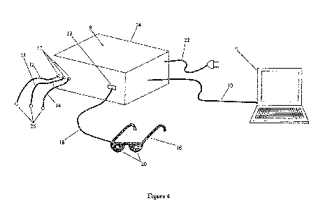
As shown in Figure 4, the disclosed apparatus may comprise a computing
device 8 that is operatively coupled to a neurostimulator 9 that is configured
to also
obtain neuroimaging data by, for example, measuring biopotential data such as
that
arising from EEG signals or other biopotential signals. Examples of suitable
computing devices are microprocessors or computers. However, any suitable
processing unit can be used as a computing device 8. These components are
coupled
to each other via electrical conduction paths such as a peripheral cable 10.
For
example, the neurostimulator 9 could be coupled to the computing device 8 with
an
RS232 cable, USB cable, etc.
As shown in Figure 1, the neurostimulator 9 may comprise a biopotential
acquisition device 15, at least one filtering unit 26, an isolation amplifier
27, and a
microcontroller 28. The neurostimulator 9 may be configured to transmit
biopotential
data such as EEG signal data to the biopotential acquisition device 15.
Additionally,
the biopotential acquisition device 15 may be configured to transmit the
biopotential
data through at least one filtering unit 26 and through the isolation
amplifier 27, with
the isolation amplifier 27 being operatively coupled to the microcontroller
28.
Furthermore, it is preferred that the isolation amplifier 27 be capable of
performing
"notch" filtering (i.e., to eliminate 60 Hz line noise). The isolation
amplifier 27 may
be of any suitable type known in the art. It is preferred that the filtering
unit 26
includes a circuit configured to filter data and/or a numerical filter.
The neurostimulator 9 may further comprise a series of electrical conductors
such as EEG sensors 11. The EEG sensors 11 may be configured to be attached to
a
subject, to monitor EEG signals of the subject, and/or to administer
neurostimulation
signals to the subject. Additionally, each EEG sensor 11 may comprise contact
electrodes 25 that may be disposed at the ends, and may include at least one
positive
lead 12, one negative lead 13 and one ground lead 14.
31
CA 02845688 2014-02-18
WO 2013/025997 PCT/US2012/051359
Employing multiple sets of the EEG sensors 11 simultaneously and multiple
biopotential acquisition devices 15 can accomplish acquisition of EEG signals
from
multiple sites on the scalp. For clarity, the apparatus is described herein
for
acquisition of EEG signal from one scalp site. One or more of the EEG sensors
11
may be connected to the neurostimulator 9 via electrical connectors such as
the EEG
sensor connectors 17.
The neurostimulator 9 may therefore comprise a biopotential acquisition
device 15 that may comprise an electric circuit configured to acquire
biopotential data
such as from the EEG signals obtained by the EEG sensors 11 attached to the
subject.
It is preferred that the subject be a mammal. It is further preferred that the
subject be a
primate and further preferred that the subject be a human being.
Additionally, the neurostimulator 9 may comprise an inductor 32 that may be
configured and positioned to act as a transformer, whereas the stimulation
signal may
be induced in the neurostimulator 9 by inducing electrical current into the
inductor 32,
which further induces electrical current in the EEG sensors 11 via
electromagnetic
coupling, and thereby into the subject.
The neurostimulator 9 may further comprise an optical unit, as shown at 16 in
Figure 4, as a possible means of delivering the stimulation signal. The
optical unit 16
may be electrically coupled to the neurostimulator 9 via optical device sensor
connectors 19 and an optical device cable 18. However, other means of
connecting
the optical unit to the neurostimulator are acceptable. The optical unit 16
further
comprises light generating devices 20 located to be in close proximity to the
subject's
eyes. In the preferred embodiment, the light generating devices 20 may be
light
emitting diodes.
With further reference to Figure 1 and Figure 4, the neurostimulator 9 is
operated by any number of possible power supply 22 sources. To assure
electrical
isolation for the patient's safety, an isolated power supply 23 is utilized in
the
32
CA 02845688 2014-02-18
WO 2013/025997 PCT/US2012/051359
preferred embodiment. Further, the neurostimulator 9 is housed in a protective
outer
enclosure 24.
The neurostimulator 9 preferably internally comprises the biopotential
acquisition device 15 and the biopotential acquisition device 15 is preferably
designed
to acquire biopotential data such as from EEG signal data, specifically
patient EEG, to
provide a means for analysis and data storage of the biopotential data through
computational means, generate a neurostimulation signal and deliver the
neurostimulation signal to the patient.
EEG signals may be acquired with EEG sensors 11 attached to a patient's
scalp. The contact electrodes 25 may be located at the ends of the EEG sensors
11 in
positions to be attached to the patient. The EEG signal is delivered to the
neurostimulator 9 via the EEG sensors 11, connected to the biopotential
acquisition
device 15 through EEG lead connectors 17, and operatively coupled to a
neuroimaging device such as a biopotential acquisition device 15. To minimize
the
effect of external electrical noise, any number of filtering units 26 may be
employed
in the preferred embodiment. To assure patient safety, the biopotential data
are passed
through the isolation amplifier 27. The output of the biopotential data, after
passing
through the biopotential acquisition device 15, filters 26 and isolation
amplifier 27 is
acquired by the microcontroller 28 through analog-to-digital ports 29. The
microcontroller 28 is operatively coupled to the computing device 8. One
method of
coupling the microcontroller 28 to the computing device is to use a peripheral
cable
10. Control of the neurostimulator 9 is accomplished by communication between
the
microcontroller 28 and the computing device 8. Further, the objective of
biopotential
data analysis and storage is accomplished computationally via communication
between the microcontroller 28 and the computing device 8.
After analysis of the acquired biopotential data such as the EEG signal, the
computing device 8 may communicate proper stimulation signal parameters to the
microcontroller 28. These parameters may include signal energy level,
frequency of
the low frequency component of an AMPWM signal, phase offset of multiple
signals,
start time, frequency offset and duration through a user interface. Utilizing
a digital-
33
CA 02845688 2014-02-18
WO 2013/025997 PCT/US2012/051359
to-analog port 30 on the microcontroller 28, the stimulation signal is output
from the
microcontroller 28 to transistors 31 or similar switching devices capable of
managing
the current levels of the stimulation signal. Depending on the mode of
stimulation
chosen by a clinician, the stimulation signal will be routed to the different
means of
stimulation signal delivery, alone or in combination. The parameters for the
clinician's
choice are set forth above.
If optical stimulation is desired, the stimulation signal will be sent to the
optical unit 16 featuring the light generating devices 20 to be worn by the
patient. Any
unit capable of emitting light may be used as a light generating device. This
includes,
but is not limited to a LED, a light bulb, a low-power laser, etc.
Alternately, if EEG
lead 11 stimulation is desired, where the stimulation signal is delivered to
the patient's
scalp via the attached electrodes 25, then the stimulation signal is sent to
the inductor
32, which may be configured and positioned to induce current in the EEG
sensors 11
from the stimulation signal generated by the microcontroller 28. A plurality
of
stimulation delivery modes may be warranted to allow for clinician choice to
further
effect successful treatment based on individual patient needs.
To assure patient safety, all electronics in the neurostimulator 9, including
the
biopotential acquisition device 15, the filter 26, the isolation amplifier 27,
the
microcontroller 28 and the transistors 31 may be supplied electricity by the
aforementioned isolation power supply 23.
Finally, regarding the coupling of the components, if a computing device is
used it is preferably operatively coupled to the processor of the
neurostimulator via
any of a number of means of commonly used peripheral communications
techniques,
such as serial communication, USB port communication or parallel communication
10. All remaining electronics are preferably operatively coupled to the
processing
device (e.g. microcontroller) in the neurostimulator. The data acquisition
circuit
preferably comprises the biopotential acquisition device 15, filters 26 and
isolation
circuitry (amplifier) 27. The isolation amplifier is preferably coupled to an
analog-to-
digital input port on the microcontroller 28, via electrical conduction paths
such as
wires or printed circuit board conductors. The filters 26 are preferably
operatively
34
CA 02845688 2014-02-18
WO 2013/025997 PCT/US2012/051359
coupled to the isolation amplifier 27 via electrical conduction paths such as
wires or
printed circuit board conductors. Further, the biopotential acquisition device
15 is
preferably operatively coupled to the filters 26 via electrical conduction
paths such as
wires or printed circuit board conductors.
A stimulation circuit is preferably coupled to a digital-to-analog port 30 on
the
microcontroller, in all cases via electrical conduction paths such as wires or
printed
circuit board conductors. It is preferred that an isolated power supply 23
supplies all
operative power for neuro stimulation outputs such as that to the optical
device 16 or
the EEG lead stimulation inducing circuitry 32. Electrical output from the
digital-to-
analog port 30 is preferably conducted to a transistor 31 that is further
coupled to the
isolated power supply 23. When a signal is received at the base of the
transistor 31
from the microcontroller 28, the transistor operates to switch on electricity
from the
isolated power supply 23 which is further conducted via electrical coupling to
the
inductor (stimulation inducing circuitry) 32. Current flow in the inductor 32
induces a
current in the EEG lead, as described in the specification.
Alternately, for photic stimulation, the isolated power supply 23 is
preferably
coupled via electrical coupling to two more transistors 31, which are
preferably
operatively coupled via electrical coupling to independent digital-to-analog
ports 30
on the microcontroller 28. Electricity conducted from the digital-to-analog
ports 30 to
the base of the transistors 31 in the photic stimulation circuit has the
effect of
switching on these transistors, further allowing for conduction of electricity
to the
photic stimulation devices, such as LEDs 21. The photic stimulation devices
are
preferably coupled to the transistors 31 via electrical connectors 19, thus
providing for
current flow to the photic stimulation devices such as LEDs 21.
Finally, it is preferred that the apparatus operate on a 12 volt power supply.
It
is more preferred that the apparatus operate on a 6 volt power supply. It is
most
preferred that the apparatus operate on a power supply equivalent to the
lowest power
supply requirement of the components used.
CA 02845688 2014-02-18
WO 2013/025997 PCT/US2012/051359
With reference to Figures 5-7, a form of electrical signal for stimulating
tissues is disclosed wherein an electrical signal of relatively high frequency
(Figure 5)
is amplitude modulated by an electrical signal of relatively low frequency
(Figure 6),
combining to form an electrical signal of the general form shown in Figure 7.
As
discussed above, using pulse width modulation for the purpose of varying the
duty
cycle of the electrical signal of relatively high frequency, the time-averaged
current
deliverable by that signal can be controlled Hence, Figure 7 shows an example
of one
embodiment of an amplitude modulated pulse width modulated (AMPWM) signal in
which the signal of relatively low frequency shown in Figure 6 and the signal
of
relatively high frequency shown in Figure 5 form an AMPWM signal shaped
similar
to a square wave pulse train.
However, an AMPWM signal may combine signals of shapes other than
square waves. For example, Figure 8 shows a signal of relatively low frequency
that
has a general sinusoidal form. When used to amplitude modulate a signal of
relatively
high frequency, a resulting AMPWM signal equivalent is that shown in Figure 9.
An AMPWM signal may also be created from multiple relatively low
frequency components. A signal with multiple frequency components can be
created
using methods such as inverse Fourier Transform theory. Figure 10 shows an
example
of a composite sinusoidal signal with three relatively low frequency
components that
are created using an inverse Fourier Transform. Such relatively low frequency
components may be selected to provide therapeutic electrical stimulation. One
anticipated benefit of creating such a composite signal is to provide for
therapeutic
electrical stimulation that has multiple frequency-dependent beneficial
effects on the
tissues to which it is applied. When a composite signal such as that
illustrated in
Figure 10 is used to amplitude modulate a signal of relatively high frequency,
a
resulting AMPWM signal equivalent is that shown in Figure 11.
In other words, the tissue stimulation signal may comprise one or more
parametric values tailored in response to neuroimaging data obtained from
tissues in a
target region of the brain of a subject suffering from one or more symptoms
associated with central sensitivity, and deliverable to the target region of a
subject's
36
CA 02845688 2014-02-18
WO 2013/025997 PCT/US2012/051359
brain for use in alleviating the symptoms. The stimulation signal may be
adapted for
use in alleviating symptoms associated with central sensitivity by tailoring
the signal
to mitigate a central sensitivity condition causing the symptoms, and may be
an
amplitude modulated pulse width modulated (AMPWM) signal for use in
penetrating
tissues between the signal source and the target region of the subject's
brain. The
signal may also or alternatively be a composite signal comprising two or more
components configured for use in providing two or more frequency-dependent
beneficial effects on tissues in a target region of a subject's brain.
The stimulation signal may be tailored in response to neuroimaging data
obtained from tissues in a target region of the brain of a subject suffering
from one or
more of the following central sensitivity symptoms: pain, musculoskeletal
pain, pain
at multiple sites, generalized hyperalgesia, stifthess, swollen feeling in
soft tissues,
fatigue, poor sleep, paresthesia, anxiety, chronic headaches, tension
headaches,
dysmenorrhea, irritable bowel syndrome, periodic limb movements, symptoms of
restless leg syndrome, depression, symptoms of Sjogren's syndrome, symptoms of
Raynaud's Phenomenon, symptoms of female urethral syndrome, impaired memory,
impaired concentration, cognitive impairment, tender cervical lymph nodes,
tender
axillary lymph nodes, post-exertion malaise, tender points, sensory
hypersensitivity,
sleep disturbances, immune dysfunction, history of viral illness,
neurohormonal
dysfunction, neuroendocrine dysfunction, or a lack of macroscopic or
microscopic
pathological findings in peripheral tissues. The stimulation signal may
alternatively be
tailored in response to neuroimaging data obtained from the brain of a subject
exhibiting, in one or more regions of the subject's brain in response to one
or more
applied peripheral stimuli, any one or more of the following abnormal brain
activities:
abnormal function, abnormal response, abnormal regions of activation, abnormal
network connectivity, abnormal release of neurochemicals, abnormal uptake of
neurochemicals, abnormal electrical activity or abnormal metabolism. The
signal may
be tailored for use in altering any one or more of the brain activities
exhibited by the
subject's brain.
Additional apparatus that provide for the generation of electrical tissue
stimulation signals, such as AMPWM signals, that reduce tissue impedance and
37
CA 02845688 2014-02-18
WO 2013/025997 PCT/US2012/051359
increase depth of signal penetration are disclosed herein. A first embodiment
of a
tissue stimulation apparatus for providing an electrical tissue stimulation
signal that
reduces tissue impedance and increases depth of signal penetration is shown in
Figure
12, as comprising a stimulation device 101 and a computing device 102 is
provided.
Power for the stimulation device 101 may be provided by an external power
source
105, such as a line connection or an adapter for providing a conditioned
electrical
source, electrically coupled to the stimulation device 101 through a power
connector
111.
Where, as shown, the stimulation device 101 is an electrical stimulation
device, it may include a battery charger and switching circuit 107
electrically coupled
to the power connector 111, enabling the receipt of electricity from the
external power
source 105. A battery 108 may also be electrically coupled to the battery
charger and
switching circuit 107. The battery 108 may further be connected to other
circuits of
the electrical stimulation apparatus through the battery charger and switching
circuit
107 and used to provide electrical power to the other circuits at times when
isolation
from line current is required or advantageous for operation of the apparatus,
such as in
times when the apparatus is being used to provide electrical stimulation to a
subject.
In practice, electrical isolation may be accomplished through a switching
portion of
the battery charger and switching circuit 107, which may be further
electrically
coupled to a controller or processor 103 configured to control various
functions of the
electrical stimulation device 101 such as electrical signal generation and as
is further
described herein. Programmed firmware, associated with processor technologies,
for
example, may provide for electrical signals to be sent from the processor 103
to
control the switching portion of the battery charger and switching circuit 107
and to
electrically decouple the electrical stimulation device 101 from the external
power
source 105 when isolation is required or desirable. At times when isolation is
not
required or desirable, such components as the processor 103, external power
source
105 and battery charger, and switching circuit 107 may be used to recharge the
battery
108 in preparation for subsequent use. In other words, the processor 103 may
be
configured to command the switching portion of the battery charger and
switching
circuit 107 to couple the external power source 105 to the battery 108 when
isolation
of the electrical stimulation device 101 is not required or desirable and to
decouple the
38
CA 02845688 2014-02-18
WO 2013/025997 PCT/US2012/051359
external power source 105 from the battery 108 when isolation is required or
desirable. This coupling may be accomplished either as a result a signal being
sent to
a processor 103 arising from a manual input such as the manual decoupling of
an
external power source 105 from line power, or automatically arising from a
software
signal being sent to a processor 103 whenever an operator utilizes a software
interface
for using the apparatus to electrically stimulate a subject. In other words,
the
processor 103 may be programmed to automatically decouple external power in
response to an operator's use of a software interface to use the apparatus to
electrically
stimulate a subject.
The battery 108 or other power source may subsequently energize a power
regulation circuit 109 that further provides conditioned power to other
circuits of the
electrical stimulation device 101 and a common reference ground that may be
used by
all circuits. A ground connector 112 may be used to provide electrical
coupling to
external circuits, such as those described herein, for common grounding
purposes.
As is also shown in the embodiment of Figure 13, conditioned power from the
power regulation circuit 109 may further be used to energize the processor
103,
whereupon a circuit for creating or generating an electrical signal for
stimulating
tissues is realized. This stimulation signal generation circuit may comprise
the
processor 103, a digital-to-analog (D/A) converter 104, a signal conditioning
and
amplification circuit 106, a stimulation switching circuit 110, and a first
ground
switching circuit 119. Further, the tissue stimulation apparatus may include a
computing device 102 coupled to the processor 103 through any suitable
computer
data cable 118 or similar interface, such as a wireless interface. The
computing device
102 may, as shown, be an external computing device and may provide and be used
as
a user interface via software, and may provide for communication between a
user and
the processor 103, such communication comprising the flow of any and all forms
of
data and control signals to set and modify operational parameters of the
electrical
stimulation device 101. In other words, the computing device 102 is programmed
to
exchange data and control signals with the processor and to allow a user to
modify
operational parameters of the electrical stimulation apparatus.
39
CA 02845688 2014-02-18
WO 2013/025997 PCT/US2012/051359
In other words, a tissue stimulation apparatus is provided for use in
alleviating
symptoms associated with central sensitivity in a subject. The apparatus may
comprise a neuroimaging device configured to obtain neuroimaging data from
tissues
in a target region of a brain of a subject suffering from one or more symptoms
associated with central sensitivity, a stimulation device 101 including a
stimulation
signal generation circuit configured to generate and deliver a tissue
stimulation signal
to the target region of the subject's brain, and a computing device 102
configured to
set one or more parametric values of the electrical tissue stimulation signal
in
response to neuroimaging data obtained by the neuroimaging device. The
stimulation
device 101 may be configured to administer magnetic and/or electrical
stimulation to
a target region of the brain of the subject. Where electrical stimulation is
administered, the electrical stimulation signal may be an AMPWM signal. The
stimulation device may be configured to deliver the stimulation signal to a
target
region of the brain from outside the subject and to transmit the signal
through
intervening tissues.
The apparatus may be used in alleviating an one or more of the following
symptoms associated with central sensitivity: pain, musculoskeletal pain, pain
at
multiple sites, generalized hyperalgesia, stifthess, swollen feeling in soft
tissues,
fatigue, poor sleep, paresthesia, anxiety, chronic headaches, tension
headaches,
dysmenorrhea, irritable bowel syndrome, periodic limb movements, symptoms of
restless leg syndrome, depression, symptoms of Sjogren's syndrome, symptoms of
Raynaud's Phenomenon, symptoms of female urethral syndrome, impaired memory,
impaired concentration, cognitive impairment, tender cervical lymph nodes,
tender
axillary lymph nodes, post-exertion malaise, tender points, sensory
hypersensitivity,
sleep disturbances, immune dysfunction, history of viral illness,
neurohormonal
dysfunction, neuroendocrine dysfunction, or a lack of macroscopic or
microscopic
pathological findings in peripheral tissues. The apparatus may also or
alternatively be
used in alleviating one or more symptoms associated with central sensitivity
in a
subject exhibiting, in one or more regions of the subject's brain in response
to one or
more peripheral stimuli, any one or more of the following brain activities:
abnormal
function, abnormal response, abnormal regions of activation, abnormal network
connectivity, abnormal release of neurochemicals, abnormal uptake of
CA 02845688 2014-02-18
WO 2013/025997 PCT/US2012/051359
neurochemicals, abnormal electrical activity or abnormal metabolism; the
apparatus
being configurable to alter the one or more brain activities exhibited by the
subject's
brain by stimulating the target region of the brain.
The disclosed apparatus and methods may be implemented using hardware,
software or a combination thereof and may be implemented in one or more
computer
systems or other processing systems. According to one embodiment, one or more
computer systems capable of carrying out the functionality described herein
are
contemplated. An example of such computer system is shown at 200 in Figure 27.
The computer system 200 includes at least one processor 204 that is connected
to a communication infrastructure 206 (e.g., a communications bus, cross-over
bar, or
network). Any suitable software embodiments may be used with this exemplary
computer system, and the disclosed apparatus and methods may be implemented
using any suitable computer system and/or architectures.
The computer system 200 may include a display interface 202 that forwards
graphics, text, and other data from the communication infrastructure 206 or
from a
frame buffer (not shown) for display on a display unit 230. The computer
system 200
may also include a main memory 208, preferably random access memory (RAM), and
may also include a secondary memory 210. The secondary memory 210 may include,
for example, a hard disk drive 212 and/or a removable storage drive 214 such
as a
floppy disk drive, a magnetic tape drive, or an optical disk drive, etc. The
removable
storage drive 214 may be configured to read from and/or writes to a removable
storage unit 218 in a well-known manner. The removable storage unit 218 may
include a floppy disk, magnetic tape, optical disk, etc., which may be read by
and
written to the removable storage drive 214. The removable storage unit 218 may
include a computer usable storage medium having stored therein computer
software
and/or data.
In alternative embodiments, the secondary memory 210 may include other
similar devices for allowing computer programs or other instructions to be
loaded into
computer system 200. Such devices may include, for example, a removable
storage
41
CA 02845688 2014-02-18
WO 2013/025997 PCT/US2012/051359
unit 222 and an interface 220. Examples of such may include a program
cartridge and
cartridge interface (such as that found in video game devices), a removable
memory
chip (such as an erasable programmable read only memory (EPROM), or
programmable read only memory (PROM)) and associated socket, and other
removable storage units 222 and interfaces 220, which allow software and data
to be
transferred from the removable storage unit 222 to the computer system 200.
The computer system 200 may also include a communications interface 224.
The communications interface 224 may be configured to allow software and data
to
be transferred between the computer system 200 and external devices. The
communications interface 224 may include a modem, a network interface (such as
an
Ethernet card), a communications port, a Personal Computer Memory Card
International Association (PCMCIA) slot and card, etc. Software and data
transferred
via communications interface 224 are in the form of signals 228, which may be
electronic, electromagnetic, optical or other signals capable of being
received by
communications interface 224. These signals 228 are provided to communications
interface 224 via a communications path (e.g., channel) 226. This path 226
carries
signals 228 and may be implemented using wire or cable, fiber optics, a
telephone
line, a cellular link, a radio frequency (RF) liffl( and/or other
communications
channels. In this document, the terms "computer program medium" and "computer
usable medium" are used to refer generally to media such as a removable
storage
drive 214, a hard disk installed in hard disk drive 212, and signals 228.
These
computer program products provide software to the computer system 200. The
disclosed apparatus and methods may include such computer program products.
Computer programs (also referred to as computer control logic) are stored in
main memory 208 and/or secondary memory 210. Computer programs may also be
received via communications interface 224. Such computer programs, when
executed,
enable the computer system 200 to perform according to the features of the
disclosed
apparatus and methods. The computer programs, when executed, enable the
processor
204 to perform according to the features of the disclosed apparatus and
embodiments.
Accordingly, such computer programs serve as controllers of the computer
system
200.
42
CA 02845688 2014-02-18
WO 2013/025997 PCT/US2012/051359
In an apparatus or method embodiment that includes the use of software, the
software may be stored in a computer program product and loaded into computer
system 200 using the removable storage drive 214, the hard drive 212, or the
communications interface 224. The control logic (software), when executed by
the
processor 204, causes the processor 204 to perform according to the functions
of the
disclosed apparatus and methods. The disclosed apparatus and methods may be
implemented primarily in hardware using, for example, hardware components,
such
as application specific integrated circuits (ASICs). Implementation of the
hardware
state machine to perform the functions described herein will be apparent to
persons
skilled in the relevant art(s).
Alternatively, the disclosed apparatus and methods may be implemented using
a combination of both hardware and software.
In some embodiments, and as shown in Figure 12, generating an electrical
signal for stimulating tissues may begin with signal parameters being
established
through various software methods used in an external computing device 102 and
communicated to a processor 103 via any suitable data cable 118 or similar
interface,
such as a wireless interface. In other words, the external computing device
102 is
configured to establish parameters of the electrical signals generated by the
electrical
stimulation device 101. Such signal parameters include, but are not limited to
waveform, frequency components, phase, pulse width, duty cycle, and amplitude
components such as minimum amplitude, maximum amplitude, and offset voltage.
Various methods of establishing signal parameters may be used with the
electrical
stimulation device 101.
Upon establishment of signal parameters in a processor 103, along with
establishment of other operational parameters, such as the aforementioned
decoupling
of an external power source 105, signals are sent from the processor 103 to a
D/A
converter 104, whereupon an analog voltage representing an electrical signal
for
stimulating tissues is first achieved. The analog voltage is further provided
to an
electrically coupled signal conditioning and amplification circuit 106, where
a
43
CA 02845688 2014-02-18
WO 2013/025997 PCT/US2012/051359
substantially equivalent signal is created with advantageous enhancements such
as,
but not limited to, increased voltage amplitude, decreased signal-to-noise
ratio, and
increased current capability.
In some embodiments, provisions may be made to the electrical stimulation
apparatus for the selective control of the delivery of an electrical signal
for
stimulating tissues to a plurality of stimulation connectors 113. A
stimulation
switching circuit 110 is electrically coupled to the processor 103, whereupon
control
signals from the processor 103 allow for the signal from the signal
conditioning and
amplification circuit 106 to be advantageously switched to any number of
independent electrical conductors or conduction paths. Further, the
independent
electrical conductors or conduction paths are electrically coupled with a
first ground
switching circuit 119, the first ground switching circuit 119 being further
electrically
coupled to the processor 103. Control signals from the processor 103 allow for
selective switching of the independent conductors to an apparatus ground
point,
providing advantageous control of the independent conductors' use as either a
conduction path for an electrical signal for stimulating tissues or a ground.
Further
electrical conduction paths are provided for each independent conductor
passing
through a first ground switching circuit 119, with each independent conductor
terminating at one of a plurality of stimulation connectors 113.
The apparatus may include a number of electrical conductors that provide
electrical coupling between a number of connectors and input/output (I/O)
ports of a
processor 103 in the electrical stimulation device 101 for the embodiments
shown.
Specifically, an auxiliary power supply connector 114 may be provided. The
apparatus may include a switch comprising an electrical conductor first
connected to
an auxiliary power supply connector 114 then to a switch, then via another
electrical
conductor to an auxiliary I/O connector 116. The switch may be used for
various
purposes to indicate an event to the processor 103. One exemplary purpose is
the use
of the switch by a subject receiving electrical stimulation to mark a point in
time of
any particular interest.
44
CA 02845688 2014-02-18
WO 2013/025997 PCT/US2012/051359
The electrical stimulation device 101 may also include a plurality of
conductors or control I/O connectors 115 that provide electrical coupling to
I/O ports
of the processor 103. Specifically, the control I/O connectors 115 may provide
control
signals between the processor 103 and various electrical apparatus or
peripheral
devices coupled to the electrical stimulation device 101, examples of which
are
described further herein. The apparatus may further include a number of lead
test
ports 117 electrically coupled to the processor 103 for electrically coupling
electrical
conductors or other couplings, to the processor 103 for the purpose of testing
the
electrical conducting integrity of any combination of such electrical
conductors, or
other couplings, such as wires combined with sensors, such as surface
electrodes,
henceforth referred to as "leads", used to conduct electrical energy between
tissues
and the electrical stimulation device 101.
As is also shown in Figure 13, the electrical stimulation device 101 may
include one or more ground leads 120, a plurality of stimulation leads 121,
and
provision at a terminating end of all leads for an electrode 122 adapted to be
placed on
tissues in either an invasive or non-invasive way. The apparatus also has
provision for
one or more external stimulation devices, such as an optical device 123,
electromagnetic device 170, electromechanical device 171 or an audio device
172,
electrically coupled by one or more external stimulation device cables 124. As
shown
in Figure 13 the external stimulation devices may include an optical device
123
comprising eyeglasses adapted with illuminating or similar photic devices,
such as
light emitting diodes, or with displays for showing digital images to a
subject
undergoing therapy. The external stimulation devices may include an audio
device
172 adapted to play music during therapeutic activity.
In operation, the apparatus of Figure 13 provides stimulation from the
electrical stimulation device 101 to tissues disposed between stimulation
leads 121
and ground leads 120 such that an approximate vector path of electrical
current flow
extends between electrodes 122 associated with the stimulation leads 121 and
electrodes 122 associated with the ground leads 120.
CA 02845688 2014-02-18
WO 2013/025997 PCT/US2012/051359
The processor 103 may be programmed to provide control signals that
selectively control the stimulation switching circuit 110 and the first ground
switching
circuit 119 to cause the leads 121 to serve as either stimulation leads
delivering
stimulation or as ground leads serving as ground sources in such a way as to
create
multiple spatial paths of electrical stimulation through tissues.
In addition, as shown in Figure 13, stimulation may be provided by an external
stimulation device 123 operatively coupled to a stimulation connector 113 that
is
being used as an active stimulation electricity source through control of a
stimulation
switching circuit 110 by signals from a processor 103.
In addition, in the apparatus shown in Figure 13, electrical conducting
integrity of any stimulation lead 121, any ground lead 20, or any external
stimulation
device 123 may be tested by effecting physical contact between a lead,
preferably by
providing mechanical connection between a lead's conduction interface such as
an
electrode 122 and a lead test port 117. In testing for electrical conducting
integrity, a
processor 103 may be selectively used to output an electrical signal of known
properties to a lead 121 being tested, whereupon the electrical signal
conducted by the
lead being tested can be acquired by the processor 103 through a lead test
port 117.
Any number of suitable analyses may be conducted, whereupon processor
firmware,
for example, makes a comparison between the electrical signal of known
properties
and the signal conducted through a lead being tested in order to determine the
electrical conducting integrity of the lead.
As shown in Figure 14, a second embodiment of tissue stimulation apparatus
for providing an electrical tissue stimulation signal that reduces tissue
impedance and
increases depth of signal penetration is shown as comprising an electrical
stimulation
device 101 and a neuroimaging device such as a biopotential acquisition device
that
measures biopotential voltage in tissue to be stimulated. Where the
neuroimaging
device is a biopotential acquisition device, the device may include a
biopotential
amplifier module 127 comprising a biopotential amplifier 130, an impedance
testing
circuit 131, a second ground switching circuit 129 and a series of inductors
128
operatively coupled to conductors extending from the second ground switching
circuit
46
CA 02845688 2014-02-18
WO 2013/025997 PCT/US2012/051359
129 and terminating at biopotential acquisition lead connectors 126 and thus
operatively coupled to biopotential acquisition leads 125 coupled to the
connectors
126. Further provisions may be made for any number of biopotential acquisition
leads
125, and any number of ground leads 120, each lead 125, 120 including a sensor
such
as a surface electrode 122 adapted to be placed on tissues. Further provisions
may be
made for electrical coupling of a biopotential amplifier module 127 to the
electrical
stimulation device 101 through stimulation lead connectors 113, an auxiliary
power
supply connector 114, control I/O connectors 115, and auxiliary I/O connectors
16 of
the electrical stimulation device 101.
In an exemplary operation, the apparatus of Figure 14 provides stimulation
from the electrical stimulation device 101 to tissues, whereupon a
biopotential voltage
is measured by the biopotential amplifier 130 operatively coupled to any
number of
biopotential acquisition leads 125 and any number of ground leads 120 having
electrodes 122 adapted to be placed on tissues, the biopotential voltage
including, but
not being limited to, electroencephalographic (EEG) voltage, electromyographic
(EMG) voltage, and electrocardiographic voltage.
In the apparatus of Figure 14, an electrical signal for stimulating tissues
may
be induced using the inductors 128 disposed adjacent the independent
conductors
extending from the second ground switching circuit 129 and terminating at
biopotential acquisition lead connectors 126, the electrical signal being
provided by
the electrical stimulation device 101, and the inductors 128 being
electrically coupled
to the electrical stimulation device 101 at stimulation connectors 113,
whereupon
selective control of the electrical signal for stimulating tissues is
accomplished as
previously disclosed herein. In other words, the biopotential acquisition
device
includes one or more inductors 128 electrically coupled to the electrical
stimulation
device 101 and operatively coupleable to one or more respective biopotential
acquisition leads 125, the electrical stimulation device and inductors being
configured
to selectively deliver tissue stimulation signals through the one or more
biopotential
acquisition leads of the biopotential acquisition device.
47
CA 02845688 2014-02-18
WO 2013/025997 PCT/US2012/051359
In the apparatus of Figure 14, data transfer of acquired biopotential voltage
may be provided between the processor 103 and the biopotential amplifier 130
through any I/O port, such as a control I/O connector 15 or an auxiliary I/O
connector
116. In certain embodiments, the biopotential voltage data may be used at any
time to
determine or alter parametric values of an electrical signal for stimulating
tissues,
such as via analysis using software in an external computing device 102 with
subsequent control data being sent from the external computing device 102 to a
processor 103 in an electrical stimulation device 101. In other words, the
external
computing device 102 is configured to determine parametric value of an
electrical
tissue stimulation signal in response to biopotential voltage data obtained by
the
biopotential acquisition device and to send corresponding control data to the
processor 103.
In the apparatus of Figure 14, the processor 103, for example, of the
electrical
stimulation device 101 may selectively sample biopotential voltage data from
the
biopotential amplifier 130 of the biopotential acquisition device at times of
minimal
electrical stimulation signal amplitude, preferably zero amplitude, within the
period of
a high frequency signal component of an AMPWM signal. Thus, the biopotential
acquisition leads 125 may be used for the dual purpose of both acquiring
biopotential
voltage and delivering an electrical signal for stimulating tissues at
overlapping, or
simultaneous, times. The frequencies of a high frequency signal component of
an
AMPWM signal may be selected to be multiples of integral powers of two,
including
but not limited to integral multiples of 256 (i.e. 28) such as for example
14,336 hertz
(256×56) and 16,384 hertz (256×64). Such selection of frequencies
facilitates mathematical analysis of acquired biopotential voltage data. Such
mathematical analysis may include a Fourier Transform analysis whereupon a
number
of samples per second equal to an integral power of two may be preferred. In
the
examples of AMPWM signal high frequency component frequencies of 14,336 hertz
and 16,384 hertz given, sampling rates for biopotential voltage data of 2,048,
1,024,
512, 256 and 128 samples per second are readily achieved within equally spaced
intervals of minimal electrical stimulation signal amplitude in the AMPWM
signal.
48
CA 02845688 2014-02-18
WO 2013/025997 PCT/US2012/051359
In the apparatus of Figure 14, the second ground switching circuit 129 may be
operatively coupled to the electrical stimulation device 101 using a control
I/O
connector 15. Operationally, the second ground switching circuit 129 receives
control
signals from the processor 103, which allows for selective switching of any
biopotential acquisition lead 125 to an apparatus ground point, permitting
advantageous control of the biopotential acquisition lead's 125 use as either
a
conduction path for an electrical signal for stimulating tissues, a conduction
path for a
biopotential voltage to the biopotential amplifier 130, or a ground. Among
other
things, such selective switching of a biopotential acquisition lead 125
permits
selective use as a reference lead to the biopotential amplifier 130 or as a
differential
lead to the biopotential amplifier 130, facilitating differential comparison
of
biopotential voltages at more than one acquisition site on a tissue.
In the apparatus of Figure 14, an impedance testing circuit 131 may be
included in the biopotential acquisition device and operationally coupled to
the
biopotential amplifier 130. The impedance testing circuit 131 may also be
coupled to
the electrical stimulation device 101 using auxiliary I/O connectors 16. In
such use,
the impedance testing circuit 131 may be used to monitor the impedance of
tissues in
mechanical contact with biopotential acquisition leads 125 and a ground lead
20, each
comprising an electrode 122 adapted to be placed on the tissues. Data
representing the
impedance of tissues is transferred to the processor 103 of the electrical
stimulation
device 101 via electrical coupling, for example. The data representing
impedance of
tissues may be used to determine or alter parametric values of an electrical
signal for
stimulating tissues through, for example, analysis using software in the
external
computing device 102, with subsequent control data being sent from the
external
computing device 102 to the processor 103 in the electrical stimulation device
101.
The data representing impedance of tissues and ongoing monitoring for
biopotential voltage integrity, such as, but not limited to, EEG measurement
integrity,
may be used to determine or alter parametric values of an electrical signal
for
stimulating tissues, such as an AMPWM signal.
49
CA 02845688 2014-02-18
WO 2013/025997 PCT/US2012/051359
The use of methods to monitor for biopotential voltage integrity accomplishes
various means of guiding a user and assuring improved biopotential signal data
throughout an acquisition time period. For example, the apparatus may include
an
alert for notifying a user if integrity is lost during treatment. Such alert
may be
provided, for example, via software analysis in an external computing device
102. In
another embodiment, such alert may be sent to a remote indicator such as a
pager
worn by a user. Further, the apparatus may include various means of indicating
to a
user when good biopotential voltage integrity is achieved as biopotential
acquisition
leads 125 and ground leads 120 are first being applied to tissues, prior to
the
acquisition of data. Such indicators may be provided, for example, via graphic
user
interface software in an external computing device 102 or via any number of
hardware indication means.
With reference to Figure 15, another embodiment of a tissue stimulation
apparatus for providing an electrical tissue stimulation signal that reduces
tissue
impedance and increases depth of signal penetration is shown as comprising an
electrical stimulation device 101 and an adjunct electrical stimulation
apparatus 132
to be used with an independent biopotential voltage measurement apparatus,
such as,
but not limited to, an EEG measurement apparatus 137. Under normal operating
conditions, an EEG measurement apparatus 137 is typically used only for the
purposes of acquiring EEG voltage data and for providing such data to an
external
computing device 102 through any data cable 138 or other coupling capable of
sufficiently transferring the data. Acquisition of the EEG voltage is normally
accomplished through any number of leads electrically coupled to an EEG
measurement apparatus 137 at an interface 139, for example. Such number of
leads
may include an EEG sensor set 136 comprising, but not being limited to, a
series of
conductors, a series of electrodes and features for positioning the
electrodes, such as
via integration of such sensors in a cap adapted to be worn by a subject. In
other
words, the tissue stimulation apparatus may comprise a sensor set 136, an
independent
biopotential voltage measurement apparatus 137, and an adjunct electrical
stimulation
apparatus 132 operatively connected between the sensor set 136 and the
independent
biopotential voltage measurement apparatus. The independent biopotential
voltage
measurement apparatus 137 may be operatively coupled to the electrical
stimulation
CA 02845688 2014-02-18
WO 2013/025997 PCT/US2012/051359
device 101, and may be configured to transmit through stimulation connectors
113 to
the sensor set, electrical tissue stimulation signals received from the
electrical
stimulation device 101, to transmit biopotential voltage from the sensor set
136 to the
independent biopotential voltage measurement apparatus 137, and to receive
control
signals from the processor 103 of the electrical stimulation device 101
through control
I/O connectors 115.
The exemplary apparatus illustrated in Figure 15 enables use of an
independent biopotential voltage measurement apparatus, such as, but not
limited to,
an EEG measurement apparatus 137, within an apparatus for providing an
electrical
signal for stimulating tissues. This use may be accomplished by placing an
adjunct
electrical stimulation apparatus 132 operatively between an EEG sensor set 136
and
an EEG measurement apparatus 137. The adjunct electrical stimulation apparatus
132
may include an adjunct switching control 135 operatively coupled to a
processor 103
of an electrical stimulation device 101 using control I/O connectors 115. The
adjunct
electrical stimulation apparatus may also include a series of EEG lead
conductors 142
and matched transfer conductors 140, for example, along with a series of
adjunct
switching circuits 133 operatively coupled to the adjunct switching control
135 via
switching control conductors 141, and further operatively coupled to
stimulation
connectors 113 of the electrical stimulation device 101.
In operation, the apparatus of Figure 15 provides for an adjunct electrical
stimulation apparatus 132 operatively coupled to an electrical stimulation
device 101
to both receive electrical signals through stimulation connectors 113 for
stimulating
tissues and to transfer control signals to a processor 103 through control I/O
connectors 115. The adjunct electrical stimulation apparatus 132 may be
further
operatively coupled to an EEG sensor set 136 at a cable interface connector
134 for
receiving EEG voltage. The adjunct electrical stimulation apparatus 132 may be
further operatively coupled to an EEG measurement apparatus 137 at an
interface 139
such as the same connecting features provided by an EEG sensor set 136.
With reference to Figures 15 and 16, a series of adjunct switching circuits
133
may be provided, each comprising any substantial circuit for switching 143,
for
51
CA 02845688 2014-02-18
WO 2013/025997 PCT/US2012/051359
example, that provides a selectable conduction pathway for an EEG lead
conductor
142 between (a) an electrical signal for stimulating tissues, such as provided
by an
electrical stimulation device 101 through electrical coupling at stimulation
connectors
113, (b) a transfer conductor 140 terminated at an interface 139 and further
provided
to an independent EEG measurement apparatus 137, or (c) a ground. Further
provision made in the adjunct switching circuit 133 may include switching
control
conductors 141 electrically coupled to an adjunct switching circuit 135, which
may be
used, for example, to determine the state of the adjunct switching circuit 133
and
therefore the conduction path provided to the EEG lead conductor 142.
As shown in Figure 15, the electrical stimulation device 101 may be combined
with an adjunct electrical stimulation apparatus 132 and biopotential voltage
measurement apparatus, such as an EEG measurement apparatus 137. At times, for
example, when a biopotential voltage measurement is required, biopotential
voltage
from a particular EEG lead conductor 142 may be directed to a transfer
conductor 140
by selective switching via an adjunct switching control 135 operated by the
processor
103 in the electrical stimulation device 101. Alternately, at times, such as
when an
electrical signal for stimulating tissues is required, the signal may be
directed from a
stimulation connector 113 to a particular EEG lead conductor 142 by selective
switching from an adjunct switching control 135 operated by the processor 103
in the
electrical stimulation device 101. Alternately, at times, such as when a
particular EEG
lead conductor 142 is to be grounded, selective switching from an adjunct
switching
control 135 operated by the processor 103 in the electrical stimulation device
101 may
be used to electrically couple the EEG lead conductor 142 to ground. In other
words,
the processor 103 of the electrical stimulation device 101 and the adjunct
switching
control may direct biopotential voltage from selected electrodes of the sensor
set 136
to the biopotential measurement apparatus 137 by selective switching via the
adjunct
switching control 135 operated by the processor 103 when a biopotential
voltage
measurement is required, may direct tissue stimulation signals from the
electrical
stimulation device 101 through selected stimulation connectors 113 to
corresponding
electrodes of the sensor set 136 through respective EEG lead conductors 142 by
selective switching via the adjunct switching control 135 operated by the
processor
103 when tissue stimulation is required, and may couple selected electrodes of
the
52
CA 02845688 2014-02-18
WO 2013/025997 PCT/US2012/051359
sensor set 136 to ground by selective switching via the adjunct switching
control 135
operated by the processor 103 when grounding of an electrode is required.
As shown in Figure 14, inductors 128 and a second ground switching circuit
129 of the apparatus of Figure 14 may be replaced, for example, by an adjunct
switching circuit 133 and an adjunct switching control 135 to control the use
of
individual leads. In other words, the biopotential acquisition device of
Figure 14 may
be modified to include at least one adjunct switching circuit 133 and an
adjunct
switching control 135 electrically coupled to the electrical stimulation
device 101,
with the adjunct switching circuit 133 being operatively coupled to at least
one
biopotential acquisition lead 125, the electrical stimulation device 101 and
an adjunct
switching control 135 selectively connecting the electrical stimulation device
101 to
selected leads to transmit tissue stimulation signals to the selected leads
and
connecting selected leads to the biopotential amplifier 130 to transmit
biopotential
voltages to the biopotential amplifier 130.
Accordingly, as shown in Figure 17, the tissue stimulation apparatus may
comprise an electrical stimulation device 101 and a biopotential amplifier and
switching module 155, and the module may further comprise a biopotential
amplifier
130, an impedance testing circuit 131, a series of EEG lead conductors 142
operatively coupled to conductors terminating at biopotential acquisition lead
connectors 126, matched transfer conductors 140, a series of adjunct switching
circuits 133 operatively coupled to the adjunct switching control 135 via
switching
control conductors 141, and further operatively coupled to stimulation
connectors 113
of an electrical stimulation device 101. Further provisions may be made for
any
number of biopotential acquisition leads 125, and any number of ground leads
120,
and a mechanism that may be used with the leads to provide for electrodes 122
adapted to be placed on tissues. Further provisions may be made for electrical
coupling of a biopotential amplifier and switching module 155 to the
electrical
stimulation device 101 through stimulation connectors 113, auxiliary power
supply
14, control I/O connectors 115 and auxiliary I/O connectors 16.
53
CA 02845688 2014-02-18
WO 2013/025997 PCT/US2012/051359
In an exemplary operation, the apparatus of Figure 17 provides stimulation
from the electrical stimulation device 101 to tissues, whereupon a
biopotential voltage
may be measured by a biopotential amplifier 130 operatively coupled through an
adjunct switching circuit 133, transfer conductor 140 and EEG lead conductor
142 to
any number of biopotential acquisition leads 125, any number of ground leads
120
and the electrode 122 adapted to be placed on tissues. The biopotential
voltage may
include, but is not limited to including, electroencephalographic (EEG)
voltage,
electromyographic (EMG) voltage, and/or electrocardiographic voltage.
As shown in Figure 17, an electrical signal for stimulating tissues may be
electrically coupled to any number of biopotential acquisition leads 125, any
number
of ground leads 120 and the electrode 122 adapted to be placed on tissues, the
electrical signal being provided by the electrical stimulation device 101,
through an
adjunct switching circuit 133, transfer conductor 140 and EEG lead conductor
142,
where the adjunct switching circuit 133 is operatively coupled to an adjunct
switching
control 135 via switching control conductors 141, and further operatively
coupled to
stimulation connectors 113 of the electrical stimulation device 101, whereupon
selective control of the electrical signal for stimulating tissues may be
accomplished
as previously disclosed herein.
Further, and with particular reference to Figure 18, the adjunct switching
circuit 133 and an adjunct switching control 135 of the apparatus of Figure 15
may be
replaced by inductors 128 and a second ground switching circuit 129, as taught
in
Figure 14 to control the use of individual leads. In other words, the adjunct
electrical
stimulation apparatus 132 may be modified to include a ground switching
circuit 129
operatively coupled to the processor 103 of the electrical stimulation device
101, to
the biopotential amplifier 130, and by conduction paths to respective
electrodes of the
sensor set, a plurality of inductors 128 operatively coupled to the electrical
stimulation device 101 and to the conduction paths, and the processor and
ground
switching circuit may be configured to provide selectable conduction pathways
for
tissue stimulation signals between the electrical stimulation device 101 and
the
electrodes of the sensor set, and for biopotential voltages between the
electrodes of
the sensor set and the biopotential voltage measurement apparatus 137.
54
CA 02845688 2014-02-18
WO 2013/025997 PCT/US2012/051359
Accordingly, as shown in Figure 18, as the tissue stimulation apparatus may
comprise a basic electrical stimulation apparatus 1 and an adjunct electrical
induction
and switching apparatus 156 to be used with an independent biopotential
voltage
measurement apparatus, such as, but not limited to, an EEG measurement
apparatus
137. Under normal operating conditions, an EEG measurement apparatus 137 is
typically utilized only for the purposes of acquiring EEG voltage data and for
providing such data to an external computing device 102 through any data cable
138
or other coupling capable of sufficiently transferring the data. Acquisition
of the EEG
voltage may be accomplished through any number of leads electrically coupled
to an
EEG measurement apparatus 137 at an interface 139, for example. Such number of
leads may include an EEG sensor set 136 comprising, but not being limited to,
a
series of conductors, a series of electrodes and features for positioning the
electrodes,
such as a cap adapted to be worn by a user and into which the electrodes may
be
integrated.
The exemplary apparatus illustrated in Figure 18 enables use of an
independent biopotential voltage measurement apparatus, such as, but not
limited to,
an EEG measurement apparatus 137, within the tissue stimulation apparatus.
This use
may be accomplished by placing an adjunct electrical induction and switching
apparatus 156 operatively between an EEG sensor set 136 and an EEG measurement
apparatus 137, whereupon said adjunct electrical induction and switching
apparatus
156 comprises a second ground switching circuit 129 operatively coupled to any
number of transfer conductors 140 and EEG lead conductors 142. In the system
of
Figure 18, a second ground switching circuit 129 may be further operatively
coupled
to an electrical stimulation device 101 using a control I/O connector 15.
Operationally, the second ground switching circuit 129 receives control
signals from a
processor 103, which allows for selective switching of any EEG lead conductor
142 to
a system ground point, permitting advantageous control of the EEG lead
conductor's
142 use as either a conduction path for an electrical signal for stimulating
tissues, or a
conduction path for an EEG measurement apparatus 137, or a ground. Further
provisions may be made for electrical coupling of an adjunct electrical
induction and
switching apparatus 156 to a basic electrical stimulation apparatus 1 through
CA 02845688 2014-02-18
WO 2013/025997 PCT/US2012/051359
stimulation connectors 113, auxiliary power supply 14, control I/O connectors
115
and auxiliary I/O connectors 16.
In operation, the apparatus of Figure 18 provides for an adjunct electrical
induction and switching apparatus 156 operatively coupled to the electrical
stimulation device 101 to both receive electrical signals through stimulation
connectors 113 for stimulating tissues and to transfer control signals between
a
processor 103 and a second ground switching circuit 129 through control I/O
connectors 115. The adjunct electrical induction and switching apparatus 156
may
further be operatively coupled to an EEG sensor set 136 at a cable interface
connector
134 for receiving EEG voltage. The adjunct electrical stimulation apparatus
132 may
further be operatively coupled to an EEG measurement apparatus 137 at an
interface
139 such as the same connecting features provided by an EEG sensor set 136.
With reference to Figure 19, another embodiment of a tissue stimulation
apparatus 144 for providing an electrical signal for stimulating tissues
comprises an
electrical stimulation device 101, may comprise an external computing device
102,
and comprises one or more circuits adapted to provide electrical stimulation
signals
from the electrical stimulation device to tissues of a subject in accordance
with
features and operations of the embodiments, or substantial equivalents, such
as are
illustrated in Figures 12-18 and taught herein. With further reference to
Figure 19, the
tissue stimulation apparatus 144 for providing an electrical signal for
stimulating
tissues may include a mobile apparatus 146 such as a wheeled cart or a wheeled
stand
for transportability, and a material supplies storage and use apparatus 147
that carries
consumable supplies for use in administering tissue stimulation signals to a
subject.
In operation, the tissue stimulation apparatus 144 of Figure 19 provides a
mobile system for providing an electrical signal for stimulating tissues,
wherein the
mobile apparatus 146 facilitates movement of the tissue stimulation apparatus
144 to a
subject, and wherein a tissue stimulation apparatus 144 may provide
stimulation
through composite stimulation leads 145, such composite stimulation leads 145
comprising any combination of stimulation leads 121, ground leads 120, and/or
external stimulation device cables 124.
56
CA 02845688 2014-02-18
WO 2013/025997 PCT/US2012/051359
In the tissue stimulation apparatus 144 shown in Figure 19, a number of
consumable supplies may be used with the tissue stimulation apparatus to
provide an
electrical signal for stimulating tissues, the supplies including, but not
being limited to
conductive pastes, conductive gels, cleaning materials, such as cotton or
gauze,
cleaning agents, such as rubbing alcohol, and/or any number of supporting
materials.
In the tissue stimulation apparatus 144 of Figure 19, the material supplies
storage and
use apparatus 147 may be operatively coupled to or carried by the mobile
apparatus
146, for example, to enable presenting the consumable supplies during use and
storing
the consumable supplies during non-use. Specifically, the material supplies
storage
and use apparatus 147 may comprise, for example, a plurality of receptacles
and
storage features, including, but not limited to, a waste storage receptacle
148, a
conductive gel receptacle 149, a conductive paste receptacle 150, a cleaning
materials
receptacle 151, an alcohol receptacle 152, any number of other supporting
materials
receptacles 153, and/or an electrode storage receptacle 154.
In the tissue stimulation apparatus 144 shown in Figure 19, provisions may be
made for any method of sensing the quantities of materials stored in
receptacles such
as, but not limited to, the waste storage receptacle 148, the conductive gel
receptacle
149, the conductive paste receptacle 150, the cleaning materials receptacle
151, the
alcohol receptacle 152, and/or any further number of supporting materials
receptacles
153. The method is further realized using any suitable computing device 102
integral
to operate with the composite electrical stimulation apparatus 144 to acquire
signals
from sensors 60 using software to manage inventory. In other words, the tissue
stimulation apparatus 144 may include one or more sensors 60 carried by the
material
supplies and use apparatus 147 and configured to sense the quantities of
materials
stored in receptacles of the material supplies storage and use apparatus 147.
The tissue
stimulation apparatus 144 may include a computing device 102 coupled to the
one or
more sensors and configured to manage inventory in response to signals
acquired
from the one or more sensors. The method may further include use of, for
example,
various alerts when inventory of any material reaches a predetermined low
point. In
other words, the tissue stimulation apparatus 144 may be configured to
generate an
alert when inventory of any material reaches a predetermined low point. The
method
57
CA 02845688 2014-02-18
WO 2013/025997 PCT/US2012/051359
may further include interfacing, such as via software, to provide orders to
replenish
material inventory when a pre-determined low point is reached. In other words,
the
tissue stimulation apparatus 144 may be configured to order materials
necessary to
replenish inventory when a pre-determined low point is reached. The method may
further provide for interfacing with a network, such as the Internet 62, and
to enable
ordering by a remote supply entity for the purposes of replenishing material
inventory
when a pre-determined low point is reached. In other words, the tissue
stimulation
apparatus 144 may be configured to order materials by interfacing with a
communications network such as the internet 62.
In the tissue stimulation apparatus 144 shown in Figure 13, the electrode
storage receptacle 154 may be configured to provide storage for electrodes 122
for
leads, the electrodes made of, for example, photosensitive materials, such as
silver-
silver/chloride. In practice, the electrode storage receptacle 154 allows the
electrodes
122 to be covered so as to block access of ambient light during periods of non-
use.
In tissue stimulation apparatus such as those shown in a number of the
figures,
the use of leads may be dynamically altered between (a) conducting
biopotential
voltages, (b) conducting an electrical signal for stimulating tissues and (c)
a ground,
in conjunction with the use of computational analysis of the acquired data,
such as
biopotential data, providing indication of a region of tissue to be
stimulated. Based on
such analysis, sufficient leads may be identified and appropriately placed so
as to
provide a number of possible conduction paths passing in near proximity to the
region
of tissue of interest. Then, control signals from a processor 103 of an
electrical
stimulation device 101 may be used to selectively switch use of the leads, in
accordance with methods taught herein, to provide any number of dynamically
controlled conductors and grounds for an electrical signal for stimulating
tissues. The
electrical stimulation device 101 may then be used to deliver the electrical
signal to
the appropriate region of tissues and may further be used to assess
subsequently
acquired data for the purpose of subsequent altering of lead use. In other
words,
tissues of a subject may be stimulated by first providing a tissue stimulation
apparatus
configured to dynamically alter the use of leads between conducting
biopotential
voltages, conducting an electrical signal for stimulating tissues, and
grounding, in
58
CA 02845688 2014-02-18
WO 2013/025997 PCT/US2012/051359
response to a computational analysis of biopotential data acquired from a
region of
tissue to be stimulated, acquiring biopotential data from a region of tissue
to be
stimulated, performing a computational analysis of the acquired biopotential
data, in
response to the analysis, identifying and placing sufficient leads so as to
provide a
number of possible conduction paths passing in near proximity to a region of
tissue of
interest, and dynamically controlling electrical signal delivery to the region
of tissue
of interest by selectively switching the use of the leads as conductors and
grounds. In
addition subsequently acquired data may be assessed for the purpose of
subsequent
altering of lead use.
In place of a battery 108 any one of a number of circuit embodiments known
in the art may be used to provide electrical isolation from an external power
source
105 and may further be used to provide isolated electrical power to one or
more
circuits of the electrical stimulation device 101.
An external computing device 102 may functionally interface with other
network computing devices, including but not limited to computing devices
coupled
to or otherwise accessible via the Internet. Such interfaces to other network
computing devices may be used, for example, to facilitate the determination or
alteration of parametric values of an electrical signal for stimulating
tissues through
analysis using software in a network computing device, with subsequent control
data
being sent from the network computing device via the functional interfaces to
an
external computing device 102, further operationally coupled to a processor
103 in an
electrical stimulation device 101. In other words, the external computing
device 102
may be configured to functionally interface with at least one other network
computing
device to determine parametric values of an electrical tissue stimulation
signal; and to
receive subsequent corresponding control data from the other network computing
device via the functional interfaces. The external computing device 102 may be
configured to functionally interface with the other network computing device
via the
Internet.
According to the disclosed apparatus and methods, the time-averaged current
flow of an electric signal for stimulating tissues may be varied by modifying
the duty
59
CA 02845688 2014-02-18
WO 2013/025997 PCT/US2012/051359
cycle of the high frequency component of an AMPWM signal. This method of
varying the time-averaged current flow may include varying stimulation
intensity
provided to a subject by an external stimulation device 123 such as, but not
limited to,
the light intensity of an optical stimulation device, the magnetic field
strength of an
electromagnetic device, the mechanical action of an electromechanical
stimulation
device or the sound intensity of an audio stimulation device.
The apparatus for providing electrical signals for stimulating tissues may be
integrated with other instruments used during periods of therapy. For example,
such
instruments may be electrically coupled to an electrical stimulation device
101
through auxiliary I/O connectors 16. In other words, the tissue stimulation
apparatus
may include data collection instruments configured to collect data on a
subject during
periods of therapy and electrically coupled to the electrical stimulation
device 101.
Among other things, this approach allows simultaneous collection of instrument
data
during periods of therapy.
A software program may be used to execute various means of identifying a
subject. Such means may include, but are not limited to, electronic or
magnetic
identification media. Such means may also include, but are not limited to, the
use of
digital photographs of a subject to both aid in identification of the subject
and to
provide visual support to aid in proper location for the placement of any
leads
associated with the apparatus.
Software may also be used to facilitate the playing of music through an
external stimulation device 123 for the subject during therapy, with the music
being
chosen, for example, to enhance therapeutic effect.
Software may also be used to facilitate the playing of educational audio or
video media clips for the subject at any time associated with therapy, with
the media
clips being chosen, for example, to enhance therapeutic effect.
A number of methods have been described for deriving quantities such as the
frequency, phase, pulse width duty cycle, and amplitude of electrical signals
for
CA 02845688 2014-02-18
WO 2013/025997 PCT/US2012/051359
stimulating tissues, e.g., signals such as AMPWM signals, that reduce tissue
impedance and increase depth of signal penetration. Such derivations are
anticipated
through either manual means such as those performed by a human, or automatic
means such as those performed by computational methods in software, or by any
combination of both means. In various methods taught herein, the term
"frequency"
refers to any singular value or to any range of values that change over a
period of time
during therapeutic activity (e.g. a "frequency sweep").
Such signals may be used to stimulate brain tissue. According to one method
61
CA 02845688 2014-02-18
WO 2013/025997 PCT/US2012/051359
Parametric determination may further rely on making comparisons between
the findings of the EEG analysis and similar measures known to represent
normal
brain activity in a healthy normal population of living beings such as human
beings.
Such a comparison may be performed, for example, for the purpose of
quantifying
differences between the measured EEG of a subject and the EEG expected in
normal
brain activity. Such differences are used to identify particular brain sites
or regions
where frequency and amplitude components of the subject's EEG are either
excessive;
that is, where they exhibit greater values than normal; diminished; that is,
where they
exhibit values lower than normal; or highly variable; that is, where they
exhibit values
that fluctuate more than normal.
Parametric determination may include selecting quantities such as the
frequency, amplitude, and phase components of the low frequency component of
an
AMPWM signal based on such comparisons in an attempt to achieve normal EEG
presentation. By using pulse width modulation for the purpose of varying the
duty
cycle of the electrical signal of relatively high frequency, the time-averaged
current
deliverable by that signal can be controlled. Therefore, further to this
embodiment, the
pulse width duty cycle of the high frequency component of an AMPWM signal is
selected based on such comparisons to affect the time averaged current
delivered by
the AMPWM signal in an attempt to achieve normal EEG presentation.
In one embodiment of this method of parametric determination, the
frequencies for the low frequency signal components of the electrical signal,
such as
an AMPWM signal, are selected to modulate either excessive or diminished EEG
activity, as determined by the aforementioned comparative analysis. In other
words,
determining parametric values may include selecting frequencies for low
frequency
signal components of an electrical tissue stimulation signal to modulate
either
excessive or diminished EEG activity, as determined by the comparative
analysis. In
this embodiment, if excessively high frequency EEG activity were found in a
region
of the brain, a lower frequency may be used as the low frequency component of
the
electrical signal for stimulating that region of the brain. In other words,
selecting
frequencies for low frequency signal components may include selecting a lower
frequency as the low frequency component of the electrical signal for
stimulating a
62
CA 02845688 2014-02-18
WO 2013/025997 PCT/US2012/051359
region of the brain where excessively high frequency EEG activity is found,
with a
"lower frequency" being defined as between 1 and 20 hertz lower than the value
of the
identified excessively high EEG frequency. In practice, a progressively lower
frequency might be used in therapeutic activity until the excessive EEG
activity in a
region of the brain reduces to a more normal level. The EEG of the brain may
be
continually monitored during therapeutic activity, providing an indication of
the
effectiveness of the therapeutic activity.
Electrical stimulation signals such as AMPWM signals may be directed
through desired tissues or tissue regions by introducing such signals so as to
cause
current to flow through the desired tissues or tissue regions. As shown in
figure 24,
this may be accomplished by first placing any number of stimulating leads 121
in
proximity to the tissues or tissue regions to be stimulated, and further
placing any
number of ground leads 120 in another proximity to the tissues or tissue
regions to be
stimulated such that a vector path extends between stimulating leads and
ground leads
and passes through the particular tissues meant to receive electrical
stimulation. In
other words, at least one stimulating lead 121 and one ground lead 20 are
placed in
proximity to a tissue region to be stimulated such that a vector path
extending
between the stimulating lead and the ground lead passes through the tissue
region to
be stimulated. An electrical stimulation signal is then introduced through the
at least
one stimulating lead such that current is caused to flow along the vector path
through
the tissue region between the stimulating lead and the ground lead.
Thus, any number of stimulating leads may, for example, be placed in
proximity to the brain tissues where abnormal EEG activity has been determined
to
exist. Further, any appropriate number of ground leads may be placed in
further
proximity to the brain tissues so as to create a vector that extends between
stimulating
leads and ground leads and that passes through the brain tissue to be
stimulated. In
this arrangement, application of an electrical signal for stimulating brain
tissues will
cause a current flow through such brain tissue, in an approximate vector
direction
between stimulating leads and ground leads.
63
CA 02845688 2014-02-18
WO 2013/025997 PCT/US2012/051359
Parametric determination for the purpose of stimulating a brain may include a
plurality of desirable stimulation frequencies being determined by EEG
analysis as
detailed above. As previously taught, a form of an AMPWM signal may be
generated
by, for example, creating a low frequency component waveform featuring
multiple
Parametric determination for the purpose of stimulating a brain may
alternatively include acquiring EEG data from brain tissue during therapeutic
tissue
stimulation signal application activity and analyzing the data at a time
generally
concurrent to the stimulation signal being applied. In other words, obtaining
biopotential voltage data may include acquiring EEG data of brain tissue
during
therapeutic stimulation signal application activity, and determining
parametric values
may include analyzing the EEG data as the stimulation signal is being applied.
Analysis of the EEG may include the use of one or more of those methods
previously
described for EEG acquired from brain tissue prior to stimulating the brain,
for
example. Based on this analysis, comparisons may be made between the acquired
EEG presentation and a desired EEG in a normal presentation. In this alternate
embodiment, quantities such as the frequency, amplitude and phase components
of
the low frequency component of an AMPWM signal may be altered based on these
comparisons in an attempt to achieve a normal EEG presentation. In this
implementation, the pulse width duty cycle of the high frequency component of
an
AMPWM signal may be altered based on the comparisons to affect the time
averaged
current delivered by the AMPWM signal in an attempt to achieve normal EEG
presentation.
64
CA 02845688 2014-02-18
WO 2013/025997 PCT/US2012/051359
Parametric determination for the purpose of stimulating a brain may
alternatively include substituting in the disclosed methods any number of
sensory
inputs other than EEG data to enable quantifying of the condition of tissues
or any
other functional state of a subject. In other words, determining parametric
values may
include obtaining sensory inputs quantifying the functional state of a
subject, and then
determining parametric values for the purpose of stimulating brain tissue in
response
to the sensory inputs. Such sensory inputs may include, but are not limited
to, tissue
impedance, temperature, oxygen saturation, EMG activity, electrocardiographic
activity, biochemical levels, and/or measures involving respiration patterns.
Further to the methods disclosed for deriving quantities such as the
frequency,
phase, pulse width duty cycle, and amplitude of electrical signals for
stimulating
tissues, such as an AMPWM signal, a number of methods may be used for
controlling
the application time of the signals.
For example, the amount of time that an electrical signal for stimulating
tissues is to be applied to a subject may be predetermined and set
programmatically
based on empirical evidence gained from clinical experience, and then
controlled by
software to start and stop the application of the signal.
Alternatively, software may be provided to start an electrical signal for
stimulating tissues and to stop the signal application automatically, as
certain
measures in tissue electrical properties are achieved. In other words,
controlling signal
application time may include starting and then automatically stopping an
electrical
tissue stimulation signal in response to the achievement of certain desired
measures of
tissue electrical properties. With reference to the method of stimulating
brain tissues
taught herein, the EEG of the brain may be further acquired during the
therapeutic
activity and analyzed at a time generally concurrent with the stimulation
signal being
applied. The electrical signal application may be stopped when any number of
predetermined EEG properties is achieved. In other words, controlling signal
application time may include acquiring EEG data from brain tissue during
therapeutic
electrical tissue stimulation activity, analyzing the acquired EEG data as the
CA 02845688 2014-02-18
WO 2013/025997 PCT/US2012/051359
stimulation signal is being applied, and stopping the electrical signal
application when
one or more predetermined EEG properties are achieved. This alternative method
may
include termination of signal application in response to one or more other
measures of
sensory input including, but not limited to, tissue impedance, temperature,
oxygen
saturation, EMG activity, electrocardiographic activity, biochemical levels,
and
measures involving respiration patterns.
Alternatively, and as shown in Figure 21, automation of signal termination
based on sensory input may be combined with predetermination of a time for
signal
application, such that the electrical signal will not exceed a predetermined
time if
desired electrical properties of the tissue are not achieved.
Generally, each of the methods disclosed can be applied to tissues that are
not
brain tissues, such as tissues including, but not limited to, muscles, bones,
tendons,
ligaments, cartilage, fascia, dermis (i.e., layers of skin), and/or internal
organs.
Parametric determination generally relies on first taking measures of tissue
electrical
properties prior to application of any electrical signal for the purposes of
stimulating
the tissues. Upon collection of tissue electrical property data, an analysis
for the
purpose of making statistical comparisons between the findings and measures
known
to represent normal tissue electrical properties in a healthy normal
population of
living beings, including human beings, may be performed. In other words, a
method is
provided for electrically stimulating tissue in which parametric values of an
electrical
tissue stimulation signal may be determined by first taking measures of
electrical
properties of a region of tissue to be stimulated, making statistical
comparisons
between the measures and measures known to represent normal tissue electrical
properties in a healthy normal population of living beings, determining
parametric
values of an electrical tissue stimulation signal in response to the
comparisons, and
then generating and applying to the region of tissue an electrical stimulation
signal
having the determined parametric values.
As shown in Figure 22, the method of parametric determination may be
completed as quantities such as the frequency, amplitude and phase components
of
the low frequency component of an AMPWM signal are selected based on such
66
CA 02845688 2014-02-18
WO 2013/025997 PCT/US2012/051359
comparisons, in an attempt to achieve normal tissue electrical property
presentation.
By using pulse width modulation for the purpose of varying the duty cycle of a
high
frequency component of an AMPWM signal, the time-averaged current deliverable
by
that signal can be controlled. Thus, the pulse width duty cycle of the high
frequency
component of an AMPWM signal may be selected, based on these comparisons, to
affect the time averaged current delivered by the AMPWM signal, in an attempt
to
achieve normal tissue electrical property presentation.
As described further above, in directing the electrical signals for the
purpose
of stimulating tissues, the electrical signal may be introduced so as to cause
current to
flow through such tissues, involving first placement of any number of
stimulating
leads 121 in proximity to the tissues, and further by placing any suitable
number of
ground leads 120 in another proximity to the tissues. In one placement
pattern, a
vector direction between stimulating leads 121 and ground leads 120 passes
through
the particular tissues meant to receive electrical stimulation.
Thus, stimulation of tissues other than a brain may be accomplished by
placing any appropriate number of stimulating leads 121 in proximity to the
tissues.
Correspondingly, any suitable number of ground leads 120 is placed in further
proximity to the tissues, so as to create a vector direction between
stimulating leads
121 and ground leads 120 that passes through the particular tissue to be
stimulated. In
this arrangement, application of an electrical signal for stimulating tissues
will cause a
current flow through the tissues, in an approximate vector orientation between
electrodes 122 of stimulating leads 121 and ground leads 120.
As a further alternative, parametric determination for the purpose of using
electrical signals for stimulating tissues, including brain tissues and
tissues that are
not brain tissues, a measure of biochemicals, particularly neurochemicals and
neurotransmitters, may first be taken from tissues and/or fluids relevant to
the tissues
to be stimulated. The measures may then be analyzed by, for example, making
comparisons between the findings of the measure of biochemicals and similar
measures known to represent normal levels of the biochemicals in a healthy
normal
population of living beings, including human beings. Such comparisons may be
done
67
CA 02845688 2014-02-18
WO 2013/025997 PCT/US2012/051359
for the purpose of quantifying differences that indicate either excessive,
that is,
greater amounts of certain biochemicals than normal, or diminished, that is,
lower
amounts of certain biochemicals than normal. In other words, a method is
provided
that may include determining parametric values of an electrical tissue
stimulation
signal by taking measures of biochemicals from tissues and/or fluids relevant
to the
tissues to be stimulated, analyzing the measures, and determining parametric
values of
an electrical tissue stimulation signal in accordance with the analysis of the
measures.
An electrical stimulation signal may then be generated and applied to the
region. The
applied signal may have the determined parametric values and may be configured
to
reduce tissue impedance and increase depth of signal penetration.
As shown in Figure 23, parametric determination may further include
determination of molecular resonant frequencies associated with biochemicals
determined to be excessive or diminished in a subject. An electrical signal
for
stimulating tissues may be applied for the purpose of affecting abnormal
biochemical
levels. In other words, determining parametric values in response to the
comparisons
may include determining electrical signal parameters that will tend to
normalize
abnormal biochemical levels when such a signal is generated and applied to the
subject.
Parametric determination may include selecting quantities such as the
frequency, amplitude, and/or phase components of the low frequency component
of
an AMPWM signal, based on the molecular resonant frequencies associated with
biochemicals to be used, in an attempt to achieve normal biochemical
presentation.
The pulse width duty cycle of the high frequency component of an AMPWM signal
may be selected based on such comparisons, to affect time averaged current
delivered
by the AMPWM signal, in an attempt to achieve normal biochemical presentation.
The involved biochemical levels may be continually or periodically monitored
during
therapeutic activity, providing an indication of the effectiveness of the
therapeutic
activity.
Parametric determination may rely on making comparisons between the
findings of abnormal biochemical levels in a subject, and the determination of
the
68
CA 02845688 2014-02-18
WO 2013/025997 PCT/US2012/051359
frequencies for the low frequency signal component of an electrical signal,
such as an
AMPWM signal, may be made based on empirical findings of frequencies that are
known to be relevant to stimulating the biochemicals, the frequencies being
those
potentially different than resonant frequencies associated with the
biochemicals. For
example, the frequencies for the low frequency signal component of an
electrical
signal, such as an AMPWM signal, may be selected to modulate diminished levels
of
the neurotransmitter serotonin, the diminished levels being common to such
conditions as depression and chronic pain, as determined by the aforementioned
comparative analysis. In various examples of published literature, production
of
serotonin has been shown to be increased by stimuli at a frequency of between
about
one and 60 hertz, more preferably at about 10 hertz. In accordance with the
method
taught herein, the low frequency component of an AMPWM signal may therefore be
selected to be between about one and 60 hertz, more preferably about 10 hertz,
in an
attempt to increase serotonin production.
A number of methods are provided for deriving, setting and altering quantities
or parameters such as the frequency, phase, pulse width duty cycle, and/or
amplitude
of electrical signals for stimulating tissues, such as an AMPWM signal,
wherein
information may be transmitted between an electrical stimulation apparatus as
taught
herein and a remote location.
According to one such method, measures of electrical parameters used to
quantify the condition of tissues or any other appropriate functional state of
a subject
may first be obtained as described above. Such electrical parameters may
include, but
are not limited to, tissue impedance, temperature, oxygen saturation, EEG
activity,
EMG activity, electrocardiographic activity, biochemical levels, and/or
measures
involving respiration patterns. These measures may be transmitted to a remote
location, via a network, such as the Internet or via another communication
medium.
As shown in Figure 25, analysis and comparisons, similar to those described
above, may be performed at the remote location for the purpose of determining
quantities such as the frequency, phase, pulse width duty cycle, amplitude,
start time,
and stop time parameters of electrical signals for stimulating tissues, such
as an
69
CA 02845688 2014-02-18
WO 2013/025997 PCT/US2012/051359
AMPWM signal. The parameters for an electrical signal for stimulating tissues
may
then be transmitted from the remote location, via a network, such as the
Internet or via
other communication medium, to an electrical stimulation apparatus as taught
herein,
and used in the therapeutic application of the electrical signal on a subject.
In other
words, a method is provided for electrically stimulating tissue that may
include the
determination of parametric values of an electrical tissue stimulation signal
by taking
measures of electrical properties of a subject, then transmitting the measures
to a
remote location via a network such as the Internet, analyzing the measures at
the
remote location by, for example, making statistical comparisons between the
measures and measures known to represent normal tissue electrical properties
in a
healthy normal population of living beings, remotely determining parametric
values
of an electrical tissue stimulation signal in response to the analysis,
transmitting the
parametric values from the remote location via a network such as the Internet
to an
electrical stimulation apparatus, and causing the electrical stimulation
apparatus to
generate and apply to a region of the subject's tissue an electrical
stimulation signal,
e.g., a signal, such as an AMPWM signal, configured to reduce tissue impedance
and
increase depth of signal penetration, and having the remotely determined
parametric
values.
Alternatively, according to this method, measures of electrical parameters
that
are used to quantify the condition of tissues or other appropriate functional
state of a
subject may be acquired during the therapeutic activity at a time generally
concurrent
to the application of the stimulation signal. Such electrical parameters may
include,
but are not limited to, tissue impedance, temperature, oxygen saturation, EEG
activity,
EMG activity, electrocardiographic activity, biochemical levels, and/or
measures
involving respiration patterns. These measures may be transmitted to a remote
location, via a network, such as the Internet or via other communication
medium.
Analysis and comparisons as described herein may be performed at the remote
location for the purpose of altering quantities such as the frequency, phase,
pulse
width duty cycle, amplitude, start time, and/or stop time parameters of
electrical
signals for stimulating tissues, such as an AMPWM signal. The determined
parameters for altering an electrical signal for stimulating tissues may be
transmitted
CA 02845688 2014-02-18
WO 2013/025997 PCT/US2012/051359
from a remote location, via a network, such as the Internet or via other
communication medium, to an electrical stimulation apparatus as taught herein,
and
used in the further therapeutic application of the altered electrical signal
on a subject.
In other words, taking measures may include acquiring measures of electrical
parameters from a subject as a stimulation signal is being applied to the
subject, and
remotely determining includes altering quantities such as the frequency,
phase, pulse
width duty cycle, amplitude, start time, and/or stop time parameters of
electrical tissue
stimulation signals in response to such measures taken as a stimulation signal
is being
applied.
The analysis and comparisons as taught herein may be performed at the
remote location for the purpose of determining changes in the electrical
parameters
over time, in accordance with the application of therapeutic activities.
Parameter
changes over time may be transmitted from a remote location, via a network,
such as
the Internet, or via other communication medium, to a subject or a person of
sufficient
competence such as a physician, and used to provide an indication of changes
in the
electrical parameters over time, in accordance with the application of
therapeutic
activities.
In addition, symptom data may be acquired from a subject and transmitted via
a network, such as the Internet, or via another communication medium, from a
subject
or a person of sufficient competence, such as a physician, to the remote
location for
the purpose of tracking changes in symptoms associated with a condition of the
subject over time, in accordance with the application of therapeutic
activities. In other
words, symptom data may be acquired from a subject, transmitted to the remote
location via a communication medium such as the Internet, and recorded at the
remote
location. Changes in the subject's symptoms may be tracked by repeating the
acquiring, transmitting, and recording of data on the subject's symptoms. This
symptom data may be compared to measures of electrical parameters acquired,
and
transmitted to a remote location either (a) periodically during the
therapeutic activity,
or (b) at a time generally concurrent with the stimulation signal being
applied, as
taught herein. A comparison of symptom data and changes in electrical
parameters
may be made and transmitted from a remote location, via a network, such as the
71
CA 02845688 2014-02-18
WO 2013/025997 PCT/US2012/051359
Internet, or via other communication medium, to a subject or a person of
sufficient
competence such as a physician, and used for the purpose of providing
indication of
changes in the symptoms over time in accordance with the application of
therapeutic
activities.
In accordance with the methods taught herein for providing feedback and
information about changes in electrical parameters and/or symptoms, such
feedback
may include, but is not limited to, methods involving statistics or graphical
representations of such changes, any method of visually illustrating the
changes, and
any method of audibly illustrating the changes.
A number of methods are provided for treatment of various conditions using
electrical signals for stimulating tissues, such as an AMPWM signal.
Figure 26 shows an exemplary flow diagram of exemplary action in
accordance with one such method. As shown in Figure 26, in step Si,
biophysical
activity such as but not limited to biopotential voltages such as EEG and EMG
may
be measured in a portion of the subject's body that is to be treated. This
portion of the
body to be treated may include a portion of the subject's brain, the subject's
entire
brain, body tissue containing an injury, body tissue near a bone injury, body
tissue
near a muscle injury, body tissue involved in or near a painful condition,
and/or body
tissue near a nerve causing health issues for example.
As shown in step S2, the measured biophysical activity may be compared to
normal biophysical activity for that portion of the body. The analysis of
biophysical
activity may involve either biophysical values from individual sites or
multiple sites.
The analysis may include statistical analyses of biophysical voltages, their
frequency
components, and/or their phase components. In addition, the statistical
analysis may
include measures of variance, correlation, and/or coherence. This step, either
alone or
in connection with steps S3 and S4, as described further below, may be
performed
either at the location in which the measurements are taken, or at a remote
location to
which the measurements have been transmitted.
72
CA 02845688 2014-02-18
WO 2013/025997 PCT/US2012/051359
As shown in step S3, the site to which electrical stimulation will be applied
may be determined, based on, for example, regions where the measured
biophysical
levels differ from the normal, desired biophysical activity. The differences
in the
biophysical levels are quantified and treatment sites may include regions
where the
frequency or amplitude components of the subject's biophysical levels exhibit
greater
values than normal, lower values than normal, and/or values that fluctuate
more than
normal. The site to which the electrical signal is to be applied may include
muscles,
bones, tendons, ligaments, cartilage, fascia, dermis, and/or internal organs.
As shown in step S4, electrical parameters including, but not limited to, the
frequency, phase, pulse width duty cycle, and amplitude may be determined for
the
electrical signal to be applied to the subject, based on, for example, the
analysis
performed in step S2, to attempt to bring the subject's biophysical values for
the
determined site to more normal, desired values.
As shown in step S5, at least one stimulating lead may be placed in proximity
to the determined site. As shown in step S6, at least one ground lead may be
placed so
as to create a vector direction between the stimulating lead and the ground
lead that
passes through the site to be treated. In this manner, the path of the
electrical
stimulation will pass through the site to be treated. Any suitable number of
stimulation and ground leads may be used.
As shown in step S7, an electrical signal may be applied through the leads,
the
electrical signal having the determined parameters such as, but not limited
to,
frequency, phase, pulse width duty cycle, and/or amplitude. The electrical
signal may
be, for example, an AMPWM signal, general examples of which are shown in
Figures
7, 9, and 11, wherein the signal includes a high frequency signal component
that is
amplitude modulated by one or more low frequency components and further pulse
width modulated. The high frequency signal component may be selected, for
example,
to overcome tissue impedance, and a low frequency signal component may
preferably
be selected for its therapeutic effect. By using pulse width modulation for
the purpose
of varying the duty cycle of the electrical signal of relatively high
frequency, the time-
averaged current deliverable by that signal can be controlled. Therefore, the
pulse
73
CA 02845688 2014-02-18
WO 2013/025997 PCT/US2012/051359
width duty cycle of the high frequency component may be selected, based on the
analysis in S2, to affect the time averaged current delivered by the AMPWM
signal.
The low frequency component of the electrical signal may be selected to
modulate the
excessive, diminished, and/or variable biophysical activity at the determined
site. The
low frequency component of the AMPWM signal may include multiple frequency
components. An AMPWM signal with multiple low frequency components is shown
in Figure 11.
As shown in step S8, information may be acquired from a sensory input
generally concurrent with the application of the electrical signal, to
quantify the
condition of either the site being treated with the electrical signal or the
functional
state of the subject being treated. Such sensory inputs may include measures
of
biophysical activity, including but not limited to EEG, EMG, tissue impedance,
temperature, oxygen saturation, electrocardiographic activity, biochemical
levels,
and/or respiratory patterns. This monitoring of sensory inputs may occur as a
continual process throughout the therapeutic application of the electrical
signal.
Biophysical activity of the subject may be sampled at times of minimal
electrical
stimulation signal amplitude, such as at zero amplitude.
As shown in step S9, at least one characteristic parameter of the electrical
signal may be altered based on a comparison of the information acquired from
the
sensory input and a desired value in a normal subject. Electrical signal
parameters
such as, but not limited to, the frequency, phase, pulse width duty cycle,
and/or
amplitude of the electrical signal may be altered. The application of the
electrical
signal may be stopped based on certain measures in tissue electrical
properties being
achieved. In addition, the particular leads used to apply the electrical
stimulation may
be varied. The comparison/analysis of the information acquired in step S8 may
occur
at the location at which the measurements are taken or at a remote location to
which
the sensory input information has been transmitted.
A central nervous system condition of a subject may be treated by stimulating
tissues in close proximity to the vagus nerve using an AMPWM signal. In one
arrangement of lead placement, an electrode 122 of any stimulating lead 121
may be
74
CA 02845688 2014-02-18
WO 2013/025997 PCT/US2012/051359
adapted to be placed at the posterior base of the neck of the subject near the
first,
second, or third cervical vertebrae. An electrode 122 of a ground lead 20 may
be
adapted to be placed on tissue in a position creating a vector between
electrodes 122
that passes near the vagus nerve.
A brain of a subject may be treated by stimulating tissues in close proximity
to
the vagus nerve using an AMPWM signal. In one arrangement of lead placement,
an
electrode 122 of any stimulating lead 121 may be adapted to be placed at the
posterior
base of the neck of the subject near the first, second, or third cervical
vertebrae. An
electrode 122 of a ground lead 20 is adapted to be further placed on tissue,
creating a
vector between electrodes 122 that passes near the vagus nerve.
Alternatively, a brain of a subject may be treated using an AMPWM signal. In
one arrangement of lead placement, an electrode 122 of any stimulating lead
121 may
be adapted to be placed on tissue of the subject near an area of the brain
identified as
having a dysfunction, such as, but not limited to, identification by EEG
analysis. An
electrode 122 of a ground lead 20 may be adapted to be further placed on
tissue near
the area of the brain identified as having a dysfunction, creating a vector
between
electrodes 122 that passes through the area of the brain identified as having
the
dysfunction.
Tissues containing an injury may also be treated using an electrical tissue
stimulation signal that reduces tissue impedance and increases depth of signal
penetration, such as an AMPWM signal. In one arrangement of lead placement, an
electrode 122 of any stimulating lead 121 may be adapted to be placed on
tissue of the
subject near the location of the injury. An electrode 122 of a ground lead 20
may be
adapted to be further placed on tissue near the location of the injury,
creating a vector
between electrodes 122 that passes through the injury.
Tissues containing an injury involving a bone may also be treated using a
signal, such as an AMPWM signal, configured to reduce tissue impedance and
increase signal penetration depth. In one arrangement of lead placement, an
electrode
122 of any stimulating lead 121 may be adapted to be placed on tissue of a
subject
CA 02845688 2014-02-18
WO 2013/025997 PCT/US2012/051359
near the bone injury. An electrode 122 of a ground lead 20 may be adapted to
be
further placed on tissue near the bone injury, creating a vector between
electrodes 122
that passes through the bone injury.
Tissues containing an injury involving a muscle may also be treated using a
signal, such as an AMPWM signal, configured to reduce tissue impedance and
increase signal penetration depth. In one arrangement of lead placement, an
electrode
122 of any stimulating lead 121 may be adapted to be placed on tissue of the
subject
near the muscle injury. An electrode 122 of a ground lead 20 may be adapted to
be
further placed on tissue near a muscle injury, creating a vector between
electrodes 122
that passes through the muscle injury.
Muscle tissues containing a painful condition for a subject, such as a
myofascial trigger point, may also be treated using a signal, such as an AMPWM
signal, configured to reduce tissue impedance and increase signal penetration
depth.
In one arrangement of lead placement, an electrode 122 of any stimulating lead
121
may be adapted to be placed on tissue of the subject near the muscle
containing a
painful condition, such as a myofascial trigger point. An electrode 122 of a
ground
lead 20 may be adapted to be further placed on tissue near the muscle
containing a
painful condition, creating a vector between electrodes 122 that passes
through the
muscle containing a painful condition; i.e., through the myofascial trigger
point.
A myofascial trigger point may also be treated using a signal, such as an
AMPWM signal, configured to reduce tissue impedance and increase signal
penetration depth. In one arrangement of lead placement, an electrode 122 of
any
stimulating lead 121 may be adapted to be placed on tissue of a subject near a
myofascial trigger point. An electrode 122 of a ground lead 20 may be adapted
to be
further placed on tissue near a myofascial trigger point, creating a vector
between
electrodes 122 that passes through the myofascial trigger point.
Myofascial pain may also be treated using an electrical tissue stimulation
signal that reduces tissue impedance and increases depth of signal
penetration, such as
an AMPWM signal. In one arrangement of lead placement, an electrode 122 of any
76
CA 02845688 2014-02-18
WO 2013/025997 PCT/US2012/051359
stimulating lead 121 may be adapted to be placed on tissue of a subject near
the
location of myofascial pain. An electrode 122 of a ground lead 20 may be
adapted to
be further placed on tissue near the location of myofascial pain, creating a
vector
between electrodes 122 that passes through the tissue involved in myofascial
pain.
Conditions associated with central nervous system dysfunction may be treated
with an electrical tissue stimulation signal, such as an AMPWM signal, which
reduces
tissue impedance and increases depth of signal penetration. Such conditions
may
include but are not limited to fibromyalgia syndrome, chronic pain, traumatic
brain
injury, affective disorders, such as attention deficit disorder (ADD) and
attention
deficit hyperactivity disorder (ADHD), chronic fatigue, sleep disorders,
obsessive
compulsive disorder, Tourette Syndrome, depression, anxiety, and addiction.
Conditions associated with abnormal levels of biochemicals including, but not
limited to neurotransmitters and/or neurochemicals in tissues, may also be
treated
with an electrical signal of this type. Such conditions may include, but are
not limited
to, fibromyalgia syndrome, chronic fatigue, obesity, chronic pain, muscle
pain,
myofascial pain, myofascial trigger points, and psychological conditions, such
as
depression.
An electrical tissue stimulation signal of this type may also be used to
enhance
a body's own healing mechanisms in treating such conditions as broken bones,
injured
tissues, post-surgical wounds, cuts, muscle pain associated with strains, and
spasms.
Such a signal may also be used to stimulate tissue in a manner that reduces
fatigue, increases alertness, and/or increases mental clarity. In other words,
a body's
function can be improved by applying an electrical tissue stimulation signal
to a
subject, where the signal is configured and applied in such a way as to
produce one or
more beneficial effects such as reducing fatigue, increasing alertness, and
increasing
mental clarity.
An electrical tissue stimulation signal of a type describe above may also be
used to enhance performance associated with, but not limited to, sporting
activities,
77
CA 02845688 2014-02-18
WO 2013/025997 PCT/US2012/051359
academic activities, and similar competitive endeavors. Such a signal may also
be
used for tissue stimulation for purposes of advantageously enhancing the
function of
organs. In one illustrative method, an AMPWM signal may be used to stimulate
pancreatic tissues so as to enhance production of insulin, thereby affecting
conditions
such as diabetes.
For the various methods and apparatus taught herein, treatment times may
range between about 1 second and about 60 minutes, with low frequency
components
of an AMPWM signal ranging between about 1 hertz and about 200 hertz, and high
frequency components of an AMPWM signal ranging between about 100 hertz and
about 1,000,000 hertz. The duty cycle of an AMPWM signal may range between
about 1 percent and about 99 percent, and assessment periods used for the
purpose of
analyzing acquired biopotential voltages and selectively switching the use of
leads
may range between about 1 second and about 60 seconds.
Symptoms may also be alleviated; conditions treated and brain activities
involving central sensitivity in a subject altered, by applying brain
stimulation to the
subject. More specifically, one or more conditions in a subject that involve
central
sensitivity, one or more symptoms of such conditions, or one or more brain
activities
associated with central sensitivity, may thus be alleviated. The alleviation
of the
conditions, symptoms or brain activities may be accomplished by administering
a
stimulation signal to tissues such that the stimulation signal is transmitted
to one or
more regions of the subject's brain, which are at least one part of a pathway
of central
sensitivity. These regions of the subject's brain can include those regions
that possess
any function or physiological state that is at least one part of a pathway of
central
sensitivity. These regions of the subject's brain can also include regions of
the brain
that do not possess any function or physiological state that is at least one
part of a
pathway of central sensitivity, but are otherwise functionally interrelated
with those
regions that do possess such a function or physiological state. One of skill
in the
neurological arts would recognize which regions of the brain are interrelated
with
other regions of the brain.
78
CA 02845688 2014-02-18
WO 2013/025997 PCT/US2012/051359
Where the method is directed toward alleviating symptoms associated with
central sensitivity in a subject, or treating a condition associated with
central
sensitivity in a subject, the method may include selecting a subject suffering
from one
or more symptoms or conditions associated with central sensitivity,
determining the
presence of central sensitivity in the subject, identifying at least one
target region of
the subject's brain as being related to the central sensitivity, and
stimulating the at
least one target region of the brain of the subject. The method may further
include
administering one or more pharmaceutical agents to the subject. The symptoms
associated with central sensitivity that a selected subject may be suffering
from may
include any one or more of the symptoms and similar indications that are known
in
the art to be associated with central sensitivity. The conditions associated
with central
sensitivity that a selected subject may be suffering from may include any one
or more
of the conditions known in the art to be associated with central sensitivity.
Where the method is directed toward altering brain activity associated with
central sensitivity in a subject, the method may include selecting a subject
exhibiting,
in one or more regions of the subject's brain in response to one or more
peripheral
stimuli, one or more brain activities associated with central sensitivity; and
stimulating a target region of the brain of the subject. The method may
further include
determining the presence of brain activity associated with central sensitivity
in the
subject, identifying at least one target region of the subject's brain as
being involved
in the brain activity associated with central sensitivity, and administering
one or more
pharmaceutical agents to the subject. The exhibited brain activity associated
with
central sensitivity may be any one or more of the brain activities known in
the art to
be associated with central sensitivity.
The stimulation of a target region of the brain can be accomplished in either
an
invasive or a noninvasive manner, whether for the purpose of alleviating
symptoms,
treating a condition, or altering brain activity associated with central
sensitivity. Such
stimulation may include at least one administration of electrical stimulation
to the
target region of the brain of the subject and may include at least one
administration of
magnetic stimulation to a target region of the brain of the subject.
Stimulation may be
administered in a noninvasive manner in which stimulation is applied to a
target
79
CA 02845688 2014-02-18
WO 2013/025997 PCT/US2012/051359
region of the brain from outside the subject and transmitted through
intervening
tissues.
Electrical stimulation may include administration of a stimulating signal that
is
configured to minimize outer tissue impedance, such as an AMPWM signal, to
provide increased conduction of the stimulating signal through such tissues.
The
administration of an AMPWM signal may be accomplished by placing noninvasive
cutaneous electrodes in an arrangement that allows for successful delivery of
the
AMPWM stimulating signal to a target region of the brain of the subject. This
may be
done using an apparatus configured to generate and deliver an AMPWM signal to
the
cutaneous electrodes.
To determine the presence of central sensitivity in a subject, or the presence
of
brain activity associated with central sensitivity in a subject, or to
identify at least one
target region of the subject's brain, one skilled in the art of medical
assessment may
administer and interpret one or more assessments designed to detect central
sensitivity, or indications of central sensitivity such as abnormally
heightened
sensitivity to one or more peripheral stimuli. Such assessments may include
any one
or more known neuroimaging tests. Such assessments may also be used for
detecting
the presence and identifying the location of one or more abnormal brain
functions
through interpretation.
The administration of a pharmaceutical may include administering at least one
pain alleviating pharmaceutical agent, and/or at least one central sensitivity
alleviating
or treatment agent, and/or at least one central sensitivity symptom
alleviating or
treatment agent. The administration of a pharmaceutical may further include
administering at least one pharmaceutical agent formulated to treat or
alleviate
symptoms of a condition associated with central sensitivity. Further, the
pharmaceutical administering step is preferably timed such that the one or
more
pharmaceutical agents are present in the subject during at least a portion of
a time
during which the stimulating step is executed.
CA 02845688 2014-02-18
WO 2013/025997 PCT/US2012/051359
The invention is not limited in any way to the embodiments described herein.
In this regard, no attempt is made to show structural details of the invention
in more
detail than is necessary for a fundamental understanding of the method of the
invention. The description is intended only to make apparent to those skilled
in the art
how the several forms of the invention may be embodied in practice.
81