Note: Descriptions are shown in the official language in which they were submitted.
CA 02846741 2015-11-04
PATENT
Atty Dkt No. 20029.0004.CPUS01
NEURAL INJECTION SYSTEM AND RELATED METH ODS
SUMMARY OF THE INVENTION
Endoscopic surgery is widely practiced throughout the world today and its
acceptance is
growing rapidly. In general, endoscopic surgery involves one or more incisions
made by trocars
(cannulae carrying sharp obturators) where the cannulae are left in place to
provide instrument
ports so that endoscopic surgical instruments may be inserted into the body.
An endoscope is
often inserted through one cannula, while a cutter, dissector, or other
surgical instrument is
inserted through another cannula for purposes of manipulating and/or cutting
an internal organ.
Sometimes it is desirable to have several cannulae in place at once in order
to receive several
surgical instruments. In this manner, organs or tissue may be grasped with one
surgical
instrument, and simultaneously may be cut with another surgical instrument,
all under view of
the surgeon via the endoscope. In order to enlarge the closed space
surrounding the surgical site,
a pneumoneedle (or Veress needle) is inserted into the body cavity and the
cavity is insufflated
by injecting gas (typically CO into the cavity through the Veress needle. At
the conclusion of the
- 1 -
CA 02846741 2014-03-14
procedure, the body cavity is desufflated (aspirated) by opening a valve on
one or more trocar
cannulae.
A very successful embodiment of a needle with an opening at the distal end
portion and
with a side port is disclosed in US 5,817,074 ("the '074 patent"). The '074
patent discloses a
stellate ganglion sympathetic block needle having a side port positioned at a
predetermined
distance from the needle distal. The stellate ganglion sympathetic block
needle allows for an
effective stellate ganglion sympathetic block even if the needle is placed
such that the needle
distal opening is under the anterior longitudinal ligament, which results in
the needle distal
opening being constricted, thereby interfering with the injection of the
anesthesia. When this
interference occurs, the side port of the novel stellate ganglion sympathetic
block needle allows
directional injection onto the surface of the anterior longitudinal ligament
in order to spread onto
the surface of the longus coli muscle toward the stellate ganglion, thereby
achieving an effective
stellate ganglion sympathetic block. This invention has found wide
applicability, however,
certain procedures might prefer or require a blunt needle without an opening
at the distal end
portion.
It is known to have needles with a blocked tip (distal end) and with a side
opening, Such
needles are used for procedures such as thoracentesis, which involves
inserting the needle
through the thoracic cage into the pleural space between the lung and the
chest wall to draw off
fluid for diagnostic or therapeutic purposes. Thoracentesis needles generally
consist of an orifice
free, sharp conical end and a circular side hole for draining fluid.
US Patent No. 5,573,519 discloses an elongated, hollow spinal needle having a
modified,
pencil-like point with a rounded shoulder at the juncture between the
modified, pencil-like point
and the body of the spinal needle. A side port formed in the hollow needle is
adjacent the
rounded shoulder, extending from the rounded shoulder. Further, the leading
edge of the side
port is located not more than 1.5 times the external diameter of the hollow
needle from the tip
(distal end) of the pencil-like point to reduce the potential bending moment
between the tip and
the side port. The cross sectional area of the side port is configured to be
about equal to the cross
sectional area of the lumen of the hollow needle.
US Patent No. 5,800,407 discloses an epidural catheter that purportedly
combines the
benefits of the open-end and closed-end three lateral holes type catheters.
The epidural catheter
- 2 -
CA 02846741 2014-03-14
is stated as being designed to include an end hole and a plurality of lateral
holes. The combined
end and multiple lateral holed epidural catheter of the present invention
includes seven holes
within a 1.5 cm head. The holes are positioned along the head such that one
hole is provided at
the tip, three lateral holes are circumferentially arranged about the head at
approximately 1 mm
from each other, and three additional holes are axially displaced from one
another by
approximately 4 mm. All the holes are within the 1.5 cm from the tip. The
radius of each of the
lateral holes is the same, thus, the anesthetic solution stream is the same
from each hole.
Further, needles with retractable stylets and/or blades have been long known
in the art.
For example, US 4,909,793 (the '793 patent) discloses an intravenous catheter
apparatus having
a retractable stylet, comprising a cannula member positioned at the end of a
catheter, affixed to
an end portion of a stylet body and engaged to an end portion of a tubular
stylet protector
chamber. The stylet protector chamber of the '793 patent includes a stylet
body plunger member
having a stylet body and stylet on the distal end thereof, the stylet
insertable in the bore of the
cannula, and extending past the distal end of the cannula when the stylet is
in the full extended
position.
US 4,924,881 discloses an implantable endocardial lead with retractable
fixation device.
The fixation device comprises a sharpened helix which can be repeatedly both
retracted within a
distal end of the lead and displaced outside the lead. A threaded stylet
passes through a lumen
from a proximal end of the lead to the distal end of the lead, where the
threaded stylet is screwed
into a piston supporting the helix. When the helix is in an exposed position,
torque can be
transmitted from the proximal end of the lead through the distal end to the
piston and thence to
the helix to screw the helix into the endocardial tissue.
US 4,967,766 discloses an implantable endocardial lead with retractable
fixation
apparatus. The fixation apparatus comprises a sharpened helix that can be
repeatedly both
retracted within an electrode at a distal end of the lead and displaced
outside the electrode by
action of a flexible, tubular lanyard. The lanyard passes through a lumen from
a proximal end of
the lead to the distal end of the lead, where the lanyard is attached to a
piston supporting the
helix. At the proximal end of the lead, a jig moves the lanyard with respect
to a longitudinal axis
of the lead. When the helix is in an exposed position, torque can be
transmitted from the
proximal end of the lead to the distal end thereof, through the electrode to
the piston and then to
- 3 -
CA 02846741 2014-03-14
,
,
the helix to screw the helix into the endocardial tissue. To stiffen the lead
during implantation, a
stylet can be inserted into a lumen in the lanyard.
US Patent No. 5,129,404 discloses an implantable endocardial lead with
retractable
sharpened helix. The piston has a central bore for receiving a specialized
stylet. The stylet
comprises a flexible wire having an enlarged distal end or tip. An elastomeric
sliding sleeve fits
over the wire. When the proximal end of the stylet is inserted into the bore
in the piston, the wire
can be withdrawn slightly, pulling the enlarged tip into the tube, and wedging
the tube against
the walls of the bore. By manipulating the stylet, the helix can be exposed
outside of the lead, or
retractable into the lead, as desired.
US Patent 5,228,455 discloses an implant tool for use with an endocardial or
other
implantable lead having an extendable/retractable positive fixation tip
including a hollow
cylindrical housing and a custom designed stylet. The cylindrical housing
attaches to a proximal
end of the implantable lead. The stylet is inserted through the implant tool
and into a lumen of
the lead. The presence of the stylet, made from a length of relatively stiff
wire, helps guide the
distal tip of the lead to a desired implant location. The cylindrical housing
includes a body
portion and an end portion. The end portion is rotatable relative to the body
portion. The stylet
includes a knob on its proximal end, and also includes a stub pin slightly
forward of its proximal
end. The stub pin is received within a slot along the side of the cylindrical
housing. Two spaced-
apart recesses along the length of the slot allow the stub pin to be
respectively held therein.
When the stub pin is held in a first recess, the stylet wire has advanced
sufficiently far into the
lead to facilitate implantation of the lead, but retains the positive fixation
tip in its retracted
position. When the stub pin is locked in a second recess, the stylet wire has
advanced sufficiently
far into the lead to engage the positive fixation tip and move it to its
extended position. A method
of using the implant tool is also disclosed.
US Patent No. 5,344,439 discloses a catheter with a retractable anchor
mechanism for
providing drugs and/or electrical stimulation to the human body. The catheter
has a flexible
tubular casing consisting of an outer member and an inner member which enclose
at least one
lumen. The retractable anchor mechanism is located near the distal end of the
catheter and is
moveable from a first extended position to a second retracted position. A
capture member is
connected to the anchor mechanism. The capture member has a socket portion and
a necked
- 4 -
CA 02846741 2014-03-14
=
down portion for guiding the enlarged tip of a stylet into the socket portion
for releasable
engagement therewith. The anchor mechanism is moved from the first extended
position to the
second retracted position by inserting a stylet and applying pressure in the
distal direction and is
moved from the second retracted position to the first extended position by
partially withdrawing
the stylet thereby applying pressure in the proximal direction. The anchor
mechanism consists of
a plurality of lobes in one embodiment and of a continuous diaphragm in
another.
Further examples of retractable stylets and/or cutting portions include, but
are not limited
to, US Patent No. 4,781,692; US Patent No. 4,909,793; US Patent No. 6,383,145;
US Patent No.
6,551,253; and US Patent No. 6,968,238.
However, none of these prior art solutions significantly reduce a risk of
injury from a
sharp blade and/or edge of an injection system or provide an enhanced medical
instrument for
neural injection.
Therefore, it would be advantageous to design a needle/neural injection
system, which
can effectively inject a medicament with less concern for an intraneural
and/or an intravascular
injection.
Accordingly, various embodiments of the present invention comprise a neural
injection
system comprising an at least partially hollow cannula being defined by a
first inside diameter, a
first outside diameter, and a first length, and a side port located coaxially
along the cannula for
fluid communication from inside said at least partially hollow cannula to
outside said at least
partially cannula, wherein said cannula has a rounded open blunt distal end;
and a stylet, wherein
the stylet is capable of being releasably locked in a first position within
said cannula. Further
embodiments comprise an introducer, the introducer being defined by a second
inside diameter, a
second outside diameter, and a second length, wherein the said second inside
diameter is larger
than said first outside diameter, wherein the introducer is used for access
across a tissue of a
patient that cannot be punctured by the rounded open blunt distal end during a
normal insertion
procedure.
Alternative embodiments comprise an at least partially hollow cannula being
defined by a
first inside diameter, a first outside diameter, and a first length, and a
side port located coaxially
along the cannula for fluid communication from inside said at least partially
hollow cannula to
outside said at least partially cannula; wherein said cannula has an open
distal end; wherein the
- 5 -
CA 02846741 2014-03-14
.,
cannula is used as an introducer for access across a tissue of a patient. The
cannula as an
introducer is able to make a slit and/or hole to provide access across a
tissue of a patient. In an
embodiment, the distal end of the cannula may be tapered or slanted.
Various embodiments of the present invention provide for at least one benefit
of
enhanced injection characteristics, increased operational efficiency, reduced
cost per unit,
reduced incidence of injury through intraneural/intravascular injection,
reduced incidence of
injury through pricking/piercing, and/or the like.
Various embodiments of the present invention further comprise methods of use.
In an
exemplary, non-limiting embodiment of a method of the present invention
wherein a patient is in
need of treatment by a medicament, the method comprises the steps of placing
the patient in a
supine position and/or extended position; locating a site for injection;
inserting at least a portion
of the blunt needle into the site; and injecting the medicament into the
patient. Further
embodiments comprise stimulating the site and/or ablating the site.
In further embodiments, the patient is in need of access to a particular
tissue.
In various other embodiments, the invention's blunt surgical needle and/or
blunt surgical
assemblage is ideally suited for injection into tissue of medicaments
containing nucleic acid
encoding a therapeutic agent (or cells containing such nucleic acid), For
example, the invention
needle (when attached to an appropriate catheter) or invention surgical
assemblage can be used
to inject medicament(s) into the wall of a beating heart or other internal
organ, without
substantial loss of the medicament at the surface of the body wall and without
substantial
damage to tissue at the injection site caused by injectate. In an embodiment,
the cannula may act
as an introducer.
BRIEF DESCRIPTION OF THE FIGURES
In order that the manner in which the above-recited and other enhancements and
objects of the
invention are obtained, a more particular description of the invention briefly
described above will
be rendered by reference to specific embodiments thereof which are illustrated
in the appended
drawings. Understanding that these drawings depict only typical embodiments of
the invention
and are therefore not to be considered limiting of its scope, the invention
will be described with
additional specificity and detail through the use of the accompanying drawings
in which:
- 6 -
CA 02846741 2014-03-14
Figure 1 is an illustration of a distal portion of a blunt needle of an
embodiment of a neural
injection system of the present invention;
Figure 2 is an illustration of a distal portion of a blunt needle of a neural
injection system of an
alternate embodiment of the present invention;
Figure 3 is an illustration of an alternate embodiment of a distal portion of
a blunt needle of a
neural injection system of an embodiment of the present invention;
Figure 4 is an illustration of an alternate embodiment of a distal portion of
a blunt needle of a
neural injection system of an embodiment of the present invention;
Figure 5 is an illustration of an alternate embodiment of a blunt needle of
the present invention;
Figure 6 is an illustration of an alternate embodiment of a blunt needle of
the present invention;
Figure 7 is an illustration of a cross-section of an alternate embodiment of a
blunt needle of the
present invention with an incorporated introducer; and,
Figure 8 is an illustration of a cross-section of an alternate embodiment of
the present invention
wherein the stylet is capable of being used as the introducer.
Figure 9 is an illustration of a cross-section of an alternate embodiment of
the present invention
wherein the cannula acts as an introducer.
Figure 10 is an illustration of a cross-section of an alternate embodiment of
the present invention
wherein the cannula acts as an introducer.
DETAILED DESCRIPTION OF THE INVENTION
The particulars shown herein are by way of example and for purposes of
illustrative
discussion of the preferred embodiments of the present invention only and are
presented in the
cause of providing what is believed to be the most useful and readily
understood description of
the principles and conceptual aspects of various embodiments of the invention.
In this regard, no
attempt is made to show structural details of the invention in more detail
than is necessary for the
fundamental understanding of the invention, the description taken with the
drawings making
apparent to those skilled in the art how the several forms of the invention
may be embodied in
practice.
- 7 -
CA 02846741 2015-11-04
PATENT
Atty Dkt No. 20029.0004.CPUS01
The following definitions and explanations are meant and intended to be
controlling in
any future construction unless clearly and unambiguously modified in the
following examples or
when application of the meaning renders any construction meaningless or
essentially
meaningless. In cases where the construction of the term would render it
meaningless or
essentially meaningless, the definition should be taken from Webster's
Dictionary 3'd Edition.
As used herein the term, "air" means and refers to a gaseous mixture that
comprises at
least about 20 mole percent 02,
As used herein, the term "attached," or any conjugation thereof describes and
refers to the
at least partial connection of two items.
Exemplary, non-limiting embodiments of medical instrument, neural injection
systems,
and/or the like that can be modified according to various teachings of the
present invention
include, but are not limited to, US 6,949,087; US 6,855,132; US 6,558,353; US
6,547,769; US
6,387,163; US 6,245,044; US 5,871,470; US 5,865,806; US 5,836,914; US
5,817,074; US
5,800,445; US 5,730,749; US 5,669,882; US 5,628,734; US 5,573,519; US
5,571,091; US
5,480,389; US 5,466,225; US 5,336,191; US 5,312,360; US 5,304,141; US
5,250,035; US
5,242,410; US 5,106,376; US 4,994,034; US 4,973,313; US 4,629,450; US
4,317,445; US
4,308,875; US 4,230,123; US 3,856,009; US 3,565,074; and, US 2,922,420. In
general, any
catheter may be used with the various embodiments of the present invention.
As used herein, the term "CO" means and refers to carbon dioxide in any form,
i.e., gas
or liquid.
As used herein, the term "fixation apparatus" means and refers to an apparatus
for
connecting to and at least partially rigidly supporting fixation structure
further including a
support for rigidly connecting the cast to the frame and biasing structure
operable between the
head and the frame and/or the fixation structure to bias the cast into
positive contact with the
upper teeth and/or palate. The apparatus is used in stereotatic diagnosis and
treatment.
As used herein, a "fluid" is a continuous, amorphous substance whose molecules
move
freely past one another and that has the tendency to assume the shape of its
container, for
example, a liquid or a gas.
- 8 -
CA 02846741 2014-03-14
As used herein, the term "medicament(s)" means and refers to all types of
fluidic
substances that have a beneficial, desired or therapeutic effect. Non-limiting
examples of
medicaments suitable for use in the invention methods include anesthesia,
biologically active
agents, such as small molecule drugs, proteinaceous substances,
polynucleotides or nucleic acids
(e.g. heterologous DNA, or RNA) and vectors, liposomes, and the like,
containing such nucleic
acids or polynucleotides, as well as liquid preparations or formulations
thereof.
As used herein, the term "medical instrument" means and refers to any item,
instrument
or structure capable of connecting to a catheter, such as, but not limited to
a stimulation device,
tubing, piping, a medicament delivery system, a meter, a liquid repository
(such as an I.V. bag),
a syringe, and/or the like.
As used herein, the term "normal insertion procedure" means and refers to a
typical
surgical and/or insertion procedure as disclosed in Heavner et al., "Sharp
Versus Blunt Needle: A
Comparative Study of Penetration of Internal Structures and Bleeding in Dogs",
2003, World
Institute of Pain, Pain Practice, 3:3, 226-231.
As used herein, the term "stylet" means and refers to a small poniard. Stylets
of the
present invention are capable of being hollow, but such is not required.
Other than in the operating examples, or where otherwise indicated, all
numbers
expressing quantities of ingredients or reaction conditions used herein are to
be understood as
modified in all instances by the term "about".
In general, various embodiments of a device of the present invention comprise
neural
injection and/or treatment systems and related methods with at least one
benefit of enhanced
injection characteristics, increased operational efficiency, reduced cost per
unit, reduced
incidence of injury through intraneural/intravascular injection, reduced
incidence of injury
through pricking/piercing, and/or the like.
More particularly, embodiments of the present invention generally comprise a
neural
injection system comprising an at least partially hollow cannula being defined
by a first inside
diameter, a first outside diameter, and a first length, and a side port
located coaxially along the
cannula for fluid communication from inside said at least partially hollow
cannula to outside said
at least partially hollow cannula, wherein said cannula has a rounded open
blunt distal end; a
- 9 -
CA 02846741 2014-03-14
=
stylet, wherein the stylet is capable of being releasably locked in a first
position within said
cannula; and, an introducer being defined by a second inside diameter, a
second outside
diameter, and a second length, wherein the introducer is used for access
across a tissue of a
patient that cannot be punctured by the rounded open blunt distal end during a
normal insertion
procedure. In further embodiments, the neural injection system further
comprises a medical
instrument, i.e., any item, instrument or structure capable of connecting to a
connector, such as,
but not limited to a stimulation device, tubing, piping, a medicament delivery
system, a meter, a
liquid repository (such as an I.V. bag), a syringe, and/or the like connected
to the connector end.
In alternative embodiment of the present invention generally comprise a neural
injection system
comprising an at least partially hollow cannula being defined by a first
inside diameter, a first
outside diameter, and a first length, and a side port located coaxially along
the cannula for fluid
communication from inside said at least partially hollow cannula to outside
said at least partially
cannula; wherein said cannula has an open distal end; wherein the cannula is
used as an
introducer for access across a tissue of a patient; and a stylet, wherein the
stylet is capable of
being releasably locked in a first position within said cannula. In an
embodiment, the distal end
of the cannula may be tapered or slanted.
Various embodiments of the invention methods and devices are designed for
injection of
minute amounts of fluid medicaments into tissue or a body wall, for example,
an interior body
wall. The therapeutic amount of the medicament to be administered according to
the invention
method will vary depending upon the therapeutic goal to be accomplished, the
size and age of
the subject, the pharmacokinetics of the injectate, and the like. However, a
therapeutic amount
according to the present invention is typically in the range from about 0.1 cc
to about 5.0 cc.
Various other embodiments are designed for treatment of a target tissue(s). In
an
embodiment, treatment of a tissue comprises at least one of probing, ablation,
stimulating, and/or
the like. In general, treatments capable with various embodiments of the
present invention can be
any treatment common in the art and should not be limited by the present
disclosure.
A cannula associated with various embodiments of a neural injection system is
generally
a cylindrical structure extending from a proximal end to a distal end. In an
embodiment, the
cannula is of a generally constant circumference. The cannula is capable of
being differentiated
by an inside diameter and an outside diameter. In an embodiment, an outside
diameter is between
- 10 -
CA 02846741 2014-03-14
=
about 0.0355 to about 0.03600 mm and an inside diameter is between about
0.0230 to about
0.0245 mm. In an alternate embodiment, an outside diameter between about
0.0205 to 0.280 mm
and an inside diameter between about 0.0155 to 0.0170 mm. Generally, an inside
diameter and
an outside diameter are capable of being any desired length and any particular
length should not
be construed as a limitation on the scope of the appended claims.
In an embodiment of the present invention, a tip end or distal end of the at
least partially
hollow cannula comprises a rounded portion, such as a shoulder, about the end
of the first length,
generally extending circumferentially around the cannula. In an embodiment,
the rounded
portion is smooth. In an alternate embodiment, the portion is at least
partially roughened. In an
alternate embodiment, at least one generally beveled edge extends along the
portion. In an
embodiment of a portion with a bevel, the beveled edge extends
circumferentially about the
shoulder whereby the point of the tip is still capable of being inserted into
at least one tissue of a
patient, but the exposed edge or point is not as sharp as a needle point.
In another embodiment of the present invention, a tip end or distal end of the
at least
partially hollow cannula comprises a shoulder that is not rounded. In an
embodiment, the
shoulder may have sharp edges.
In an embodiment, a connector is about the proximal end of the cannula. A
connector of
the present invention comprises an attachment means for attaching the cannula
and an optional
further medical instrument. The connection or connections at the connector end
may be any type
of connection common in the art, such, as for example, and not by way of
limitation, a luer lock
connector, a threaded attachment, an interference fit attachment, a clamp, a
system utilizing a
dowel, two or more of the aforesaid in combination, and/or the like.
A stylet of various embodiments of the present invention generally extends
through at
least a portion of the hollow portion of the cannula. In an embodiment, a
stylet of the present
invention is generally characterized by an outside diameter and a length
extending from a
proximal end to a distal end, wherein the outside diameter is smaller than the
first inside
diameter of the cannula. In various embodiments, the stylet comprises a side
port and is at least
partially hollow. In an embodiment, the stylet and the cannula define a
passageway for passage
of at least one medicament.
-11 -
CA 02846741 2014-03-14
=
In general, various embodiments of stylets of the present invention are
capable of being
inserted to any desired location within the cannula. In an embodiment, the
distal end of the stylet
is flush with the shoulder of the distal end of the cannula. In an alternate
embodiment, the distal
end of the stylet extends past the shoulder of the distal end of the cannula.
In an alternate
embodiment, the shoulder of the distal end of the cannula extends past the
distal end of the stylet.
Various embodiments of the present invention either fixedly connect,
releasably connect, or
leaves unconnected the stylet and the cannula. Accordingly, in an embodiment,
the stylet is
capable of sliding within the cannula. In an alternate embodiment, the stylet
is releasably secured
within the cannula by a locking mechanism, such as, but not limited to a luer
lock, an
interference fit, a snap, screw threads, and/or the like. In an embodiment of
a luer lock system,
internal male luer threads are located in or about the stylet adjacent to
receive and engage a
cannula having female luer threads thereon. In alternate embodiments, the luer
lock is reversed.
In an alternate embodiment, the cannula is welded to or otherwise fixedly
connected to the
cannula.
A side port of the present invention is generally a port extending from the
exterior of the
cannula to the interior of the cannula and/or stylet. The shape of the side
port can vary. In an
embodiment, a port is circular. In an alternate embodiment, a port is ovular.
In an alternate
embodiment, a port is a quadrangular port, such as a rectangle or a square. In
an alternate
embodiment, the port is triangular. In general, ports of the present invention
can be any shape
sufficient to permit fluid aspiration therethrough.
A side port of the present invention may be further characterized by the
associated edge
of the port on the cannula and/or stylet. In an embodiment, a port has a
slightly inwardly beveled
edge extending from the exterior surface of the cannula to the interior
surface. In an alternate
embodiment, a port has a slightly outwardly beveled edge extending from the
interior surface of
the cannula to the exterior surface. In an embodiment, the degree of bevel can
be used to change
the pressure of the medicament as it enters the target tissue; facilitate a
change in the degree of
spread of the medicament; and, allow for a smooth surface as the cannula is
inserted to the target
tissue.
Further embodiments of the present invention comprise a cannula and/or stylet
with
multiple ports arranged in any orientation about the shaft. Further
embodiments comprise a
- 12 -
CA 02846741 2014-03-14
. '
'
porous portion, such that the port is one of multiple ports and a medicament
introduced into the
neural injection system would tend to ooze out of the cannula into the target
tissue. Accordingly,
an embodiment comprises a blunt cannula with a weeping tip portion for
microinjection of
medicaments into a tissue.
In an embodiment, the neural injection system comprises a nonporous hollow
cannula
having a connector end adapted to mate with a neural injection system, a
porous distal end in
fluid-tight connection to the cannula, and a closed distal portion. The porous
distal end of the
neural injection system is adapted to cause a liquid injectate to weep or ooze
therefrom
multidirectionally under injection pressure while the distal portion is
inserted into a tissue. In an
embodiment, the porous distal end oozes at a substantially uniform rate.
In an embodiment, a stylet comprises, in application, a side port across a
cannula and a
side port across the stylet. In various embodiments, the side ports are
positioned such that
reasonable alignment of the side ports occurs at a desired position of the
stylet within the cannula
such that a medicament may pass from across the reasonably aligned side ports.
Certain tissues in a patient's body require an introducer to make a slit
and/or hole prior to
insertion of a novel blunt cannula of the present invention. In an embodiment,
a separate
introducer is included, the introducer being defined by a second inside
diameter, a second outside
diameter, and a second length, wherein the said second inside diameter is
larger than said first
outside diameter, wherein the introducer is used for access across a tissue of
a patient that cannot
be punctured by the rounded open blunt distal end during a normal insertion
procedure and
wherein the second inside diameter is larger than the first outside diameter.
In an embodiment, the introducer is integral with a neural injection system of
the present
invention. In an embodiment, an introducer is positioned surrounding a cannula
and is slidable
from within about a body of a neural injection system to engage a patient's
tissue. Typically in
such an embodiment, the introducer slides from the neural injection system to
a position past a
distal end of the cannula to engage a tissue of the patient. In an embodiment,
the introducer is
maintained within a housing of the neural injection system such that the
distal portion of the
introducer is only exposed during a procedure requiring the introducer to
puncture a tissue,
thereby minimizing the injuries resulting from prior art exposed introducers.
- 13 -
CA 02846741 2014-03-14
In embodiments where the introducer is a portion of the neural injection
system, the
introducer can be extended by various methods, such as, but not limited to
physical means,
pneumatic means, mechanical means, hydraulic means, electronic means, and/or
the like.
Generally, any method of extending a medical instrument can be used.
Typically, the same
method used for extension is capable of being used for retraction of the
introducer. In preferred
embodiments, the introducer is maintained in relation to the cannula such that
no exposed sharp
edge(s) of the introducer is capable of being contacted by a user, thereby
minimizing accidental
punctures and/or pricks by the introducer.
In yet an alternate embodiment, the introducer is positioned inside or within
the cannula,
thereby creating a slit in the tissue slightly smaller than the cannula, but
minimizing the hole and
trauma to the patient's tissue.
In various embodiments, the size of the neural injection system is important.
To minimize
size, an introducer is capable of being a separate medical instrument.
Alternately, to minimize
size and reduce the need for multiple medical instruments, a stylet can be
used as an introducer.
In another alternate embodiment, the cannula can be used as an introducer. In
an embodiment,
the distal end of the cannula is not blunt. In a further embodiment, the
distal end of the cannula
is not rounded. In an embodiment, the distal end of the cannula is tapered or
slanted. In an
embodiment, the cannula and the stylet may be an introducer.
Accordingly, in various embodiments, at least a portion of a stylet is edged,
such that at
least a portion of the distal edge of the stylet is capable of penetrating a
tissue that a normal
insertion procedure could not penetrate. In an embodiment, the distal end of
the stylet has at least
one bevel. In an alternate embodiment, the distal end of the stylet is a
point. Generally, in
embodiments characterized as such, at least a portion of the distal end of the
stylet can be of any
shape capable of penetrating a tissue that a normal insertion procedure could
not penetrate.
Embodiments of connectors of the present invention optionally further comprise
at least
one wing. A wing of the present invention may be any structure. In an
embodiment, the wing is
an extension of the connector, such as, but not limited to, a rigid flap, a
rigid bar, a flexible flap,
a flexible bar, and/or the like. In further embodiments, at least two wings
extend from about the
connector, such as, for example, and not by way of limitation, two wings
extending evenly
- 14 -
CA 02846741 2014-03-14
spaced circumferentially about the connector. Further embodiments have various
numbers of
wings extending from, attached to, or secured to the connector in various
arrangements.
In an embodiment, one wing extends from about a proximal end of a cannula. A
single
wing connector or hub has certain benefits that are appealing for a variety of
applications. It is
common in the medical field that neural injection systems are inserted into
patients for a variety
of procedures. An issue that arises is how to secure or stabilize the neural
injection system
before, during and/or after insertion. A hemostat, pliers, and/or the like are
often used to secure
an area about the connector to secure it, However, the use of a hemostat,
pliers, and/or the like is
capable of occluding visualization down and/or about the needle and/or
catheter. Further, the use
of any metal tends to degrade the quality of images produced through NMR, X-
ray, and/or the
like. As well, the use of an instrument to secure the needle that is capable
of interfering with an
image through or about the needle is not desired in many applications.
Accordingly, numerous
procedures, such as, but not limited to fluoroscopic guidance procedures, NMR
procedures, X-
ray procedures, direct viewing procedures, and/or the like would find a
benefit in a single winged
structure. Solutions in the art comprise attaching wings to the connector, in
most cases, two
wings. However, the inventors are unaware of the art field containing a neural
injection system
comprising an elongated at least partially hollow shaft comprising a sharp
needle at least
partially occluded, closed distal end; a connector end; and, at least one side
port located coaxially
along the shaft, with a single wing. Double winged structures exist in the
art, but are more
expensive to manufacture and the double wings do not add appreciable
stability. Accordingly,
benefits can be realized with a one winged design that can be secured by a
hemostat, pliers and
or the like, without requiring bending or manipulation of the needle and/or
any associated
connector and/or wing.
The inventor believes that the double winged structures were created so that,
among other
attributes, medical personnel could stabilize the needle by pinching the two
wings together as the
needle is inserted. However, pinching the two wings together creates inherent
instability.
Various embodiments of the present invention further comprise a wire or other
means of
conveying stimulation to a target tissue. In an embodiment, the wire extends
along the cannula
from about the proximal end to the shoulder. In an alternate embodiment, the
wire is integral
(attached to) with the cannula. In an alternate embodiment, the wire extends
along the outside of
- 15 -
CA 02846741 2014-03-14
=
the cannula. In yet an alternate embodiment, a wire extends, along, through,
and is integral with
the stylet. No matter where the wire is contained or attached, it is
preferable to have the wire
secured so that the wire is not loose.
Design considerations that are capable of being implemented with various
embodiments
of the present invention include, but are not limited to designing the wire
and connector such that
they may be utilized as a plug and use type of arrangement. A plug and use
arrangement is
beneficial because it reduces the complexity of the device and reduces loose
wires. In an
embodiment, the wire is formed into the connector such that when the connector
is connected to
another medical instrument, the wire is able to communicate with the
instrument. However, any
connection common in the art that would allow the wire to communicate with a
medical
instrument is contemplated within the various embodiments of the present
invention.
Further modifications of embodiments of a neural injection system with a wire
comprise
the introduction of insulation or at least one form insulation about the
cannula, stylet, and/or
wire. In general any method of insulation could be used such as, but not
limited to, a plastic, a
metal, and/or the like.
Further modifications of embodiments of a neural injection system with a wire
comprise
the introduction of a probe about the shaft and/or wire. In general any method
of insulation could
be used as described previously. Various probes capable of use with
embodiments of the present
invention include temperature probes, stimulation probes, cameras, and/or the
like.
Accordingly, a further embodiment of the present invention comprises a neural
injection
system comprising an at least partially hollow cannula being defined by a
first inside diameter, a
first outside diameter, and a first length, and a side port located coaxially
along the cannula for
fluid communication from inside said at least partially hollow cannula to
outside said at least
partially cannula, wherein said cannula has a rounded open blunt distal end; a
stylet, wherein the
stylet is capable of being releasably locked in a first position within said
cannula; and, an
introducer being defined by a second inside diameter, a second outside
diameter, and a second
length, wherein the introducer is used for access across a tissue of a patient
that cannot be
punctured by the rounded open blunt distal end during a normal insertion
procedure; and, a
connector end connected through a connection to an instrument.
- 16 -
CA 02846741 2014-03-14
Also contemplated in various embodiments of the present invention are methods
of use
and manufacture of a neural injection system of the present invention.
An exemplary embodiment of a method of the present invention comprises a
method for
administering a medicament to a patient comprising the steps of locating a
site for injection;
inserting at least a portion of a neural injection system into the site, the
neural injection system
comprising an at least partially hollow cannula being defined by a first
inside diameter, a first
outside diameter, and a first length, and a side port located coaxially along
the cannula for fluid
communication from inside said at least partially hollow cannula to outside
said at least partially
hollow cannula, wherein said cannula has a rounded open blunt distal end; a
stylet, wherein the
stylet is capable of being releasably locked in a first position within said
cannula; and, injecting
the medicament into the patient. In an embodiment, the medicament is
anesthesia. Further
embodiments comprise the use of an introducer, the introducer being defined by
a second inside
diameter, a second outside diameter, and a second length, wherein the
introducer is used for
access across a tissue of a patient that cannot be punctured by the rounded
open blunt distal end
during a normal insertion procedure. In an alternative embodiment, the cannula
acts as an
introducer. In an embodiment, the cannula is not blunt. In another embodiment,
the cannula is
not rounded. In a further embodiment, the cannula may be tapered.
Further embodiments of a method of the present invention further comprise
stimulating a
tissue. Yet further embodiments of a method of the present invention comprise
ablating at least a
portion of the tissue about a site. Other embodiments comprise probing a
tissue. And yet further
embodiments of a method of the present invention comprise preparing a patient
for administering
a medicament.
In an embodiment of an administration of a block, such as a nerve block, the
method
further comprises preparing the patient. In an embodiment, preparing the
patient comprises
placing a patient in a supine position or extended position, without a pillow,
with the patient's
head in a neutral position.
In an embodiment of administration of a medicament, while standing on the side
of the
body that is to be blocked, the physician tactilely locates the cricoid
cartilage. The neural
injection system is then inserted in a position approximately one finger
breadth below the cricoid
cartilage, between the carotid sheath and the trachea on the side to be
blocked, while aiming
- 17 -
CA 02846741 2014-03-14
slightly medially until bony contact is made with the ventral lateral side of
the body of the
seventh cervical vertebra. When the neural injection system is in said
position, the anesthesia is
injected. The new design allows for directional injection out the side port
onto the surface of the
anterior longitudinal ligament. This allows the anesthesia to spread onto the
surface of the longus
coli muscle toward the stellate ganglion. Thus, an injection via the side port
will achieve an
effective stellate ganglion sympathetic block.
In another embodiment according to the present invention, there are provided
methods
for injecting a medicament into tissue in a subject in need thereof. The
invention injection
method comprises inserting the distal portion of an embodiment of the neural
injection system
into the tissue of the subject and causing a therapeutic amount of the
medicament to ooze
multidirectionally from a distal end into the tissue without substantial
leakage or loss of the
medicament at the surface of the tissue. The invention method using the
invention with a porous
distal end is designed for injection of minute amounts of fluid into tissue or
a body wall, hence
the use of the term "microinjection" herein. However, other amounts may also
be injected.
In an alternate embodiment according to the present invention, there are
provided
methods for injecting a medicament into a subject in need thereof comprising
inserting the distal
portion of the neural injection system into an interior body wall or tissue of
the subject and
applying sufficient pressure to a liquid medicament in fluid communication
with the distal
portion of the neural injection system to expel the medicament such that the
medicament weeps
multidirectionally from the pores in the distal end thereof into the interior
body wall or tissue
without substantial leakage or loss of the medicament at the surface of the
body wall.
Various embodiments of the present methods are particularly useful for
injecting
medicament(s) into an interior body wall or tissue that is subject to motion,
for example, the wall
of a beating heart during electrophysiologic testing, transmyocardial
revascularization, and the
like. The blunt cannula reduces incidence of inadvertent puncture and/or
prick.
In an embodiment, the cannula acts as an introducer. In an embodiment, the
cannula is
not blunt. In another embodiment, the cannula is not rounded. In a further
embodiment, the
cannula may be tapered.
In yet another embodiment, the present invention provides a method for
injecting a
medicament into tissue in a subject in need thereof comprising: inserting the
distal portion of the
- 18 -
CA 02846741 2015-11-04
PATENT
Atty Dkt No. 20029.0004.CPUS01
invention system into the tissue of the subject and causing a therapeutic
amount of the
medicament to ooze multidirectionally from the distal end into the tissue
without substantial
damage to the tissue of the subject caused by injectate.
The invention may be embodied in other specific forms without departing from
its spirit
or essential characteristics. The described embodiments are to be considered
in all respects only
as illustrative and not restrictive. The scope of the invention is, therefore,
indicated by the
appended claims rather than by the foregoing description. All changes to the
claims which come
within the meaning and range of equivalency of the claims are to be embraced
within their scope.
Example
Figure 1 illustrates a neural injection system 1 having a cannula 10, a distal
portion 5,
shoulder 3, a side port 7, and a stylet (not shown) 2. Cannula 10 is capable
of being characterized
by a distance from the absolute tip of distal portion 5 to side port 7 and is
represented by distance
9. Distance 9 is about 1.1 mm in this embodiment. However, distance 9 is
capable of being any
distance as dictated by the procedure and/or patient. Distal portion 5
typically comprises a blunt
portion 11 and a stylet 2.
A neural injection system 1 of the present invention can be of any length. In
various
embodiments, the length is the same or similar to that of a 20 or a 22 gauge
needle. A 20 gauge
needle has an outside diameter of about 0.0355 to 0.0360 mm and an inside
diameter of about
0.0230 to 0.0245 mm. A 22 gauge needle has an outside diameter of about 0.0280
to 0.0205 mm
and an inside diameter of about 0.0155 to 0.0170 mm.
Now referring to Figure 2, a perspective view of the embodiment of Figure 1,
an
arrangement of distal portion 5 becomes more apparent. In this embodiment,
distal portion 5
comprises a stylet 2 and blunt portion 11. Shoulder 3 is rounded such that
there are no sharp
edges. Stylet 2 and shoulder 3 are flush in this embodiment.
Now referring to Figure 3, an embodiment of a neural injection system of the
present
invention is disclosed wherein side port 27 is quadrangular.
- 19 -
CA 02846741 2014-03-14
'
Now referring to Figure 4, an illustration of an alternate embodiment of a
neural injection
system of the present invention is disclosed. Neural injection system 40
comprises a distal
portion 45, cannula 50, and a circular side port 47, wherein distal portion 45
comprises a
shoulder 43, blunt portion 51, and stylet 48. In this embodiment, stylet 48 is
not flush with blunt
portion 51. Stylet 48 extends past blunt portion 51.
Now referring to Figure 5, an illustration of a cross section of an alternate
embodiment of
a neural injection system of the present invention is disclosed. Neural
Injection System 60
comprises a distal portion 65, a cannula 75, a side port 70, an insulation 77,
a wire 79, a
connector 72, a wing 76, and a medical instrument 74, wherein distal portion
65 comprises a
stylet 71, a shoulder 63, and a blunt portion 71. Wire 79 is capable of
treatment of a tissue.
Insulation 77 assists in localizing treatment.
Now referring to Figure 6, an illustration of an alternate embodiment of a
neural injection
system of the present invention is disclosed. Neural injection system 80
comprises a cannula 95,
distal portion 85, a square side port 90, a connector 92, a medical instrument
94, and at least two
wings 96.
Now referring to Figure 7, a cross-section of an alternate embodiment of a
neural
injection system of the present invention is disclosed. Neural Injection
system 100 comprises a
distal portion 105, a stylet 107, a cannula 115, an introducer 117, a housing
116, and a medical
instrument 114. Distal portion 105 comprises a stylet 107, comprising a solid
portion 104 and a
generally hollow portion 106.
Side port 110 is not visible in this embodiment, but has been represented by
dashed lines
for illustration purposes. Side port 110 generally extends across at least
cannula 115 and a
surface of stylet 107. Therefore, stylet 107 comprises, in application, a side
port across cannula
115 and a side port across stylet 107. In various embodiments, the side ports
are positioned such
that reasonable alignment of the side ports occurs at a desired position of
the stylet within the
cannula.
Housing 116 is at least partially capable of maintaining introducer 117.
Figure 7
illustrates an at least partially extended introducer 117. Introducer 117 has
an edge 118 capable
of penetrating a tissue.
- 20 -
CA 02846741 2014-03-14
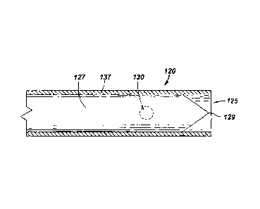
Now referring to Figure 8, an illustration of a distal portion of an alternate
embodiment of
a neural injection system of the present invention is disclosed. Neural
injection system 120
comprises a cannula 137 and a stylet 127. Side port 130 has been represented
by dashed lines for
illustrative purposes. In this embodiment, stylet 127 is capable of being
extended to contact a
tissue wherein point 129 can penetrate the tissue, thereby removing the need
for an introducer.
Now referring to Figure 9, an illustration of a cross-section of an alternate
embodiment of
a neural injection system of the present invention is disclosed. Neural
injection system 200
comprises a distal portion 205, a stylet 207, and a cannula 215. The distal
portion 205 of cannula
215 comprises shoulders 243. Side port 210 has been represented by dashed
lines for illustrative
purposes. In this embodiment, cannula 215 is capable of penetrating the
tissue, thereby removing
the need for an introducer.
Now referring to Figure 10, an illustration of a cross-section of an alternate
embodiment
of a neural injection system of the present invention is disclosed. Neural
injection system 200
comprises a distal portion 205, a stylet 207, and a cannula 215. The distal
portion 205 of cannula
215 comprises shoulders 243. Side port 210 has been represented by dashed
lines for illustrative
purposes. In this embodiment, cannula 215 is capable of penetrating the
tissue, thereby
removing the need for an introducer.
- 21 -