Note: Descriptions are shown in the official language in which they were submitted.
CA 02846827 2014-02-26
WO 2013/033084
PCT/US2012/052645
METHOD FOR LASER CUTTING A CORNEAL POCKET
CROSS REFERENCE TO RELATED APPLICATION
[0001] This application claims priority to earlier filed U.S. Patent
Application No. 12/281,749, filed on January 8, 2009, the disclosure of which
is hereby incorporated by reference, in its entirety.
FIELD OF THE INVENTION
[0002] The present invention relates generally to ocular surgery. More
particularly, the present invention relates to a method for laser cutting a
corneal pocket.
BACKGROUND OF THE INVENTION
[0003] Presbyopia is the gradual loss of near vision, which often
accompanies the aging process. The eyes of a person suffering from
presbyopia have a diminished ability to focus on near objects such as books,
magazines, or a computer screen. Symptoms of presbyopia can include
difficulty reading fine print and blurred vision when transitioning the focus
of
the eye between near and distant objects.
[0004] There are several common treatments for presbyopia. A
dedicated pair of reading glasses is one such treatment. Reading glasses
provide magnification of near objects to provide for improved vision.
However, if a person also needs glasses to focus on distant objects switching
between reading glasses and distance glasses can be inconvenient. Another
treatment is bifocal glasses, which provide a portion of the glasses lens for
assisting with distance vision and a portion for assisting with near vision.
While bifocals provide a single pair of glasses for both near and distance
1
CA 02846827 2014-02-26
WO 2013/033084
PCT/US2012/052645
vision correction, they can cause disorientation. Contact lenses for the
surface
of the eye have also been developed which provide vision correction for both
near and distance vision. Although these treatments provide vision correction
for a person suffering from presbyopia, each requires at least one an
additional
accessory or pair of contact lenses that must be worn or used daily.
Additionally, very small lenses for insertion into the eye are being
developed.
However, a small pocket must be made in the cornea into which the lens can
be inserted.
[0005] Accordingly, it is desirable to provide method for creating such
a small pocket in the cornea into which the lens can be inserted.
SUMMARY OF THE INVENTION
[0006] The foregoing needs are met, to a great extent, by the present
invention, wherein in one aspect an apparatus is provided that in some
embodiments includes a method for laser cutting a corneal pocket into which a
lens can be inserted.
[0007] In accordance with one aspect of the present invention, a
method for creating a corneal pocket includes providing a low-energy
femtosecond or nanosecond laser configured to create a corneal pocket. The
method can also include positioning the laser proximate to a cornea such that
it can be used to create the corneal pocket and determining a movement path
for the laser, in order to form the corneal pocket having a specific pocket
shape wherein the movement path follows a generally curvilinear path.
Additionally, the method can include focusing a laser beam from the laser to a
predetermined depth within the cornea between an anterior surface and a
2
CA 02846827 2014-02-26
WO 2013/033084
PCT/US2012/052645
posterior surface of the cornea such that the laser beam cut corneal tissue at
the predetermined depth. The method can also include moving the laser beam
in the movement path in order to create the corneal pocket having the specific
pocket shape.
[0008] In accordance with another aspect of the present invention, the
method can include moving the laser toward the middle of the cornea to
compensate for astigmatic effect. The method can also include using a laser
with an energy output in a range between approximately 0.2 microjoules and
1.5 microjoules. The laser can also have a spot size in a range of
approximately 0.2 to 4.0 microns and the corneal pocket can be positioned at a
depth in a range of approximately 220 microns to 350 microns. Additionally,
the laser with multiple laser beam spots and the space between the spots can
be eliminated. The method can further include programming the laser to
create the specific pocket shape.
[0009] In accordance with still another embodiment of the present
invention a method for creating a corneal pocket includes providing a low-
energy femtosecond or nanosecond laser configured to create a corneal pocket.
The method can also include positioning the laser proximate to a cornea such
that it can be used to create the corneal pocket and determining a movement
path for the laser, in order to form the corneal pocket having a specific
pocket
shape wherein the movement path follows a generally curvilinear path. The
method can include using positioning software in order to create the specific
shape. Additionally, the method can include focusing a laser beam from the
laser to a predetermined depth within the cornea between an anterior surface
3
CA 02846827 2014-02-26
WO 2013/033084
PCT/US2012/052645
and a posterior surface of the cornea such that the laser beam cuts and
separates corneal tissue at the predetermined depth. The method can also
include moving the laser beam in the movement path in order to create the
corneal pocket having the specific pocket shape.
[0010] In accordance with still another embodiment of the present
invention a method for creating a corneal pocket includes providing a low-
energy femtosecond or nanosecond laser configured to create a corneal pocket.
The method can also include positioning the laser proximate to a cornea such
that it can be used to create the conical pocket and determining a three-
dimensional movement path for the laser, in order to form the corneal pocket
having a specific pocket shape wherein the movement path follows a generally
curvilinear path. The method can include programming a computer to control
the laser such that it follows the three-dimensional movement path to form the
specific shape. Additionally, the method can include focusing a laser beam
from the laser to a predetermined depth within the cornea between an anterior
surface and a posterior surface of the cornea such that the laser beam cuts
and
separates corneal tissue at the predetermined depth. The method can also
include moving the laser beam in the movement path in order to create the
corneal pocket having the specific pocket shape.
[0011] In accordance with another aspect of the present invention, the
method can include moving the laser toward the middle of the cornea to
compensate for astigmatic effect. The method can also include using a laser
with an energy output in a range between approximately 0.2 microjoules and
1.5 microjoules. The laser can also have a spot size in a range of
4
CA 02846827 2014-02-26
WO 2013/033084
PCT/US2012/052645
approximately 0.2 to 4.0 microns and the corneal pocket can be positioned at a
depth in a range of approximately 220 microns to 350 microns. Additionally,
the laser can have multiple laser beam spots and the space between the spots
can be eliminated.
[0012] There has thus been outlined, rather broadly, certain
embodiments of the invention in order that the detailed description thereof
herein may be better understood, and in order that the present contribution to
the art may be better appreciated. There are, of course, additional
embodiments of the invention that will be described below and which will
form the subject matter of the claims appended hereto.
[0013] In this respect, before explaining at least one embodiment of
the invention in detail, it is to be understood that the invention is not
limited in
its application to the details of construction and to the arrangements of the
components set forth in the following description or illustrated in the
drawings. The invention is capable of embodiments in addition to those
described and of being practiced and carried out in various ways. Also, it is
to
be understood that the phraseology and terminology employed herein, as well
as the abstract, are for the purpose of description and should not be regarded
as limiting.
[0014] As such, those skilled in the art will appreciate that the
conception upon which this disclosure is based may readily be utilized as a
basis for the designing of other structures, methods and systems for carrying
out the several purposes of the present invention. It is important, therefore,
that the claims be regarded as including such equivalent constructions insofar
CA 02846827 2014-02-26
WO 2013/033084
PCT/US2012/052645
as they do not depart from the spirit and scope of the present invention.
BRIEF DESCRIPTION OF THE DRAWINGS
[0015] FIG. 1 illustrates a laser surgery apparatus for laser surgery to
create an intracorneal pocket in accordance with an embodiment of the
invention.
[0016] FIG. 2 is a sectional view of the anterior portion of the eye
having an intracorneal lens disposed therein, according to an embodiment of
the invention.
[0017] FIG. 3 illustrates a sectional view of the anterior portion of an
eye having an implant disposed within the cornea of the eye according to an
embodiment of the invention.
[0018] FIG. 4 illustrates a series of steps involved in a method for
inserting a lens into the cornea of the patient.
[0019] FIGS. 5A and 5B illustrate incisions in a cornea and a corneal
pocket in accordance with an embodiment of the invention.
[0020] FIGS. 6A and 6B illustrate incisions in a cornea in accordance
with an embodiment of the invention.
[0021] FIG. 7 illustrates a top down view of a corneal pocket in
accordance with an embodiment of the invention.
[0022] FIGS. 8A and 8B illustrate a 3 dimensional path for the laser
beam in accordance with an embodiment of the invention.
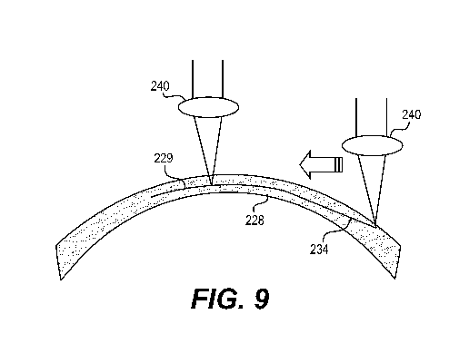
[0023] FIG. 9 illustrates a path for movement of the laser beam in
accordance with an embodiment of the invention.
DETAILED DESCRIPTION
[0024] The invention will now be described with reference to the
6
CA 02846827 2014-02-26
WO 2013/033084
PCT/US2012/052645
drawing figures, in which like reference numerals refer to like parts
throughout. An embodiment in accordance with the present invention
provides an apparatus and method for creating a flap or pocket in the cornea.
This lens or pocket preferably is created by a laser used in conventional
lasilc
surgery.
[0025] FIG. 1 illustrates a laser surgery apparatus 10 for laser surgery
to create an intracorneal pocket in accordance with an embodiment of the
invention. The laser surgery apparatus 10 can include a laser source 12 which
can generate and control, using software, a source beam 14 having a
continuous train of laser pulses of substantially constant pulse duration and
pulse energy. In one embodiment of the laser surgery apparatus 10, a source
beam 14 can take the form of a femtosecond or a nanosecond laser. The
source beam 14 can also have a wavelength greater than 800 nanometers and a
pulse energy in a range of approximately 0.2 mu.J. to 1.5 mu.J. Using less
energy for the pulse is preferable, but can be any level of energy suitable
for
creating the corneal pocket.
[0026] The laser surgery apparatus 10 further includes an optical
system 16 for forming a shaped laser beam 18 and directing the shaped laser
beam 18 toward and into the cornea 28 of an eye 22. The laser beam 18 can
be programmed with a computer to determine the path of the laser beam 18
over the patient's eye. Additionally, the laser beam 18 can be configured to
follow a three-dimensional path to cut and separate the cornea to form a
pocket for the insertion of the lens.
[0027] FIG. 2 is a sectional view of the anterior portion of the eye 22
7
CA 02846827 2014-02-26
WO 2013/033084
PCT/US2012/052645
having an intracorneal lens 26 disposed therein, according to an embodiment
of the invention. In the embodiment of the invention shown in FIG. 2,
intracorneal lens 26 may be disposed within a cornea 28 of the eye 22, which
may partially enclose the anterior chamber 30 of the eye 22. Also shown in
FIG. 2 is an iris 32. In accordance with an embodiment of the invention, lens
26 may be inserted within cornea 28 following formation of a corneal pocket
29, which may be formed using a laser surgery apparatus 10 as shown in FIG.
1.
[0028] Intracorneal lens 26 is not restricted to the configuration shown
in the drawings, but may have various shapes, such as circular or oval. In
some embodiments, intracorneal lens 26 may have a doughnut-like
configuration. The size and shape of intracorneal lens 26 may, in some cases,
determine the size and shape of the corneal pocket.
[0029] The intracorneal lens 26 preferably may be formed of a
biocompatible material that permits sufficient gas diffusion to allow adequate
oxygenation of internal eye tissues. Such materials may include silicone,
hydrogels, urethanes or acrylics. It also may be desirable that the lens be
made
of a hydrophilic material which swells somewhat when hydrated. Such
materials, for example, hydrogels, are well known and are used in some
present contact lenses.
[0030] The optical characteristics of intracorneal lens 26 may be
selected for correcting various visual deficiencies, including without
limitation: myopia (short sightedness), hypermetropia (long sightedness),
presbyopia and astigmatism. As an example, intracorneal lens 26 may have a
8
CA 02846827 2014-02-26
WO 2013/033084
PCT/US2012/052645
diopter power or value in the range of from +15 to -30. Intracorneal lens 26
may be customized for a particular patient to provide optical characteristics
to
correct a specific visual defect of a patient. Intracorneal lens 26 may be
multi-
focal, may be provided as an off-the-shelf unit with pre-determined optical
characteristics and may have zones with optical power and zones without
optical power. It is to be understood that the present invention is not
limited to
treatment of the aforementioned visual defects, and that treatment of other
eye
conditions is also within the scope of the invention.
[0031] FIG. 3 shows a cross section of a cornea 28 having a corneal
pocket 29 formed by a laser surgery apparatus 10 in accordance with one
embodiment of the invention. Cornea 28 has an anterior surface 31 and a
posterior surface 33. Corneal pocket 29 may be formed by photo disruption
using laser beam 18 from a laser source 12.
[0032] The corneal pocket 29 may be formed with a thickness and
shape that conforms to the surfaces of the intracorneal lens 26. For example,
the interior surfaces of the corneal pocket 29 may be convex, concave, planar
or irregular. The edges of the corneal pocket 29 may form an outline having
various shapes depending on the desired outcome and the shape of the
intracorneal lens 26. The various configurations of corneal pockets can be
adapted to be used with lenses of various shapes and sizes. The corneal pocket
can also be configured to facilitate the insertion of the lens and minimize
the
size of the incision for improved post-surgical healing of the cornea. The
corneal pocket can also include an entry channel 34 that may be cut into the
cornea 28 after the corneal pocket 29 is formed. Entry channel 34 may permit
9
CA 02846827 2014-02-26
WO 2013/033084
PCT/US2012/052645
the insertion of the intracorneal lens 26 into the corneal pocket 29.
[0033] FIG. 4 schematically represents a series of steps involved in a
process for creating a corneal pocket and inserting a lens in the cornea of a
patient, according to one embodiment of the invention. The process may begin
with the step 74 of providing an intracorneal lens 26. The intracorneal lens
26
may or may not have optical power depending on the purpose of the
intracorneal lens 26. In step 78 a corneal pocket 29 may be formed. This may
be done using the laser surgery apparatus 10 shown in FIG. 1. In particular, a
laser source 12 being controlled by an optical system 16 may be used to focus
a laser beam 18 within the corneal tissue. The laser beam 18 will cut and
separate a region of the cornea tissue in the area of the focus of the laser
beam
18. The focus of the laser beam 18 may then be moved laterally by hand to cut
a layer of corneal tissue. While the focus of the laser beam 18 is being moved
laterally, it may be maintained a fixed depth within the cornea using known
laser surgical techniques. The focus of the laser beam 18 may be easily,
quickly and accurately moved laterally by controlled software within the
confines of the pocket region without the risk of cutting outside the desired
area defined by the software.
[0034] The thickness of the corneal pocket created using the above
techniques will be about the size of the diameter of the laser beam 18 focal
point. In some cases, depending on the thickness and shape of the intracorneal
lens 26, additional tissue may be cut at different depths within the cornea
28.
[0035] In step 80 an entry channel 34 may be formed. This may be
accomplished using the laser source 12 or may be formed using a conventional
CA 02846827 2014-02-26
WO 2013/033084
PCT/US2012/052645
scalpel. Entry channel 34 may provide a means for insertion of the
intracorneal lens 26 and also will allow the release of gasses created by
laser
ablation when the intracorneal pocket 29 is formed.
[0036] The intracorneal lens 26 may then be inserted into the
intracorneal pocket 29 in step 82. Step 82 may further involve temporarily
deforming the intracorneal lens 26 before it is introduced into the cornea 28.
The intracorneal lens 26 may be deformed by rolling, folding, and the like.
The intracomeal lens 26 may have prescribed memory characteristics that
allow it to return to its original size and configuration after insertion into
the
cornea 28, while retaining its desired optical characteristics. The
intracorneal
lens 29 may be made of a hydrophilic material which swells when hydrated.
The lens may be inserted fully hydrated to elastically fit into a conical
pocket,
or while at least partly dehydrated such that subsequent hydration helps
secure
the tit in the pocket.
[0037] FIGS. 5A and 5B illustrate incision patterns in a cornea, in
accordance with an embodiment of the invention. As illustrated in FIGS. 5A
and 5B, an entry incision 102, 202 can be made on the cornea 100, 200. The
entry incision 102, 202 is shown as being positioned on a rightward edge of
the cornea 100, 200, in FIGS. 5A and 5B. However, the entry incision 102,
202 can be positioned in any suitable portion of the cornea 100, 200. A
circular pocket 104, 204 can also be formed in the cornea. An insertion tunnel
106, 206 can be positioned between the entry incision 102 and the pocket 104.
Additionally, as illustrated in FIG. 5A, a second tunnel 108 can be positioned
to the left of the circular pocket 104. Alternately, as shown in FIG. 5B,
11
CA 02846827 2014-02-26
WO 2013/033084
PCT/US2012/052645
relaxing incisions 210 can be made in the cornea 200, in order to ease the
insertion of the corneal lens and reduce astigmatism.
[0038] FIGS. 6A and 6B also illustrate incision patterns in a cornea, in
accordance with an embodiment of the invention. As illustrated in FIGS. 6A
and 6B, an entry incision 302, 402 can be made on the cornea 300, 400. The
entry incision 302, 402 is shown as being positioned on a rightward edge of
the cornea 300, 400, in FIGS. 6A and 6B. However, the entry incision 302,
402 can be positioned in any suitable portion of the cornea 300, 400. An
insertion tunnel 306, 406 can be positioned leftward of the entry incision
302,
402, and can extend across the cornea 300, 400. Additionally, as shown in
FIG. 6B, relaxing incisions 410 can be made in the cornea 400, in order to
ease the insertion of the corneal lens and reduce a preexisting astigmatism.
[0039] FIG. 7 illustrates a top down view of the same corneal pocket
29. The pocket and the relaxing incisions can be made with a femtosecond or
nanosecond laser having an energy profile in a range of approximately 0.2
microjoules to 1.5 microjoules. Any suitable energy level can be used,
however lower energy output is preferable. Additionally, the laser beam can
have a spot size in a range of approximately 0.2 microns to 4.0 microns. The
depth of the cut can be in a range of approximately 220 microns to 350
microns. It should be noted that if the cut is too deep the structure of the
cornea can become less stable. The pocket profile 29 shown in FIG. 8 can be
used to minimize distortion of the patient's vision through the newly
implanted lens. However, if the patient suffers from astigmatism the cut can
be moved toward the middle of the cornea in order to minimize the astigmatic
12
CA 02846827 2014-02-26
WO 2013/033084
PCT/US2012/052645
effect.
[0040] FIGS. 8A, 8B, and 9 illustrates a path for the laser beam and a
direction for the movement of the laser beam, in accordance with an
embodiment of the invention. More particularly, FIG. 8A illustrates a side
view of the path for the laser beam and FIG. 8B illustrates a top down view of
the path The pocket 229 can be formed and an adjacent entry channel 234
can be formed in order to allow the insertion of the intracorneal lens into
the
corneal pocket 229. While FIGS. 8A and 8B illustrate a path for the laser
beam, this is simply one example of the path that can be used to form the
pocket 229 and the entry channel 234. Any path that is suitable for the
purpose of forming a pocket can be used. Preferably, the path the laser is
moved in is curvilinear to follow the natural curvature of the eye. FIG. 9
illustrates the laser beam 240 moving across an axis of the eye. The laser
beam 240 can have a single beam or multiple beams creating a single laser
spot or multiple laser spots respectively. Additionally, if the laser beam
used
has multiple spots, preferably there is no space between the spots of the
laser
beam.
[0041] As can be appreciated by those skilled in the art, the present
invention may provide a method for correcting the vision of a patient with an
intracorneal lens 26 that may be easily inserted into a corneal pocket 29. The
corneal pocket 29 may be created using a laser source 12 or may be created
using other forms of electromagnetic radiation. The creation of the corneal
pocket 29 is facilitated by the use of software that prevents the laser beam
18
from cutting and separating tissue outside the boundary of a desired shape. A
13
CA 02846827 2014-02-26
WO 2013/033084
PCT/US2012/052645
variety of corneal pocket configurations may be used to accommodate various
corneal lens shapes and sizes. Other surgical procedures, such as arcuate
cuts,
may also be made using the techniques of the invention.
[0042] The many features and advantages of the invention are apparent
from the detailed specification, and thus, it is intended by the appended
claims
to cover all such features and advantages of the invention, which fall within
the true spirit, and scope of the invention. Further, since numerous
modifications and variations will readily occur to those skilled in the art,
it is
not desired to limit the invention to the exact construction and operation
illustrated and described, and accordingly, all suitable modifications and
equivalents may be resorted to, falling within the scope of the invention.
14