Note: Descriptions are shown in the official language in which they were submitted.
CENTRIFUGALLY-ENHANCED CAPTURE METHOD AND DEVICE
Cross-reference to Related Applications
This application claims the benefit of United States Provisional Patent
Application
serial number USSN 61/528,883 filed August 30, 2011.
Field of the Invention
The present invention relates to methods and devices for capturing and
detecting
target molecule in a sample. In particular, the present invention related to
microfluidic
devices having centrifugally enhanced capture capability.
Background of the Invention
The capture and isolation of biological targets (pathogens, bacteria, cells,
functionalized micro-beads, etc.) are critically important in many clinical
diagnostic,
screening, environmental assessment and quality control applications. For many
of these
applications, there is a need for rapid and low-cost detection/identification
assays. In the
area of food safety, foodborne disease is a serious public health threat and
thus rapid
detection of potentially life-threatening pathogens remains a major public
health challenge
(Yang 2006). Similar challenges are found in the field of clinical diagnostics
where rapid
detection of pathogens in a patient's blood is sought; and in environmental
and
biosecurity applications where identification of bacteria and other
contaminants from
water samples are desired. Over the past several years, a variety of methods
have been
investigated for the detection of bacteria and other biological targets in
food or water, for
example, immunological assays (Koubova 2001; Vaughan 2001; Sewell 2003),
nucleic
acid-based tests (Ingianni 2001; Choi 2002; Amagliani 2004) and
physicochemical tests
based on bacterial growth (Wawerla 1999; Firstenberg-Eden 2000).
Among the above mentioned methods, immuno-capture based assays are of
great interest due to the high sensitivity and specificity of antigen-antibody
immuno-
interaction. Antigens present on surfaces of species/objects of interest
(pathogens,
bacteria, microbeads) suspended in a biological fluid/sample are captured by
specific
antibodies immobilized on to a surface. While the antigen-antibody interaction
have been
primarily used in the immuno-capture assays, various techniques can replace
this
interaction with other moieties such as aptamers (peptides/oligonucleotide
sequences)
and biophages that are thought be provide better capture (Zourob 2008). In
such assays,
1
CA 2847215 2019-03-26
WO 2013/029153
PCT/CA2012/000794
CA 02847215 2014-02-28
the probability of capture of the targets is directly related to the velocity
of the fluid above
the functionalized surface (antibody, biophage, aptamer-coated surface), with
higher
probability of capture being obtained at lower flow rates. The order of
magnitude of the
liquid velocity at which reasonable values for probability of capture are
obtained ranges in
the tens of micrometers per second. At these relatively slow flow rates and
with the
typical sample volumes in use in many biological protocols (milliliter to
hundreds of
microliters), an assay or analysis can take significant time, thus preventing
rapid
detection.
The main reason these extremely low flow rates are used in innmuno and other
capture assays originates in the hydrodynamic interaction of species or
objects with the
functionalized surface. Particles flowing near a rigid surface undergo a 'wall
effect" where
an asymmetric wake of the particles near the surface leads to lift forces away
from the
surface (Zeng 2005). Thus, the "natural" tendency of functionalized rigid
surfaces is to
repel particles flowing near the surface, the repelling force being higher at
higher
velocities of the particles. Consevently, the velocity of the liquid must be
as small as
possible in order to allow particles to attach to the functionalized surfaces.
The forces that
naturally push the particles against the capture area of the surface are
thermal,
gravitational and diffusive effects in the biological liquid sample.
In order to increase the efficiency of species binding to functionalized
capture
sites, several methods have been proposed. One of them employs an array of
interdigitated metallic electrodes and the dielectrophoretic force to give
pathogens an
additional push against the capture sites (Li 2002; Yang 2006). The
dielectrophoretic
force acting on pathogens originates in the ability of the pathogens to
polarize in the
presence of electric fields. This force can be adjusted by tuning the
amplitude and
frequency of the applied AC fields. An equivalent method employs
electromagnetic
cellular polarization and optical scattering for direct detection but without
the use of any
biochemical marker (Choi 2006).
One and the most important drawback of dielectrophoresis-based capture
approaches is related to the short range action of the dielectrophoretic force
itself, which,
in practical microfluidic applications is only on the order of tenths of
micrometers (Li
2002). This limits the size of the microfluidic channels, thus the overall
throughput of the
device. Moreover, the use of complicated arrays of electrodes increases the
number of
fabrication steps (thus the cost per unit device) associated with the
electronics needed to
generate the necessary high frequency AC voltages. This is detrimental when
single-use,
low-cost and portable devices for point-of-care applications are intended.
2
W02013/029153
PCT/CA2012/000794
CA 02847215 2014-02-28
Another approach is based on immuno-magnetic capture and separation (Dwivedi
2011). Instead of forcing particles to bind to rigid (fixed) walls, the
antibodies are
deposited onto the surface of superparamagnetic beads. These beads become
magnetic
only in the presence of external magnetic fields and return immediately to the
non-
magnetized state as the magnetic field is removed. This is an important
property for
immuno-magnetic capture since the beads will freely interact with the target
antigens
(pathogens) in stagnant liquid suspensions without clustering together by
mutual
magnetic interactions. The process of capture can be slightly accelerated if
moderate
vortexing (agitation) of liquid suspensions is induced. Commercial devices,
such as the
well known BeadRetrieverTM from Dynal Biotech Ltd. (Wirral, UK) based on the
inverse
magnetic particle processing principle, are able to reduce the capture time
further by
moving the particles along small tubes containing the sample with the aid of a
magnetic
bar. Related methods further decrease the detection time by adding features
such as
quantum dots for enhanced fluorescence (Su 2004), magnetic relaxation
(Kaittanis 2007)
and time-of-flight mass spectrometry (Madonna 2001).
In immuno-magnetic capture using superparamagnetic beads, the time needed by
functionalized beads to bind to specific pathogens present in the sample may
be lowered
by stirring the solution to increase the probability of capture. However, the
stirring speed
is limited to the same fluid-to-solid relative velocities as in the static
case, mainly due to
the same hydrodynamic wall effect that manifests at the surface of moving
beads.
Consequently, the fundamental problem related to the wall effects that repel
particles
from functionalized surfaces is not addressed.
Immune-magnetic capture using superparamagnetic beads may be implemented
in microfluidic devices (e.g. Lee 2010; Lee 2011). The beads are used as a
carrier
surface for the capture of a target molecule. In these cases, centrifugal
force generated
by the rotating device is used to pump fluids through the device and to move
the beads
from chamber to chamber. Centrifugal force is not used to directionally
immobilize target
particles on to an immobile capture surface.
There remains a need for increasing capture efficiency of a target molecule in
a
capture assay in a microfluidic device.
Summary of the Invention
In one aspect of the present invention there is provided a centrifugal
microfluidic
device for conducting capture assays, the device comprising a rotating
microfluidic
3
W02013/029153
PCT/CA2012/000794
CA 02847215 2014-02-28
platform that rotates in a plane of rotation, the platform having at least one
capture
surface for immobilizing a target particle of interest in the device, the
capture surface
oriented so that it is not parallel to the plane of rotation of the device,
the capture surface
positionally fixed in the device during operation of the device, and
centrifugal force arising
from rotation of the device forces the target particles against the capture
surface.
A method of capturing a target particle of interest for an assay in a
centrifugal
microfluidic device, the method comprising: introducing a fluid containing the
target
particle into a rotatable microfluidic platform of the microfluidic device;
rotating the
microfluidic platform in a plane of rotation to generate centrifugal force in
the device; and,
using the centrifugal force to direct flow of the fluid to a capture surface
in the device
thereby pushing the target particle against the capture surface to increase
probability of
the target particle interacting with the capture surface, wherein capture
efficiency of the
capture surface for the target particle is independent of rate of flow of the
fluid and
independent of rate of rotation of the microfluidic platform.
In existing centrifugal microfluidic devices centrifugal force generated by
rotation
of the platform is used exclusively to pump liquids from one place to another.
Capture
surfaces in the device are located on the bottom surface of the device
parallel to the
plane of rotation and parallel to the centrifugal force in the device. Target
particles flow
over the top of the capture surface but target particle/capture site
interactions depend on
thermal, gravitational and diffusive effects to occur. As previously stated,
particles flowing
near a rigid surface undergo a "wall effect" where an asymmetric wake of the
particles
near the surface leads to lift forces away from the surface. Thus, the
"natural" tendency of
rigid surfaces is to repel particles flowing near the surface, the repelling
force being
higher at higher velocities of the particles. Consequently, the velocity of
the liquid must be
as small as possible in order to allow particles to attach to the capture
surface. Since the
forces that naturally push the particles against the capture surface are
thermal,
gravitational and diffusive effects, existing centrifugal microfluidic devices
are hampered
by poor capture efficiency and slow assay times.
In contrast, in the present invention, centrifugal force is also used to push
and
guide target particles to and against the capture surface, which increases
target
particle/capture site interaction thereby increasing surface capture
efficiency and
permitting faster fluid flow which leads to more rapid assays. In devices of
the present
invention, the capture surface is oriented so that it is not parallel to the
plane of rotation of
the platform and is positionally fixed in the device during operation of the
device. Both the
non-parallel orientation and positional fixing of the capture surface lead to
improved
4
capture efficiency. Thus, the direction at which the capture surface is
oriented
forms a non-zero angle with the plane of rotation, i.e. it is out of the plane
of rotation of
the platform, and therefore also forms a non-zero angle with the direction of
the
centrifugal force in the device. This facilitates increased interaction
between the capture
surface and the target particles moving in the fluid flow. Since the capture
surface is also
positionally fixed it is rigid and does not move around in the device thereby
maintaining its
non-parallel orientation. Further, the non-parallel orientation of the capture
surface with
respect to the plane of rotation leads to decoupling of the capture efficiency
from fluid flow
rate and rotational rate. Such independence of capture efficiency permits the
use of faster
fluid flow rates which speeds up assay time, and minimizes the need to control
the
rotational rate of the platform thereby simplifying operation. These are
considerable
advantages over existing devices.
Preferably, the angle formed between the capture surface and the plane of
rotation (or the direction of centrifugal force) is in a range of from 300 to
150 , more
preferably from 60 to 120 . Yet more preferably, the angle is about 90 . When
the angle
is 900, the capture surface is oriented orthogonally to the plane of rotation
and therefore
orthogonally to the direction of centrifugal force. When the capture surface
is oriented
orthogonally to the plane of rotation, the capture surface is parallel to the
axis of rotation
of the platform. To further enhance capture efficiency, the capture surface is
preferably
oriented parallel to the circumferential direction of the rotating platform.
Target particles are entities on which a detection assay is desired to be
performed. Such target particles may include biological or non-biological
entities.
Biological targets are preferred. Target particles may comprise viral
particles, cells (e.g.
bacterial, fungal or eukaryotic cells) or microparticles (e.g. microbeads,
magnetic
microparticles). Microparticles may be vehicles for carrying molecules of
interest to which
the assay is directed, for example, biological molecules such as proteins,
carbohydrates,
nucleic acids and the like. Target particles are preferably pathogens, for
example viruses
or cellular pathogens (e.g. bacteria or fungi), especially cellular pathogens.
The capture surface may be unfunctionalized or may be functionalized with
capture moieties that bind to the target particles. If an unfunctionalized
capture surface is
used, the surface will have structures to participate in target particle
capture. If a
functionalized capture surface is used, the capture surface may be
unstructured or
structured. In the case of a functionalized capture surface, the type of
capture moiety is
selected based on the nature of the target particle. The target particle must
be able to
interact physically, chemically or biologically with the capture moiety. Some
examples of
5
CA 2847215 2019-03-26
WO 2013/029153
PCT/CA2012/000794
CA 02847215 2014-02-28
capture moieties include small molecular entities that react with specific
chemical
functional groups on the target particle, antibodies, biophages and aptamers.
For small .
molecular entities, functional group pairs that interact chemically are
generally known, for
example catalytic reaction of COOH and NH2 or COOH and OH, where the capture
moiety is selected to have one group of the pair to complement the other group
of the pair
on the target particle. Immuno-capture-based assays are of particular interest
due to their
high sensitivity and specificity. In immuno-capture-based assays the capture
moiety may
be, for example, a biomolecule (e.g. antibody, aptamer), a biophage, a metal
or a mixture
thereof. lmmuno-capture-based assays are particularly useful for target
particles that
comprise a biological component.
The capture surface may be unstructured or structured. In the case of a
structured
capture surface, the surface comprises features that can capture target
particles based
on physical properties of the target particles, for example size, shape, mass,
magnetic
properties or combination thereof. Structural features include any micro-
and/or nano-
structured features, for example holes, posts, blazed gratings, etc. The
capture surface
may comprise a combination of structures for physical capture of target
particles and
functionalization with a capture moiety to increase specificity and efficiency
of capture. In
addition to facilitating physical capture of the target particles, structural
features on the
capture surface can increase surface area of the capture surface to increase
density of
capture moieties coated thereon.
The capture surface is positionally fixed in the centrifugal microfluidic
device. The
capture surface is preferably one or more immovable walls of a chamber or
channel in the
device. When the platform is rotated to generate centrifugal force, the one or
more
immovable walls do not move, maintaining the same orientation in respect of
the
rotational plane. Preferably, the capture surface is part of a capture chip
comprising one
or more inlets, outlets, channels and/or chambers. Flowing fluid in the device
would enter
the chip through the inlet, flow through the channels and/or chambers and then
flow out of
the chip through the outlet. One or more of the interior walls of the channels
and/or
chambers in the chip would be the capture surface to capture target particles
flowing
along with the fluid in the chip.
Microfluidic devices may comprise one or more capture surfaces designed in
accordance with the present invention. Further, more than one microfluidic
device may be
multiplexed to form a hybrid or more complex interconnected system capable of
performing multiple tasks. One or more of the devices in the system may
comprise
6
WO 2013/029153
PCT/CA2012/000794
CA 02847215 2014-02-28
capture surfaces designed in accordance with the present invention, providing
a great
deal of flexibility in performing biological assays of various sorts.
Microfluidic devices generally comprise a microfluidic circuit having at least
one
micro-scale channel in fluid communication with at least one microfluidic
chamber.
Channels include, for example, sample loading channels, cell loading channels,
medium
perfusion channels, mixing channels, particle separation or fractionation
channels,
gradient generating channels and high resistance perfusion conduits, which may
have
different channel dimensions dictated by the specific application.
Microfluidic chambers
include, for example, cell culture chambers, capture chambers, biomolecular
interaction
chambers or mixing chambers. Other microfluidic structures may also be
present, for
example valves and pumps for controlling fluid flow, conduits, inlets,
outlets, and the like.
Channels are preferably no larger than 1 mm, at least in one direction, and
the total
length of the device is preferably on the order of a few centimeters to tens
of centimeters.
The depth of chambers, including the reservoir and siphoned chamber, may be
larger
than the depth of the channels in order to accommodate larger volumes of
fluid, and may
exceed 1 mm in size. Microfluidic devices can be readily fabricated by any of
the actual
microfabrication techniques known in the art, for example, machining, hot
embossing, 3D
printing, etc.
The device and method of the present invention is useful in many diagnostic,
screening, environmental assessment and quality control applications,
especially those in
which there is a need for rapid and low-cost detection/identification assays.
Some
examples of applications include food safety, clinical diagnostics,
environmental sample
screening and biosecurity, where identification of bacteria and other
contaminants from
water samples are desired.
The present invention has several distinct advantages over the prior art. The
capture efficiency is decoupled from the flow rate of the fluid near the
capture surface,
which his in contrast to all other known devices and methods where capture
efficiency is
still dependent on the fluid flow rate. In the present invention, the
centrifugal force
pushing target particles against the capture surface is scaled to the velocity
of the flow,
increasing at higher flow rates and keeping the capture efficiency flow-
independent with a
capture location determined by the microfluidic configuration and the target
particle
parameters (density, size, etc.). Moreover, this centrifugal force has a very
long range of
action compared to dielectrophoretic or even magnetic forces, acting
identically upon all
species approaching the capture surface. Consequently, a given device is
characterized
by a specific value of capture length and the same capture efficiency will be
obtained
7
WO 2013/029153
PCT/CA2012/000794
CA 02847215 2014-02-28
regardless the speed of the fluid flow. Thus, the device can be easily adapted
to fit
regular centrifuge machines since rotation protocols and precise control of
the rotation
speed are not necessary. High-throughput and efficient capture of target
particles is the
result.
Devices of the present invention may be used in a clinical setting for rapid
diagnosis of infections (in humans and carriers, such as insects) and various
other
diseases. Other applications include detection of, and characterization
(relative to drug
resistance, for example) of pathogens in various media (food, water, air) or
substances
(medications, devices, equipment), especially for detection of infectious
agents in hospital
or community settings.
The present invention is particularly appropriate for the detection of rare
biomarkers or pathogens in a complex sample that is constituted of various
particles
(size, composition, density). The centrifugal assisted capture allows for
rapid separation
of the biomarker or pathogen of interest from the other constituents. One
example would
be for the detection of cancerous cell in a blood stream, where rare
circulating tumour
cells (CTCs) are present in mixture with red and white blood cells. The
present invention
also allows for rapid separation of the red blood cell and with the addition
of surface
functionalization can isolate/capture the CTCs from the white blood cells.
When detached
from a primary tumour and circulating in the bloodstream, CTCs may constitute
seeds for
subsequent growth of additional tumours (metastasis) in different tissues. As
a "cancer
blood test," this would be extremely useful to determine cancer stage, spread
and
response to treatment, thereby improving the efficiency of treatment planning.
The advantage of detecting agents that are small in number compared to
components in the sample applies to most applications, including the detection
of
pathogens (bacteria) from a swab sample or a physiological sample.
Additionally, the
device and method can effectively be used for capture of bacteria or viruses
from food
and water samples pending sample preparations that can reduce the volume.
The present invention can be used for any kind of application in which
enhanced
dynamic capture is needed. Since this invention is amenable to applications in
automated
analysis, it may find additional, cost-effective applications in food safety,
bioprocess
control, defence, and veterinary medicine, and other areas.
Further features of the invention will be described or will become apparent in
the
course of the following detailed description.
8
WO 29131029153
PCT/CA2012/000794
CA 02847215 2014-02-28
Brief Description of the Drawinos
In order that the invention may be more clearly understood, embodiments
thereof
will now be described in detail by way of example, with reference to the
accompanying
drawings, in which:
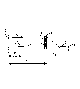
Fig. 1 depicts a schematic diagram of a centrifugal microfluidic device for
centrifugally enhanced capture of target particles in accordance with the
present
invention.
Fig. 2 depicts a vertical cross-sectional view of a capture chip in accordance
with
the present invention illustrating centrifugally enhanced target particle
capture.
Fig. 3 depicts a schematic diagram of spatially-tuned capture of different
types of
target particles (Type 1, Type 2 and Type 3) on a surface, where Lcaptur91,
Lcapture2 and
Lcapture3 are capture lengths of each type of particle on the surface, Fel,
Fcf2 and Fcf3 are
the centrifugal forces acting on each type of particle and Fro, F,72 and F,73
are the forces
due to fluid flow acting on each type of particle..
Fig. 4 depicts schematic drawings of capture surfaces for target particles in
biological fluid flows over: (A) an antibody functionalized unstructured
surface; (B) a
micro-structured surface; and (C) an antibody functionalized micro/nano-
structured
surface.
Description of Preferred Embodiments
Referring to Fig. 1 and Fig. 2, a centrifugal microfluidic device for
centrifugally
enhanced capture of a pathogen comprises holding blade 11 that rotates around
rotation
axis 12. Top surface 13 of the holding blade is perpendicular to the rotation
axis and
therefore parallel to the rotation plane and parallel to direction of
centrifugal acceleration
ao. Mounted rigidly on the top surface of the holding blade are capture chip
14, sample
reservoir 20 and waste reservoir 21. Capture chip 14 comprises capture surface
15
located in capture chamber 16. Under the influence of centrifugal force
generated by
rotation of the blade, a biological fluid containing the pathogen flows from
sample
reservoir 20 via a channel to capture chip 14, enters capture chamber 16
through inlet 17,
flows through capture chamber 16 where the fluid encounters capture surface
15, and
then flows out of capture chamber 16 through outlet 18 to be carried by a
channel into
waste reservoir 21. Because capture chip 14 is oriented perpendicularly to
holding blade
11, capture surface 15, which is the bottom wall of the capture chip, is
oriented
9
WO 2013/029153
PCT/CA2012/000794
CA 02847215 2014-02-28
orthogonally to the plane of rotation. When the biological fluid flows into
the capture
chamber it is forced to flow up the chip in a direction orthogonal to the
plane of rotation.
However, since centrifugal acceleration ao is still parallel to the plane of
rotation,
pathogen particle 19 in capture chamber 16 experiences centrifugal force af
parallel to
the plane of rotation that pushes the pathogen particle toward capture surface
15, even
though the fluid is flowing with velocity U and exerting a force Fn on the
pathogen particle
in a direction perpendicular to the plane of rotation. As a consequence of the
two
opposed forces Fcf and Fn, pathogen particle 19 follows a curved path before
encountering capture surface 15.
Fa is a long range force field that acts identically on all objects entering
the
capture chip and will force the objects in the flow (e.g. pathogen particles,
cells, debris,
etc.) to cross fluid streamlines and curve their trajectories towards the
capture surface.
The centrifugal force Fc, and fluid flow rate Q (the scalar component of fluid
flow velocity
U) are responsible for distance Lcapture traveled by pathogen particles from
inlet 17 to
capture point 25 on capture surface 15. These two important quantities
(centrifugal force
Fcf and flow rate Q) can easily be tuned by the positions of sample reservoir
20 and
capture chip 14 on holding blade 11 (Ro and Rc, respectively) and the
hydrodynamic
resistance Rhyd of the microfluidic circuit between the sample reservoir and
the capture
chip. Capture length Lcapture is given by the analytical expression:
2 Rr. - R,2 P
= 2 Eq. (1)
4r BS,,, , RõRõ, - P
whereas the flow rate Q is
o Fe - R72
= Pa) Eq. (2)
yJ
In the two equations above q is the dynamic viscosity of the fluid, h the
thickness of the
capture chip, r8 and Pb the radius and density of the pathogen particle
respectively, p the
density of the fluid, Sol, the cross-sectional area of the capture chip and
(.0 the angular
velocity of the microfluidic device. The condition for a 100% probability of
capture is that
Lowe s L, where L is the length of the capture chip in the direction of the
fluid flow.
It can be seen from Eq. (1) that Lcapture is independent of co whereas Q is
not. This
means that the Lcapture depends only on the device's geometrical setup (i.e.
position of
reservoirs, position of the capture chip, geometry and hydrodynamic resistance
of the
WO 2013/029153
PCT/CA2012/000794
CA 02847215 2014-02-28
microfluidic circuits, etc.) and it is the same regardless of rotational
speed. In contrast, the
fluid flow rate Q, as shown in Eq. (2), can be tuned by adjusting the
rotational speed_
Consequently, the capture efficiency is decoupled from the rate of fluid flow,
and for a
specific geometry of the device, there is the same capture probability
regardless of the
rotational speed and the fluid flow rate of the biological fluid above the
capture surface.
Further, it is evident from Eq. 1 that Lcapture is a function of the radius
and density
of the particle. Thus, in complex sample with multiple species, particles,
debris of different
sizes and densities, the capture of these different objects will occur at
different points
along the capture surface, providing a spatially distributed or tuned
immobilization and
separation (Fig. 3) providing the ability to separate along the flow
trajectory the capture
position of known target particles in the fluid. This is especially
advantageous in
applications such as the capture of target particles (e.g. bacteria or other
cells) from
complex food/water samples or the simultaneous detection of multiple
pathogens.
Referring to Fig. 4, the capture surface in a device of the present invention
may be
unfunctionalized (Fig. 4B) or functionalized with antibodies (Fig. 4A and Fig.
4C) that bind
to the pathogen particles. Further, the capture surface may be unstructured
(Fig. 4A) or
structured with micro-scale features (Fig. 4B and Fig. 4C). Fig. 4A depicts an
unstructured capture surface functionalized with antibodies that interact with
antigens on
the surface of the pathogen particle. The pathogen particle experiences
centrifugal force
Fa pushing the pathogen particle toward the capture surface, even though the
fluid is
flowing with velocity U and exerting a force F,7 on the pathogen particle in a
direction
perpendicular to the centrifugal force. Further, the "wall effect" exerts a
force Fh in an
opposite direction as the centrifugal force pushing the pathogen particle away
from the
capture surface. Provided Fcr is greater than rh, the pathogen particle will
eventually
encounter the functionalized capture surface and be captured. In Fig. 48, the
unfunctionalized capture surface has micro-scale grooves angled against the
fluid flow so
that pathogen particles can be captured physically in the grooves. In Fig. 40,
the capture
surface is both functionalized with antibodies and has a micro-scale grating.
The grating
captures pathogen particles physically while the antibodies bind to antigens
on the
surface of the pathogen particle thereby increasing capture efficiency.
11
References:
Amagliani G, Brandi G, et al. (2004) Direct detection of Listeria
monocytogenes from milk
by magnetic based DNA isolation and PCR. Food Microbiology. 21(5), 597-603.
Andersson P, Thorsen G, Kylberg G. (2003) Functional Unit Enabling Controlled
Flow in a
Microfluidic Device. United States Patent Publication US 2003-0053934
published March
20, 2003.
Carvalho BL, Sheppard Jr. NF, Feakes C, Kellogg GJ. (2004) Microfluidics
Devices and
Methods for Performing Cell Based Assays. United States Patent US 6,818,435
issued
November 16, 2004.
Choi J-W, Pu A, et al. (2006) Bacteria Detection in a Microfluidic Channel
Utilizing
Electromagnetic Cellular Polarization and Optical Scattering. 2006 Digest of
the LEOS
Summer Topical Meetings. 2, 17-18.
Choi W. (2002) Rapid enumeration of Listeria monocytogenes in milk using
competitive
PCR. International journal of food microbiology. 84, 79-85.
Desmond SM, Shigeura J. (2006) Micro-channel Design Features that Facilitate
Centripetal Fluid Transfer. United States Patent US 7,041,258 issued May 9,
2006.
Dwivedi HP, Jaykus LA. (2011) Detection of pathogens in foods: The current
state-of-the-
art and future directions. Critical Reviews in Microbiology. 37(1), 40-63.
Firstenberg-Eden R, Shelef LA. (2000) A new rapid automated method for the
detection
of Listeria from environmental swabs and sponges. International journal of
food
microbiology. 56(2-3), 231-237.
Garcia Da Fonseca J, Esteves Reis NA, Burger R. (2010) Analytical Rotors and
Methods
for Analysis of Biological Fluids. International Patent Publication WO 2010-
077159
published July 8, 2010.
Garcia-Cordero JL, Dinnov IK, O'Grady J, Ducree J, Barry T, Ricco AJ. (2009)
Monolithic
Centrifugal Microfluidic Platform for Bacteria Capture and Concentration,
Lysis, Nucleic-
Acid Amplification, and Real-Time Detection. MEMS 2009 - 22nd IEEE
International
Conference on Micro Electro Mechanical Systems, pp. 356-359.
12
CA 2847215 2019-03-26
W02013/029153
PCT/CA2912/000794
CA 02847215 2014-02-28
Gui JY, Tian W-C, Phukan A, Thutupalli S, Samper V. (2007) Rotation-based
Microsampler, System and Method of Using the Same. United States Patent
Publication
US 2007-0224591 published September 27, 2007.
Hurt SN, Gordon JF, McIntyre KR. (2006) Method and Apparatus for Blood Typing
with
Optical Bio-discs. United States Patent US 7,026,131 issued April 11, 2006.
Inganas M, Soderman T, Hogstrand E. (2008) Liquid Detection and Confidence
Determination. United States Patent Publication US 2008-0138247 published June
12,
2008.
Ingianni A, Floris M, et al. (2001) Rapid detection of Listeria monocytogenes
in foods, by
a combination of PCR and DNA probe. Molecular and cellular probes. 15(5), 275-
280.
Jung EKY, Leuthardt EC, Levien RA, Lord RW, Malamud MA, Rinaldo JD, Wood LL.
(2008) Systems for Pathogen Detection. United States Patent Publication US
2008-
0241000 published October 2, 2008.
Kaittanis C, Naser SA, Perez JM. (2007) One-Step, Nanoparticle-Mediated
Bacterial
Detection with Magnetic Relaxation. Nano Letters. 7(2), 380-383.
Kido H, Norton JR, Coombs JH. (2005) Fluidic Circuits for Sample Preparation
Including
Bio-discs and Methods Relating Thereto. United States Patent Publication US
2005-
0047968 published March 3, 2005.
Kim S-h. (2010) Optical Detection Apparatus, Optical Detection Method, and
Microfluidic
System Including the Optical Detection Apparatus. United States Patent US
7,692,794
issued April 6, 2010.
Kouboµth V, Brynda E, et al. (2001) Detection of foodborne pathogens using
surface
plasmon resonance biosensors. Sensors and Actuators B: Chemical. 74(1-3), 100-
105.
Lee B-s, Cho Y-k, Lee J-g, Park J-m. (2010) Centrifugal Force-Based
Microfluidic Device
for Protein Detection and Microfluidic System Including the Same. United
States Patent
US 7,776,267 issued August 17, 2010.
Lee B-s, Cho Y-k, Lee J-g, Park J-m. (2011) Centrifugal Force-Based
Microfluidic Device
for Protein Detection and Microfluidic System Including the Same. United
States Patent
Publication US 2011-020194 published January 27, 2011.
13
W02013/029153
PCT/CA2012/000794
CA 02847215 2014-02-28
Li H, Bashir R. (2002) Dielectrophoretic separation and manipulation of live
and heat-
treated cells of Listeria on microfabricated devices with interdigitated
electrodes. Sensors
and Actuators B: Chemical. 86(2-3), 215-221.
Li PCH, Peng XY, Yu HZ, Parameswaren M, Chen H, Chou WL. (2010) Microfluidic
Microarray Assemblies and Methods of Manufacturing and Using. United States
Patent
Publication US 2010-0041562 published February 18, 2010,
Madonna AJ, Basile F, et al. (2001) Detection of bacteria from biological
mixtures using
immunomagnetic separation combined with matrix-assisted laser
desorption/ionization
time-of-flight mass spectrometry. Rapid communications in mass spectrometry :
RCM.
15(13), 1068-1074.
Niwa D. (2011) Separation Purification Method and Microfluidic Circuit. United
States
Patent Publication US 2011-0003285 published January 6, 2011.
Nolte DD. (2009) Review of centrifugal microfluidic and bio-optical disks.
Review of
Scientific Instruments. 80, 101101 (22 pages).
Ostlin H, Eriksson L, Ljungstrom M, Agren T. (2007) Detector Arrangement Based
on
Surfaces Plasmon resonance. United States Patent US 7,295,320 issued November
13,
2007.
Sewell AM, Warburton DW, et al. (2003) The development of an efficient and
rapid
enzyme linked fluorescent assay method for the detection of Listeria spp. from
foods.
International journal of food microbiology. 81(2), 123-129.
Su X-L, Li Y. (2004) Quantum dot biolabeling coupled with immunomagnetic
separation
for detection of Escherichia coli 0157:H7. Analytical chemistry. 76(16), 4806-
4810.
Tooke NE, Andersson PX. (2008) Integrated Microfluidic Disc. United States
Patent US
7,332,126 issued February 19, 2008.
Vaughan R, O'Sullivan CK, Guilbault GG. (2001) Development of a quartz crystal
microbalance (QCM) immunosensor for the detection of Listeria monocytogenes.
Enzyme
and microbial technology. 29(10), 635-638.
Wawerla M, Stolle A, et al. (1999) Impedance Microbiology: Applications in
Food
Hygiene. Journal of Food Protection. 62, 1488-1496.
14
WO 2013/029153
PCT/CA2012/000794
CA 02847215 2014-02-28
Yang L, Banada PP, et al. (2006) A multifunctional micro-fluidic system for
dielectrophoretic concentration coupled with immuno-capture of low numbers of
Listeria
monocytogenes. Lab on a chip. 6(7), 896-905.
Zeng L., Balanchadar S, Fischer P. (2005) Wall-induced forces on a rigid
sphere at finite
Reynolds number. Journal of Fluid Mechanics, 536, 1-25.
Zourob M, Elwary S, et al. (2008) Principles of Bacterial Detection -
Biosensors,
Recodnition Receptors and Mvcrosystems. New York, Springer Science+Business
Media,
LLC.
Zucchelli P, Van de Vyver B. (2006) Devices and Methods for Programmable
Microscale
Manipulation of Fluids. United States Patent US 7,152,616 issued December 26,
2006.
Other advantages that are inherent to the structure are obvious to one skilled
in
the art. The embodiments are described herein illustratively and are not meant
to limit the
scope of the invention as claimed. Variations of the foregoing embodiments
within the
scope of the claimed and generally disclosed invention will be evident to a
person of
ordinary skill and are intended by the inventor to be encompassed by the
following
claims.