Note: Descriptions are shown in the official language in which they were submitted.
CA 02949255 2014-03-19
=
54236-19
A COMPOSITION AND METHOD FOR TREATING AN AUTOIMMUNE DISEASE
Field of the Disclosure
The present disclosure relates to the use of a composition for the treatment
of an
autoimmune disease. More specifically, the composition comprises a combination
of antibiotics
which may be used to treat autoimmune diseases including multiple sclerosis
Background,
Multiple sclerosis (MS) is a chronic autoimmune and demyelinating disease that
primarily
affects the central nervous system. MS is characterised by the infiltration of
myelin-specific CD4+ T
cells that attack the axonal myelin sheath and other elements of the central
nervous system (CNS),
destroying myelin and the basal axon.
The present inventors have found that a combination of antibiotics, previously
used in the
treatment of inflammatory bowel disorders have an effect on the inflammatory
response of a subject
suffering from an autoimmune disease including MS and other autoimmune
diseases.
Any discussion of documents, acts, materials, devices, articles or the like
which has been
included in the present specification is not to be taken as an admission that
any or all of these
matters form part of the prior art base or were common general knowledge in
the field relevant to
the present disclosure as it existed before the priority date of each claim of
this application.
1
=
CA 02949255 2014-03-19
WO 2013/041963 PCT/IB2012/002252
Summary of the Disclosure
The disclosure provides a combination of rifabutin, clarithromycin, and
clofazimine for
the treatment of an auto-immune disease
The disclosure, in one aspect, provides a composition including rifabutin,
clarithromycin,
and clofazimine for the treatment of an auto-immune disease.
The present disclosure also provides a composition including rifabutin,
clarithromycin, and
clofazimine for the treatment of multiple sclerosis.
In a further aspect, there is provided a composition comprising rifabutin,
clarithromycin and
clofazimine for the treatment of an autoimmune disease.
In a further aspect, there is provided a composition comprising rifabutin,
clarithromycin and
clofazimine for the treatment of multiple sclerosis (MS).
In a further aspect, there is provided a composition comprising a combination
of antibiotic
agents for the treatment of multiple sclerosis, said composition comprising
rifabutin,
clarithromycin and clofazimine.
In a further aspect, there is provided a composition comprising a combination
of two or
more antibiotic agents for the treatment of an autoimmune disease, said two or
more antibiotic
agents selected from rifabutin, clofazimine and at least one macrolide.
In a further aspect, there is provided a composition comprising a combination
of two or
more antibiotic agents for the treatment of an autoimmune disease, said two or
more antibiotic
agents selected from rifabutin, clofazimine and clarithromycin.
In a further aspect, there is provided a composition comprising a combination
of two or
more antibiotic agents for the treatment of an autoimmune disease, said two or
more antibiotic
agents selected from clofazimine, clarithromycin and at least one antibiotic
having bactericidal
activity.
2
= CA 02949255 2014-03-19
54236-19
In another aspect, the present disclosure provides a method of treating an
autoimmune
disease in a patient comprising administering a composition including
rifabutin, clarithromycin,
and clofazimine to said patient.
In a further aspect there is a method of treating a patient suffering from an
autoimmune
disease, and having, or susceptible to, infection by a Mycobacterium,
comprising administering .
to the patient a composition including rifabutin, clarithromycin, and
clofazimine.
In another aspect, there is a method of treating a patient suffering from
multiple sclerosis,
said patient also testing positive for a mycobacterial infection comprising
administering to the
patient a composition including rifabutin, clarithromycin, and clofazimine.
= In another aspect, the present disclosure provides a method of treating
an auto-immune
disease in a patient comprising administering a composition comprising a
combination of
antibiotics selected from the group rifabutin, clarithromycin, and clofazimine
to said patient.
In another aspect, the present disclosure provides a method of treating
multiple sclerosis
in a patient comprising administering a composition comprising a combination
of antibiotics
selected from the group rifabutin, clarithromycin, and clofazimine to said
patient.
=
3
81778119
The invention as claimed relates to:
- a pharmaceutical composition, comprising: rifabutin; clarithromycin; and
clofazimine, for the treatment of multiple sclerosis;
- use of the composition as described herein, for the treatment of a subject
.. suffering from multiple sclerosis; and
- use of a combination of rifabutin, clarithromycin, and clofazimine, for the
treatment of multiple sclerosis.
Brief Description of the Drawings
Figure 1 is a graph showing the effects of administration of RHB 104 on the
concentration of
cytokine IL-17 in a mouse model;
Figure 2 is a graph showing the effects of administration of RHB 104 on the
concentration of
cytokine TNF-alpha in a mouse model;
Figure 3 is a graph showing the effects of administration of RHB 104 on the
concentration of
cytokine IFN-gamma in a mouse model;
Figure 4 is a graph showing the effects of administration of RHB 104 on the
concentration of
cytokine IL-6 in a mouse model;
3a
CA 2849255 2019-01-15
CA 02949255 2014-03-19
WO 2013/041963 PCT/IB2012/002252
Figure 5 is a graph showing the effects of administration of RHB 104 on the
concentration of
cytokine IL-2 in a mouse model;
Figure 6 is a graph showing EAE severity in various treatment groups in a
recognised MS mouse
model;
Figure 7 is a graph showing the change in body weight in various treatment
groups in a
recognised MS mouse model;
Figure 8 is a graph showing the average number of inflammatory foci detected
histologically (in
H&E sections) in both a control and treatment group in a recognised MS mouse
model;
Figure 9 is Graph 9 is a graph showing the average demyelination score from a
histological
analysis (from Luxol fast blue sections) in both a control and treatment group
in a recognised MS
mouse model;
Figure 10 is a graph showing the average demyelination score determined
histologically (from
H&E sections) in both a control and treatment group in a recognised MS mouse
model;
Figure 11 is a graph showing the average number of apoptotic cells detected
histologically (in
H&E sections) in both a control and treatment group in a recognised MS mouse
model; and
Figure 12 is a graph showing the severity of relapse disease in various
treatment groups of a
recognised mouse model.
Description of Exemplary Embodiments of the Disclosure
By the term "multiple sclerosis", multiple sclerosis variants such as
Neuromyelitis Optica
(Devic's Disease), Diffuse Sclerosis, Transitional Sclerosis, Acute
Disseminated
Encephalomyelitis, and Optic Neuritis are also incorporated.
Use of the term "subject" includes both human and non-human animals.
4
CA 02949255 2014-03-19
WO 2013/041963 PCT/IB2012/002252
"Treatment" is meant that at least an amelioration of the symptoms associated
with the
condition (eg MS) afflicting the subject is achieved, where amelioration is
used in a broad sense
to refer to at least a reduction in the magnitude of a parameter, e.g.,
symptom, associated with the
condition being treated. As such, treatment also includes situations where the
condition, or at
least symptoms associated therewith, are completely inhibited, e.g., prevented
from happening,
or stopped, e.g. terminated, such that the subject no longer suffers from the
condition, or at least
the symptoms that characterize the condition. "Treatment" also includes the
prevention of a
relapse episode in a subject or should the relapse episode occur then the term
"treatment" is as
above.
A variety of subjects are treatable according to the subject methods. In many
embodiments the subjects are "mammals" or "mammalian", where these terms are
used broadly
to describe organisms which are within the class mammalian, including the
orders carnivore
(e.g., does and cats), rodentia (e.g., mice, guinea pigs, and rats), and
primates (e.g., humans,
chimpanzees, and monkeys). In many embodiments, the subjects are humans. While
the present
invention may be used for the treatment of a human subject, it is to be
understood that the subject
methods may also be carried-out on other animal subjects such as, but not
limited to, mice, rats,
dogs, cats, livestock and horses, etc. Accordingly, it is to be understood
that any subject in need
of being treated according to the subject invention is suitable.
Moreover, suitable subjects of this invention include those who have and those
who have
not previously been afflicted with a condition, those that have previously
been determined to be
at risk of suffering from a condition, and those who have been initially
diagnosed or identified as
being afflicted with or experiencing a condition.
Treatment may be assessed using any one or more of a number of criteria. The
assessment of said treatment may be either or both quantitative or
qualititative. Assessment of
treatment may be made based on a clinical scale of severity of a disease. In
subjects being
treated for an autoimmune disease such as MS, the treatment may be assessed
using a number of
scales such as the Expanded Disability Status (EDSS), the Ambulation Index
(Al) or the Scripps
Neurologic Rating Scale (SNRS).
Assessment of treatment may include the assessment of one or more symptoms
associated
with a particular disease. In the example of MS, symptoms include: weakness
and/or numbness
CA 02949255 2014-03-19
WO 2013/041963 PCT/IB2012/002252
in one or more limbs; tingling of the extremities and tight band-like
sensations around the trunk
or limbs; dragging or poor control of one or both legs to spastic or ataxic
parepesis; hyperactive
tendon reflexes; disappearance of abdominal reflexes; Lhermitte's sign;
retrobulbar or optic
neuritis; unsteadiness in walking; brain stem symptoms (diplopia, vertigo,
vomiting); disorders
of micturition; hemiplegia; trigeminal neuralgia; other pain syndromes;
nystagmus and ataxia;
cerebellar-type ataxia; Charcot's triad; diplopia; bilateral internuclear
ophthalmoplegia;
myokymia or paralysis of facial muscles; deafness; tinnitus; unformed auditory
hallucinations;
vertigo and vomiting; transient facial anesthesia or of trigeminal neuralgia;
bladder dysfunction;
euphoria; depression; dementia, dull, aching pain in the low back; sharp,
burning, poorly
localized pains in a limb or both legs and girdle pains: abrupt attacks of
neurologic deficit;
dysarthria and ataxia; paroxysmal pain and dysesthesia in a limb; flashing
lights; paroxysmal
itching; and/or tonic seizures, taking the form of flexion (dystonic) spasm of
the hand, wrist, and
elbow with extension of the lower limb.
In MS, ameliorating symptoms of the disease further include reducing the
number of
inflammatory episodes ("episode" includes any or a combination of at least the
above clinical
manifestations), slowing the progression of the disease, or reducing/slowing
down the
appearance of brain lesions (identified by magnetic resonance imaging). The
recurrence of
diseases including MS can be ameliorated by decreasing the severity of the
symptoms (eg the
symptoms described above) associated with an MS episode, or by lengthening the
time period
between the occurrence of episodes.
In MS and associated diseases, quantitative analysis may also be used to
assess treatment.
Examples of quantitative analysis techniques include the identification of
biological markers.
Examples include but are not limited to biomarkers which reflect alteration of
the immune
system; biomarkers of blood-brain barrier disruption, of demyelination, of
oxidative states and
excitotoxicity, of gliosis or of remyelination and repair. A panel of various
markers may be
measured to reflect various stages of disease including various stages of
inflammation,
demyelination, axonal degeneration and remyelination.
It is to be appreciated that the assessment of treatment may result from a
number of
techniques and may rely on both clinical manifestation and the analysis of
various non-clinical
markers such as biomarkers. In diseases such as MS which is a complex disease
with several
pathophysiological mechanisms which are not uniform in MS patient sub-groups,
there is a need
6
CA 02949255 2014-03-19
WO 2013/041963 PCT/IB2012/002252
to assess treatments based on various and differing criteria and markers and
it is to be understood
that the above examples provided for the assessment of treatment is not an
exhaustive list but
merely provides example of the means by which treatment may be evaluated.
Throughout this specification the word "comprise", or variations such as
"comprises" or
"comprising", will be understood to imply the inclusion of a stated element,
integer or step, or
group of elements, integers or steps, but not the exclusion of any other
element, integer or step,
or group of elements, integers or steps.
The composition of the present disclosure may further comprise at least one
antibiotic
active against Gram positive bacteria "Gram positive antibiotic". The Gram
positive antibiotic
may be selected from one or more of the group comprising daptomycin,
clindamycin, rifampicin,
erythromycin, oleandomycin, roxithromycin, azithromycin, kanamycin,
gentamycin,
tombramycin, streptomycin, neomycin, paromomycin, ethambutol, isoniazid,
minocyclin.
tetracycline.
The term "one or more" antibiotic agents includes but is not limited to one,
two, three,
four, five, six etc. antibiotic agents. It is to be understood that the
skilled artisan is able to
empirically determine the specific number of antibiotic agents needed for use
according to the
embodiments provided herein and as known in the art.
The present compositions may be used for treating a patient suffering from an
auto-
immune disease wherein said patient also tests positive for infection with the
bacterium
Mycobacterium avium paratuberculosis (MAP).
The auto-immune disease may be multiple sclerosis.
Still further, the autoimmune disease may be Hashimoto's Thyroiditis,
Melkersson-
Rosenthal syndrome. Sarcoidosis or other similar diseases.
In another embodiment, the term auto-immune disease includes any of a large
group of
diseases characterized by abnormal functioning of the immune system resulting
in antibodies
against self tissue.
7
CA 02949255 2014-03-19
WO 2013/041963 PCT/IB2012/002252
The antibiotics or compositions presently disclosed may be administered
orally.
Alternatively, the antibiotics may be administered intravenously.
Other routes of administration are contemplated including, but not limited to,
intramuscular and intrao s seou s routes.
Each antibiotic may be administered separately. Alternatively two or more
antibiotics may
be administered together.
In one embodiment, the compositions provided herein comprise at least two
antibiotic agents
that are co-formulated in a single dosage form. In another embodiment, the
composition provided
herein comprises at least three antibiotics that are co-formulated in a single
dosage form.
In one embodiment, each of rifabutin, clarithromycin, and clofazimine are co-
formulated
into a single dosage form.
Alternatively, each antibiotic agent may be formulated in a dosage form
separately to the
other antibiotic agents. In this embodiment, it is envisaged that the separate
dosage forms would be
packaged together in a kit to ensure that each was taken at generally the same
time by a patient. In
another embodiment, two of the antibiotic agents may be formulated in a single
first dosage form and
the remaining antiobiotic agent(s) may be separately formulated in a second
dosage form to be taken
with the first dosage form.
In one embodiment the present antibiotics and compositions may be available in
the form
of a tablet containing at least one of rifabutin, clarithromycin, and
clofazimine in a powdered
form. In some instances two of or all three of rifabutin, clarithromycin, and
clofazimine are in a
powdered form. Alternatively, present compositions may be in the form of a
tablet capsule
containing at least one of rifabutin, clarithromycin, and clofazimine in a
microencapsulated form.
In one embodiment, two or all of rifabutin, clarithromycin, and clofazimine
are in a
microencapsulated form.
In a further embodiment, present compositions may be in the form of a tablet
capsule
containing at least one of rifabutin, clarithromycin, and clofazimine in a
powdered form, and the
remaining agents present in a microencapsulated form. As a further
possibility, present
8
CA 02949255 2014-03-19
WO 2013/041963 PCT/IB2012/002252
compositions may be in the form of a tablet capsule containing one or more of
rifabutin,
cl arithrom ycin, and clofazimine present in a microgranulated form. In
additional embodiments,
present compositions may be in the form of a tablet containing one or more of
rifabutin,
clarithromycin, and clofazimine within a capsule, a capsule containing one or
more of rifabutin,
clarithromycin, and clofazimine within a tablet, a capsule containing one or
more of rifabutin,
clarithromycin, and clofazimine within an outer capsule containing the other
agents, or any
combination of the above.
In a further embodiment, the present compositions comprise an inner capsule
containing
rifabutin, within an outer capsule containing clarithromycin and clofazimine,
wherein clarithromycin
and clofazimine may be present in powdered, microencapsulated, or
microgranulated forms.
Still further, the present compositions may comprise liposome-encapsulated, un-
encapsulated or
polymer-coated lipo some-encapsulated forms.
The present methods may be carried out by administration of one or more
tablets/capsules
containing rifabutin, clarithromycin, and clofazimine as described above, or
through the
administration of each of these separately. In preferred embodiments,
rifabutin, clarithromycin,
and clofazimine are administered simultaneously in one dose.
The present compositions may be prepared by means known in the art for the
preparation of
pharmaceutical compositions including blending, grinding, homogenizing,
suspending, dissolving,
emulsifying, dispersing, and, where appropriate, mixing of rifabutin,
clarithromycin, and clofazimine
together with selected excipients, diluents, carriers and adjuvants.
For oral administration, the present compositions may be in the form of
tablets, lozenges,
pills, troches, capsules, elixirs, powders, including lyophilized powders,
solutions, granules,
suspensions, emulsions, syrups and tinctures. The present compositions may
comprise slow-release,
or delayed-release forms for example in the form of coated particles, multi-
layer tablets or
micro granules.
Solid forms of the present compositions for oral administration may contain
pharmaceutically
acceptable binders, sweeteners, disintegrating agents, diluents, flavorings,
coating agents,
preservatives, lubricants, and/or time delay agents. Suitable binders include
gum acacia, gelatin,
corn starch, gum tragacanth, sodium alginate, carboxymethylcellulose or
polyethylene glycol (PEG).
9
CA 02949255 2014-03-19
WO 2013/041963 PCT/IB2012/002252
Suitable sweeteners include sucrose, lactose, glucose, aspartame or
saccharine. Suitable
disintegrating agents include corn starch, methylcellulose,
polyvinylpyrrolidone, xanthan gum,
bentonite, alginic acid or agar. Suitable diluents include lactose, sorbitol,
mannitol, dextrose, kaolin,
cellulose, calcium carbonate, calcium silicate or dicalcium phosphate.
Suitable flavoring agents
include peppermint oil, oil of wintergreen, cherry, orange, or raspberry
flavoring. Suitable coating
agents include polymers or copolymers of acrylic acid and/or methacrylic acid
and/or their esters,
waxes, fatty alcohols, zein, shellac or gluten. Suitable preservatives include
sodium benzoate,
vitamin E, alpha-tocopherol, ascorbic acid, methyl paraben, propyl paraben or
sodium bisulphite.
Suitable lubricants include magnesium stearate, stearic acid, sodium oleate,
sodium chloride or talc.
Suitable time delay agents include glyceryl monostearate or glyceryl
distearate.
Liquid forms of the present compositions for oral administration may contain,
in addition to
the above agents, a liquid carrier. Suitable liquid carriers include water,
oils, such as olive oil, peanut
oil, sesame oil, sunflower oil, safflower oil, arachis oil, coconut oil,
liquid paraffin, ethylene glycol,
propylene glycol, polyethylene glycol, ethanol, propanol, isopropanol,
glycerol, fatty alcohols,
triglycerides, or mixtures thereof.
Suspensions of the present compositions for oral administration may further
include
dispersing agents and/or suspending agents. Suitable suspending agents include
sodium
carboxymethylcellulose, methylcellulose, hydroxypropylmethyl-cellulose, poly-
vinyl-
pyrrolidone, sodium alginate or ceryl alcohol. Suitable dispersing agents
include lecithin,
polyoxyethylene esters of fatty acids such as stearic acid, polyoxyethylene
sorbitol mono- or di-
oleate, -stearate or -laurate, polyoxyethylene sorbitan mono- or-dioleate, -
stearate or -laurate, and
the like.
Emulsions of the present compositions for oral administration may further
include one or
more emulsifying agents. Suitable emulsifying agents include dispersing agents
as exemplified
above or natural gums such as gum acacia or gum tragacanth.
Each antibiotic may be administered daily. Alternatively, each antibiotic may
be
administered twice a day. In another embodiment, each antibiotic may be
administered three times a
day. In a further embodiment, each antibiotic may be administered from the
following: every 3
hours, every 4 hours, every 5 hours, every 6 hours, every 7 hours, every 8
hours, every 9 hours, every
hours, every 11 hours or every 12 hours. The administration of said
antibiotics may be for a
CA 02949255 2014-03-19
WO 2013/041963 PCT/IB2012/002252
period of 1 week, 2 weeks, 3 weeks, 4 weeks, 5 weeks, 6 weeks, 7 weeks, 8
weeks or greater. It
should be appreciated that the treatment period may continue for 3 months, 4,
months, 5 months, 6
months, 7 months, 8 months, 9 months, 10 months, 11 months or 1 year or more.
The dosage of clarithromycin may be from 250mg to 1.5g per day, more typically
about
950mg per day. Said 950mg may be administered in 95mg capsules, requiring ten
capsules per
day. The typical dosage of rifabutin is from 150mg to 750mg per day, more
typically about
450mg per day. The typical dosage of clofazimine is from 50 to 500mg per day.
Typically the
dosage of clofazimine is around 100mg/day. Said 100mg may be administered in
10mg
capsules, ten times per day. The dosage of clofazimine may further be
calculated by weight and
maybe from about lmg/kg to about 6mg/kg, more typically about 2mg/kg.
In children, the following doses (in mg/day) are envisaged:
Child Weight (kg) 15-30 30-45
Clarithromycin 225-550 450-675
Clofazimine 50 75
Rifabutin 258 180
In a further embodiment, ramp-up dosing may be followed in children.
11
CA 02949255 2014-03-19
WO 2013/041963 PCT/IB2012/002252
For example:
Antibiotic Number of Capsules
Clarithromycin 95 190 285 380 475 570 665 760 855 950
Clofazimine 10 20 30 40 50 60 70 80 90 100
Rifabutin 45 90 135 180 225 270 315 360 405 450
Weight of child 15-29.9 kg
Weeks 1, 2& 3= 1 capsule daily
Weeks 4&5 = 1 capsule twice daily (BID)
Weeks 6&7 = 3 capsule daily
Weeks 8 onward= 2 capsule twice daily (BID)
Wight of child 30-45 kg
Weeks 1= 1 cap daily
Weeks 2 & 3 = 1 caps twice daily (BID)
Weeks 4 & 5 = 3 caps daily
Weeks 6 & 7 = 2 caps twice daily (BID)
Weeks 8 onward = 5 caps daily
Weight of child >45 kg
Week 1 = 1 cap twice daily (BID)
Weeks 2&3 = 2 caps twice daily (BID)
Weeks 4&5 = 3 caps twice daily (BID)
Weeks 6&7 = 4 caps twice daily (BID)
Weeks 8 onward dose of 5 caps twice daily (BID).
At least one antibiotic may be co-formulated with an absorption enhancer that
may improve
bioavailability of said antibiotic. The amount of absorption enhancer may be
between 300-700% w/w
relative to the amount of antibiotic. In certain embodiments, the absorption
enhancer is
polyethylene glycol. In one example, the polyethylene glycol has an average
molecular weight
of between 200-20.000 (such as, between 1000-15000, 5000-12000, 7000-9000, or
7500-8500).
In a further embodiment, a method of formulating the present compositions
includes
dispersing at least said clofazimine in PEG to form a PEG/clofazimine
dispersion and
subsequently mixing said PEG/clofazimine dispersion with at least one of said
other antibiotic
agents. In one embodiment, the PEG/clofazimine dispersion is mixed with both
clarithromycin
and rifabutin. Similarly, the clarithromycin or the rifabutin may be first
dispersed in PEG and
subsequently mixed with the remaining antibiotics.
12
CA 02949255 2014-03-19
WO 2013/041963 PCT/IB2012/002252
The present compositions may further include a vitamin. In a particular
embodiment, the
present compositions include Vitamin D.
The present compositions may further include an anti-inflammatory agent. The
anti-
inflammatory agent may include 5-aminosalicylic acid. Alternatively the anti-
inflammatory may
comprise Azathioprine. Another anti-inflammatory may comprise Methotrexate.
The present compositions may further comprise a cyclin dependent kinase
inhibitor. An
example includes R-roscovitine. A further example includes Flavopiridol.
Further, the present compositions may comprise an activated T-cell
transcription
inhibitor. An example includes Tacrolimus.
MS patients have been found to display immunological and cytokine elevations
consistent
with those found in chronic infections. The present disclosure relates to the
use of immuno-
modulatory properties of antibiotics as one therapeutic approach for
attenuating a host's
inflammatory response, particularly in instances of autoimmune responses with
a view to treating
autoimmune diseases.
Bacteriolytic antibiotics such as p-lactams work by inhibiting bacterial cell
wall synthesis,
leading to lysis of the pathogen and, therefore, to the release of pro-
inflammatory bacterial
components which result in an increasing mortality and sequelae. Contrary to
the above,
bactericidal antibiotics such as rifabutin prevent the initial inflammatory
burst. In vitro data
suggest that therapy with non-bacteriolytic antibiotics causes less
inflammation and could
ameliorate the outcome of severe infections.
Macrolide antibiotics have a superior immunomodulatory action. Clarithromycin
reduces
the bacterial viability correlated with a decline in bacterial protein
synthesis as shown by a time-
dependent intracellular accumulation established in a number of bacterial
infections. Macrolides
such as clarithromycin inhibit synthesis of reactive oxygen species and/or
secretion of pro-
inflammatory cytokines in vitro while exerting variable effects on the release
of anti-
inflammatory cytokines.
13
CA 02949255 2014-03-19
WO 2013/041963 PCT/IB2012/002252
The role of inflammatory cytokines in human inflammatory diseases was
investigated and
the effects of combination antibiotics on cytokine protein levels assessed.
As noted previously, multiple sclerosis is an autoimmune disease that involves
the
destruction of the myelin sheath that surrounds the neurons in the brain and
spinal cord. It
affects movement, sensation and bodily functions and is characterized by the
infiltration of
inflammatory cells in the CNS. Its etiology includes a combination of genetic
and environmental
factors. While its pathogenesis needs to be researched further, viral and/or
microbial infections
seem to contribute to the disease. It commonly affects young adults, women and
Caucasians of
Northern European ancestry.
In multiple sclerosis, major histocompatibility complex (MHC) class II
proteins expressed
on the surface of antigen presenting cells bind to myelin proteins or myelin
related proteins.
causing Th0 cells to undergo activation and differentiation. Thl cells then
cross the blood brain
barrier into the CNS, engage antigen-MHC complexes and produce pro-
inflammatory cytokines.
Experimental autoimmune encephalomyelitis (EAE) is the most commonly used
mouse
model of human multiple sclerosis (MS). Because of its many similarities to
MS, EAE is used to
study pathogenesis of autoimmunity, CNS inflammation, demyelination, cell
trafficking and
tolerance induction.
Recent research involving EAE animal models points to the role of a
proinflammatory
cascade of Th17 cells, IL-6 and TGF-I3 in the central nervous system in the
pathogenesis of both
EAE and MS. EAE shows clinical and pathological similarities to MS. The EAE
model is
central to the determination of therapeutic treatments (validity of the
target, assessment of
potential drug candidates, accelerated mode of study, analysis of
histopathology).
Example 1: Cytokine experiment
Mouse Model - immunization with M0G35_55/CFA
This study objective was to determine the effects of a composition comprising
rifabutin,
clarithromycin and clofazimine hereinafter referred to as formulation RHB 104
on the cytokine
14
CA 02949255 2014-03-19
WO 2013/041963 PCT/IB2012/002252
production by T lymphocytes from draining lymph nodes and spleen after
immunization with
M0G3555/CFA.
RHB-104 capsules comprise 10mg clofazimine. 95 mg of clarithromycin and 45 mg
of
rifabutin and together with various excipients.
The embodiment of the composition used in this study and referred to as RHB
104 is provided
below.
Composition of RHB-104 Capsules
Ingredient (Grade) Function
mg per capsule %
Clofazimine (USP/Ph.Eur.). Active 10.00 3.23
Rifabutin (USP/Ph.Eur.) Active 45.00 14.53
Clarithromycin (USP/Ph.Eur.) Active 95.00 30.67
Polyethylene Glycol 8000 Dispersing 50.00 16.14
(NF/Ph.Eur.) Agent
Polysorbate 80 (NF/Ph.Eur.) Wetting Agent 6.66 2.15
Microcrystalline Cellulose 200 Diluent 28.00 9.04
(NF/Ph.Eur.)
Magnesium Stearate, vegetable Lubricant 4.68 1.51
grade (NF/Ph.Eur.)
Sodium Lauryl Sulfate Wetting Agent 10.00 3.23
(NF/Ph.Eur.)
Microcrystalline Cellulose 200 Diluent 60.42 19.51
Hard Gelatin Capsule (Mfg.Std) - 1 unit
Total 309.76 100
Experimental Design
There were 3 experimental groups with 4 mice/group.
Disease was induced by immunising mice on Day 0 with myelin oligodendrocyte
glycoprotein, peptide 33-55 (M0G35_55) emulsified in complete Freund's
adjuvant (CFA) and
treatment started on the same day. Eleven days after immunization, mice were
euthanized,
spleens and lymph nodes collected and cell suspensions prepared. Cell
suspensions were
cultured for 3 days in the presence of multiple concentrations of MOG35_55.
The culture
supernatants were collected at the end of this 3-day culture period. The
concentrations of 7
cytokines (IL-2, IL-4, IL-6, IL-10, IL-17A, TNF-alpha and IFN-y) were
determined in the
CA 02949255 2014-03-19
WO 2013/041963 PCT/IB2012/002252
culture supernatants using Th1/Th2/Th17 cytokine bead assay (CBA) kits from
Becton
Dickinson.
Mice and immunization
The study used a total of 12 female C57BL/6 mice (Taconic Farms, 14 weeks
old).
On Day 0, mice were immunized at two sites in the back s.c. with M0G35_55/CFA.
Groups and treatment
Treatment started on Day 0 (day of immunization) and continued until mice were
sacrificed on Day 11.
Group 1 ¨ Vehicle, 10 mL/kg, p.o., BID (negative control)
Group 2¨ RHB-104, 36 mg/k2, p.o., BID, 10 mL/kg
Group 3 ¨ RHB-104, 36 mg/kg, p.o., QD, 10 mL/kg
AM dosing: RHB-104, 36 mg/kg. p.o., QD, 10 mL/kg
PM dosing: Vehicle, QD, 10 mL/kg (control for dosing stress)
All dosing was performed at the same time (+/- 1 hour) each day. There was at
least 10
hours between morning and evening dosing and not more than 14 hours between
evening and
morning dosing.
All mice were sacrificed 1 to 4 hours after the morning dose on Day 11.
Spleen and lymph node cell cultures
Spleens from all mice were collected, pooled for each group, and cell
suspensions
prepared.
Inguinal lymph nodes from all mice were collected, pooled for each group, and
cell
suspensions prepared.
16
CA 02949255 2014-03-19
WO 2013/041963 PCT/IB2012/002252
From each cell suspension, cultures were set up in 96-well plates with five
concentrations
of MOG35 55; none, 0.7. 2.2, 6.7, and 20.0 [rg/mL, all in triplicates.
After 72 hours of culture, supernatants were collected.
Cytokine concentrations in each culture were measured using CBA Thl/Th2/Th17
kit
(Mouse Th1/Th2/Th17 BD'm Cytometric Bead Array (CBA) kit, Becton Dickinson).
This kit
allows simultaneous measurement of 7 different cytokine concentrations (IL-10,
IL-4, IL-2, IL-
17A, IFN-y, TNF-alpha, IL-6).
Results
1) Cytokine IL-10
IL-10 was below standard range, so any changes could not be observed.
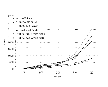
2) Cytokine IL-17 A
Dosing with RHB-104 BID demonstrated a reduction in the cytokine levels in the
lymph
nodes and to a lesser extent in the spleen as seen in Figure 1.
3) Cytokine TNF-alpha
RHB-104 BID and QD dosing reduced TNF-alpha in the spleen. Additionally, BID
dosing reduced TN-alpha in the lymph nodes. BID dosing reduction of the TNF-
alpha in the
spleen was almost 50% as shown in Figure 2.
4) Cytokine IFN-gamma
RHB-104 reduced IFN-gamma for BID dosing in the spleen as shown in Figure 3:
5) Cytokine IL-6
RHB 104 BID dosing in the spleen showed an almost 50% reduction of IL-6 in the
spleen
as shown in Figure 4.
6) Cytokine IL-4
IL-4 was below standard detection.
17
CA 02949255 2014-03-19
WO 2013/041963 PCT/IB2012/002252
7) Cytokine IL-2
RHB-104 reduced IL-2 in QD dosing in both spleen and lymph nodes as shown in
Figure
5.
The effect of the formulation RHB 104 comprising rifabutin, clarithromycin and
clofazimine on cytokine levels in the above mouse model supported further
analysis in an EAE
mouse model which is a well recognised model for human MS.:
Experiment 2: Evaluation of the efficacy of RHB 104 administered in a mouse
model of EAE.
Background and Overview of EAE model
EAE induction
Chronic EAE develops in C57BL/6 mice after immunization with an emulsion of
M0G35_55/CFA or M0G1_125/CFA followed by injection of pertussis toxin. This
model is used to
test the potential of compounds to prevent or mitigate EAE disease. It can be
run with the
compound dosed from the time of immunization (prophylactic treatment), or with
the aim of
reversing the course of disease and facilitating recovery by dosing the
compound from the time
of EAE onset (therapeutic treatment).
The model uses female C57BL/6 mice of age 10 to 14 weeks at the start of the
study.
Typically, EAE develops 8-18 days after immunization. EAE development is
usually followed
for 4 weeks (28 days) after immunization.
Stress reduces mouse susceptibility to EAE. Aside from any compound effects,
the
administration of treatment during the disease induction period (-0-10 days
after immunization)
postpones disease onset and reduces disease severity. This is due to the
stress of compound
administration and the effects of the vehicle on the mice. The more frequent
the administration
and the less tolerated the vehicle, the greater the impact on disease
development.
The stress of treatment and administration of vehicle has much less effect on
disease
development after clinical signs of EAE have appeared.
18
CA 02949255 2014-03-19
WO 2013/041963 PCT/IB2012/002252
Prophylactic treatment
In prophylactic studies, treatment begins before disease onset, at the time of
immunization
and group assignment. Mice are assigned to treatment groups in a balanced
manner to achieve
groups with similar distributions of body weights.
Prophylactic studies assess if treatment will affect the course of disease
both before and
after the first clinical signs of EAE.
To compensate for the stress of treatment in prophylactic treatment studies
and achieve
the target disease severity, EAE is induced with a higher dose of pertussis
toxin than used in
therapeutic studies. The dose of pertussis toxin is based on the expected
stress due to dosing
(route, frequency, and formulation of vehicle).
In prophylactic studies, median time to disease onset is usually the most
sensitive measure
of compound efficacy.
Small changes in the immune response can result in postponed disease onset -
suppression
of T cell activation and proliferation, antigen presentation, differentiation
into Thl and/or Th17
cells will all result in postponed onset of EAE.
Delayed onset of EAE accompanied with lower maximum severity indicates overall
efficacy of treatment compared to the negative control group.
Therapeutic treatment
In therapeutic treatment studies, treatment begins at the time of EAE onset.
Mice are
distributed into different treatment groups as they develop EAE (rolling
enrolment) in a balanced
manner to achieve groups with similar time of EAE onset and similar onset
scores.
Therapeutic studies assess if treatment will reverse the course of disease or
improve
recovery from EAE.
The most important readout in this model is the average end clinical EAE
score. This is
the clinical outcome of the experiment; a reduction compared to the negative
control group
indicates treatment efficacy.
19
CA 02949255 2014-03-19
WO 2013/041963 PCT/IB2012/002252
Course of EAE development in untreated mice
Individual mice will have differing courses of disease. Most mice show initial
signs of
EAE between 9 and 14 days after immunization. Once EAE starts, the peak of
disease almost
always occurs 3-4 days later. The maximum score continues for several days and
then mice
partially recover. In some mice, disease will stay at maximum severity until
the end of the study.
Less often, a mouse will stay at the peak severity for only one day and then
start recovering.
The extent of recovery largely depends on the maximum severity reached by the
mouse.
Most untreated or vehicle-treated mice will not fully recover, but their end
score will usually be
0.5 to 1.5 points lower than their maximum score. About 25% of untreated or
vehicle-treated
mice show worsening EAE between 24 and 28 days after immunization, resembling
a relapse.
Spinal cords of these mice at the time of EAE worsening have a large number of
inflammatory
foci (> 7 foci per section), similar to histological findings at the time of
EAE onset and peak,
suggesting that these are true relapses with a new wave of inflammation in the
spinal cords.
When mice are followed for a longer period of time, disease slowly increases
in severity,
resembling the chronic progressive course of disease observed in human MS
patients.
During the course of EAE, changes in body weight reflect disease severity.
Mice often
lose a small amount of weight on the day following immunization. This appears
to be due to
effects of the administered adjuvant and pertussis toxin. Mice then steadily
increase their body
weight until disease onset. On the day of EAE onset, mice consistently lose 1-
2 g of their body
weight (5-10% of body weight). The weight loss continues with the progression
of EAE
severity, with the loss reaching around 20% of their pre-onset body weight at
the peak of disease.
The weight loss is most likely due to both paralysis and reduced food intake
as well as high
production of pro-inflammatory cytokines such as TNF during the acute phase of
inflammation.
After the peak of disease is reached, mice slowly gain weight, even if their
clinical score does
not improve. This increase in weight may be due to down regulation of
inflammation which
results in lower levels of pro-inflammatory cytokines in blood. Untreated or
vehicle-treated mice
usually have around 90% of their pre-immunization body weight 28 days after
immunization.
CA 02949255 2014-03-19
WO 2013/041963 PCT/IB2012/002252
Histology
Inflammation in EAE normally starts in the lumbar region of the spinal cord,
spreading to
the entire spinal cord by the peak of disease.
At onset of disease the number of inflammatory foci correlates strongly with
disease
severity. The number of foci increases until the peak of disease, when 6-15
inflammatory foci
/section are typically found throughout the spinal cord. In the chronic stage
of EAE (starting
several days after the peak of disease), many inflammatory foci resolve,
typically resulting in 3-4
inflammatory foci in each spinal cord section by approximately 28 days after
immunization.
Because the largest numbers of inflammatory foci are present early in the
course of
disease, if histological analysis is performed at the end of the study, mice
which have late EAE
onset often have more inflammatory foci in their spinal cords than might be
expected from their
clinical score. For example, in a 28 day study a mouse with EAE onset on 27
days after
immunization and an end clinical score of 2 will likely have more inflammatory
foci than a
mouse with EAE onset 9 days after immunization and an end score of 3.5.
Similarly, a mouse
which relapses shortly before the end of the study (relapse is defined as 1 or
more points of
increase in clinical score) will usually have more inflammatory foci at the
end of the study than a
mouse with stable chronic disease, even if the two have the same clinical
score at the end of the
study.
Demyelination is usually not found during the first two days after disease
onset, but is
found at the peak of disease (4-5 days after EAE onset) and continues during
the chronic phase
of EAE. Demyelination scores do not change much between the peak and 28 days
after
immunization and usually average between 1.2 and 2.5.
Demyelination is scored in both Luxol fast blue stained sections (LFB) and in
H&E
sections.
In LFB sections, spinal cord white matter stains dark blue and demyelinated
areas are a
lighter blue colour, and are associated with large vacuoles.
In H&E stained sections disruption of normal structure with large vacuoles is
indicative
of demyelination.
21
CA 02949255 2014-03-19
WO 2013/041963 PCT/IB2012/002252
Apoptotic cells are identified in H&E sections, and are usually not found
during the first
two days of disease development. They are found at the peak and during the
chronic stage of
EAE. The average number of apoptotic cells is usually between 2 and 4 per
section.
Experimental Design
Mice were weighed before the start of the study and then assigned to groups in
a balanced
manner. Compound treatment started on the day of immunization (Day 0 of the
study).
Disease was induced by immunizing mice on Day 0 with myelin oligodendrocyte
glycoprotein, peptide 35-55 (M0G35_55) emulsified in complete Freund's
adjuvant (CFA),
followed by two injections of pertussis toxin (administered on Days 0 and 1).
To assess disease development, mice were weighed three times per week (Monday.
Wednesday and Friday) from the time of immunization and scored daily for
clinical signs of
EAE starting on Day 7.
Materials and Methods
Mice
The study used a total of 24 female C57BL/6 mice (Taconic Farms, 10 weeks
old).
Groups and treatment
Mice were assigned to groups in a balanced manner to achieve similar weight at
the start
of the study.
Table 1 below shows which treatment was administered to each group.
22
CA 02949255 2014-03-19
WO 2013/041963 PCT/IB2012/002252
Table 1 - Immunization and treatment regimen
Group Compound Dose Frequency Purpose
1 Vehicle BID Negative control
2 RHB- 104 36 mg/kg BID Test compound
Each group consisted of 12 mice.
Treatment of all groups was p.o., BID at a volume of 10 mL/kg.
Treatment started on the day of immunization (Day 0) and lasted until Day 27
after
immunization. All dosing was performed at the same time (+/- 1 hour) each day.
There were no
more than 14 hours between the evening and morning dose and no less than 10
hours between
the morning and evening dose.
EAE induction
EAE was induced in 24 female C57BL/6 mice (10 weeks old) as follows:
Day 0, Hour 0 ¨ Immunization with MOGi5-55/CFA
Day 0, Hour 2 ¨ Injection of pertussis toxin
Day], Hour 0 ¨ 2"3 injection of pertussis toxin (24 hours after initial
immunization)
Mice were injected subcutaneously at two sites in the back with the modified
emulsion
component of the kit (containing MOG35_55). One site of injection was in the
area of upper back.
approximately 1 cm caudal of the neck line. The second site was in the area of
lower back.
approximately 2 cm cranial of the base of the tail. The injection volume was
0.1 mL at each site.
Within 2 hours of the injection of emulsion, and then again 24 hours after the
injection of
emulsion, the pertussis toxin component of the kit was administered
intraperitoneally. The
volume of each injection was 0.1 mL.
23
CA 02949255 2014-03-19
WO 2013/041963 PCT/IB2012/002252
Scoring and readout
Readouts were EAE scores and body weight at the end of the study.
Mice were scored daily from Day 7 until the end of the study, and body weight
was
measured three times/week (Monday, Wednesday and Friday), starting on Day -1.
The last day of scoring was Day 28 after immunization.
Scoring was performed blind, by a person unaware of both treatment and of
previous
scores for each mouse.
EAE Scoring
EAE was scored on scale 0 to 5:
Score of 0.
No obvious changes in motor functions of the mouse in comparison to non-
immunized
mice.
When picked up by the tail, the tail has tension and is erect. Hind legs are
usually spread
apart.
When the mouse is walking, there is no gait or head tilting.
Score of 1.
Limp tail.
When the mouse is picked up by the tail, instead of being erect, the whole
tail drapes over
finger.
Score of 2.
Limp tail and weakness of hind legs.
When mouse is picked up by tail, legs are not spread apart, but held closer
together. When
the mouse is observed walking, it has a clearly apparent wobbly walk.
24
CA 02949255 2014-03-19
WO 2013/041963 PCT/IB2012/002252
Score of 3.
Limp tail and complete paralysis of hind legs (most common); or
Limp tail with paralysis of one front and one hind leg; or
ALL of:
Severe head tilting,
Walking only along the edges of the cage,
Pushing against the cage wall,
Spinning when picked up by the tail.
Score of 4.
Limp tail, complete hind leg and partial front leg paralysis.
Mouse is minimally moving around the cage but appears alert and feeding.
Usually, euthanasia is recommended after the mouse scores level 4 for 2 days.
When the
mouse is euthanized because of severe paralysis, score of 5 would be entered
for that mouse for
the rest of the experiment.
Score of 5.
Complete hind and complete front leg paralysis, no movement around the cage;
or
Mouse is spontaneously rolling in the cage; or
Mouse is found dead due to paralysis.
In-between scores were assigned when the clinical signs fell between two above
defined
scores.
Histological analysis of spinal cords
On Day 28 (end of the study) all mice were sacrificed for histological
analysis.
Mice were perfused with PBS and spines were collected in 10% buffered
formalin.
For each mouse, 3 Luxol fast blue stained sections and 3 H&E sections, from
lumbar,
thoracic, and cervical spinal cord, were prepared and analysed.
CA 02949255 2014-03-19
WO 2013/041963 PCT/IB2012/002252
The histological analysis was performed by a pathologist blinded to the
experimental
groups and all clinical readouts.
Count of inflammatory foci
Inflammatory foci of approximately 20 cells were counted in each H&E stained
section.
When inflammatory infiltrates consisted of more than 20 cells, an estimate was
made of how
many foci of 20 cells were present.
Estimation of demyelinated area
The demyelination score represents an estimate of demyelinated area for each
section as
follows:
0 ¨ no demyelination (less than 5% demyelinated area)
1 ¨ 5 to 20% demyelinated area
2 ¨ 20 to 40% demyelinated area
3 ¨ 40 to 60% demyelinated area
4 ¨ 60 to 80% demyelinated area
¨ 80 to 100% demyelinated area
For Luxol fast blue stained slides, the size of the demyelinated area was
estimated based
on less intense blue staining of myelin.
For H&E stained sections, the demyelinated area was estimated by looking for
interruption of normal structure ¨ pallor and vacuolation consistent with
edema and
demyelination, and dilated axons.
Count of apopiotic cells
The number of apoptotic cells in each of the three H&E sections was
determined. The
apoptotic cells are neurons and their number correlates with disease stages.
Apoptotic cells
appear soon after disease onset, so at EAE onset there will be many
inflammatory foci, but few
apoptotic cells. Then, the number of apoptotic cells increases until the peak
of disease, then
remains elevated.
26
CA 02949255 2014-03-19
WO 2013/041963 PCT/IB2012/002252
Statistical analysis
Statistical analysis was performed as follows:
Disease incidence compared using Chi-square test
Median day of EAE onset compared using Wilcoxon's survival test
Mean day of EAE onset compared using two-tailed Student's t-test
Mean maximum score (MMS) compared using Wilcoxon rank sum test
End score compared using Wilcoxon rank sum test
Change in body weight compared using two-tailed Student's t-test
Demyelination scores (LFB) compared using Wilcoxon's non-parametric test
Demyelination scores (H&E) compared using Wilcoxon's non-parametric test
Number of apoptotic cells compared using 2-tailed Student's t-test
Results and interpretation of data
EAE development was evaluated by comparing:
EAE incidence,
median and mean of day of EAE onset (MME),
mean maximum score (MMS),
average EAE scores at the end of the study, and
average body weight at the end of the study relative to initial weight
between the vehicle group (negative control) and the RHB-104 group
27
CA 02949255 2014-03-19
WO 2013/041963 PCT/IB2012/002252
Summary of results - Clinical Findings
Table 2
End %
EAE End
MMS p body
Treatment incidence MME p value score
value value +I- SD value weight
p value
(%) +1- SD
+/- SD
3.08 +/- 2.54 +/- 90.7 +/-
Vehicle 100.0% 15.0
0.87 1.05 7.5
33 +/- 0.92 +/- 105.2
RHB-104 100.0% 1.0000 15.0 0.7617 2'0.65 0.0031
0.67 0.0011 0.0000
3.8
Group 1: Vehicle group, p.o., BID (negative control)
Most mice in this group developed severe EAE (Table 1 and Figure 6).
Most mice in this group lost weight during this study, which was expected
(Table 1 and
Figure 7).
No mice died in this group.
Group 2: RHB-104, 36 mg/kg, p.o., BID
Most mice in this group developed milder disease than observed in the vehicle
group.
This group was significantly improved in most clinical readouts of EAE
compared to the
vehicle group (Table 1 and Figures 6 and 7).
No mice died in this group.
The above results observed show a marked effect in relation to the severity of
the disease
by the abovementioned indicators upon treatment with RHB 104 when compared to
a control in
the recognised human MS mouse model.
28
CA 02949255 2014-03-19
WO 2013/041963 PCT/IB2012/002252
Summary of Results - Histological findings
Table 3
Inflammator Demyelination Demyelinalion Apoptotic
Treatment (LFB) (H&E) cells
foci y p
value value value value
+1-SD +1-SD +/-SD
+/- SD
Vehicle 3.2 +/- 2.2 1.7 +/- 0.7 1.4 +/- 0.6 3.1
+/- 1.2
RHB-104 1.8 +/- 1.6 0.0867 0.6 +1- 0.6 0.0018 0.8 +/-
0.5 0.0198 1.4 +/- 1.7 0.0093
Vehicle treated mice
Histological findings in the vehicle-treated mice were typical for this stage
and severity of
EAE. Low magnification images of representative thoracic and lumbar spinal
cord sections from
vehicle treated mice showed that inflammation was present in the leptomeninges
and in the white
matter. No mice died in this group.
RHB 014 treated mice
Consistent with the clinical findings, most histological readouts in these
mice were
indicative of significantly less severe disease than in the vehicle-treated
mice. Low
magnification images of representative thoracic and lumbar spinal cord
sections from RHB-104
treated mice showed fewer inflammatory foci in these sections than in sections
from vehicle
treated mice. In addition, the inflammatory foci were smaller in the RHB-104
treated mice than
in vehicle treated mice.
Demyelinated areas were significantly smaller in the RHB-104 treated mice than
in
vehicle treated mice. No mice died in this group.
The average number of inflammatory foci detected in H&E sections is shown in
Figure 8
and the average demyelination score from Luxol fast blue sections shown in
Figure 9.
Consistent with the clinical findings, the histological readouts in these mice
were
indicative of significantly less severe disease than in the vehicle-treated
mice. Fewer
inflammatory foci were found in the RHB 104 treated mice than in sections from
vehicle treated
mice. In addition, the inflammatory foci were smaller in the RHB-104 treated
mice than in
vehicle treated mice.
29
CA 02949255 2014-03-19
WO 2013/041963 PCT/IB2012/002252
Dem yelinated areas were significantly smaller in the RHB-104 treated mice
than in
vehicle treated mice. All these findings confirm the clinical observation that
RHB-104 treated
mice had significantly less severe EAE than vehicle-treated mice at the end of
the MS mouse
model study.
Experiment 3: EAE relapse study.
The model most strongly resembles the remitting-relapsing form of MS (the most
common form of MS).
As a background to the model, it is to be understood that mice develop a first
episode of
paralysis 11-14 days after immunization in an EAE model and, similar, to most
MS patients, they
fully or almost fully recover from this first wave of paralysis. After a
disease-free period of 1-2
weeks, 50 to 100% of the mice develop a second wave of paralysis (relapse).
This model is used for testing the effect of compounds on the development of
EAE
relapses (therapeutic treatment). Treatment can be initiated at the onset of
clinical signs of EAE,
or at the start of recovery from the first wave of EAE. This model is
typically run for 5 to 7
weeks, but mice are sometimes observed longer
Experiment Design
Disease was induced by immunizing mice on Day 0 with PLP139_151 peptide
emulsified in
complete Freund's adjuvant (CFA).
To assess disease development, mice were weighed three times per week (Monday.
Wednesday and Friday) from the time of immunization and scored daily for
clinical signs of
EAE starting on Day 9.
Enrollment of mice into groups occurred on the second day of clinical signs of
EAE for
each mouse. Mice were enrolled into treatment groups as they develop signs of
EAE (rolling
enrolment).
CA 02949255 2014-03-19
WO 2013/041963 PCT/IB2012/002252
Group assignment and treatment
All mice were initially considered a single group. Daily scoring started on
Day 9 after
immunization.
Enrolment of mice into groups occurred on the second day of clinical signs of
EAE for
each mouse. Mice were enrolled into treatment groups as they develop signs of
EAE (rolling
enrolment).
45 mice were enrolled into 3 groups of 15 and the treatment started on the day
of
enrolment. Assignment was balanced to achieve similar scores between the
groups at enrolment.
7 mice which develop disease latest, or which had unusual symptoms, were not
enrolled
into groups and were not used in the study.
Groups
Group 1 ¨ Vehicle (PBS), p.o., BID, 5 mL/kg (negative control)
Group 2 ¨ treated with FTY-720 (Fingolimod, Gilenya) 3 mg/kg, p.o., QD (a drug
used for
treatment of MS and used as a positive control)
Group 3 ¨ treated with RHB-104, p.o., BID, 5 mL/kg
Treatment
Treatment started on the day of enrolment and continued until Day 39.
All dosing was performed at the same time (+/- 1 hour) each day. There was at
least 10
hours between morning and evening dosing and not more than 14 hours between
evening and
morning dosing.
The last day of dosing was Day 39 for all mice.
31
CA 02949255 2014-03-19
WO 2013/041963 PCT/IB2012/002252
Scoring and readout
Mice were scored daily from Day 9 to Day 40, and body weight was measured
three
times/week (Monday, Wednesday and Friday), starting before immunization (Day -
1).
Scoring was performed blind, by a person unaware of both treatment and of
previous
scores for each mouse.
Readouts were EAE scores on the scale 0-5 in 0.5 unit increments, and changes
in body
weight.
EAE Scoring
EAE was scored on scale 0 to 5 as described above.
Statistical analysis
Statistical analysis was performed as follows:
Median day of EAE onset compared using Wilcoxon's survival test
Mean day of EAE onset compared using two-tailed Student's t-test
Mean maximum score (MMS) of first wave compared using Wilcoxon rank sum test
Relapse incidence compared using Chi-square test
Mean maximum score (MMS) of relapse compared using Wilcoxon rank sum test
End score compared using Wilcoxon rank sum test
Change in body weight compared using two-tailed Student's t-test
Results
8 mice in Group 1 showed relapse as indicated in their clinical scores whereas
only 2
mice from each of Groups 2 and 3 demonstrated relapse.
Further, the severity of the relapse disease in Group 3 was significantly less
than the
severity in Group 1 as evidenced by the graph shown in Figure 12.
32
CA 02949255 2014-03-19
WO 2013/041963 PCT/IB2012/002252
Conclusion
In addition to reducing the symptoms in an initial onset of EAE, the present
RHB-104
composition has been shown to protect against relapse of the disease in the
well recognised
mouse model used above and in the cases where relapse occurs, the severity of
the disease is
significantly reduced when compared to a negative control group. Collectively,
these results
indicate that RHB-104 was highly efficacious in reducing disease severity in
this study.
It will be appreciated by persons skilled in the art that numerous variations
and/or
modifications may be made to the above-described embodiments, without
departing from the
broad general scope of the present disclosure. The present embodiments are,
therefore, to be
considered in all respects as illustrative and not restrictive.
33