Note: Descriptions are shown in the official language in which they were submitted.
CA 02849335 2014-03-19
WO 2013/052652
PCT/US2012/058732
METHODS AND SYSTEMS FOR IDENTIFYING AND TREATING ANTI-
PROGESTIN SENSITIVE TUMORS
BACKGROUND
[0001] This application claims priority to U.S. Provisional Patent Number
61/542,931, filed on October 4, 2011, the disclosure of which is incorporated
by
reference herein in its entirety.
[0002] The progesterone receptor (PR) is present in cells in two major
isoforms,
PR-A and PR-B. In the presence of a bound progestin ligand, such as
progesterone, the
PR is phosphorylated at specific sites, dimerizes, forms a complex with a
number of
different cellular elements (e.g., p300 and the steroid receptor coactivator),
and binds to
specific DNA sequences known as progesterone responsive elements (PREs) to
initiate
DNA transcription into RNA. The PR-ligand complex also attracts numerous other
co-
activators and co-repressors, which form the cellular elements which in turn
transcribe
particular genes. These PR complexes (also referred to as foci) can be
visualized in the
nuclei of cells which contain the progesterone receptor as fluorescent
aggregates using
immunohistofluorescence techniques and as dense and dark stained nuclear
aggregates
using the immunohistochemistry techniques described in this patent.
[0003] In premenopausal women, during the proliferative phase (the first
part of
the menstrual cycle) when estrogen is the dominant hormone and progesterone is
minimally secreted, staining of normal endometrial cells for PR-A and PR-B
(e.g., using
immunofluorescent techniques and confocal microscopy) reveals a diffuse
progesterone
receptor nuclear staining pattern. In the secretory phase (the second part of
the menstrual
cycle) when progesterone is the dominant hormone, using the same
immunofluorescent
techniques and confocal microscopy, staining for PR-A and PR-B appears as
readily
detectable fluorescent nuclear foci.
[0004] RNA transcription inhibitors have been shown to prevent formation
of PR
foci, and 26S proteasome inhibitors have been shown to disrupt the PR nuclear
foci. It is
1
CA 02849335 2014-03-19
WO 2013/052652
PCT/US2012/058732
therefore believed that the presence of PR foci in cells corresponds to active
transcriptional complexes, and indicates the activation of the PR and
subsequent gene
expression. Conversely, diffuse nuclear staining or the absence of PR foci
indicates the
presence of PR which is transcriptionally inactive. Upon exposure of normal
breast and
endometrium tissues (which are physiologically responsive to progesterone) to
progestin
ligands, a change from a diffuse nuclear staining pattern to focal subnuclear
structures
can be observed, indicating the activation of the progesterone receptor.
[0005] Whereas estrogens are mitogenic (e.g., cause cellular
proliferation) for
normal breast epithelial and endometrial cells, the effects of progestins are
more
complex. In the endometrium, progestins inhibit estrogen-induced cell cycle
progression
early in the G1 phase, whereas in the breast progestins may both stimulate and
inhibit
proliferation. In normal breast tissue biopsies it has been shown that
proliferative activity
is stimulated by progesterone (Am J Obstet Gynecol, 1997). This complexity has
led to
confounding experimental observations in breast cancer. For example,
progestogens
appear to have a direct proliferative effect on breast cancer cell in vitro
when phenol red¨
free media is used. H. J. Kloosterboer,J. Steroid Biochem. Molec. Biol. Vol.
49, No. 4-6,
pp. 311-318, 1994. However, when the same contraceptive progestogens that
induced
proliferation in breast cancer cell lines were studied in an estrogen-
dependent DMBA rat
breast cancer model, these progestogens inhibited tumor progression. Id.. It
has been
shown recently that many such in vitro experimental models are inadequate.
See, e.g.,
Lange C. et al. Progesterone Receptor Action: Translating Studies in Breast
Cancer
Models to Clinical Insights. Chapter 7 in Innovative Endocrinology of Cancer;
94-111
(2010). While progesterone-induced proliferation has been shown in these
experimental
models, the majority of proliferating cells were not expressing the PR. Thus,
these
models do not necessarily predict the efficacy of treatment with
antiprogestins.
[0006] Malignant cells also exhibit nuclear PR foci, but they are
different in size
and composition from the foci of normal cells. PR foci observed in cancer
indicate a
specific role for the PR which is pertinent to the malignant nature of the
cells. For
example, the genes activated by the PR in malignant (cancer) breast cells are
different
2
CA 02849335 2014-03-19
WO 2013/052652
PCT/US2012/058732
than the genes activated by the PR in normal breast cells; in endometrial
cancers PR foci,
but not PR levels, are associated with malignant characteristics; foci in
cancer cells are
larger, which may be due to alterations in the chromatin remodeling which are
common
in cancer, and; PR foci in breast cancer are observed regardless of hormonal
status (e.g.,
in the presence and absence of circulating progesterone in premenopausal and
post-
menopausal women respectively). PR foci
have been observed (e.g., using
immunofluorescent techniques and confocal microscopy) in the tumor cells of
approximately 50% of PR-receptor positive human breast cancer biopsies. Other
patient's tumor samples exhibited a diffuse PR nuclear staining pattern in the
tumor cells
using immunofluorescent techniques and confocal microscopy, indicative of a
non-
activated or non-functional form of the PR.
[0007] The
majority of breast cancers can be treated with hormonal treatments
(i.e., anti-estrogens or aromatase inhibitors), which are currently some of
the most
effective medications used in breast cancer therapy. Hormonal treatment is
usually
indicated based on the identification of hormone receptors within the cancer
cells.
Onapristone (ONA) is an anti-progestin drug which was originally developed for
contraceptive use. However, it has demonstrated substantial activity in
advanced breast
cancer, with a 10% response rate in a study of 101 poor prognosis patients
with breast
cancer in whom prior hormonal therapy had failed (e.g., breast cancer
progressed despite
the patient receiving the antiestrogen tamoxifen). In a small breast cancer
study using
ONA as a first line hormone treatment, ONA produced a 56% objective response
rate, an
efficacy in the upper range of the best available treatments in this disease.
ONA binds to
the PR, does not induce PR phosphorylation and does not allow the PR to
dimerize. The
PR-ONA complex binds weakly, or not at all, to its target DNA segment and
therefore
does not activate the chromatin remodeling which is a necessary process for
DNA
transcription. In in vitro systems, ONA has been shown to reverse the PR
nuclear
aggregates produced by binding of an artificial ligand to the PR. Gene
activation studies
have consistently shown that, while progestins and other anti-progestins
activate
progesterone responsive genes, ONA has minimal activation (i.e., 3 genes).
3
CA 02849335 2014-03-19
WO 2013/052652
PCT/US2012/058732
[0008] In addition, ONA is a pure PR antagonist at concentrations which
can be
physiologically achieved. ONA does not interfere with other steroid receptors
and does
not increase estrogen secretion in human subjects, which is an undesirable
side-effect for
breast cancer therapy exhibited by other anti-progestins such as mifepristone.
[0009] While onapristone has previously been investigated as a potential
therapeutic agent for breast cancer, its development was stopped due to
toxicity concerns.
Robertson et al., Onapristone, a Progesterone Receptor Antagonist, as First-
line Therapy
in Primary Breast Cancer European J. of Cancer 35(2) 214-218 (1999). It is
important to
identify the subset of the patients with tumors most likely to respond and
equally as
important to identify the subset of the patients with tumors least likely to
respond to
treatment with ONA and other anti-progestins. Identifying these subsets of
patients will
allow those patients with APF access to a potentially effective cancer
treatment and will
avoid exposing patients with those cancers for which ONA or other anti-
progestins may
not provide benefit to unnecessary toxicity.
[0010] Currently, only the presence or absence of the estrogen or
progesterone
receptor is considered when making therapeutic decisions on whether to use an
endocrine
treatment in certain cancers (e.g., breast cancer). Accordingly, conventional
assays for
PR classify the tumors from patients with cancer into two categories: PR-
positive or PR-
negative. One type of assay quantitates the amount of PR per total protein of
the cell.
These methods can be automated and are quantitative, but are not satisfactory
with
respect to accuracy, sensitivity and analysis of cellular subnuclear receptor
structures. A
second type of assay includes immunohistochemical methods using formalin fixed
tissue
specimens and fluorescent or chromophore labeled monoclonal antibodies
targeting the
receptor (either an antibody for each of PR-A and PR-B, or a single antibody
that
recognizes both). With immunohistochemical methods, any microscopically
detectable
nuclear staining reaction in more than a certain percentage of cells
(typically > 1%), is
reported as being PR positive as per professional society guidelines.
Typically, a clinical
cut off of >10% ER or PR positive cells is used to make therapeutic decisions
regarding
the use of anti-hormone treatments. No consideration is given to the pattern
of cellular or
4
CA 02849335 2014-03-19
WO 2013/052652
PCT/US2012/058732
nuclear staining. Relative staining intensity (i.e., low, medium, or high) is
also use as a
qualitative measure of hormone receptor positivity. This second type of assay
is more
labor intensive and it is not standardized. Typically, low magnification
microscopic
examination is used for the IHC analysis to identify the presence of the
hormone receptor
(either estrogen receptor (ER) or PR). Using conventional methods, no analysis
of
cellular distribution is done other then an estimate of the percentage of the
tumor cells
expressing the identified hormone receptor. Analysis of the subnuclear
distribution
pattern of the PR requires high powered microscopy. In contrast, high powered
microscopy is not needed for standard IHC determination of hormone receptors
in tumor
tissue. These conventional methods of hormone receptor determination are thus
unable
to provide information regarding subnuclear PR distribution.
[0011] Progestins have complex actions in the breast and other hormone
sensitive
tissues by targeting distinct cells and having indirect effects on cells not
expressing the
PR. PR foci complexes are not qualitatively the same in normal tissue and
cancerous
tissue, and they do not necessarily activate the same progesterone receptor
associated
genes. Available clinical data does not fully support the position that
conventional
techniques for identifying hormone receptor positive cells are predictive of
anti-hormone
efficacy, whether it be for anti-estrogen or anti-progestin directed
treatments. Currently,
the decision to utilize a hormone treatment (e.g., antiestrogens or aromatase
inhibitors)
for patients with breast cancer and other hormone sensitive tumors is based on
the simple
presence of hormone-receptors in tumor samples. The presence of hormone
receptors
(ER or PR) does not fully predict for response to hormone treatment, as only
50-60% of
hormone-receptor positive tumor cases are expected to benefit from treatment.
[0012] There is a need for a consistent method for predicting the
efficacy of ONA
and other anti-progestins with respect to heterogeneous "naturally occurring"
tumors.
Further, there is a need for an assay which is predictive of therapeutic
efficacy of ONA
and other anti-progestins against the cancers in individual patients.
CA 02849335 2014-03-19
WO 2013/052652
PCT/US2012/058732
SUMMARY
[0013] An important question pertinent to anti-progestin treatment is how
to
identify activated PRs that are relevant clinical therapeutic targets. The
present
exemplary methods are aimed at characterizing PRs that are present in a
functional
(activated) state in the human tumor tissue routinely obtainable in the
clinical setting. As
antagonizing non-active PR with a specific anti-progestin is therapeutically
pointless, the
present methods provide new and critical information to guide treatment of
patients with
anti-progestins. Such a predictive diagnostic test would provide (1)
consistent methods
to support therapeutic decision-making with respect to ONA and other anti-
progestins,
(2) guide selection of individual patients and patient populations that are
likely to respond
to treatment, and (3) exclude those individual patients that are least likely
to respond or
benefit from an anti-progestin treatment.
[0014] In one aspect, a method for identification and treatment of a
subset of
progesterone receptor (PR) positive tumors most susceptible to treatment with
an anti-
progestin such as onapristone (ONA) is provided. Progesterone receptor
positive tumors
exhibiting a dense, focal PR nuclear distribution pattern, as described
herein, are more
susceptible to treatment with anti-progestins such as onapristone. Results
from in vitro
homogeneous, experimental models are not necessarily predictive of the
properties of
naturally-occurring heterogeneous tumors.
[0015] In another aspect, a method of inhibiting the growth of a tumor
susceptible
to growth inhibition by anti-progestins is provided. A tissue sample suspected
of being
tumorigenic or cancerous can be obtained from a patient. Progesterone receptor
positive
cells in the tissue sample can be identified. The degree of distribution of
the progesterone
receptor foci in nuclei of the progesterone positive cells from the tissue
sample can then
be determined and an anti-progestin can be administered to the patient if the
degree of
focal distribution in the tissue sample is greater than about 5% of the
progesterone
receptor positive cells.
6
CA 02849335 2014-03-19
WO 2013/052652
PCT/US2012/058732
[0016] These patients are more likely to benefit from treatment with an
anti-
progestin that inactivates activated progesterone foci (APF) (e.g., ONA) and
prevents
further formation of APF than patients whose tumors do not express activated
PR. The
non-activated form of the PR is typically seen as diffuse nuclear PR staining.
Inactivation of the APF by an anti-progestin may occur by any of a variety of
mechanisms, including dissociation of the foci and inhibition of activation of
the foci
without substantially altering their structure. In one aspect, APF formation
can be
inhibited or prevented by an anti-progestin through several mechanisms. For
example,
onapristone may not allow the individual progesterone receptors to dimerize
and prevent
the PR from being phosphorylated at the ligand phosphorylation sites. The PR-
ONA
complex may bind weakly, or not at all, to its target DNA segment (PREs) and
fail to
induce the chromatin remodeling which is a necessary process for DNA
transcription. In
another example, other anti-progestins may allow the PR to dimerize and form
complexes
with co-activators or co-repressors which do not induce DNA transcription.
[0017] In this example, DNA binding may occur at the PRE, but
transcription
does not occur. Identification of APF may inform the decision of any anti-
progestin
treatment as long as the agent interferes with the PR pathway. In one aspect,
identification of APF determines the status of the PR pathway as activated or
not. For
example, the use of mifepristone, or any progestin that complexes with PR and
binds to
the DNA, could be informed by the identification of APF. The activity of other
agents,
including those which would inhibit PR phosphorylation and thus interfere with
PR
activation, would be predicted by the presence of APF in various cancers.
Thus,
identification of APF could be used to inform treatment recommendations for
various
classes of compounds which act by inhibiting the function of the PR.
[0018] Patient tumors that do not express activated PR foci (APF) may
include
those that are PR-negative by the conventional assay, or those that are PR-
positive by the
conventional assay. In one aspect, any tumor/cancer which exhibits APF is a
candidate
for treatment with such anti-progestins, including breast, brain, meningiomas,
prostate,
ovarian, endometrial, uterine leiomyoma, lung, and uterine cancers. Pulmonary
7
CA 02849335 2014-03-19
WO 2013/052652
PCT/US2012/058732
leiomyomatosis which has yet to be formally classified as a cancerous
condition would
also be likely to benefit if APF is expressed in the abnormal tissue. In
another aspect,
benign tumors not manageable with standard treatment, but presenting APF, can
be
treated by an antiprogestin as the presence of APF indicates that the tumor is
driven by
aberrant activation of PR, i.e. by the progestin pathway.
[0019] Another aspect provides a method of treating patient with a tumor
susceptible to growth inhibition by anti-progestins by obtaining a tissue
sample suspected
of being tumorigenic or cancerous from a patient and exposing the tissue to an
anti-
progesterone receptor antibody. Progesterone receptor positive cells in the
tissue sample
can be identified. The degree of focal binding distribution of the
progesterone receptor in
nuclei of cells from the tissue can be determined. If the focal binding
distribution is
greater than about 5% of the progesterone receptor positive cells in the
tissue sample, an
anti-progestin is administered to the patient in a dosage range of about 10 to
about 200
mg per day depending upon the potency, bioavailability, and safety profile of
the
antiprogestin.
[0020] In another aspect, the tissue is a specimen of a tumor tissue
selected from
the group consisting of breast, brain, meningiomas, prostate, ovarian,
endometrial, uterine
leiomyoma, lung, and uterine tissue.
[0021] In another aspect, the presence or absence of focal distribution
is detected
by fluorescence, a colorimetic reaction (e.g., an enzymatic reaction), imaged
with a
counter staining antibody (e.g., chromophore), radioactivity, and Western blot
(e.g.,
differential phosphorylation of the PR).
[0022] In yet another aspect, the anti-progestin is selected from the
group
consisting of onapristone, lonaprisan, mifepristone, PF-02413873,
telapristone,
lilopristone, 0RG2058, asoprisnil, and ulipristal.
[0023] The presence of active progesterone receptor focal distribution is
indicated
by a degree of nuclear focal distribution of greater than about 5% of the
progesterone
receptor positive cells. In another aspect, a tumor may be heterogeneous with
respect to
8
CA 02849335 2014-03-19
WO 2013/052652
PCT/US2012/058732
focal distribution and exhibit an active binding pattern (A) with distinct
progesterone
receptor foci, a diffuse binding pattern (D) without distinct progesterone
receptor foci, or
a mixture of an A pattern and a D pattern (AD) in various areas of the tumor.
[0024] In any of the foregoing aspects, when focal distribution (A or AD
pattern)
is present, the intensity or density of such focal distribution may be
quantitated. For
example, progesterone receptor antibodies may be radiolabeled, fluorescently
labeled,
imaged with a counter staining antibody (chromophore), imaged with a
colorimetic
reaction (e.g., an enzymatic reaction), or labeled in another manner where the
intensity of
the label can be measured and quantified.
FIGURES
[0025] FIGS. 1A and 1B shows exemplary immunohistochemical brown nuclear
staining patterns in human breast cancer samples derived from formalin-fixed
and
paraffin-embedded biopsies using antibodies directed to the progesterone
receptor;
[0026] FIGS. 2A and 2B show exemplary green nuclear staining patterns in
human breast cancer samples derived from formalin-fixed and paraffin-embedded
biopsies using antibodies directed to the progesterone receptor;
[0027] FIGS. 3A and 3B show exemplary immunohistochemical brown nuclear
staining patterns with HES background counterstaining in human breast cancer
samples
derived from formalin-fixed and paraffin-embedded biopsies using antibodies
directed to
the progesterone receptor; and
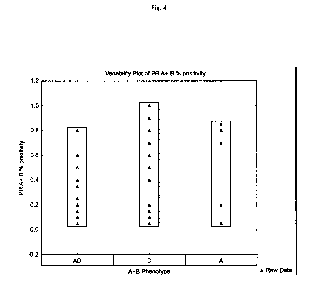
[0028] FIG. 4 shows the percent of breast cancer samples positive for PR-
A and
PR-B for three binding patterns, A, AD, and D.
DETAILED DESCRIPTION
[0029] Before describing several exemplary aspects described herein, it
is to be
understood that the invention is not limited to the details of construction or
process steps
set forth in the following description. The aspects described herein are
capable of being
practiced or being carried out in various ways.
9
CA 02849335 2014-03-19
WO 2013/052652
PCT/US2012/058732
[0030] As used herein, the phrases "treating a tumor" and "treatment of a
tumor"
mean to inhibit the replication of tumor cells, inhibit the spread of the
tumor, decrease
tumor size, lessen or reduce the number of tumor cells in the body, or
ameliorate or
alleviate the symptoms of the disease caused by the tumor, decrease the growth
of the
tumor (increase the time it takes the tumor to progress) or improve the
survival of the
patient when death is due to the cancer or secondary effects of the cancer.
The term also
includes treatment of cancer. Tumors include both cancers and non-cancerous
tumors.
The treatment is considered therapeutic if there is a decrease in mortality
and/or
morbidity, improvement of tumor-related symptoms, or there is a decrease in
disease
burden as may be manifested by reduced numbers of tumor cells in the body,
decreased
tumor size or improvement in the time to progression, improvement of
progression free
survival or improvement of disease free survival.
[0031] As used herein, the term "APF-active anti-progestin" and its
equivalents
refer to an anti-progestin drug which exhibits an ability to dissolve or
dissociate activated
PR foci (APF) in the nuclei of cells or inhibit the formation of APF in the
nuclei of cells,
indicating that its mechanism of action is via the PR activation pathway of
the cell.
[0032] The terms "APF-positive", "PR foci positive", "activated PR", "PRs
in a
functional state" and the like refer to the presence of progesterone receptor
aggregates in
the nuclei of cells.
[0033] The term "focal distribution" refers to the distribution of "foci"
(i.e.,
aggregation of progesterone receptors) in the nuclei of progesterone positive
cells.
Speckled or hyperspeckled pattern are terms that can be used referring to
steroid nuclear
receptor foci pattern in biology.
[0034] The term "degree of focal distribution" refers to the relative
amount of PR
foci present in the nuclei of progesterone positive cells. The degree of focal
distribution
can be determined quantitatively or qualitatively.
[0035] For example, the use of a colorimetric, enzymatic, or radiolabeled
ligand
such as a progesterone receptor antibody, can be used to bind to progesterone
receptors in
CA 02849335 2014-03-19
WO 2013/052652
PCT/US2012/058732
cell nuclei. The degree of focal distribution can be determined
quantitatively, for
example, by measuring color intensity, fluorescence or quantifying the level
of
radioactivity emitted by the labeled antibody. The degree of focal
distribution can
determined qualitatively by comparing the intensity of binding between a
control sample
and a labeled sample using a light microscope at an appropriate magnification
or
techniques including, but not limited to, DNA microarray, protein profiling,
radiolabeling, or other surrogates for measuring APF.
[0036] The term " diffuse pattern" refers to a finely granular pattern
which is
indicative of the absence of focal distribution.
[0037] The term "progestin" refers to a natural or synthetic
progestational
substance that mimics some or all of the actions of progesterone, also
referred to as
progesterone receptor modulators (PRM) or selective progesterone receptor
modulators
(SPRM).
[0038] The term "anti-progestin" refers to a substance that inhibits the
formation,
transport, or action of or inactivates progestational agents, including, but
not limited to,
onapristone, lonaprisan, mifepristone, PF-02413873, telapristone,
lilopristone, 0RG2058,
asoprisnil, and ulipristal. A PRM or SPRM may have some anti-progestin
properties, and
be considered an anti-progestin or a progestin depending on the context of
use.
[0039] The term "antibody" or "antibodies" refers to a protein which is
capable of
specifically binding to an antigen and includes any substance, or group of
substances,
which has a specific binding affinity for an antigen to the exclusion of other
substances.
Generally, the term "antibody" includes polyclonal antibodies, monoclonal
antibodies,
antibodies derived from humans or animals, humanized antibodies (e.g., non-
binding
portions derived from a human, binding portions derived from an animals) and
fragments
thereof.
[0040] The terms "anti-PR-A" and "anti-PR-B" antibodies refer to
antibodies
directed to isoforms of the progesterone receptor ¨ PR-A and PR-B
respectively. Anti-
PR-AB" refers to an antibody capable of binding to both PR-A and PR-B.
Specific
11
CA 02849335 2014-03-19
WO 2013/052652
PCT/US2012/058732
antibodies suitable for use in accordance with aspects herein include, but are
not limited
to, PgR636 and PgR1294 (M. Press, et al. (Steroids (2002) 67:799-813)),
Novacastra
clone 16, clone 5AN27, clone 1A6, Dako clone PgR636, Ventana, clone 1E2, Novus
Biologicals Progesterone Receptor [p Ser162] Antibody Clone 1064-E2; Novus
Biologicals Progesterone Receptor [p Ser190] Antibody Clone EP1516Y, Novus
Biologicals Progesterone Receptor [p Ser294] Antibody Clone 608, Abcam
Progesterone
Receptor [p Ser400] Antibody Ref ab60954, and Genetex Progesterone Receptor [p
Ser554] Antibody Ref. GTX118987.
[0041] The term "administer" refers to providing a drug or drugs,
prescribing one
or more drugs, or placing one or more drugs on a formulary. The term
"providing" refers
to dispensing the drug directly to patient through any suitable route of
administration
(e.g., oral, injection, intravenous, intramuscular, and transdermal etc.) or
providing
instructions to a patient to do the same.
[0042] One aspect provides a method of inhibiting the growth of a tumor
susceptible to growth inhibition by anti-progestins by obtaining a tissue
suspected of
being tumorigenic from a patient and determining the degree of focal
distribution of anti-
progesterone receptor in nuclei of cells from the tissue. If the degree of
focal distribution
is greater than about 5%, an ant-progestin (e.g., onapristone, lonaprisan,
mifepristone,
PF-02413873, telapristone, lilopristone, 0RG2058, asoprisnil, and ulipristal)
can be
administered to the patient.
[0043] While the role of PR, progestins and anti-progestins in breast and
other
cancers has previously been studied, the results have been inconclusive
leading to
difficulties in diagnosing and treating patients. Multiple models have shown
the
numerous and complex interactions of species, strains, cancer type,
carcinogens, and
tumor environment among other factors. Without being bound by theory, the PR
may be
pathologically activated with altered physiological properties affecting the
activation
potential of the ligand resulting in abnormal or uncontrolled stimulation of
cell growth
and proliferation. However, the most commonly studied models originate from a
small
number of original tumors, and therefore do not accurately represent the
physiological
12
CA 02849335 2014-03-19
WO 2013/052652
PCT/US2012/058732
variability between tumor types or the tumors of different patients. That is,
the limited
number of cancer models is insufficient to cover the complexity of heterogenic
cancers in
a human population.
[00441 Studies of the formation of PR foci have been used to test
compounds for
their ability to induce PR translocation from the cytoplasm to the nucleus in
genetically
engineered cell lines. These assays, such as the Thermo Scientific PR
(Progesterone
Receptor) Redistribution Assay, use image analysis and fluorescence
microscopy to
quantitate nuclear accumulation of PR in the presence of the test compound. In
contrast,
aspects provided herein are designed for analysis of PR foci in primary tumor
tissue,
irrespective of the presence of a PR ligand or a drug. In one aspect, the
exemplary
methods described herein relate to the presence of PR foci in the nuclei of
cells in
naturally-occurring tumors indicating an anomaly that can be used to predict
the efficacy
in that patient of an anti-progestin that has PR antagonist properties. In
another aspect,
the characterization of constitutively activated PR in the clinic has now been
found to
indicate that tumors and cancers are susceptible to treatment with anti-
progestins,
including onapristone.
[00451 Onapristone, (e.g., (8S,11R,13R,14S,17S)-1144-
(dimethylamino)pheny11-
17-hydroxy-17-(3-hydroxypropy1)-13-methyl-1,2,6,7,8,11,12,14,15,16-
decahydrocyclopentala]phenanthren-3-one) has the following chemical structure:
H
OHO
0 =
13
CA 02849335 2014-03-19
WO 2013/052652
PCT/US2012/058732
[0046] Other anti-progestins include: progestational 3-(6,6-ethylene-17B-
hydroxy-3-oxo-17A-pregna-4-ene-17A-YL)propionic acid G-lactones, 3-(6,6-
ethylene-
17.beta.-hydroxy-3-oxo-17.alpha.-pregna-4-ene-17.alpha.-y- 1)propionic acid
.gamma. -
lactone and the following:
[0047] Mifepristone
(10S,11S,14S,15 S,17R)-1744-(dimethylamino)pheny11-14-hydroxy-15-methy1-14-
(prop-
1-yn-l-y1)tetracyclo [8.7Ø0'{2,7 } .0'{11,15 } Theptadeca-1 ,6-dien-5 -one
C
0 C`
141
",===
[0048] Lilopristone
(11-beta,17-beta,17(z))-ropenyl);estra-4,9-dien-3-one,11-(4-
(dimethylamino)pheny1)-17-
hydroxy-17-(3-hydroxy-1-p;111344-(Dimethylamino)phenyl] -17 P-hydroxy-17-[(Z)-
3 -
14
CA 02849335 2014-03-19
WO 2013/052652
PCT/US2012/058732
hydroxy-1-propenyl]estra-4,9-dien-3-one
1
11 =
I 0 ,
r \ kl.....
[0049] 0RG2058
(8R,9S,10R,13S,14S,16R,17S)-16-ethy1-17-(2-hydroxyacety1)-13-methyl-2,6,
7,8,9,10,11,12,14,15,16,17-dodecahydro-1H-cyclopenta[a]phenanthren-3-one
o-H
o, 1
¨I
,-...
...-
<71-'44
0 -
[0050] Lonaprisan
(8S,11R,13S,14S,17S)-11-(4-acetylpheny1)-17-hydroxy-13-methy1-17-(1,1,2,
2,2-pentafluoroethyl)-1,2,6,7,8,11,12,14,15,16-
decahydrocyclopenta[a]phenanthren-3-
one
CA 02849335 2014-03-19
WO 2013/052652
PCT/US2012/058732
o H
o
F
NI#1
[0051] Asoprisnil
(8 S,11R,13 S,14S,17 S)-1144-[(E)-hydroxyiminomethyl]pheny11-17-methoxy-17-
(methoxymethyl)-13-methy1-1,2,6,7,8,11,12,14,15,16-
decahydrocyclopenta[a]phenanthren-3-one
HH
[0052] Ulipristal
(8 S,11R,13 S,14S, 17R)-17-acety1-1144-(dimethylamino)pheny1]-17-hydroxy-13-
methy1-
1,2,6,7,8,11,12,14,15,16-decahydrocyclopenta[a]phenanthren-3-one
16
CA 02849335 2014-03-19
WO 2013/052652
PCT/US2012/058732
1
H R
õ.õ-- .............õ ,......
, ,-- ,,---1.-----)
rio
=-.--õ,,,-..:,-- -,-)
0
[0053] PF-2413873
4- [3 -Cyclopropyl- 1 -(mesylmethyl)-5 -methyl- 1 H-pyrazol-4-y 1] oxy,-2 ,6-
dimethylbenzonitrile
A
,14,,, z o /
:--
.,..,
(
0-
[ \
I
C
III
N
[0054] In another aspect, focal PR binding provides a more sensitive and
predictive test than currently-used conventional PR assays. Patients
classified in
conventional PR assays as PR-negative as well as those that are conventionally
PR-
positive may test positive for focal PR nuclear binding and therefore be
candidates for
treatment with anti-progestins such as onapristone. Thus, a patient previously
identified
as PR negative using previous methods would not have been considered a
candidate for
17
CA 02849335 2014-03-19
WO 2013/052652
PCT/US2012/058732
treatment with anti-progestins such as onapristone. The presence of PR foci in
patients
conventionally tested as PR-negative would explain the apparently anomalous
result that
onapristone is active in some of these patients. Aspects described herein will
therefore
make hormonal treatment potentially available to a greater number of patients
with
cancer, including potentially those patients with breast cancer that are
classified as "triple
negative" (i.e., negative for estrogen receptor (ER), PR and Her2).
[0055] Exemplary suitable immunohistochemical methods for use in aspects
described herein are described by M. Press, et al. (Steroids (2002) 67:799-
813) and M.
Nadji (Anatomic Pathol. (2005) 123:21-27) hereby incorporated by reference in
their
entirety. By way of example, primary cancer tissue specimens for analysis may
be
prepared as paraffin sections or fine needle aspiration smears of the cancer
tissue as is
known in the art for conventional PR assays. If paraffin sections are used,
the paraffin is
first melted by heating the slides, and dewaxed with xylene. Slides are then
rehydrated in
decreasing grades of ethanol and exposed to an antibody, preferably a
monoclonal
antibody that specifically binds to PR-A, PR-B, or both. Binding of the
antibody is then
detected using any one of the methods known in the art for detection of
antibody binding,
examples of which are described below.
[0056] One exemplary suitable method for detection of binding of an
antibody to
its target is a colorimetric assay, typically an enzymatic colorimetric assay.
One such
method employs peroxidase to produce a colored stain visible under the light
microscope.
Endogenous peroxidase in the tissue specimen is blocked using hydrogen
peroxide and
endogenous biotin is blocked using a biotin-blocking reagent prior to
incubation with the
antibody or antibodies. If the primary antibody is a mouse antibody, it is
subsequently
bound to a biotinylated antimouse immunoglobulin. Streptavidin-peroxidase
conjugate is
added to bind the enzyme to the antibody-target complex. Color is developed by
addition
of diaminobenzidine and cupric sulfate. The tissue specimen may be
counterstained with
fast green to increase visibility of the peroxidase stain.
[0057] Alternatively, a fluorescence method may be used to detect
antibody
binding to PR-A, PR-B or both. In this case, a fluorescently-labeled primary
antibody
18
CA 02849335 2014-03-19
WO 2013/052652
PCT/US2012/058732
may be bound to the PR target and detected directly under a fluorescence
microscope.
However, a method employing binding of an unlabeled primary antibody to the PR
followed by binding a fluorescently-labeled secondary (e.g., antimouse
immunoglobulin)
antibody to the primary antibody may reduce non-specific fluorescence. Any
fluorescent
label known for use in immunohistochemical assays may be used in the aspects
described
herein, for example FITC (fluorescein isothiocyanate); fluorescein FITC 520 nm
green
Alexa 488 515 rim green phycoerythrin PE 565 nm yellow; phycoerythrin-Texas
Red
ECD 620 nm red; phycoerythrin-cyanine5 PC5 665 nm deep red; Peridinin
chlorophyll
PerCP 670 nm deep red; phycoerythrin-cyanine 5.5 PC5.5 703 nm far red;
phycoerythrin-
cyanine 7 PC7 755 far red; E allophycocyanin APC 660 nm deep red;
Allophycocyanin-
cyanine 7 APC-CY7.
[0058] Both monoclonal and polyclonal antibodies may be useful in aspects
described herein. A non-exhaustive list of suitable monoclonal antibodies is
described by
M. Press, et al. supra, including two antibodies which are resistant to
formalin fixation
and paraffin embedding (PgR636 and PgR1294). Specific antibodies suitable for
use in
accordance with aspects herein include, but are not limited to, PgR636 and
PgR1294 (M.
Press, et al. (Steroids (2002) 67:799-813)), Novacastra clone 16, clone SAN27,
clone
1A6, Dako clone PgR636, Ventana, clone 1E2, Novus Biologicals Progesterone
Receptor
[p Ser162] Antibody Clone 1064-E2; Novus Biologicals Progesterone Receptor [p
Ser190] Antibody Clone EP1516Y, Novus Biologicals Progesterone Receptor [p
Ser294]
Antibody Clone 608, Abeam Progesterone Receptor [p Ser400] Antibody Ref
ab60954,
and Genetex Progesterone Receptor [p Ser554] Antibody Ref. GTX118987.
[0059] In one aspect, binding of the antibody to PR is detected by
observation of
the stained slide under a light microscope or fluorescence microscope as
appropriate.
Magnification is typically about 200X or 400X to evaluate, for example, the
percentage
of cells positive for binding to an antibody. However, to improve sensitivity
for detection
of APF it may be desirable to evaluate the slides at 800X-1000X to facilitate
study of
subnuclear structures.
19
CA 02849335 2014-03-19
WO 2013/052652
PCT/US2012/058732
[00601 Samples that are apparently PR negative by microscopy may be
evaluated
by flow cytometry to detect positive samples below the threshold of light or
fluorescence
microscopy. If flow cytometry indicates rare positive cells, high
magnification X800-
X1000 microscopy may be used to study subnuclear structures and identify
activated
progesterone receptor foci (APF). However, if the positive cells detected by
flow
cytometry are too rare to be reliably detected by microscopy for analysis of
APF, a
fluorescence-activated cell sorter (FACS) can be used to separate positive
cells from the
cells in suspension based on their fluorescence (e.g., Sony Cell Sorter SH800,
Siemens
Immulite 2000). As positive cells are concentrated but not damaged by this
process, the
reliability and probability of successfully visualizing APF on subsequent
microscopic
evaluation is substantially increased.
[00611 The presence or absence of APF in individual tumor cell nuclei may
be
detected visually under a light or fluorescence microscope, or by any other
appropriate
means, such as fluorescence or colorimetric measurements. In one aspect,
visual means
for detection will be used. The results of staining may be quantitated by
noting presence
or absence of APF, or by counting the number or percentage of positive cells.
Alternatively, specific characteristics of the staining may be quantitated.
For example,
detection may include notation of whether or not focal binding in the form of
APF is
accompanied by diffuse nuclear staining, quantitation of positive cells by
number or
percentage, and/or quantitation of intensity or number/density of APF.
Quantitation of
APF density may be determined as the average number of foci/cell, or using an
arbitrary
scale (e.g., "few", "moderate" or "many"). Intensity may similarly be
determined using
an arbitrary scale, e.g., low/medium/high or a numerical scale such as 1-5. In
another
aspect, the results of the analysis of the patient's tumor tissue will be
compared to
positive and/or negative controls.
[00621 In one aspect, a tumor tissue specimen is judged as APF-positive
when 1-
100%, 5-100%, 25-100% or 50-100% of the nuclei of progesterone positive cells
in the
specimen exhibit APF. In yet another aspect, the therapeutic efficacy of an
APF-active
anti-progestin may also be correlated with the intensity of APF staining or
with the
CA 02849335 2014-03-19
WO 2013/052652
PCT/US2012/058732
number or density of APF, these parameters may also be used to determine the
sensitivity
of the tumor to treatment with the APF-active anti-progestin. In general, and
without
being bound by theory, the sensitivity of a tumor to treatment with APF-active
anti-
progestin will increase with increasing number or percentage of positive
cells, increasing
intensity of APF and/or increasing number of APF in the cells of the tumor
tissue
specimen.
[0063] In further aspects, methods for determining the sensitivity of a
tumor to
APF-active anti-progestins may be either manual (e.g., visual detection using
a
fluorescence microscope) or they may be automated or semi-automated using
methods
for rapid scanning, detection and quantitation of colorimetrically- or
fluorescently-labeled
tissue specimens. For example, a fully automated scanning and analysis system
may be
developed and used in certain aspects. While manual selection of specific
regions of the
tumor to be analyzed may be used in one aspect, (e.g., InScape0
immunohistochemistry
system ((e.g., InScape0 immunohistochemistry system (Quest Diagnostics 3
Giralda
Farms Madison, NJ 07940), an automated system for scanning and analysis of APF
in
cell nuclei can be used to provide automated whole-specimen scanning and
analysis of
the antigen-specific immunohistochemistry stained specimen. In another aspect,
image
recognition can be used to create a digital image of the entire stained tissue
section. An
antigen-specific computer algorithm can be used to analyze the results of the
digital
image representing the whole specimen. In yet another aspect, the software can
configured to distinguish foci from diffuse background staining in the
nucleus, and
measure fluorescence intensity and size of foci on a cell-by-cell or cluster-
by-cluster
basis, repeating the process for each cell or cluster over the entire
specimen. These
automated methods can, in certain aspects, result in improved accuracy by
performing a
function that is not possible manually, with reduced cost. Full automation can
also make
the test accessible to non-expert medical centers.
[0064] In one aspect, the decision whether to treat the patient based on
the results
of the diagnostic assay is based on the number/percentage, intensity and/or
density of
APF when they are present. Without being bound by theory, it is anticipated
that the
21
CA 02849335 2014-03-19
WO 2013/052652
PCT/US2012/058732
efficacy of treatment with an APF-active anti-progestin will increase with
increasing
number or percentage of positive cells, increasing intensity of APF and/or
increasing
number of APF in the cells of the tumor tissue specimen. Based on these
parameters the
medical practitioner may also determine the dosing, timing and length of
treatment.
Accordingly, another aspect relates to use of an APF-active anti-progestin for
treating an
APF-positive tumor.
[0065] The tumor to be identified or treated according to the above
methods may
include any cancerous or non-cancerous tumor in which APF occur, and in which
the
presence of APF can be determined. Such cancers or tumors include breast
cancer, lung,
uterine cancer, uterine leiomyoma, ovarian cancer, prostate cancer, brain, and
angiomas.
Benign tumors which can be identified or treated according to certain aspects
include
meningiomas, 70% of which express PR by conventional analysis.
[0066] The APF-active anti-progestin of the foregoing methods may be any
anti-
progestin drug having the ability to inactivate APF (for example by dissolving
or
dissociating the aggregates or preventing formation of APF or forming inactive
APF).
Such drugs include onapristone (ONA), but others with a similar mechanism of
action are
also suitable for use in aspects described herein.
[0067] Another aspect provides methods of identifying a tumor susceptible
to
growth inhibition by anti-progestins by obtaining a tissue suspected of being
tumorigenic
or cancerous from a patient and exposing the tissue to an anti-progesterone
receptor
antibody. Progesterone positive cells in the tissue sample can be identified.
The degree
of focal distribution of the progesterone receptor in nuclei of the
progesterone positive
cells from the tissue sample can be determined and an antiprogestin can be
administered
to the patient if the degree of focal distribution in the tissue sample is
greater than about
5% of the progesterone receptor positive cells.
[0068] In yet another aspect, a method of treating a patient with a tumor
susceptible to growth inhibition by anti-progestins is provided. The method
comprises
obtaining a tissue sample suspected of being tumorigenic from a patient and
exposing the
tissue to an anti-progesterone receptor antibody. The progesterone receptor
positive cells
22
CA 02849335 2014-03-19
WO 2013/052652
PCT/US2012/058732
in the tissue sample can be identified and the focal binding distribution of
the
progesterone receptor in nuclei of cells from the tissue can be determined. If
the focal
binding distribution is greater than 5% A or AD binding pattern of the
progesterone
receptor positive cells in the tissue sample, an anti-progestin is
administered to the patient
in a dosage range of about 10 to about 200 mg per day depending upon the
potency,
bioavailability, and safety profile of the anti-progestin.
[0069] In another aspect, the degree of focal distribution can be
determined by
suitable method as discussed herein including immunochemical,
immunofluorescence,
DNA microarray, protein profiling, radiolabeling, or other surrogates for
measuring APF.
[0070] In another aspect, the tumor tissue is selected from the group
consisting of
breast, meningiomas, prostate, ovarian, endometrial, uterine leiomyoma, lung,
and uterine
tissue.
[0071] In yet another aspect, the anti-progestin is selected from the
group
consisting of onapristone, lonaprisan, mifepristone, PF-02413873,
telapristone,
lilopristone, 0RG2058, asoprisnil, and ulipristal.
[0072] In another aspect, the degree of focal distribution is determined
by
identifying the binding pattern of progesterone receptor in the nuclei of
progesterone
positive tissue cells. Heterogeneous tumors include cells which may have
active
progesterone receptor foci or inactive progesterone receptor foci. Therefore,
there may
be cellular regions containing active foci as shown by distinct clumps in the
cellular
nuclei, and cellular regions which exhibit a more diffuse pattern.
[0073] For example, Figure 1 depicts two exemplary binding patterns from
brown
nuclear staining obtained with anti-progesterone antibodies in human breast
cancer
samples formalin-fixed and paraffin-embedded tissue samples obtained from
biopsies of
breast cancer patients. Figure 1A shows a diffuse, granular pattern (D)
indicative of cells
which are not likely to be susceptible to treatment with anti-progestins. In
contrast,
Figure 1B shows a mottled binding pattern (A) indicative of cells which are
likely to be
23
CA 02849335 2014-03-19
WO 2013/052652
PCT/US2012/058732
susceptible to treatment with anti-progestins. A mixed pattern exhibits both A
and D
patterns and is termed AD.
[0074] In another aspect, the anti-progesterone antibody is selected from
the
group consisting of anti-PR-A antibody, anti-PR-B antibody, and a mixture of
anti-PR-A
and anti-PR-B antibodies, and bispecific anti-PR AB antibodies.
[0075] In yet another aspect, the anti-progestin is administered in an
amount from
to about 200 mg per day depending upon the potency, bioavailability, and
safety
profile of the anti-progestin.. Without being bound by theory, it is believed
that by
identifying patients with tumors that are susceptible to treatment with
progestins, a lower
dose of the anti-progestin may be used resulting in a lower risk of toxic side
effects.
Thus, a lower dosage range can be used for patients exhibiting greater than 5%
focal
distribution of the progesterone receptor. In one aspect, the A or AD
classification could
result in different doses, while D pattern would indicate that treatment with
an anti-
progestin treatment is not warranted.
[0076] In yet another aspect, methods for screening antitumor drugs for
the ability
to inactivate APP are provided. These methods are useful, for example, to
identify
additional anti-progestins which may be candidates for use in treating of APP-
positive
tumors according to the methods described herein. In one aspect, the method
provides a
method of screening a drug candidate for the ability to decrease focal
distribution of the
progesterone receptor in the nuclei of progesterone receptor positive cells in
a tumor. At
least two tumor tissue specimens from the same tumor can be obtained. One
tumor tissue
specimen can be exposed to a drug candidate. The tumor tissue specimens can
then be
exposed to anti-progesterone receptor antibodies and the degree of focal
distribution of
progesterone receptors in the nuclei of the progesterone receptor positive
cells from the
tumor tissue specimens can be determined. If the focal distribution of the
progesterone
receptor in the tumor tissue specimen exposed to the drug candidate is
decreased
compared to tumor tissue specimens not exposed to the drug candidate, the drug
candidate is capable of decreasing focal distribution of the progesterone
receptor in
progesterone receptor positive cells of the tumor.
24
CA 02849335 2014-03-19
WO 2013/052652
PCT/US2012/058732
[0077] Another aspect provides a system for classifying a tumor
susceptible for
treatment with an anti-progestin, comprising a tissue sample and at least one
antibody or
antibody binding fragment capable of detecting the progesterone receptor. The
antibody
or antibody binding fragment can be used to determine the degree of focal
distribution of
the progesterone receptor in the progesterone receptor positive nuclei of
cells from a
tumor tissue specimen. In another aspect, the tumor is susceptible to
treatment with an
anti-progestin if the degree of focal distribution in the cell nuclei of the
progesterone
positive cells is greater than about 5%.
[0078] In another aspect, detecting a decrease in detectable staining of
the APF is
an indication of APF inactivating activity of the antitumor drug. Detecting no
substantial
decrease in detectable staining of the APF is an indication of lack of APF
inactivation of
the antitumor drug.
[0079] In another aspect, an APF-active anti-progestin may be used in
combination with additional hormonal treatment that does not act by an APF
inactivation
mechanism (e.g., antiestrogens) to achieve improved therapeutic efficacy as
compared to
either agent alone. Alternatively, an APF-active anti-progestin may be used in
combination with one or more conventional chemotherapeutic agents which are
negative
for APF activity in the screening assay to achieve improved therapeutic
efficacy as
compared to either agent alone (e.g., everolimus, trastuzumab, TM1-D, anti-
HER2 drugs,
bevacizumab, or chemotherapy with agents such as paclitaxel, docetaxel,
taxanes,
doxorubicin, liposomal doxorubicin, pegylated liposomal doxorubicin,
anthracyclines,
anthracenediones, carboplatin, cisplatin, 5-FU, gemcitabine and
cyclophosphamide). For
example, everolimus is an mTor inhibitor that is indicated in combination with
an
aromatase inhibitor and may, in the future, be indicated in combination with
an anti-
progestin.
[0080] In yet another aspect, detecting the presence of focal
distribution of the
antibody to progesterone receptors in the nuclei may be used as an indication
that the
tumor of a patient previously treated with an antitumor drug, which has become
resistant
to that drug, is still sensitive to an APF-active anti-progestin such as
onapristone. In one
CA 02849335 2014-03-19
WO 2013/052652
PCT/US2012/058732
aspect, the method can be adapted to determine whether chemoresistance of a
tumor
resulting from previous chemotherapy can be reversed by treatment with an APF-
active
anti-progestin. Reversal of such chemoresistance may be based on the different
mechanisms of action of the previous chemotherapy and the APF-active anti-
progestin.
[0081] Another aspect is directed to a system for classifying a tumor
susceptible
for treatment with an anti-progestin. The system comprises a tissue sample and
at least
one antibody or antibody binding fragment capable of detecting the
progesterone receptor
wherein the antibody or antibody binding fragment is used to determine the
degree of
focal distribution of the progesterone receptor in the nuclei of cells from a
tumor tissue
specimen and wherein the tumor is susceptible to treatment with an anti-
progestin if the
degree of focal distribution is greater than about 5%.
[0082] Example 1
[0083] Tumor specimens from patients with breast cancer (invasive ductal
carcinoma) and endometrial cancer were selected from the archives of Oscar
Lambret
Cancer Center (Lille, France), anatomical pathological department. Patients
had
previously provided consent for the use of their tissues for research
purposes. Samples of
breast or endometrial tumor tissues which had been fixed in 4% formalin
fixative and
embedded in paraffin were obtained.
[0084] Immunohistochemistry (IHC) was performed on 3-4 lam sections of
the
archival breast or endometrial tumor tissues. The sections were
deparaffinized, hydrated
and washed in working buffer (0.05 mol/L Tris/HCI, 0.15 mol/L NaC1, 0.05%
Tween 20,
pH 7.6, Dako, Denmark, code S3006). Antigen retrieval was carried out with the
Dako
Target Retrieval Solution (modified citrate buffer, pH 6.1, Dako, Denmark,
code S1699)
in a water bath at 98 C for 20 min. Then, the sections were covered with the
Dako
Peroxydase Block solution to block endogenous peroxides at room temperature
(RT) for
min (Dako EnVision() +/HRP Mouse (DAB+) Kit, Dako, Denmark, code K4007),
washed and incubated with the primary antibodies at the appropriate optimal
dilutions at
RT for 60 min in a humidified chamber (Table 1). Following a 5-min. wash with
working buffer, the Dako Labelled Polymer (Dako EnVision() +/HRP Mouse (DAB+)
26
CA 02849335 2014-03-19
WO 2013/052652 PCT/US2012/058732
Kit, Dako, Denmark, code K4007) was used for the detection of the primary
antibody
binding at RT for 30 min. Chromogen (DAB) was then used with Substrate-Batch
at
room temperature for 5-10 min and the sections were lightly counterstained
with Gill's
hematoxylin.
[0085] Negative controls were obtained by substitution of the primary
antibodies
with isotype control mouse IgG1 (Table 1) or with antibody diluent alone (wash
buffer
negative control) in the immunohistochemical staining procedure.
Table 1. Antibodies used for immunohistochemistry
Antibody Clone Dilutions Host / Isotype Supplier Code
against
PR, A form 16 1:100 (3.6 Mouse IgG1
Novocastra PGR-312-L-
lAg/m1) CE
1:200 (1.8
p g/m1)
PR, B form SAN27 1:100 (0.4 Mouse IgG1K
Novocastra PGR-B-CE
g/ml)
1:200 (0.2
g/m1)
PR, A/B forms 1A6 1:40 (1.2 Mouse IgG1
Novocastra PGR-L-CE
g/m1)
1:80 (0.6
g/m1)
PR, A/B forms 16SAN27 1:100 (2 g/nil) Mouse IgG1 Novocastra PGR-AB-L-
1:200 (1 gimp CE
Negative control DAK- 1:25 (4 gimp Mouse IgG1 Dako X0931
GO1 1:100 (1 Kg/m1)
1:200 (0.5
gimp
[0086] Immunohistochemistry analysis was performed using a Zeiss Axioscope
microscope, equipped with an Imaging Model ROHS digital camera. Immunoreactive
signals were classified as unequivocal brown labeling of tumor cell nuclei.
The intensity
of labeling was defined as 0 for negative, + for weak, ++ for moderate and +++
for
strong.
27
CA 02849335 2014-03-19
WO 2013/052652
PCT/US2012/058732
[0087] Example 2
[0088] 12 breast cancer samples were analyzed with 3 different antibodies
and 4
methods in IHC. 6 samples could be processed for further
immunohistofluorescence
(IHF) analysis.
[0089] Immunohistofluorescence was performed using a Zeiss fluorescent
microscope equipped with a CCD camera and Smart Capture software, specific for
capture of fluorescent images. IHF was performed on 3-4 p.m sections of the
archival
breast tumor tissues. The sections were deparaffinized, hydrated and washed in
working
buffer (0.05 mol/L Tris/HC1, 0.15 mol/L NaCl, 0.05% Tween 20, pH 7.6, Dako,
Denmark, code S3006). Antigen retrieval was carried out with the Dako Target
Retrieval
Solution (modified citrate buffer, pH 6.1, Dako, Denmark, code S1699) in a
water bath at
98 C for 20 min. Then, the sections were incubated with the primary antibodies
at the
appropriate optimal dilutions at RT for 60 min in a black humidified chamber
(Table 2).
Following a 5-minute wash with working buffer, appropriate secondary antibody
conjugated to Alexa Fluor 488 was used for the detection of the primary
antibody binding
at RT for 30 min (Anti-mouse IgG (H+L), F(ab')2, Cell Signaling, USA, code
4408S,
dilution 1:1000 ; Anti-rabbit IgG (H+L), F(ab')2, Cell Signaling, USA, code
4412S,
dilution 1:1000). All slides were then washed and coverslipped using
Vectashield0
HardSet Mounting Medium (Vector Labs, USA, code I-1-1400) and stored
refrigerate in
the dark until analysis, to preserve fluorescence. Negative controls were
obtained by
substitution of the primary antibodies with isotype control mouse IgG1 or
rabbit serum
(see IHC table) or with antibody diluent alone (wash buffer negative control)
in the
immunohistofluorescence staining procedure.
[0090] All tumor samples were PR Positive for the three different
antibodies.
However, the analysis of the nuclear pattern was inconclusive in 6 out of 11
PR positive
cases with the bispecific A and B antibody (1 case was PR negative with this
antibody
only). Six cases were subjected to IHF analysis with all of the antibodies. In
two cases,
the IHF procedure could not be performed with all antibodies because not
enough tumor
tissue remained available. The four cases could be analyzed with the PR B
antibody.
28
CA 02849335 2014-03-19
WO 2013/052652
PCT/US2012/058732
The IHF analysis with the other antibodies (PRA and PRA + B) was inconclusive
in one
instance for characterizing the nuclear pattern. The IHF PR nuclear
distribution and
binding patterns observed were concordant with IHC.
[0091] Thereafter, a larger sample was analyzed in IHC with the Anti-PR A
antibody, Anti-PR B antibody, or the mixture of both (called thereafter A+B).
[0092] 75 breast cancers and 25 endometrial cancer samples were
processed. For
each labeled tumor sample, positive focal distribution was defined as the
percentage of
labeled tumor cells in the entire tumor tissue, excluding necrotic areas.
[0093] The two basic patterns found are presented in Figure 1. These
images
show the staining of tissue samples with anti-PR antibodies using (IHC).
Figure 1 A
shows a brown, finely granular, and diffuse D pattern. Figure 1B shows a
mottled,
clumped pattern representing a positive focal binding A pattern. Figure 2
shows the same
samples processed using IHF. Figure 2A shows a diffuse D pattern similar to
the IHC
result in Figure 2A. Figure 2B shows a similar mottled, clumped, focal binding
pattern as
in Figure 2B. The diffuse D pattern of Figures 1A and 2A are similar to the
results
obtained in gene-engineered cells that express a fluorescent receptor when no
progesterone or no progesterone-agonist is present (Arnett-Manfield et Al,
2004, 1C
Control, 1D, and 1E) and in normal human endometrial tissue and in endometrial
cancer
(Arnett-Manfield et Al, 2004, 1A, 1B, 1C, 1D, 1E, 1F).
[0094] The active A pattern observed in formalin fixed, paraffin embedded
tumor
tissue may differ from images obtained in fresh cells. This is expected
because formalin-
fixation and paraffin embedding tissue will result in changes to the cellular
contents,
thereby resulting in a different pattern of PR. Another difference relative to
the research
publications which utilized IHF, is related to the method. In the research
setting, a
confocal microscope (i.e. using two laser beams) provides high resolution and
3D
images; thin slices of tissue samples (e.g., 2 microns) are utilized. The IHC
pattern
results from a chemical reaction that modifies the cellular content. In
contrast for IHC, a
traditional wide-field microscope is used for reading the standard thicker
tumor slices
(e.g., 4 microns). The IHC technique described results in some loss of
resolution.
29
CA 02849335 2014-03-19
WO 2013/052652
PCT/US2012/058732
[0095] The IHF technique is less chemically aggressive for tumor tissues,
in that
it does not alter the microscopic cellular architecture. IHF
requires specialized,
equipment, a pathologist experienced with the technique, and is much more time-
consuming. IHF cannot be easily coupled with other pathology analyses such as
standard
histology that requires formalin-fixed paraffin embedded tissues. Thus, in one
aspect,
IHC may be used as a routine pathological laboratory procedure. In the
developed IHC
technique used herein, 4 micrometer tissue sections (a commonly used thickness
for
routine clinical analysis ) were used for all analysis.
[0096] Figures 3A and 3B are equivalent to Figures 1A and 1B with
background
staining. The diffuse pattern observed in 5A, or in immunofluorescence, is
darkened by
the counterstaining. Likewise, 5B demonstrates gross nuclear anomalies.
However, the
even, diffuse pattern of 5A is still characteristic with 5A with homogeneous
nuclei, while
1B translated in dysformed nuclei in 5B.
[0097] Thus, two basic patterns are found: a diffuse PR nuclear staining
indicating an absence of activated PRs, or and heterogeneous staining where
aggregates,
called PR foci, can be recognized within the nucleus of the cells. PR foci are
larger than
elements of a diffuse pattern that are substantially smaller (see Figures).
[0098] Example 3
[0099] Three categories or phenotypes have been identified for use with
aspects
described herein and which are observed at higher magnification (800X). In
contrast,
standard magnification (400X) is used in for conventional IHC PR status
determination.
[00100] Categories (observed at high magnification)
[00101] D : Diffuse Staining, no PR Foci (e.g., Figure 1A)
[00102] AD : Area associating A and D cells, or heterogeneous
distribution of
PR foci with smaller sizes than A.
[00103] A Large Foci distributed in an heterogeneous manner (e.g.,
Figure
1B)
CA 02849335 2014-03-19
WO 2013/052652
PCT/US2012/058732
[00104] This
classification (D, AD, and A) was evaluated on 100 additional cases
(75 breast cancer and 25 endometrial cancer tissue samples). In some cases the
samples
were positive for one PR isotype and not the other (e.g., positive for PR-A
but not for PR-
B).
[00105] Breast
Cancer Samples (61 cases are analyzed for standard PR expression,
12 cases were PR negative for all antibodies, 2 cases had missing data).
Table 2
Breast Cancer Tumor Cells Positive for Indicated Antibody
Number
In Percentages Mean Min Max
of Cases
Anti-PR A + B 54 34% 5% 90%
Anti-PR A Alone 51 31% 5% 90%
Anti-PR B Alone 52 32% 5% 85%
Either A or B * 58 36% 5% 95%
* Each antibody gives statistically similar data with the same average percent
(31-
36%) of PR Positive cells and varying within the same range (5-95%).
* This is a computation that selects the highest percentage of PR A or PR B,
as it
was apparent that with the antibodies used, the rate of positive progesterone
receptor cells
was not the same for both antibodies in a same biopsy.
Table 3
Endometrial Cancer Cells Positive for Indicated Antibody
25 Cases (3 Negative Cases for All PR Antibodies)
Number
In Percentages Mean Min Max
of Cases
Anti-PR A + B 19 31% 2% 100%
Anti-PR A Alone 18 21% 5% 90%
Anti-PR B Alone 18 23% 5% 85%
Either A or B * 20 27%% 5% 90%
[0100] C. Focal Distribution
[01011 The
section below describe the frequencies of A, AD, D patterns and N
(negative, no PR staining). All cases were analyzed at high magnification
(800X). Two
breast cancer cases were not evaluable. The data in table 4 demonstrate that
the
31
CA 02849335 2014-03-19
WO 2013/052652
PCT/US2012/058732
classification varies with the antibody (PRA or PRB) used, and that there is
more
variability among the antibodies for the AD pattern. This most likely reflects
the inherent
deregulation of the two PR receptors (A and B) in cancer tissue. In certain
aspects,
antibodies targeted at each of the PR isoforms may be used to provide
additional
information for interpreting the results of the analysis. For example, a case
may be "D"
with an anti-PR A antibody and "AD" with the second anti-PR B antibody. Based
on the
later classification of "AD", a treatment with a anti-progestin would be
potentially
appropriate. Similarly, a case may be "A" with an antibody against PR A and
"AD" with
an antibody against PR B, which could potentially require a different (higher)
dose of the
anti-progestin because of the greater degree of malignant cell growth
indicated by the
aberrant PR activity. Conventional IHC methods to determine PR cannot provide
this
information because they only indicate the presence or absence of hormone
receptors
(i.e., ER and PR). In one aspect, the activated PR foci pattern based on
analysis with 1 or
more separate antibodies would provide additional information for analyzing
the
activated PR foci pattern.
Table 4¨ PR Focal Distribution for Breast Cancer Cells
Number
A AD D Neg
In Number of cases of cases
Anti-PR A + B 71 4 21 29 17
Anti-PR A Alone 67 3 19 29 16
Anti-PR B Alone 69 1 24 17 27
In Percentages % A AD D Neg
Anti-PR A and B 101% 6% 30% 41% 24%
Anti-PR A Alone 99% 4% 28% 43% 24%
Anti-PR B Alone 100% 1% 35% 25% 39%
[0102] Example 4
[0103] In the data set outlined in the tables below, a given tumor sample
could be
APF negative for one antibody and APF positive for another and show a
different APF
pattern for one antibody versus the other antibody. However, the results were
generally
32
CA 02849335 2014-03-19
WO 2013/052652 PCT/US2012/058732
concordant between PR-A and PR-B antibodies. This concordance is shown on the
diagonal of the cross-tabulations that follow below. The concordance between
the two
sets of conditions is highlighted in the shaded text box of the table. These
results
illustrate that in certain aspects, more than one antibody would provide
additional
information to identify the APF nuclear distribution pattern.
[0104] Table 5 below compares the APF patterns with the PR A antibody in
relationship to the PR A+B antibody mixture in the breast cancer samples. A:
Aggregated Pattern with large foci, AD: mix of A Cells and D cells, or
heterogeneous
medium-medium size foci. D: diffuse pattern or absence of Activated PR. The
columns
classify the cases according to the indicated binding pattern using only the
PR-A
antibody while the rows classify the cases using PR-A + PR-B antibodies. The
diagonal,
highlighted row shows the number of concordant cases, i.e., cases with the
same binding
pattern using both methods. Other cells show discordant results, i.e., cases
with different
binding patterns for each method.
[0105] Table 5: Comparison of the APF patterns with PR A versus PR A+B
PR A
Breast Cancer
Total A AD D Neg N/A
Total 7
3 19 29 16 4
A 4 2 2 0 0 0
AD 21 1 12 6 2 0
PR A+B
29 0 5 18 5 1
Neg 17 0 0 5 9 3
N/A 0 0 0 0 0 0
[0106] Table 6: Breast cancer samples: Cross-tabulation of results obtained
with an
anti-PR B antibody (PR B) vs the mixture of anti-PR A and anti-PR B (PR A+B).
A:
Aggregated Pattern with large foci, AD: mix of A Cells and D cells, or
heterogeneous
medium-medium size foci. D: diffuse pattern or absence of Activated PR. The
columns
classify the cases according to the indicated binding pattern using only the
PR-B antibody
while the rows classify the cases using PR-A + PR-B antibodies. The diagonal,
33
CA 02849335 2014-03-19
WO 2013/052652 PCT/US2012/058732
highlighted row shows the number of concordant cases, i.e., cases with the
same binding
pattern using both methods. Other cells show discordant results, i.e., cases
with different
binding patterns for each method.
[0107] Table 6: Comparison of the APF patterns with PR B versus PR A+B
PR B
Breast Cancer
Total A AD D Neg N/A
Total 71 1 24 27 17 2
A 4 1 3 0 0 0
AD 21 0 14 5 2 , 0
PR A+B -
D 29 0 7 m 19m 3 0
Neg 17 0 0 3 1 12 2
N/A 0 0 0 0 0 0
k
[01081 Table 7: Breast cancer samples: Cross-tabulation of results obtained
with an
anti-PR B antibody (PR B) vs an antibody anti-PR A (PR A). A: Aggregated
Pattern
with large foci, AD: mix of A Cells and D cells, or heterogeneous medium-
medium size
foci. D: diffuse pattern or absence of Activated PR. The columns classify the
cases
according to the indicated binding pattern using only the PR B antibody while
the rows
classify the cases using PR A antibody. The diagonal, highlighted row shows
the number
of concordant cases, i.e., cases with the same binding pattern using both
methods. Other
cells show discordant results, i.e., cases with different binding patterns for
each method.
34
CA 02849335 2014-03-19
WO 2013/052652 PCT/US2012/058732
[0109] Table 7 Comparison of the APF patterns with PR A versus PR B
PR B
Breast Cancer
Total A AD D Neg N/A
Total 71 2 24 17 27 1
A 3 0 1 0 1 1
AD 19 0 16 2 1 0
PRA
29 0 5 4 20 0
Neg 16 2 2 9 5 0
N/A 4 0 0 2 0 0
[0110] Endometrial Cancer
[0111] Similar patterns of PR nuclear distribution are observed in
endometrial
cancer samples. Importantly, normal fibroblasts were found in biopsy samples
and were
noted to be PR positive. These normal fibroblasts had a D PR nuclear
distribution
phenotype indicating that the PR in these normal cells were not activated,
most likely
because the patients are post menopausal and thus are not producing
physiologic levels of
progesterone. Therefore, the fibroblasts are not exposed to endogenous
progesterone. In
contrast, cancer tissue was presenting activated form of PR (APF) even in
absence of
physiological progesterone as indicated by the fibroblast pattern.
[0112] Table 8: Endometrial cancer samples: Cross-tabulation of results
obtained
with an anti-PR A antibody (PR A) vs the mixture of Anti-PR A and an antibody
Anti-PR
B (PR A+B). A: Aggregated Pattern with large foci, AD: mix of A Cells and D
cells, or
heterogeneous medium-medium size foci. D: diffuse pattern or absence of
Activated PR.
The columns classify the cases according to the indicated binding pattern
using only the
PR A antibody while the rows classify the cases using PR A and PR B
antibodies. The
diagonal, highlighted row shows the number of concordant cases, i.e., cases
with the
same binding pattern using both methods. Other cells show discordant results,
i.e., cases
with different binding patterns for each method.
CA 02849335 2014-03-19
WO 2013/052652 PCT/US2012/058732
[0113] Table 8: Comparison of the APF patterns with PR A versus PR A+ B
PR A
Endometrial Cancer
Total A AD D Neg N/A
Total 23 ' 0 12 5 6 0
A 1 0 1 0 0 0
AD 11 0 9 2 0 0
PR A+B
D 4 0 0 -_.) 1 0
Neg 7 0 2 0 5 0
N/A 0 0 0 0 0 0
[0114] Table 9: Endometrial cancer samples: Cross-tabulation of results
obtained
with an anti-PR B antibody (PR B) vs the mixture of Anti-PR A and an antibody
Anti-PR
B (PR A+B). A: Aggregated Pattern with large foci, AD: mix of A Cells and D
cells, or
heterogeneous medium-medium size foci. D: diffuse pattern or absence of
Activated PR.
The columns classify the cases according to the indicated binding pattern
using only the
PR B antibody while the rows classify the cases using PR A and PR B
antibodies. The
diagonal, highlighted row shows the number of concordant cases, i.e., cases
with the
same binding pattern using both methods. Other cells show discordant results,
i.e., cases
with different binding patterns for each method.
[0115] Table 9: Comparison of the APF patterns with PR B versus PR A+B
PR B
Endometrial Cancer _
_
Total A AD D Neg N/A
Total 23 0 16 4 3 0
,--
A 1 I o 1 o 0 __ 0
AD 11 0 ii k 0 0 0
PR A+B ,
D 4 0 2 g 0 2 0
Neg 7 0 2 4 1 0
N/A 0 0 0 0 0 0
36
CA 02849335 2014-03-19
WO 2013/052652 PCT/US2012/058732
[0116] Table 10: Endometrial cancer samples: Cross-tabulation of results
obtained
with an anti-PR B antibody (PR B) vs an antibody Anti-PR A (PR A). A:
Aggregated
Pattern with large foci, AD: mix of A Cells and D cells, or heterogeneous
medium-
medium size foci. D: diffuse pattern or absence of Activated PR. The columns
classify
the cases according to the indicated binding pattern using only the PR B
antibody while
the rows classify the cases using PR A antibody. The diagonal, highlighted row
shows
the number of concordant cases, i.e., cases with the same binding pattern
using both
methods. Other cells show discordant results, i.e., cases with different
binding patterns
for each method.
[0117] Table 10: Comparison of the APF patterns with PR B versus PR A
PR B
Endometrial Cancer
Total A AD D Neg N/A
Total 25 0 17 5 3 0
A 0 0 0 0 0 0
AD 12 0 12 0 0 0
PR A
6 0 4 Cl 2 0
Neg 7 0 1 5 1 0
N/A 0 0 0 0 0 0
[0118] In one aspect, the use of antibodies directed to PR-A and PR-B or bi-
specific antibodies directed to PR-A and PR-B can be used together to identify
the AD
pattern PR nuclear distribution pattern where use of a single antibody (e.g.,
PR-A or PR-
B) may not identify the AD pattern in certain cases.
[0119] In another aspect, the methods disclosed herein describe a PR
nuclear
pattern in cancer biopsies shown using, for example, IHC, and confirmed using
fresh
tissues and IHF. The diffuse pattern is found in normal cells/tissues that are
not exposed
to progestins under experimental and physiological conditions. The diffuse
nuclear
distribution pattern indicates that the PR of the tumor cells is not
activated, and therefore
treatment of the tumor with an antiprogestin is unlikely to be effective. In
contrast, the
37
CA 02849335 2014-03-19
WO 2013/052652
PCT/US2012/058732
presence of the AD or A pattern is similar to what is observed when
experimental models
or normal cells are exposed to progestins. These patterns signal that PRs are
activated
and transcriptionally activate in some cells and that treatment with
antiprogestins is likely
to be effective in these cases.
[0120] Expression of these patterns (e.g., A and AD) is heterogeneous in
tumors
and across different samples, which is a characteristic of cancers. In
contrast, the D
phenotype is homogeneous, a pattern consistent with a lack of PR biologic
function. The
expression of PR and the phenotype we have described vary according to the
expressed
PR Isotype (A or B) and the antibody used (e.g., bispecific AB, A only, B only
and the
mixture of A + B). This variability of the PR nuclear distribution pattern is
not
unexpected in naturally occurring human cancers which are inherently
heterogenous
[0121] Example 5
[0122] The plot of FIG. 4 shows the percent of breast cancer samples
positive for
PR-A and PR-B for the three binding patterns, A, AD, and D. The results
support the
conclusion that a positive progesterone receptor status determined by
conventional
methods does not correlate with the presence of PRF distribution as described
herein.
[0123] Example 6
[0124] Table 11
[0125] Table 11 shows the percentage of "A" binding pattern cells for
tissue
samples exhibiting both "A' and "D" binding pattern cells. The column labeled
"APR"
indicates the overall pattern observed for the tissue sample while the "A%"
column
indicates the percentage of cells in the sample that exhibit the "A" binding
pattern. Each
row shows the results for one case using both anti-PR-A and anti-PR-B
antibodies or
each antibody alone.
38
CA 02849335 2014-03-19
WO 2013/052652 PCT/US2012/058732
[0126] Table 11 Percentage of Cells expressing the APF pattern with
different
antibodies
PR A+B PRA PRB
APR % of Cells with APF APR % of Cells with APF
APR % of Cells with APF
AD 30% AD AD
A AD 5% AD
D D AD 5%
AD AD , 0% AD 20%
D AD 0% AD 10%
AD 10% D AD
A AD 15% A
A AD 40% AD 20%
A A AD 10%
A AD 20% AD 20%
AD 50% D D
AD 5% AD D
AD 5% D D
AD 70% A AD 40%
AD 5% D AD
AD 10% Neg AD 40%
AD 60% D AD 10%
AD 30% AD D
A AD 20% D
AD AD 20% AD
AD 50% AD 30% AD 20%
A AD 20% AD , 20%
A A AD 5%
[01271 Although the invention herein has been described with reference to
particular embodiments, it is to be understood that these embodiments are
merely
illustrative of the principles and applications of the present invention. It
will be apparent
to those skilled in the art that various modifications and variations can be
made to the
method and systems described herein without departing from the spirit and
scope of the
39
CA 02849335 2014-03-19
WO 2013/052652
PCT/US2012/058732
invention. Thus, it is intended that the present invention include
modifications and
variations that are within the scope of the appended claims and their
equivalents.