Note: Descriptions are shown in the official language in which they were submitted.
CA 02850162 2014-03-26
WO 2013/049167
PCT/US2012/057288
INTRA G A STRI C IMPLANT DEVICES
CROSS-REFERENCES TO RELATED APPLICATIONS
100011 The present application is a non-provisional of, and claims the benefit
of U.S.
[00021 The present application is related generally to U.S. Patent Application
No. 12/568,899,
filed on September 29, 2009,,U.S. Provisional Patent Application No.
61/317,710 filed on March
26, 2010 and U.S. Non--Provisional Application No. 13/073,762 filed on March
28, 2011, of
FIELD OF THE INVENTION
100031 The present invention relates generally to medical devices, system and
methods.
Exemplary embodiments provide devices, systems, and methods for anchoring a
treatment or
diagnostic device in the stomach of a patient. Specific embodiments provide
devices, systems,
BACKGROUND OF THE INVENTION
[00041 Obesity and other metabolic related disorders affect millions of
patients, and the
number of patients suffering from such disorders has increased in recent
years. Morbid obesity
conditions. Although bariatric surgical procedures, such as the Roux-en-Y
gastric bypass and
1
CA 02850162 2014-03-26
WO 2013/049167
PCT/US2012/057288
gastric sleeve resection, have proven beneficial, these procedures are highly
invasive and
typically involve removal of portions of the stomach, stapling or suturing,
which generally result
in permanent irreversible changes to the patient's digestive tract and carry a
substantial risk of
surgical complications or death.
[00051 Although more recently, endoscopic procedures have been developed to
deliver less
invasive therapies, such as the gastric bypass sleeve, into the a patient's
gastrointestinal system,
anchoring of such devices has proven difficult as the stomach and
gastrointestinal tract is fairly
flexible and may contort significantly during digestion. Additionally, the
harsh digestive
environment within the stomach can tend to break down foreign objects, such as
an anchoring
device, when placed in the stomach for any length of time. The unique anatomy
of the stomach
also presents challenges in anchoring a treatment device as devices large
enough to resist passage
from the stomach through the gastrointestinal tract may block the passage of
nutrients and food
particles through the digestive system, while devices small enough to allow
passage of food and
nutrients are often passed through the digestive system. Due to these
difficulties in anchoring
treatment devices in the stomach, many procedures still rely on invasive
techniques, such as
suturing or penetrating tissues.
[00061 In light of the above, it would be beneficial to provide improved
devices, systems and
methods for anchoring treatment, sensing and/or monitoring devices, and in
particular anchoring
of such devices in a stomach of a patient for use in treatrnent of obesity and
other metabolic
related disorders. It would be desirable to provide a device and method for
anchoring a
treatment and/or sensing device in a gastrointestinal tract or stomach of a
patient that does not
require suturing, stapling, or resection of tissue, while allowing the passage
of nutrients and food
particles through the digestive system. It would also be beneficial to provide
systems and
methods of treatment that allow for treatment of metabolic disorders by
limiting intestinal
contact with stomach chyme and stomach secretions thereby influencing
secretion of certain
hormones, both of which contribute to metabolic disorders including diabetes
and obesity. It
would be further beneficial to provide systems and methods that allow for
passage of such
devices through the gastrointestinal tract after treatment monitoring is
complete. It is further
desirable that such devices and methods are robust enough to withstand the
harsh environment of
the stomach while providing adequate anchoring in the unique morphology of the
stomach.
2
CA 02850162 2014-03-26
WO 2013/049167
PCT/US2012/057288
BRIEF SUMMARY OF THE INVENTION
100071 The present invention generally provides improved medical devices,
systems and
methods of treatment. Exemplary embodiments of these devices and techniques
can be used to
anchor a treatment device in a stomach of a patient, which is particularly
useful in treating
Advantageously, the present invention allows for anchoring of a treatment
device, such as a
i0008) In one aspect, the invention comprises an anchor having an elongate
element extending
between a proximal end and a distal end and further having a proximal and
distal atraumatic
[0009] In another aspect, the treatment device comprises an intestinal bypass
sleeve having a
3
CA 02850162 2014-03-26
WO 2013/049167
PCT/US2012/057288
food particles, the flow being in contact with the walls of the stomach before
passing into the
lumen. The proximal opening of the lumen is supported by the anchor distally
of the proximal
end of the anchor so as to direct the flow of ingested matter from the stomach
into the lumen
thereby reducing contact of the ingested matter with the walls of the
gastrointestinal tract. In an
embodiment particularly useful in treating diabetes, the sleeve may prevent
ingested matter from
contacting a portion of the duodenum, thereby influencing production of
certain hormones that
affect diabetes, such as ghrelin, a hormone that stimulates hunger.
[0010J In many embodiments, the distal atraumatic feature is sized and
configured to be
advanced through a pyloric valve such that when implanted the =distal feature
is disposed distal of
the pyloric valve and the proximal feature is disposed within the stomach. In
other
embodiments, the distal atraumatic feature may be disposed in a distal portion
of the stomach
and the proximal feature may also be disposed in the stomach albeit proximal
of the distal
feature. Typically, in embodiments where the treatment device includes a
sleeve, the elongate
element between the features at each end has a profile smaller than the
sleeve, and optionally, the
distal feature may radially support the proximal opening of the sleeve so that
a majority of a flow
of ingested food advancing around the anchor passes into the opening of the
sleeve. In other
embodiments, the anchor is attached to the treatment device, often a sleeve,
by a tether or other
such coupling structure and the proximal opening of the sleeve tnay be
supported by a separate
structure, such as a sliding seal, which supports the proximal opening of the
seal, typically by
exerting an outward radially force so as to seal the proximal end of the
sleeve against a wall of
the gastrointestinal tract or the stomach. Such seals and support members may
include
expandable rings or other expandable structures, such as those described
herein. Preferably, the
seal is slidable so as to allow atraumatic movement within the
gastrointestinal tract or the
stomach.
10011] In some embodiments, one or both of the atraumatic features include
expandable
members having a collapsed configuration suitable for delivery through the
stomach, such as in
an endoscopic procedure, and an expanded configuration, so as to distribute
anchoring loads to
inhibit tissue damage by an end of the elongate element or member and may also
prevent passage
of the distal feature across the pyloric valve or to help maintain a position
of the anchor. The
expandable members may include balloons, rigid, or non-rigid members,
expandable wire loop
structures, sinusoidal-type structures, or any structures that may expand when
released from a
collapsed configuration, may be expanded by inflation, or by movement of a
drawstring, by
4
CA 02850162 2014-03-26
WO 2013/049167
PCT/US2012/057288
retraction of a constraining outer sheath, or other expanding mechanism. The
distaï atraumatic
feature may include an expandable structure sized so as to substantially fill
the duodenal bulb
and supporl the proximal opening of the sleeve in a position nearest the
pyloric valve.
(00121 In many embodiments, the elongate element is sufficiently long to
prevent end-to-end
rotation in the stomach so as to maintain a relatively stable position in the
stomach for anchoring
the treatment or diagnostic device. Typically, the elongate element is at
least 10 cm in length. In
a preferred embodiment, when the distal atraumatic feature is disposed in a
proximal portion of
the duodenum a proximal portion of the anchor engages a distal portion of the
stomach wall such
that engagement of a proximal and distal portion of the anchor with the
tissues of the stomach or
gastrointestinal tract prevent the anchor from passing from the stomach
through the duodenum.
In some embodiments, the length of the elongate element and the distal and
proximal features are
configured such that displacement of a longitudinal axis of the anchor is lin-
nted by the
engagement of the proximal and distal portions of thc anchor with tissues of
the stomach and the
gastrointestinal tract so as to substantially maintain a position of the
anchor and to maintain a
position of the sleeve, and/or the distal feature, thereby maintaining the
position of the anchor as
well as the tlovv of ingested matter from the stomach through the sleeve.
[00131 In many embodiments, the elongate element includes one or more sensors
so as to
allow for diagnostic procedures and/or medical monitoring. The elongate
element may be sized
so that the proximal atraumatic tip passed beyond the pyloric valve and sized
so as to anchor at a
point in the gastrointestinal tract distal of the pyloric valve, such as at
the duodenal-jejunal
juncture where the Ligament of Treitz produces a sharp, relatively fixed tarn
in the small bowel.
[00/41 Also disclosed are methods for treating a patient having an obesity or
diabetes related
disorder using the claimed anchor implant. An exemplary method includes
deploying an implant
within a gastrointestinal tract of the patient so that a proximal end of an
anchor of the implant is
disposed in the stomach, wherein the anchor comprises an elongate element
having a proximal
end and a distal end, and an atraumatic feature disposed near each end;
supporting a therapeutic
device, such as a sleeve, with the anchor so that the sleeve extends along an
intestine of the
patient, and so that ingested matter enters a lumen of the sleeve =from the
stomach, the ingested
matter being in contact with a wall of the stomach; inhibiting advance of the
anchor around a
bend of the intestine by engaging a proximal portion of the anchor against a
distal surface area of
the stomach and by engaging a distal portion for the anchor against a proximal
surface area of
5
CA 02850162 2014-03-26
WO 2013/049167
PCT/US2012/057288
the intestine and by resisting bending of the elongate element between the
proximal and distal
ends; and advancing a flow of the ingested matter, such as ingested nutrients
and particles of
food, along the intestine within the lumen of the sleeve.
[0911.5] These and other embodiments are described in further detail in the
following
BRIEF DESCRIPTION OF THE DRAWINGS
10016j Figure IA is a view of a sectioned stomach and small bowel with an
intragastric anchor
implant, in accordance with many embodiments;
embodiment of an intragastric anchor implant and detachable fill-tube, in
accordance with many
embodiments;
10018j Figure IC is a view of an intragastric anchor implant with a rigid
element conforming
to an acutely-angled toroidal balloon, in accordance with many embodiments;
to an obtusely-angled toroidal balloon, in accordance with many embodiments;
100201 Figure lE depicts multi-chamber balloon configurations for intragastric
anchor
implants, in accordance with many embodiments;
100211 Figure 2 is a view of a sectioned stomach and small bowel with an
intragastric anchor
[0022] Figure 3A is a view of a sectioned stomach and small bowel with an
intragastric anchor
itnplant supporting a sleeve with an antral opening via a flexible connector,
in accordance with
many embodiments;
implant supporting a sleeve with a duodenal opening via a flexible connector,
in accordance with
many embodiments;
6
CA 02850162 2014-03-26
WO 2013/049167
PCT/US2012/057288
[0024] Figure 3C depicts sliding seal embodiments for sleeves, in accordance
with many
embodiments;
[0025] Figure 3D depicts a sliding seal for sleeves with a composite
structure, in accordance
with many embodiments;
[0026] Figure 4 is a view of a sectioned stomach and small bowel with an
inflatable
embodiment of an intragastric anchor implant, in accordance with many
embodiments;
[0027] Figure 5 is a view of a sectioned stomach and small bowel with an
intragastric anchor
implant with a rigid element passing through the pylorus, in accordance with
many
embodiments;
[0028] Figure 6 is a view of a sectioned stomach and small bowel with an
intragastric anchor
implant with an antral sliding seal, in accordance with many embodiments;
[0029] Figure 7 is a view of a sectioned stomach and small bowel with an
intragastric anchor
implant with a duodenal sliding seal, in accordance with many embodiments;
[0030] Figure 8 is a view of a sectioned stomach and small bowel with an
intragastric anchor
implant with an atraumatic feature including loops, in accordance with many
embodiments;
[0031] Figure 9A is a view of a sectioned stomach and small bowel with an
intragastric anchor
implant with a sliding seal including leaflets, in accordance with many
embodiments;
[0032] Figure 99 is a view of an intragastric anchor implant with a sliding
seal including
leaflets, in accordance with many embodiments;
[0033] Figure 10A is a view of a sectioned stomach and small bowel with a one-
piece
intragastric anchor implant, in accordance with many embodiments;
[0034] Figure 10B is a view of a one-piece intragastric anchor implant, in
accordance with
many embodiments;
[0035] Figure IOC is a view of a one-piece intragastric anchor implant with
attachment tabs, in
accordance with many embodiments;
[0036] Figure 1 IA is a proximal view of an intragastric anchor implant with
struts angling in
opposite directions, in accordance with many embodiments;
7
CA 02850162 2014-03-26
WO 2013/049167
PCT/US2012/057288
[00371 Figure 11B is a distal view of an intragastric anchor implant with
struts angling in
opposite directions, in accordance with many embodiments;
[0038] Figure 12 is a view of a sectioned stomach and small bowel with an
intragastric anchor
implant with a single-piece atraumatic feature-expandable sleeve support, in
accordance with
many embodiments;
[00391 Figure 13 is a view of a sectioned stomach and small bowel with an
intragastric anchor
implant with a flexibly-attached sleeve support, in accordance with many
embodiments;
[00401 Figure 14A is a view of a sectioned stomach with a parallel endoscopic
delivery system
for an intragastric anchor implant, in accordance with many embodiments;
[0041] Figure 14B is a view of a sectioned stomach with a parallel endoscopic
delivery system
for an intragastric anchor implant extending a sleeve, in accordance with many
embodiments;.
042] Figure 14C is a view of a sectioned stomach with a parallel endoscopic
delivery system
for an intragastric anchor implant deploying an expandable sleeve support, in
accordance with
many embodiments;
100431 Figure 15A is a view of a sectioned distal stomach with an endoscope
and overtube, in
accordance with many embodiments;
[00441 Figure 15B is a view of a sectioned distal stomach and intestine with
an endoscope and
overtube with inflated balloon, in accordance with many embodiments;
[00451 Figure 15C is a view of a sectioned stomach and intestine with an
intragastric anchor
implant being delivered distally through an endoscope overtube, in accordance
with many
embodiments;
[00461 Figure 15D is a view of a sectioned stomach and intestine with an
intragastric anchor
implant being delivered distally through an endoscope overtube, in accordance
with many
embodiments;
[00471 Figure 16A is a view of a sectioned stomach and intestine with an over
the wire
intragastric anchor implant delivery system, in accordance with many
embodiments;
[0048] Figure 16B is a view of a sectioned stomach and intestine with an
intragastric anchor
implant delivery system with a split introducer, in accordance with many
embodiments;
8
CA 02850162 2014-03-26
WO 2013/049167
PCT/US2012/057288
[0049] Figure 16C is a view of a sectioned stomach and intestine with an
intragastric anchor
implant delivery system with a split introducer releasing a sleeve and
expandable sleeve support,
in accordance with many embodiments;
100501 Figure 17A is a view of a with an intragastric anchor implant having
space-filling
struts, in accordance with many embodiments;
[0051.j Figure 17B is a view of a sectioned stomach and intestine with the
intragastric anchor
implant with space-filling struts, in accordance with many embodiments;
[0052] Figure 18A is a view of a sectioned stomach and intestine with an
intragastric anchor
implant having a recurve strut configuration, in accordance with many
embodiments;
[0053] Figure 1SB is a view of the intragastric anchor implant with the
recurve strut
configuration under compression, in accordance with many embodiments;
[0054] Figure 18C is a view of a sectioned stomach and intestine with the
intragastric anchor
showing flexure of the recurve strut configuration, in accordance with many
embodiments;
[0055] Figure 19 is a view of a sectioned stomach and intestine with a C-
shaped intragastric
anchor implant, in accordance with many embodiments;
[0056] Figure 20 is a view of a sectioned stomach arid intestine with an
intragastric anchor
having a flexible portion, in accordance with many embodiments;
[0057] Figure 21 is a view of a sectioned stomach and intestine with an
intragastric anchor
having a proximal atraumatic tip and a proximal and distal expandable
structure, in accordance
with many embodiments;
[0058] Figure 22A is a view of a an intragastric anchor having at least one
sensor, in
accordance with many embodiments;
[00591 Figure 22B is a diagram showing an inductive coupling between a
transmitter and an
intragastric anchor having at least one sensor, in accordance with many
embodiments;
[0060] Figure 22C is a view of a sectioned stomach and intestine with a
frangible intragastric
anchor having a at least one sensor, in accordance with many embodiments;
9
CA 02850162 2014-03-26
WO 2013/049167
PCT/US2012/057288
100611 Figure 221) is a view ()fa sectioned stomach and intestine with a
frangible intragastric
anchor having a at least one sensor sized for passing out of the stomach and
anchoring at the
duodenal-jejunal juncture, in accordance with many embodiments.
DETAILED DESCRIPTION OF THE INVENTION
100621 The present invention generally provides improved medical devices,
system and
methods for treatment of patients. As described herein, the term "proximal"
means nearest the
point of origin, within the context of the flow of food particles through the
digestive system, and
"distal" means situated farthest from the point of origin. For example, in
reference to the
stomach, a "proximal" portion of the stomach refers to the portion nearest the
esophagus where
the flow of food into the stomach originates, whereas the "distal" portion of
the stomach refers to
the portion nearest the pyloric valve where the flow of food particles leaves
the stomach.
Similarly, the "proximal" portion of the duodenum refers to the portion
nearest the pyloric valve,
from which the flow of food particles entering the duodenum originates.
[00631 Exemplary embodiments of the present invention can be used to anchor a
treatment
device in a stomach of a patient in treating metabolic related disorders such
as diabetes,
hypertension, and obesity (along with related disorders). Such treatments
typically include
anchoring a sleeve placed in the gastrointestinal tract in the stomach, such
that the sleeve reduces
the absorption of ingested matter flowing from the stomach through the
gastrointestinal tract.
Advantageously, the present invention allow for anchoring of a treatment
device, such as a
sleeve, in the stomach of a patient while still allowing sufficient flow of
food and nutrients
through the gastrointestinal tract via the sleeve without requiring stapling,
suturing, resection, or
other such invasive modification of the gastrointestinal tissues. The
invention described herein
exploits the geometry of the gastric and intestinal anatomy to maintain an
intragastric implant in
a relatively fixed position within the gastrointestinal tract without
attachment to the gastric wall.
Specifically, the present invention is directed to a gastric implant that
comprises an intragastric
anchor and a therapeutic or diagnostic device coupled to the anchor. The
intragastric anchor of
the invention limits the movements of a device attached to it to the
displacement available to the
anchor within the stomach and/or the duodenum.
CA 02850162 2014-03-26
WO 2013/049167
PCT/US2012/057288
[00641 in one embodiment of the invention, the intragastric anchor has an
elongate shape
extending along the long axis of the stomach from the antrum to the fundus
such that
displacement of the anchor along the long axis due to gastric umtracti lily or
other causes is
limited by the gastric anatomy of the &Indus and pylorus. Similarly, the
elongate anchor is
configured to be longer than the transverse width of the stomach and thus too
long to be flipped
end over end within the stomach by gastric contractility. Thus, the
intragastric anchor provides a
relatively stable platform on which to anchor bariatric or other therapeutic
or diagnostic devices.
Once deployed within the stomach, the anchor is configured to be larger than
the pyloric valve to
prevent its passing out of the stomach and into the small bowel. This
configuration enables the
intragastric anchor to maintain a relatively fixed position and orientation
within the
gastrointestinal tract. The present invention is further directed to a method
for anchoring a
therapeutic device or a diagnostic device within the stomach in a relatively
fixed position, that is
with a relatively stable position and orientation, while being free from
attachment to the stomach
wall, the method comprising positioning an anchor according to the invention
in the stomach of a
patient between the fundus and the pyloric valve and coupling a therapeutic
device or a
diagnostic device to the anchor.
[00651 In another embodiment, the intragastric anchor has an elongate shape
extending from
the antrum through the pylorus to the duodenum such that displacement of the
anchor along the
long axis due to gastric or intestinal motility or other causes is limited by
the anatomy of pylorus
and proximal duodenum. Similarly, the elongate anchor is configured to be
longer than can be
accommodated by the relatively fixed curvature of the proximal duodenum. Thus
configured a
transpyloric intragastric anchor is too long to be fully passed out of the
stomach and into the
small bowel and may serve as a relatively stable platform on which to anchor
ba.riatric or other
therapeutic or diagnostic devices within the gastrointestinal tract. Once
deployed across the
pylorus, a narrow portion of the anchor is configured small enough, -typically
5 mrn or smaller, to
allow food particles and nutrients to pass around the anchor through the
pylorus and into the
duodenum. This configuration enables the transpyloric intragastric anchor to
maintain a
relatively fixed position and orientation within the gastrointestinal tract
without compromising
the flow of nutrients out of the stomach. The present invention is further
directed to a method
for anchoring a therapeutic device or a diagnostic device within the stomach
in a relatively fixed
position, that is with a relatively stable position and orientation, while
being free from
attachment to the stomach wall, the method comprising positioning an anchor
according to the
11
CA 02850162 2014-03-26
WO 2013/049167
PCT/US2012/057288
invention across the pylorus of a patient between the antrum and the duodenum;
and coupling a
therapeutic device or a diagnostic device to the anchor.
[00661 Having secured the anchoring implant within the gastrointestinal tract,
any number of
devices may be attached to it. In one embodiment, the device is a therapeutic
device. For
example, a bypass sleeve may be secured to extend from the esophagus to the
jejunum,
supported by a relatively fixed intragastric anchor. Having secured the
anchoring implant within
the gastrointestinal tract, any number of devices may be attached to it. In
one embodiment, the
device is a therapeutic device. For example, a bypass sleeve may be secured to
extend from the
body of the stomach to the jejunum, supported by a geometrically-fixed
intragastric anchor.
Similarly, a duodenal-jejunal bypass sleeve extending from the duodenal bulb
into the jejunum
may be supported by such an anchor. Also similarly, devices restricting
gastric inflow and/or
outflow may be supported by an intragastric anchor.
[0067] Similarly, diagnostic devices, such as for example a pH sensor, may be
supported
within the gastrointestinal tract by an intragastric anchor. 13y way of
example, when configured
to maintain a pH sensor within the duodenum: an intragastric anchor may
include a duodenal
extension, extending from the body of the anchor through the first bend of the
duodenum and to
the arnpulla of Vater to support the sensor in a relatively fixed position
within the intestine. To
minimize the risk of ductal blockage the sensor is preferably allowed some
degree of motion
within the small bowel. Furthermore, since the intragastric anchor will move
in a restricted
fashion in response to normal gastric and intestinal motility and likewise
move the pH sensor
affixed to it, the pH sensor may be slidably coupled to the intestine by an
atraumatic sliding
apposition structure. Regular sliding along the intestinal wall reduces the
possibility of
hyperplastic tissue ingrowth, minimizes pressure on healthy mucosa, minimizes
the likelihood of
duct blockage, and enhances the removability of the sensor. The combination of
elongate
intragastric anchor and sliding apposition structure improves upon stenting,
stapling, suturing,
and other fixation methods for implanted diagnostic devices by avoiding the
tendency of such
implants to migrate or to become non-removable through tissue ingrowth and
scarring.
[00681 In a preferred embodiment, the intragastric anchor revolves around
bariatric therapy. A
specific implementation described in this disclosure mimics the mechanisms by
which Roux-en-
Y gastric bypass surgery is thought to operate while advantageously avoiding
the severe,
invasive, and permanent surgical changes to the patient's anatomy associated
with such
12
CA 02850162 2014-03-26
WO 2013/049167
PCT/US2012/057288
procedures. An anchored bypass sleeve isolates a section.of the small bowel
from gastric chyme
and secretions, delays exposure to digestive enzymes by bypassing the ampulla
of Vater, and
reduces nutrient absorption by bypassing a section of the small bowel.
[00691 Configured to secure an anchored bypass sleeve within the duodenum, the
intragastric
anchor may support the proximal opening of the sleeve in the duodenal bulb or
duodenum distal
to the pylorus, or in the antrum or body of the stomach proximal to the
pylorus such that it is
prevented from migrating distally into the small bowel. Since the intragastric
anchor will move
in a restricted fashion during normal gastric motility and will likewise move
the bypass sleeve
affixed to it, the proximal opening of the anchored bypass sleeve is
preferably slidably coupled
to the surrounding lumen by an atraumatic sliding seal. The combination of
elongate intragastric
anchor and sliding seal improves upon stenting, stapling, suturing, and other
fixation methods by
avoiding the tendency of gastrointestinal tract implants to migrate, cause
perforations, or to
become non-removable through tissue ingrowth and scarring.
[00701 One embodiment of an anchored bypass sleeve device includes a radially
compliant
proximal section of bypass sleeve extending from the duodenum to an
intragastric anchor
implant in the stomach. The radially compliant section of sleeve maintains
outward radial
pressure on the pylorus and aids in guiding food particles to the more distal
portions of the
sleeve. Radial compliance may be inherent in the construction of the proximal
sleeve or it rnay
be imparted by an expandable sleeve support such as a sinusoid or strut
structure. The distal
section of the bypass sleeve extends into the small bowel.
[00711 The anchored bypass sleeve is delivered to the target site via a
prepositioned guidewire,
in parallel with an endoscope, through an overtube slid over an endoscope, or
with a flexible
delivery enclosure The device ma.y include multiple radiopaque markers along
its length to aid
deployment under fluoroscopic guidance. For example, markers may be placed at
the distal end
of the bypass sleeve, at the distal end of the anchor, at the proximal end of
the anchor, and at the
sliding seal. Portions of the anchor such as the rigid element may be
configured -for relative
radiopacity.
100721 In a first specific aspect of the present invention, an intragastric
anchor implant
comprises an elongate anchor adapted to extend substantially from the fundus
to the pyloric
valve in a patient and a therapeutic or diagnostic device, such as a bariatric
sleeve, coupled to the
anchor. The elongate anchor will usually be adapted to remain positioned in
the stomach without
13
CA 02850162 2014-03-26
WO 2013/049167
PCT/US2012/057288
the need for suturing, stapling, or other forms of attachment. The length and
geometry of the
anchor will typically be selected to assure that the anchor remains within the
stomach without
being ejected through the pyloric valve or otherwise adversely affecting the
patient. The
therapeutic or diagnostic device will be either fixedly or movably coupled to
the anchor, and may
be coupled at one or more points. In the case of a bariatric sleeve, the
bariatric sleeve will be
configured to act in a manner similar or analogous to the Roux-en-Y gastric
bypass, typically
having a central passage with an upper or proximal opening positionable in the
esophagus and a
lower or distal outlet positionable in the intestines, or in some instances
within the stomach. In
any of the embodiments described herein, the anchor may have a variety of
particular
configurations and geometries: it may be either rigid, flexible, or have
portions of each; it may be
either straight, curved, include one or more bends, or have various other
combinations of
geometries; and/or it may comprise nestable or hinged links in order to have a
shape-lock
configuration which facilitates introduction and subsequent reconflauration
within the stomach.
The elongate anchor will usually have upper and lower atraurnatic ends or
features, where the
atratimatic end may be a bulbous geometry, a looped structure, or the like.
[00731 The bariatric sleeve will usually have a resilient sliding seal at its
proximal end, which
is adapted to slide against or slidably engage the inner wall of the
esophagus. The sliding seal
may be an inflatable balloon or cuff structure, or it may be a resilient
flared structure, or it may
be a stented structure to provide a resilient opening force, or the like.
Usually, the bariatric
sleeve will also slidably extend through the pyloric valve so that both the
upper and lower ends
of the sleeves may move within the gastroesophageal junction and pyloric valve
as the stomach
changes positions.
[0074j In another aspect of the present invention, a method for treating
obesity comprises
positioning an anchor across the pylorus of a patient. The anchor will
typically be positioned
between the antrum and the duodenal bulb and will usually be free from
attachment to the
intestinal or stomach wall. A bypass sleeve is coupled to the anchor, and the
anchor positions a
proximal opening of the sleeve in the antrum, duodenal bulb, or duodenum and a
distal outlet of
the sleeve in the small bowel. The proximal opening of the sleeve is
preferably slidably disposed
against a lumenal wall to guide food particles into the sleeve and inhibit
food bypass.
[0075] Figure IA describes an intragastric anchor implant 100 supporting an
intestinal bypass
sleeve I 01 that is substantially impenetrable to nutrients and food
particles, in accordance with
14
CA 02850162 2014-03-26
WO 2013/049167
PCT/US2012/057288
many ernbodiinents. The sleeve is configured to line the intestine such that
nutrients and food
particles pass through the sleeve lumen and are prevented from contacting the
intestinal wall
while passing inside the sleeve. A preferred embodiment of the sleeve is
configured to line the
duodenum and proximal jejunum at least to the ligament of Treitz. Alternate
embodiments may
include shorter or longer bypass sleeves. An embodiment of the intragastric
anchor implant
includes proximal and distal atraumatic tips or features, 103 and 104
respectively, connected by a
substantially rigid element 102. In an embodiment in which atraumatic features
103 and 104 are
balloons, rigid element 102 may serve as a conduit to distribute pressurized
fluid between the
balloons as well as acting as preventing the anchor's passage out of the
stomach in the event of
accidental balloon deflation. A preferred embodiment of the rigid element is
at least 10 cm long
such that it will be unable to pass through the tight and relatively fixed
turns of the proximal
duodenum, although alternate embodiments may employ rigid elements that are
shorter and
longer than 10 cm. Some embodiments of the rigid element may be configured for
substantial
radiopacity such that the position of the rigid element within an anatomical
lumen may be
confirmed fluoroscopically. These embodiments may include barium, tantalum,
gold, metallic
particles, or any suitable radio-opaque material in their construction.
100761 The embodiment of the intragastric anchor implant depicted in Figure lA
is configured
to geometrically limit movement of the distal atraumatic feature and sleeve
opening relative to
the pylorus by extending substantially between the antrum and fundus of the
stomach. Although
this embodiment includes a toroidal distal atraumatic feature 104 which
supports and holds open
the proximal sleeve opening 106, alternate embodiments may include U-shaped
distal features,
multiple balloons, or any other suitable form which includes a space which
holds open the
proximal end of an intestinal bypass sleeve and be connects it to the anchor
implant 100. The
distal atraumatic feature is configured to be engaged by peristaltic action of
the antrum such that
it is pulled towards the pylorus while being sufficiently large, approximately
25 mm in diameter
although larger and smaller configurations are possible, not to pass through
the pylorus. A
proximal portion 105 of the intestinal bypass sleeve 101 is configured to
slidably engage the
gastrointestinal tract such as one or more of the pylorus, bulb of the
duodenum, duodenum, and
jejunum. The proximal portion of the intestinal bypass sleeve may be
configured to retain
sufficient compressive strength to slide back and forth through the pyloric
valve as gastric
peristalsis displaces the intragastric anchor implant within the stomach.
CA 02850162 2014-03-26
WO 2013/049167
PCT/US2012/057288
E00771 A preferable embodiment of a sleeve would include a portion that is
capable of
smoothly changing diameter to accommodate stomach and small bowel peristalsis
as well as the
opening and closing of the pyloric sphincter forming a seal such that food
particles exit the
stomach through the proximal opening 106 of the intestinal bypass sleeve 101.
To achieve such
compressive strength and the ability to change diameters a portion of the
sleeve as shown in
Figure 3D may be constructed of a braided material embedded in an elastomer
such as silicone,
polyurethane, thermoplastic elastomer such as Santoprene, or any suitably
compliant material.
The braided material may be polypropylene monofilament, stainless steel,
nickel-titanium alloy,
fluoropolymer, or any suitable braid material. More preferably a braid-
reinforced proximal
portion of the sleeve may be configured to provide sufficient radial
compliance to expand when
the pyloric sphincter relaxes while exerting minimal surface pressure on the
sphincter when it is
closed. A bypass sleeve thus constructed will also offer significantly
improved resistance to
twisting and kinking along its length within the small bowel, preventing
possible blockages.
[0078] The embodiment of an intragastric anchor implant depicted in Figure 1
includes a
toroidal distal atraumatic feature 104 angled with respect to the main axis of
the anchor towards
the pylorus such that the bypass sleeve exits the atraumatic feature more
directly in the direction
of the pylorus. Another embodiment of an intragastric anchor implant may
include a distal
atraumatic feature rotated 90 degrees such that its central axis is orthogonal
to the main axis of
the intragastric implant.
[0079) Figure 1B depicts an embodiment of an intragastric anchor implant
supporting an
intestinal bypass sleeve attached circumferentially to a substantially rigid
element 102 which
includes a lumen 108 to which bypass sleeve 101 is connected and through which
chyme
(partially-digested food particles) may pass. A rigid element 102 included in
any embodiment of
an intragastric anchor implant may include a one-way valve and port 106
through which air,
carbon dioxide, saline, or another suitable fluid may be introduced via a fill
tube 107 into
proximal and distal atraumatic features 103 and 104 when configured as
balloons.
[00801 Figure IC depicts an embodiment of an intragastric anchor implant
supporting an
intestinal bypass sleeve 101 attached to the central opening 111 of a toroidal
distal feature
balloon 104 which is itself attached to a flattened, spoon-shaped section 109
of rigid element
102. The sleeve 101, balloon 104, and rigid element 102 may be attached with
UV curing glue,
silicone glue, RF welding, or any other suitable technique. The flattened-
section 109 may extend
16
CA 02850162 2014-03-26
WO 2013/049167
PCT/US2012/057288
proximally to make room for food particles to reach the mouth of the bypass
sleeve 101. The
rigid element may include a lumen 110 to serve as a conduit distributing
pressure between the
balloons. The sleeve opening supported by the distal balloon may be angled
acutely relative to
the main axis of the intragastric anchor implant.
100811 Figure 1D depicts an embodiment of an intragastric anchor implant
supporting an
intestinal bypass sleeve 101. The sleeve opening 120 supported by the distal
balloon may be
angled obtusely relative to the main axis of the intragastric anchor implant
to separate the sleeve
opening 120 from the rigid member and improve exposure of the sleeve opening
to gastric
peristalsis carrying food particles.
[00821 Figure lE depicts various examples of embodiments of both the proximal
115 and
distal 116 balloons configured to be manufactured by RF welding or similar
techniques starting
from a flat pattern 119 of multiple layers of material such as polyurethane
thermoplastic.
Chambers 118 in a distal balloon may be arranged around an intestinal bypass
sleeve opening
117 such that they support it when inflated. Such balloons may be attached to
a rigid element
102 via UV curing glue, heat-staking, or any other suitable attachment method.
[00831 Figure 2 depicts an embodiment of an intragastric anchor implant 200
with a flexible
section 205 connecting distal and proximal atraumatic feature balloons, 204
and 203
respectively, supporting a therapeutic device, such as an intestinal bypass
sleeve 201 as shown in
this embodiment. The flexible section 205 may be constructed of flexible
material such as
eiastomer, polymer, or fiber, or may be constructed with a hinge or plurality
of hinged or linked
joints. This flexible configuration allows the distal balloon 204 of an
intragastric anchor implant
200 to rotate to face the pylorus, accommodating the curvature of the stomach,
while the entire
structure retains sufficient 4iffness to limit the movement of the distal
balloon and sleeve
opening relative to the pylorus. The distal balloon 204 may include a smooth
outer surface or it
may be ridged and include several sub-chambers. A preferred embodiment of an
intragastric
anchor implant may include a fill-valve 206 connector for a detachable fill
tube naounted on the
proximal balloon. Flexible connecting tubes 207 and 208 may connect the fill-
valve to rigid
element 202 and distal atraumatic feature 204.
10084j Figure 3A depicts an embodiment of an intragastric anchor implant 300
supporting an
intestinal bypass sleeve 301 via a flexible connector 305. The flexible
connector may connect
the proximal end, outer surface, or inner surface of the bypass sleeve 301 to
a rigid element 302
17
CA 02850162 2014-03-26
WO 2013/049167
PCT/US2012/057288
or to distal atraumatic feature 304. In a preferred embodiment the connector
connects the rigid
element 302 to the inner surface of the bypass sleeve at a point distal to the
sleeve's sliding seal
306 via a coupling structure 308 such that the sleeve proximal opening is free
to expand with
sliding contact against the surrounding stomach, pylorus, or intestinal wall.
In the embodiment
depicted in Figure 3A, the length of the flexible connector 305 and distance
from the sleeve
connection point are sized to maintain the sleeve opening in a variable but
relatively constant
position within the antrum portion of the stomach. In alternate embodiments,
the connector
length and connection point may be sized to maintain the position of a
slidably-coupled sleeve
opening within the pylorus, duodenal bulb, duodenum, or any advantageous
position within the
gastrointestinal tract. In the embodiment depicted in Figure 3A, the proximal
opening 306 of the
bypass sleeve 301 is configured to slidably couple to the mucosal surface of
the antrum such that
substantial contact is maintained as the intragastric anchor implant is
displaced within the
stomach by gastric and intestinal motility. The friction of sliding contact
may be reduced by
coating a portion of the external surface of the bypass sleeve 301 with a
hydrophilic polymer,
parylene, or other suitable friction-reducing coating. In the embodiment
depicted in Figure 3A,
the proximal opening of the bypass sleeve 301 is configured with a radiused
edge 307 such that
the sleeve presents an atraumatic surface compatible with sliding contact.
Alternate embodiments
may position a slidably coupled proximal sleeve opening within the duodenal
bulb, duodenum,
or any advantageous position within the gastrointestinal tract.
[00851 Figure 3B depicts an embodiment of an intragastric anchor implant
supporting an
intestinal bypass sleeve with a flexible connector 305. The connector may be
constructed of a
polymeric elastomer, fiber reinforced elastomer, polymer monolilament, or any
suitable material.
In this embodiment, a proximal portion 310 of the sleeve 301 is configured to
be slidably
coupled to the inner surface of the duodenum and the connector 305 attaches to
the sleeve's
proximal opening with a coupling element 311 leaving the opening substantially
patent such that
the passage of nutrients and food particles is substantially unimpeded. The
coupling element
may be an integrally molded part of connector 305 or may be constructed of
nickel-titanium
alloy or stainless steel wire struts, or any suitable material of sufficient
strength to resist forces
imparted by peristalsis while being sufficiently compressible to allow the
bypass tube and anchor
assembly to be delivered in a small diameter package.
100861 Figure 3C depicts embodiments of a slidably coupled bypass sleeve 342
configured for
atraumatic sliding upon the surface of an anatomical lumen while accommodating
varying lumen
18
CA 02850162 2014-03-26
WO 2013/049167
PCT/US2012/057288
diameters and shapes. In these embodiments, a sliding seal section of the
intestinal bypass
sleeve 342 is configured for substantial apposition against and atraumatic
sliding upon the
surface of anatomical lumen 348. In order for the opening of a bypass sleeve
to conform to the
internal topography of a section of the gastrointestinal tract, it may be
configured to have a
mechanical compliance similar to or less than that of the lumenal wall against
which it will slide
while at the same time generating enough outward radial pressure to maintain
substantial
apposition against that wall. This may be achieved with a stent-like
structure, such as
compressible ring 340, incorporated into the proximal sleeve which may also
include a radiused
proximal edge 341. The compressible ring 340 may include at least one
sinusoidal element to
allow compression and expansion such that outward radial pressure may be
applied to an
anatomical lumen so as to create a sliding seal when combined with a flexible
circumferential
tube 347 made of material such as polypropylene, fluoropolymer film or the
like. The ring may
be made of polymer such as polypropylene or fluoropolymer, nickel-titanium
alloy, stainless
steel, or any suitable material. An alternate embodiment of a sliding seal
section of a bypass
sleeve includes a radiused low durometer elastomeric cuff 345 attached to a
bypass sleeve
opening 346, Another alternate embodiment includes a tapered low durometer
elastomeric wiper
343 attached to the proximal sleeve opening 344, or any suitable structure
that provides
appropriate compliance and sleeve apposition.
10087] A preferred embodiment of a sliding seal, as shown in Figure 3C,
employs overlapping
leaflets 331 attached to a coupling ring 330 which provides circumferential
mechanical support
and which may attach to intestinal bypass sleeve 332. The leaflets, while
fixed to coupling ring
330 at their bases, may slide past one another along the rest of their lengths
to increase or
decrease the effective diameter of the sealing surface. The leaflets may be
configured with
outward curvature such that effective diameter D is larger than the diameter
of the coupling ring
and may interfere somewhat with an anatomical lumen to provide a slideably
coupled seal. The
leaflets may be made of flexible material such as thermoplastic elastomer,
silicone,
fluoropolyrner, polypropylene, or any suitable material.
[00881 A preferred embodiment of a sliding seal section of intestinal bypass
sleeve 320, which
typically includes a proximal opening of the intestinal bypass sleeve,
incorporates a compliant
composite structure as shown in Figure 3D and in Figure 5 (in sectioned and
partially-sectioned
cutaway views, respectively) in which a fibers in a tubular braid 321 are
embedded in an
elastomeric matrix such as silicone rubber, polyurethane, therrrioplastic
elastomer such as
9
CA 02850162 2014-03-26
WO 2013/049167
PCT/US2012/057288
Santoprene, or any suitable elastomeric matrix material. Such a structure
enables a bypass sleeve
of varying diameters to be constructed such that a larger diameter may form a
duodenal or antral
sliding surface while the entire structure may be contracted into a smaller
diameter for delivery
into a target site within the gastrointestinal tract, maintaining
substantially smooth inner and
outer surfaces. Similarly, a smaller diameter section forming a pyloric
sliding surface 324 may
be defined and set into the elastomeric composite structure, limiting the
reaction force exerted on
particular sections of the gastrointestinal tract. Sliding surfaces defined by
the smaller diameter
section may include pyloric and duodenal sliding surfaces. A relatively
smaller diameter section
at the proximal opening of the bypass sleeve may also be set into the
elastomeric composite
structure such that an atraumatic radiused edge 322 of a sliding seal is
formed. Such an
atraumatic leading edge may also be formed through molding, heat-setting, or
any suitable
process. A bend 323 may also be set into such an elastomeric composite
structure such that the
flexible sleeve tends to settle into specific bends in the gastrointestinal
tract such as the first bend
of the duodenum. Sliding surfaces constructed in this manner may also include
a coating of
hydrophilic polymer, parylene, or other suitable friction reducing coating.
[0089] Figure 4 depicts an embodiment of an inflatable intragastric anchor
implant 400
supporting via a flexible tether 403 an intestinal bypass sleeve 401 having a
proximal end
(shown in sectioned cutaway view) configured as a sliding seal 406 with a
curved opening edge
404 configured for sliding and positioned within and slidably coupled to the
duodenal bulb and
duodenum. As shown in Figure 4, rigid element 402 may be attached to an outer
surface of
inflatable portion 405, although the rigid element may also be attached to an
inner surface of the
inflatable portion or in an intermediate position. The rigid element 402 and
may include an
inflation port 407 with a one-way valve to retain pressure within the implant.
The rigid element
is configured to be at approximately 10 cm long to prevent the intragastric
anchor implant from
exiting the stomach should the inflatable portion 405 deflate, although longer
and shorter
embodiments of the rigid element are possible. Tether 403 may connect directly
to the rigid
element as shown in Figure 4, or alternately may connect to the inflatable
portion.
[0090] Figure 5 depicts an embodiment of an intragastric anchor implant 500
configured to
position a substantially rigid element 502 through the pyloric valve such that
it straddles the
antrurn and proximal duodenum and supports an intestinal bypass sleeve 501
that is substantially
impenetrable to nutrients and food particles. The sleeve 501 is configured to
line the intestine
such gastric chyme passes through the sleeve lumen and is prevented from
contacting the
CA 02850162 2014-03-26
WO 2013/049167
PCT/US2012/057288
intestinal wall while passing inside the sleeve. A preferred embodiment of the
sleeve is
configured to line the duodenum and proximal jejunum, such as a 40 cm to 80 cm
portion of the
small bowel, at least to the ligament of Treitz. Alternate embodiments may
include shorter or
longer intestinal bypass sleeves. A preferred embodiment includes a rigid
element 502 that is at
least 10 cm long such that it will be unable to pass through the tight and
relatively fixed turns of
the proximal duodenum, although alternate embodiments may employ rigid
elements that are
shorter or longer. A preferred embodiment of the rigid element 502 is
approximately 3 to 5 mrn
in diameter, although practicable embodiments of larger and smaller diameters
are possible
depending upon material selection. The rigid element 502 may be constructed of
acid-resistant
inaterials such as polypropylene, fluoropolymer, nickel-titanium alloy,
stainless steel, or any
suitable material. Some embodiments of the rigid element 502 may be configured
for substantial
radiopacity such that the position of the rigid element within a biological
lumen may be
confirmed fluoroscopically. These embodiments may include bariurn, tantalum,
gold, metallic
particles, or any suitable radio-opaque material in their construction. Since
an intragastric
anchor implant, although remaining in a relatively stable position, will be
subject to movement
imparted by gastric and intestinal motility it is advantageous for the rigid
element 502 to have a
substantially smooth and even surface such that it may slide relatively
unimpeded against pyloric
tissue. Alternate embodiments of the anchor may include a friction-reducing
coating such as a
hydrophilic polymer, parylene, or the like, on the outer surface of the rigid
element 502 to
reduce the possibility of mucosal erosions in the pylorus and surrounding
tissues. The rigid
element 502 is located between proximal and distal atraumatic features, 503
and 504
respectively, which spread the mechanical loads imparted by gastric motility
over relatively large
surface areas.
[00911 Embodiments of these atraumatic features, 503 and 504, may include
pressurized fluid-
filled balloons, or non--pressurized hollow or solid compliant projections
including ribs, fins,
rods, domes, bulbs, prismatic shapes, or other flexible features capable of
spreading mechanical
loads through mechanical deflection over a relatively large area. The
compliant projections may
be constructed of acid-resistant low durometer elastomer such as silicone
rubber, polyurethane,
Santoprene, and the like, or they may be constructed of loops, arcs, braids,
or networks of
polymer such as polypropylene or fluoropolymer or of springy metal such as
superelastic nickel-
titanium alloy or stainless steel. Wire embodiments of the atraumatic features
may be encased in
acid-resistant flexible membrane such as silicone elastonaer, thermoplastic
elastomer such as
21
CA 02850162 2014-03-26
WO 2013/049167
PCT/US2012/057288
Santoprene, polyurethane, PTFE, and the like to protect tissue or they may be
exposed,
particularly in the case of a distal atraumatic feature 504 which may be
contained within an
intestinal bypass sleeve 501. Alternately the proximal and distal atraumatic
features may be
rigid as the proximal atraumatic feature 1003 depicted in Figure 10B and
configured to distribute
mechanical loads over relatively large surface areas without substantially
deforming. The
proximal atraumatic feature resides in the stomach and serves to shield
gastric tissue from the
rigid element's proximal end. In the embodiment depicted in Figure 5, the
distal atraumatic
feature 504 resides in the duodenal bulb or proximal duodenum and serves to
connect the
intragastric anchor implant to the bypass sleeve as well as shielding the
small intestine from the
distal end of the rigid element. The distal atraumatic feature may be
configured to substantially
fill the duodenal bulb as shown in Figure 8 or to be suspended within the
bypass tube's proximal
opening, contacting the bypass tube only at a few connection points with fins
505 as shown in
Figure 5 or with struts 1006 as shown in Figure -10B, or any suitable
connection element.
[00921 The embodiment of the intestinal bypass sleeve shown in the partially-
sectioned
cutaway view in Figure 5 is configured to slidably couple with the duodenum.
As shown in
Figure 5, this coupling with the duodenal wall is achieved by a sliding seal
507 forming the
proximal portion of the intestinal bypass sleeve 501 constructed with braid
506 embedded in
silicone elastomer, thermoplastic elastomer such as Santoprene, or other
suitable flexible matrix
material set into a fully expanded state which is slightly larger in diameter
than the expected
diameter of the proximal duodenum or duodenal bulb. Compliance fins 505 may
also provide
radial expansion force to aid in coupling sliding seal 508 to an anatomical
lumen. In an
embodiment of the invention including a sliding seal made as a composite
structure with braided
fibers embedded in an elastomeric matrix, a contact zone of larger diameter
than the majority of
the intestinal bypass sleeve 501 and a curved proximal opening edge 507
configured for sliding
may be defined in manufacturing. This larger diameter contact zone forms the
sliding seal which
maintains a constant gentle pressure against the intestinal wall such that the
contact zone
conforms to differences in diameter and shape as it slides back and forth
within the duodenum, in
response to gastric and intestinal motility.
[00931 As shown in Figure 5, a proximal section of an intestinal bypass sleeve
may be pre-
forrned into an curved or bent configuration such that the proximal section of
the sleeve will tend
to settle into the first bend of the duodenum. A preferred embodiment includes
a proximal
portion cif the bypass sleeve formed of flexible mesh or braided inaterial 506
embedded in an
22
CA 02850162 2014-03-26
WO 2013/049167
PCT/US2012/057288
elastomeric matrix such as silicone, polyurethane, thermoplastic elastomer
such as Santoprene,
or the like. The braid serves to maintain smooth, wrinkle-resistant bends and
accommodates
changes in diameter while also helping to spread the thrust load produced by
stomach motility,
such as peristalsis, acting upon the intragastric anchor implant.
intestinal bypass sleeve 601 (shown in partially-sectioned view) with a
proximal opening
configured as a sliding seal 603 slidably coupled to the antrum and distal
stomach. The
intragastric anchor implant may include a rigid element 602 which may be
coupled to the bypass
sleeve 601 by flexible internal ribs 604 formed of elastomeric material such
as Santoprene or
100951 Figure 6 further illustrates a mechanism by which this embodiment of
the anchor
implant, as well as many of the other embodiment described herein,
substantially maintains its
anchoring position within the stomach. As shown in Figure 6, a distal portion
of the anchor
implant near the distal feature is engaged with the tissue at the pyloric
valve at Point A and a
23
CA 02850162 2014-03-26
WO 2013/049167
PCT/US2012/057288
[0096] Figure 7 depicts an embodiment of an intragastric anchor implant 700
supporting an
intestinal bypass sleeve 701 with a flexible connector 702 such that the
opening of the sleeve 701
remains slidably coupled in relatively constant position within the duodenum
distal to the
duodenal bulb. A flexible connector 702 may be constructed of polymeric
elastomer, a fiber
reinforced elastomer, a polymer monofilament, or any suitable material or
combination of
materials. The flexible connector 702 may be configured with sufficient
rigidity to transmit
compressive mechanical loads as well as tensile loads or it may bc configured
such that it
transmits only tensile loads. A connector 702 may be coupled to a bypass
sleeve with a
compliant coupling structure 703 allowing temporary compression to a smaller
diameter for
cleliverability and a substantially open lumen such that nutrients and food
particles may pass
relatively unimpeded. The connector coupling structure 703 may include an
elastomeric
polymer fin structure, a superelastic nickel-titanium wire structure, or
employ any suitably
flexible material. The coupling structure 703 may support a sliding seal
configured to slideably
couple the proximal opening of intestinal bypass sleeve 701 to an anatomical
lumen. The distal
atraumatic feature 705 covers the rigid element's distal end providing
relatively large, compliant
surfaces within the duodenum while retaining a relatively open structure such
that nutrients and
food particles may pass through to the sleeve opening relatively unimpeded. A
proximal
atraumatic feature 706 may be configured with folded-over fins to present a
larger compliant
surface area to the surrounding tissues. The atraumatic features or caps, 705
and 706, may be
made of any suitable elastomeric material or may include a combination of
materials such as
silicone and superelastic nickel-titanium, silicone and stainless steel,
silicone and polymer
monofilaments such as polypropylene, or any suitable compliant 'material or
combination of
materials. Alternate embodiments may include at least one fluid-pressurized
balloon or non-
pressurized bulb as one or both of the atraumatic features.
[00971 Figure 8 depicts an embodiment of an intraga.stric anchor implant 800
supporting an
intestinal bypass sleeve 801 (shown in cutaway view) that is slidably coupled
with the duodenum
and duodenal bulb via sliding seal 803. In this embodiment, the anchor is
coupled to the bypass
sleeve 8C/I via slender loops 804, such as wire loops, which serve as an
atraumatic feature to
distribute and transfer mechanical loads from the anchor to the bypass sleeve
and duodenum
while presenting a very small cross-sectional area and little resistance to
the passage of nutrients
and food particles. The slender loops 804 may also be configured to interfere
with the
surrounding bypass sleeve 801 such that they provide radial compliance to the
sliding seal 803 to
24
CA 02850162 2014-03-26
WO 2013/049167
PCT/US2012/057288
aid in maintaining apposition of the proximal sleeve opening to the duodenal
wall. Alternately
the slender loops may be configured to connect only to a portion of the bypass
sleeve 801 such as
the proximal opening or a portion distal to the opening, allowing the
unconnected portions of the
loop structure free to move within the bypass sleeve 801. In a preferred
embodiment, the slender
loops 804 are made of superelastic nickel-titanium alloy whereas alternate
embodiments may
employ stainless steel alloys, polymer monofilaments, thermopolymer such as
polypropylene,
fluoropolymer, thermoplastic elastomer such as Santoprene, or any suitable
material.
[0098] Figures 9A and 9B depict an embodiment of an intragastric anchor
implant 900
supporting an intestinal bypass sleeve 901. The implant includes a rigid
element 902 configured
to pass from the antrum section of the stomach through the pyloric valve and
into the duodenal
bulb. Proximal and distal atraumatic features, 903 and 904 respectively, serve
to spread and
transfer mechanical loads preventing tissue erosion and reducing the
likelihood of perforation. A
sliding seal 905 which slideably couples the proximal opening of the
intestinal bypass sleeve 901
to the intestinal lumen may include a plurality of overlapping leaflets 906
configured to conform
the sliding seal's diameter to the intestinal lumen's diameter. The leaflets
906 and atraumatic
features may be made of a flexible thermoplastic elastomer such as Santoprene,
silicone,
thermoplastic polymer such as polypropylene, fluoropolymer, or any suitable
material.
Alternately, they may include multiple materials such as polypropylene and
Santoprene,
fluoropolymer and nickel-titanium alloy, or any suitable combination of
materials that results in
appropriate compliance and conformability. The distal atraumatic feature 904
may include a
coupling ring 907 configured to aid coupling of the intestinal bypass sleeve
901 to the anchor
implant assembly via thermal bonding such as heatstaking, insert molding, or
other suitable
connection means. The coupling ring 907 may be coupled to the distal
atraumatic feature 904
via fins 909 (as shown in Figure 9B), loops, struts, or other suitable
connection elements. A
preferred embodiment of the intragastric anchor implant includes an intestinal
bypass sleeve 901
made of polypropylene film and a distal feature 904 made of melt-bonding
compatible material
such as polypropylene polymer or Santoprene thermoplastic polymer. Some
embodiments of the
intestinal bypass sleeve 901 may include near the distal opening a stiffening
element 908
configured to engage intestinal peristalsis to apply distal axial tension to
the bypass sleeve 901
and aid in deployment and extension of the bypass sleeve 901 into the
intestine. In alternate
embodiments, stiffening element 908 may be configured to also prop open a
portion of the sleeve
901 and locally maximize its cross-sectional area. The stiffening element 908
may be heatstaked
CA 02850162 2014-03-26
WO 2013/049167
PCT/US2012/057288
to the sleeve 901, insert molded with the sleeve, bonded with adhesive or may
employ any
suitable attachment technique.
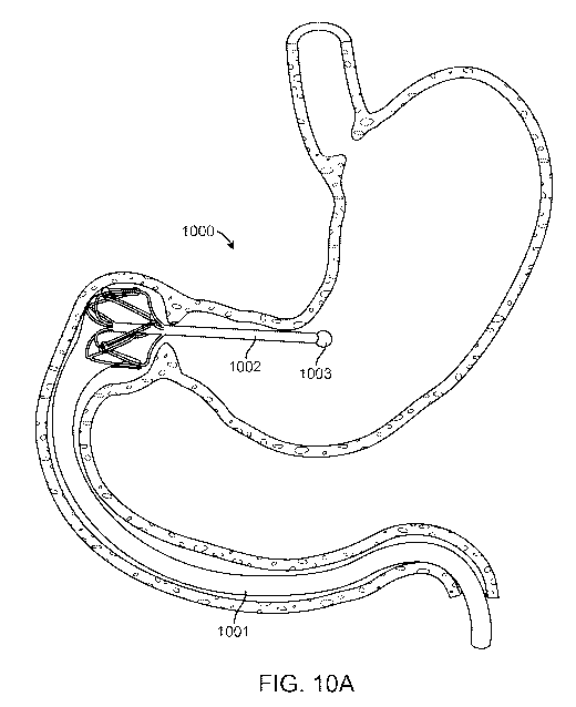
[00991 Figure 10A, 10B, and IOC depict an embodiment of an intragastric anchor
implant
1000 with a sliding seal 1004 configured to directly support the proximal
opening of an intestinal
bypass sleeve 1001. The sliding seal 1004 in this embodiment may include an
expandable sleeve
support 1005 configured to expand radially from a collapsed configuration to
an expanded
configuration, the support slidably coupling the proximal sleeve opening
against an anatomical
lumen wall when in the expanded configuration. The expanded sleeve support
1005 creates a
substantially continuous circumferential seal that causes most nutrients and
food particles exiting
the stomach to proceed down the lumen of the intestinal bypass sleeve. The
expandable sleeve
support 1005 may include a sinusoid, diamond, multi-link, or any suitable
expandable ring
configuration. The expandable sleeve support may be coupled to a rigid element
1002 via struts
1006, fins, or other suitable connecting structures. In a preferred
embodiment, flexible struts
1006 connect the rod to at least one apex of an expandable sleeve support
sinusoid ring such that
the struts to allow the sinusoid to expand and compress radially while
maintaining a connection
to the rigid element 1002. In a more preferred embodiment, the struts 1006
retain a distal slope
such that an expanded sleeve support distal to a narrowed section of lumen
such as a sphincter
may be radially compressed by withdrawing the rigid element 1002 proximally
such that the
struts press against the narrowed lumen and flex radially towards the rigid
element, drawing the
sleeve support inward. The proximal end of the rigid element 1002 may include
an atraumatic
proximal feature 1003 with an enlarged radius of curvature configured so as to
provide a blunt,
atraumatic surface. Although the embodiment shown in Figures 10A and 10B
depicts an
intragastric implant 1000 in which rigid element 1002, proximal 1003
atraumatic feature, struts
1006 and expandable sleeve support 1005 are a single-piece structure made of a
single material
such as polypropylene or fluoropolymer, alternate embodiments may include
multiple
components made of different materials such as struts or an expandable sleeve
support made of
nickel-titanium or stainless steel alloy. Fabricating the anchor implant from
a single-piece of
material is advantageous as it allows the implant to resist wear, breakage and
fatigue from the
cyclical movements exerted on the anchor implant by gastric motility over the
life of the implant
while also reducing its manufacturing cost.
101001 Figure 10C depicts an embodiment of an intragastric anchor implant
whereby the
expandable sleeve support includes tabs 1007 to which the proximal opening of
intestinal bypass
26
CA 02850162 2014-03-26
WO 2013/049167
PCT/US2012/057288
sleeve 1001 may be attached via thermal bonding, riveting, clamping,
heatstaking, gluing, or
other suitable means of attachment.
[01011 Figures 11A and 11B depict an alternate embodiment of an expandable
sleeve support
1100 includes struts 1104 and 1105 angled in opposite directions connecting
the rigid element
1102 to alternating expandable sleeve support 1106 sinusoidal troughs. This
configuration
allows expansion of the sinusoidal ring without translation relative to the
rigid element which is
advantageous in that relative to other embodiments it reduces the overall
length of the expanding
support.
[01021 Drawstrings are commonly employed in removable stents as a means of
compressing
the stent diameter such that it may be withdrawn from an anatomical lumen. As
depicted in
Figures 11 A and 11B, an alternate embodiment of an expandable sleeve support
includes at least
one drawstring, 1107, attached to each proximal sinusoid apex and running
through at least one
lateral opening 1111 in the rigid element 1102 and through a central lumen
1010. An alternate
embodiment may also include a drawstring 1108 attached to each distal sinusoid
apex and
running through central lumen 1110. Each drawstring may be withdrawn through
the central
lumen to compress the expandable sleeve support 1106 by pulling on drawstring
loop 1109. The
drawstring loop may be actuated by pulling into a lumen of a support structure
such as the
working channel of an endoscope, the tip of which may support the rigid
element 1102 providing
a counterforce for tension on the drawstring loop which is transmitted to and
compresses the
expandable sleeve support 1106 reducing its diameter in preparation for
removal of the device
from an anatoinical lumen. The device may be removed by applying a loop snare
to the rigid
element distal to atraumatic proximal feature 1103.
[0103] Figure 12 depicts an alternate embodiment of an intragastric anchor
implant 1200
including an atraumatic distal feature 1204 that includes an expandable sleeve
support 1205 to
which intestinal bypass sleeve 1201 is attached. In a preferred embodiment,
the distal atraumatic
feature 1204 and expandable sleeve support are rnade of flexible polymer as a
single piece and
the expandable sleeve support 1205 is configured to collapse and compress when
the anchor
implant is proximally retracted and withdrawn from a luminal narrowing such as
the pylorus.
The feature and sleeve support may be made of silicone, polypropylene,
thermoplastic elastomer
such as Santoprene, silicone, fluoropolymcr, or any suitable flexible
material. In an further
27
CA 02850162 2014-03-26
WO 2013/049167
PCT/US2012/057288
preferred embodiment, the distal atraumatic feature 1204 includes radial fins
1206 or struts
which are sized to interfere with the lumen into which the feature is placed.
[0104] Figure 13 depicts an alternate embodiment of an intragastric anchor
implant 1300 in
which an expandable sleeve support 1305 is flexibly attached to distal
atraumatic feature 1304
via a flexible coupling section 1306. The expandable sleeve support 1305
slidably couples with
an anatomical lumen such as the intestine to form a sliding seal and, with the
flexible coupling
section, guides food particles into intestinal bypass sleeve 1301 while
allowing rigid element
1302 with attached proximal and distal atraumatic features, 1303 and 1304
respectively, to move
relatively freely with respect to the expandable sleeve support 1305 in
response to gastric and
intestinal motility. Expandable sleeve support 1305 may be made of nickel-
titanium alloy,
stainless steel, polymer such as polypropylene or fluoropolymer, or any
suitable material.
Flexible coupling section 1306 may be made of polymer film such as
polypropylene or
fluoropolymer, or elastomer such as silicone or thermoplastic elastomer such
as Santoprene. An
embodiment of the intragastric anchor implant, as shown in Figure 13, may
include a drawstring
configured to radially compress expandable sleeve support 1305. A preferred
drawstring
embodiment may be configured similarly to drawstring 1107 in Figure 11 A such
that the
drawstring is directed through a lumen in the rigid element 1302 such that the
expandable sleeve
support 1305 may be radially compressed by pulling the drawstring from a point
on the anchor
implant proximal to the pylorus. This method of compressing an expandable
sleeve support
distal to a narrowed section of anatomical lumen such as the pylorus is
advantageous in that no
additional compression aid need be passed through the pylorus in order to
collapse and withdraw
the device.
[01051 Figure 14A depicts an embodiment of a parallel endoscopic delivery
system 1400 for
deployment of an intragastric anchor implant, in accordance with many
embodiments. The
delivery system 1400 is advanced into the stomach in parallel with an
endoscope 1401, which is
placed into a long position along the greater curvature of the stomach.
Endoscope connector
1402 attaches the delivery system to the distal end of the endoscope via an
elastic loop into
which the endoscope tip has been inserted. The endoscope connector 1402 may be
made of
thermoplastic elastomer, silicone, polymer, or any suitable material. A
delivery enclosure 1403
contains an intragastric anchor implant 1407 with an attached intestinal
bypass sleeve 1406
compressed axially and radially and contained within the delivery enclosure
1403. The delivery
enclosure 1403 is located distally to endoscope connector 1402 such that it
may be visualized
28
CA 02850162 2014-03-26
WO 2013/049167
PCT/US2012/057288
through the endoscope as the entire assembly is advanced and the delivery
enclosure 1403 is
placed through the pylorus. In a preferred embodiment, the delivery enclosure
includes an
optically transparent or translucent wall such that the implant itself is
visible to the endoscopist
as it is advanced and deployed. Parallel placement of the endoscope and
delivery enclosure 1403
allows continuous visual monitoring of the intragastric anchor implant is as
it is delivered and
deployed. A flexible pushrod 1404 runs through a housing 1405 and is used to
transmit axial
force to push the intragastric anchor implant 1400 out of delivery enclosure
1403, deploying it
into the duodenum and stomach.
[0106] As shown in Figure 1413, deployment of intragastric anchor implant 1407
through
parallel endoscopic delivery system 1400 may proceed with advancement of
pushrod 1404 so
that bypass sleeve 1406 is advanced out of delivery enclosure 1403. The
endoscopist may
advance the bypass sleeve 1406 in increments, viewing progress through
endoscope 1401,
allowing intestinal peristalsis to engage and extend the sleeve 1406 distally
within the duodenum
and jejunum. Bypass sleeve extension may be confirmed fluoroscopically or
radiographically.
As shown in Figure 14C, the anchor implant's distal atraumatic feature 1408
may be advanced
out of the delivery enclosure and expanded once the bypass sleeve 1406 has
completed its distal
extension. The delivery system 1400 and endoscope 1401 may then be withdrawn
while pushrod
1404 is advanced further EO deploy the anchor implant's proximal atraumatic
feature 1409. Full
and correct deployment may be confirmed endoscopically before the endoscope
1401 and
delivery system are withdrawn from the patient's gastrointestinal tract.
[0107] Figures 15A, 15B, 15C, and 15D depict an embodiment of an alternate,
coaxial,
delivery system 1500 for an intragastric anchor implant in which an endoscope
1501 with an
overtube 1502 including distal balloon 1503 is delivered into the stomach and
inserted through
the pylorus. As shown in Figure 15A, the endoscope is initially inserted into
the stomach orally
and its tip 1507 is guided through the pylorus and into the duodenum while the
overtube 1502
remains positioned proximally to the pylorus. The overtube 1502 is then slid
distally over the
endoscope through the pylorus and distal balloon 1503 is inflated distal to
the pylorus as shown
in Figure 15B. The endoscope 1501 is then withdrawn and overtube 1502 is
partially withdrawn
as shown in Figure 15C placing the stomach and the overtube 1502 into a
relatively straightened
position, thus reducing the radius of curvature of the overtube. A straighter
overtube 1502 may
be advantageous in reducing the force required to advance an intragastric
anchor implant through
the overtube's central lumen. Intragastric anchor implant 1504 with intestinal
bypass sleeve
29
CA 02850162 2014-03-26
WO 2013/049167
PCT/US2012/057288
1505 is introduced into the proximal opening of overtube 1502 and advanced
distally in a
compressed configuration while the endoscope 1501 is reintroduced and is used
to advance the
anchor implant. In a specific embodiment of the delivery system, a pushrod
1508 may run
through the endoscope's tool channel 1506 and frangibly engage the anchor
implant so as to
enable the endoscopist to advance the anchor implant while maintaining a
relatively steady
endoscope position. The pushrod 1508 may also be advantageous in maintaining a
physical
space between the anchor implant and the tip of the endoscope which may aid in
anchor implant
deployment by enabling advancement of the implant separate from advancement of
the
endoscope 1501 as shown in Figure I 5D arid by enhancing visibility through
the endoscope.
Figure 151) depicts an intragastric anchor implant 1504 partially advanced out
of overtube 1502
such that intestinal bypass sleeve 1505 has become uncompressed and
has.extended distally into
the intestine and compressible ring 1510 has expanded into the expanded
configuration, no
longer constrained by the overtube 1502 in the constrained configuration. In
the expanded
configuration, compressible ring 1510 is in substantial apposition and sliding
contact with the
lumen of the duodenum. Fluid such as water or saline may be flushed through
the endoscope or
overtube to aid in the extension of the bypass sleeve. A radiographic contrast
agent such as
barium may be included in the fluid to aid in fluoroscopic visualization of
sleeve extension.
Some embodiments of the intestinal bypass sleeve may include stiffening
element 1509 near the
distal opening, the stiffening element configured to engage intestinal
peristalsis to apply distal
axial tension to the bypass sleeve 1505 and aid in deployment and extension of
the bypass sleeve
into the intestine. In alternate embodiments, stiffening element 1509 may be
configured to also
prop open a portion of the sleeve 1505 and locally maximize its cross-
sectional area. Stiffening
element 1 509 may be configured for substantial radiopacity such that full
sleeve extension may
be confirmed fluoroscopically and may include barium, tantalum, gold, metallic
particles, or any
suitable radio-opaque material in its construction. The stiffening element
1509 may be heat
staked to the sleeve, thermally bonded with the sleeve, insert molded with the
sleeve, bonded to
the sleeve with adhesive or rnay employ any suitable attachment technique.
[01081 Figures 16A, 16B and 16D depict an alternate delivery system 1600 for
an intragastric
anchor implant 1602 in which a flexible sheath 1603 encloses a compressed
anchor implant and
intestinal bypass sleeve 1605. An endoscope is initially placed into the
duodenum and a
guidewire 1601 is advanced distally into the duodenum and jejunum through the
endoscope's
working channel per standard endoscopic practice. As shown in Figure 16A the
flexible sheath
CA 02850162 2014-03-26
WO 2013/049167
PCT/US2012/057288
may be delivered over guidewire 1601 and may include an introducer tip 1604
whose distal end
tapers to a relatively small diameter, preferably approximately to the
diameter of the guidewire,
to ease passage through narrowed lumens such as the pylorus. The guidewire may
pass through
the introducer tip 1604, through the intestinal bypass sleeve 1605 and
compressible ring 1611,
and through the central lumen of the flexible sheath and through the lumen
1608 of the plunger
1606 such that the entire assembly may be advanced over the guidewire 1601.
[01091 As shown in Figure 16B, the introducer tip 1604 may include at least
one split 1609
along a portion of its length such that it may expand radially to allow the
anchor implant to exit
when advanced distally relative to the introducer tip. The split may be held
together with a tether
1607 such as suture, monofilament, wire, or the like, threaded through
openings 1610 along
adjacent edges of the split such that removal of the tether allows the
introducer tip 1604 to open
and expand. Alternately, the split may be held together with frangible
connections such as
perforations, scoring, thermal bonds, adhesive, or any suitable connection
means. In some
embodiments, the introducer tip may be larger in diameter than the flexible
sheath 1603, such
that it may help to retain the sheath's position within the gastrointestinal
tract when fully
advanced past a narrow section oflumen such as the pylorus. When advanced
relative to the
flexible sheath 1603 plunger 1606 transmits axial force to the anchor implant
to advance it
distally and deploy it. The plunger may include a lumen 1608 along a portion
of its length
through which a guidewire 1601 extends.
101101 As shown in Figure 16C, split 1609 in introducer tip 1604 is
disconnected such that it
may expand radially when tether 1607 is withdrawn relative to the flexible
sheath 1603, allowing
compressible ring 1611 to expand and intestinal bypass sleeve 1605 to expand
radially and to
longitudinally extend along and over guidewire 1601. The flexible sheath and
introducer t may
be withdrawn over plunger 1606 to fully release intragastric anchor implant
1600, leaving the
guidewire temporarily in place while intestinal peristalsis applies distal
axial tension to the
bypass sleeve and extends it to its full length. The presence of the guidewire
during bypass
sleeve deployment may be advantageous in preventing kinking and longitudinal
folding.
[01111 Figures 17A and 17B depict a preferred embodiment of an intragastric
anchor implant
1700 with a rod 1702 separating a proximal atraumatic tip 1'703 and a distal
atraumatic tip 1707
which is configured to substantially fill the duodenal bulb. The distal
atraumatic tip 1707 may
include on a proximal section a sliding seal 1704 configured to directly
support the proximal
31
CA 02850162 2014-03-26
WO 2013/049167
PCT/US2012/057288
opening of an intestinal bypass sleeve 1701 in close proximity to the pyloric
valve. The sliding
seal in this embodiment may include an expandable sleeve support 1705
configured to expand
radially and slidably couple the proximal sleeve opening against an anatomical
lurnen wall,
creating a substantially continuous circumferential seal that causes most
nutrients and food
particles exiting the stomach to proceed down the lumen of the intestinal
bypass sleeve 1701.
The expandable sleeve support 1705 may include a sinusoid, zigzag, diamond,
multi-link, a
plurality of flexible arms, or any suitable expandable ring configuration. The
distal atraumatic
tip 1707 may also include space-filling struts 1708 configured to support
sleeve 1701 and
maintain a patent path for nutrients and food particles through the sleeve as
they negotiate the
first turn of the duodenum 1710. Sleeve 1701 may include a section of enlarged
diameter 1711
immediately distal to sliding seal 1704. This enlarged section may enable
sleeve 1701 to
accommodate a tight turn such as the first bend of the duodenum 1710 while
maintaining a
patent path for chyme through the distal atraumatic tip 1707 without pinching
or obstruction.
[01121 Figure 18A,18B, and 18C depict an embodiment of an intragastric anchor
implant 1800
with connecting struts 1806 which angle distally at rod 1802 and curve
proximally to reach the
edge of expandable sleeve support 1805. As shown in Figure 18A, this recurve
strut
configuration enables sliding seal 1804 to be positioned immediately adjacent
to the pyloric
valve.
101131 As shown in Figure 18B, distal atraumatic tip 1807 may be compressed
radially for
delivery by flexing connecting struts 1806 proximally. This compression
configuration is
advantageous in that it produces an effectively flat surface at the proximal
end of the atraumatic
tip which may be placed directly against the pyloric valve. Additionally,
proximal tension on the
rod 1802 will tend to expand the distal atraurnatic tip 1807, helping to seat
it against the
duodenal lumen.
[011z11 As shown in Figure 18C, when the anchor implant 1800 is withdrawn the
connecting
struts 1806 may flex distally as they are pressed against the pyloric valve,
presenting a tapered,
conical surface in aggegate which will tend to radially compress the distal
atraumatic tip 1807
as the struts 1806 pass through the pyloric valve.
101151 Figure 19 depicts a gastroduodenal anchor 1900 with a proximal
atraumatic feature
1901 and a distal atraumatic feature 1902 and a sliding seal 1904 disposed
therebetween
supporting an intestinal bypass sleeve 1903. As shown, the sliding seal 1904
may be configured
32
CA 02850162 2014-03-26
WO 2013/049167
PCT/US2012/057288
as a radially expandable structure supporting the proximal end of the
intestinal bypass sleeve
1903 to allow atraumatic movement within the gastrointestinal tract.
Typically, the sliding seal
1904 is adapted to sit in or near the duodenal bulb. The elongate element
extending between the
proximal and distal atraumatic features may be either rigid, flexible, or have
portions of each; it
may be either straight, curved, include one or more bends, or have various
other combinations of
geometries. For example, as shown in Figure 19, the elongate element may
include two bends or
curved portions so as to conform to the shape approximate shape of the
gastrointestinal tract and
improve anchoring and comfort for the patient. Although the proximal and
distal atraumatic
features are shown as bulbous features and the sliding seal is shown as an
expandable structure,
it is appreciated these features may include any of the atraumatic features
and sliding seals
described herein.
101161 The anchor may be configured in various shapes as desired. For example,
as shown
Figure 19, the gastroduodenal anchor 1900 may be configured in a C-shape that
includes three
sections: a Section A that is substantially parallel with the lumen of the
proximal duodenum and
antrum, a Section B that is substantially parallel with the descending
duodenum, and a Section C
that is substantially parallel with the third part of the duodenum and extends
into the fourth part
of the duodenum. In some embodiments, the bypass sleeve 1903 surrounds a
portion of at least
one anchor section, other embodiments in which the sleeve lies alongside the
anchor sections are
within the scope of this invention. At least one sliding seal 1904 may be
configured to directly
support the proximal opening of intestinal bypass sleeve 1903 via an
expandable sleeve support
1905. The anchor 1900 supports sliding seal 1904 largely within the duodenal
bulb, however
other embodiments are possible in which the sliding seal is located in the
antrum of the stomach,
or more distally in the duodenum. A connecting structure 1906 may attach one
anchor section to
an expandable sleeve support 1905.
101171 Figure 20 depicts a gastroduodenal anchor 2000 including a relatively
flexible section
2001 that may deflect to conform to the approximate shape of the duodenum and
a relatively
rigid section 2002 that resists deformation. The duodenal anchor may extend
from the pylorus to
the distal duodenum. Rigid section 2002 should be approximately 30 mm to 80 mm
in length to
prevent passage through the tight turn and relatively fixed geometry of the
duodenal-jejunal
juncture. Flexible section 2001 is configured to provide longitudinal support
to intestinal bypass
sleeve 2003 holding its proximal opening in the proximal duodenum or gastric
antrum.
33
CA 02850162 2014-03-26
WO 2013/049167
PCT/US2012/057288
[01181 Figure 21 depicts an embodiment of an intragastric anchor implant 2100
with a rod
2102 separating a proximal atraumatic tip 2103 and a distal atraumatic tip
2107, The proximal
atraumatic tip 2103 may include an expandable 2104 structure which may
increase its surface
area and serve to spread mechanical loads. The expandable structure may
include supporting
struts 2105 which are biased distally such that the expandable structure may
radially compress as
it is withdrawn through narrow portions of the GI tract. The proximal
atraumatic tip may also
include a snaring feature 2108 which may easily be endoscopically snared such
that tension may
be applied to the anchor implant 2100 such that it may be withdrawn from the
GI tract.
[0119] In certain circumstances it may be advantageous to implant an
intragastric anchor
I 0 without an intestinal bypass sleeve. Such an anchor may serve as a
platform to support one or
more sensors such as a camera, pH sensor, pressure sensor, or any type of
sensor which ITI ight be
advantageous to place within the GI tract. In its simplest form, a platform
anchor may consist of
a distal atraumatic tip and proximal atraumatic tip separated by a rod whereby
either atraumatic
tip, the rod, or a combination thereof may contain sensors, power supplies, or
control electronics.
in a more preferable embodiment, as shown in Figure 22A, the platform anchor
implant 2200
may be frangible into two or more segments after a desired period of time, de-
anchoring the
anchor implant such that relatively small segments of the anchor that may pass
naturally out of
the GI tract,
[01201 Electrolytic erosion may be employed to selectively time the de-
anchoring of an anchor
implant. As shown in Figure 22A, at least one segment 2203 rnay include a
local power source
2201 which may be a chemical battery such as a button battery, or which may be
an inductive
coil 2209 configured to be energized by an inductively-coupled external
transmitter (as shown
schematically in Figure 22B). The local power source 2201 may provide
electrical power to at
least one sensor 2208 and may also provide power for de-anchoring through
electrolytic erosion,
The time of de-anchoring may be determined by electronics 2201 which may
include at least one
on-board microprocessor or de-anchoring may be initiated externally via
induction whereby at
least one segment contains a coil or antenna capable of inductive coupling.
Segment joints 2204
may be held together by a small-gauge insulated wire 2205 such as 0.005 inch
diameter stainless
steel anchored to a segment at a distal portion of the wire. An uninsulated
portion 2206 of
insulated wire 2205 at or near a joint may be exposed to GI tract fluid such
that an electrically
conductive path 2210 is formed between the un insulated portion of the wire
2206 and a relatively
large electrode 2207 located on another part of the anchor implant. The joint
may be separated
34
CA 02850162 2014-03-26
WO 2013/049167
PCT/US2012/057288
by electrolytic erosion of the uninsulated section of wire by applying a
voltage of +2.5V with a
current of 5mA to it relative to the large electrode 2207 for several minutes.
Multiple joints may
be separated approximately simultaneously or sequentially.
[0121] As depicted in Figure 22C, a frangible anchor implant may be sized to
anchor a sensor
package across the pyloric valve. In such a configuration it may be
advantageous to include
sensors such as a camera or a pH sensor at the proximal atraumatic tip 2211,
or at the distal
atraumatic tip 2212.
[0:122] As depicted in Figure 22D, a frangible anchor implant may be
configured to pass out of
the stomach and geometrically anchor at the duodenal-jejunal juncture where
the Ligament of
Treitz produces a sharp, relatively fixed turn in the small bowel. This
position, distal to the
Ampulla of Vater, may be advantageous for sensor placement as the
characteristics of pancreatic
and bile juices may be monitored.
[01231 Although the exemplary embodiments have been described in some detail
for clarity of
understanding and by way of example, a variety of additional modifications,
adaptations and
changes may be clear to those of skill in the are One of skill in the art will
appreciate that the .
various features described herein may be combined with one another or
substituted with one
another. Hence, the scope of the present invention is limited solely by the
appended claims.