Note: Descriptions are shown in the official language in which they were submitted.
CA 02850912 2014-04-02
WO 2013/057568 PCT/1B2012/002091
FETAL CHROMOSOMAL ANEUPLOIDY DIAGNOSIS
Field of the invention
This invention generally relates to the diagnostic testing of a fetal
chromosomal aneuploidy by
determining imbalances between different nucleic acid sequences, and more
particularly to the
identification of aneuploidy in chromosomes 13, 18, 21, X and/or Y via testing
a maternal
sample such as blood.
Introduction to the invention
Aneuploidy refers to an abnormal number of chromosomes (or part of
chromosomes) that is a
common cause of birth defects. In aneuploidy, genes can be present in three
copies "trisomy"
or in only one copy "monosomy". These changes in chromosome number, resulting
from
_
nondisjunction of chromosomes during meiosis, have dramatic effects on the
affected persons
and result in well-known syndromes. The majority of trisomies and monosomies
are lethal to
the fetus and cause spontaneous abortions or death immediately after birth.
Some
aneuploidies, however, are viable and result in syndromes. The most occurring
aneuploidies
among live births are chromosomes 21, 18, 13 trisomy's and a distorted number
of sex
chromosomes. The most common autosomal aneuploidy that infants can survive
with is
trisomy 21 (Down syndrome), affecting 1 in 800 births. Trisomy 18 (Edwards
syndrome) affects
1 in 6,000 births, and trisomy 13 (Patau syndrome) affects 1 in 10,000 births.
Sex chromosome
aneuploidy (SCA) affects 1 in 400 newborns and is therefore, as a whole, more
common than
Down syndrome. While SCA include a variety of abnormalities of the sex
chromosomes, by far
the most commonly occurring SCA is the deletion of chromosome X (45,X-Turner
syndrome)
or the addition of an X or Y chromosome (47,XXY-Klinefelter syndrome, 47,XYY,
47,XXX). Of
these conditions, only Turner syndrome results in an easily identifiable
physical phenotype.
However, subtle language and learning difficulties have been identified in
most forms of SCA.
The most important risk factor for aneuploidy is maternal age since the
majority of children with
aneuploidy are born to mothers over the age of 35, so the prevalence is
increasing as more
women choose or need to delay childbearing. Contemporary prenatal screening
programs
typically include the common fetal chromosomal aneuploidies 21, 18 and 13. The
risk of a
pregnancy is assessed by a number of means. For the chromosomal aneuploidies
non-
invasive screening tests based on ultrasonography and the measurement of
markers in
maternal serum have been implemented to identify high-risk pregnancies in the
first 3 months
of pregnancy (11-14 weeks). The sonogram measures the fluid underneath the
skin along the
back of the baby's neck, called the nuchal translucency (NT). The sonogram
will also
determine if the baby's nasal bone is present or absent. A maternal blood
sample is used to
1
CONFIRMATION COPY
CA 02850912 2014-04-02
WO 2013/057568 PCT/1B2012/002091
analyze two serum markers called free beta-human chorionic gonadotropin (hCG)
and
pregnancy associated plasma protein-A (PAPP-A), which are found in the blood
of all pregnant
women. In aneuploidy pregnancies there is extra fluid behind the baby's neck
and/or the hCG
and PAPP-A results are higher or lower than average. Additionally, a baby's
nasal bone may
be absent in some pregnancies with a chromosome abnormality. Combining age-
related risk
with the NT measurement, nasal bone data, and blood markers provide a risk
figure for Down
syndrome and one risk figure for trisomy 13 or trisomy 18. The first trimester
screen's detection
rate is approximately 90% with a 5% false positive rate for pregnancies in
which the baby has
Down syndrome, and is somewhat higher for pregnancies with trisomy 13 or
trisomy 18. A
nuchal translucency sonogram can be performed without measuring hCG and PAPP-
A. In this
case, however, the aneuploidy detection rate is reduced to about 70%.
Prenatal diagnosis is an integral part of obstetric practice. To perform a
genetic diagnosis
prenatally, genetic material from the unborn fetus is required.
Conventionally, fetal-DNA1S-
sampled by invasive procedures such as amniocentesis or chorionic villus
sampling. These
procedures are associated with a risk of miscarriage of respectively 0,5 and 1-
2%. Hence, it is
routine to reserve the invasive diagnostic procedures for pregnancies
estimated to be at high
aneuploidy risk which represent about 5-10% of all women screened for
aneuploidy risk. Given
the procedure related risks of conventional prenatal diagnosis, it would be
ideal if genetic
analysis of the fetus could be performed non-invasively. To perform non-
invasive prenatal
diagnosis, a source of fetal genetic material without harming the fetus is
therefore required. A
major breakthrough to this end was reported by Lo et al. (1997)1 and
W098/39474 describing
the existence of free floating fetal DNA in maternal plasma. They subsequently
showed that
fetal-derived DNA contributed -10% of the free-floating DNA in maternal
plasma. Fetal DNA
can be detected in maternal plasma just weeks after conception and is rapidly
cleared from
maternal plasma and disappears within hours after delivery. As a result, free
floating fetal DNA
in maternal plasma is a promising source of fetal genetic material for the
development of a
non-invasive prenatal test. However, fetal DNA represents only a minor
fraction of the total free
floating DNA in plasma with the remaining portion of DNA contributed by the
mother, mainly
derived from maternal white blood cells.
Given the enormous potential, several non-invasive methodologies for
aneuploidy detection
have been described in the last decade. One method is to focus on the analysis
of nucleic acid
molecules that are fetal-specific in maternal plasma and hence overcome the
interference
caused by the background maternal DNA. One could target the detection of
placental
expressed mRNA or placenta-specific epigenetic signatures originating from the
chromosome
of interest. In a series of developments since 2000, the basis for plasma RNA
as a prenatal
diagnostic tool has been established. Poon et al. (2000)2 showed that mRNA
transcribed from
2
CA 02850912 2014-04-02
WO 2013/057568 PCT/1B2012/002091
the Y chromosome could be detected in the plasma of women carrying male
fetuses. Later, it
was shown that the placenta is a major source of fetal-derived RNA in maternal
plasma using
human placental lactogen mRNA and mRNA coding for the beta subunit of human
chorionic
gonadotrophin as examples3.
In 2007, a placental-specific mRNA, transcribed from a gene located on
chromosome 21,
PLAC4, was identified using a microarray-based approach and was shown to be
detectable in
maternal plasma and cleared following delivery of the fetus4. To determine the
dosage of
chromosome 21 using PLAC4 mRNA in maternal plasma the RNA-SNP allelic ratio
approach
was used. This method is based on the presence of a SNP in the coding region
of the PLAC4
gene. If a fetus is heterozygous for this SNP, it possesses two alleles that
are distinguishable
by DNA sequence. If the fetus is euploid, the ratio of these two SNP alleles
is 1:1. Conversely,
if the fetus has trisomy 21, then the RNA-SNP allelic ratio would become 1:2
or 2:1. Lo et al.
-(2007)-4-demonstrated-tha this strategy could be applied to non-invasively
determine the
chromosome 21 trisomy status of a fetus. Similarly, the RNA-SNP approach was
also applied
for the non-invasive detection of trisomy 18 through the analysis of the
allelic ratio of
SERPINB2 mRNA5.
The main limitation of the RNA-SNP allelic ratio approach, however, is that
only fetuses
heterozygous for the analyzed SNP can be successfully diagnosed. For example,
with the use
of the single SNP in PLAC4, approximately 45% of fetuses are expected to be
heterozygous
and thus diagnosable using this approach. Consequently, several markers are
needed for full
diagnostic coverage. To this end, a number of investigators have described new
polymorphic
SNP markers that can be analyzed using this approach. One preliminary report
describes ten
markers with a combined heterozygosity rate that covers up to 95% of the US
general
population6. The evaluation of these markers in large-scale clinical trials is
expected over the
next few years.
Placenta-specific epigenetic signatures, such as DNA methylation, originating
from the fetal
chromosome of interest have also been investigated. As tissues in the body
have different
gene expression profiles, the methylation status of certain genes also
exhibits tissue-specific
patterns. Evidence shows that fetal DNA in maternal plasma originates from the
placenta and
that the maternal DNA background is derived from maternal blood cells.
Therefore, one way to
develop epigenetic fetal DNA markers is to identify genes whose methylation
status differs
between placental tissues and maternal blood cells. Chim et al. (2005)7
studied the methylation
profile of the SERPINB5 (maspin) promoter and showed that it was
hypomethylated in
placental tissues but hypermethylated in maternal blood cells. Using
methylation specific PCR,
the placental-derived hypomethylated SERPINB5 could be detected and
distinguished from
the maternally derived hypermethylated molecules in maternal plasma. This made
SERPINB5,
3
CA 02850912 2014-04-02
WO 2013/057568 PCT/1B2012/002091
located on chromosome 18, the first universal circulating fetal DNA marker
that could be used
for all pregnancies regardless of fetal gender and genotype. Since the
SERPINB5 gene is
located on chromosome 18, it allowed the development of a strategy that is
analogous to the
RNA-SNP allelic ratio approach, the so-called epigenetic allelic ratio
approach. Thus, if a fetus
is heterozygous for an SNP located in the promoter region of SERPINB5,
measuring the ratio
of the SNP alleles in the hypomethylated version of the gene, allows
ascertainment of the
fetus's trisomy 18 status.
However, methylation-specific PCR requires the use of a bisulphite conversion,
which alters
unmethylated cytosines to uracil nucleotides. But, bisulphite conversion
degrades up to 95% of
the DNA molecules in a sample and therefore substantially reduces the amount
of fetal DNA in
a maternal plasma sample and may result in false-negative detection.
Consequently,
researchers developed fetal epigenetic markers that could be detected in
maternal plasma
without the need_for_bisulphite_conversion¨To-this endr-Chan et al. (2006)8
used the prombter
of RASSF1, located on chromosome 3, which is hypermethylated in placental
tissues but
hypomethylated in maternal blood cells. Consequently, the hypomethylated
RASSF1
sequences derived from the maternal blood cells can be removed from maternal
plasma using
methylation sensitive restriction enzyme digestion. Indeed, after restriction
enzyme digestion,
fetal RASSF1 sequences could be detected in maternal plasma before delivery
but completely
disappeared from maternal plasma within 24h after delivery. Chan et al.
(2006)9 used the
differential methylation pattern of the RASSF1 promoter as the positive
control for fetal DNA
detection in a non-invasive prenatal fetal rhesus D blood group typing for 54
early-gestation
RhD-negative women.
The RNA-SNP allelic ratio approach and the DNA methylation approach target
subsets of
nucleic acid molecules present in maternal plasma in a molecular fashion. An
alternative is
using physical methods that result in the relative enrichment of fetal DNA
present in the
maternal plasma.
Recently , it was shown that the length of free floating fetal DNA in the
maternal plasma is -20
bp shorter than the maternally derived free-floating DNA. Therefore, size
fractioning methods
such as gel electrophoresis allow size-fractionation of plasma DNA and
enrichment of the
shorter, fetal DNA fragments. This approach has been used successfully to
enrich for free
floating fetal DNA. While this approach has been shown empirically to be
useful for the
qualitative detection of disease causing mutations, for example those causing
beta-
thalassemia, it is yet unknown whether the degree of enrichment might be
sufficient for fetal
chromosomal aneuploidy detection requiring quantitative measurement of
chromosome
dosage. Dhallan et al. (2006)10 reported another approach for the enrichment
of fetal DNA in
4
CA 02850912 2014-04-02
WO 2013/057568 PCT/1B2012/002091
maternal plasma. They hypothesized that a significant portion of maternal
derived free-floating
DNA in maternal plasma is released by maternal white blood cells following
phlebotomy.
Therefore it was proposed that if maternal nucleated blood cells could be
fixed, using
formaldehyde, then this dilution of fetal DNA in maternal plasma could be
avoided. Dhallanl
demonstrated the benefit of this approach for the noninvasive prenatal
diagnosis of trisomy 21
showing a mean proportion fetal DNA of 34% in an experiment comprising 60
pregnant
women. However, the beneficial effects of formaldehyde treatment could not be
replicated by
several other groups.
The above-mentioned approaches are based on the assumption that the low
fractional
concentration of fetal DNA in maternal plasma makes it challenging to pursue
the direct
detection of fetal chromosomal aneuploidies. This is based on the limited
precision of
conventional methods for circulating fetal DNA detection, for example by real-
time_ECR._
The recent availability of single molecule counting techniques allows
detection of fetal
aneuploidy without the need to restrict the analysis to fetal-specific nucleic
acids in maternal
plasma. Digital PCR and massively parallel sequencing are both single molecule
counting
methods, which allow the quantification of nucleic acids by counting molecules
and have
superior analytical precision compared to conventional PCR based detection
methods. Digital
PCR refers to the performance of multiple PCRs in parallel in which each PCR
typically
contains either a single or no target molecule. Through the counting of the
number of positive
reactions at the end of amplification it is possible to determine the number
of input target
molecules. Thus, they can precisely quantify small increments in the total
(maternal vs. fetal)
amount of DNA molecules derived from the aneuploid chromosome. Indeed, Lo et
at. (2007)11
demonstrated that the aneuploidy status detection is possible even when the
trisomic DNA is
present as a minor (10%) fraction. The lower the fetal DNA concentration, the
smaller the
expected increment in the amount of aneuploidy chromosome DNA. For digital
PCR,
quantitative precision improves with increasing number of PCR analyses
performed. Lo et at.
(2007)11 showed that accurate fetal trisomy 21 detection in a maternal plasma
sample
containing 25% fetal DNA requires about 8000 digital PCRs to be performed,
requiring the use
of automated platforms in the clinical setting. Such automated platforms using
microfluidics are
available (e.g. Fluidigm) but are expensive. Several groups demonstrated that
non-invasive
detection of fetal trisomy 21 could be achieved with the use of massively
parallel, or next-
generation, sequencing (e.g. W02009/013496). Massively parallel sequencers
allow analysis
of nucleotide sequences of millions to billions of DNA molecules in each run.
Therefore, in
addition to the identity, a frequency distribution of the DNA molecules in the
analyzed sample
can be obtained. Since free floating DNA in maternal plasma is fragmented in
nature it can be
used directly to identify the chromosomal origin of each DNA molecule and
determine the
5
CA 02850912 2014-04-02
WO 2013/057568 PCT/1B2012/002091
proportion of molecules derived from a potentially aneuploid chromosome.
Several groups
showed that the proportion of chromosome 21 DNA molecules in plasma of women
pregnant
with a trisomy 21 fetus was elevated compared with that of euploid
pregnancies. This
approach was highly accurate for the direct detection of fetal trisomy 21 from
maternal plasma
among small cohorts of pregnancies.
Recently, two clinical validation studies were performed applying the above-
described method.
In one study 449 samples were analyzed of which 39 were trisomic for
chromosome 2112. A
second study analyzed blood samples from 1014 at risk pregnancies collected in
13 US clinic
locations before they underwent an invasive prenatal procedure13. Of these 119
samples
underwent massively parallel DNA sequencing. Fifty-three sequenced samples
were classified
correctly as having an abnormal fetal karyotype. Both clinical validation
studies showed
excellent sensitivity and specificity. These data demonstrate that plasma DNA
sequencing is a
viable method for noninvasive detection of fetal trisomy 21 and warrants
clinical validation in
larger multicenter study.
On the other hand, it has been shown that the measurement of the proportional
amounts of
sequences derived from chromosomes with higher or lower GC contents then
chromosome 21
was not as robust. Therefore, the measurements for chromosomes 18 and 13 are
less precise
and suffer from quantitative bias using trisomy 21 protocols. Thus, to achieve
reliable non-
invasive detection of trisomy 18 and trisomy 13, sequencing and data analysis
protocols that
are less susceptible to the chromosomal GC content effects need to be
developed and further
validated. A recent study partially solved the above problem using a non-
repeat masked
reference genome and a bioinformatics approach to correct GC content bias in
the sequencing
data14. Using this approach all trisomy 13 fetuses (25 out of 25) were
detected at a specificity
of 98.9% and 92% (34 out of 37) of the trisomy 18 fetuses at 98.0%
specificity. These data
indicate that with appropriate bioinformatics analysis, noninvasive prenatal
diagnosis of trisomy
13 and trisomy 18 by maternal plasma DNA sequencing is not as reliable as
trisomy 21.
In addition, the cost of massively parallel sequencing is high and the
throughput is low. Only a
handful of cases can be analyzed per run, which takes several days. Further
work is needed to
develop more cost-effective protocols with higher throughput.
Recently, target enrichment was used to obtain a more efficient and cost-
effective massive
parallel sequencing approach15. This study investigated the applicability in
enriching selected
genomic regions from plasma DNA and the quantitative performance of this
approach. The
experiment showed that the mean sequence coverage of the enriched samples was -
200-fold
higher than that of the non-enriched samples and more importantly that
maternal and fetal
DNA molecules were enriched evenly. Furthermore, by using SNP data the authors
were able
6
CA 02850912 2014-04-02
WO 2013/057568 PCT/1B2012/002091
to show that the coverage of fetus-specific alleles within the targeted region
increased from
3.5% to 95.9%. Overall, targeted sequencing of maternal plasma DNA allows
efficient and
unbiased detection of fetal alleles and is a powerful method for measuring the
proportion of
fetal DNA in a maternal plasma sample. Based on this single scientific paper
target enrichment
shows great promise since it can reduce the sequencing cost substantially. At
the same time it
requires an extra, enrichment step that will add an extra cost to the final
test and also will delay
the test since a typical enrichment protocol takes about 24-36 hours to
complete.
Summary of the invention
The present invention provides a non-invasive diagnostic DNA test for
aneuploidy detection of
chromosomes 21 and/or 18 and/or 13 and/or X and/or Y by combining multiplex
PCR based
amplification of specific DNA sequences¨(i,e,targets)¨which contain at least
one SNP
combination with sequencing technologies.
In another aspect the invention provides a non-invasive diagnostic DNA test
for aneuploidy
detection of chromosomes 21 and 18 and 13 and X and Y by combining multiplex
PCR based
amplification of specific DNA sequences (i.e. targets) which contain at least
one SNP in
combination with sequencing technologies.
Briefly, the present invention is directed to a method of differential
detection of a
predetermined set of target sequences in a mixture of maternal and fetal
genetic material.
Thus, the methods and materials described herein apply techniques for
analyzing numerous
nucleic acids contained in a biological sample (preferably serum or plasma)
containing free
floating DNA which is a mixture of DNA from both the mother and the fetus, and
allowing
detection of statistically significant difference between euploid and triploid
fetuses. In contrast
to the current massive parallel sequence methods, based on whole genome or
enriched
samples, which do not achieve a sufficient sensitivity and specificity, in
particular, for
chromosome 13, the present invention provides a non-invasive diagnostic assay
with a
specificity and sensitivity close to 100% (respectively 99.99% specificity and
99.5% sensitivity)
for the simultaneous detection of chromosome 13, 18, 21, X and Y aneuploidies.
Without
limiting the invention to a particular theory or explanation, one reason why
multiplex-PCR was
not considered before in the development of non-invasive diagnostic aneuploidy
tests is the
presence of high GC-rich regions particularly in chromosome 13. Yet another
reason is that the
use of multiplex-PCR was discouraged by one of the leading inventors (i.e.
Dennis Lo) in
US2010/0112590. Indeed, in the latter application on paragraphs 116-117 it is
recommended
to apply locus-independent assays rather than locus-dependent assays such as
for example
the targeted amplification carried out by the methods of the present
invention.
7
CA 02850912 2014-04-02
WO 2013/057568 PCT/1B2012/002091
Thus in one aspect the invention provides a method for determining the
presence or absence
of fetal aneuploidy in a biological sample comprising fetal and maternal
nucleic acids present
in free floating DNA from said maternal biological sample, amplifying a
selected set of target
DNA sequences in a quantitative (i.e. amplifying the template DNA such that
the amplified
DNA is reproducing the original template DNA ratios) multiplex PCR reaction,
conducting DNA
sequencing of said amplified selected set of target DNA sequences to determine
the sequence
of said DNA sequences, using the obtained sequence data to compare an amount
of amplified
sequences derived from at least one first chromosome in said mixture of
maternal and fetal
DNA to an amount of amplified DNA sequences derived from at least one second
chromosome
in said mixture of maternal and fetal DNA, wherein said at least one first
chromosome is
presumed to be euploid in the fetus, wherein said at least one second
chromosome is
suspected to be aneuploid in the fetus, thereby determining the presence or
absence of said
fetal aneuploidy.
In another aspect the invention provides a method for determining the presence
or absence of
fetal aneuploidy in a biological sample comprising fetal and maternal nucleic
acids (such as
free floating DNA) from said maternal biological sample, amplifying a selected
set of target
DNA sequences in a quantitative multiplex PCR reaction wherein each amplified
DNA
sequence comprises at least one SNP which is considered informative in case
the pregnant
female is heterozygous for this SNP, conducting DNA sequencing of said
amplified selected
set of target DNA sequences to determine the sequence of said DNA sequences,
using the
obtained sequence data to compare an amount of amplified sequences which carry
an
informative SNP derived from at least one first chromosome in said mixture of
maternal and
fetal derived DNA to an amount of amplified DNA sequences which carry an
informative SNP
derived from at least one second chromosome in said mixture of maternal and
fetal derived
DNA, wherein said at least one first chromosome is presumed to be euploid in
the fetus,
wherein said at least one second chromosome is suspected to be aneuploid in
the fetus,
thereby determining the presence or absence of said fetal aneuploidy and/or
determining in
said determined DNA sequences the allelic ratios of the informative SNPs
wherein a distorted
allelic ration is indicative for the presence of a fetal chromosomal
aneuploidy in said pregnant
female.
Figure legends
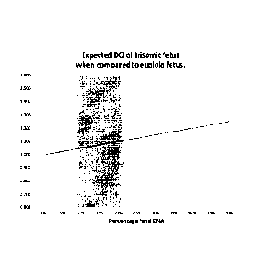
Figure 1:
Dosage Quotients (DO) of trisomic fetus when compared to euploid fetus. The
grey shaded
area indicates the expected percentages of fetal DNA.
8
CA 02850912 2014-04-02
WO 2013/057568 PCT/1B2012/002091
Figure 2:
Number of SNPs needed to gent minimally a given number of informative SNPs,
plotted per
Minor Allele Frequency (MAF). The calculations are done for a minimal
probability of 99%.
Figure 3: plot of expected vs. observed normalized read counts for chromosome
21 in a Down
syndrome (trisomy 21) DNA samples (square) and 4 euploid DNA samples
(circles).
Figure 4: plot of expected vs. observed normalized read counts for two ATP
samples
(representing 20% trisomy 21) DNA samples (squares) and 4 euploid DNA samples
(circles).
Figure 5: schematic representation of first multiplex PCR reaction of the
MASTR assays
_procedure. Reverse and forward primers-are-amplicon--specific--primersTagt
and Tag2 are
universal sequencing that are used in the second PCR reaction of the MASTR
assay
procedure to incorporate
Figure 6: schematic representation of the second PCR reaction of the MASTR
procedure. In
this step the MID sequences (barcodes) and A and B adaptors (for 454 emulsion
PCR) are
incorporated in the resulting amplicons from the first PCR reaction.
Detailed description of the invention
The prior art has shown the feasibility of massive parallel sequencing as an
analysis platform
for free floating DNA based aneuploidy testing. However, current protocols
result in expensive
and low throughput tests when used as a molecular diagnostic tool. The main
reason for this is
the fact that current tests are based on genome wide sequencing of free
floating DNA resulting
in the production of huge sequencing datasets of which only a small fraction (-
5%) is used to
determine the ploidy status of the fetus. With this genome wide approach it is
obligatory to use
a substantial part of the capacity of a massive parallel sequencer resulting
in sequencing of a
limited number of individuals per run, which takes several days to complete.
Furthermore,
huge sequencing datasets are generated per individual that hamper efficient
data storage and
analysis.
The present invention offers a solution for this problem by using a multiplex-
PCR based
approach to amplify a number of selected chromosomal regions. Selected
chromosomal
regions are amplified in a multiplex PCR reaction from one or more chromosomes
which are
presumed to be aneuploid and selected set of chromosomal regions are
amplified, preferably
9
CA 02850912 2014-04-02
WO 2013/057568 PCT/1B2012/002091
in the same multiplex PCR reaction, from one or more chromosomes which are
presumed to
be euploid. Chromosomes which are presumed to be euploid are herein further
designated as
a 'reference chromosome'.
Accordingly the present invention provides in a first embodiment a method for
the detection of
a fetal chromosomal aneuploidy in a pregnant female comprising i) receiving a
biological
sample from said pregnant female, ii) preparing nucleic acids from said
biological sample, iii)
amplifying a selected set of target DNA sequences in a quantitative multiplex
PCR reaction
wherein at least one amplified DNA sequence comprises at least one SNP which
is considered
informative if the pregnant female is heterozygous for this SNP, iv)
sequencing of the amplified
target DNA sequences and v) calculating the sum of read counts for all
amplified DNA
sequences of a suspected chromosomal aneuploidy followed by normalization,
against the
sum of read counts for all amplified DNA sequences of a reference chromosome
to determine
by statistical methods a set score---indicative for the presence of a fetal
chromosomal¨
aneuploidy and/or determining the allelic ratios of the informative SNPs
wherein a distorted
allelic ratio is indicative for the presence of a fetal chromosomal aneuploidy
in said pregnant
female.
The term "biological sample" as used herein refers to any sample that is taken
from a subject
(e.g. such as a pregnant female or a pregnant woman) and contains one or more
nucleic acid
molecule(s) of interest.
Accordingly a biological sample comprises for example blood, sputum, urine,
cerebrospinal
fluid (CSF), tears, plasma, serum, saliva or transcervical lavage fluid.
The term "nucleic acid" or "polynucleotide" refers to a deoxyribonucleic acid
(DNA) or
ribonucleic acid (RNA) and a polymer thereof in either single- or double-
stranded form. Unless
specifically limited, the term encompasses nucleic acids containing known
analogs of natural
nucleotides that have similar binding properties as the reference nucleic acid
and are
metabolized in a manner similar to naturally occurring nucleotides. Unless
otherwise indicated,
a particular nucleic acid sequence also implicitly encompasses conservatively
modified
variants thereof (e.g., degenerate codon substitutions), alleles, orthologs,
SNPs, and
complementary sequences as well as the sequence explicitly indicated.
Specifically,
degenerate codon substitutions may be achieved by generating sequences in
which the third
position of one or more selected (or all) codons is substituted with mixed-
base and/or
deoxyinosine residues The term nucleic acid is used interchangeably with gene,
cDNA, mRNA,
small noncoding RNA, micro RNA (miRNA), Piwi-interacting RNA, and short
hairpin RNA
(shRNA) encoded by a gene or locus.
CA 02850912 2014-04-02
WO 2013/057568 PCT/1B2012/002091
The term "gene" means the segment of DNA involved in producing a polypeptide
chain. It may
include regions preceding and following the coding region (leader and trailer)
as well as
intervening sequences (introns) between individual coding segments (exons).
The term "reaction" as used herein refers to any process involving a chemical,
enzymatic, or
physical action that is indicative of the presence or absence of a particular
polynucleotide
sequence of interest. An example of a "reaction" is an amplification reaction
such as a
polymerase chain reaction (PCR), preferably a multiplex PCR reaction. Another
example of a
"reaction" is a sequencing reaction, either by synthesis or by ligation. The
term "clinically
relevant nucleic acid sequence" as used herein can refer to a polynucleotide
sequence
corresponding to a segment of a larger genomic sequence whose potential
imbalance is being
tested or to the larger genomic sequence itself. Examples include chromosome
18, 13, 21, X
and Y. Yet other examples include mutated genetic sequences or genetic
polymorphisms or
copy number_variations _that-a-fetus may inherit¨from one- or both of Its-
parents. The term
"background nucleic acid sequence" as used herein may refer to nucleic acid
sequences
originating from the mother or originating from the chromosome not tested for
aneuploidy in a
particular analysis.
The term 'free-floating DNA" is DNA which is derived from genomic DNA, free-
floating DNA is
in fact degraded genomic DNA and occurs in the extra-cellular space. As such
free-floating
DNA can be isolated from body fluids (e.g. serum, plasma, sputum).
The term "quantitative data" as used herein means data that are obtained from
one or more
reactions and that provide one or more numerical
values.
The term "parameter" as used herein means a numerical value that characterizes
a
quantitative data set and/or a numerical relationship between quantitative
data sets. For
example, a ratio (or function of a ratio) between a first amount of a first
nucleic acid sequence
and a second amount of a second nucleic acid sequence is a parameter.
The term "cutoff value" as used herein means a numerical value whose value is
used to
arbitrate between two or more states (e.g. diseased and non-diseased) of
classification for a
biological sample. For example, if a parameter is greater than the cutoff
value, a first
classification of the quantitative data is made (e.g. diseased state); or if
the parameter is less
than the cutoff value, a different classification of the quantitative data is
made (e.g. non-
diseased state).
The term "imbalance" as used herein means any significant deviation as defined
by at least
one cutoff value in a quantity of the clinically relevant nucleic acid
sequence from a reference
quantity.
11
CA 02850912 2014-04-02
WO 2013/057568 PCT/1B2012/002091
The term "chromosomal aneuploidy" as used herein means a variation in the
quantitative
amount of a chromosome from that of a diploid genome. The variation may be a
gain or a loss.
It may involve the whole of one chromosome or a region of a chromosome.
Examples of
The term "random sequencing" as used herein refers to sequencing whereby the
nucleic acid
fragments sequenced have not been specifically identified or targeted before
the sequencing
procedure. Sequence-specific primers to target specific gene loci are not
required when
whether an increase or decrease (diseased state) of a clinically-relevant
chromosomal region
exists compared to a non-diseased state. This determination may be done by
using a
parameter of an amount of a clinically-relevant chromosomal region in relation
to other non-
clinically-relevant chromosomal regions (background regions) within a
biological sample.
30 values.
The clinically relevant chromosomal region (also called a clinically relevant
nucleic acid
sequence or suspected aneuploid chromosome or chromosomal region) and the
background
nucleic acid sequence may come from a first type of cells and from one or more
second types
of cells. For example, fetal nucleic acid sequences originating from
fetal/placental cells are
12
CA 02850912 2014-04-02
WO 2013/057568 PCT/1B2012/002091
fetal nucleic acid sequences are derived from free-floating DNA. In one
embodiment, the cutoff
value is determined based at least in part on a percentage of the first type
of cells in a
biological sample. Note the percentage of fetal sequences in a sample may be
determined by
any fetal-derived loci and not limited to measuring the clinically-relevant
nucleic acid
sequences.
In another embodiment the methods of the invention use cell (e.g. blood cells)
stabilizing
chemicals in the preparation of the nucleic acids present in the biological
sample which is
received from the pregnant female. Indeed, one of the major technical
challenges in using free-
floating fetal DNA from maternal blood is the low fraction of fetal DNA
present in the sample.
This fraction is typically between 10 and 20% in the first trimester of
pregnancy (week 11-14),
which corresponds with the stage where an aneuploidy DNA test is best
performed. This low
Iraction_oLfetal DNA is-even for-molecular-counting methods challenging with
respect to the
sensitivity and specificity of the test. Therefore it is important to maximize
the ratio
fetal/maternal free floating DNA. The present invention provides different
solutions for this
problem.
In a particular embodiment the disruption of nucleated blood cells is
prevented during the
collection, storage or transport of the biological material, in particular a
maternal blood sample
prior to plasma isolation. This is important to prevent dilution of fetal DNA
resulting in a
decreased ratio fetal/maternal free floating DNA. Several commercial cell
stabilizing blood
collection tubes are available which stabilize blood cells for at least 14
days at room
temperature allowing convenient sample collection, transport and storage
(available for
example at www.streck.com).
In yet another particular embodiment a size fractionation is used in the
methods of the
invention to prepare maternal and fetal nucleic acids.
Indeed, the prior art shows that fetal and maternal free-floating DNA have
different size
distributions. Free floating fetal DNA is generally 20 bp shorter than the
maternal free floating
DNA and this observation can be used to further enrich the free-floating fetal
DNA fraction if
this smaller sized fraction is specifically separated from the maternal
fraction. One way to
accomplish this is by means of gel electrophoresis. In a particular
embodiment, a gel
electrophoresis based size-fractionating device is used as marketed by Sage
Science
(www.sagescience.com). This device is a fully automated system enabling tight
size selection
and a high recovery rate. Furthermore, it eliminates the cross contamination
risk completely
since all samples are separated from each other during the whole size
fractionation process.
13
CA 02850912 2014-04-02
WO 2013/057568 PCT/1B2012/002091
In a particular embodiment the amplified DNA sequences obtained in the
quantitative multiplex
PCR reaction in the methods of the invention have a size between 80 and 140
base pairs.
In view of the size distributions of the fetal and maternal free floating DNA
populations it is
essential to keep the amplified DNA sequence lengths below 140 bp to ensure
efficient
amplification of the shorter fetal free-floating DNA fraction.
Preferred amplified DNA sequence lengths are between 80 and 140 basepairs.
In yet another embodiment the amplified DNA sequences obtained in one single
multiplex PCR
reaction are between 30 and 60.
In yet another embodiment the amplified DNA sequences obtained in one single
multiplex PCR
reaction are between 60 and 80.
In yet another embodiment the amplified DNA sequences obtained in one single
multiplex PCR
reaction are between 70 and 80.
Preferably only one quantitative multiplex_RCR reaction-is applied-to
practice the methods-of-
the invention.
In yet another embodiment the GC-content of the target DNA sequences (i.e. the
DNA
sequences which are amplified with the quantitative multiplex PCR reaction) is
between 30%
and 70%. Our experimental data point out that a range of 40%-60% GC is optimal
for a close
to 100% sensitivity and specificity of the methods of the invention.
An essential step in the methods of the present invention is the 'sequencing
of the amplified
target DNA sequences. As a high number of sequencing reads, in the order of
hundred
thousand to millions or even possibly hundreds of millions or billions can
theoretically be
generated from each sample in each run, the resultant sequenced reads form a
representative
profile of the mix of nucleic acid species in the original biological sample.
However, the person
skilled in the art would know how many runs to perform based on the stage of
pregnancy
(which is correlated with the amount of free-floating fetal DNA in the
biological sample) and
based on the origin of the biological sample derived from a pregnant female.
The most
important aspect is that a high degree of statistical confidence is obtained.
In order to improve
statistical confidence, it is preferable to perform a large number of reads,
preferably between
10.000 and 100.000 or more reads, depending on the percentage of fetal DNA
present in the
mixture. A commonly used measure of statistical significance when a highly
significant result is
desired is p<0.01, i.e. a 99% confidence interval based on a chi-square or t-
test.
In a preferred embodiment massive parallel sequencing methods are used. In
particular
embodiments, the sequencing is done using massively parallel sequencing.
Massively parallel
sequencing, such as for example on the 454 platform (Roche) (Margulies, M. et
al. 2005
Nature 437, 376-380), Illumina Genome Analyzer (or Solexa platform) or SOLiD
System
14
CA 02850912 2014-04-02
WO 2013/057568 PCT/1B2012/002091
(Applied Biosystems) or the Helicos True Single Molecule DNA sequencing
technology (Harris
T D et al. 2008 Science, 320, 106-109), the single molecule, real-time
(SMRTTm) technology of
Pacific Biosciences, and nanopore sequencing (Soni G V and MeIler A. 2007 Clin
Chem 53:
1996-2001), allow the sequencing of many nucleic acid molecules isolated from
a specimen at
high orders of multiplexing in a parallel fashion. Each of these platforms
sequences clonally
expanded or even non-amplified single molecules of nucleic acid fragments.
An important advantage of the limited set of amplified nucleotide sequences
which is
generated by the methods of the present invention is that emerging low cost
and lower
capacity massive parallel sequencers can be used such as the 454 junior
(Roche), PGM (Life
Technologies) or MiSeq (IIlumina). The combination of the methods of the
invention and the
low end sequencers results in a fast turnaround time per test since these
platforms typically
take only a few hours per sequencing run. In addition, the lower cost is also
an important
improvement over the_methods used in-the-prior-art.
In a particular embodiment the massive parallel sequencing data are analyzed
by calculating
the sum of read counts for all amplified DNA sequences of a suspected
chromosomal
aneuploidy (e.g. all amplified DNA sequences derived from chromosome 21 and/or
chromosome 13 and/or chromosome 18 and/or chromosome X and/or chromosome Y)
are
counted (i.e. the number of times a specific amplified chromosomal sequence is
present in the
biological sample). The sum of read counts for the amplified DNA sequences
derived from a
particular suspected aneuploid chromosome (e.g. chromosome 13 or 18 or 21 or X
or Y) is
then normalized against the sum of read counts for the amplified DNA sequences
derived from
a reference chromosome (i.e. a chromosome for which no aneuploidy is
reported). Thus, the
multiplex PCR allows the calculation of dosage quotients (D0s) by comparing
(target region
read count, i.e. the suspected aneuploidy chromosome or chromosomal
region)/(control region
read count, i.e. the reference chromosome or chromosomal region) ratios
between the
pregnant female and the fetus. The DQs in function of the percentage fetal DNA
is depicted in
Figure 1.
An essential element of the methods of the present invention is that the
amplified target DNA
sequences are reflecting identical ratios of the amounts of maternal and fetal
free floating
nucleic acids in the biological sample and hence the methods require
quantitative
amplification. Based on multiplex PCR assays and the PCR conditions used to
amplify
samples (limited number of cycles) we previously showed that template DNA is
amplified
quantitatively16. If there is a normal distribution between the two read
counts then a score (e.g.
a Z-score or a dosage quotient) is obtained. A Z-score of 1 means that there
is no aneuploidy
CA 02850912 2014-04-02
WO 2013/057568 PCT/1B2012/002091
for the suspected aneuploidy chromosome. A Z-score higher than 1,
preferentially higher than
2, more preferentially higher than 3, is an indication for the presence of an
aneuploidy of the
chromosome. It is understood that Z-scores are determined for all the
suspected aneuploidy
chromosomes for which a selected set of target DNA sequences are obtained by
the methods
of the invention. The normalization and the calculation of the Z-score is
assisted by the use of
statistical methods. Useful statistical methods which can be used in the
context of the present
invention include Bayesian-type likelihood method, sequential probability
ratio testing (SPRT),
false discovery, confidence interval and receiver operating characteristic
(ROC).
In yet another particular embodiment the massive parallel sequencing data of
the amplified
target DNA sequences are analyzed based on the determination of the allelic
ratios of the
informative SNPs wherein a distorted ratio is indicative for the presence of a
fetal
.c_hromosomal_aneuploidy-in--the pregnant female. The allelic ratio
1sdistorted for informative
SNPs on aneuploid chromosomes. This distortion can be measured when the mother
is
heterozygous for a given SNP (referred herein as "informative SNP").
Therefore, sequence
analysis of the MASTR assay will result in a number of informative SNPs that
can be used to
determine the fetal ploidy status on top of the fetal ploidy status
determination by molecular
counting as described above. Figure 2 shows the result of a calculation of the
number of
informative SNPs with a 99% probability provided a minor allele frequency
(MAF) between
0,25 and 0,50. Based on this calculation it is depicted in Figure 2 that with
a minimal MAF of
0,25 at least 7 informative SNPs are present in a set of 35 amplified target
DNA sequences,
while 10 informative SNPs are identified for SNPs with a MAF of 0,50.
In yet another particular embodiment the massive parallel sequencing data of
the amplified
target DNA sequences are analyzed based on the determination of the allelic
ratios of the
informative SNPs wherein a distorted ratio is indicative for the presence of a
fetal
chromosomal aneuploidy in the pregnant female in combination with calculating
the sum of
read counts for all amplified DNA sequences of a suspected chromosomal
aneuploidy (e.g. all
amplified DNA sequences derived from chromosome 21 and/or chromosome 13 and/or
chromosome 18 and/or chromosome X and/or chromosome Y) are counted (i.e. the
number of
times a specific amplified chromosomal sequence is present in the biological
sample).
In yet another embodiment based on carrying out the methods of the invention a
classification
of whether a fetal chromosomal aneuploidy exists for one or more suspected
aneuploid
chromosomes determined. In one embodiment, the classification is a definitive
yes or no. In
yet another embodiment, a classification may be unclassifiable or uncertain.
In yet another
16
CA 02850912 2014-04-02
WO 2013/057568 PCT/1B2012/002091
embodiment, the classification may be a score that is to be interpreted at a
later date, for
example, by a medical doctor.
In particular embodiments the bioinformatics, computational and statistical
approaches used to
determine if a biological sample obtained from a pregnant woman conceived with
an aneuploid
chromosome or chromosomal region or euploid fetus could be compiled into a
computer
program product used to determine parameters from the sequencing output. The
operation of
the computer program would involve the determining of a quantitative amount
from the
potentially aneuploid chromosome as well as amount(s) from one or more of the
other
chromosomes. A parameter would be determined and compared with appropriate cut-
off
values to determine if a fetal chromosomal aneuploidy exists for the
potentially aneuploid
chromosome.
In yet another embodiment the invention provides a diagnostic kit for carrying
out the method
of the invention. Such a diagnostic kit comprises at least a set of primers to
amplify target
maternal and target fetal nucleic acids wherein these target nucleic acids are
derived from
chromosome 13 and/or chromosome 18 and/or chromosome 21 and/or chromosome X
and/or
chromosome Y. Preferentially the kit comprises primers for amplifying target
nucleic acids
derived from chromosomes 13, 18, 21, X and Y. In addition, the diagnostic kit
comprises a set
of primers which are able to identify target DNA sequences of a reference
chromosome or a
reference chromosomal part. It is understood that such a reference chromosome
or part
thereof is an euploid chromosome. Euploid refers to the normal number of
chromosomes.
Other reagents which can optionally be included in the diagnostic kit are
instructions and a
polymerase and buffers to carry out the quantitative polymerase multiplex PCR
reaction.
Examples
The following examples are offered to illustrate, but not to limit the claimed
invention.
1. Prenatal diagnosis of fetal trisomy 21
The DNA samples used in the present examples are samples prepared by mixing a
diploid
DNA sample derived from a female (representing the maternal DNA) with either a
male DNA
sample sample euploid for chromosome 21 (referred to as artificial euploid
pregnancy or AEP)
or with a male DNA sample triploid for chromosome 21 (referred to as
artificial trisomy
pregnancy or ATP). Each artificial sample was comprised of a mixture of 80%
maternal DNA
17
CA 02850912 2014-04-02
WO 2013/057568 PCT/1B2012/002091
and 20% of male DNA. In addition, included in the analysis was a DNA sample
derived from a
Down syndrome individual, having 3 copies of chromosome 21.
Measurements were performed on 4 AEP samples, 2 ATP samples and 1 Down
syndrome
DNA sample. For each measurement, approximately 50 ng of DNA was used in a
standard 2-
step MASTR assay PCR amplification procedure (see Materials and Methods). The
fetal
chromosome 21 MASTR assay is comprised of 20 primer pairs derived from
chromosome 21
and 10 primer pairs derived from chromosome 18. The resulting amplicons from
each MASTR
amplified individual DNA sample contained a specific barcode. The resulting
barcoded
amplicons of each DNA sample were equimolarly mixed and subjected to the 454
junior
emulsion PCR protocol as described by the manufacturer. After emulsion PCR,
beads were
isolated and loaded on a 454 junior according to the manufacturer's protocol.
A total of two 454
junior runs were performed in order to obtain sufficient reads to reach a per
amplicon coverage
between 300 and_500.
Since the Down syndrome DNA sample contains 3 chromosome 21 copies, it should
provide
50% more chromosome 21 reads then the AEP samples. To calculate this, the
following
calculation steps were performed on the Down sample and on the AEP samples:
(i)
Read counts for each chromosome 18 and 21 amplicon was divided by the total
number of chromosome 18 derived read counts
(ii) For each
chromosome 18 and 21 amplicon, the average read count over the
different AEP samples was calculated
(iii) For each chromosome 18 and 21 amplicon, (i) was divided by (ii)
(iv) For each chromosome and each sample, the average value of (iii) was
calculated
(v) The observed normalized ratio chromosome21/chromosome18 was calculated
by
dividing averages calculated under (iv) per AEP and ATP
Figure 3 shows a plot of the observed (calculated as above) and expected (i.e.
theoretical
values) number of read counts for chromosome 21 amplicons of the Down DNA
sample.
These data show that a clear distinction can be made between a normal, euploid
DNA sample
and a trisomy (i.e. Down syndrome), chromosome 21 DNA sample.
To evaluate the feasibility to distinguish between an euploid sample
(represented by the AEP
artificial samples) and an artificial chromosome 21 aneuploidy sample
containing 20%
chromosome 21 trisomy derived DNA, the above calculations were performed on
the ATP
samples relative to the AEP samples.
18
CA 02850912 2014-04-02
WO 2013/057568
PCT/1B2012/002091
A presence of 20% of trisomy DNA in the ATP samples should result in a 10%
increase in
chromosome 21 amplicon read count compared to the AEP samples. Indeed using
the above
calculations, figure 4 shows a clear distinction between the AEP and ATP
samples reflecting
an approximately 10% increase in chromosome 21 in the two ATP samples.
Material and methods
1. Primer sequences used in the examples
phcon Chrom Forw Rev
NITT_089 ch r18 AAGACTCGGCAGCATCTCCATTTGGAGTTAGCTTGACTTTGG
GCGATCETCACTUTCTCCAGAGATGGTATTAGGAAGGTTTGGT
NITT_092 ch r18 AAGACTCGGCAGCATCTCCACACTTTCTCCTAACACCCTTGG
GCGATCGTCACTGTTCTCCAGTGGGTGTCCTTAGGGGTCT
NITT_096 ch r18 AAGACTCGGCAGCATCTCCATCAGCACTCCCTCCATG A
GCGATCGTCACTGTTCTCCACTCAAAGAAATGGAAGAGAATACAAAA
N1T7_097 ch r18 AAGACTCGGCAGCATCTCCACCTGCATCTTGACACAGTCG
GCGATCGTCACTGTTCTCCAGGCATCCAGGAGGAGAAAA
NITT_093 chr18 AAGACTCGGCAGCATCTCCAGGATGGTCACAGTGGGTCA
GCGATCGTCACTGTTCTCCAGAAGAGGGGAGAAGTAGAGGTTAAA
NITT_094 ch r18 AAGACTCGGCAGCATCTCCACCAGAGIGGAATIGCTGAGAC
¨GCGATCGTCACTGTTCTCCACTCCTICTOTTCTTCTTCTTCTAAGC
NIT7_009 chr21 AAGACTCGGCAGCATCTCCAGAACAGCATTCCTCCTCCTAGT
GCGATCGTCACTGTTC7CCATTGAACCATAAATGTCAGC7CTTG
NITT_072 ch r21
AAGACTCGGCAGCATCTCCAGAAAGCTGGGCGTATTGG
GCGATCGTCACTGTTCTCCAGAACATTCTGAACATCTGGAATG A
Table 1: list of 30 primer pairs composing the chromosome 21 aneuploidy
detection MASTR
assay
2. MASTR assay principle
Primerpairs were first tested in simplex PCR reactions on 20 ng of genomic DNA
using 10
pmol per primer; the other parameters were equal to those of the multiplex
PCR. The multiplex
PCR reactions were performed on 50 ng genomic DNA in a 25-ml reaction
containing
Titaniumw Taq PCR buffer (Clontech, Palo Alto, CA) with a final concentration
of 0.25mM for
each dNTP (Invitrogen, Carlsbad, CA) and a total of 0.125 ml of TitaniumTm Taq
DNA
Polymerase (Clontech). Primer concentrations were optimized and varied between
0.05
pmol/ml and 0.2 pmol/ml final concentration.
19
CA 02850912 2014-04-02
WO 2013/057568 PCT/1B2012/002091
The final multiplex assay (MASTR assay) was used to amplify all DNA samples.
The first PCR
reaction was performed on 50 ng of DNA with following settings: initial sample
denaturation 10
min at 95 C followed by 20 cycles each consisting of: 45 sec at 95 C, 45 sec
at 60 C and 2
min at 68 C ending with a final extension step of 10 min of 72 C (see Figure
5).
The resulting PCR fragments were 1000 times diluted followed by a second PCR
step to
incorporate the individual barcode. The PCR conditions of this step are
identical to the
conditions of the first PCR step (see Figure 6).
The resulting barcoded amplicons are equimolarly mix and used in an emulsion
PCR reaction
as described by the manufacturer (Roche diagnostics).
20
CA 02850912 2014-04-02
WO 2013/057568 PCT/1112012/002091
References
1 Lo Y, Corbetta N, Chamberlain P, Rai V, Sargent I, Redman C, and Wainscoat J
(1997)
Presence of fetal DNA in maternal plasma and serum. The Lancet 350: 485-487
2 Poon L, Leung T, Lau T, Lo Y (2000) Presence of fetal RNA in maternal
plasma. Clin Chem
46: 1832-1834
3 Ng E, Tsui N, Lau T, Leung T, Chiu R, Panesar N, et al. (2003) mRNA of
placental origin is
readily detectable in maternal plasma. PNAS 100: 4748-4753
4 Lo Y, Tsui N, Chiu R, Lau T, Leung T, Heung M, et al. (2007) Plasma
placental RNA allelic
ratio permits noninvasive prenatal chromosomal aneuploidy detection. Nat Med
13:218-23
5 Tsui N,2, Wong B, Leung T, Lau T, Chiu R and Lo Y (2009) Non-invasive
prenatal detection
of fetal trisomy 18 by RNA-SNP allelic ratio analysis using maternal plasma
SERPINB2
-m RNA: a-feasibility-study.- Prenat-Diagn-29:-1031-1037
6 Yang Y, Ding J, Lee M, Loria 0, Mohsenian F, Tang M, et al. (2008)
Identification of mRNA-
SNP markers for a noninvasive prenatal trisomy 21 (T21) test. Prenat Diagn
2008: 28-S12
7 Chim S, Tong Y, Chiu R, Lau T, Leung T, Chan L, et al. (2005) Detection of
the placental
epigenetic signature of the maspin gene in maternal plasma. PNAS 102: 14753-
14758
8 Chan K, Ding C, Gerovassili A, Yeung S, Chiu R, Leung T et al. (2006)
Hypermethylated
RASSF1A in Maternal Plasma: A Universal Fetal DNA Marker that Improves the
Reliability of
Noninvasive Prenatal Diagnosis. Clin Chem 52:2211-2218
9 Lo D, Chan A, Sun H, Chen E, Jiang P, Lun F et al. (2010) Maternal Plasma
DNA
Sequencing Reveals the Genome-Wide Genetic and Mutational Profile of the
Fetus. Sci Transl
Med 2: 6
19 Dhallan R, Guo X, Emche S, Damewood M, Bayliss P, Cronin M et al. (2007) A
non-
invasive test for prenatal diagnosis based on fetal DNA present in maternal
blood: a
preliminary study. Lancet 369: 474-481
11 Lo D, Lun F, Chan A, Tsui Y, Chong C, Lau T, et al. (2007) Digital PCR for
the molecular
detection of fetal chromosomal aneuploidy. PNAS 104:13116-131121
12 Ehrich M, Deciu C, Zwiefelhofer T; Tynan J, Cagasan L, Tim R et al. (2011)
Noninvasive
detection of fetal trisomy 21 by sequencing of DNA in maternal blood: a study
in a clinical
setting. Am J Obstet Gynecol 204:205.e1-11
13 Sehnert A, Rhees B, Comstock D, de Feo E, Heilek G,1 Burke J and Raval P
(2011) Optimal
Detection of Fetal Chromosomal Abnormalities by Massively Parallel DNA
Sequencing of Cell-
Free Fetal DNA from Maternal Blood. Clin Chem 57: 1042-1047
14 Chen E, Chiu R, Sun H, Akolekar R, Chan A, Leung T et al. (2011)
Noninvasive Prenatal
21
CA 02850912 2014-04-02
WO 2013/057568 PCT/1B2012/002091
Diagnosis of Fetal Trisomy 18 and Trisomy 13 by Maternal Plasma DNA
Sequencing. PLoS
ONE 6: e21791
15 Liao G, Lun F, Zheng Y, Chan A, Leung T, Lau T et al. (2011) Targeted
Massively Parallel
Sequencing of Maternal Plasma DNA Permits Efficient and Unbiased Detection of
Fetal
Alleles. Clin Chem 57: 92-101
22