Note: Descriptions are shown in the official language in which they were submitted.
CA 02853189 2014-04-22
WO 2013/066992
PCT/US2012/062777
SYSTEMS AND METHODS FOR A WIRELESS VASCULAR PRESSURE
MEASUREMENT DEVICE
BACKGROUND
[0001] The present invention relates to a system and methods for a
vascular
pressure measurement device.
[0002] Pressure wire has been used in the catheterization laboratory as
part of
the Percutaneous Coronary Intervention (PCI) procedure since the late 1980's.
The
form factor most commonly used is that of the 0.014" diameter guide wire.
[0003] A typical construction of a pressure wire involves a radio opaque
spring tip in the distal end, a housing or holder for the pressure sensor
itself a few
centimeters proximal to the distal end and a lumen, which is a hollow channel,
to
accommodate the electrical conductors or optical fibers depending on whether
the
pressure sensor is electrical or optical in its theory of operation.
[0004] At the proximal end where the pressure wire exits the human body,
an
electrical interface is typically provided for signal acquisition, processing
and display.
Some user input interface can also be provided.
[0005] There are times the pressure wires are used like a guide wire on
which
other interventional device like balloon or stent deployment system can be
delivered.
Consequently, the profile of a pressure wire needs to be maintained throughout
the
length of the body of the pressure wire. This requirement also applies to the
electrical
contacts where the above electrical interface for acquisition is located.
Having
electrical contacts that remain flushed with the pressure wire body profile is
therefore
important.
[0006] The electrical interface where the pressure signal is acquired
and/or
processed also needs to be removable when the pressure wire is to be used as a
guide
wire for delivery of other interventional devices.
[0007] Some clinicians, for tactile familiarity, have a preference to use
a
particular guide wire to begin the interventional procedure. These guide wires
are
- 1 -
CA 02853189 2014-04-22
WO 2013/066992
PCT/US2012/062777
also referred to as the primary guide wires. If a separate pressure wire is
used for
subsequent pressure measurement, it would then involve a wire exchange step
which
is sometimes undesirable especially if it is a very difficult lesion, a
narrowing or
obstruction, in the vessel, to cross the first time.
[0008] It would then be preferable to measure the pressure with a catheter
over the guide wire that is already in place. The disadvantage is that the
accuracy of
the pressure measurement relative to that from a pressure wire might be
reduced due
to the presence of the catheter. It is therefore important to have a micro-
catheter as
small as possible.
[0009] The trade off between measuring the pressure in the form of a guide
wire or a stand-alone micro-catheter will be discussed when the present
invention is
further described below.
[0010] While the sensing technology continues to make progress in terms of
sensor miniaturization and improved processing and manufacturing method can
achieve better performance and cost, many limitations remain.
[0011] Some of the limitations of prior art pressure wire are described
here.
[0012] It is common to have a lumen in the region proximal to the sensor
to
accommodate the electrical or optical transmission lines. Unfortunately, this
reduces
somewhat the ability to provide a 1:1 torque transmission from the proximal
end to
the distal end of the pressure wire. Consequently, many physicians tend to use
their
preferred guide wire to cross the lesion in the vessel and only when they want
to
perform pressure measurement, they would do a wire exchange to deploy a
pressure
wire.
[0013] A culprit lesion that is responsible for the symptoms that bring
the
patient into the catheterization laboratory in the first place is often times
one that has a
severe narrowing of the vessel lumen. Many physicians may see no need to
further
measure the pressure gradient caused by that culprit lesion to assess its hemo-
dynamic
significance. In addition, it would be challenging to deploy a pressure wire
there
since it usually will not perform as well as one designed to be a primary
guide wire.
- 2 -
CA 02853189 2014-04-22
WO 2013/066992
PCT/US2012/062777
[0014] On the other hand, if there are multiple lesions, one may appear to
be
only marginally constrictive from the appearance of the angiogram. The
decision to
intervene will then be based upon the hemo-dynamic of the lesion and pressure
gradient measurement will be very helpful.
[0015] The pressure wire is also tethered to a non-sterile electronic
equipment
which as described above will acquire and process the signal from the sensor.
The
electronic equipment typically will also have a user input device to
facilitate the
procedure and provide a display for the signal as well as any processed
results.
[0016] This need for electronic equipment near the sterile field in the
catheterization laboratory can impede a smooth work flow in the
catheterization
laboratory. One solution is to have the electrical interface located far
enough from the
sterile field to avoid accidental contamination. However, this arrangement
comes at
the expense of degraded signal quality due to the parasitic noise induced by
the
extended connection length.
[0017] U.S. Patent 7,724,148 provides a wireless interface which is
attached at
the proximal end of the pressure wire. Pressure signals are processed and
transmitted
from the proximal end of the pressure wire wirelessly to a wireless receiver
in the
non-sterile area. The size is such that while it can function as a handle for
the
pressure wire, it is too large to function adequately like a torque device,
known
sometimes as a torquer, commonly used to manipulate a 0.014" guide wire.
[0018] The position of the wireless transceiver is also fixed by the
location of
the electrical contacts on the pressure wire and would not allow the operator
to
manipulate the guide wire in a way that is similar to a torque device. A
regular torque
device can be used at an arbitrary position along the proximal region of the
guide wire
according to personal preference and the requirement of the anatomy involved
at the
procedure.
[0019] Implementing the wireless transceiver in the form factor of a
torque
device allows it to move to a location along the guide wire closer to where it
enters
the touhy borst. This will allow better control of the wire movement.
[0020] With prior art pressure wire, it is common to have only a single
sensor
at the distal tip as described above. In some procedure, it is desired to
measure both
-3 -
CA 02853189 2014-04-22
WO 2013/066992
PCT/US2012/062777
the pressure distal to the lesion in a coronary vessel as well as the pressure
in the
aorta, the ratio of which is a useful ratio to estimate a parameter known as
Fractional
Flow Reserve
[0021] This desire to measure pressures at two locations requires a
pullback
operation to move the sensor from a location distal to a lesion in a coronary
vessel to
a location proximal to the lesion. Having multiple sensors would typically
increase
the number of transmission lines and can be a difficult task given the small
space of a
guide wire form factor.
[0022] It is therefore apparent that an urgent need exists for an improved
pressure measuring device that includes one or more of the following
improvements:
(i) elimination of the hollow lumen in the body of the guide wire, (ii)
wireless
transmission, (iii) multiple sensors and (iv) stand-alone micro-catheter
compatible
with primary guide wires, resulting in better handling characteristics, better
measurements, and shortened invasive procedures.
SUMMARY
[0023] To achieve the foregoing and in accordance with the present
invention,
a system and method for measuring at least one physiological parameter of at
least
one location inside a human is provided. In particular, a wireless vascular
pressure
measurement device for measuring parameter(s) at one or more vascular
locations
inside a human is provided.
[0024] In one embodiment, the vascular measuring system includes an
elongated sleeve configured to be delivered over a standard guide wire
configured to
be threaded into a vascular pathway of the human, and may include at least one
sensor
operatively coupled to a distal end of the sleeve, wherein the at least one
sensor is
configured to measure the at least one physiological parameter of the human.
[0025] In some embodiments, the at least one sensor may be located at the
distal end of a guide wire without a sleeve. The system may include a
connector
operatively coupled to the proximal end of the sleeve or guide wire. The
connector is
configured to receive the at least one measured parameter from the at least
one sensor,
- 4 -
CA 02853189 2014-04-22
WO 2013/066992
PCT/US2012/062777
and display the result of processing from the at least one parameter from the
at least
one location. The connector may also include a wireless transmitter and is
adequately
light weight such that it can be attached onto the guide wire in substantially
arbitrary
location along a proximal portion of the guide wire thereby enabling the
connector to
function like a torque device for manipulating the guide wire inside the
vascular
pathway of the human.
[0026] Note that the various features of the present invention described
above
may be practiced alone or in combination. These and other features of the
present
invention will be described in more detail below in the detailed description
of the
invention and in conjunction with the following figures.
BRIEF DESCRIPTION OF THE DRAWINGS
[0027] In order that the present invention may be more clearly
ascertained,
some embodiments will now be described, by way of example, with reference to
the
accompanying drawings, in which:
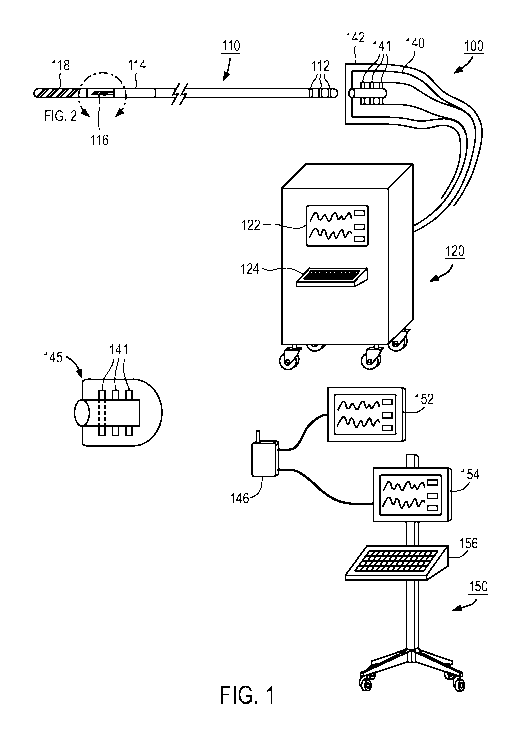
[0028] Figure 1 is a schematic showing the key components making up a
pressure wire measurement system;
[0029] Figure 2 is a schematic showing the conductors between the sensor
and
proximal electrical contacts in a prior art embodiment;
[0030] Figure 3 illustrates one preferred embodiment of the electrically
conductive structures of the present invention;
[0031] Figure 4 illustrates the cross-sectional view of Figure 3;
[0032] Figures 5 a, b, c and d illustrate a torque device in accordance to
one
embodiment of the invention;
[0033] Figure 6 illustrates one preferred embodiment of the pressure wire
to
provide a guiding mechanism so that the torque device will engage the
conductive
traces at the appropriate orientation;
[0034] Figure 7 illustrates another preferred embodiment of the pressure
wire
measurement system where there are two sensors deployed on a sleeve that can
be
-5 -
CA 02853189 2014-04-22
WO 2013/066992
PCT/US2012/062777
delivered over a traditional guide wire, 110, not shown, and a torque device
can
wirelessly activate the sensors and shows the results from the signals return
by these
two sensors; and
[0035] Figure 8 illustrates another embodiment where the stand alone
sleeve
catheter with two sensors is in a rapid exchange catheter configuration with
guide
wire, 110, and a catheter handle, 810, now serving as the display for either
the
waveforms from the two sensors or the results after processing of the two
waveforms
or both, depending on the display size available. Two switches to control the
electronics in the handle are also shown in this illustration.
DETAILED DESCRIPTION
[0036] The present invention will now be described in detail with
reference to
several embodiments thereof as illustrated in the accompanying drawings. In
the
following description, numerous specific details are set forth in order to
provide a
thorough understanding of embodiments of the present invention. It will be
apparent,
however, to one skilled in the art, that embodiments may be practiced without
some or
all of these specific details. In other instances, well known process steps
and/or
structures have not been described in detail in order to not unnecessarily
obscure the
present invention. The features and advantages of embodiments may be better
understood with reference to the drawings and discussions that follow.
[0037] Figure 1 shows one embodiment of a pressure wire measurement
system, 100, not to scale. It includes a pressure wire, 110. The distal end,
designated
118, is usually radio-opaque to allow for visualization under X-ray and is
usually
implemented as a coil to make it floppy and atraumatic. The pressure sensor is
designated 116 and is often followed by another coil section 114 for desired
stiffness.
The remaining body of the pressure wire often has a hollow lumen to
accommodate
the electrical transmission lines (not shown) connecting the sensor 116 with
the
electrical contacts 112 at the proximal end.
[0038] The hollow lumen in the proximal portion of the pressure wire
designed to accommodate the electrical or optical transmission conductors
reduces the
- 6 -
CA 02853189 2014-04-22
WO 2013/066992
PCT/US2012/062777
fidelity of the torque transmission due to the reduced rigidity of the body of
the
pressure wire. System 100 addresses this issue by having thin conductive
traces on
the central core wire.
[0039] Figure 1 also shows a connector 140 that couples to the proximal
end
of the pressure wire 110. Internal to connector 140, there are electrical
contacts 141
that mate with the counterpart 112 on the pressure wire. The connector 140
being
non-sterile needs to be enclosed with a sterile barrier, 142, typically a
sterile bag, to
prevent contamination of the sterile field during the PCI procedure.
[0040] It is also possible to have a long pressure wire such that the
connect
140 is far remove from the sterile field where the risk of contamination is
low and a
sterile barrier 142 may not be needed. However, if long transmission lines are
used as
a consequence of having a long pressure wire, signal quality may be degraded.
[0041] The connector 140 is coupled to an electronic equipment, 120, where
the signals from the sensor can be acquired, processed and display with the
display
122. If user input is needed, an input device 124 can also be located on the
electronic
equipment 120.
[0042] In another embodiment, a wireless implementation is described. In
this
embodiment, a wireless transceiver 145 is coupled to the pressure wire such
that the
electrical contacts, 141, in the transceiver 145, mates with the electrical
contact 112
on the pressure wire 110. The signals are then wirelessly received by a
wireless
transceiver 146 which can then display the information on a display 152 or
couple to
the electronic equipment 150 which may take the form of an Intravenous pole
with a
display 154 and an input device 156.
[0043] Figure 2 shows a close up view of the sensor 116 with the
electrical
transmission conductors 210. These conductors terminate at the electrical
contacts
112 at the proximal end of the pressure wire 110. With this construction, the
mating
connector, whether in the form of a connector 140, or in the form of a
wireless
transceiver 145 is located at the proximal end of the pressure wire 110 where
the
electrical contacts 112 are located on the pressure wire 110.
[0044] This arrangement for the wireless transceiver 145 can be an
impediment to the work flow as transceiver should be smaller and light weight
and
- 7 -
CA 02853189 2014-04-22
WO 2013/066992
PCT/US2012/062777
ideally should perform like a torque device. A torque device, not shown, also
needs
to be able to be positioned anywhere proximal to where the pressure wire exits
the
human body and not be constrained to the proximal end or a particular fixed
location.
[0045] Referring to Figure 3, the conductors that electrically connect the
sensor to the equipment for acquisition, processing and display have been
replaced
with electrically conductive traces, 304, embedded in insulating layers, 305.
Three
such insulating layers are illustrated in Figure 3.
[0046] In some embodiments, the traces are terminated in pads, 303, which
are connected to pads, 301, on the sensor chip via wire bonding with gold
wires, 302.
Other connection schemes known to persons skilled in the art are also
possible.
[0047] The traces 304 are distinguished from one another by the number of
insulating layers 305 as well as the circumferential locations as indicated in
the cross-
sectional representation in Figure 4.
[0048] Shielding layers, not shown, can also be implemented to improve the
electrical performance of these conductive traces if needed.
[0049] These traces 304 can be metallization via various depositing
process or
conductive polymer and the insulating layers 305 can be various insulating
polymers,
like polyimide film.
[0050] It is also possible to print conductive polymer onto an insulating
substrate and achieve similar results. Beside these additive processes, it is
also
possible to start with a conductive layer on top of an insulating layer,
subtractive
processes can then be used where the conductive material is removed to result
in
conductive traces remaining on the insulating layer to serve as conductors.
[0051] It is possible to have variations along this theme. For example,
multiple conductive traces can reside in the same layer underneath one
insulating
layer if they can be separated adequately apart. This may be an advantage in
the case
of multiple sensor chips. One sensor chip can have its conductive traces
residing in
one layer, while the other can have its conductive traces in another layer.
[0052] In Figure 5a, an exemplary torque device 500, is shown with a cap
501
and collet 502, an arrangement where as the cap is advanced, the fingers 503
of the
- 8 -
CA 02853189 2014-04-22
WO 2013/066992
PCT/US2012/062777
collet 502 will close on and grip on the pressure wire 110. Pressure wire 110
is not
shown.
[0053] Different ways to implement a torque device are possible.
[0054] In Figure 5a, some of the fingers have a tapered tip 510, capable
of
penetrating the insulation layers 305, and making contact with the appropriate
traces
304, thereby forming electrical connection(s). Different shape and arrangement
for
the finger 503 to make electrical contacts with the conductive traces 304 are
also
possible.
[0055] Different fingers 503 can have different length tapered tip 510
capable
of penetrating to the correct depth to make contact with the conductive trace
304
through the various insulating layers 305.
[0056] Figures 5b and Sc show two close up views of one embodiment of a
finger with a tapered tip configuration designed to simultaneously penetrate
two
insulating layers 305 to make contact with conductive traces 304 lying at two
different depths.
[0057] The configuration is such that while making contact with the deeper
layer, it avoids shorting with the shallower layer.
[0058] This implementation of the tapered tips is useful where multiple
sensor
chips 116 are present at the distal end of the pressure wires and the
conductive traces
are embedded in separate layers at different depths. Different length tapered
tip 510
can engage different sensor chip signals at different depth levels with no
ambiguity.
Even if the number of conductive traces is small enough to fit with in the
circumference of a single layer of insulating layer, it may still be
advantageous to
keep the number of fingers 503 small but utilize multiple tapered tips 510 to
engage
the conductive traces at different depths. Such flexibility is provided for in
these
embodiments.
[0059] Other configurations and methods for the tapered tips to engage the
conductive traces are also possible.
[0060] In Figure 5d, a view from B-B of Figure 5a, the body of the collet
502
has a guiding track 520 to guide the insertion of the torque device such that
the
- 9 -
CA 02853189 2014-04-22
WO 2013/066992
PCT/US2012/062777
orientation of the fingers 503 remain aligned with the conductive traces 305
correctly.
In Figure 6, the portion of the pressure wire 110 that accepts the torque
device has a
corresponding guiding ridge 610 that allows the torque device to slide along
it once
the guiding track 520 is aligned with the guiding ridge 610.
[0061] This is one example of a mechanical means to ensure a proper
orientation of the torque device. Using a visible strip marking on the guide
wire for
aligning with a counterpart marking on the torque device is an example of a
visual
means for achieving correct alignment.
[0062] Other ways to provide orientation guidance are known for those
skilled
in the art.
[0063] In Figure 5a, a display 504 is also shown, where result derived
from
the sensor can be made available to the user of the torque device.
[0064] This torque device being able to make electrical connection with
the
sensor 116 can now provide the needed signal acquisition, processing and
wireless
transmission to a receiver outside the sterile area of the catheterization
laboratory.
[0065] In this embodiment, it is important to make the transceiver unit
small
and light weight as well as being able to position freely along a much larger
range in
the proximal portion of the pressure wire and behave like a torque device.
[0066] To achieve this behavior, some parts of the acquisition and
processing
are partitioned off the transceiver 145 and locate on the pressure wire body
proper.
The constraint is to maintain the profile such that the diameter of the entire
pressure
wire can still accept delivery of other device designed to be delivered over a
guide
wire, e.g. balloon and stent, usually 0.014 inch in diameter.
[0067] In one embodiment, a piece of signal processing component can be
interposed and embedded in the envelope of the proximal portion of the
pressure wire
such that a partially processed signal emerges on the continuation of a
conductive
trace.
[0068] In another embodiment, multiple such interposed segments can be
implemented in the proximal portion of the pressure wire in order to reduce
the size
and weight of the transceiver 145 to better perform like a torque device.
- 10 -
CA 02853189 2014-04-22
WO 2013/066992
PCT/US2012/062777
[0069] In another embodiment, transceiver 145 only sends out the processed
results for display without the pressure signals derived from the sensor chip
116.
[0070] The proximal portion of the pressure wire 110 is more tolerant of
having any stiff sections that are required to implement signal conditioning
and
processing components. These components are being off-loaded from the torque
device to enable a smaller form factor for the torque device that also doubles
as a
transceiver.
[0071] Note that this proximal portion of the pressure wire does not enter
the
human body.
[0072] In a modern catheterization laboratory, many pieces of equipment
vie
for the limited space available around the sterile patient table. Able to
provide a
minimally invasive pressure measurement device that conforms as much as
possible
to other interventional device like a balloon improves the work flow
immensely.
[0073] As all the communication between the sensor chip and the torque
device takes place in between the insulating layers and the conductive traces,
the
pressure sensing can also be implemented in the form of a stand-alone sleeve
that is
delivered over the preferred guide wire that the user has chosen.
[0074] This approach of performing the pressure measurement differs from
the approach of implementing a pressure wire. The advantage of this approach
is that
the operator can use his preferred guide wire without any possible compromise
on the
wire performance but with the possible disadvantage that an additional
catheter,
however small, needs to be delivered over the guide wire and subsequently
removed
to allow for other device to be delivered over the same guide wire again for
the next
steps in the procedure.
[0075] Figure 7 illustrates the concept of this embodiment where sensor
701
and sensor 702 are located on a sleeve and are in communication, wireless or
wired,
with torque device 500. A display 504 is also shown on the torque device 500.
This
torque device 500 can also optionally communicate, via a wireless receive 146,
with
equipment 150 with its display 154 and input device 156 or a stand alone
remote
display.
- 11 -
CA 02853189 2014-04-22
WO 2013/066992
PCT/US2012/062777
[0076] In one embodiment, sensors 701 and 702 are wireless. Sensor 701 is
distal to a stenosis in a coronary artery, sensor 702 is in the aorta.
Together, they
provide two independent pressure measurements that are transmitted to the
torque
device 500. The display, 504, on the torque device can then, as an example,
display
the measured Fractional Flow Reserve value which is a ratio of the mean of the
distal
pressure over the mean of the proximal pressure.
[0077] In one embodiment, the torque device 500 itself can activate the
two
sensors, 701 and 702, as indicated in Figure 7. Sensor 701 is deployed distal
to a
stenosis in the coronary artery while sensor 702 remains in the aorta such
that upon
activation by the torque device via an electromagnetic wave, they send out
their
respective pressure measurement signals wirelessly. These signals are received
by the
torque device and any computation result based on these two measurement
signals is
then shown on the display 504. No other capital equipment in required and both
pressure signals needed to generate the ratio for Fractional Flow Reserve
(FFR) is
obtained simultaneously without the need for a pullback.
[0078] It is also possible to implement sensor using
MicroElectroMechanical
Systems (MEMS) technology and they can be piezo-resistive or capacitive in
their
principle of operation. It is also possible to implement the sensor using
piezo-electric
polymer or ceramic.
[0079] The use of piezo-electric polymer is of particular value since it
does
not require the use of rigid sensor chip and can be conformable to the shape
of a guide
wire geometry.
[0080] The choice of the specific sensor technology for 701 and 702
depending on process complexity and cost of manufacturing with corresponding
pro's
and con's.
[0081] It should be appreciated that it is possible to have a hybrid
system
where the sensors 701 and 702 can have wired connections between them and then
wirelessly communicate with torque device via wireless means. This has a
certain
advantage when the pressure sensing is implemented as a stand alone device to
be
delivered over an existing guide wire. Sensor 702 which resides in the aorta
as
opposed to the coronary artery would have more room to accommodate a wireless
- 12 -
CA 02853189 2014-04-22
WO 2013/066992
PCT/US2012/062777
transceiver to transmit both pressure measurements. This will then not impact
the
need to have a small form factor in the distal sensor 701 to have accurate
pressure
measurement.
[0082] In one embodiment, the sensor 701 is implemented with a piezo-
electric polymer that generates a voltage when experience a change in
pressure. The
capacitance of sensor 701 can also be a function of pressure as it changes
dimension.
This voltage or capacitance change is measured via conductive traces or other
wired
transmission means to a proximal sensor 702 which resides in the aorta. Sensor
702
itself senses pressure at the aorta as well as handling any needed
conditioning and
processing of pressure signal from sensor 701 and together wirelessly provides
the
result or partial result to the torque device 500 on its display 504.
[0083] It is contemplated that this invention is applicable to
physiological
parameters other than pressure. One characteristics of this invention is the
use of a
low cost, disposable transceiver. It can be made small if the data rate and
power
consumption are low ¨ which dictates the kind of information and type of
signal
acquisition and processing that can be accomplished.
[0084] Physiologic parameters like pressure, temperature, pH value, etc.,
are
slow varying parameters that can be acquired with low sampling frequency,
simple
processing, if any, and low data transmission rate. The power consumption is
also
correspondingly low.
[0085] The improvement described here affords a better torque transmission
as it removes the need to have a lumen to accommodate the electrical or
optical
transmission lines. In particular, the electrical connection scheme also
improves the
electrical performance as the parasitic capacitance is reduced by increasing
the
separation of the transmission lines. The improved construction also allows
for better
integration of multiple sensors.
[0086] The improvement with a wireless transfer of the physiologic signal
allows for a more compatible operation with how a guide wire is used in the
PCI
procedure. A wireless embodiment also improves the work flow and avoids the
need
to have a large instrument near the patient's bed during the procedure.
Wireless
- 13 -
CA 02853189 2014-04-22
WO 2013/066992
PCT/US2012/062777
communication between the sensor and the torque device also makes for a
compact
system when a simple display on the torque device is adequate for the
procedure.
[0087] Multiple sensors eliminate the need to perform a pullback procedure
to
obtain pressure information from multiple locations.
[0088] A stand alone embodiment allows pressure measurement with an
existing primary guide wire and eliminates the need for a wire exchange
procedure.
[0089] Several variations of the stand alone sleeve with multiple sensors
as
illustrated by Figure 7 are possible. For example, the distance between the
two
sensors, 701 and 702, can be made variable to accommodate different lesion
locations
in the coronary arteries while keeping the proximal sensor in the aorta..
[0090] The sleeve can also be constructed such that a guide wire exit port
allows for a rapid exchange catheter configuration as described in US patent
5451233
"Angioplasty Apparatus Facilitating Rapid Exchanges" by Paul Yock.
[0091] The sleeve in the above configuration can now have a catheter
handle,
as opposed to a torque device, where a larger display can be accommodated.
This
larger display can display both waveforms and numerical results from
processing of
the waveforms.
[0092] In this configuration, as shown in Figure 8, the connection between
the
sensors (701, 702) and the electronics in the handle, 810, will not require
embedding
the conductors in insulating layers and are self contained within the stand-
alone sleeve
catheter.
[0093] Having the sensors implemented on the sleeve itself allows for
integration with other interventional devices that could benefit from a
pressure
measurement to monitor the progress of the interventional procedure. For
example, if
this pressure measuring sleeve is integrated with a Chronic Total Occlusion
(CTO)
device, the pressure monitoring can indicate when the CTO device has succeeded
in
entering the distal true lumen as opposed to entering a false lumen in the
intima of the
vessel wall. This can reduces the use contrast medium and radiation from the
angiogram.
- 14 -
CA 02853189 2014-04-22
WO 2013/066992
PCT/US2012/062777
[0094] Other applications can include integration with percutaneous valve
implantation where the reduction of the pressure gradient across the valve is
an
important parameter. Having a sleeve approach for pressure measurement allows
for
relatively easy integration with such percutaneous valve devices.
[0095] In sum, the present invention provides a system and methods for an
improved pressure measurement device. The advantages of such a system include
the
ability to manipulate the pressure wire more like a guide wire and perform the
pressure measurement in a way more compatible with other catheterization
laboratory
procedures.
[0096] While this invention has been described in terms of several
embodiments, there are alterations, modifications, permutations, and
substitute
equivalents, which fall within the scope of this invention. Although sub-
section titles
have been provided to aid in the description of the invention, these titles
are merely
illustrative and are not intended to limit the scope of the present invention.
[0097] It should also be noted that there are many alternative ways of
implementing the methods and apparatuses of the present invention. It is
therefore
intended that the following appended claims be interpreted as including all
such
alterations, modifications, permutations, and substitute equivalents as fall
within the
true spirit and scope of the present invention.
- 15 -