Note: Descriptions are shown in the official language in which they were submitted.
MATERIALS AND METHOD FOR IMMOBILIZING, ISOLATING, AND
CONCENTRATING CELLS USING CARBOXYLATED SURFACES
BACKGROUND
A. Field of the Invention
The present invention relates to materials in methods for use in concentrating
and/or
isolating cells from a sample using carboxylated surfaces.
B. Brief Description of Related Art
Carboxylated surfaces have found multiple uses in the biotechnological arts.
Frequently, the surfaces are used to isolate specific biomolecules. For
example, WO
2007-004687 discloses the formation of agglutinates of lipoproteins, other
than a specific
lipoprotein fraction, using magnetic nanoparticles which have anionic
functional groups, such
as a carboxylic acid group, on their surfaces.
Carboxylated surfaces also have been used as supports for biomolccule- and
biomaterial-specific ligands, which may be used to immobilize the biomolecule
or
biomaterial. In the typical scenario, the carboxyl group is activated, for
example, using a
carbodiimide. The activated carboxyl is then reacted with a reactive group
(such as an
amine) in an entity capable of binding to a target cell or class of target
cells, such as an
antibody, thereby immobilizing the entity to the surface. The cell may then be
bound to the
surface via an interaction between the immobilized entity and the cell. For
example, WO
2007-095279 utilizes carboxy-modified nanoparticles coated with DNA aptamers
having a
high affinity for a target cell to immobilize the cell and separate it from
the sample by flow
cytometry. In US 7,713,627, "probe-bonded particles" are disclosed for use in
separating
bacteria, viruses, and cells. The "probe" specific for the target may be
attached to the particle
via chemical reaction with a surface functionalized with a carboxylic acid-
bearing group. In
EP 1118676, microorganisms are isolated using binding between the
microorganism and a
ligand, such as a carbohydrate to which the microorganism is known to bind, a
nutrient for
the microorganism, or an iron chelating compound.
CA 2854165 2019-02-07
CA 02854165 2014-04-30
WO 2013/067399
PCT/US2012/063385
Others have described the use of carboxylated surfaces to directly isolate
viruses from
biological samples. For example, US 2003-0087284 discloses the use of
particles coated
with at least one cationic group and at least one anionic group to bind,
separate, and detect
viruses.
SUMMARY OF THE INVENTION
The present disclosure relates to immobilization of a cell using a
carboxylated
surface.
A method of immobilizing a cell is disclosed, comprising contacting a
carboxylated
surface with a sample, the sample comprising the cell and a fixative, for a
sufficient time to
permit the cell to bind to the carboxylated surface.
A method of isolating a cell from a sample is disclosed comprising: (a)
contacting a
carboxylated surface with a sample comprising the cell and a fixative agent
under conditions
sufficient to induce binding between the cell and the surface; and (b)
separating the surface
from the sample, thereby isolating the cell.
A method of isolating a biomolecule from a cell is also disclosed, the method
comprising: (a) contacting a carboxylated surface with a sample comprising the
cell under
conditions sufficient to induce binding between the cell and the surface; (b)
separating the
surface from the sample, thereby isolating the cell; and (c) lysing the cell,
thereby releasing
the biomolecule.
A method of determining the presence of a biomolecule in a sample is also
disclosed,
the method comprising: (a) contacting a carboxylated surface with a sample
comprising the
cell under conditions sufficient to induce binding between the cell and the
surface; (b)
separating the surface from the sample, thereby isolating the cell; (c) lysing
the cell, thereby
releasing the biomolecule into a lysate; and (d) detecting the biomolecule in
the lysate.
A method of determining the presence of a nucleic acid in a sample is also
disclosed,
the method comprising: (a) contacting a carboxylated surface with a sample
comprising the
cell under conditions sufficient to induce binding between the cell and the
surface; (b)
separating the surface from the sample, thereby isolating the cell; (c) lysing
the cell, thereby
releasing the nucleic acid; and (d) detecting the nucleic acid by a method
comprising
generating a DNA:RNA hybrid between the nucleic acid and a nucleic acid probe
specific for
the nucleic acid.
A method of concentrating a cell in a sample is also disclosed, said method
comprising: (a) contacting a carboxylated surface with the sample under
conditions sufficient
2
CA 02854165 2014-04-30
WO 2013/067399
PCT/US2012/063385
to induce binding between the cell and the surface; and (b) removing at least
a portion of the
sample that is not bound to the surface, thereby concentrating the cell.
Also disclosed is a complex between a cell and a carboxylatcd surface in the
presence
of a fixative agent.
Also disclosed is a carboxylated surface for use in the methods disclosed
herein.
BRIEF DESCRIPTION OF THE DRAWINGS
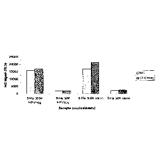
Fig. 1 is a bar graph comparing HPV16 nucleic acid recovery using carboxylate
beads to concentrate the cell ("COOmag") compared to centrifugation ("MC).
SiHa cells
(either 300,000 or 30,000) were used as a source of HPV16, spiked in either
HPV-negative
clinical samples or clean PRESERVCYT liquid cytology medium. The y-axis
represents a
signal (in relative light units ("RCU")) directly correlating to the
concentration of the HPV 16
nucleic acid.
Fig. 2 is a bar graph comparing HPV16 nucleic acid recovery using carboxylate
beads to concentrate the cell ("COOmag") compared to centrifugation ("MC).
SiHa cells
(either 300,000 or 30,000) were used as a source of HPV16, spiked in either
HPV-negative
clinical samples or clean PRESERVCYTq_z, liquid cytology medium. The y-axis
represents a
signal (in RCU) directly correlating to the concentration of the HPV 16
nucleic acid.
Fig. 3 is a bar graph comparing HPV16 nucleic acid recovery using different
amount
of carboxylate beads (10 Ill and 5 I) and different incubation times (1, 10,
15 and 30 min.) to
concentrate the cell.
Fig. 4 is a bar graph comparing HPV16 nucleic acid recovery when various
volumes
(50, 100, and 200 ul) of the supernatant removed after immobilization of the
cells was
reserved and added back to the lysate before being assayed. "PC Sup" indicates
that an
indicated volume of a supernatant was added before the HC2TM assay. "STM-
F/DNR"
indicates positive controls using an indicated volume of a 2:1 mixture of
STM/DNR in place
of the added supernatant.
Fig. 5A and Fig. 5B are bar graphs comparing HPV16 nucleic acid recovery using
carboxylate beads to concentrate 20,000, 10,000, and 0 SiHa cells spiked in
SUREPATH
HPV negative cervical samples. "MC" indicates that samples were processed
according to
Protocol 4, while "C00-" indicates that samples were processed according to
Protocol 3.
Fig. 6 is a bar graph comparing HPV16 nucleic acid recovery in SUREPATH ,
samples at pH 4 or pH 7.
3
CA 02854165 2014-04-30
WO 2013/067399
PCT/US2012/063385
Fig. 7 is a bar graph comparing HPV16 nucleic acid recovery in SUREPATHOR
samples at pH 4.5 or pH 7, with or without methanol (at 24% and 42%). "SP"
indicates a
sample in SUREPATH4), while "PC" indicates a sample in PRESERVCYT m, which was
used as a positive control.
Fig. 8 is a bar graph comparing recovery of nucleic acid from heat inactivated
Chlamydia trachomatis elementary bodies spiked into clean PRESERVCYTTm ("ebs
in PC
clean") or HPV¨negative cervical specimen pool in PRESERVCYTTm ("ebs in PC
Pool"),
using a carboxylated bead to concentrate the elementary bodies before nucleic
acid isolation
and analysis. Comparative examples without the bead concentration are
indicated by "ebs in
PC clean sup MC" and "ebs in PC Pool sup MC".
Fig. 9 is a bar graph comparing recovery of nucleic acid from heat inactivated
Chlamydia trachomatis elementary bodies were spiked into HPV¨negative cervical
specimen
pool in PRESERVCYTTm ("ebs in PC Pool") or fresh urine, and carboxylated beads
were to
concentrate the elementary bodies before nucleic acid isolation and analysis.
Comparative
examples without the bead concentration are indicated by "PC Pool sup MC".
Chlamydia
trachomatis elementary bodies added directly to a 2:1 mixture of STM and DNR
were used
as a positive control ("STM/DNR").
DETAILED DESCRIPTION OF THE INVENTION
The present disclosure includes methods, compositions, reagents, systems, and
kits
related to immobilization of cells using a carboxylated surfaces. The methods,
compositions,
reagents, systems, and kits are useful for, for example, laboratory research
and clinical
diagnostic purposes, including but not limited to isolation of biomolecules
from cells for use
in the detection and identification of pathogenic organisms and the detection
of a genetic
predisposition to a particular disease.
Immobilizing the cell
In an aspect, a method of immobilizing a cell is disclosed, said method
comprising
contacting a sample comprising the cell with a carboxylated surface under
conditions
sufficient to induce binding between the cell and the surface. In an aspect,
the cell is a
mammalian cell.
In principle, any cell can be used. In another aspect, the mammalian cell is a
human
cervical epithelial cell. In another aspect, the cell is a unicellular
organism, such as a
bacterium of the genus Mycobacterium, Chlamydia, Staphylococcus, or Neisseria;
or a
protozoa of the genus Trichomonas; or a yeast of the genus Saccharomyces or
Candida. In
4
CA 02854165 2014-04-30
WO 2013/067399
PCT/US2012/063385
another aspect, the bacterium is of the genus chlamydia, for example, in the
form of an
elementary body.
As used herein, the term "carboxylate compound" shall refer to any compound or
portion thereof comprising at least one free carboxylic acid (COOH) or
carboxylate anion
(COO).
As used herein, the term "carboxylated surface" shall refer to a surface of a
solid or
quasi-solid, the surface comprising a free carboxylic acid group, carboxylate
anion, or
carboxylate salt.
In an aspect, the "surface" as used herein in particular refers to the portion
of a solid
phase which comes into contact with a solution when the solid phase is
contacted therewith.
The solid phase provides the surface that provides, e. g. carries, the
carboxylate group,
carboxylate anion, or carboxylate salt. Suitable solid phases may be made of
or may
comprise in particular at their surface a material selected from the following
group:
a material comprising or consisting of silicon such as silica and polysilicic
acid materials, quartz, borosilicates, silicates, diatomaceous earth or
glasses,
- a material comprising or consisting of a polymer such as
poly(meth)acrylate,
polyurethane, polystyrene, polystyrol, polyacrylamide, a divinylbenzene
polymer, a styrene divinylbenzene polymer, polyethylene, polypropylene,
polyvinylidene fluoride, polyacrylonitrile, polyvinylchloride, polyacrylate,
polyacrylamide, polymethacrylate or a methyl methacrylate polymer;
- a material comprising or consisting of a polysaccharide such as agarose,
cellulose, dextrans or sepharose;
a material comprising or consisting of a mineral;
- a material comprising or consisting of a metal oxide such as aluminum
oxide,
magnesium oxide, titanium oxide or zirconium oxide;
- a material comprising or consisting of a metal such as gold or platinum;
and
- a material comprising or consisting of a derivative of the foregoing.
Furthermore, the solid phase may also comprise more than one of the above
described
materials. In an aspect, at least the surface comprising the carboxylate
moieties is composed
of one of the described materials or a mixture thereof. Also any solid phase
suitable for ion
5
CA 02854165 2014-04-30
WO 2013/067399
PCT/US2012/063385
exchange chromatography may be used as solid phase to provide the surface that
comprises,
e.g. is functionalized with, carboxylate groups.
Exemplary formats of the solid phase include but are not limited to particles
such as
beads, membranes, filters, plates, columns and dipsticks. According to one
aspect, the surface
comprising carboxylate moieties is provided by a vessel, for example the inner
surface of a
vessel that is intended to receive the sample. The inner surface or portions
thereof can be
functionalized, e.g. coated, with carboxylate moieties. Examples of respective
vessels include
but are not limited to microtubes and wells of a microplate. In this aspect,
the cells bind due
to the provided carboxylate moieties to the inner surface of the vessel or
well and are thereby
collected.
According to an exemplary aspect, the surface is provided by a solid phase
that can be
provided as suspension and can be separated from a liquid phase. When
contacted with a
liquid phase such as for example a cell containing liquid based cytology
medium, the surface
comprising the carboxylate moieties is in contact with the liquid phase in
order to allow cell
-- binding. In an aspect, the surface comprising carboxylate moieties is
provided by particles
which comprise carboxylate moieties at their surface. The particles may have
an average size
that is selected from a range of 100 nm to 50 gm, 200 nm to 40 gm, 300 nm to
35 gm, 400
nm to 30 gm, 450 nm to 25 gm, 500 nm to 20 gm, 550 nm to 15 gm, 600 nm to 12.5
gm, 650
nm to 10 gm, 700 nm to 7.5 gm, 750 nm to 5 gm, 800 nm to 3.5 gm, 800nm to 3
gm, 800 to
-- 2.5 gm, 800nm to 2 gm and 800 nm to 1.5 gm. Particles of the respective
sizes and in
particular of a smaller size such as Sum or less, 2.5 gm or less or 1.5 m or
less are easy to
handle and can be well resuspended in the cell sample. Furthermore, respective
small
particles provide a large surface area that can bind and accordingly can
efficiently collect the
cells from the remaining sample such as e.g. a liquid-based cytology
collection medium.
Suitable materials for providing or making the particles are described above.
The particles
may also comprise more than one of the above described materials, e.g.
comprising two or
more layers comprising or consisting of different materials to provide the
particle body.
In an aspect, magnetic particles are used to provide a carboxylated surface
that binds
cells. Using magnetic particles has the advantage, that they can be processed
and moved by
the aid of a magnetic field. The magnetic particles can for example have
superparamagnetic,
paramagnetic, ferrimagnetic or ferromagnetic characteristics. The magnetic
particles may
comprise a magnetic material that is incorporated in the particles and/or is
associated with the
particles. To avoid leaching of the magnetic material, the magnetic material
may be
6
CA 02854165 2014-04-30
WO 2013/067399
PCT/US2012/063385
completely encapsulated e. g. by the material providing the surface such as
e.g. silica,
polysilicic acid, glass or a polymeric material such as polyacrylate.
Numerous suitable solids and quasi-solids arc well-known in the art, including
but not
limited to surfaces useful in cation exchange chromatography. The solid or
quasi-solid
should be of such a character that it can be placed in suspension in, and
separated from, a
liquid phase. Moreover, the solid or quasi-solid should be of such a character
that, when
contacted with a liquid phase, the surface comprising the carboxylate-
containing compound is
in contact with the liquid phase. Exemplary solid or quasi-solid surfaces
include, but are not
limited to polycarbonate and/or magnetic beads, including paramagnetic,
diamagnetic,
.. ferromagnetic, ferrimagnetic, and diamagnetic beads; columns; plates;
filter paper;
polydimethylsiloxane (PDMS); and dipsticks. In an exemplary aspect, the
carboxylated
surface comprises a magnetic bead. Exemplary magnetic beads include those
which are
described in the German patent application DE 10 2005 058 979.9. Such magnetic
beads are
commercially available.
In an exemplary aspect, the surface is coated with a carboxylated polymer.
Examples
of carboxylated polymers which arc suitable as coating material arc described
in detail in the
German patent application DE 10 2005 040 259.3. Examples of compounds which
may be
bound to the surface are glycine, aspartic acid, 6-aminocaproic acid, NTA
(nitrilotriacetic
acid), polyacrylic acid (PAA), diglyme (diethylene glycol dimethyl ether), or
combinations of
these, without being limited thereto.
In an aspect, the carboxylated surface having a weakly negative overall charge
is
employed. In a further aspect, the carboxylated surface comprises a carboxyl
content as
determined by conductometric titration with sodium hydroxide of: at least 0.1
mEq/g; at least
0.2 mEq/g; at least 0.3 mEq/g; at least 0.4 mEq/g; from about 0.1 to about 0.7
mEq/g; from
about from about 0.1 to about 0.6 mEq/g; from about 0.1 to about 0.5 mEq/g;
from about 0.2
to about 0.7 mEq/g; from about 0.2 to about 0.6 mEq/g; from about 0.2 to about
0.5 mEq/g;
from about 0.3 to about 0.7 mEq/g; from about 0.3 to about 0.6 mEq/g; from
about 0.3 to
about 0.5 mEq/g; from about 0.4 to about 0.7 mEq/g; from about 0.4 to about
0.6 mEq/g; or
from about 0.4 to about 0.5 mEq/g.
In a further aspect, the carboxylated surface has a negative overall charge.
In a further aspect, the carboxylated surface may comprise additional anionic
functional groups, such as phosphonate and/or sulfate groups.
In another aspect, a carboxylated surface suitable for use in cation exchange
chromatography is used. Such surfaces are well known in the art, such as, for
example,
7
CA 02854165 2014-04-30
WO 2013/067399
PCT/US2012/063385
SERADYN DS-MGCM beads. In a further aspect, the surface is suitable for use
in cation
exchange chromatography, but not anion exchange chromatography.
In an aspect, binding of the cell to the carboxylated surface is not mediated
by a
ligand-receptor interaction or an antibody-antigen interaction. As such, a
carboxylated
surface comprising at least one compound that is not modified with a receptor
ligand or an
antibody capable of binding the cell may be used. In a further aspect, the
cell is immobilized
by a direct association between the cell and the compound comprising the
carboxylate group.
Any sample may be used in which cells may be present, including, without
limitation:
a specimen or culture (e.g., cellular and tissue cultures) including clinical
and laboratory
biological samples; food and agricultural samples; and forensic samples,
including urine,
semen, hair, blood, skin, and saliva samples. Samples may be from any mammal,
including a
human, and explicitly include fluid, solid (e.g., stool) or tissue, as well as
liquid and solid
food and feed products and ingredients such as dairy items, meat and meat by-
products, and
waste. Explicitly included are samples taken directly from a mammal, as well
as samples that
have been stored in a preservative, including but not limited to paraffin-
embedded tissue
samples and cellular and tissue samples stored in a liquid-based cytology
medium.
According to one aspect, the carboxylated surface is added to a liquid sample
comprising the cell. In an aspect, the particle comprising the carboxylated
surface is added to
the liquid sample in a volume to achieve a particle concentration after
resuspension that lies
in a range selected from: at least 10 p..g/ml, at least 20 pg/m1; at least 30
pg/m1; at least
40 pg/m1; at least 50 g/ml; 10 pg/mlto 1000 p.g/m1; 10 pg/m1 to 500 pgml; 10
pg/m1 to 300
1.ig/m1; 10 g/m1 to 200 g/m1; 20 1.ig/m1 to 1000 pg/m1; 20 pg/ml to 500
[tg/m1; 20 g/m1 to
300 g/m1; 20 p.g/m1 to 200 g/m1; 30 g/m1 to 1000 pg/m1; 30 tig/m1 to 500
g/m1; 30
pg/ml to 300 pg/ml; 30 pg/m1 to 200 pg/m1; 40 pg/ml to 1000 iig/m1; 40 p ginal
to 500 p..g/m1;
40 p..g/m1 to 300 g/ml; 40 pg/m1to 200 pg/m1; 40 pg/m1 to 1000 g/m1; 40
g/m1 to 500
g/m1; 50 g/ml to 300 1.tg/m1; and 50 g/ml to 200 g/ml.
In an aspect, the sample comprises at least one fixative agent. In an aspect,
the
sample comprises a cross-linking and/or non-cross-linking fixative agent.
Cross-linking
fixatives function by making chemical bonds between proteins in the tissue
sample, leading
to their precipitation and immobilization within the tissue. Exemplary
crosslinking fixatives
include, but are not limited to, formaldehyde and paraformaldehyde. Non-cross-
linking
fixatives do not chemically alter the proteins in the sample; rather they
simply precipitate
them where they are found in the tissue sample. Non-cross-linking fixatives
include ethanol,
acetone, methanol and mixtures thereof.
8
CA 02854165 2014-04-30
WO 2013/067399
PCT/US2012/063385
In another aspect, the sample is a liquid sample comprising at least one
fixative agent.
In an aspect, the sample comprises a cross-linking and/or non-cross-linking
fixative agent.
Cross-linking fixatives function by making chemical bonds between proteins in
the tissue
sample, leading to their precipitation and immobilization within the tissue.
Exemplary
crosslinking fixatives include, but are not limited to, formaldehyde and
paraformaldehyde.
Non-cross-linking fixatives do not chemically alter the proteins in the
sample; rather they
simply precipitate them where they are found in the tissue sample. Non-cross-
linking
fixatives include ethanol, acetone, methanol and mixtures thereof
In an aspect, the sample is in a liquid cytology medium. Exemplary liquid
cytology
media include, but are not limited to PRESERVCYT (Hologic, Inc., Bedford, MA)
(a
methanol-based liquid cytology medium); DIGENE Specimen Transport Medium
(Qiagen
Gaithersburg, Inc., Gaithersburg, MD) (a guanidinium-based specimen transport
medium);
and SUREPATHTm (Becton, Dickinson and Company, Franklin Lakes, NJ) (a liquid
cytology
medium comprising alcohol and aldehyde).
PRESERVCYT is a methanol-based liquid cytology medium comprising ¨42%
methanol; ¨5mM EDTA, at a pH 4.5).
DIGENE Specimen Transport Medium (Qiagen Gaithersburg, Inc., Gaithersburg,
MD) is a a guanidinium-based specimen transport medium.
SUREPATHTm (Becton, Dickinson and Company, Franklin Lakes, NJ) is a liquid
cytology medium comprising ¨22% ethanol and a trace cell fixative/cross-linker
(formaldehyde-like), pH 7.
In an aspect, the pH of the liquid cytology medium is in a range selected from
the
group consisting of: not more than 9; not more than 8; not more than 7; from 2
to 9; from 2 to
8; from 2 to 7; from 3 to 9; from 3 to 8; from 3 to 7; from 4 to 9; from 4 to
8; from 4 to 7. In
an aspect, the pH of the liquid cytology medium is adjusted to improve
immobilization of the
cell to the carboxylated surface.
In an aspect, the liquid sample is urine.
In an aspect, the cells are brought into contact with the carboxylated surface
in the
presence of the liquid cytology medium or urine over a sufficiently long
period of time, i.e. a
9
CA 02854165 2014-04-30
WO 2013/067399
PCT/US2012/063385
period of time which suffices to allow the cells to bind/attach themselves to
the carboxylated
surface. Such a period of time should be at least 30 s, preferably at least 1
min, further
preferably at least 3 min, further preferably at least 10 minutes.
Concentrating a cell from the sample and/or isolating the cell from the sample
In an aspect, the carboxylated surface may be used to concentrate or isolate
the cell.
In an aspect, a method is provided comprising: (a) immobilizing the cell as
set forth above;
and (b) separating the surface the sample, thereby isolating the cell.
In an aspect, a method of concentrating a cell in a sample is provided
comprising: (a)
immobilizing the cell as set forth above; and (b) separating the surface from
at least a portion
of the sample, thereby isolating the cell.
Different modes of operations are feasible in order to separate the surface
with the
bound cells from the sample.
In an aspect, a vessel is used, wherein the inner wall of the vessel is at
least partially
functionalized with carboxylated moieties as described herein, the remaining
sample can be
discarded by decanting or it can be removed by aspiration or similar methods.
The bound
cells remain associated to the inner surface of the vessel due to the
carboxylated moieties.
Respective separation steps can be performed using a robotic system.
As another example, particles comprising carboxylated moieties are used to
provide
the carboxylated surface. If the particles are non-magnetic they can be
collected for example
by filtration or sedimentation which can according to one aspect be assisted
by centrifugation.
It is preferred though to use magnetic particles, because the magnetic
particles including the
bound cells can be processed easily by the aid of a magnetic field, e.g. by
using a permanent
magnet. This aspect is preferred as it is compatible with established robotic
systems capable
of processing magnetic particles. Here, different robotic systems exist in the
prior art that can
.. be used in conjunction with the present invention to process the magnetic
particles to which
the cells were bound. According to one aspect, the magnetic particles are
collected at the
bottom or the side of the reaction vessel and the remaining liquid sample is
removed from the
reaction vessel, leaving behind the collected magnetic particles to which the
cells are bound.
Removal of the remaining sample can occur by decantation or aspiration. Such
systems are
well known in the prior art and thus need no detailed description here.
In an alternative system that is known for processing magnetic particles the
magnet
which is usually covered by a cover or envelope plunges into the reaction
vessel to collect the
magnetic particles. The magnetic particles that carry the bound cells can then
be transferred
for example into a new reaction vessel e.g. comprising a resuspension
solution, preferably a
CA 02854165 2014-04-30
WO 2013/067399
PCT/US2012/063385
denaturing composition as will be described in the following. As respective
systems are well-
known in the prior art and are also commercially available (e.g. QIAsymphony;
QIAGEN),
they do not need any detailed description here.
In a further alternative system that is known for processing magnetic
particles, the
sample comprising the magnetic particles can be aspirated into a pipette tip
and the magnetic
particles can be collected in the pipette tip by applying a magnet e.g. to the
side of the pipette
tip. The remaining sample can then be released from the pipette tip while the
collected
magnet particles which carry the bound cells remain due to the magnet in the
pipette tip. The
collected magnetic particles can then be processed further. Such systems are
also well-known
in the prior art and are also commercially available (e.g. BioRobot EZ1,
QIAGEN) and thus,
do not need any detailed description here.
It is within the scope of the present invention and preferred to contact the
bound cells
after step b) with a liquid composition. This aspect has the advantage that it
may inter alia
support the collection of the cells if magnetic particles are used, in
particular if a robotic
system is used for collecting the magnetic particles wherein the magnet
plunges into the
reaction vessel. By contacting the collected cells which are still bound to
the magnetic
particles with a liquid composition, it is ensured in this aspects that the
particles are
efficiently removed from the magnet and are redispersed in the liquid
composition. However,
contacting the collected cells with a liquid composition has additional
general advantages
also when using other magnetic separation systems or other carboxylated
surfaces as
described herein. The liquid composition is compatible with the subsequent
downstream
processing, for example, when forming double stranded DNA:RNA hybrids as
described
below. The liquid composition may be added to the collected cells that are
bound to the
carboxylated surface, e.g. it may be added to the magnetic particles carrying
the bound cells
or vice versa. According to one aspect contacting the carboxylated surface
carrying the bound
cells with the liquid composition results in that the collected cells are at
least partially
released and thus eluted from the carboxylated surface. If desired, the
carboxylated surface
can then be separated from the released cells. Here, any mode of separation is
feasible and
suitable modes include but arc not limited to collecting the liquid
composition comprising the
released cells e.g. by aspiration or, which is feasible if magnetic particles
are used, collecting
and separating the magnetic particles from the remaining liquid composition
comprising the
released cells using a magnetic field. However, as is shown by the examples,
it is not
mandatory to remove the carboxylated surface prior to steps c) and d) and this
is a particular
11
CA 02854165 2014-04-30
WO 2013/067399
PCT/US2012/063385
advantage of the present invention as this again saves handling steps. The
cells may even
remain bound to the carboxylated surface as long as the nucleic acids are
released therefrom.
Releasing an entity from the immobilized cell
A method of isolating a biomolecule from a cell is further provided
comprising: (a)
isolating the cell as set forth above; and (b) lysing the cell, thereby
releasing the biomolecule
into a lys ate.
In an aspect, an entity may be released from a cell that has been immobilized
as
described above, optionally after the cell has been concentrated and/or
isolated from the rest
of the sample. By way of example and not limitation, the entity may include,
but is not
limited to: a biomolecule, such as nucleic acids (including but not limited to
DNA and RNA),
peptides (including but not limited to oligopeptides and polypeptides),
lipids, sugars, et
cetera; a virus or viral particle; an organelle or other subcellular
structure; and a
microorganism, such as bacteria, mycobacteria, protozoa, and fungi. Methods of
releasing
the foregoing entities are well known in the art.
In an aspect, the entity is a biomolecule selected from the group consisting
of peptides
and nucleic acids. In an aspect, the biomolecule is a cell-surface peptide and
is released
without lysing the cell, such as by extraction from the plasma membrane,
elution of a ligand
from a cell surface receptor, or enzymatic release of a protein anchored to
the plasma
membrane.
In another aspect, the biomolccule is an intracellular, and is released by
lysing the
cell. Any manner of lysing the cell can be used in the disclosed method,
including without
limitation: mechanical lysis, such as by sonication or cytolysis; and chemical
lysis,
including use of detergents such as 3-[(3-cholamidopropyl)dimethylammonio]-1-
propanesulfonate (sold commercially as CHAPS ); octylphenoxypolyethoxyethanol
(also
.. known as NONIDET P-40 or IGEPAL CA-630g); deoxycholate; C14F1220(C2H40)5
(sold
commercially as TRITON X-100); sodium dodecyl sulfate (sold commercially as
SDS);
and/or polysorbate surfactants (sold commercially as TWEENk). In a further
aspect, lysis
is performed in the presence of heat.
In a further aspect, the biomolecule is a nucleic acid or a protein.
In a further aspect, a nucleic acid, is released by a method comprising: (b)
isolating or
concentrating the cell as set forth above; (c) contacting the cell with a
liquid composition that
lyses the cell, thereby releasing the nucleic acid into a lysate, and (d)
optionally, denaturing
the released nucleic acid. According to one aspect, the collected cells are
lysed by contacting
them with a liquid composition.
12
CA 02854165 2014-04-30
WO 2013/067399
PCT/US2012/063385
In an aspect, the liquid composition for lysing the cells comprises a
chaotropic agent.
Any chaotropic agent can be used for this purpose that causes disorder in a
protein or nucleic
acid by, for example, but not limited to altering the secondary, tertiary or
quaternary structure
of a protein or a nucleic acid. Preferably, a chaotropic salt is used. The
chaotropic salt
preferably comprises guanidinium, thiocyanate, isothiocyanate, perchlorate,
trichloroacetate
and/or trifluoroacetate as chaotropic ion. Preferably, the chaotropic agent is
selected from the
group consisting of guanidinium hydrochloride, guanidinium thiocyanate,
guanidinium
isothiocyanate, sodium thiocyanate, sodium iodide, sodium perchlorate, sodium
trichloroacetate, sodium trifluroacetate and urea. Also a mixture of
chaotropic agents can be
used. Preferably, guanidinium hydrochloride, guanidinium thiocyanate or
guanidinium
isothiocyanate is used as chaotropic agent in the composition that assists the
release of the
nucleic acids in step c). The liquid composition may comprise the chaoptropic
agent, which
preferably is a chaotropic salt as mentioned above, in a concentration that
lies in a range
selected from about 0.1M up to the saturation limit, about 0.2M to 8M, about
0.3M to 4M,
about 0.4M to 3M, about 0.5M to 2.5M, about 0.6M to about 2M, about 0,6M to
about 1.5M
and 0.6M to about 1M. Preferably, the liquid composition is a solution such as
a lysis buffer.
According to one aspect, the liquid composition for lysing the cells comprises
a
chelating agent, preferably EDTA. A chelating agent is an organic compound
that is capable
of forming coordinate bonds with metals through two or more atoms of the
organic
compound. Suitable chelating agents include, but are not limited to
diethylenetriaminepentaacetic acid (DTPA), ethylenedinitrilotetraacetic acid
(EDTA),
ethylene glycol tetraacetic acid (EGTA) and N,N-bis(carboxymethyl)glyeine
(NTA).
According to a preferred aspect, EDTA is used. As used herein, the term "EDTA"
indicates
inter alia the EDTA portion of an EDTA compound such as, for example, K2EDTA,
K3EDTA or Na2EDTA.
According to one aspect, the liquid composition for lysing the cell may
comprise a
detergent. The detergent may be non-ionic, ionic, anionic, cationic or
zwitterionic.
In an aspect, the liquid composition for lysing the cells may comprise a
preservative
such as sodium azidc. Furthermore, the liquid composition for lysing the cells
may comprise
a buffering agent. Preferably, a biological buffer such as HEPES, MES, MOPS,
TRIS, BIS-
TRIS Propane and others is comprised in the composition. Preferably, a Tris
buffer is used.
In an aspect, the released nucleic acid is also denatured. This can be
achieved e.g. by
adding a denaturation agent such as a base and/or heating as will be described
in the
following. For cases in which the released nucleic acid is double stranded and
is to be
13
CA 02854165 2014-04-30
WO 2013/067399
PCT/US2012/063385
hybridized to a nucleic acid probe, it is preferred that the double-stranded
target nucleic acid
is converted to a be at least partially single stranded to make the nucleic
acids accessible to
hybridization.
In an aspect, the liquid composition for lysing the cells has an alkaline pH
value to
achieve or support the denaturation of the released nucleic acids. According
to one aspect,
the liquid composition has a pH value that is selected from a pH value of 10
or more, a pH
value of 11 or more, a pH value of 11.5 or more, a pH value of 12 or more, a
pH value of
12.5 or more, a pH value of 12.75 or more, a pH value of 13 or more and a pH
value of 13.25
or more. A high alkaline pH value supports the release of the nucleic acids
and furthermore,
denatures the released nucleic acids, thereby preparing the nucleic acids for
detection (dl).
This aspect is particularly preferred if the target nucleic acid is DNA
because such basic pH
will both nick and degrade a majority of the internal RNA in the specimen. In
an aspect, the
denaturation is supported by heating as will be described below. In addition,
alkaline
treatment can disrupt interactions between peptides and nucleic acids to
improve accessibility
of the target nucleic acid and degrade protein. Furthermore, alkaline
treatment of proteins
effectively homogenizes the specimen to ensure reproducibility of analysis
results for a given
sample. It can also reduce the viscosity of the sample to increase kinetics,
homogenize the
sample, and reduce background by destroying any endogenous single stranded RNA
nucleic
acids, DNA-RNA hybrids or RNA-RNA hybrids in the sample. It also helps
inactivate
enzymes such as RN ascs and DNases that may be present in the sample. One
skilled in that
art would appreciate that if RNA is the target nucleic acid (as opposed to
DNA), different
reagents may be preferable. For establishing an alkaline pH value as described
above, a base,
preferably a chemical base such as e.g. sodium or potassium hydroxide can be
added,
respectively may be comprised in the liquid composition.
Using a strong alkaline liquid composition as described above is particularly
suitable
if the target nucleic acid is DNA. If the target nucleic acid is RNA, more
moderate conditions
are preferred in order to preserve the integrity of the RNA. E.g. the liquid
composition may
have a pH value of 7.5 to 10,8 to 9.5 or 8.5 to 9.
The liquid composition for lysing the cells can be provided by one
composition, e.g.
one solution or may be prepared by contacting the cells with two or more
separate
compositions. According to one aspect, the collected cells are contacted with
two or more
separate compositions in order to provide the release and denaturation
conditions described
above. According to one aspect, two or more compositions are mixed to provide
a liquid
composition as described above prior to contracting said composition with the
carboxylated
14
CA 02854165 2014-04-30
WO 2013/067399
PCT/US2012/063385
surface to which the cells are bound (preferably magnetic particles).
According to one aspect,
a first composition comprises the chaotropic agent and optionally the other
components
described above (e.g. the composition STM can be used as first composition,
see examples)
and a separate second composition comprises the denaturation agent (e.g. DNR
as second
composition, see examples). In an aspect, said second composition is an
alkaline solution.
Any alkali that can bring the liquid composition that results from the mixing
of the first and
second composition to a pH range described above is suitable. Suitable
concentrations of
alkali include from about 1.0 N to about 2.0 N or from about 1.25 N to about
1.75 N. Without
being limited, suitable alkali include NaOH and KOH. Mixing the two (or more)
compositions together provides the liquid composition that assists the release
and
denaturation of the nucleic acids and thus provides the conditions described
above and in
particular establishes the alkaline pH value described above. Preparing the
liquid composition
by mixing two or more separate compositions to establish the conditions
described above is
preferred and it is in particular preferred to add the denaturation agent as
described above
separately.
According to one aspect, the surface is provided by magnetic particles, and
the
collected cells that are bound to the magnetic particles are contacted with an
amount of the
liquid composition described above that is selected from 251.11 to 5001.11,
30111 to 4000, 35111
to 3000, 40 1to 2501.11, 50u1 to 2000, 601.11 to 1751.11, 701.11 to 1501.11,
80111 to 1251.11 and 85111
to 1001.11. According to one aspect, the liquid composition is added in a
volume to achieve a
particle concentration after resuspension that lies in a range selected from
2000 g/m1 to
150001g/ml, 2500 g/m1 to 125001g/ml, 30001g/m1 to 100001g/m1; 35001g/m1 to
95001g/ml, 4000 g/m1 to 9000 g/ml, 4500n/1111 to 8750uginil and 50001g/m1 to
8500 g/ml.
Instead of or in addition to the denaturation agent described above, other
methods of
denaturation may be employed such as utilizing a heating step, for example,
heating the
sample to at least 80 C or at least 95 C to separate the nucleic acid strands.
Adding an
alkaline denaturation agent, which can be comprised in the liquid composition
as described
above, and performing a heating step is preferred to efficiently denature the
released nucleic
acids.
According to one aspect, after the cells were contacted with the liquid
composition for
lysing the cells, the resulting mixture is heated to assist the denaturation
of the nucleic acids.
Preferably, said mixture is heated to at least 55 C, preferably at least 60 C.
Suitable
temperature ranges include 55 C to 90 C, preferably 60 C to 85 C and more
preferred 65 C
CA 02854165 2014-04-30
WO 2013/067399
PCT/US2012/063385
to 80 C. Preferably, heating occurs for at least 30min, preferably at least
35min, more
preferred at least 40min. Suitable time periods can be selected from 30min to
150min, 35min
to 130min, 40min to 120min and 45min to 100min. The described time and
temperature
conditions shall provide an efficient denaturation of the nucleic acids in an
acceptable amount
of time, while leaving the target nucleic acid in a suitable condition for
carrying out (dl). The
suitable time period also depends on the processed sample. E.g. when
processing cell
containing samples comprising specific fixatives such as SUREPATH samples,
longer
incubation times of 90min (or longer if desired) at a temperature of at least
60 C, preferably
at about 65 C are preferred. When processing cell-containing samples that do
not comprise
cross-linking fixatives such as PRESERVCYT samples, shorter incubation times
of e.g.
45min (or longer if desired) at a temperature of at least 60 C, preferably at
about 65 C are
sufficient. Suitable time periods can also be determined by the skilled
person. It will be
readily understood by one of ordinary skill in the art that longer periods of
incubation at
lower temperatures, or shorter periods of incubation at higher temperatures,
may be balanced
to provide a similar effect to the conditions described herein. The release of
the nucleic acids
can also be assisted by shaking. E.g. the sample treated with the liquid
composition described
above can be mixed by hand mixing or mechanical shaking at about 800 rpm,
about 900 rpm,
about 1000 rpm, between about 600 and about 1000 rpm, or between about 600 and
1200
rpm.
According to one aspect, the sample mixture comprising the liquid composition
for
lysing the cells, the cells and optionally the carboxylated surface, e.g. the
magnetic particles,
or an aliquot of said mixture is transferred into a new vessel, preferably a
multi-well device
such as a 96 well plate, prior to heating. This aspect is advantageous as it
can be easily
integrated in established assay work-flows for generating and isolating
DNA:RNA hybrids
and also automated processing systems as therein respective multi-well devices
are processed
and accordingly, existing equipment can be used.
After performing the above, a sample is obtained which comprises the released
nucleic acid. Said sample optionally also comprises the carboxylated surface,
e.g. magnetic
particles if the magnetic particles were not separated prior to or during the
isolation.
The sample obtained after may be stored e.g. at 4 C or below prior to
performing
subsequent processing steps, if any.
Detecting a released nucleic acid
In one exemplary aspect, a nucleic acid is detected in a sample by isolating
the nucleic
acid as set forth above; and detecting the nucleic acid in the lysate. Any
method of detecting
16
the nucleic acid may be used, including gel electrophoresis, PCR-related
techniques including reverse
transcriptase PCR and real time PCR, sequencing, sub-cloning procedures,
Southern blotting, northern
blotting, fluorescent in situ hybridization, and various mutational analyses
including HYBRID
CAPTURETm and multiplex analysis. In one exemplary aspect, a nucleic acid
comprising a specific
sequence may be detected by hybridizing it to a nucleic acid probe
complementary to the specific
sequence. In one aspect, the nucleic acid probe is bound to a solid phase or
adapted to be bound to a solid
phase. In another aspect, hybridization of the nucleic acid probe to the
nucleic acid molecule results in a
DNA:RNA hybrid between the probe and the nucleic acid molecule. The resulting
hybrid may then be
bound by an antibodies known to bind specifically to DNA:RNA hybrids ("DNA:RNA-
binding
antibody"), which in turn may be bound to a solid phase or adapted to be bound
to a solid phase. In either
case, hybridization of the probe with the nucleic acid results in the nucleic
acid being associated with a
solid phase, which may then be separated from the lysate using mechanical
means. By way of example
and not limitation, such methods are described in U.S. Pat. No. 6,228,578 and
U.S. Patent Application
Ser. No. 12/695,071. Exemplary DNA:RNA-binding antibodies include, but are not
limited to, those
disclosed in U.S. Pat. Nos. 4,732,847 and 4,865,980. In other exemplary
methods, the nucleic acid is
detected by, inter alia, amplifying the nucleic acid. Exemplary amplification
methods include, but are not
limited to, polymerase chain reaction ("PCR"), reverse transcriptase PCR ("RT-
PCR"), real time PCR,
real-time RT-PCR.
In an aspect, a method for detecting a target nucleic acid in a sample is
disclosed, said method
comprising: (c) releasing the nucleic acid from the cell as described above;
(d) optionally denaturing the
released nucleic acid; and (e) detecting the released nucleic acid.
In a further aspect, an automated method for screening clinical samples for a
disease state is
provided, said method comprising: (a ) immobilizing a cell comprised in the
samples to the carboxylated
surface as set forth above; (b) isolating or concentrating the cell as set
forth above; (c) lysing the cells to
create a lysate as set forth above; (d) optionally denaturing the released
nucleic acid; and (e) detecting the
presence of a target nucleic acid in the lysate, wherein the presence or
absence of the target nucleic acid in
the lysate is indicative of the disease state. Any detection method compatible
with automation may be
used. By way of example and not limitation, the detection method may comprise
hybridizing a nucleic
acid probe to the nucleic acid from the lysate. By way of example and not
limitation,
17
CA 2854165 2019-02-07
CA 02854165 2014-04-30
WO 2013/067399
PCT/US2012/063385
hybridization results in a DNA:RNA hybrid. In a further aspect, the DNA:RNA
hybrid is
detected by binding an antibody specific for DNA:RNA hybrids to the DNA:RNA
hybrid
between the nucleic acid probe and the nucleic acid from the lysate.
Detecting a released nucleic acid using double stranded nucleic acid hybrids
In an aspect, detection comprises generation of a double stranded nucleic acid
hybrid
between the target nucleic acid and a nucleic acid probe specific therefor. In
an aspect, the
nucleic acid hybrid is a DNA:RNA hybrid.
According to one aspect, detection comprises:
(el) contacting the released and optionally denatured target nucleic acid
with one or more probes specific for the target nucleic acid under
conditions that allow the probes and target nucleic acid to hybridize
forming double-stranded nucleic acid hybrids; and
(e2) detecting the presence or absence of double-stranded nucleic acid
hybrids.
In an aspect, the double stranded nucleic acid hybrids are detected by a
method comprising:
(e2a) capturing the double stranded nucleic acid hybrids to a solid
support;
(e2p) optionally separating the double-stranded nucleic acid hybrids
bound to the solid support from un-bound nucleic acids; and
(e2y) detecting the presence or absence of double-stranded nucleic
acid hybrids.
In another aspect:
(e2a) the double stranded nucleic acid hybrids are captured to the
solid support by contacting the double stranded hybrids with a
first binding agent that is bound to or adapted to be bound to
the solid phase to form a double-stranded nucleic acid/first
binding agent complex; and
(e2y) the presence or absence of double-stranded nucleic acid hybrids
is detected by (a) binding said double-stranded nucleic
acid/first binding agent complex with a further binding agent
that is labelled with a detectable marker to form a double-
stranded nucleic acid hybrid/first binding agent/labelled
binding agent complex; (b) optionally washing the double-
stranded nucleic acid hybrid/first binding agent/labelled
18
CA 02854165 2014-04-30
WO 2013/067399
PCT/US2012/063385
binding agent complex; and (c) detecting the presence or
absence of the label of the further binding agent thereby
indicating the presence or absence of the target nucleic acid.
In the foregoing aspects, the carboxylated surface preferably is disposed on a
magnetic
particle, which magnetic particle may optionally be present throughout the
detection.
In one aspect, detection comprises the use of an analyzer comprising: (1) a
heating
element; and (2) a device for detecting a detectable signal, such as a
fluorimeter or a
luminometer.
In the foregoing methods, after release and optional denaturation of the
nucleic acids
as is described above, the target nucleic acids are contacted with one or more
probes under
conditions suitable for the one or more probes to hybridize to the target
nucleic acid to form a
double-stranded nucleic acid hybrid. The probe is preferably a polynucleotide
probe. The
probe can be full length, truncated, or synthetic DNA or full length,
truncated, or synthetic
RNA. Furthermore, probes comprising RNA and DNA nucleotides or comprising
modified
nucleotides and/or analogs of nucleotides can be used, as long as a hybrid is
formed. If the
target nucleic acid is DNA, then preferably the probe is RNA and if the target
nucleic acid is
RNA, then preferably the probe is DNA. Accordingly, a RNA/DNA hybrid is
preferably
formed. The probes are designed to hybridize or bind with the target nucleic
acid molecules.
In one aspect, the probes are capable of hybridizing or binding to HPV and HPV
high
risk variants. In an additional aspect, the probes are specific for HPV and
HPV high risk
variants. High risk (HR) probes can include probes for HPV high risk types 16,
18, 31, 33,
35, 39, 45, 51, 52, 56, 58, 59, 66, 68 and 82. The probes may vary in amount
from about 7.5
ng to about 60 ng per HPV type per assay, or from about 20 ng to about 45 ng
per HPV type
per assay, or about 30 ng of probe for each HPV type per assay is used. Thus,
in one aspect
the HR probes consist of or consist essentially of one or more probes for HPV
high risk types
16, 18, 31, 33, 35, 39, 45, 51, 52, 56, 58, 59, 66, 68, and 82 or low risk HPV
types 6, 11,40,
43, 53, 61, 67, 69, 70, 71, 72, 81, and 83. The RNA probes may be short
synthetic RNA
probes that specifically bind only to the target nucleic acid molecule.
In a non-limiting aspect, the one or more probe used is capable of hybridizing
or
binding to target nucleic acid molecules that are at least 70 percent, at
least 80 percent, at
least 85 percent, at least 90 percent, at least 95 percent, at least 96
percent, at least 97 percent,
at least 98 percent, at least 98 percent, at least 99 percent, or 100 percent
identical to nucleic
acid molecules associated with HPV, genetic variants of HPV, HPV DNA of a high
risk HPV
19
CA 02854165 2014-04-30
WO 2013/067399
PCT/US2012/063385
type, or HPV RNA of a high risk HPV type, or any one of high risk HPV types
16, 18, 31,
33, 35, 39, 45, 51, 52, 56, 58, 59, 66, 68, and 82 or any one of low risk HPV
types 6, 11,40,
43, 53, 61, 67, 69, 70, 71, 72, 81, and 83. In another aspect, the one or more
probes used is
complementary to HPV, genetic variants of HPV, HPV DNA of a high risk HPV
type, HPV
RNA of a high risk HPV type, or any one of high risk HPV types 16, 18, 31, 33,
35, 39, 45,
51, 52, 56, 58, 59, 66, 68, and 82 or any one of low risk HPV types 6, 11, 40,
43, 53, 61, 67,
69, 70, 71, 72, 81, and 83. Also DNA or RNA fragments of the target nucleic
acids can be
used.
According to one aspect, the denatured sample is neutralized prior to or
during the
addition of the probes if denaturation occurred under alkaline conditions.
According to one
aspect, the one or more probes are diluted in a probe diluent that also can
act as a neutralizing
hybridization buffer. This aspect is advantageous in order to neutralize the
alkaline pH value
that was used in step c) to release and denature the nucleic acids. The probe
diluent used for
DNA or RNA probes will differ due to the different requirements necessary for
DNA versus
RNA stability. For example, if the probes are RNA, it is preferred to
neutralize the sample
first and then add the one or more probes or alternatively, add the RNA probe
and a
neutralizing agent (probe diluent) to the sample at the same time as strong
alkaline pH values
can destroy RNA. The probe diluent can be used to dissolve and dilute the
probe and also
help restore the sample to about a neutral or weakly alkaline pH, e.g., about
pH 6 to about pH
9, preferably 6.5 to 8, more preferred 6.5 to about 7.5 to provide a more
favorable
environment for hybridization. Sufficient volume of probe diluent, e.g. one-
half volume of
the sample, may be used to neutralize a base-treated sample.
In an aspect, the probe diluent comprises a buffer, polyacrylic acid, NaOH and
sodium azide. The probe diluent may comprise acetic acid. In one aspect, the
probe diluent
comprises 2.2 M BES (N,N-Bis(2-hydroxyethyl)-2-aminoethanesulfonic acid), 2.6
percent
polyacrylic acid (PAA), 0.7 N NaOH and 0.05 percent sodium azide. The probe
diluent may
contain from about 1.2 M to about 2.6 M BES, from about 1.5 M to about 2.5 M
BES; from
about 1.75 M to about 2.25 M BES; from about 2 M to 2.4 M BES, or about 2.2 M
BES, as
well as any number within the recited ranges. In one aspect the probe diluent
may contain
from about 2 percent to about 3.0 percent PAA or, as well as any number within
the recited
ranges. In another aspect, the PAA concentration is from about 2.2 percent to
about 2.7
percent. In yet another aspect, the PAA concentration is about 2.6 percent. In
a further aspect
the probe diluent may contain from about 0.6 N to about 0.8 N NaOH, for
example, about 0.7
N NaOH. The concentration of NaOH generally increases as the amount of BES
increases.
CA 02854165 2014-04-30
WO 2013/067399
PCT/US2012/063385
For large probes, a heated alkaline solution may be added to the sample, then
probe
diluent may be added to the sample at room temperature, and then the sample
may be
reheated. Such a process can inhibit secondary structure from forming. Binding
agents such
as antibodies tend to bind to structures with secondary structure. When using
non-full length
probes such as truncated or synthetic probes, heating the solutions or sample
may not be
necessary because secondary structures issues are not present. In an aspect,
the sample is not
heated when used with truncated or synthetic probes. After treatment with the
denaturation
reagent, an aliquot of neutralization buffer, in an aspect the probe diluent
described, in which
the one or more probes are dissolved, can be added to the sample under
appropriate
conditions to allow hybridization or binding of the probe and the target
nucleic acid to occur.
The neutralization buffer may contain a single buffering salt. In an aspect,
the neutralization
buffer does not contain more than a single buffering salt. The hybridization
condition is
sufficient to allow the one or more polynucleotide probes to anneal to a
corresponding
complementary nucleic acid sequence, if present, in the sample to form a
double-stranded
nucleic acid hybrid.
Hybridization conditions suitable for the particular probes and diluents used
are
employed. For example, the probes and sample nucleic acids can be incubated
for a
hybridization time, preferably at least about 5 to about 120 minutes, about 10
to about 100
minutes, or from about 20 to about 80 minutes, or from about 30 minutes to
about 60
minutes, as well as any number within the recited ranges sufficient to allow
the one or more
polynucleotide probes to anneal to a corresponding complementary target
nucleic acid
sequence. The hybridization conditions can include a hybridization temperature
of at least
about 55 C, at least about 60 C, preferably from about 60 C to about 75 C,
preferably 65 C
to about 70 C as well as any number within the recited ranges. For a given
target nucleic acid
and a given probe, one of ordinary skill in the art can readily determine
desired hybridization
conditions and hybridization times by routine experimentation. One of ordinary
skill in the art
will further appreciate that the time and temperature of hybridization must be
optimized, one
with respect to the other. Without being limited, stringent hybridization
conditions may be
controlled by increasing the temperature, increasing the ionic conditions to
above 0.5M (for
example, NaCl), or reducing the concentration of PAA. As a non-limiting
example, stringent
hybridization conditions may include performing a hybridization reaction at
elevated
temperatures, such as of at least about 65 C.
After the one or more probes were allowed to hybridize to the target nucleic
acid
molecule and to form a double-stranded nucleic acid hybrid, the hybrid is
captured by a
21
CA 02854165 2014-04-30
WO 2013/067399
PCT/US2012/063385
molecule that binds to the double-stranded nucleic acid hybrid formed. Such a
molecule is
referred to herein as binding agent. Thereby, a double-stranded nucleic acid
hybrid/binding
agent complex is formed. Binding agents specific for the double stranded
nucleic acid hybrids
include, but are not limited to, monoclonal antibodies, polyclonal antibodies,
proteins such as
but not limited to RNAse H, nucleic acids including but not limited to
aptamers, or sequence
specific nucleic acids. In one aspect an antibody binding the formed double-
stranded nucleic
acid hybrid is used as binding agent, respective antibodies are also known as
anti-hybrid
antibodies. Accordingly, the double-stranded nucleic acid hybrids formed in
accordance with
the present invention can be captured and detected using antibodies or
antibody fragments
that are specific to double-stranded nucleic acid hybrids. Subsequently, we
will describe
suitable and preferred aspects by referring to antibodies. However, said
description equally
applies to antibody fragments such as Fab fragments capable of binding the
hybrids or other
suitable binding agents.
The antibody is specific to double-stranded hybrids, such as but not limited
to
RNA/DNA hybrids; DNA/DNA hybrids; RNA/RNA hybrids; and mimics thereof, where
mimics refer to molecules that behave similarly to RNA/DNA, DNA/DNA, or
RNA/RNA
hybrids. The anti-double-stranded nucleic acid hybrid antibody, i.e., the anti-
hybrid antibody
that is utilized will depend on the type of double-stranded nucleic acid
hybrid formed. In one
aspect, the anti-hybrid antibody is immunospecific to RNA/DNA hybrids. It will
be
understood by those skilled in the art that either polyclonal or monoclonal
anti-hybrid
antibodies can be used and/or coupled and/or immobilized on a support in the
present method
as described below. In one aspect, monoclonal antibodies support high
stringency incubation
temperatures during the capture step. The first and further binding agents,
which preferably
are antibodies may be the same for capture and detection or may be different
from each other.
In one aspect, the first and further binding agent (which can be labelled, see
below), which
preferably are both monoclonal antibodies, used for capture and/or detection
are the same and
are specific for RNA-DNA hybrids. As described above, also suitable as binding
agents are
immunofragments or derivatives of antibodies that are specific for double-
stranded hybrids
where such fragments or derivatives contain the binding regions of the
antibody.
In an aspect of the present invention, a monoclonal anti-RNA/DNA hybrid
antibody
derived from a hybridoma cell line is used. Such hybridoma cell lines are
described in U.S.
Pat. No. 4,865,980, U.S. Pat. No. 4,732,847, and U.S. Pat. No. 4,743,535.
Hybrid-specific
monoclonal antibodies may also be prepared using techniques that are standard
in the art. The
hybrid-specific monoclonal antibody may be used for both capturing and
detecting the target
22
CA 02854165 2014-04-30
WO 2013/067399
PCT/US2012/063385
nucleic acid. Also other binding agents suitable of specifically binding the
formed hybrid can
be used.
in one aspect, a first anti-hybrid binding agent such as an anti-hybrid
antibody is
immobilized onto a support using techniques that are standard in the art.
Examples of suitable
immobilization technologies include covalent linkages or adsorption, for
example, protein-
protein interactions, protein-G beads, biotin- streptavidin interaction, EDAC
to link to a
carboxyl or tosyl group, etc., or hybridization directly onto the solid
support using, for
example, sequence specific nucleic acids in an affinity column.
Supports include but are not limited to reaction vessels, including microtiter
plates
wherein one or more wells are functionalized with the molecule that binds the
hybrid,
preferably an anti-hybrid antibody, particles, magnetic particles, columns,
plates, filter paper
and dipsticks. Any support can be used as long as it allows removal, e.g.
extraction of a liquid
phase. Magnetic particles are useful in that they can be left in the solution
and the liquid
phase can be extracted or decanted, if a magnetic field is applied to
immobilize the particles
or the magnetic particles with the bound hybrid can be removed using a system
as described
above. Particles that are small and have a high surface area are preferable,
such as particles
about 0.5i.tm to 10i.tm, 0.75t.tm to 7.5um, 0.75 m to 5 m, 0.75 m to 2.5um and
most
preferred lium in diameter. However, when using magnetic particles as solid
support for the
first binding agent that binds the hybrid, it is preferred to perform a final
magnetic separation
step prior to performing (d2) in order to ensure that the magnetic particles
do not interfere
with the detection.
Preferably, the support that is used for immobilising the first binding agent
which
binds the generated hybrid is a reaction vessel. Preferably, the support is
provided by a multi-
well device such as a microtiter plate, wherein the wells are at least
partially functionalized
with the first binding agent. This aspect is advantageous, as it allows to
easily remove the
particles that were used for binding the cells in the course of the assay, if
said particles were
not separated prior to (dl). The generated hybrid is captured by the binding
agent and thus is
immobilized to the reaction vessel, so that the remaining sample including the
particles that
were used for cell binding can be easily removed e.g. by aspiration and/or
washing.
The hybrids are incubated with the anti-hybrid binding agent attached to the
support
for a sufficient amount of time to allow capture of the double-stranded
nucleic acid hybrids
by the immobilized anti-hybrid binding agent. Thereby, a double-stranded
nucleic acid
hybrid/solid support complex is formed, which also comprises the first binding
agent that is
used for capturing the hybrid. As described above, in a preferred aspect, the
support is a
23
CA 02854165 2014-04-30
WO 2013/067399
PCT/US2012/063385
reaction vessel, preferably a microtiter plate functionalized with one or more
anti-hybrid
binding agents such as anti-hybrid antibodies. The anti-hybrid antibody may be
monoclonal
or polyclonal. In one aspect the antibody is monoclonal. In one aspect, the
antibody is
coupled to the support by an 1-ethyl-3-[3- dimethylaminopropyl] carbodiimide
hydrochloride
(EDAC) linker.
In one aspect, the support is a polystyrene bead. In an aspect, the support or
bead
coupled to the binding agent, which preferably is an antibody, is diluted in a
bead dilution
buffer. The bead dilution buffer is helpful in minimizing protein denaturation
on the bead.
One example of a bead dilution buffer comprises 6 percent casein, 100 mM Tris-
HC1, 300
mM NaCl, and 0.05 percent sodium azide.
In an aspect, the support coated with the anti-hybrid antibody is incubated
with the
sample. Incubation may be performed at room temperature or at elevated
temperatures. The
incubation time can range from about 5 to about 120 minutes, about 10 to about
100 minutes,
or from about 20 to about 80 minutes, or from about 30 minutes to about 60
minutes, as well
as any number within the recited ranges sufficient to allow capture. The same
incubation
times are suitable if the first binding agent that is used for binding the
formed hybrid is not
bound to a solid support. The sample can be shaken during said incubation. It
will be
understood by those skilled in the art that the incubation time, temperature
and/or shaking
conditions can be varied to achieve alternative capture kinetics as desired.
Following binding and thus capture of the target nucleic acid/probe hybrid,
the
captured hybrid may be separated from the rest of the sample. Separation is
particularly easy
if the fist binding agent is immobilized to a solid support. In this case, the
unbound sample
can e.g. simply be aspirated as is also described in the examples. According
to one aspect,
one or more washing steps are performed to wash away non-captured nucleic
acids and
sample remainders. As described above, if the particles that were used for
binding the cells
are still present during the generation and capture of the hybrid, they will
at least be partially
removed during said separation step. Advantageously, it is not necessary to
specifically
remove the particles e.g. by the aid of a magnet in case of magnetic particles
as they will be
automatically removed when separating the sample remainders. This saves
handling steps.
According to one aspect, a further binding agent is used. The further binding
agent
may comprise a detectable label. The further binding agent is used to allow
detecting the
presence of double- stranded nucleic acid hybrids. The further binding agent
can be bound
directly or indirectly to the complex that is formed when the first binding
agent binds and
thus captures the formed hybrid, thereby providing a double-stranded nucleic
acid hybrid/first
24
binding agent/labelled binding agent complex or a double-stranded nucleic acid
hybrid/solid
support/labelled binding agent complex if the first binding agent was
immobilized to a solid support. In
one aspect, the further binding agent comprises a label that must react with a
substrate to provide a signal
that can be detected. The further binding agent may be dissolved in a suitable
buffer. In one aspect the
buffer comprises 100 mM TrisIIC1, pH 7.4, 0.5 M NaC1, 0.1 mM ZnC12, 1.0 mM
MgCl2, 0.25 percent
Tween 20, 0.2 mg/ml RNase A, 4 percent hydroxypropyl-b-cyclodextrin
(cyclodextrin), 30 percent bead
dilution buffer as discussed previously, 0.05 percent goat IgG, 0.05 percent
sodium azide. Preferably, the
further binding agent is an antibody or fragment thereof, preferably a
monoclonal antibody.
According to one aspect, the further binding agent comprises a detectable
label and binds to the
double-stranded nucleic acid hybrid. Alternatively, the further binding agent
which comprises a detectable
label binds the first binding agent. Alternatively, the formed double-stranded
nucleic acid hybrids can be
detected with a second binding agent that is not directly labelled. In this
aspect, a second binding agent is
used which may bind to the double stranded nucleic acid hybrid or to the first
binding agent and said
second binding agent can be bound by a further binding agent which comprises a
detectable label. For
example, the second binding agent can be a mouse immunoglobulin that is
detected by a labelled third
antibody, e.g. a goat anti-mouse antibody.
In an aspect, the binding reaction of the labelled binding agent to the
complex comprising the
captured hybrid takes place at room temperature. In an aspect, the binding
reaction takes place at room
temperature for between about 15 minutes and 120 minutes, 20minutes and 100
minutes, 25 minutes and
80 minutes, 30 minutes and 60 minutes or 30 minutes and 45 minutes. The
binding reaction may take
place at room temperature or at elevated temperatures.
It will be understood by those skilled in the art that any detectable label
such as, but not limited
to, an enzyme, radioactive molecule, fluorescent molecule, or metal particle
such as gold particle can be
used. In certain aspects, the detectable label is alkaline phosphatase.
Methods of conjugating a label to an
antibody are known. For example, an antibody can be reduced with
dithiothreitol (DTT) to yield
monovalent antibody fragments. The reduced antibody can then be directly
conjugated to maleinated
alkaline phosphatase by the methods of Ishikawa et al, J. Immunoassay 4:209-
237 (1983) and Means et
al, Chem. 1 : 2-12 (1990), and the resulting conjugate can be purified by
HPLC. The conjugate may also
be purified using any type of size-exclusion chromatography. One benefit of
purification is that the
conjugates of
CA 2854165 2019-02-07
one protein to one antibody can be separated from those conjugates with other
ratios of protein to
antibody.
Following binding with the further binding agent comprising a detectable
label, the sample can be
washed with a wash buffer. The wash buffer may contain one or more detergents
or may be free of a
detergent. If the wash buffer contains a detergent, the detergent preferably
is an ionic or a non-ionic
detergent. One example of a non-ionic detergent is Triton-X. The detergent may
be present in the wash
buffer at a concentration of about 0.05 percent to about 1.5 percent, or from
about 0.075 percent to about
1.0 percent, or from about 0.1 percent to about 0.75 percent, or about 0.5
percent or any number within
the recited ranges. One example of a suitable wash buffer comprises 40 mM
Tris, p11 8.2, 100 mM NaC1,
0.5 percent Triton-X 100 and 0.05 percent sodium azide.
The sample may be washed with the wash buffer from one to ten times, or from
three to seven
times, or from four to six times, or five times, or any number within the
recited ranges. The sample may
also be washed with a single wash buffer or with multiple wash buffers. Each
wash may use the same
wash buffer or a different wash buffer. For example, a detergent-containing
wash buffer may be used for
one wash while a detergent-free wash buffer may be used for another wash. In
an aspect, one of the wash
buffers does not include Triton.
Performing said one or more washing steps has the advantage that any remaining
traces of
magnetic particles that were used for binding the cells can be washed away
efficiently and thus are
removed prior to performing detection. The particles can be particularly
easily washed away if they have
a size of 5um or less, preferably 2.511M or less.
The label present on the further binding agent is detected to thus indicate
the presence or absence
of the target nucleic acid molecule. Methods for detecting various labels are
known in the art. For
example, colorimetry, radioactive, surface plasmon resonance, or
ehemilumineseence methods are
described by e.g., Coutlee et at, J. Clin. Microbiol. 27: 1002-1007 (1989).
For example, a bound alkaline phosphatase conjugate can be detected by
ehemiluminescence with a reagent such as a LUMI-PHOS 530 reagent (Lumigen,
Detroit, MI) or DR2
(Applied Biosystems, Foster City, CA) using a detector such as an FVLUMINA
luminometer (Source
Scientific Systems, Inc., Garden Grove, CA), an OPTOCOMP I Luminometer (MGM
Instruments,
Hamden, CT), or the like, such as a Vcritas Microplate Luminometer by Turner
Biosystems. Multiple
detection techniques can also be used in
26
CA 2854165 2019-02-07
CA 02854165 2014-04-30
WO 2013/067399
PCT/US2012/063385
sequence or in parallel. For example, the conjugate may be detected by
chemiluminescence
and fluorescence. In another aspect, the conjugate can be detected by
chemiluminescence.
As described herein, detection of the label is indicative of the presence of
one or more
of the target nucleic acid molecules in the sample that are complementary to
the one or more
probes. Following washing (see above), the sample is suspended in a detection
buffer that for
example, contains the substrate for the label on the labelled binding agent.
One reason why the presence of HPV or other target nucleic acid molecules can
be
determined in short periods of time is because the method does not require an
amplification
of the target nucleic acid molecule prior to detection. Instead of target
amplification, signal
amplification may be used to accurately detect the presence of HPV or other
target nucleic
acid molecules. In an aspect, the methods of the present invention may include
a signal
amplification step. In an aspect, the methods of the present invention do not
include a target
amplification step and in particular do not include a PCR amplification step
in (dl). In
another aspect, the methods of the disclosure may include a signal
amplification step and no
target nucleic acid amplification step.
The present disclosure also provides methods and assays for detecting cancer,
for
example cervical cancer, by detecting the presence of a target nucleic acid
molecule, such as
HPV, in a sample using the method discussed above.
It will be understood to those skilled in the art that the detection can be
carried out on
.. a number of platforms including, but not limited to, tubes, dipsticks,
microarrays,
microplates, 384 well plates, other microliter plates and microfluidic
systems.
In an exemplary aspect, the target nucleic acid is derived from a pathogen
such as a
microorganism or virus. In a further example, the nucleic acid is derived from
a virus. In a
further aspect, the viral nucleic acid is a virally-derived DNA molecule
association with the
cell in an intact virus or a viral caspid; maintained in the cell episomally;
or integrated in a
cellular DNA molecule, such as a chromosome of the cell. In a further aspect,
the viral
nucleic acid is an mRNA encoded by a viral gene or a cDNA molecule derived
from such an
mRNA. In a further aspect, the target nucleic acid is derived from a human
papillomavirus.
In a further aspect, the automated method is a method of screening clinical
samples for the
presence of a high-risk human papillomavirus.
In one aspect, the target nucleic acid molecules are at least 70 percent, at
least 80
percent, at least 85 percent, at least 90 percent, at least 95 percent, at
least 96 percent, at least
97 percent, at least 98 percent, at least 98 percent, at least 99 percent, or
100 percent identical
to nucleic acid molecules associated with or comprised in any one of cervical
samples (e.g., a
27
CA 02854165 2014-04-30
WO 2013/067399
PCT/US2012/063385
sample obtained from a cervical swab) or cervical cell samples, adenoid cells,
anal epithelial
cells, blood, saliva, cerebral spinal fluid, pleural fluid, milk, lymph,
sputum, urine and semen,
other viral, bacteria, mycobacteria or plasmodia, for example cytomegalovirus
HPV, (CMV),
herpes, HIV, H1N1, chlamydia, gonorrhea, Neisseria gonorrhoeae (GC), Chlamydia
.. trachomatis (CT), Trichomonas vaginalis, Staphylococcus aureus,
tuberculosis, SARS-
associated coronavims or influenza.
In one aspect, the target nucleic acid molecules are human papillomavirus
(HPV) and
include genetic variants of HPV. A variant includes polymorphisms, mutants,
derivatives,
modified, altered, or the like forms of the target nucleic acid. In one
aspect, the target nucleic
acid is an HPV nucleic acid. In another aspect, the HPV nucleic acid is HPV
DNA of a high
risk HPV type. In another aspect, the HPV nucleic acid is HPV RNA of a high
risk HPV
type. In another aspect the target nucleic acids are any one of high risk HPV
types 16, 18, 26,
31, 33, 35, 39, 45, 51, 52, 56, 58, 59, 66, 68, and 82 or any one of low risk
HPV types 6, 11,
40, 43, 53, 61, 67, 69, 70, 71, 72, 81, and 83. In one aspect, the target
nucleic acid molecule is
at least 70 percent, at least 80 percent, at least 85 percent, at least 90
percent, at least 95
percent, at least 96 percent, at least 97 percent, at least 98 percent, at
least 98 percent, at least
99 percent, or 100 percent identical to nucleic acid molecules associated with
any one of
HPV, genetic variants of HPV, HPV DNA of a high risk HPV type, or HPV RNA of a
high
risk HPV type. In another aspect the target nucleic acids are at least 70
percent, at least 80
percent, at least 85 percent, at least 90 percent, at least 95 percent, at
least 96 percent, at least
97 percent, at least 98 percent, at least 98 percent, at least 99 percent, or
100 percent identical
to nucleic acid molecules associated with any one of high risk HPV types 16,
18, 26, 31, 33,
35, 39, 45, 51, 52, 56, 58, 59, 66, 68, and 82 or any one of low risk HPV
types 6, 11, 40, 43,
53, 61, 67, 69, 70, 71, 72, 81, and 83.
The target nucleic acid molecule may be DNA or RNA. In one aspect, the target
nucleic acid to be detected is DNA (e.g., HPV genomic DNA or cDNA) or RNA
(e.g.,
mRNA, ribosomal RNA, nuclear RNA, transfer RNA, viral RNA, heterogeneous
nuclear
RNA, small non-coding RNA, siRNA, miRNA), wherein the one or more
polynucleotide
probes are polyribonucleotides or polydeoxyribonucleotides, respectively or
mixtures thereof.
The probes may also comprise modified nucleotides. When the target nucleic
acid molecule
is DNA, the polynucleotide probe is preferably RNA and when the target nucleic
acid is
RNA, the polynucleotide probe is preferably DNA. However, a DNA probe can be
used with
DNA target nucleic acid molecule and an RNA probe can be used with RNA target
nucleic
acid molecule. In a preferred aspect, the double-stranded nucleic acid hybrids
are DNA-RNA
28
CA 02854165 2014-04-30
WO 2013/067399
PCT/US2012/063385
hybrids formed by hybridization of target DNA and probe RNA, and can be
detected using an
antibody that is immunospecific to RNA-DNA hybrids.
In a preferred aspect, the target nucleic acid is derived from a pathogen such
as a
microorganism or virus. In a further example, the target nucleic acid is
derived from a virus.
In a further aspect, the viral nucleic acid is a virally-derived DNA molecule
and can be
present in an intact virus or a viral capsid, can be comprised in the cell in
an episomal form or
can be present integrated in a cellular DNA molecule, such as a chromosome of
the cell. In a
further aspect, the viral nucleic acid is an mRNA encoded by a viral gene or a
cDNA
molecule derived from such an mRNA. In one aspect, the target nucleic acid is
derived from
a human papillomavirus. In a one aspect, the automated method is a method of
screening
clinical samples for the presence of a high-risk human papillomavirus.
The methods disclosed herein are particularly advantageous in that they can be
fully
automated. Using a surface comprising carboxylated moieties in particular in
form of
magnetic beads comprising carboxylated moieties allows cell isolation, lysis,
and nucleic acid
analysis to be performed without a centrifugation step, thereby permitting a
high-throughput
sample processing and analysis.
In a further aspect an automated system for use in analyzing samples is
provided, said
system comprising: (1) a paramagnetic, superparamagnetic, ferromagnetic or
ferrimagnetic
carboxylated surface for use in binding cells from a liquid sample for
analysis; (2) at least one
magnet adapted to immobilize the carboxylated surface; (3) at least one
aspiration unit
adapted to remove a liquid from the carboxylated surface; (4) at least one
reagent supply unit
adapted to contact the carboxylated surface with at least one reagent; and (5)
an analytical
unit adapted to perform an analytical test on the sample. By way of example
and not
limitation, the reagent supply unit may be adapted to provide, for example, a
lysis buffer; a
wash buffer; and/or an analytical reagent, such as a nucleic acid
amplification reagent, an
antibody solution, or a luminescence reagent. By way of example and not
limitation, the
analytical unit may be a thermocycler suitable for performing real-time PCR, a
fluorimeter
suitable for performing fluorescence detection and quantification, or analytic
units suitable
for colorimetric analysis.
Detecting a released protein
In one aspect, the biomolecule is a protein. The released protein may
optionally be
further purified before or during detection. Protein purification methods
include without
limitation ammonium sulfate precipitation, differential solubilization,
sucrose gradient
centrifugation, immunoprecipitation, and chromatography. Chromatographic
protein
29
CA 02854165 2014-04-30
WO 2013/067399
PCT/US2012/063385
isolation methods include without limitation size exclusion, ion exchange,
hydrophobic
interaction, affinity, immuno-affinity, and metal binding chromatography.
Proteins obtained
with the disclosed methods and compositions may be used in subsequent
molecular analytical
methods including without limitation sequencing, immunoprecipitation, western
blots,
.. ELISA assays, dot blots, and enzyme-activity assay, such as assays for
kinase activity or
phophatase activity.
By way of example and not limitation, the biomolecule detected in the lysate
is a
peptide or protein. By way of example and not limitation, the protein may be
measured using
immunological methods, such as ELISA, ELISPOT, Western Blot. By way of example
and
not limitation, the protein may be detected by enzyme activity assays.
In a further aspect an automated system for use in analyzing samples is
provided, said
system comprising: (1) a paramagnetic, superparamagnetic, ferromagnetic or
ferrimagnetic
carboxylated surface for use in binding cells from a liquid sample for
analysis; (2) at least one
magnet adapted to immobilize the carboxylated surface; (3) at least one
aspiration unit
adapted to remove a liquid from the carboxylated surface; (4) at least one
reagent supply unit
adapted to contact the carboxylated surface with at least one reagent; and (5)
an analytical
unit adapted to perform an analytical test on the sample. By way of example
and not
limitation, the reagent supply unit may be adapted to provide, for example, a
lysis buffer; a
wash buffer; and/or an analytical reagent, such as a nucleic acid
amplification reagent, an
antibody solution, or a luminescence reagent. By way of example and not
limitation, the
analytical unit may be a fluorimeter suitable for performing fluorescence
detection and
quantification, a mass spectrometer, or an analytic unit suitable for
colorimetric analysis such
as ELISAs.
Automated detection
In a further aspect, an automated method for screening clinical samples for a
disease
state is provided, said method comprising: (a) immobilizing a cell comprised
in the samples
to the carboxylated surface as set forth above; (b) isolating or concentrating
the cell as set
forth above; (c) lysing the cells to create a lysate as set forth above; (d)
detecting the presence
of a biomoleculc in the lysate, wherein the presence or absence of the
biomolecule in the
lysate is indicative of the disease state. By way of example and not
limitation, the
biomolecule is a nucleic acid.
In one exemplary embodiment, the nucleic acid is derived from a gene of the
cell,
included but not limited to the gene, an mRNA encoded by the gene, or a cDNA
derived from
CA 02854165 2014-04-30
WO 2013/067399
PCT/US2012/063385
the mRNA. In a further embodiment, the gene, mRNA, and/or cDNA comprises a
mutation
indicative of a disease state, such as cancer.
In another exemplary embodiment, the nucleic acid is derived from a pathogenic
microorganism or virus. In a further example, the nucleic acid is derived from
a virus. In a
further aspect, the viral nucleic acid is a virally-derived DNA molecule
association with the
cell in an intact virus or a viral caspid; maintained in the cell episomally;
or integrated in a
cellular DNA molecule, such as a chromosome of the cell. In a further
embodimentaspect,
the viral nucleic acid is an mRNA encoded by a viral gene or a cDNA molecule
derived from
such an mRNA. In a further embodimentaspect, the target nucleic acid is
derived from a
human papillomavirus. In a further embodimentaspect, the automated method is a
method of
screening clinical samples for the presence of a high-risk human
papillomavirus.
Any detection method compatible with automation may be used. By way of example
and not limitation, the detection method may comprise hybridizing a nucleic
acid probe to the
nucleic acid from the lysate. By way of example and not limitation,
hybridization results in a
DNA:RNA hybrid. In a further embodiment, the DNA:RNA hybrid is detected by
binding an
antibody specific for DNA:RNA hybrids to the DNA:RNA hybrid between the
nucleic acid
probe and the nucleic acid from the lysate.
By way of example and not limitation, the biomolecule detected in the lysate
is a peptide or
protein. By way of example and not limitation, the protein may be measured
using
immunological methods, such as ELISA, ELISPOT, Western Blot. By way of example
and
not limitation, the protein may be detected by enzyme activity assays.
EXAMPLES
Example 1
A total of 30,000 or 300,000 SiHa cells (2 copies HPV 16 per cell) were spiked
into
3mL of clean PRESERVCYTTm ("clean PC") liquid cytology medium or HPV¨negative
cervical specimen pool. Each sample was then processed according to either
Protocol 1 or
Protocol 2, set forth below.
Protocol 1: 30111_, of SERADYN DS-MGCM magnetic beads (50 mg/mL stock;
carboxyl content: ¨0.5 mEq/g; 1 im dia.; 5% solids) were added to each 3mL
sample, and
incubated at room temperature for 30 minutes. The sample was then placed on a
magnetic
stand and the supernatant was removed from the beads by aspiration. The beads
were then
resuspended in 150 pL of a 2:1 mixture of Sample Transfer Medium ("STM") (a
buffered
solution comprising 1M guanidinium-HCL; 10mM EDTA; 10mM Tris-HC1, pH-8.2-8.6;
31
CA 02854165 2014-04-30
WO 2013/067399
PCT/US2012/063385
0.05% sodium azide) and Denaturation Reagent ("DNIC) (aqueous sodium hydroxide
solution comprising 1.75M NaOH in deionized water) and incubated for 45
minutes at 65 C
to lysc the cell and release the nucleic acids. An HC2'm assay (Qiagen
Gaithersburg, Inc.,
Gaithersburg, MD) was then used to detect HPV16 DNA. The HC2TM Test is a
nucleic acid
hybridization assay with signal amplification that utilizes microplate
chemiluminescent
detection. Specimens containing the target DNA hybridize with a specific HPV
RNA probe
cocktail. The resultant RNA:DNA hybrids are captured onto the surface of a
microplate well
coated with antibodies specific for RNA:DNA hybrids. Immobilized hybrids are
then reacted
with alkaline phosphatase conjugated antibodies specific for the RNA:DNA
hybrids, and
detected with a chemiluminescent substrate. The intensity of the light emitted
denotes the
presence or absence of target DNA in the specimen. An RLU measurement equal to
or
greater than the Cutoff Value (CO) indicates the presence of HPV DNA sequences
in the
specimen. An RLU measurement less than the Cutoff Value indicates the absence
of the
specific HPV DNA sequences tested or HPV DNA levels below the detection limit
of the
assay.
Protocol 2 (Comparative): 300 1_, of sample conversion buffer was added to
each
3mL sample. Sample conversion buffer is part of a commercial kit, 5100-1400,
HC2TM
Sample Conversion kit (Qiagen Gaithersburg, Inc., Gaithersburg, MD). It
comprises a cell
binder to help pellet the specimen cells, polyacrylic acid, and cosine dye to
help visualize the
pellet. The cells were then pelleted at 2900g and the supernatant poured off.
The cell pellet
was then resuspended in 150 !IL of a 2:1 mixture of STM and DNR and incubated
for 45
minutes at 65 C. HPV16 DNA was then detected using an HC2TM assay as
described in
Protocol 1.
Results comparing Protocol 1 and Protocol 2 are shown in Fig. 1.
Example 2
Example 1 was repeated using the following samples: (1) 10,000 SiHa in HPV-
negative PC cervical sample; (2) 5,000 SiHa in HPV-negative PC cervical
sample; (3) 2,500
SiHa in HPV-negative PC cervical sample; (4) HPV-negative PC cervical sample
(SiHa-
free); and (5) 5,000 SiHa in clean PC.
Results comparing protocol 1 and protocol 2 are shown in Fig. 2.
Example 3
Protocol 1 was repeated with two different amount of beads added (10jil and 5
pl) and
different incubation times (1, 10, 15 and 30min). Results are shown at Fig. 3.
Example 4
32
CA 02854165 2014-04-30
WO 2013/067399
PCT/US2012/063385
Protocol 1 was repeated using 10111 of beads, with the proviso that various
volumes
(50, 100, and 200 1) of the supernatant removed after immobilization of the
cells was
reserved and added back to the lysate. Equal volumes of 2:1 STM:DNR were used
as
controls. Results are shown at Fig. 4. Although addition of supernatant
affected overall
signal, the assay can nonetheless be performed without completely separating
the cells from
the sample.
Example 5
A total of 10,000 or 20,000 SiHa cells (2 copies HPV 16 per cell) preserved in
SUREPATHTm ("SP") liquid cytology medium were spiked into a 3mL SP
HPV¨negative
cervical specimen pool. Each sample was then processed according to either
Protocol 3 or
Protocol 4, set forth below.
Protocol 3: 10 1i1_, of SERADYN DS-MGCM magnetic beads (50 mg/mL stock;
carboxyl content: ¨0.5 mEq/g; 1 gm dia.; 5% solids) were added to each 3mL
sample, and
incubated at room temperature for 10 minutes with shaking (900rpm). The sample
was then
placed on a magnetic stand and the supernatant was removed from the beads by
aspiration.
The beads were then resuspended in 150 gr, of a 2:1 mixture of Sample Transfer
Medium
("STM") (a buffered solution comprising guanidinium hydrochloride) and
Denaturation
Reagent ("DMZ") (aqueous sodium hydroxide) and incubated for 45 minutes at 65
C,
vortexed, and then incubated for an additional 45 minutes at 65 C to lyse the
cell and release
the nucleic acids. An HC2'" assay (Qiagen Gaithersburg, Inc., Gaithersburg,
MD) was then
used to detect HPV16 DNA.
Protocol 4 (Comparative): 300 gL of sample conversion buffer was added to each
3mL sample. Sample conversion buffer is part of a commercial kit, 5100-1400,
HC2TM
Sample Conversion kit (Qiagen Gaithersburg, Inc., Gaithersburg, MD). It
comprises a cell
binder to help pellet the specimen cells, polyacrylic acid, and eosine dye to
help visualize the
pellet. The cells were then pelleted at 2900g and the supernatant poured off.
The cell pellet
was then resuspended in 150 litL of a 2:1 mixture of STM and DNR and incubated
for 45
minutes at 65 C. HPV16 DNA was then detected using an HC2TM assay as
described in
Protocol 1.
Results comparing Protocol 3 and Protocol 4 are shown in Figs. 5A and 5B.
Example 6
Protocol 3 was repeated as above, except 50,000 SiHa cells were used and the
pH of
the SP clinical pools was brought down to pH 4 using HC1. Results are shown at
Fig. 6. As
33
CA 02854165 2014-04-30
WO 2013/067399
PCT/US2012/063385
can be seen, reducing the pH improved the ability of the carboxylated surface
to bind to the
cells.
Example 7
Protocol 3 was repeated as above, except 50,000 SiHa cells were used, SP
clinical
pools were diluted with various volumes of methanol (24%, 42%) and the pH was
brought
down to pH4.5 using HCl. Results are shown at Fig. 7.
Example 8
A total of 50,000 heat inactivated Chlamydia trachomatis elementary bodies (5
cryptic plasmid copies per cell; 250,000 copies per assay) were spiked into
3mL of clean
PRESERVCYTTm ("clean PC") liquid cytology medium or HPV¨negative cervical
specimen
pool in PRESERVCYTTm. Each sample was then processed according to Protocol 5,
set
forth below.
Protocol 5: 10 pt. of SERADYNk DS-MGCM magnetic beads (50 mg/mL stock;
carboxyl content: ¨0.5 mEq/g; 1 !um dia.; 5% solids) were added to each 3mL
sample, and
incubated at room temperature for 10 minutes with shaking at 900 rpm. The
sample was then
placed on a magnetic stand and the supernatant was removed from the beads by
aspiration
and saved for processing according to protocol 6, below. The beads were then
resuspended
in 150 tiL of a 2:1 mixture of Sample Transfer Medium ("STM") (a buffered
solution
comprising guanidinium hydrochloride) and Denaturation Reagent ("DNR")
(aqueous
.. sodium hydroxide) and incubated for 45 minutes at 65 C to lyse the cell
and release the
nucleic acids. 75 !IL of each sample was then assayed using a Chlamydia
trachomatis
specific HC2TM assay.
Protocol 6 (Comparative): 300 IttL of sample conversion buffer was added to
each
supernatant from Protocol 5. The cells were then pelleted at 2900g and the
supernatant
poured off. The cell pellet was then resuspended in 150 L. of a 2:1 mixture
of STM and
DNR and incubated for 45 minutes at 65 C. 75 1.1L of each sample was then
assayed using a
Chlamydia trachomatis specific HC2TM assay.
Results comparing Protocol 5 and Protocol 6 are shown in Fig. 8.
Example 9
Chlamydia trachomatis elementary bodies (5 cryptic plasmid copies per cell;
250,000
copies per assay) were spiked into 3mL of an HPV¨negative cervical specimen
pool in
PRESERVCYTTm or fresh urine (stored less than 24 hours at 4 C). Each sample
was then
processed according to Protocol 5 or Protocol 6, set forth below. Chlamydia
trachomatis
34
CA 02854165 2014-04-30
WO 2013/067399
PCT/US2012/063385
elementary bodies added directly to a 2:1 mixture of STM and DNR were used as
a positive
control.
Protocol 7: 10 uL of SERADYNg DS-MGCM magnetic beads (50 mg/mL stock;
carboxyl content: ¨0.5 mEq/g; 1 um dia.; 5% solids) were added to each 3mL
sample, and
incubated at room temperature for 10 minutes with shaking at 900 rpm. The
sample was then
placed on a magnetic stand and the supernatant was removed from the beads by
aspiration
and saved for processing according to protocol 6, below. The beads were then
resuspended
in 150 uL of a 2:1 mixture of Sample Transfer Medium ("STM") (a buffered
solution
comprising guanidinium hydrochloride) and Denaturation Reagent ("DNR")
(aqueous
sodium hydroxide) and incubated for 45 minutes at 65 C to lyse the cell and
release the
nucleic acids. 75 uL of each sample was then assayed using a Chlamydia
trachomatis
specific HC2TM assay.
Protocol 8 (Comparative): 300 tiL of sample conversion buffer was added to
each
sample. The cells were then pelleted at 2900g and the supernatant poured off.
The cell pellet
was then resuspended in 150 uL of a 2:1 mixture of STM and DNR and incubated
for 45
minutes at 65 C. 75 mt of each sample was then assayed using a Chlamydia
trachomatis
specific HC2TM assay.
Results comparing Protocol 7 and Protocol 8 are shown in Fig. 9.
35