Language selection

Search

Patent 2854378 Summary

Third-party information liability

Some of the information on this Web page has been provided by external sources. The Government of Canada is not responsible for the accuracy, reliability or currency of the information supplied by external sources. Users wishing to rely upon this information should consult directly with the source of the information. Content provided by external sources is not subject to official languages, privacy and accessibility requirements.

Claims and Abstract availability

Any discrepancies in the text and image of the Claims and Abstract are due to differing posting times. Text of the Claims and Abstract are posted:

  • At the time the application is open to public inspection;
  • At the time of issue of the patent (grant).
(12) Patent: (11) CA 2854378
(54) English Title: ELECTROSURGICAL INSTRUMENT
(54) French Title: INSTRUMENT ELECTROCHIRURGICAL
Status: Granted and Issued
Bibliographic Data
(51) International Patent Classification (IPC):
  • A61B 18/00 (2006.01)
  • A61B 18/12 (2006.01)
(72) Inventors :
  • HOODLESS, RICHARD JOHN (United Kingdom)
  • KEOGH, RICHARD JOHN (United Kingdom)
(73) Owners :
  • GYRUS MEDICAL LIMITED
(71) Applicants :
  • GYRUS MEDICAL LIMITED (United Kingdom)
(74) Agent: NORTON ROSE FULBRIGHT CANADA LLP/S.E.N.C.R.L., S.R.L.
(74) Associate agent:
(45) Issued: 2021-08-10
(22) Filed Date: 2014-06-16
(41) Open to Public Inspection: 2014-12-24
Examination requested: 2019-03-28
Availability of licence: N/A
Dedicated to the Public: N/A
(25) Language of filing: English

Patent Cooperation Treaty (PCT): No

(30) Application Priority Data:
Application No. Country/Territory Date
1311197.6 (United Kingdom) 2013-06-24

Abstracts

English Abstract

An electrosurgical instrument is provided for the treatment of tissue, the instrument (3) including a shaft (14) and a tip portion including at least one electrode (16), located at the distal end of the shaft. A fluid impermeable sheath (25) covers at least a proportion of the shaft and extends to the tip portion where it terminates in a distal end portion (26). A metallic shroud (29) is provided, comprising an annular ring portion (30) and a rearwardly extending cylindrical portion (31). The ring portion (30) is connected to the tip portion, and the cylindrical portion (31) overlies the distal end portion (26) of the sheath (25) so as to prevent ingress of fluids at the distal end portion of the sheath.


French Abstract

Un instrument électrochirurgical est décrit pour le traitement de tissu, linstrument (3) comprenant un arbre (14) et une partie pointe comprenant au moins une électrode (16), située à lextrémité distale de larbre. Une gaine imperméable aux fluides (25) recouvre au moins une proportion de larbre et sétend jusquà la partie dextrémité où elle se termine dans une partie dextrémité distale (26). Une enveloppe métallique (29) comprend une partie bague annulaire (30) et une partie cylindrique sétendant vers larrière (31). La partie bague (30) est reliée à la partie pointe, et la partie cylindrique (31) recouvre la partie dextrémité distale (26) de la gaine (25) de façon à empêcher lentrée de fluides au niveau de la partie dextrémité distale de la gaine.

Claims

Note: Claims are shown in the official language in which they were submitted.

CLAIMS
1. An electrosurgical instrument, including
a) a shaft having a longitudinal axis defining a proximal and a
distal direction,
b) a tip portion located at the distal end of the shaft, the tip portion
including at
least one electrode,
c) a fluid impermeable sheath covering at least a proportion of the shaft
and
extending to the tip portion where it terminates in a distal end portion, and
d) a metallic shroud comprising an annular ring portion and a rearwardly
extending cylindrical portion,
the arrangement being that the ring portion is connected to the tip portion,
and
the cylindrical portion overlies the distal end portion of the sheath so as to
prevent
ingress of fluids at the distal end portion of the sheath, a cross-sectional
thickness of the
ring portion being greater than a cross-sectional thickness of the cylindrical
portion,
thereby allowing clearance for the distal end portion of the sheath to be
received under
the cylindrical portion of the metallic shroud so that the cylindrical portion
overlies the
distal end portion so as to prevent the ingress of fluids at the distal end
portion.
2. An electrosurgical instrument according to claim 1, wherein
the ring
portion is connected to the tip portion at a location distal of the point
where the sheath
terminates in a distal end portion.
3. An electrosurgical instrument according to claim 1 or claim 2,
wherein the
ring portion is connected to the tip portion by a process involving heating
the ring
portion.
4. An electrosurgical instrument according to claim 3, wherein
the ring
portion is welded to the tip portion.
5. An electrosurgical instrument according to claim 4, wherein the ring
portion is laser welded to the tip portion.
- 6 -
Date Recue/Date Received 2020-07-31

6. An electrosurgical instrument according to any one of claims 3
to 5,
wherein the fluid impermeable sheath is melted in the region of the
cylindrical portion
of the metallic shroud so as to be sealed against the cylindrical portion.
7. An electrosurgical instrument according to any one of claims 1 to 6,
wherein the tip portion includes a bipolar electrode assembly comprising at
least one
active electrode and at least one return electrode.
8. An electrosurgical instrument according to claim 7, wherein the ring
portion is connected to the return electrode.
9. An electrosurgical instrument according to any one of claims 1 to 8,
wherein the instrument is an endoscopic surgical instrument.
10. A method of manufacturing the electrosurgical instrument according to
claim 1, the method comprising the steps of:
a) providing the shaft, the tip portion including the at least one electrode,
the
fluid impermeable sheath, and the metallic shroud comprising the annular ring
portion
and the rearwardly extending cylindrical portion,
b) securing the tip portion at the distal end of the shaft,
c) applying the fluid impermeable sheath to the shaft such that it covers
at
least a proportion of the shaft and extends to the tip portion where it
terminates in a
distal end portion,
d) applying the metallic shroud such that the cylindrical portion covers
the
distal end portion of the sheath, and
e) welding the annular ring portion to the tip portion such that it is secured
thereto, in such a way that heat from the welding process also heats the
cylindrical
portion and melts the fluid impermeable sheath in the location thereof,
sealing the fluid
impermeable sheath to the cylindrical portion.
- 7 -
Date Recue/Date Received 2020-07-31

Description

Note: Descriptions are shown in the official language in which they were submitted.


CA 02854378 2014-06-16
ELECTROSURGICAL INSTRUMENT
Technical Field
This invention relates to an electrosurgical instrument for the treatment of
tissue.
Such systems are commonly used for the vaporisation and/or coagulation of
tissue in
surgical intervention, most commonly in "keyhole" or minimally invasive
surgery, but
also in "open" surgery.
Background to the Invention and Prior Art
One type of electrosurgical procedure is known as "underwater" electrosurgery,
in which the instrument is operated submerged in an electrically conductive
fluid such as
normal saline. Even in dry field electrosurgery, the surgical site is often
wet, either due
to the presence of blood or irrigating fluid. There is often therefore a
requirement for the
electrosurgical instrument to be sealed such that fluid does not penetrate the
instrument
and reach the internal components. Fluid ingress can cause unwanted arcing
between
components, and for this reason electrosurgical instruments are frequently
covered with a
fluid impermeable sheath. However, even with such a sheath, unwanted fluid
ingress
can still occur.
Summary of the Invention
In order to address the above, embodiments of the invention provide an
electrosurgical instrument with improved resistance to fluid ingress.
Accordingly, from
one aspect an electrosurgical instrument is provided for the treatment of
tissue, the
instrument including
a) a shaft having a longitudinal axis defining a proximal and a distal
direction,
b) a tip portion located at the distal end of the shaft, the tip portion
including at
least one electrode,
c) a fluid impermeable sheath covering at least a proportion of the shaft
and
extending to the tip portion where it terminates in a distal end portion, and
d) a metallic shroud comprising an annular ring portion and a rearwardly
extending cylindrical portion,
- 1 -

the arrangement being that the ring portion is connected to the tip portion,
and the
cylindrical portion overlies the distal end portion of the sheath so as to
prevent ingress of
fluids at the distal end portion of the sheath.
In one aspect, a cross-sectional thickness of the ring portion is greater than
a cross-
sectional thickness of the cylindrical portion, thereby allowing clearance for
the distal end
portion of the sheath to be received under the cylindrical portion of the
metallic shroud so that
the cylindrical portion overlies the distal end portion so as to prevent the
ingress of fluids at
the distal end portion.
The metallic shroud covers the distal end of the fluid impermeable sheath and
prevents
to fluid from encroaching at the point where the sheath meets the tip
portion of the instrument.
Conveniently, the ring portion of the metallic shroud is connected to the tip
portion at a
location distal of the point where the sheath terminates in a distal end
portion. In this way, the
rearwardly extending cylindrical portion overlies the point where the sheath
terminates, so as
to shield it from fluid ingress.
The ring portion is preferably connected to the tip portion by a process
involving
heating the ring portion, typically by being welded to the tip portion.
Conveniently, the ring
portion is laser welded to the tip portion. Preferably, the heat process is
such that the fluid
impermeable sheath is melted in the region of the cylindrical portion of the
metallic shroud so
as to be sealed against the cylindrical portion. In this way, not only does
the shroud cover the
distal end portion of the sheath, but also melts the sheath such that it is
sealed up against the
shroud. By melting the sheath in the region of the cylindrical portion, the
barrier against fluid
ingress is improved.
Typically, the tip portion includes a bipolar electrode assembly comprising at
least one
active electrode and at least one return electrode. In this construction, the
ring portion is
preferably connected to the return electrode. This means that the metallic
shroud is
electrically connected to the return electrode, and forms an extension of the
return electrode in
order to conduct current flowing from the active electrode.
The electrosurgical instrument is conveniently an endoscopic surgical
instrument.
Such instruments are typically used in arthroscopic, laparoscopic or
gynaecological surgery,
together with an endoscope or other visualisation instrument in a minimally
invasive process.
Embodiments of the invention also reside in a method of manufacturing an
electrosurgical instrument comprising:
a) providing a shaft, a tip portion including at least one
electrode, a fluid
impermeable sheath, and a metallic shroud comprising an annular ring portion
and a
rearwardly extending cylindrical portion,
- 2 -
Date Recue/Date Received 2020-07-31

CA 02854378 2014-06-16
b) securing the tip portion at the distal end of the shaft,
c) applying the fluid impermeable sheath to the shaft such that it covers
at least
a proportion of the shaft and extends to the tip portion where it terminates
in a distal end
portion,
d) applying the metallic shroud such that the cylindrical portion covers the
distal end portion of the sheath, and
e) welding the annular ring portion to the tip portion such that it
is secured
thereto, in such a way that the heat from the welding process also heats the
cylindrical
portion and melts the fluid impermeable sheath in the location thereof,
sealing it to the
cylindrical portion.
Brief Description of the Drawings
Embodiments of the invention will now be further described, by way of example
only, with reference to the accompanying drawings, in which:
Figure 1 is a schematic diagram of an electrosurgical system using an
electrosurgical instrument in accordance with an embodiment of the present
invention,
and
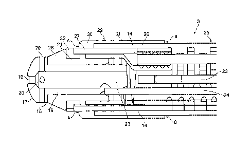
Figure 2 is a cross-sectional side view of the tip of an electrosurgical
instrument
in accordance with the embodiment of the present invention.
Description of the Embodiments
Referring to the drawings, Figure 1 shows electrosurgical apparatus including
a
generator 1 having an output socket 2 providing a radio frequency (RF) output,
via a
connection cord 4, for an electrosurgical instrument 3. Activation of the
generator 1 may
be performed from the instrument 3 via a handswitch (not shown) on the
instrument 3, or
by means of a footswitch unit 5 connected separately to the rear of the
generator 1 by a
footswitch connection cord 6. In the illustrated embodiment, the footswitch
unit 5 has
two footswitches 7 and 8 for selecting a desiccation mode and a vaporisation
mode of the
generator 1 respectively. The generator front panel has push buttons 9 and 10
for
respectively setting desiccation and vaporisation power levels, which are
indicated in a
display 11. Push buttons 12 are provided as an alternative means for selection
between
- 3 -

CA 02854378 2014-06-16
the desiccation and vaporisation modes.
The electrosurgical instrument 3 comprises a housing 13 with an elongate shaft
14, and tissue treatment electrodes at the distal end of the shaft, as will be
described
below. A movable handle 15 associated with the housing can be actuated to
cause the
shaft to bend. This instrument is particularly suited to the treatment of the
hip joint,
where a relatively long shaft with articulation capability is needed to access
the area to
the treated.
Figure 2 shows the tip of the electrosurgical instrument 3, and includes an
active
tissue treatment electrode 16 comprising a hemispherical end face 17 and a
cylindrical
side face 18. The electrode is typically formed from tungsten (or an alloy of
tungsten
and platinum), and can be formed from a single integral component or from two
components welded one to the other. The end face 17 is provided with a single
aperture
19 located at the centre thereof, while the side face 18 is provided with a
plurality of
apertures 20 spaced at equal distances around its circumference. The tissue
treatment
electrode is located on a ceramic component 21, and held in place by a split
ring retainer
22 which is laser welded in place. The electrode 16 and ceramic component 21
are both
hollow so as to form a suction lumen 23, and also to accommodate a lead 24 to
supply
RF energy to the tissue treatment electrode 16.
The majority of the shaft 14 is covered by a fluid impermeable insulating
sheath
25, the insulating sheath having and end portion 26 leaving a portion of the
shaft
uncovered in order to form a return electrode 27. The return electrode 27 is
separated
from the tissue treatment electrode 16 by a portion of the ceramic component,
forming an
insulator 28. A metallic shroud 29 is located over the end of the sheath 25,
the shroud
comprising an annular ring portion 30 and a rearwardly extending cylindrical
portion 31.
The ring portion 30 is laser welded to the exposed part of the shaft 14, at
that part
forming the return electrode 27. The cylindrical portion 31 overlies the end
portion 26 of
the sheath, and the heat from the laser welding of the ring portion 30 causes
the sheath
material to melt and form a seal against the cylindrical portion 31.
In use the instrument 3 is introduced into the body of a patient and
manoeuvred
into position, typically adjacent a hip joint. RF energy is supplied to the
tissue treatment
electrode 16, which is used to vaporise or coagulate tissue depending on the
type of RF
energy supplied from the generator 1. The instrument is typically used
submerged in an
electrically conductive fluid, such as normal saline, with the RF energy
flowing from the
- 4 -

CA 02854378 2014-06-16
=
tissue treatment electrode 16, through the conductive liquid to the return
electrode 27,
with any tissue entering the region of the tissue treatment electrode 16 being
vaporised or
coagulated depending on the circumstances.
Saline ingress into the interior of the instrument 3 is prevented at point A
by the
laser welding of the annular ring portion 30 to the shaft 14. Similarly,
saline ingress is
prevented at point B by the sealing of the end portion 26 of the sheath 25 to
the
cylindrical portion 31 of the shroud 29. The only points at which saline can
flow into the
interior of the instrument is through the apertures 19 & 20, which lead to the
sealed
suction lumen 23 which controls the saline flow. In this way, saline does not
penetrate
the other areas of the instrument, and unwanted arcing is therefore prevented.
Alternative embodiments will be envisaged by those skilled in the art without
departing from the scope of the present invention. For example, the
electrosurgical
instrument can also be used as a monopolar instrument, with just the electrode
16 being
connected to the generator 1 together with a remote patient return pad (not
shown).
Whichever type of instrument is employed, the shroud 29 helps to prevent
saline ingress
to the interior of the instrument, avoiding unwanted heating of components and
arcing
therebetween.
- 5 -

Representative Drawing
A single figure which represents the drawing illustrating the invention.
Administrative Status

2024-08-01:As part of the Next Generation Patents (NGP) transition, the Canadian Patents Database (CPD) now contains a more detailed Event History, which replicates the Event Log of our new back-office solution.

Please note that "Inactive:" events refers to events no longer in use in our new back-office solution.

For a clearer understanding of the status of the application/patent presented on this page, the site Disclaimer , as well as the definitions for Patent , Event History , Maintenance Fee  and Payment History  should be consulted.

Event History

Description Date
Letter Sent 2021-08-10
Inactive: Grant downloaded 2021-08-10
Grant by Issuance 2021-08-10
Inactive: Cover page published 2021-08-09
Pre-grant 2021-06-21
Inactive: Final fee received 2021-06-21
Notice of Allowance is Issued 2021-03-16
Letter Sent 2021-03-16
Notice of Allowance is Issued 2021-03-16
Inactive: Q2 passed 2021-03-05
Inactive: Approved for allowance (AFA) 2021-03-05
Common Representative Appointed 2020-11-07
Amendment Received - Voluntary Amendment 2020-07-31
Change of Address or Method of Correspondence Request Received 2020-07-31
Examiner's Report 2020-04-30
Inactive: Report - QC passed 2020-04-17
Common Representative Appointed 2019-10-30
Letter Sent 2019-04-02
Request for Examination Received 2019-03-28
Request for Examination Requirements Determined Compliant 2019-03-28
All Requirements for Examination Determined Compliant 2019-03-28
Inactive: Cover page published 2014-12-30
Application Published (Open to Public Inspection) 2014-12-24
Inactive: IPC assigned 2014-09-15
Inactive: First IPC assigned 2014-09-15
Inactive: IPC assigned 2014-09-15
Inactive: Filing certificate - No RFE (bilingual) 2014-07-02
Filing Requirements Determined Compliant 2014-07-02
Application Received - Regular National 2014-06-18
Inactive: QC images - Scanning 2014-06-16
Inactive: Pre-classification 2014-06-16

Abandonment History

There is no abandonment history.

Maintenance Fee

The last payment was received on 2021-06-07

Note : If the full payment has not been received on or before the date indicated, a further fee may be required which may be one of the following

  • the reinstatement fee;
  • the late payment fee; or
  • additional fee to reverse deemed expiry.

Please refer to the CIPO Patent Fees web page to see all current fee amounts.

Fee History

Fee Type Anniversary Year Due Date Paid Date
Application fee - standard 2014-06-16
MF (application, 2nd anniv.) - standard 02 2016-06-16 2016-05-19
MF (application, 3rd anniv.) - standard 03 2017-06-16 2017-05-23
MF (application, 4th anniv.) - standard 04 2018-06-18 2018-05-18
Request for examination - standard 2019-03-28
MF (application, 5th anniv.) - standard 05 2019-06-17 2019-05-22
MF (application, 6th anniv.) - standard 06 2020-06-16 2020-06-08
MF (application, 7th anniv.) - standard 07 2021-06-16 2021-06-07
Final fee - standard 2021-07-16 2021-06-21
MF (patent, 8th anniv.) - standard 2022-06-16 2022-06-07
MF (patent, 9th anniv.) - standard 2023-06-16 2023-06-05
MF (patent, 10th anniv.) - standard 2024-06-17 2023-12-13
Owners on Record

Note: Records showing the ownership history in alphabetical order.

Current Owners on Record
GYRUS MEDICAL LIMITED
Past Owners on Record
RICHARD JOHN HOODLESS
RICHARD JOHN KEOGH
Past Owners that do not appear in the "Owners on Record" listing will appear in other documentation within the application.
Documents

To view selected files, please enter reCAPTCHA code :


To view images, click a link in the Document Description column. To download the documents, select one or more checkboxes in the first column and then click the "Download Selected in PDF format (Zip Archive)" or the "Download Selected as Single PDF" button.

List of published and non-published patent-specific documents on the CPD .

If you have any difficulty accessing content, you can call the Client Service Centre at 1-866-997-1936 or send them an e-mail at CIPO Client Service Centre.


Document
Description 
Date
(yyyy-mm-dd) 
Number of pages   Size of Image (KB) 
Representative drawing 2021-07-14 1 8
Abstract 2014-06-16 1 16
Description 2014-06-16 5 220
Drawings 2014-06-16 2 27
Claims 2014-06-16 2 63
Representative drawing 2014-11-28 1 8
Cover Page 2014-12-30 1 38
Claims 2020-07-31 2 71
Description 2020-07-31 5 231
Cover Page 2021-07-14 1 38
Filing Certificate 2014-07-02 1 178
Reminder of maintenance fee due 2016-02-17 1 110
Reminder - Request for Examination 2019-02-19 1 115
Acknowledgement of Request for Examination 2019-04-02 1 174
Commissioner's Notice - Application Found Allowable 2021-03-16 1 557
Electronic Grant Certificate 2021-08-10 1 2,527
Request for examination 2019-03-28 3 99
Examiner requisition 2020-04-30 4 182
Amendment / response to report 2020-07-31 12 527
Change to the Method of Correspondence 2020-07-31 3 71
Final fee 2021-06-21 5 162