Note: Descriptions are shown in the official language in which they were submitted.
DEMANDE OU BREVET VOLUMINEUX
LA PRESENTE PARTIE DE CETTE DEMANDE OU CE BREVET COMPREND
PLUS D'UN TOME.
CECI EST LE TOME 1 DE 2
CONTENANT LES PAGES 1 A 57
NOTE : Pour les tomes additionels, veuillez contacter le Bureau canadien des
brevets
JUMBO APPLICATIONS/PATENTS
THIS SECTION OF THE APPLICATION/PATENT CONTAINS MORE THAN ONE
VOLUME
THIS IS VOLUME 1 OF 2
CONTAINING PAGES 1 TO 57
NOTE: For additional volumes, please contact the Canadian Patent Office
NOM DU FICHIER / FILE NAME:
NOTE POUR LE TOME / VOLUME NOTE:
CA 02854432 2014-05-02
DESCRIPTION
Title of Invention: Fluorescence immunoassay using polypeptide complex
containing fluoro-labeled antibody variable region
Technical Field
[0001]
The present invention relates to a kit for measuring and/or detecting the
concentration of an antigen, and a method for measuring and/or detecting the
concentration of an antigen, by which a low molecular weight compound can be
detected with high sensitivity without the need of immobilization and washing
steps.
Background Art
[0002]
Immunoassays using antibody-antigen binding are broadly employed for
the detection of substances in specimens or the measurement of the
concentrations
thereof. The measurement method most broadly employed for clinical diagnosis,
basic research, environmental research, and the like among these methods for
measuring the concentrations of antigens and antibodies is a type of
immunoassay
referred to as a sandwich ELISA method (or sandwich RIA method). This method
uses 2 types of monoclonal antibody that recognizes different epitopes of the
same
antigen, or a monoclonal antibody and a polyclonal antibody. The sandwich
method is as specifically described below. The
1st stage involves immobilizing
a monoclonal or polyclonal antibody referred to as a primary antibody on a
measurement plate, injecting a sample containing an antigen into the plate,
and
then allowing the plate to stand for a period of time for reaction so as to
bind the
antibody and the antigen. Next, the 2" stage involves removing contaminants
bound to the antibody and any antigen that has nonspecifically bound to the
plate
1
CA 02854432 2014-05-02
by washing with a washing solution. The 3rd stage involves injecting a
solution
of a labeled secondary antibody to which a reporter molecule such as an
enzyme, a
fluorescent dye, or a radioisotope has been bound in advance for a reaction to
occur for a period of time, so that the labeled secondary antibody will bind
to the
antigen captured by the primary antibody. After this reaction, excess labeled
antibody is removed by washing with a washing solution, the amount of the
reporter molecule bound to the measurement plate is measured with the use of
enzyme activity, fluorescence, a radioisotope, or the like, and thus the
amount of
the antigen in the sample is measured.
[0003]
As described above, a general sandwich ELISA method requires two
types of antibodies; the epitopes for which are different. When, for example,
a
low molecular weight compound or the like is used as an antigen, it is
difficult to
prepare a plurality of antibodies that recognize different epitopes.
Accordingly,
Ueda et al., have established a highly accurate immunoassay for low molecular
weight compounds, which is referred to as an open sandwich method. This
method uses a light chain variable region (VL) and a heavy chain variable
region
(VH) of a single antibody (Patent Documents 1 and 2, Non-patent Documents 1
and 2). This method is used to measure the concentration of an antigen and
comprises preparing a VH-region polypeptide and a VL-region polypeptide of an
antibody that specifically recognizes an antigen, labeling one of the
polypeptides
with a reporter molecule to prepare a labeled polypeptide, immobilizing the
other
polypeptide on a solid phase to prepare an immobilized polypeptide, bringing a
specimen containing the antigen and the labeled polypeptide into contact with
the
immobilized polypeptide, and then measuring the amount of the reporter
molecule
of the labeled polypeptide bound to the immobilized polypeptide. Another
example of a method for measuring a low molecular weight compound is a liquid
chromatography method, in addition to immunoassays. However, such method is
problematic in that it requires a highly accurate measuring instrument, a
large
2
CA 02854432 2014-05-02
amount of a test sample, and much time for measurement, and it has low general
versatility.
[0004]
Moreover, as immunoassays for measuring the concentration of an
antigen using an antibody labeled with a fluorescent dye, an immunoassay that
involves labeling an antibody and an antigen with different fluorescent dyes,
and
then using changes in fluorescence resonance energy transfer (FRET) efficiency
taking place between the fluorescent dyes as indicators (Non-patent Documents
3
and 4), an immunoassay that involves using changes in efficiency due to
quenching (and specifically, such method makes use of the phenomenon whereby
the fluorescence of an antibody, which has been quenched by mixing a
fluoro-labeled antibody in advance with a quenching substance, is increased
through the introduction of a substance to be detected), and an immunoassay
that
involves measuring a decrease in fluorescence intensity resulting from the
aggregation of an antibody (fluoro-labeled antibody) labeled with a
fluorescent
dye and a substance to be measured (Patent Document 3) are known.
[0005]
However, most immunoassays require a step of immobilizing an antibody
or an antigen and a washing step for eliminating the adsorption of a non-
specific
labeling compound.
Since these steps require complicated procedures, are
time-consuming, and produce variable measurement results, the development of a
liquid phase immunoassay that requires neither an immobilization step nor a
washing step is required. Accordingly, the present inventors have developed
"homogenous fluorescence immunoassay" (also referred to as a "homogenous
fluorescent immunoassay method," "Quenchbody assay," or "Q-body assay") that
is a liquid phase system requiring neither an immobilization step nor a
washing
step, enables the rapid and convenient quantitative measurement of a target
substance, and allows visualization of an antigen (Patent Document 4, Non-
patent
Document 5, Figs. 1-3).
3
CA 02854432 2014-05-02
[0006]
The above "homogenous fluorescence immunoassay" is a measurement
method using technology that utilizes the quenching phenomenon, relating to:
(1) a kit for measuring and/or detecting the concentration of an antigen,
which
enables the measurement of the concentration of an antigen or the
visualization of
an antigen, using a positive correlation between the concentration of an
antigen
and the fluorescence intensity of a fluorescent dye in a liquid phase, as an
indicator, wherein
the kit is provided with an antibody light chain variable region (referred to
as
"VL") polypeptide and an antibody heavy chain variable region (referred to as
"VH") polypeptide, and either the antibody light chain variable region
polypeptide
or the antibody heavy chain variable region polypeptide is labeled with a
fluorescent dye; and
(2) the kit for measuring and/or detecting the concentration of an antigen
according to (1), wherein the VL polypeptide and the VH polypeptide are bound
to
form a single-chain antibody. Specifically, this is a fluorescent immunoassay
that comprises: mixing two antibody fragments in which either a VL polypeptide
or a VH polypeptide is fluoro-labeled, or a single-chain antibody (scFv) in
which a
fluoro-labeled VL polypeptide and a VH polypeptide are bound to each other
(with
(1) and (2) being referred to as "Quenchbody" or "Q-body," (1) also being
referred
to as "VH+VL-type Q-body," and (2) also being referred to as "scFv-type
Q-body") in a test specimen solution to be tested to determine the presence of
an
antigen; and enabling the measurement of the concentration of an antigen or
the
visualization of an antigen using a positive correlation between the
fluorescence
intensity of the fluorescent dye and the concentration of an antigen as an
indicator
(Figs. 1-3). The
principle of such a measurement method is that highly
conservative tryptophan residues within an antibody molecule interact with a
fluorescent dye so as to quench the fluorescent dye, and the quenching is
canceled
in an antigen-dependent manner as a result of the addition of the antigen.
4
CA 02854432 2014-05-02
[0007]
The "homogenous fluorescence immunoassay" technique using the
Q-body is advantageous in that: 1) it is an extremely convenient measurement
technique that requires no washing step, and complete measurement is possible
with only the mixture of a small amount of a sample and the measurement of
fluorescence intensity; 2) the quenching effect can be exhibited even when the
antibody type is varied because of the use of highly conservative tryptophan
residues existing in an antibody for quenching, and the technique has
excellent
general versatility such that it is broadly applicable to detection of various
substances with the use of various antibodies; and 3) it is applicable in
principle to
a low molecular weight compound, since it requires only a single antigen site
(see
Patent Document 4, Fig. 1), for example.
Moreover, the homogenous
fluorescence immunoassay requires no washing step and is a convenient
measurement method, and thus a measurement device therefor can be designed in
a
very compact size such that downsizing to a portable palm-sized device is
possible.
Therefore, even a general user who has never been professionally trained may
be
able to perform on-site measurements. Although this fluorescence immunoassay
is very useful, as described above, the method is problematic in that the
ratio of
fluorescence intensity in the absence of an antigen to that in cases in which
the
antigen reaches a saturation point is as high as 1:6 and is as low as about
1:1.2.
Hence, it has been expected that the dynamic range of measurement results
would
be extended, so as to increase the sensitivity and further improve the
performance
of the assay.
Prior Art Document
Patent Documents
[0008]
Patent Document 1: JP Patent Publication (Kokai) No. H10-78436 A (1998)
Patent Document 2: JP Patent Number 3784111
CA 02854432 2014-05-02
Patent Document 3: JP Patent Publication (Kokai) No. H10-282098 A (1998)
Patent Document 4: W02011/061944
Non-patent Documents
[0009]
Non-patent Document 1: Hiroshi Ueda, Yakugaku Zasshi 27: 71-80 (2007)
Non-patent Document 2: Lim SL, et al., Anal Chem. 79 (16): 6193-200 (2007)
Non-patent Document 3: Iijima I. and Hohsaka T., Chembiochem. 17; 10 (6):
999-1006 (2009)
Non-patent Document 4: Kajihara D, et al., Nat Methods. 3 (11): 923 (2006)
Non-patent Document 5: Abe R, et al., J. Am. Chem. Soc. 133 (43): 17386-17394
(2011)
Summary of the Invention
Problem to Be Solved by the Invention
[0010]
An objective of the present invention is to provide an immunoassay,
which:
enables rapid and convenient detection and/or quantitative measurement of a
target substance in a liquid phase without the need for immobilization and
washing
steps, and allows visualization of an antigen; and
is a fluorescence immunoassay that exhibits an even wider dynamic range for
measurement results and high sensitivity.
Means for Solving the Problem
[0011]
A fluoro-labeled single-chain antibody (scFv) that had been quenched in
an antigen-free solution and then denatured with guanidine hydrochloride had
almost the same fluorescence intensity as that in a case in which the antigen
reached a saturation point. Based on this, the present inventors considered
that
6
CA 02854432 2014-05-02
a
increasing the quenching efficiency in the absence of an antigen would be an
effective means for increasing the dynamic range. Thus, the following
examination was performed.
[0012]
Specifically, the present inventors speculated that:
quenching might mainly result from contact of tryptophan residues highly
conserved in a VL polypeptide and a VH polypeptide with a fluorescent dye(s)
used for labeling; and
such fluorescent dye(s) might be moved out from an antigen-binding pocket as a
result of stabilization of the structure of a variable region accompanying
antigen
binding, thereby canceling the quenching state and increasing fluorescence
intensity.
It was predicted that a VL polypeptide and a VH polypeptide existing as 2
different types of protein would result in lower contact efficiency between
fluorescent dye(s) and tryptophan residues and lower the quenching level
because
of dissociation and weak interaction between the VL polypeptide and the VH
polypeptide. Also, in the case of a single-chain antibody (scFv), the
interaction
between a VL polypeptide and a VII polypeptide can be increased through
linking
of the VL polypeptide and the VH polypeptide with an artificial peptide linker
to
form a single-chain antibody. However, it was thought that this could decrease
original antibody functions such as its activity to bind to an antigen and its
stability because of the addition of such an artificial peptide linker. The
present
inventors speculated that a Fab fragment might retain its original antibody
functions, if a polypeptide comprising an antibody light chain variable region
(VL) and an antibody light chain constant domain and a polypeptide comprising
an
antibody heavy chain variable region (VH) and an antibody heavy chain constant
domain form the Fab (Fragment, antigen binding) comprising 1 molecule of a
hetero dimer protein wherein such polypeptides are bound via a disulfide bond.
The present inventors discovered that when either a VH-containing polypeptide
or
7
CA 02854432 2014-05-02
a VL-containing polypeptide of a Fab fragment was fluoro-labeled, the
fluorescent
dye was more strongly quenched in the absence of the antigen so as to allow
lowering of the background. Moreover, the present inventors discovered that
when a VU-containing polypeptide and a VL-containing polypeptide of a Fab
fragment were each labeled with fluorescent dyes of the same color, a
quenching
effect (H-dimer) due to the interaction between dyes was obtained in addition
to
quenching due to contact between the fluorescent dyes and tryptophan residues,
and thus higher detection sensitivity could be obtained. The present inventors
discovered that when a VU-containing polypeptide and a VL-containing
polypeptide of a Fab fragment were each further labeled with fluorescent dyes
of
different colors, a quenching effect resulting from the FRET effect was
obtained in
addition to quenching due to the interaction between fluorescent dyes and
tryptophan residues and the quenching effect due to contact between dyes, and
thus even higher detection sensitivity could be obtained. Furthermore, the
present inventors discovered that when a VH-containing polypeptide and a
VL-containing polypeptide of a Fab fragment were labeled with a fluorescent
dye
and a quencher for quenching the fluorescent dye, respectively, quenching due
to
the interaction between the fluorescent dye and tryptophan residues and the
quenching effect between the fluorescent dye and the quencher, and thus high
detection sensitivity, could be obtained.
[0013]
The present inventors further discovered that when the thermal stability
of a conventional fluoro-labeled single-chain antibody (scFv) and the same of
the
fluoro-labeled Fab complex of the present invention were measured, the
fluoro-labeled Fab complex was excellent in terms of heat resistance and
preservation such that it was denatured at 73 C, although the fluoro-labeled
single-chain antibody (scFv) was denatured at 61 C. The present invention was
completed based on these findings (Fig. 4).
[0014] Specifically, the present invention relates to:
8
CA 02854432 2014-05-02
[1] A kit for measuring and/or detecting the concentration of an antigen,
which is
characterized by enabling the measurement of the concentration of an antigen
or
the visualization of an antigen using a positive correlation between the
concentration of an antigen and the fluorescence intensity of a fluorescent
dye in a
liquid phase, as an indicator, wherein,
either or both a polypeptide containing an antibody light chain variable
region
and a polypeptide containing an antibody heavy chain variable region are
labeled
with a fluorescent dye, and
the kit is provided with a complex comprising the polypeptide containing the
antibody light chain variable region and the polypeptide containing the
antibody
heavy chain variable region;
[2] The kit for measuring and/or detecting the concentration of an antigen
according to [1] above, which is characterized in that the polypeptide
containing
the antibody light chain variable region and the polypeptide containing the
antibody heavy chain variable region are each labeled with the same
fluorescent
dye;
[3] The kit for measuring and/or detecting the concentration of an antigen
according to [1] above, which is characterized in that the polypeptide
containing
the antibody light chain variable region and the polypeptide containing the
antibody heavy chain variable region are each labeled with a different type of
fluorescent dye;
[4] The kit for measuring and/or detecting the concentration of an antigen
according to [1] above, which is characterized in that either the polypeptide
containing the antibody light chain variable region or the polypeptide
containing
the antibody heavy chain variable region is labeled with a fluorescent dye and
the
other is labeled with a quencher for quenching the fluorescent dye;
[5] The kit for measuring and/or detecting the concentration of an antigen
according to [1] above, which is characterized in that either the polypeptide
containing the antibody light chain variable region or the polypeptide
containing
9
CA 02854432 2014-05-02
the antibody heavy chain variable region is labeled with a fluorescent dye;
[6] The kit for measuring and/or detecting the concentration of an antigen
according to any one of [1] to [5] above, which is characterized in that a
complex
comprising the polypeptide containing the antibody light chain variable region
and
the polypeptide containing the antibody heavy chain variable region is a Fab
(Fragment, antigen binding);
[7] The kit for measuring and/or detecting the concentration of an antigen
according to any one of [1] to [6] above, which is characterized in that the
fluorescent dye is selected from a rhodamine-based fluorescent dye and an
oxazine-based fluorescent dye;
[8] The kit for measuring and/or detecting the concentration of an antigen
according to [7] above, which is characterized in that the fluorescent dye is
selected from carboxy rhodamine 110, carboxytetramethyl rhodamine, and ATTO
655 (trade name); and
[9] The kit for measuring and/or detecting the concentration of an antigen
according to any one of [4] to [8] above, which is characterized in that the
quencher is 7-nitrobenzofurazan (NBD).
[0015] The present invention further relates to:
[10] A method for measuring and/or detecting the concentration of an antigen,
which is characterized by comprising the following steps (a) to (c) in
sequence,
(a) a step of bringing a complex into contact with an antigen in a specimen
for
measurement, wherein
the complex comprises a polypeptide containing an antibody light chain
variable region and a polypeptide containing an antibody heavy chain variable
region, in which either or both polypeptides are labeled with a fluorescent
dye;
(b) a step of detecting the fluorescence of the fluorescent dye, or measuring
the
fluorescence intensity of the fluorescent dye; and
(c) a step of calculating the amount of the antigen contained in a sample or
visualizing the antigen, using a positive correlation between the
concentration of
CA 02854432 2014-05-02
the antigen and the fluorescence intensity of the fluorescent dye as an
indicator;
[11] The method for measuring and/or detecting the concentration of an antigen
according to [10] above, which is characterized in that the polypeptide
containing
the antibody light chain variable region and the polypeptide containing the
antibody heavy chain variable region are each labeled with the same
fluorescent
dye;
[12] The method for measuring and/or detecting the concentration of an antigen
according to [10] above, which is characterized in that the polypeptide
containing
the antibody light chain variable region and the polypeptide containing the
antibody heavy chain variable region are each labeled with a different type of
fluorescent dye;
[13] The method for measuring and/or detecting the concentration of an antigen
according to [10] above, which is characterized in that either the polypeptide
containing the antibody light chain variable region or the polypeptide
containing
the antibody heavy chain variable region is labeled with a fluorescent dye,
and the
other is labeled with a quencher for quenching the fluorescent dye;
[14] The method for measuring and/or detecting the concentration of an antigen
according to [10] above, which is characterized in that either the polypeptide
containing the antibody light chain variable region or the polypeptide
containing
the antibody heavy chain variable region is labeled with a fluorescent dye;
[15] The method for measuring and/or detecting the concentration of an antigen
according to any one of [10] to [14] above, which is characterized in that the
complex comprising the polypeptide that contains the antibody light chain
variable
region and the polypeptide that contains the antibody heavy chain variable
region
is a Fab (Fragment, antigen binding);
[16] The method for measuring and/or detecting the concentration of an antigen
according to any one of [10] to [15] above, which is characterized in that the
antigen is a low molecular weight compound; and
[17] The method for measuring and/or detecting the concentration of an antigen
11
CA 02854432 2014-05-02
according to any one of [10] to [15] above, which is characterized in that the
antigen is human osteocalcin, bisphenol A, serum albumin, clenbuterol,
ractopamine, cotinine, influenza A virus hemagglutinin, a morphine, a
methamphetamine, cocaine, tetrahydrocannabinol, or ketamine.
Effect of the Invention
[0016]
According to the present invention, a target substance can be detected
and/or quantitatively measured rapidly and conveniently in a liquid-phase
system.
Moreover, a highly-sensitive immunoassay capable of measuring a low molecular
weight compound and a kit for measuring an antigen by this immunoassay can be
provided. The measurement method of the present invention enables detection
and/or measurement of the binding of an antigen with a complex (hereinafter,
also
referred to as the "fluoro-labeled complex of the present invention")
comprising a
polypeptide (hereinafter, also referred to as "VL-containing polypeptide")
that
contains an antibody light chain variable region and a polypeptide
(hereinafter,
also referred to as "VH-containing polypeptide") that contains an antibody
heavy
chain variable region, wherein either or both polypeptides are labeled with a
fluorescent dye(s), with the use of the fluorescence intensity of the
fluorescent dye
as an indicator. Since the above fluorescent dye(s) is in an effectively
quenched
state when the fluoro-labeled complex of the present invention is not bound to
an
antigen, the antigen can be detected and/or measured with good sensitivity.
Brief Description of the Drawings
[0017]
Fig. 1 shows the characteristics of Q-body assay. The concentration of
an antigen can be rapidly measured with high sensitivity only by mixing an
antigen with 2 types of polypeptide (VH+VL), an antibody heavy chain variable
region polypeptide and an antibody light chain variable region polypeptide,
one of
which is fluoro-labeled, or, a fluoro-labeled single-chain antibody (scFv)
prepared
by linking an antibody heavy chain variable region polypeptide and an antibody
12
CA 02854432 2014-05-02
light chain variable region polypeptide (see W02011/061944).
Fig. 2 shows the principle of Q-body. Fluorescence is quenched in the
absence of an antigen due to the interaction between fluorescent dyes and
amino
acids that are broadly conserved in an antibody variable region, specifically
Trp
33, Trp 47, Trp 36, and Trp 106 of the antibody heavy chain variable region
and
Trp 40 of the antibody light chain variable region. However, quenching is
canceled in an antigen concentration-dependent manner and thus fluorescence
intensity increases. Such fluoro-labeled (VH+VL) and fluoro-labeled scFv are
referred to as "Quench body (Q-body)."
Fig. 3 shows an example of the implementation of a homogenous
fluorescence immunoassay whereby antigen concentration was measured using
Q-body. TAMRA-labeled anti-BGP single-chain antibody (scFv) was used as
Q-body and antigen concentration was varied. Fig. 3 shows the results of
measuring fluorescence emission spectrum using a spectrophotofluorometer
(FluoroMax-4), and fluorescence intensity using a fluorescent image analyzer
(FM-BIOIII).
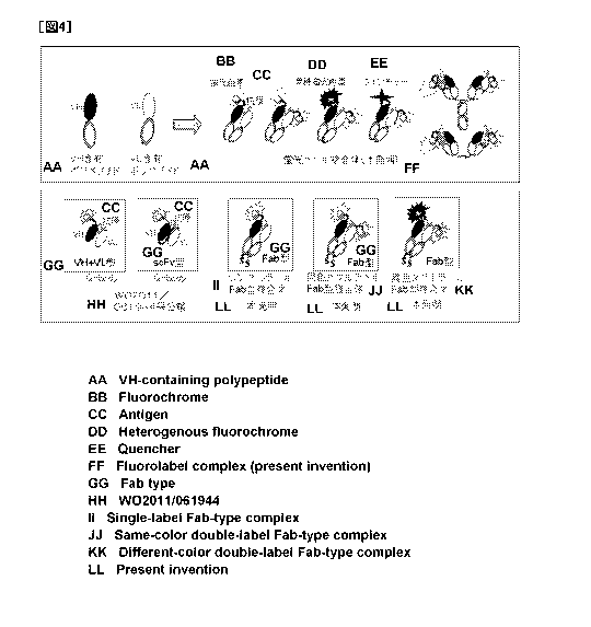
Fig. 4 schematically shows a complex comprising a polypeptide that
contains an antibody light chain variable region and a polypeptide that
contains an
antibody heavy chain variable region, either or both of which are labeled with
a
fluorescent dye. Specifically, Fig. 4 schematically shows the fluoro-labeled
complex of the present invention (upper figure). Fig. 4 also schematically
shows
Q-body used in Examples, and, the single-label Fab complex, the same-color
double-label Fab complex, and the different-color double-label Fab complex of
the
present invention (lower figure).
Fig. 5 schematically shows a method for preparing the fluoro-labeled
Fab complex of the present invention. The single-label Fab complex of the
present invention was prepared as follows. A plasmid containing ProX tag
(TAG),
VH, CHI, and a gene prepared by adding the DNA sequences of a linker and an
His
tag to the C terminus, a plasmid containing ProX tag (TTT), VL, Cx, and a gene
13
CA 02854432 2014-05-02
prepared by adding the DNA sequences of a linker and a FLAG tag to the C
terminus, and TAMRA-AF-tRNA amber (CloverDirect) were added to an E. coil
cell-free synthesis kit (Remarkable Yield Translation System Kit (RYTS)),
followed by 2 hours of reaction at 20 C, and then the TAMRA-labeled
VH-containing polypeptide and VL-containing polypeptide were synthesized via
co-expression. Thereafter, the resultant was left to stand at 4 C for 16
hours, so
as to form a complex. Protein purification was performed using FLAG and His
tags added to the C terminus. A same-color double-label Fab complex was
synthesized and purified by a method similar to that for the single-label Fab
complex using a plasmid constructed by adding a ProX tag (TAG) to the N
terminus of the VH-containing gene and the VL-containing gene. A
different-color double-label Fab complex was synthesized and purified by the
above method that involves using a plasmid constructed by adding a ProX tag
(TAG) and a ProX tag (CGGG) to the N terminus of one of the genes, and adding
an A-AF-tRNA amber dye and a B-AF-tRNA CGGG dye (CloverDirect). A
schematic diagram showing the incorporation of fluoro-labeled amino acids with
a
four-base codon is shown within the framework.
Fig. 6 shows that BGP and bisphenol A could be measured using the
single-label Fab complex of the present invention with a fluorescence
intensity
ratio higher than those of VH+VL-type Q-body and scFv-type Q-body.
Fig. 7 shows that BGP could be measured using the same-color
double-label Fab complex of the present invention with a fluorescence
intensity
ratio higher than that of the single-label Fab complex of the present
invention.
Fig. 8 shows that HSA could be measured using the same-color
double-label Fab complex of the present invention with a fluorescence
intensity
ratio higher than that of the single-label Fab complex of the present
invention.
Fig. 9 shows that BGP could be measured using the different-color
double-label Fab complex of the present invention with a fluorescence
intensity
ratio higher than that of the single-label Fab complex of the present
invention.
14
CA 02854432 2014-05-02
Fig. 10 shows that HSA could be measured using the different-color
double-label Fab complex of the present invention with a fluorescence
intensity
ratio higher than that of the single-label Fab complex of the present
invention.
Fig. 11 shows that HSA could be measured using a different-color
double-label Fab complex of an anti-SA antibody, which comprises a polypeptide
containing a CR110-labeled anti-SA antibody heavy chain variable region (VH)
and an antibody heavy chain constant domain (CHI) and a polypeptide containing
a TAMRA-labeled anti-SA antibody light chain variable region (VL) and an
antibody light chain constant domain (CIO, with a fluorescence intensity ratio
higher than that of the single-label Fab complex of the present invention.
Fig. 12 shows that BGP could be measured using the Fab complex of the
present invention comprising a polypeptide labeled with a fluorescent dye and
the
other polypeptide labeled with a quencher for quenching the fluorescent dye,
with
a high fluorescence intensity ratio. Measurement was always performed with
Ex/Em= 530/580.
Fig. 13 shows that the fluoro-labeled Fab complex of the present
invention is excellent in thermostability such that the temperature at which
the Fab
complex was thermally denatured was higher than the temperature at which the
fluoro-labeled scFv was thermally denatured, by 12 C.
Modes for Carrying Out the Invention
[0018]
The kit for measuring and/or detecting the concentration of an antigen
of the present invention is not particularly limited, as long as it is a kit
for
measuring and/or detecting the concentration of an antigen, which is
characterized
in that:
the kit is provided with a complex that comprises a polypeptide (VL-containing
polypeptide) containing an antibody light chain variable region and a
polypeptide
(VH-containing polypeptide) containing an antibody heavy chain variable
region,
CA 02854432 2014-05-02
wherein either or both the VL-containing polypeptide and the VH-containing
polypeptide are labeled with a fluorescent dye(s); and
the kit enables the measurement of the concentration of an antigen or the
visualization of an antigen using a positive correlation between the
concentration
of the antigen and the fluorescence intensity of the fluorescent dye in a
liquid
phase, as an indicator. The kit may also be provided with an antigen that can
be
used as a standard substance, reagents, tools, instruction manuals, and the
like that
are generally used for this type of immunoassay kit, in addition to contain as
a
component the above complex comprising a VL-containing polypeptide and a
VH-containing polypeptide, one of or both of which are labeled with a
fluorescent
dye(s).
[0019]
The above antigen is not particularly limited, as long as it is an antigen
that is specifically recognized by the above VH-containing polypeptide, the
above
VL-containing polypeptide, or a complex comprising these polypeptides.
Examples thereof include a protein, a peptide, a carbohydrate, a lipid, a
glycolipid,
and a low molecular weight compound, as well as proteins subjected to protein
modification such as phosphorylation or methylation. The kit for measuring
and/or detecting the concentration of an antigen of the present invention is
excellent in detection sensitivity, and thus is particularly useful for
detection of a
low molecular weight compound.
[0020]
In the kit for measuring and/or detecting the concentration of an antigen
of the present invention, either or both a VL-containing polypeptide and a
VH-containing polypeptide that compose a complex may be labeled with a
fluorescent dye(s). Specifically, the complex that may be used herein is (i) a
complex in which either the VL-containing polypeptide or the VH-containing
polypeptide is labeled with a fluorescent dye, (ii) a complex in which these
polypeptides are labeled with the same fluorescent dye, (iii) a complex in
which
16
CA 02854432 2014-05-02
these polypeptides are labeled with different types of fluorescent dye, or
(iv) a
complex in which one of these polypeptides is labeled with a fluorescent dye
and
the other polypeptide is labeled with a quencher for quenching the fluorescent
dye.
When added to either a VH-containing polypeptide or a VL-containing
polypeptide, a fluorescent dye may be added to any one of these polypeptides,
and
is preferably added to the one so that high detection sensitivity can be
obtained.
When two different types of fluorescent dye are added to a VH-containing
polypeptide and a VL-containing polypeptide, respectively, the combination of
a
polypeptide and a fluorescent dye to be added thereto is not limited, and is
preferably a combination of a fluorescent dye and a polypeptide by which high
detection sensitivity can be obtained. A VL-containing polypeptide and a
VH-containing polypeptide may be: labeled with a protein comprising an
arbitrary
amino acid sequence, a peptide tag such as a ProX tag (SEQ ID NO: 1), a FLAG
tag, a His tag, an HA tag, or an Ni tag, a linker comprising an arbitrary
amino acid
sequence, a stable radio isotope, an enzyme, or a fluorescent dye that is a
type
differing from that of the above fluorescent dye; and may further be subjected
to
modification such as sugar chain addition, phosphorylation, and methylation,
as
long as the light emission, detection, and quenching of the above fluorescent
dye
are not inhibited.
[0021]
The antibody light chain variable region (VL) is not particularly limited,
as long as it contains an amino acid sequence specific to an antibody light
chain
variable region (VL) encoded by an exon of the V and J regions of an antibody
light chain gene. The antibody light chain variable region (VL) may be
prepared
by adding an arbitrary amino acid sequence to the N-terminus and/or the
C-terminus of an amino acid sequence specific to the above antibody light
chain
variable region, or prepared by deleting, substituting, or inserting 1, 2 or
more
amino acids, as long as the affinity of the above VL-containing polypeptide or
the
fluoro-labeled complex of the present invention for an antigen is not
negatively
17
CA 02854432 2014-05-02
affected. The
affinity for an antigen can be adequately examined by a
conventional method such as ELISA or FACS. Moreover, an amino acid
sequence specific to the above antibody light chain variable region is
preferably
an amino acid sequence in which the 35th amino acid (as numbered using the
Kabat
numbering system) is tryptophan.
[0022]
The antibody heavy chain variable region (VH) is not particularly
limited, as long as it contains an amino acid sequence specific to an antibody
heavy chain variable region (VII) encoded by an exon of the V, D, and J
regions of
an antibody heavy chain gene. The antibody heavy chain variable region (VII)
may be prepared by adding an arbitrary amino acid sequence to the N-terminus
and/or the C-terminus of an amino acid sequence specific to the above antibody
heavy chain variable region, or prepared by deleting, substituting, or
inserting 1, 2
or more amino acids, as long as the affinity of the above VH-containing
polypeptide or the fluoro-labeled complex of the present invention for an
antigen
is not negatively affected. The
affinity for an antigen can be adequately
examined by a conventional method such as ELISA or FACS. Moreover, an
amino acid sequence specific to the above antibody heavy chain variable region
is
preferably an amino acid sequence in which the 36th, the 47th, or 103`d amino
acid
(as numbered using the Kabat numbering system) is tryptophan.
[0023]
Any VL-containing polypeptide may be used herein as long as it
contains an antibody light chain variable region (VL), and it can contain an
antibody light chain or a peptide comprising an arbitrary amino acid sequence
in
an antibody light chain. For example, the VL-containing polypeptide may be
prepared by adding an antibody light chain constant domain (CIO and a hinge
portion to an antibody light chain variable region (VL), and is particularly
preferably a polypeptide prepared by adding Cic to VL, for example. A specific
example of the above VL-containing polypeptide is preferably a polypeptide
18
CA 02854432 2014-05-02
comprising an amino acid sequence prepared by adding SEQ ID NO: 4 to SEQ ID
NO: 5, SEQ ID NO: 4 to SEQ ID NO: 7, SEQ ID NO: 4 to SEQ ID NO: 10, SEQ ID
NO: 4 to SEQ ID NO: 15, SEQ ID NO: 4 to SEQ ID NO: 17, SEQ ID NO: 4 to SEQ
ID NO: 19, SEQ ID NO: 4 to SEQ ID NO: 21, SEQ ID NO: 4 to SEQ ID NO: 23,
SEQ ID NO: 4 to SEQ ID NO: 25, SEQ ID NO: 4 to SEQ ID NO: 27, or SEQ ID
NO: 4 to SEQ ID NO: 29. Furthermore, a VL-containing polypeptide capable of
recognizing an antigen can be adequately prepared depending on the antigen to
be
measured.
[0024]
Any VH-containing polypeptide may be used herein, as long as it
contains an antibody heavy chain variable region (VII), and it can contain an
antibody heavy chain or a peptide comprising an arbitrary amino acid sequence
in
the antibody heavy chain. For example, the VH-containing polypeptide may be
prepared by adding an antibody heavy chain constant domain (CHI), and further
a
hinge portion and an Fc region to an antibody heavy chain variable region
(VH),
and is particularly preferably a polypeptide prepared by adding CHI to VII,
for
example. A
specific example of the above VH-containing polypeptide is
preferably a polypeptide comprising an amino acid sequence prepared by adding
SEQ ID NO: 6 to SEQ ID NO: 3, SEQ ID NO: 6 to SEQ ID NO: 9, SEQ ID NO: 6
to SEQ ID NO: 12, SEQ ID NO: 6 to SEQ ID NO: 16, SEQ ID NO: 6 to SEQ ID
NO: 18, SEQ ID NO: 6 to SEQ ID NO: 20, SEQ ID NO: 6 to SEQ ID NO: 22, SEQ
ID NO: 6 to SEQ ID NO: 24, SEQ ID NO: 6 to SEQ ID NO: 26, SEQ ID NO: 6 to
SEQ ID NO: 28, or SEQ ID NO: 6 to SEQ ID NO: 30.
Furthermore, a
VH-containing polypeptide capable of recognizing an antigen can be adequately
prepared depending on the antigen to be measured.
[0025]
The VL-containing polypeptide and the VH-containing polypeptide
preferably form a complex, and are not particularly limited, as long as
peptides
containing amino acid sequences that form the complex are bound to an antibody
19
CA 02854432 2014-05-02
light chain variable region (VL) and an antibody heavy chain variable region
(VH),
respectively. Examples of peptides that form a complex include the above
antibody constant domains (e.g., CHI and CK). Moreover, one of the peptides
forming a dimer can be added to VL and the other can be added to VH.
Furthermore, two types of proteins, which interact with each other to
contribute to
the formation of such a complex, can also be selected.
[0026]
The "complex" of the fluoro-labeled complex of the present invention
may be any complex, as long as it contains a VL-containing polypeptide and a
VH-containing polypeptide as components that form the complex. The complex
may further contain as components, a peptide, a protein, a lipid, a metal, and
other
compounds, for example, in addition to the above VL-containing polypeptide and
VH-containing polypeptide, as long as the functions of the fluoro-labeled
complex
of the present invention are not impaired.
[0027]
Furthermore, the complex of the present invention may be any structure
such that the above polypeptides are combined to be able to function
integrally.
In this case, the presence or the absence of a chemical bond between the above
polypeptides is a matter of no importance. Examples of such a bond include a
disulfide bond between the above polypeptides and a bond formed using a
cross-linking agent. These (plurality of) bonds may be used in combination in
one complex. In
particular, a preferable example thereof is a disulfide bond.
The complex of the present invention is preferably formed of the above
polypeptides located close to each other, or comprises a VL-containing
polypeptide and a VH-containing polypeptide that contain peptides having such
functions. An antibody light chain constant domain and an antibody heavy chain
constant domain in an antibody molecule interact with each other so that the
antibody light chain variable region and the antibody heavy chain variable
region
are located closer to each other, thereby serving an ancillary role to form a
strong
CA 02854432 2014-05-02
antigen-binding pocket. Accordingly, as the complex of the present invention,
a
fragment antigen-binding (Fab) fragment that is composed of two polypeptides,
which comprises one variable region and one constant domain of each of a light
chain and a heavy chain of an antibody and each of the polypeptides is bound
via a
disulfide bond, a F(ab')2 fragment wherein two Fab fragments are
disulfide-bonded via a hinge, or a full-length antibody is preferred. In
particular,
a Fab fragment is most preferred. Such fluoro-labeled complex of the present
invention that forms a Fab fragment comprising a VL-containing polypeptide and
a VH-containing polypeptide may also be referred to as "the fluoro-labeled Fab
complex of the present invention." In particular, the fluoro-labeled Fab
complex
of the present invention in which either a VL-containing polypeptide or a
VH-containing polypeptide is fluoro-labeled may also be referred to as "the
single-label Fab complex of the present invention."
Furthermore, the
fluoro-labeled Fab complex of the present invention in which both a
VL-containing polypeptide and a VH-containing polypeptide are fluoro-labeled
may also be referred to as "the same-color double-label Fab complex of the
present invention" when the two types of fluorescent dye are the same or
referred
to as "the different-color double-label Fab complex of the present invention"
when
the two types of fluorescent dye are different.
[0028]
In the present invention, a VL-containing polypeptide, a VH-containing
polypeptide, a complex containing these polypeptides, its components, and the
like
can be prepared by known chemical synthesis methods, gene recombination
techniques, methods for the denaturation of an antibody molecule using
protease,
and the like. In particular, they can be preferably prepared by gene
recombination techniques by which mass preparation is possible with relatively
simple operation. When
the above polypeptides are prepared by gene
recombination techniques, DNA containing the nucleotide sequence encoding such
a polypeptide is introduced into an appropriate expression vector to construct
a
21
CA 02854432 2014-05-02
recombinant vector, and then a target polypeptide can be expressed using an
expression system using bacterial, yeast, insect, animal/plant cells, or the
like as
host cells or a cell-free translation system (Fig. 5). When a target
polypeptide is
expressed in a cell-free translation system, for example, the target
polypeptide can
be expressed in a reaction solution prepared by adding nucleotide triphosphate
and
various amino acids to a cell-free extract of such as Escherichia coli, wheat
germ,
or rabbit reticulocytes. At this time, a tag such as a ProX tag, a FLAG tag,
or a
His tag may be added to a VL-containing polypeptide and a VH-containing
polypeptide.
These tags can be used for addition of a fluorescent dye,
purification of a polypeptide, and the like. The thus obtained VL-containing
polypeptide and VH-containing polypeptide can be caused to form a complex in
an
appropriate solvent during, before, or after labeling with a fluorescent dye.
Specifically the polypeptides are bound via a disulfide bond or using a
cross-linking agent to form a complex, for example. For example, genes
encoding the above VL-containing polypeptide and VH-containing polypeptide are
co-expressed in an Escherichia coli (E. coli) cell-free synthesis system,
followed
by 16 hours of incubation at 4 C to form a disulfide bond. Thus, a complex can
be formed. Moreover, molecular chaperon such as protein disulfide isomerase or
proline cis/trans isomerase is added to an E. coli cell-free synthesis
(reaction)
system, and thus disulfide bonding can be accelerated. Moreover, the above
cross-linking agent may be a compound that can cause the cross-linking and
binding of polypeptides.
Examples thereof include aldehydes (e.g.,
glutaraldehyde), carbodiimides, and imidoesters. A
commercially available
cross-linking agent can be adequately obtained and used according to a
conventional method. Furthermore, the complex of the present invention can
also be prepared by cleaving an antibody with an enzyme or the like. For
example, an antibody is treated with papain or pepsin, and thus a Fab fragment
or
a F(ab')2 fragment can also be prepared.
[0029]
22
CA 02854432 2014-05-02
In the present invention, a method for labeling a VL-containing
polypeptide or a VH-containing polypeptide with a fluorescent dye is not
particularly limited. A method for directly labeling using functional groups
on
both ends or a side chain of the polypeptide or indirectly labeling with a
cross-linking agent or the like, a technique for site-specifically labeling
while
synthesizing polypeptides using a cell-free translation system, or the like
can be
used herein. As a method for labeling with the use of a cell-free translation
system, an amber suppression method (Ellman J et al. (1991) Methods
Enzymo1.202: 301-36), a four-base codon method (Hohsaka T., et al., J. Am.
Chem.
Soc., 118, 9778-9779, 1996), a C-terminal labeling method (JP Patent
Publication
(Kokai) No. 2000-139468 A), an N-terminal labeling method (U.S. Patent No.
5,643,722, Olejnik et al. (2005) Methods 36: 252-260), or the like is known.
The
amber suppression method involves preparing DNA or mRNA by substituting a
codon encoding an amino acid at a labeling target site with an amber codon
that is
one of termination codons, and then synthesizing a protein from the DNA or the
mRNA using a cell-free translation system. At this time, suppressor tRNA to
which a labeled non-natural amino acid has been bound is added to a reaction
solution for protein synthesis, and thus a protein, in which such a labeled
amino
acid has been introduced into a site subjected to substitution with an amber
codon,
can be synthesized. The four-base codon method involves extending a codon
mainly to a four-base codon, CGGG, preparing DNA or mRNA by substituting a
codon encoding an amino acid with CGGG, and then synthesizing a protein from
the DNA or the mRNA using a cell-free translation system. At this time,
tRNAcGaG, to which a labeled non-natural amino acid has been bound, is added
to
the reaction solution for protein synthesis, so that a protein, in which such
a
labeled amino acid has been introduced into a site subjected to substitution
with
the four-base codon can be synthesized. For the different-color double-label
of
the present invention, co-expression is performed by a combination of the
amber
suppression method and the four-base codon method using a cell-free
translation
23
CA 02854432 2014-05-02
system, a VH-containing polypeptide and a VL-containing polypeptide are
labeled
with different fluorescent dyes, and then a complex can be formed. According
to
the C-terminal labeling method, a cell-free translation system prepared by
adding
labeled puromycin at an optimum concentration, a protein is translated from
DNA
or mRNA, and the protein, in which a label has been introduced in a C-terminus
specific manner, can be synthesized.
[0030]
Moreover, a technique that involves site-specifically introducing a
fluorescent dye by a gene-recombination technique using Escherichia coil or an
animal cell as a host can also be used herein. Azidotyrosine is introduced
site-specifically to a polypeptide using Escherichia coil as a host, into
which
aminoacyl tRNA synthase that recognizes azidotyrosine and suppressor
azidotyrosyl-tRNA have been introduced. Then a fluorescent dye can be bound
to the thus introduced azide group.
Also, azide Z lysine is introduced
site-specifically to a polypeptide using animal cells as host cells, into
which
archaebacteria-derived pyrrolidyl tRNA synthase and suppressor pyrrolidyl-tRNA
have been introduced, and thus a fluorescent dye can be bound to the thus
introduced azide group.
[0031]
In the present invention, a fluorescent dye to be used for fluorescent
labeling is not particularly limited, as long as it is a fluorescent dye that
is
quenched in the absence of an antigen under a condition where the fluoro-
labeled
complex of the present invention is formed, when a VH-containing polypeptide
and/or a VL-containing polypeptide is labeled, and it emits fluorescence when
such a complex and an antigen are bound to cancel the quenching functions.
Also,
when the same or different types of fluorescent dye are added to a VH-
containing
polypeptide and a VL-containing polypeptide, a combination is preferably
selected
so that, in addition to the above quenching, quenching between dyes and
quenching resulting from the FRET effect effectively take place in the absence
of
24
CA 02854432 2014-05-02
an antigen. Examples of a fluorescent dye to be used for fluorescent labeling
include fluorescent dyes having rhodamine, coumarin, Cy, EvoBlue, oxazine,
carbopyronin, naphthalene, biphenyl, anthracene, phenenthrene, pyrene,
carbazole,
or the like as a backbone, or derivatives of such fluorescent dyes. Specific
examples thereof include CR110: carboxyrhodamine 110: Rhodamine Green (trade
name), TAMRA: carboxytetramethylrhodamine: TMR, carboxyrhodamine 6G:
CR6G, ATTO 655 (trade name), BODIPY FL (trade name):
4,4-difluoro-5,7-dimethy1-4-bora-3a,4a-diaza-s-indancene-3-propionic
acid,
BODIPY 493/503 (trade
name):
4,4-difluoro-1,3,5,7-tetramethy1-4-bora-3a,4a-diaza-s-indancene-8-propionic
acid,
BODIPY R6G (trade name): 4,4-difluoro-5-(4-phenyl-1,3-butadienyl)
-4-bora-3a,4a-diaza-s-indancene-3-propionic acid, BODIPY 558/568 (trade name):
4,4-difluoro-5 -(2-thieny1)-4 -bora-3 a,4a-diaza-s-indancene-3 -propioni c
acid,
BODIPY 564/570 (trade
name):
4,4-difluoro-5-styry1-4-bora-3a,4a-diaza-s-indancene-3-propionic acid, BODIPY
576/589 (trade
name):
4,4-difluoro -5 -(2-pyrroly1)-4-bora-3 a,4a-diaza- s-indancene-3 -propionic
acid,
BODIPY 581/591 (trade name): 4,4-
difluoro-5-(4-pheny1-1,
3-butadieny1)-4-bora-3a,4a-diaza-s-indancene-3-propionic acid, Cy3 (trade
name),
Cy3B (trade name), Cy3.5 (trade name), Cy5 (trade name), Cy5.5 (trade name),
EvoBluel0 (trade name), EvoBlue30 (trade name), MR121, ATTO 390 (trade
name), ATTO 425 (trade name), ATTO 465 (trade name), ATTO 488 (trade name),
ATTO 495 (trade name), ATTO 520 (trade name), ATTO 532 (trade name), ATTO
Rho6G (trade name), ATTO 550 (trade name), ATTO 565 (trade name), ATTO
Rho3B (trade name), ATTO Rholl (trade name), ATTO Rhol2 (trade name), ATTO
Thio12 (trade name), ATTO 610 (trade name), ATTO 611X (trade name), ATTO
620 (trade name), ATTO Rhol4 (trade name), ATTO 633 (trade name), ATTO 647
(trade name), ATTO 647N (trade name), ATTO 655 (trade name), ATTO Oxal2
(trade name), ATTO 700 (trade name), ATTO 725 (trade name), ATTO 740 (trade
CA 02854432 2014-05-02
name), Alexa Fluor 350 (trade name), Alexa Fluor 405 (trade name), Alexa Fluor
430 (trade name), Alexa Fluor 488 (trade name), Alexa Fluor 532 (trade name),
Alexa Fluor 546 (trade name), Alexa Fluor 555 (trade name), Alexa Fluor 568
(trade name), Alexa Fluor 594 (trade name), Alexa Fluor 633 (trade name),
Alexa
Fluor 647 (trade name), Alexa Fluor 680 (trade name), Alexa Fluor 700 (trade
name), Alexa Fluor 750 (trade name), Alexa Fluor 790 (trade name), Rhodamine
Red-X (trade name), Texas Red-X (trade name), 5 (6)-TAMRA-X (trade name),
5TAMRA (trade name), and SFX (trade name). In particular, particularly
preferable examples thereof include rhodamine-based fluorescent dyes, such as
CR110 and TAMRA, and an oxazine-based fluorescent dye such as ATTO 655.
[0032]
A quencher to be used in the present invention is not particularly limited,
as long as it can quench the fluorescence of a fluorescent dye that is used
for
labeling one of components (a VL-containing polypeptide and a VH-containing
polypeptide) of the fluoro-labeled complex of the present invention in the
absence
of an antigen, when added to the other polypeptide, and its quenching function
is
canceled and fluorescence is emitted when the complex binds to an antigen.
Examples of such a quencher include quenching dyes containing NBD:
7-nitrobenzofurazan, DABCYL, BHQ, ATTO, QXL, QSY, Cy, Lowa Black,
IRDYE, and the like as backbones and derivatives thereof.
Specific examples
thereof include NBD, DABCYL, BHQ-1 (trade name), BHQ-2 (trade name),
BHQ-3 (trade name), ATTO 540Q (trade name), ATTO 580Q (trade name), ATTO
612Q (trade name), QXL490 (trade name), QXL520 (trade name), QXL570 (trade
name), QXL610 (trade name), QXL670 (trade name), QXL680 (trade name),
QSY-35 (trade name), QSY-7 (trade name), QSY-9 (trade name), QSY-21 (trade
name), Cy5Q (trade name), Cy7Q (trade name), Lowa Black FQ (trade name),
Lowa Black RQ (trade name), and IRDYE QC-1 (trade name). Of these examples,
NBD is preferred. Also, any combination of a fluorescent dye and a quencher in
the complex of the present invention can be adequately selected, as long as
the
26
CA 02854432 2014-05-02
quencher effectively quenches the fluorescent dye in the absence of an
antigen, but
the emission of the fluorescent dye is not inhibited in the presence of an
antigen.
An example thereof is a combination of a fluorescent dye TAMRA and NBD.
[0033]
In the case of the fluoro-labeled complex of the present invention; that is,
a complex comprising a VL-containing polypeptide and a VH-containing
polypeptide, either or both the VL-containing polypeptide and the VH-
containing
polypeptide are labeled with a fluorescent dye(s), the quenching of the
fluorescent
dye takes place in the absence of an antigen because of interaction between
the
above fluorescent dye and tryptophan residues conserved in the antibody
variable
region. In addition to this, in the case of the fluoro-labeled complex of the
present invention, wherein the polypeptides are labeled with fluorescent dyes
of
the same color, the quenching effect between fluorescent dyes can be obtained.
Moreover, in the case of the fluoro-labeled complex of the present invention,
wherein the above polypeptides are labeled with fluorescent dyes of different
colors, the quenching effect due to the fluorescence resonance energy transfer
(FRET) effect can be obtained in addition to the above quenching due to
tryptophan residues and quenching between the fluorescent dyes. Furthermore,
in the case of the fluoro-labeled complex of the present invention, wherein
the
above polypeptides are labeled with a fluorescent dye and a quencher for
quenching the fluorescent dye, respectively, the dynamic range can be
increased
by the quenching effect between the fluorescent dye and the quencher.
[0034]
The method for measuring and/or detecting the concentration of an
antigen of the present invention may be a method for measuring and/or
detecting
the concentration of an antigen which is characterized by comprising the
following
steps (a) to (c) in sequence:
(a) bringing a complex into contact with an antigen in a specimen for
measurement,
wherein the complex comprises a polypeptide containing an antibody light chain
27
1
CA 02854432 2014-05-02
variable region and a polypeptide containing an antibody heavy chain variable
region, in which either or both the polypeptide containing the antibody light
chain
variable region and the polypeptide containing the antibody heavy chain
variable
region are labeled with a fluorescent dye;
(b) detecting the fluorescence of the fluorescent dye, or measuring the
fluorescence intensity of the fluorescent dye;
(c) calculating the amount of the antigen contained in a sample or visualizing
the
antigen using a positive correlation between the concentration of the antigen
and
the fluorescence intensity of the fluorescent dye as an indicator.
Here, the above complex may be the fluoro-labeled complex of the present
invention. In particular, examples thereof include the fluoro-labeled Fab
complex of the present invention, more preferably the single-label Fab complex
of
the present invention, the same-color double-label Fab complex of the present
invention, and the different-color double-label Fab complex of the present
invention. In addition, the method for measuring and/or detecting the
concentration of an antigen of the present invention can be performed using
the
fluoro-labeled complex of the present invention and the kit for measuring
and/or
detecting the concentration of an antigen of the present invention.
[0035]
When the kit for measuring and/or detecting the concentration of an
antigen of the present invention is used and the method for measuring and/or
detecting the concentration of an antigen of the present invention is
performed, the
fluoro-labeled complex of the present invention is preferably brought into
contact
with an antigen in a liquid phase. Accordingly, a sample; that is a specimen
to be
measured is preferably prepared adequately as a liquid specimen or a specimen
containing liquid, or a specimen to be measured, which is immersed in liquid
and
then subjected to the above step (a) or the method for measuring and/or
detecting
the concentration of an antigen of the present invention. The use of the kit
for
measuring and/or detecting the concentration of an antigen of the present
28
CA 02854432 2014-05-02
invention, the origin of a sample to be subjected to the method for measuring
and/or detecting the concentration of an antigen of the present invention, and
the
like are not particularly limited.
Pretreatment and the like are adequately
performed and thus the above specimen for measurement can be prepared. A
liquid sample can be directly subjected to measurement as a specimen for
measurement, or can be diluted with buffer, physiological saline, or the like,
concentrated, or adequately adjusted to have a pH, a salt concentration, or
the like
to prepare a specimen for measurement, as long as an antigen is not
deteriorated or
the measurement and/or the detection of antigen concentration is not
inhibited.
Examples of such a liquid sample include body fluids that can contain a target
antigen to be measured, such as serum, blood plasma, saliva, spinal fluids,
and
urine, culture supernatants, cell extracts, microbial extracts, and industrial
wastewater.
[0036]
A non-liquid sample such as a solid sample can be directly used as a
specimen for measurement. Alternatively, such a solid sample may be adequately
treated (e.g., divided, shredded, crushed, ground, prepared into tissue
sections, or
subjected to removal or extraction of only a specific component of the
sample), as
long as the antigen is not deteriorated or the measurement and/or detection of
the
concentration of the antigen is not inhibited, and then dissolved, suspended,
or
immersed in a liquid such as a buffer or physiological saline, so that the
fluoro-labeled complex of the present invention can come into contact with the
antigen, and then the resultant can be used as a specimen for measurement. In
the above tissue section preparation, immobilization treatment can be
performed
using paraformaldehyde, glutaraldehyde or the like without deteriorating the
antigen. Moreover, blocking treatment can also be performed using BSA (bovine
serum albumin), skim milk or the like. Examples of such a solid sample include
a nitrocellulose membrane or a PVDF membrane to which ingredients such as
tissue, cells, proteins, and sugars (collected in vivo) have been blotted,
foods and
29
CA 02854432 2014-05-02
soil.
Moreover, a specimen for measurement may adequately contain an
antiseptic, a fungicide, a pH adjuster, a surfactant, an anticoagulant agent,
a
chelating agent, or the like, as long as it does not deteriorate the antigen
and not
inhibit the measurement and/or the detection of the concentration of the
antigen.
[0037]
In the present invention, furthermore, body fluids such as blood and
spinal fluid, tissue, and the like in vivo can also be used as specimens for
measurement. Specifically, the fluoro-labeled complex of the present invention
is administered to a non-human animal such as an experimental animal, so that
the
fluoro-labeled complex of the present invention can be brought into contact
with
an antigen in vivo. Such a non-human animal may be any animal other than
humans. Examples thereof include vertebrates and particularly, non-human
animals such as mammals, fishes, birds, reptiles, and amphibians. In
particular,
mammals are preferred, and mice, rats, hamsters, monkeys, pigs, and the like
are
more preferred. Also, the above administration method is not particularly
limited,
and an administration method can be adequately selected from parenteral local
administration methods including intramuscular injection, intraperitoneal
injection,
intravenous injection, subcutaneous injection, embedding, and coating, and
oral
administration methods. Moreover, another drug and the like may also be
administered before, simultaneously with, or after the administration of the
fluoro-labeled complex of the present invention. Through administration of the
fluoro-labeled complex of the present invention to a non-human animal, the
position or the transfer of an antigen in vivo, the amount of or changes in
the
amount of an antigen in vivo can also be observed. For such observation,
samples such as body fluids and tissues are collected over time, specimens for
measurement are prepared, and thus fluorescence intensity can be measured,
localization of fluorescence can be observed, or in vivo fluorescence
intensity and
changes in in vivo fluorescence intensity, and localization and the transfer
of
fluorescence can be detected and observed in real time.
CA 02854432 2014-05-02
[0038]
Reaction conditions for bringing the fluoro-labeled complex of the
present invention into contact with an antigen in a specimen for measurement
are
not particularly limited, as long as they can be generally employed for an
antigen-antibody reaction after the addition of the fluoro-labeled complex of
the
present invention to a specimen for measurement, followed by incubation
thereof.
The temperature conditions range from 1 C to 30 C, and preferably range from
18 C to 25 C, for example. The reaction time ranges from a second to 180
minutes, and preferably ranges from 1 to 90 minutes, for example. Furthermore,
when a reaction is performed in vivo in a non-human animal, incubation is
performed after administration for 5 to 180 minutes and preferably for 60 to
120
minutes, for example. If necessary, treatment such as, excision of tissue,
blood,
cells or the like, or exposition of an observation target site can be
adequately
performed. In a specimen after incubation, the quenching of the fluoro-labeled
complex of the present invention that has recognized the antigen is canceled,
fluorescence is emitted by irradiation with excitation light.
However, the
fluoro-labeled complex of the present invention that has not yet recognized
the
antigen remains being quenched, and no fluorescence is emitted even via
irradiation with excitation light. Accordingly, a specimen for measurement, to
which the above fluoro-labeled complex has been added can be directly
subjected
to the measurement and/or the detection of the concentration of an antigen
without
being subjected to a step such as a washing step. This
is a significant
characteristic of the kit for measuring and/or detecting the concentration of
an
antigen of the present invention and the method for measuring and/or detecting
the
concentration of an antigen of the present invention.
[0039]
A method for detecting fluorescence in a specimen for measurement,
which is employed in the present invention, is not particularly limited, as
long as
fluorescence emitted from a fluorescent dye can be detected, and a specimen
for
31
CA 02854432 2014-05-02
measurement after the above reaction is irradiated with excitation light and
then
the fluorescence intensity of the fluorescent dye can be measured and/or
detected.
Excitation light to be used for irradiation and the wavelength of fluorescence
to be
measured and/or detected can be adequately selected depending on the type of a
fluorescent dye to be used herein. For example, when CR110 is used as a
fluorescent dye, a combination of an excitation light wavelength of 480 nm and
a
fluorescence wavelength of 530 nm can be employed. When TAMRA is used, a
combination of an excitation light wavelength of 530 nm and a fluorescence
wavelength of 580 nm can be employed. When ATTO 655 is used, a combination
of an excitation light wavelength of 630 nm and a fluorescence wavelength of
680
nm can be employed. Moreover, when different two types of fluorescent dye are
used, a combination of an excitation light wavelength and a fluorescence
wavelength, which enables the measurement of the concentration of an antigen
and/or the detection of an antigen can be adequately selected and used. For
example, a specimen for measurement caused to react with the fluoro-labeled
complex of the present invention is irradiated with light with an excitation
wavelength suitable for one of fluorescent dyes contained in the above
fluoro-labeled complex, so as to obtain a fluorescence emission spectrum. This
procedure is performed for both two types of fluorescent dye, so that a
combination of an excitation light wavelength and a fluorescence wavelength
optimum for the measurement and/or detection of the concentration of an
antigen
can be specified. An example of a combination of an excitation light
wavelength
and a fluorescence wavelength is a combination of an excitation light
wavelength
and a fluorescence wavelength, which is suitable for any one of fluorescent
dyes.
A more preferable example thereof is a combination of an excitation light
wavelength and a fluorescence wavelength, which is suitable for a fluorescent
dye
with an excitation light wavelength and a fluorescence wavelength shorter than
the
other. In addition fluorescence may be detected as a fluorescence emission
spectrum or fluorescence intensity at a specific wavelength. For example, when
32
CA 02854432 2014-05-02
a combination of CR110 and TAMRA is used, fluorescence with a wavelength of
530 nm can be detected with an excitation light wavelength of 480 nm, and
fluorescence can also be detected as a fluorescence emission spectrum with a
wavelength ranging from 515 nm to 650 nm. With the use of a combination of an
excitation light wavelength and a fluorescence wavelength, which is suitable
for
one of different two types of fluorescent dye, a reduction in the background
resulting from the FRET (Fluorescence resonance energy transfer) effect in the
absence of an antigen can be efficiently detected and measured with higher
sensitivity.
[0040]
A light source and a measuring device to be used for fluorescence
detection in the present invention can be adequately selected. A light source
may
be any light source by which radiation with an excitation light wavelength is
possible, and examples thereof include a mercury lamp, a xenon lamp, LED, and
a
laser beam. Excitation light with a specific wavelength can be obtained using
an
appropriate filter. A device to be generally used for fluorescence observation
can
be used as a fluorescence measuring device. A microscope and the like provided
with an excitation light source, an irradiation system thereof, and a
fluorescence
image acquisition system can be adequately used, for example. Examples thereof
include MF20/FluoroPoint-Light (Olympus Corporation) and FMBIO-III (Hitachi
Software Engineering Co., Ltd.). Fluorescence intensity and the concentration
of
an antigen are in a positive correlation.
Hence, fluorescence intensity is
measured when a substance to be tested containing an antigen with a known
concentration is used, a standard curve showing the relationship between the
concentration of the antigen and the fluorescence intensity is created, and
then the
concentration of an antigen (with an unknown concentration) can be calculated
from the standard curve. Regarding such calculation of the concentration of an
antigen, the amount of an antigen can be automatically calculated using the
conversion equation or the like determined based on the standard curve created
in
33
CA 02854432 2014-05-02
advance. In addition, the detection of fluorescence may be the detection of a
fluorescence emission spectrum or the detection of fluorescence intensity at a
specific wavelength.
[0041]
Furthermore, when the fluoro-labeled complex of the present invention is
administered to a non-human animal, a region to be detected of the non-human
animal is irradiated with excitation light, and thus the fluorescence of a
fluorescent dye can be measured and/or detected two-dimensionally or
three-dimensionally, in addition to the collection of a body fluid, tissue, or
the like
thereof. In this case, a fluorescence microscope, a fluorescent image
analyzer, an
endoscope provided with a light source, and the like, can be used, for
example.
Moreover, when detection is performed, images showing the body, tissue, or
cell
structures of a non-human animal are also preferably obtained using an
endoscope,
X-ray, CT, MRI, ultrasonic wave, microscope, or the like. Measured and/or
detected fluorescence intensities and the amounts of an antigen are in a
positive
correlation. Hence, based on the thus detected two-dimensional or 3-
dimensional
fluorescence images, the localization (position) and/or the amount of the
antigen
can be found, and the results can also be compared with the images showing the
above structures, at this time. When fluorescence is detected, a specimen for
measurement or the like containing no fluoro-labeled complex of the present
invention or no analyte is preferably prepared as a negative control, and also
preferably subjected to measurement and/or detection. Moreover, the amount of
an antigen can also be measured using a fluorescence intensity ratio found by
dividing a fluorescence level measured for a specimen for measurement by a
fluorescence level measured for the negative control, for example.
Alternatively,
fluorescence intensities and the amounts of an antigen are in a positive
correlation
in the present invention, and thus when fluorescence intensity exceeding an
adequately determined threshold is obtained, the presence of the antigen in a
specimen for measurement can also be determined.
34
CA 02854432 2014-05-02
[0042]
As described above, according to the present invention, all antigens that
can be measured by immunoassays such as ELISA, immunodiffusion, latex
agglutination, immunochromatography, a surface plasmon resonance method, and
the like can be detected. For example, competitive ELISA is generally employed
for performing an immunoassay of a low-molecular-weight substance. The
method for detection and measurement of a low-molecular-weight substance
according to the present invention is better than competitive ELISA in terms
of
convenient techniques, detection/measurement sensitivity, SN ratio, and the
like,
and is capable of exhibiting its best capacity.
Examples of such
low-molecular-weight substances that can be measured and/or detected by the
method of the invention include: stimulant drugs and narcotics such as
amphetamine, methamphetamine, morphine, heroin, and codeine; mycotoxins such
as aflatoxin, sterigmatocystin, neosolaniol, nivalenol, fumonisin, ochratoxin,
and
endophyte-producing toxin; sex hormones such as testosterone and estradiol;
additives that are illegally used for feedstuffs, such as clenbuterol and
ractopamine; hazardous substances such as PCB, gossypol, histamine,
benzpyrene,
melamine, acrylamide, and dioxin; residual agricultural chemicals, such as
acetamiprid, imidacloprid, chlorfenapyr, malathion, carbaryl, clothianidin,
triflumizol, chlorothalonil, spinosad, methomyl (lannate), methamidophos, and
clorpyrifos; and environmental hormones such as bisphenol A.
[0043]
Furthermore, according to the present invention, measurement results can
be instantly obtained and the detection method thereof is so simple that
detection
instruments can be downsized resulting in its lower price. These advantages
can
be exhibited not only for detection and/or measurement of low-molecular-weight
substances, but also for on-site analyses by which measurement is performed on
site. Furthermore, measurement is so easy according to the present invention,
and thus not only a specialist but also a non-specialist can perform
measurement.
CA 02854432 2014-05-02
The method of the present invention can exhibit its ability thoroughly in the
fields
of: clinical diagnosis, specifically for detection and/or measurement of
causative
viruses and bacteria of influenza, communicable diseases, infectious diseases,
and
the like, blood drug levels, and POCT; simple medical examinations at
workplaces,
schools, day-care centers and home; security and safety, such as antiterrorism
measures, specifically for detection and/or measurement of anthrax bacillus,
botulinus toxin, sarin, and VX gas; environment, specifically for detection
and/or
measurement of environmental pollutants and house dust that should be detected
and/or measured on site; and research and development requiring immunoassay,
for example.
[0044]
Hereafter, the present invention is described in greater detail with
reference to the examples, although the technical scope of the present
invention is
not limited to the examples.
Example 1
[0045]
1. Establishment of homogenous fluorescence immunoassay using the
fluoro-labeled complex of the present invention
(Construction of expression vector)
1) Single-label Fab complex
A gene prepared by adding the DNA sequence of a ProX (trade name) tag
(the nucleotide sequence corresponding to the 9th amino acid is TTT, and
MSKQIEVNFSNET; SEQ ID NO: 1 after translation) to the N-terminus and the
DNA sequences of a linker (SEQ ID NO: 14) and a FLAG tag to the C-terminus of
a DNA sequence encoding a polypeptide containing an anti-human osteocalcin
(human Bone Gla Protein; BGP) antibody light chain variable region (VL; SEQ ID
NO: 5) and an antibody light chain constant domain (Cic; SEQ ID NO: 4) was
incorporated into a pIVEX2.3d vector (Roche Diagnostics). A gene prepared by
adding the DNA sequence of a ProX tag (the nucleotide sequence corresponding
to
36
CA 02854432 2014-05-02
the 9th amino acid is TAG and MSKQIEVNXSNET (X denotes fluoro-labeled
amino acid) after translation; SEQ ID NO: 2) containing an amber codon to the
N
terminus and the DNA sequences of a linker (SEQ ID NO: 14) and a His tag to
the
C-terminus of a DNA sequence encoding a polypeptide containing an anti-BGP
antibody heavy chain variable region (VH; SEQ ID NO: 3) and an antibody heavy
chain constant domain (CHI; SEQ ID NO: 6) was incorporated into a pIVEX2.3d
vector (Roche Diagnostics) (Fig. 5). The thus constructed expression vectors
were designed so that the ProX tag (after translation, VH was labeled and VL
was
not labeled) was added to the N-terminus, and the His tag or the FLAG tag was
added to the C-terminus of the inserted VL or VH.
[0046]
A gene prepared by adding the DNA sequences of a ProX tag (SEQ ID
NO: 1) and a GGGS5 spacer (GGGSGGGSGGGSGGGSGGGS; SEQ ID NO: 8) to
the N-terminus and the DNA sequences of a linker (SEQ ID NO: 14) and a FLAG
tag to the C-terminus of a DNA sequence encoding a polypeptide containing an
anti-bisphenol A antibody light chain variable region (VL; SEQ ID NO: 7) and
an
antibody light chain constant domain (Cx; SEQ ID NO: 4) was incorporated into
a
pIVEX2.3d vector (Roche Diagnostics). A gene prepared by adding the DNA
sequences of a ProX tag containing an amber codon (SEQ ID NO: 2) and a GGGS5
spacer (SEQ ID NO: 8) to the N-terminus and the DNA sequences of a linker (SEQ
ID NO: 14) and a His tag to the C-terminus of a DNA sequence encoding a
polypeptide containing an anti-bisphenol A antibody heavy chain variable
region
(VH; SEQ ID NO: 9) and an antibody heavy chain constant domain (CHI; SEQ ID
NO: 6) was incorporated into a pIVEX2.3d vector (Roche Diagnostics) (Fig. 5).
The thus constructed expression vectors were designed so that the ProX tag
(after
translation, VH was labeled and VL was not labeled) was added to the N-
terminus
and the His tag or the FLAG tag was added to the C-terminus of the inserted VL
or
VH.
[0047]
37
CA 02854432 2014-05-02
A gene prepared by adding the DNA sequences of a ProX tag (SEQ ID
NO: 1) and a GGGS2 spacer (GGGSGGGS; SEQ ID NO: 11) to the N-terminus and
the DNA sequences of a linker (SEQ ID NO: 14) and a FLAG tag to the C-terminus
of a DNA sequence encoding a polypeptide containing an anti-serum albumin (SA)
antibody light chain variable region (VL; SEQ ID NO: 10) and an antibody light
chain constant domain (Ck; SEQ ID NO: 4) was incorporated into a pIVEX2.3d
vector (Roche Diagnostics). Furthermore, a gene prepared by adding the DNA
sequences of a ProX tag containing an amber codon (SEQ ID NO: 2) and a GGGS5
spacer (SEQ ID NO: 8) to the N-terminus and the DNA sequences of a linker (SEQ
ID NO: 14) and a His tag to the C-terminus of a DNA sequence encoding a
polypeptide containing an anti-serum albumin (SA) antibody heavy chain
variable
region (VH; SEQ ID NO: 12) and an antibody heavy chain constant domain (CHI;
SEQ ID NO: 6) was incorporated into a pIVEX2.3d vector (Roche Diagnostics)
(Fig. 5). The thus constructed expression vectors were designed so that the
ProX
tag (after translation, VH was labeled, but VL was not labeled) was added to
the
N-terminus and the His tag or the FLAG tag was added to the C-terminus of the
inserted VL or VH.
[0048]
2) Same-color double-label Fab complex
A gene prepared by adding the DNA sequence of a ProX tag containing an
amber codon (after translation, SEQ ID NO: 2) to the N-terminus and the DNA
sequences of a linker (SEQ ID NO: 14) and a FLAG tag to the C-terminus of a
DNA sequence encoding a polypeptide containing an anti-BGP antibody light
chain variable region (VL; SEQ ID NO: 5) and an antibody light chain constant
domain (Cic; SEQ ID NO: 4) was incorporated into a pIVEX2.3d vector. A gene
prepared by adding the DNA sequence of a ProX tag (after translation, SEQ ID
NO: 2) containing an amber codon to the N-terminus and the DNA sequences of a
linker (SEQ ID NO: 14) and a His tag to the C-terminus of a DNA sequence
encoding a polypeptide containing an anti-BGP antibody heavy chain variable
38
CA 02854432 2014-05-02
region (VH; SEQ ID NO: 3) and an antibody heavy chain constant domain (CHI;
SEQ ID NO: 6) was incorporated into a pIVEX2.3d vector. The thus constructed
expression vectors were designed so that the ProX tag (amber) was added to the
N-terminus and the His tag or the FLAG tag was added to the C-terminus of the
inserted VL or VH.
[0049]
A gene prepared by adding the DNA sequences of a ProX tag containing
an amber codon (after translation, SEQ ID NO: 2) and a GGGS2 spacer (SEQ ID
NO: 11) to the N-terminus and the DNA sequences of a linker (SEQ ID NO: 14)
and a FLAG tag to the C-terminus of a DNA sequence encoding a polypeptide
containing an anti-serum albumin (SA) antibody light chain variable region
(VL;
SEQ ID NO: 10) and an antibody light chain constant domain (Cic; SEQ ID NO: 4)
was incorporated into a pIVEX2.3d vector (Roche Diagnostics). Moreover, a
gene prepared by adding the DNA sequences of a ProX tag containing an amber
codon (after translation, SEQ ID NO: 2) and a GGGS2 spacer (SEQ ID NO: 11) to
the N-terminus and the DNA sequences of a linker (SEQ ID NO: 14) and a His tag
to the C-terminus of a DNA sequence encoding a polypeptide containing an
anti-SA antibody heavy chain variable region (VH; SEQ ID NO: 12) and an
antibody heavy chain constant domain (CHI; SEQ ID NO: 6) was incorporated into
a pIVEX2.3d vector. The thus constructed expression vector was designed so
that the ProX tag (amber) was added to the N-terminus and the His tag or the
FLAG tag was added to the C-terminus of the inserted VL or VH.
[0050]
3) Different-color double-label Fab complex
A gene prepared by adding the DNA sequence of a ProX tag containing a
CGGG four-base codon (the nucleotide sequence corresponding to the 9th amino
acid is CGGG; and SEQ ID NO: 2 after translation) to the N-terminus and the
DNA sequences of a linker (SEQ ID NO: 14) and a FLAG tag to the C-terminus of
a DNA sequence encoding a polypeptide containing an anti-BGP antibody light
39
CA 02854432 2014-05-02
chain variable region (VL; SEQ ID NO: 5) and an antibody light chain constant
domain (Cic; SEQ ID NO: 4) was incorporated into a pIVEX2.3d vector.
Moreover, a gene prepared by adding the DNA sequence of a ProX tag containing
an amber codon (after translation, SEQ ID NO: 2) to the N-terminus and the DNA
sequences of a linker (SEQ ID NO: 14) and a His tag to the C-terminus of a DNA
sequence encoding a polypeptide containing an anti-BGP antibody heavy chain
variable region (VH; SEQ ID NO: 3) and an antibody heavy chain constant domain
(CHI; SEQ ID NO: 6) was incorporated into a pIVEX2.3d vector (Fig. 5). The
thus constructed expression vectors were designed so that the ProX tag was
added
to the N-terminus and the His tag or the FLAG tag was added to the C-terminus
of
the inserted VL or VH.
[0051]
A gene prepared by adding the DNA sequences of a ProX tag containing a
CGGG four-base codon (after translation, SEQ ID NO: 2) and a GGGS2 spacer
(SEQ ID NO: 11) to the N-terminus and the DNA sequences of a linker (SEQ ID
NO: 14) and a FLAG tag to the C-terminus of a DNA sequence encoding a
polypeptide containing an anti-serum albumin (SA) antibody light chain
variable
region (VL; SEQ ID NO: 10) and the antibody light chain constant domain (Cic;
SEQ ID NO: 4) was incorporated into a pIVEX2.3d vector (Roche Diagnostics).
Furthermore, a gene prepared by adding the DNA sequences of a ProX tag
containing an amber codon (after translation, SEQ ID NO: 2) and a GGGS2 spacer
(SEQ ID NO: 11) to the N-terminus and the DNA sequences of a linker (SEQ ID
NO: 14) and a His tag to the C-terminus of a DNA sequence encoding a
polypeptide containing an anti-SA antibody heavy chain variable region (VH;
SEQ
ID NO: 12) and an antibody heavy chain constant domain (Cfli; SEQ ID NO: 6)
was incorporated into a pIVEX2.3d vector. The thus constructed expression
vectors were designed so that the ProX tag was added to the N-terminus and the
His tag or the FLAG tag was added to the C-terminus of the inserted VL or VH.
[0052]
CA 02854432 2014-05-02
=
(Synthesis of fluoro-labeled Fab complex)
Fluoro-labeled amino acids were introduced into an
antibody-variable-region-containing peptide and/or the N-terminal region of an
antibody-variable-region-containing peptide based on a cell-free translation
system using an RYTS (Trade name) E. coli cell-free synthesis kit (Remarkable
Yield Translation System Kit (ProteinExpress))
[0053]
1) Single-label or same-color double-label Fab complex
A reaction solution (60 L) was prepared by the addition of 3 p.L of
Enzyme Mix, 0.6 tiL of methionine, 30 L of 2xReaction Mix, 20 L, of an E.
coli
lysate, 2 p,L of two types of plasmid DNA (200 ng each), 3 pL of a fluoro-
labeled
amino acyl-tRNA amber (480 pmol), and 1.4 I, of Nuclease Free Water.
CloverDirect (trade name) tRNA Reagents for Site-Directed Protein
Functionalization (ProteinExpress) was used as fluoro-labeled aminoacyl-tRNA
(TAMRA-X-AF-tRNA amber, CR110-X-AF-tRNA amber, and ATTO
655-X-AF-tRNA amber) for preparation of fluoro-labeled proteins. The reaction
solution was left to stand at 20 C for 2 hours for reaction, and thus protein
synthesis was performed. Thereafter, complex formation was completed by
further
16 hours of reaction at 4 C. After the completion of the reaction, 0.5 p1 of
the
reaction solution was used to perform SDS-PAGE (15%), and then protein
expression was observed with a fluorescent image analyzer (FMBIO-III; Hitachi
Software Engineering Co., Ltd.). Furthermore, Western blotting was performed
using an anti-His tag antibody or an anti-FLAG tag antibody, thereby
confirming
the synthesis of the target peptide containing the fluoro-labeled antibody
variable
region.
[0054]
2) Different-color double-label Fab complex
A reaction solution (60 pl) was prepared by the addition of 3 pL of
Enzyme Mix, 0.6 p1 of methionine, 30 pl of 2 x Reaction Mix, 20 pL, of E. coli
41
CA 02854432 2014-05-02
lysate, 2 uL of two types of plasmid DNA (200 ng each), 1.5 uL of two types of
fluoro-labeled aminoacyl-tRNA amber and CGGG (480 nmol each), 1.4 uL of
Nuclease Free Water.
CloverDirect (trade name) tRNA Reagents for
Site-Directed Protein Functionalization (ProteinExpress) were used as
fluoro-labeled aminoacyl-tRNA (TAMRA-X-AF-tRNA amber or CGGG,
CR110-X-AF-tRNA amber or CGGG, and ATTO 655-X-AF-tRNA amber or
CGGG) for preparation of fluoro-labeled proteins. A VU-region-containing
polypeptide and a VL-region-containing polypeptide were labeled with a
tRNAamber and tRNAcGoG, respectively. The reaction solution was left to stand
at
20 C for 2 hours to perform a reaction. After protein synthesis, complex
formation was competed by further 16 hours of reaction at 4 C. After the
completion of the reaction, 0.5 uL of the reaction solution was used to
perform
SDS-PAGE (15%), and then protein expression was observed with a fluorescent
image analyzer (FMBIO-III; Hitachi Software Engineering Co., Ltd.).
Furthermore, Western blotting was performed using an anti-His tag antibody or
an
anti-FLAG tag antibody, thereby confirming the synthesis of the target peptide
containing the fluoro-labeled antibody variable region.
[0055]
3) Fab complex comprising a polypeptide labeled with a fluorescent dye and the
other polypeptide labeled with a quencher for quenching the fluorescent dye
TAMRA was used as a fluorescent dye and NBD-X, SE (Anspec) was
used as a quencher. NBD-X-AF-tRNA CGGG was synthesized using NBD-X, SE
(Anspec) instead of TAMRA-X, SE of Abe et al.,'s method (Abe R, et al., J.
Biosci.
Bioeng. 110 (1): 32-38 (2010)). A reaction solution (60 p,L) was prepared by
the
addition of 3 uL of Enzyme Mix, 0.6 jtL of metionine, 30 1AL of 2xReaction
Mix,
20 L of E. coli lysate, 2 uL of two types of plasmid DNA (200 ng each), 1.5
p,L of
TAMRA-X-AF-tRNA amber and NBD-X-AF-tRNA CGGG (480 nmol each), and
1.4 111_, of Nuclease Free Water. A VH region-containing polypeptide and a VL
region-containing polypeptide were labeled with tRNAamber and tRNAcoGo,
42
CA 02854432 2014-05-02
respectively. The reaction solution was left to stand at 20 C for 2 hours for
reaction. After protein synthesis, complex formation was completed by further
16 hours of reaction at 4 C. After the completion of the reaction, 0.5 pl of
the
reaction solution was used to perform SDS-PAGE (15%), and then protein
expression was observed with a fluorescent image analyzer (FMBIO-III; Hitachi
Software Engineering Co., Ltd.). Moreover, Western blotting was performed
using an anti-His tag antibody or an anti-FLAG tag antibody, thereby
confirming
the synthesis of the target peptide containing the fluoro-labeled antibody
variable
region.
[0056]
(Purification of fluoro-labeled Fab complex)
The thus synthesized fluoro-labeled Fab complexes were each purified
using anti-FLAG M2 affinity gel (Sigma-Aldrich Corporation) or His-Spin Trap
Column (GE Healthcare). The reaction solution (60 L) was applied to a column
containing anti-FLAG M2 affinity gel. After 15 minutes of incubation at room
temperature, the resultant was washed 3 times with a wash buffer (20 mM
phosphate buffer (pH 7.4)/0.5M NaCl/0.1% polyoxyethylene (23) lauryl ether).
Next, the resultant was eluted 3 times with 200 iiL of elute buffer (20 mM
phosphate buffer (pH7.4)/0.5 M NaCl/100 lig FLAG peptide/0.1%
polyoxyethylene (23) lauryl ether). Next, the eluate was applied to a His-Spin
Trap Column. After 15 minutes of incubation at room temperature, the resultant
was washed 3 times with a wash buffer (20 mM phosphate buffer (pH 7.4)/0.5 M
NaCl/60 mM imidazole/0.1% polyoxyethylene (23) lauryl ether). Next, the
resultant was eluted 3 times with 200 pi of an Elute buffer (20 mM phosphate
buffer (pH7.4)/0.5 M NaCl/0.5 M imidazole/0.1% polyoxyethylene (23) lauryl
ether).
Furthermore, the eluate was subjected to buffer exchange and
concentration using Amicon Ultra-0.5 centrifugal filter 10 kDa (Millipore) and
PBS (+0.05% Tween20). The concentration of each sample after purification was
measured using a fluorescent image analyzer (FMBIO-III; Hitachi Software
43
CA 02854432 2014-05-02
Engineering Co., Ltd.).
[0057]
(Preparation of Q-body)
Single-chain antibodies (scFv) were prepared by linking the VH and VL
(VH was labeled, but VL was not labeled) of a TAMRA-labeled anti-BGP antibody,
and the VH and VL of TAMRA-labeled anti-BGP and the VH and VL of
TAMRA-labeled anti-bisphenol A antibodies using a
linker
(GGGGSGGGGSGGGGS) according to the method described in International
Publication W02011/061944.
Example 2
[0058]
2. Measurement by homogenous fluorescence immunoassay using the
fluoro-labeled complex of the present invention
(Fluorescence emission spectrum measurement using single-label Fab complex)
The TAMRA single-label anti-BGP antibody Fab complex, the
TAMRA-labeled anti-BGP antibody scFv, or the TAMRA-labeled anti-BGP
antibody VH + anti-BGP antibody VL prepared in Example 1, comprising a
polypeptide containing an anti-BGP (human osteocalcin) antibody light chain
variable region (VL; SEQ ID NO: 5) and an antibody light chain constant domain
(Cic; SEQ ID NO: 4), and a polypeptide containing a TAMRA fluorescence-labeled
anti-BGP antibody heavy chain variable region (VH; SEQ ID NO: 3) and an
antibody heavy chain constant domain (CHI; SEQ ID NO: 6) was used to measure
the concentration of BGP. The TAMRA single-label anti-BGP antibody Fab
complex, or the TAMRA-labeled anti-BGP scFv (70 nM, 6.25 L), or the
TAMRA-labeled anti-BGP antibody VII + anti-BGP antibody VL (70 nM/mL, 6.25
1.1L) and antigenic BGP-C7 (SEQ ID NO: 13) (0, 1, 3, 10, 25, 100, or 1,000 nM)
were prepared to a total of 50 pi using PBS containing 1% BSA (+0.05%
Tween20). The solution was left to stand at 25 C for 70 minutes, and then
subjected to fluorescence emission spectrum measurement using a
44
CA 02854432 2014-05-02
spectrophotofluorometer (FluoroMax-4; HORIBA Jobin Yvon). A He-Ne laser
was used at 543 nm, with the excitation wavelength set at 530 nm, and the
fluorescence intensity at 580 nm was measured. The ratio of the fluorescence
intensity at each antigen concentration to the fluorescence intensity when no
antigen was present is shown in the upper left graph of Fig. 6. The ratio of
the
fluorescence intensity when the concentration of BGP-C7 was 1,000 nM is shown
in the lower left table of Fig. 6.
Similarly, the TAMRA single-label
anti-bisphenol A Fab complex, or the TAMRA-labeled anti-bisphenol A antibody
scFv, comprising a polypeptide containing a TAMRA fluorescence-labeled
anti-bisphenol A antibody VL (SEQ ID NO: 7) and Cx (SEQ ID NO: 4) and a
polypeptide containing an anti-bisphenol A antibody VH (SEQ ID NO: 9) and CHI
(SEQ ID NO: 6) was reacted with bisphenol A (0, 1, 3, 10, 30, 100, or 1,000
nM),
and then fluorescence intensity was measured. The ratio of the fluorescence
intensity at each antigen concentration to the fluorescence intensity when no
antigen was present is shown in the upper right graph of Fig. 6. The ratio of
the
fluorescence intensity when the concentration of bisphenol A was 1,000 nM is
shown in the lower right table of Fig. 6. It was confirmed that the single-
label
Fab complex of the present invention makes it possible to obtain a
fluorescence
intensity ratio higher than that obtained using conventional scFv-type Q-body
or
VH + VL-type Q-body, regardless of the type of fluorescent dye or antigen
concentration, and to detect and quantify a low-molecular-weight compound and
a
protein with high sensitivity. The Fab complex of the present invention forms
a
complex wherein its components; that is, polypeptides, are located close to
each
other, compared with those in the scFv-type Q-body or the VH + VL-type Q-body,
so that the quenching effect of tryptophan in an antibody variable region on a
fluorescent dye takes place effectively.
Example 3
[0059]
(Fluorescence emission spectrum measurement using same-color double-label Fab
CA 02854432 2014-05-02
complex)
The same-color double-label Fab complex (70 nM, 6.25 I) prepared in
Example 1, comprising a polypeptide containing a light chain variable region
(VL;
SEQ ID NO: 5) of an anti-BGP antibody labeled with CR110, TAMRA, or ATTO
655 fluorescent dye and an antibody light chain constant domain (Cic; SEQ ID
NO:
4) and a polypeptide containing a heavy chain variable region (VH; SEQ ID NO:
3) of an anti-BGP antibody labeled with the same dye and an antibody heavy
chain
constant domain (CHI; SEQ ID NO: 6) and antigenic BGP-C7 (0 to 10,000 nM)
were prepared to a total of 50 L with PBS containing 1% BSA (+0.05% Tween20).
As a control, a single-label Fab complex sample wherein either a polypeptide
containing anti-BGP antibody VL (SEQ ID NO: 5) and CI< (SEQ ID NO: 4) or a
polypeptide containing anti-BGP antibody VH (SEQ ID NO: 3) and CHI (SEQ ID
NO: 6) had been fluoro-labeled was prepared.
These solutions were left to stand
at 25 C for 70 minutes, and then the fluorescence emission spectrum was
measured using a spectrophotofluorometer (FluoroMax-4; HORIBA Jobin Yvon).
When the same-color double-label Fab complex labeled with a CR110 fluorescent
dye was used, the excitation wavelength (Ex) was set at 480 nm, and then the
fluorescence intensity was measured at a fluorescence wavelength (Em) of 530
nm.
When the same-color double-label Fab complex labeled with a TAMRA
fluorescent dye was used, the excitation wavelength was set at 530 nm, and
then
the fluorescence intensity was measured at a fluorescence wavelength of 580
nm.
When the same-color double-label Fab complex labeled with an ATTO 655
fluorescent dye was used, the excitation wavelength was set at 630 nm, and
then
the fluorescence intensity was measured at a fluorescence wavelength of 680
nm.
The ratio of the fluorescence intensity at each antigen concentration to the
fluorescence intensity when no antigen was present was designated as
"fluorescence intensity ratio" and is shown in the graphs of Fig. 7.
Fluorescence
intensity ratios when the concentration of BGP-C7 was 1,000 nm or 10,000 nm
are
shown in the tables of Fig. 7.
46
CA 02854432 2014-05-02
[0060]
In a similar manner, the same-color double-label Fab complex comprising
a polypeptide containing an anti-serum albumin (SA) antibody light chain
variable
region (VL; SEQ ID NO: 10) and an antibody light chain constant domain (CK;
SEQ ID NO: 4) and a polypeptide containing a heavy chain variable region (VH;
SEQ ID NO: 12) of an anti-SA antibody labeled with the same dye and an
antibody
heavy chain constant domain (CHI; SEQ ID NO: 6) was reacted with antigenic
HSA (0 to 100 1.1M), and then fluorescence intensity was measured.
Fluorescence
intensity ratios are shown in the graphs of Fig. 8 and fluorescence intensity
ratios
when the concentration of HSA was 100 M are shown in the tables of Fig. 8.
Based on the above results, it was confirmed that the same-color double-label
Fab
complex of the present invention makes it possible to measure the amount of an
antigen with a fluorescence intensity ratio higher than that of the single-
label Fab
complex, regardless of the type of fluorescent dye and the type of antigen
used.
The same-color double-label Fab complex can reduce the background so as to
enhance the dynamic range because of the quenching effect (H-dimer) due to the
interaction between fluorescent dyes, in addition to the quenching effect of
tryptophan in an antibody variable region on a fluorescent dye(s).
Example 4
[0061]
(Fluorescence emission spectrum measurement using different-color double-label
Fab complex)
The different-color double-label Fab complex (70 nM, 6.25 'IL) of an
anti-BGP antibody prepared in Example 1, comprising a polypeptide containing a
light chain variable region (VL; SEQ ID NO: 5) of an anti-BGP antibody labeled
with CR110, TAMRA, or ATTO 655 fluorescent dye and an antibody light chain
constant domain (Cic; SEQ ID NO: 4) and a polypeptide containing a heavy chain
variable region (VH; SEQ ID NO: 3) of an anti-BGP antibody labeled with a dye
differing from the above dye and an antibody heavy chain constant domain (CHI;
47
CA 02854432 2014-05-02
SEQ ID NO: 6) and antigenic BGP-C7 (0 to 1,000 nM) were prepared to a total of
50 1.iL using PBS (+0.05% Tween20) containing 1% BSA. As a control, a
same-color double-label Fab complex sample was prepared.
Fluorescence
intensity was assumed in a manner similar to Example 3. The ratio of the
fluorescence intensity at each antigen concentration to the fluorescence
intensity
when no antigen was present was obtained. In addition, when CR110 and
TAMRA were used, the excitation wavelength was set at 480 nm, and then the
fluorescence intensity was measured at a fluorescence wavelength of 530 nm
(the
left graph of Fig. 9). When CR110, TAMRA, and ATTO 655 were used, the
excitation wavelength was set at 530 nm, and then the fluorescence intensity
was
measured at a fluorescence wavelength of 580 nm (the center graph of Fig. 9).
When TAMRA and ATTO 655 were used, the excitation wavelength was set at 630
nm, and then the fluorescence intensity was measured at a fluorescence
wavelength of 680 nm (the right graph of Fig. 9). Fluorescence intensity
ratios
when the concentration of BGP-C7 was 1,000 nM are shown in each table of Fig.
9.
[0062]
Similarly, the different-color double-label Fab complex of the anti-SA
antibody prepared in Example 1 comprising a polypeptide containing a heavy
chain variable region (VH; SEQ ID NO: 12) of an anti-SA antibody labeled with
a
CR110, TAMRA, or ATTO 655 fluorescent dye and an antibody heavy chain
constant domain (CHI; SEQ ID NO: 6), and a polypeptide containing a light
chain
variable region (VL; SEQ ID NO: 10) of an anti-SA antibody labeled with a dye
differing from the above dye and an antibody light chain constant domain (Cx;
SEQ ID NO: 4) was reacted with antigenic HSA (0 to 100 tM), and then
fluorescence intensity was measured. The ratio of the fluorescence intensity
at
each antigen concentration to the fluorescence intensity when no antigen was
present was designated as the "fluorescence intensity ratio" and is shown in
the
graphs of Fig. 10. Fluorescence intensity ratios when the concentration of HSA
48
CA 02854432 2014-05-02
was 100 p,M are shown in the tables of Fig. 10.
[0063]
Furthermore, the different-color double-label Fab complex (also denoted
as CR110 TAMRA) of an anti-SA antibody, comprising a polypeptide containing a
CR110-labeled anti-SA antibody heavy chain variable region (VH; SEQ ID NO:
12) and an antibody heavy chain constant domain (CHI; SEQ ID NO: 6) and a
polypeptide containing a TAMRA-labeled anti-SA antibody light chain variable
region (VL; SEQ ID NO: 10) and an antibody light chain constant domain (Cic;
SEQ ID NO: 4) was reacted with antigenic HSA (1x10⁻⁴, 1x10, 1x10⁶, 1x10⁻⁷M).
The resultant was irradiated with excitation light having a wavelength of 480
nm
(the upper left graph of Fig. 11) or a wavelength of 530 nm (the upper right
graph
of Fig. 11), and then the fluorescence emission spectrum was measured.
Moreover, the CR110 single-label Fab complex (CR110_no) of an anti-SA
antibody, comprising a polypeptide containing CR110-labeled anti-SA antibody
VH (SEQ ID NO: 12) and CHI (SEQ ID NO: 6) and a polypeptide containing
anti-SA antibody VL (SEQ ID NO: 10) and CI< (SEQ ID NO: 4) was reacted with
antigenic HSA (1x10, 1x10⁵, 1x10⁶, 1 x10⁻⁷M). The resultant was irradiated
with an excitation light having a wavelength of 480 nm, and then the
fluorescence
emission spectrum was measured (the lower left graph of Fig. 11). The TAMRA
single-label Fab complex (no_TAMRA) of an SA antibody comprising a
polypeptide containing anti-SA antibody VH (SEQ ID NO: 12) and CHI (SEQ ID
NO: 6) and a polypeptide containing TAMRA-labeled anti-SA antibody VL (SEQ
ID NO: 10) and CI( (SEQ ID NO: 4) was reacted with antigenic HSA (1x10⁻⁴,
1x10⁵, 1x10⁶, 1 x10⁻⁷M). The resultants were irradiated with excitation light
with a wavelength of 530 nm and then the fluorescence emission spectrum was
measured using a spectrophotofluorometer (FluoroMax-4) (the lower right graph
of Fig. 11). The curves in each graph are of data obtained from samples with
HSA concentrations of (from the top) 1 x10⁻⁴ M, 1 x10⁻⁵ M, 1 x10⁻⁶ M, 1 x10⁻⁷
M,
and 0 M.
49
CA 02854432 2014-05-02
[0064]
The above results revealed that in the case of the different-color
double-label Fab complex CR110_TAMRA, fluorescence at a wavelength of about
530 nm in the absence of an antigen was suppressed by the FRET effect, when
irradiated with excitation light with a wavelength of 480 nm, and thus a high
fluorescence intensity ratio was obtained at a wavelength of about 530 nm.
Moreover, when irradiated with excitation light with a wavelength of 530 nm,
fluorescence intensity ratios obtained at a fluorescence wavelength of about
530
nm in the case of CR110 TAMRA was higher than those of no_TAMRA, because
of the quenching effect due to the interaction between the fluorescent dyes.
Therefore, the background can be reduced by the addition of the quenching
effect
due to the interaction between fluorescent dyes and the FRET effect, in
addition to
the quenching effect of tryptophan in an antibody variable region on a
fluorescent
dye(s), thereby enhancing the dynamic range. In this experimental system,
CR110 TAMRA was capable of detecting an antigen, while exhibiting a maximum
of 75-fold fluorescence enhancement, thereby demonstrating the usefulness of
the
present invention.
Example 5
[0065]
(Fluorescence emission spectrum measurement using Fab complex comprising
fluoro-labeled polypeptide and quencher-labeled polypeptide)
The Fab complex (70 nM, 6.25 4) prepared in Example 1, consisting of
a polypeptide containing a TAMRA-labeled anti-BGP antibody heavy chain
variable region (VI-I; SEQ ID NO: 3) and an antibody heavy chain constant
domain
(CHI; SEQ ID NO: 6), and a polypeptide containing a NBD-labeled anti-BGP
antibody light chain variable region (VL; SEQ ID NO: 5) and an antibody light
chain constant domain (Cic; SEQ ID NO: 4), and antigenic BGP-C7 (0 to 10,000
nM) were prepared to a total of 50 4 with PBS (+0.05% Tween20) containing 1%
BSA. The solution was left to stand at 25 C for 70 minutes, and then the
CA 02854432 2014-05-02
fluorescence emission spectrum was measured using a spectrophotofluorometer
(FluoroMax-4; HORIBA Jobin Yvon). The excitation wavelength was set at 530
nm, and then the fluorescence intensity was measured at a fluorescence
wavelength of 580 nm. The ratio of the fluorescence intensity at each antigen
concentration to the fluorescence intensity when no antigen was present was
designated as "fluorescence intensity ratio" and shown in the graph of Fig.
12.
The fluorescence intensity ratio when the concentration of BGP-C7 was 1,000 nM
is shown in the table of Fig. 12.
[0066]
The usefulness of the present invention was demonstrated by the results
that the Fab complex TAMRA_NBD was capable of detecting BGP at a
concentration of 1 nM, the same as that in the case of the single-label Fab
complex
(TAMRA No) (Fig. 7), and detecting an antigen, while exhibiting a 27-fold
fluorescence enhancement, which was higher than that of the double-label
(TAMRA TAMRA) (Fig. 7).
Example 6
[0067]
(Measurement of thermostability)
A thermal shift assay was performed to measure the thermal stability of
the anti-BGP TAMRA single-label Fab complex and the
anti-BGP TAMRA-labeled scFv prepared in Example 1. The temperature was
increased by 1 C per minute using a StepOne real-time PCR system (Applied
Biosystems), and then fluorescence intensity was measured at each temperature.
Thermal stability was measured based on the principle that a quenching state
is
canceled when each antibody undergoes thermal denaturation to result in the
disintegration of the structure, so that fluorescence intensity increases. As
shown in Fig. 13, the Tm value (at which thermal denaturation takes place) for
the
TAMRA-labeled scFv was 61 C, however, the Tm value for the TAMRA
single-label Fab complex of the present invention was 73 C; an increase of 12
C.
51
CA 02854432 2014-05-02
This thermal stability enables the long-term storage of reagents and can
significantly contribute to industrial merits such as distribution
temperature,
storage temperature, and storage period.
Example 7
[0068]
(Measurement of clenbuterol using fluoro-labeled Fab complex)
The different-color double-label Fab complex synthesized by the method
shown in Example 1, comprising a polypeptide containing an anti-clenbuterol
antibody CR110-labeled light chain variable region (VL; SEQ ID NO: 15) and an
antibody light chain constant domain (C-K; SEQ ID NO: 4) and a polypeptide
containing a TAMRA-labeled anti-clenbuterol antibody heavy chain variable
region (VH; SEQ ID NO: 16) and an antibody heavy chain constant domain (CHI;
SEQ ID NO: 6) was reacted with antigenic clenbuterol (0 to 16 1.1g/mL), and
then
fluorescence intensity was measured following the method of Example 4.
Fluorescence intensity ratio when the concentration of clenbuterol was 16
[tg/mL
is shown in Table 1. As a result, 16 1.1g/mL clenbuterol could be measured.
[0069]
[Table 1]
Measurement of clenbuterol using fluoro-labeled Fab complex
Labeling fluorescent dye Measurement
Fluoro-labeledFluorescence
Antigen VH-containing VL-containing
Fab antibody Ex/Em enhancement
*1
polypeptide polypeptide
Anti-Clenbuterol Clenbuterol TAM RA CR110 480/530
1.5
*1 (Fluorescence intensity in the presence of 16 g/mL
Clenbuterol)/(Fluorescence intensity in the
absence of antigen)
Example 8
[0070]
(Measurement of ractopamine and cotinine using fluoro-labeled Fab complex)
A same-color double-label Fab complex comprising a polypeptide
containing an anti-ractopamine antibody TAMRA-labeled light chain variable
52
CA 02854432 2014-05-02
region (VL; SEQ ID NO: 17) and an antibody light chain constant domain (Cic;
SEQ ID NO: 4) and a polypeptide containing a TAMRA-labeled anti-ractopamine
antibody heavy chain variable region (VH; SEQ ID NO: 18) and an antibody heavy
chain constant domain (CHI; SEQ ID NO: 6) was synthesized by the method
described in Example 1. Furthermore, a same-color double-label Fab complex
comprising a polypeptide containing an anti-cotinine antibody TAMRA-labeled
light chain variable region (VL; SEQ ID NO: 19) and an antibody light chain
constant domain (Cic; SEQ ID NO: 4) and a polypeptide containing a
TAMRA-labeled anti-cotinine antibody heavy chain variable region (VH; SEQ ID
NO: 20) and an antibody heavy chain constant domain (CHI; SEQ ID NO: 6) was
synthesized.
Subsequently, each complex was reacted with antigenic
ractopamine (3.4 gg/mL) or cotinine (3.5 ug/mL), and then fluorescence
intensity
was measured following the method of Example 4. The results are shown in
Table 2. As shown in Table 2, measurement was successfully performed with
fluorescence intensity ratios (indicating fluorescence enhancement) of 2.3 and
1.9.
[0071]
[Table 2]
Measurement of ractopamine and cotinine using fluoro-labeled Fab complex
Labeling fluorescent dye Measurement
Fluoro-labeledFluorescence
Antigen VH-containing VL-containing
Fab antibodyEx/Em enhancement
polypeptide polypeptide
Anti-Ractopamine Ractopamine TAMRA TAM RA 530/580 2.3"
Anti-cotinine Cotinine TAMRA TAMRA 530/580 1.8*2
*1 (Fluorescence intensity in the presence of 3.4 f.ig/mL
Ractopamine)/(Fluorescence intensity in
the absence of antigen)
*2 (Fluorescence intensity in the presence of 3.5 p.g/mL
Cotinine)/(Fluorescence intensity in the
absence of antigen)
Example 9
[0072]
(Measurement of influenza A virus hemagglutinin (HA) using fluoro-labeled Fab
complex)
53
CA 02854432 2014-05-02
A different-color double-label Fab complex synthesized by the method
described in Example 1, comprising a polypeptide containing an anti-HA
(hemagglutinin of influenza A (H5N1, H1N1)) antibody CR110-labeled light chain
variable region (VL; SEQ ID NO: 21) and an antibody light chain constant
domain
(Cic; SEQ ID NO: 4) and a polypeptide containing a TAMRA-labeled anti-HA
(hemagglutinin of influenza A (H5N1, H1N1)) antibody heavy chain variable
region (VH; SEQ ID NO: 22) and an antibody heavy chain constant domain (CHI;
SEQ ID NO: 6) was reacted with each antigen (30 [tg/mL), and then fluorescence
intensity was measured following the method of Example 4. The results are
shown in Table 3. As shown in Table 3, measurement was successfully
performed with fluorescence intensity ratios (indicating fluorescence
enhancement) for H5N1 HA and H1N1 HA antigens were 6.1 and 7.1, respectively.
[0073]
[Table 3]
Measurement of influenza A virus HA using fluoro-labeled Fab complex
Labeling fluorescent dye Measurement
Fluoro-labeledFluorescence
Antigen VH-containing VL-containing
Fab antibodyEx/Em enhancement
polypeptide polypeptide
Anfi-HA H5N1 HA TAM RA CR110 480/530 6.1
H1N1 HA TAM RA CR110 480/530 7.1
*1 (Fluorescence intensity in the presence of 30 1.1g/mL
antigen)/(Fluorescence intensity in the
absence of antigen)
Example 10
[0074]
(Measurement of morphines, methamphetamines, and cocaine using fluoro-labeled
Fab complex)
A same-color double-label Fab complex comprising a polypeptide
containing an anti-morphine antibody TAMRA-labeled light chain variable region
(VL; SEQ ID NO: 23) and an antibody light chain constant domain (CK; SEQ ID
NO: 4) and a polypeptide containing a TAMRA-labeled anti-morphine antibody
54
CA 02854432 2014-05-02
heavy chain variable region (VH; SEQ ID NO: 24) and an antibody heavy chain
constant domain (CHI; SEQ ID NO: 6) was synthesized by the method shown in
Example 1. Similarly, a same-color double-label Fab complex comprising a
polypeptide containing an anti-methamphetamine antibody TAMRA-labeled light
chain variable region (VL; SEQ ID NO: 25) and an antibody light chain constant
domain (Cic; SEQ ID NO: 4) and a polypeptide containing a TAMRA-labeled
anti-methamphetamine antibody heavy chain variable region (VH; SEQ ID NO:
26) and an antibody heavy chain constant domain (CHI; SEQ ID NO: 6) was
synthesized. Moreover, a same-color double-label Fab complex comprising a
polypeptide containing an anti-cocaine antibody TAMRA-labeled light chain
variable region (VL; SEQ ID NO: 27) and an antibody light chain constant
domain
(Cic; SEQ ID NO: 4) and a polypeptide containing a TAMRA-labeled anti-cocaine
antibody heavy chain variable region (VH; SEQ ID NO: 28) and an antibody heavy
chain constant domain (CHI; SEQ ID NO: 6) was synthesized. Furthermore,
according to Example 1, a single-chain antibody (scFv) was prepared by linking
the TAMRA-labeled anti-morphine antibody VL and VH using a linker
(GGGGSGGGGSGGGGS) and a single-chain antibody (scFv) was prepared by
linking the anti-methamphetamine antibody VH and VL using a linker
(GGGGSGGGGSGGGGS). The thus constructed TAMRA double-label Fab
complex and the TAMRA-labeled scFv were reacted with various morphines,
methamphetamines, cocaine, and ketamine, and then fluorescence intensity was
measured following the method of Example 3 or Example 1. The results are
shown in Table 4. The three types of fluoro-labeled Fab complex specifically
recognized each antigen. Furthermore, when the fluoro-labeled morphine Fab
complex was compared with scFv, and when the fluoro-labeled methamphetamine
Fab complex was compared with scFv for fluorescence enhancement (fluorescence
intensity ratio), fluorescence enhancement was always higher in the Fab
complexes and the dynamic range was significantly increased.
[0075]
CA 02854432 2014-05-02
=
[Table 4]
Measurement of morphines, methamphetamines, and cocaine using fluoro-labeled
Fab complex
Fluorescence enhancement in the presence of 100 1.1.g/mL antigen.i
Anti-
Anti-Methamphetamin Anti-Morp
Anti-Morphine Anti-Cocaine
Methamphet
Fl =ro-labele ehine
amine
d Fab
a ibody VHn-contai VL-contai VHn-contai VL-contai VHn-contai VL-contai
Fluore ent scFv scFv
polypepti polypepti polypepti polypepti polypepti polypepti
= e
de de de de de de
Antigen
TAMRA TAMRA TAMRA TAMRA TAMRA TAMRA TAMRA TAMRA
Morphines
Morphine 7.8 ___ MEM ________________ 0.9 1.5
Heroin 8.3 1-5
Codeine 8.1 1-5
111111.111m.....¨
Thebaine 6.2
Ethylmorphin 8.4
Dihydrocodei 8.3
ne
Metharnpheta
mines
Methampheta 1.8
mine
MDMA
Amphetamine II 76..28 0
MDA
Cocaine
Cocaine
Ketamine
Ketamine 1.1 1.0 0.9 1.0
*1 (Fluorescence intensity in the presence of 100 vtg/mL
antigen)/(Fluorescence intensity in the absence of antigen)
Example 11
[0076]
(Measurement of cannabinoid component THC and ketamine using fluoro-labeled
Fab complex)
A same-color double-label Fab complex comprising a polypeptide
containing an anti-THC (cannabinoid component) antibody TAMRA-labeled light
chain variable region (VL; SEQ ID NO: 29) and an antibody light chain constant
domain (CK; SEQ ID NO: 4) and a polypeptide containing a TAMRA-labeled
anti-THC (cannabinoid component) antibody heavy chain variable region (VH;
SEQ ID NO: 30) and an antibody heavy chain constant domain (CHI; SEQ ID NO:
6) was synthesized by the method described in Example 1. Similarly, a
same-color double-label Fab complex comprising a polypeptide containing an
56
CA 02854432 2014-05-02
anti-ketamine antibody TAMRA-labeled light chain variable region (VL; SEQ ID
NO: 31) and an antibody light chain constant domain (Cic; SEQ ID NO: 4) and a
polypeptide containing a TAMRA-labeled anti-ketamine antibody heavy chain
variable region (VII; SEQ ID NO: 32) and an antibody heavy chain constant
domain (CHI; SEQ ID NO: 6) was synthesized. Subsequently, each complex was
reacted with antigenic THC (100 1.tg/mL) or ketamine (1.0 mg/mL), and
fluorescence intensity was measured following the method of Example 4. As
shown in Table 5, measurement was successfully performed with fluorescence
intensity ratios (indicating fluorescence enhancement) of 1.3 and 2Ø
[0077]
[Table 5]
Measurement of cannabinoid component THC and ketamine using fluoro-labeled Fab
complex
Labeling fluorescent dye Measurement
Fluoro-labeledFluorescence
Antigen VH-containing VL-containing
Fab antibodyEx/Em enhancement
polypeptide polypeptide
Anti-THC THC TAM RA TAM RA 530/580 1.3"
Anti-Ketamine Ketamine TAM RA TAM RA 530/580 2.0*2
*1 (Fluorescence intensity in the presence of 100 [ig/mL THC)/(Fluorescence
intensity in the
absence of antigen)
*2 (Fluorescence intensity in the presence of 1.0 mg/mL
Ketamine)/(Fluorescence intensity in the
absence of antigen)
Industrial Applicability
[0078]
The present invention can be usefully used in the fields of specimen
analyses, drug tests, and portable specimen analysis kits, for example.
57
DEMANDE OU BREVET VOLUMINEUX
LA PRESENTE PARTIE DE CETTE DEMANDE OU CE BREVET COMPREND
PLUS D'UN TOME.
CECI EST LE TOME 1 DE 2
CONTENANT LES PAGES 1 A 57
NOTE : Pour les tomes additionels, veuillez contacter le Bureau canadien des
brevets
JUMBO APPLICATIONS/PATENTS
THIS SECTION OF THE APPLICATION/PATENT CONTAINS MORE THAN ONE
VOLUME
THIS IS VOLUME 1 OF 2
CONTAINING PAGES 1 TO 57
NOTE: For additional volumes, please contact the Canadian Patent Office
NOM DU FICHIER / FILE NAME:
NOTE POUR LE TOME / VOLUME NOTE: