Note: Descriptions are shown in the official language in which they were submitted.
CA 02854941 2014-05-07
WO 2013/101745 PCMJS2012/071311
SPECTROSCOPY DATA DISPLAY SYSTEMS AND METHODS
Technical Field
[0001] This disclosure relates to spectrometer systems. In particular, this
disclosure relates to directly correlating spectroscopy data to physical
locations on a
sample and overlaying indicia of the spectroscopy data over an image of the
sample
at the corresponding locations.
Background Information
[0002] Mass spectroscopy is an analytical technique that measures the mass-
to-
charge ratio of charged particles for determining, for example, the elemental
composition of a specimen or sample of matter. Laser-assisted spectroscopy
(LAS)
involves directing laser energy at a sample in order to disassociate its
constituent
parts and make them available to a spectrometer. LAS systems apply the laser
energy to the sample while passing a fluid, typically an inert gas, over the
sample to
capture the disassociated species and carry them to a spectroscope for
processing.
Example LAS systems include laser ablation inductively coupled plasma mass
spectroscopy (LA ICP-MS), laser ablation inductively coupled plasma emission
spectroscopy (ICP-OES/ICP-AES) and laser induced breakdown spectroscopy
(LIBS).
[0003] In certain LAS systems, a laser beam path moves along a beam
trajectory
(e.g., the laser beam may be deflected relative to sample and/or the sample
may be
moved relative to the laser beam using motion stages) to ablate material from
a
selected portion or portions of the sample for analysis. For example, FIG. 1
is a
simplified schematic diagram of a sample 100 including a kerf 110 cut by a
laser
beam. In this example, the beam trajectory along the kerf 110 is in a
direction
indicated by arrow 112. The sample 100 may include more than one type of
material
and the composition or respective concentrations of elements may change along
the
kerf 110. However, mass spectrometers generally output data as tabulated text
or in
spreadsheet formats that do not correspond to physical locations of the sample
100.
The mass spectroscopy data may be displayed in the form of numbers and graphs.
For example, FIG. 2 illustrates example graphs of mass spectroscopy data for
various elements measured for the sample 100 shown in FIG. 1. In this example,
concentrations are graphed with respect to time for selected nuclides of
Sulfur (S32),
Calcium (Ca44), Manganese (Mn55), Zinc (Zn66), Mercury (Hg202), Lead (Pb208),
1
and Bismuth (Bi209). A problem with the graphs shown in FIG. 2 is that there
is no
correlation to physical locations on the surface of the sample 100 relative to
where the
material was extracted for generating the displayed data.
Summary of the Disclosure
[0004]
Spectroscopy data are correlated to physical locations on a sample. In one
embodiment, there is described a method for displaying laser-assisted mass
spectroscopy data of a sample specimen, the method comprising: scanning, using
a
laser processing system, a laser beam along a beam trajectory relative to the
sample,
wherein the sample is located in a sample chamber during the scanning, and
wherein
the laser beam disassociates material from the sample along the beam
trajectory to
produce an aerosol of the disassociated material within the sample chamber;
passing
a fluid through the sample chamber to transport the disassociated material to
a
spectrometer for determining mass spectroscopy data values of a selected
element
along the beam trajectory; correlating, using a processor, the mass
spectroscopy data
values with respective locations of the sample along the beam trajectory; and
displaying, on a display device, in real time as the laser beam continues to
disassociate the material from the sample along the beam trajectory, an image
of at
least a portion of the sample including the respective locations along the
beam
trajectory where the material was disassociated by the laser beam, the image
comprising indicia of the mass spectroscopy data values directly displayed on
the
sample at their correlated locations.
[0005] In another embodiment, there is described a laser-assisted mass
spectroscopy system, comprising: a sample chamber for holding a sample
specimen;
a laser source for producing a laser beam; a scanning subsystem for scanning
the
laser beam along a beam trajectory relative to the sample, wherein the laser
beam
disassociates material from the sample along the beam trajectory to produce an
aerosol of the disassociated material within the sample chamber, and wherein a
fluid
passing through the sample chamber transports the disassociated material to a
spectrometer for determining mass spectroscopy data values of a selected
element
along the beam trajectory; a processor for controlling the scanning subsystem
and for
correlating the mass spectroscopy data values with respective locations of the
sample
2
CA 2854941 2019-06-11
along the beam trajectory; and a display device for displaying, in real time
as the laser
beam continues to disassociate the material from the sample along the beam
trajectory, an image of at least a portion of the sample including the
respective
locations along the beam trajectory where the material was disassociated by
the laser
beam, the image comprising indicia of the mass spectroscopy data values
directly
displayed on the sample at their correlated locations.
[0005a] There is also described a laser-assisted mass spectroscopy system,
comprising: means for scanning a laser beam along a beam trajectory relative
to a
sample, wherein the sample is located in a sample chamber during the scanning,
and
wherein the laser beam disassociates material from the sample along the beam
trajectory to produce an aerosol of the disassociated material within the
sample
chamber; means for passing a fluid through the sample chamber to transport the
disassociated material to a spectrometer for determining mass spectroscopy
data
values of a selected element along the beam trajectory; means for correlating
the
mass spectroscopy data values with respective locations of the sample along
the
beam trajectory; and means for displaying, while simultaneously continuing to
determine mass spectroscopy data, an image of at least a portion of the sample
including the respective locations along the beam trajectory where the
material was
disassociated by the laser beam, the image comprising indicia of the mass
spectroscopy data values directly displayed on the sample at their correlated
locations.
[0005b] There is also described a method for displaying laser-assisted mass
spectroscopy data of a sample specimen, the method comprising: scanning, using
a
laser processing system, a laser beam along a beam trajectory relative to the
sample;
generating, using one or more mass spectrometers, mass spectroscopy data
values
along the beam trajectory; correlating, using a processor, the mass
spectroscopy data
values with respective locations of the sample along the beam trajectory; and
displaying, on a display device, while simultaneously continuing to generate
mass
spectroscopy data values, an image of at least a portion of the sample
including the
respective locations along the beam trajectory, the image comprising indicia
of the
mass spectroscopy data values directly displayed on the sample at their
correlated
locations.
2a
CA 2854941 2019-06-11
CA 02854941 2014-05-07
WO 2013/101745 PCT/US2012/071311
[0006] Additional aspects and advantages will be apparent from the
following
detailed description of preferred embodiments, which proceeds with reference
to the
accompanying drawings.
Brief Description of the Drawings
[0007] FIG. 1 is a simplified schematic diagram of a sample including a
kerf cut
by a laser beam.
[0008] FIG. 2 illustrates example graphs of mass spectroscopy data for
various
elements measured for the sample shown in FIG. 1.
[0009] FIG. 3 is a block diagram of a laser ablation sampling system
according to
one embodiment.
[0010] FIG. 4A is a simplified schematic diagram of a composite image that
may
be displayed, for example, on the display device shown in FIG. 3 according to
one
embodiment.
[0011] FIG. 4B is a simplified schematic diagram of an image that may be
displayed, for example, on the display device shown in FIG. 3 according to
another
embodiment.
[0012] FIG. 5 illustrates four composite images of a sample with indicia of
correlated spectroscopy data according to one embodiment.
[0013] FIG. 6 illustrates four composite images of a sample with indicia of
correlated spectroscopy data according to one embodiment.
[0014] FIG. 7 illustrates two composite images of a sample with user
annotations
and indicia of correlated spectroscopy data according to one embodiment.
[0015] FIG. 8 is a flow chart of a method for displaying spectroscopy data
of a
sample specimen according to one embodiment.
[0016] FIG. 9 is a flow chart of a method for correlating the concentration
values
with respective locations along the beam trajectory according to one
embodiment.
[0017] FIG. 10 graphically represents a graphical user interface according
to one
embodiment.
Detailed Description of Preferred Embodiments
[0018] Spectroscopy data are correlated to physical locations on a sample.
The
correlation may use, for example, location data (e.g., X, Y, and/or Z data) of
a laser
beam trajectory along a surface (or below the surface) of the sample, scan
velocity
data, and system delay data to accurately match spectrometer output to
geographic
locations on or within the sample. The spectroscopy data may include elemental
3
CA 02854941 2014-05-07
WO 2013/101745 PCT/US2012/071311
concentrations and/or detector responses associated with concentrations such
as
volts, counts, counts per second, frequency, and wavelength. The spectroscopy
data may also include ratios of responses such as elemental ratios or
isotropic
ratios. In certain embodiments, the spectroscopy data is acquired using a
laser-
assisted spectroscopy (LAS) system such as laser ablation inductively coupled
plasma mass spectroscopy (LA ICP-MS), laser ablation inductively coupled
plasma
emission spectroscopy (ICP-OES/ICP-AES), and laser induced breakdown
spectroscopy (LIBS)
[0019] lndicia of the spectroscopy data are directly displayed on an image
of the
sample at locations corresponding to the extraction of material from the
sample for
processing. The displayed indicia may include, for example, color variation,
hue
variation, brightness variation, pattern variation, symbols, text,
combinations of the
foregoing, and/or other graphical representations of spectroscopy data with
respect
to geographic locations on or within the sample. In certain embodiments, the
indicia
of spectroscopy data are overlaid on the image of the sample in real time as
material
is being ablated by the laser beam and processed by the spectrometer. In
addition,
or in other embodiments, the indicia may be overlaid on the image any time
after the
spectroscopy data has been generated. In certain such embodiments, one or more
fiducial marks may be added to the sample and/or to the image of the sample
for
later alignment of the indicia of the spectroscopy data with the physical
geography of
the sample.
[0020] In certain embodiments, a graphical user interface includes a
layered
environment that selectively represents the graphical buildup of various
layers of
information corresponding to one or more samples. For example, the user may be
allowed to select the display of a layer representing an empty sample chamber
where a laser induced aerosol may be produced, a layer representing an insert
loaded with one or more samples within the sample chamber, a layer
representing
sample maps from one or more system cameras, a layer representing images
imported from other systems or devices (e.g., petrographic microscope systems,
scanning electron microscope (SEM) systems, or other imaging systems), a layer
representing annotation, and/or a layer representing the indicia of
spectroscopy data.
Artisans will recognize from the disclosure herein that other layers may also
be used.
In certain embodiments, the entire layered environment can be saved to enable
the
user to load saved environments at a later time and recall all of the
information
4
CA 02854941 2014-05-07
WO 2013/101745 PCT/US2012/071311
associated with a particular experiment (e.g., scan positions, SEM data,
spectrometer raw data, reduced data such as age of the particular sample, and
other
data used in the experiment). As the user scans across the environment,
respective
data and data files become available for viewing, which enables traceability
of the
various aspects of the experiment and reduces or negates the requirement for
the
user to keep separate records. In certain embodiments, mobile device
applications
(e.g., for laptop computers, tablet computers, smart phones, or other mobile
devices)
allow the user to review selected environments at any time.
[0021] Reference is now made to the figures in which like reference
numerals
refer to like elements. For clarity, the first digit of a reference numeral
indicates the
figure number in which the corresponding element is first used. In the
following
description, numerous specific details are provided for a thorough
understanding of
the embodiments disclosed herein. However, those skilled in the art will
recognize
that the embodiments can be practiced without one or more of the specific
details, or
with other methods, components, or materials. Further, in some cases, well-
known
structures, materials, or operations are not shown or described in detail in
order to
avoid obscuring aspects of the invention. Furthermore, the described features,
structures, or characteristics may be combined in any suitable manner in one
or
more embodiments.
[0022] Embodiments may include various steps, which may be embodied in
machine-executable instructions to be executed by a general-purpose or special-
purpose computer (or other electronic device). Alternatively, the steps may be
performed by hardware components that include specific logic for performing
the
steps or by a combination of hardware, software, and/or firmware.
[0023] Embodiments may also be provided as a computer program product
including a non-transitory, machine-readable medium having stored thereon
instructions that may be used to program a computer (or other electronic
device) to
perform the processes described herein. The machine-readable medium may
include, but is not limited to, hard drives, floppy diskettes, optical disks,
CD-ROMs,
DVD-ROMs, ROMs, RAMs, EPROMs, EEPROMs, magnetic or optical cards, solid-
state memory devices, or other types of media/computer-readable medium
suitable
for storing electronic instructions.
[0024] FIG. 3 is a block diagram of a laser ablation sampling system 300
according to one embodiment. The system 300 includes a laser 310 to produce a
CA 02854941 2014-05-07
WO 2013/101745 PCT/US2012/071311
laser beam 312 directed to a sample 314 within a sample chamber 316. By way of
example, and not by limitation, the sample 314 may comprise bone, rock or
other
geological material, paint, varnish, pigment, metal, ceramic, glass, paper,
textiles, or
other types of materials. The laser beam 312 may include a plurality of laser
pulses
at a pulse repetition frequency, wavelength, pulse energy, and other laser
parameters selected to ablate or otherwise dissociate material from the sample
314.
Artisans will recognize from the disclosure herein that, in other embodiments,
a
continuous wave (CW) laser beam may be used. As illustrated, a stream of
carrier
gas enters the sample chamber 316 and picks up fine sample particles (e.g., in
an
aerosol) produced by the laser ablation process and transports them to a
spectrometer (not shown) for processing. The carrier gas may include, for
example,
Argon, Helium, or another inert gas.
[0025] The sample chamber 316 is mounted on motion stages 318 that allow
the
sample to be moved relative to the laser beam 312 in three directions (X, Y,
and Z).
A mirror 320 may be used to direct the laser beam 312 to the sample 314.
Although
not shown in FIG. 3, other optics may also be used along the path of the laser
beam
312 such as focusing optics (e.g., lenses) and beam steering optics (e.g.,
fast
steering mirrors, mirror galvanometer deflectors, electro-optic deflectors,
and/or
acousto-optic deflectors). The mirror 320 may be configured (e.g., a half-
silvered
mirror) to combine the optical axes of the laser beam 312 with a field of view
323 of a
camera 322. The camera 322 may provide still images and/or video of the sample
314 and/or sample chamber 316 for display on a display device 324. Although
not
shown, other imaging systems may also be used. For example, the system 300 may
include one or more additional video cameras (e.g., for both high resolution
and
wide-angle views), petrographic microscope systems, and/or scanning electron
microscope (SEM) systems. Further, more than one display device 324 may be
used to allow a user to control the system 300 and view selected images of the
sample 314 and/or sample chamber 316.
[0026] The system 300 further includes a controller 326 and a memory device
328. The controller 326 is configured to control the laser 310, the motion
stages
318, the camera 322, and the display device 324. The controller 326 may also
be
used, in certain embodiments, to control other devices such as the
spectrometer,
petrographic microscope systems, scanning electron microscope (SEM) systems,
or
other imaging systems. An artisan will understand from the disclosure herein
that
6
CA 02854941 2014-05-07
WO 2013/101745 PCT/US2012/071311
more than one controller may also be used. The memory device 328 stores
computer-executable instructions that may be read and executed by the
controller
326 to cause the system 300 function as described herein. The memory device
328
may also store generated spectroscopy data, data for correlating the
spectroscopy
data with geographic locations on or within the sample 314, images and/or
video of
the sample 314 and/or sample chamber 316, other imported images and/or video,
user generated annotations of the sample 314 and/or spectroscopy data, and
other
data associated with the processes described herein (e.g., scan positions, age
and/or origin of the sample 314, report files, sample chamber parameters,
laser
parameters, and other experiment or ablation parameters).
[0027] In certain embodiments, a user may select a particular portion or
portions
of the sample 314 to ablate for examination. For example, the sample may be
composed of more than one type of material and the user may desire to study
only
one of the materials or a selected group of materials. Thus, the user may
define a
laser beam path along a beam trajectory with respect to a surface of the
sample 314.
Thus, the beam trajectory may be defined in an X-Y plane. In addition, or in
other
embodiments, the laser beam trajectory may be in the Z direction (e.g., a
direction
parallel to the laser beam as it drills into the sample). The user may define
one more
single spots, a line of distinct spots, a grid of distinct spots, a line of
continuous
ablation (e.g., overlapping laser spots creating a continuous ken f such as
the kerf
110 shown in FIG. 1), and/or a raster pattern covering a two-dimensional (2D)
area
of the sample 314. In certain embodiments, multiple passes of the laser beam
312
along the same spot, line, or raster pattern may be used to cut deeper into
the
sample 314 so as to generate three-dimensional (3D) spectroscopy data.
[0028] FIG. 4A is a simplified schematic diagram of a composite image 400
that
may be displayed, for example, on the display device 324 shown in FIG. 3
according
to one embodiment. The composite image 400 includes an image 410 of the sample
314 shown in FIG. 3 (or a portion of the sample 314) overlaid with indicia 412
of
spectroscopy data. The indicia 412 of spectroscopy data are correlated to
actual
locations within a 2D area of a surface of the sample 314 where laser ablation
was
used to generate the spectroscopy data. The 20 area may correspond, for
example,
to a plurality of adjacent or partially overlapping, vertical passes of the
laser beam
312 (as opposed to the single, vertical pass the width of the laser beam spot
size
7
CA 02854941 2014-05-07
WO 2013/101745 PCT/US2012/071311
used to produce the ken f 110 shown in FIG. 1) to produce a widened kerf in
the
horizontal direction.
[0029] In the
simplified example shown in FIG. 4A, different fill patterns (e.g.,
diamond-shaped hatch, slanting lines, vertical lines, square-shaped hatch, or
no-fill)
are used to distinguish variations in spectroscopy data (e.g., concentrations
in parts
per million (ppm), counts, counts per second, volts, frequency, wavelength,
elemental ratios, and/or isotropic ratios) of a selected element within the 2D
area.
For example, a first range of concentrations (or counts, etc.) is indicated in
areas
414, a second range of concentrations is indicated in areas 416, a third range
of
concentrations is indicated in area 418, a fourth range of concentrations is
indicated
in area 420, a fifth range of concentrations is indicated in area 422, and a
sixth range
of concentrations is indicated in area 424. Although not shown in FIG. 4A, a
legend
or other indication of the particular concentration ranges or other
spectroscopy data
associated with each fill pattern may also be displayed.
[0030] In
certain embodiments, one or more fiducial marks are added to the
sample and/or the image of the sample so as to correctly align the indicia of
the
spectroscopy data either in real time as the spectroscopy data is being
generated or
at a later time. For example, the laser beam used for disassociating the
material
from the sample (e.g., the sample 314 shown in FIG. 3) may also be used to add
fiducial marks to the sample for later reference. Thus,
the indicia of the
spectroscopy data may be overlaid on later acquired images of the sample. As
another example, FIG. 4A shows fiducial marks 426 (two shown) added to the
image
410 of the sample. The fiducial marks 426 may be used to align the indicia 412
over
the image 410 during the laser ablation process as the spectroscopy data is
generated and/or at a later time, as selected by the user.
[0031] FIG. 4B
is a simplified schematic diagram of an image 430 that may be
displayed, for example, on the display device 324 shown in FIG. 3 according to
another embodiment. The image 430 includes an image 410 of the sample 314
shown in FIG. 3 (or a portion of the sample 314) and graphs 432 showing
spectroscopy data (e.g., counts) versus depth for various elements (labeled
element
A, element B, element C, element D, and element E). The sample image 410 and
the graphs 432 may be displayed together, for example, in a split screen or
picture-
in-picture format. The displayed sample image 410 includes the fiducial marks
426
discussed above. The graphs 432 show changes in spectroscopy data at a
selected
8
CA 02854941 2014-05-07
WO 2013/101745 PCT/US2012/071311
X, Y location for different depths in the Z direction. In the example shown in
FIG. 4B,
the X, Y location is in a plane corresponding to the displayed sample image
410 and
the Z direction is perpendicular to the plane (e.g., extending into the
sample). The Z
direction may also be considered as being parallel to the laser beam at the
sample.
[0032] In
certain embodiments, a user may position a cursor 434 over the
displayed sample image 430 to select the X, Y position at which spectroscopy
data is
displayed for various depths in the Z direction. In such embodiments, the
displayed
graphs 432 change as the user moves ("mouses over") the cursor 434 over the
displayed sample image 410. The spectroscopy data at different depths may be
acquired, for example, by making multiple passes of the laser beam along the
same
kerf or by using multiple pulses to drill down into the sample at a selected
location.
Information regarding the amount (depth) of material removed by each laser
pass or
each laser pulse is used to correlate the spectroscopy data to a Z location
within the
sample.
[0033] In other
embodiments, continuous changes (e.g., rather than discrete
ranges) in spectroscopy data may be indicated using, for example, a continuous
spectrum of colors, shades, or hues. For example, FIG. 5 illustrates four
composite
images of a sample 510 with indicia of correlated spectroscopy data 512, 514,
516,
518 according to one embodiment. For illustrative purposes, the spectroscopy
data
512, 514, 516, 518 is shown in FIG. 5 as various shades of gray within a 2D
area
surrounded by a dashed line. In certain embodiments, however, a continuous
spectrum of colors is used to represent variations in spectroscopy data, and
the
dashed line (or a solid) line may not be used because the colors sufficiently
distinguish the sample image from the spectroscopy data.
[0034] In a
first image shown in FIG. 5, the overlying spectroscopy data 512
represents the concentration of Lanthanum (La) within a 2D area of the sample
510,
and a displayed legend 520 indicates that the concentration of Lanthanum
within the
2D area ranges between 0 ppm and 2,500 ppm. Applying the spectrum of colors,
for
example, 0 ppm may be represented by black with trace amounts represented by
violet.
Similarly, Lanthanum in concentrations of about 1250 ppm may be
represented by green (e.g., near the center of the visible spectrum between
violet
and red), and Lanthanum concentrations of about 2500 ppm may be represented by
red. Artisan's will recognize that any relationship (e.g., linear or
nonlinear) between
concentrations and colors may also be used, and that ranges of concentrations
may
9
CA 02854941 2014-05-07
WO 2013/101745 PCT/US2012/071311
be assigned to a single color (e.g., concentrations between 2200 ppm and 2500
ppm
may all be represented by red).
[0035] In a
second image of the sample 510, the overlying spectroscopy data 514
represents the concentration of Samarium (Sm) within the 2D area of the
sample,
and a displayed legend 522 indicates that the concentration of Samarium within
the
20 area ranges between 0 ppm and 700 ppm. In certain embodiments, the
concentrations for different elements are not represented by the same colors.
For
example, whereas red represents a maximum of about 2500 ppm in the first
image,
red represents a maximum of about 700 ppm in the second image. In a third
image
of the sample 510, the overlying spectroscopy data 516 represents the
concentration
of Ytterbium (Yb) within the 2D area of the sample, and a displayed legend 524
indicates that the concentration of Ytterbium within the 2D area ranges
between 0
ppm and 400 ppm. In a fourth image of the sample 510, the overlying
spectroscopy
data 518 represents the concentration of Uranium (U) within the 2D area of the
sample, and a displayed legend 526 indicates that the concentration of Uranium
within the 20 area ranges between 0 ppm and 40 ppm.
[0036] FIG. 6
illustrates four composite images of a sample 610 with indicia of
correlated spectroscopy data 612, 614, 616, 618 according to one embodiment.
As
in FIG. 5, the spectroscopy data 612, 614, 616, 618 is shown in FIG. 6 as
various
shades of gray within a 2D area surrounded by a dashed line. In other
embodiments, however, a continuous spectrum of colors is used to represent
variations in spectroscopy data. Similar to FIG. 5, FIG. 6 includes a first
image
where the spectroscopy data 612 represents the concentration of Lanthanum
(La), a
second image where the spectroscopy data 614 represents the concentration of
Samarium (Sm), a third image where the spectroscopy data 616 represents the
concentration of Ytterbium (Yb), and a fourth image where the spectroscopy
data
618 represents the concentration of Uranium (U). Each of the composite images
includes a legend 620, 622, 624, 626 corresponding to the respective
concentrations.
[0037] In
addition to spectroscopy data, other data may be displayed along with
or overlaid on the sample images. For example, FIG. 7 illustrates two
composite
images 710, 712 of a sample 700 with user annotations and indicia of
correlated
spectroscopy data according to one embodiment. The two composite images 710,
CA 02854941 2014-05-07
WO 2013/101745 PCT/US2012/071311
712 may be displayed separately or together (e.g., side by side) on the
display
device 324 shown in FIG. 1.
[0038] In this
example, the sample 700 is an ear bone of a fish and a user has
added annotation markings 714, 716, 718 and text on a first image 710 to
highlight
various anatomical features. For
example, a first marking 714 represents a
boundary between a "vatente" and a "reservoir" of the fish ear bone, a second
marking 716 represents a boundary between the "reservoir" and a "hatchery
portion"
of the fish ear bone, and a third marking 718 represents a boundary between
the
"hatchery portion" and a "vaterite" of the fish ear bone.
[0039] A second
image 712 includes indicia of correlated spectroscopy data 720
within a 2D area of the fish ear bone. In this example, the indicia of
spectroscopy
data 720 correspond to the measured concentration of Strontium (Sr) within the
2D
area, which for illustrative purposes in FIG. 7 is shown within a dashed line.
As with
the examples shown in FIGS. 5 and 6, certain embodiments use a spectrum of
colors to represent variations in the concentration levels and a first legend
722 may
be displayed to indicate the correspondence between color and concentration
level.
As shown in FIG. 7, a second legend 724 may also be displayed to indicate a
scale
(e.g., distance or length) for the displayed images 710, 712. In this example,
indications of the distance or length are also displayed along the horizontal
or X
direction (e.g., 100 and 200) and the vertical or Y direction (e.g., 200, 400,
600, 800,
1000, 1200, 1400, and 1800) of the 2D area of the indicia of correlated
spectroscopy
data 720.
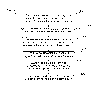
[0040] FIG. 8
is a flow chart of a method 800 for displaying spectroscopy data of
a sample specimen according to one embodiment. The method 800 includes
scanning 810 a laser beam along a beam trajectory relative to a sample (e.g.,
in X,
Y, and/or Z directions) to produce an aerosol of disassociated material within
a
sample chamber, and passing 812 a fluid through the sample chamber to
transport
the disassociated material to a spectrometer. As discussed above, the fluid
may
include an inert gas such as Argon or Helium. The method 800 also includes
processing 814 the disassociated material with a spectrometer to determine
concentration values of a selected element along the beam trajectory, and
correlating 816 the concentration values with respective locations along the
beam
trajectory (see, e.g., FIG. 9). As discussed above, the determined
concentration
values may be in parts-per-million or may be represented by detector responses
11
CA 02854941 2014-05-07
WO 2013/101745 PCT/US2012/071311
such as volts, counts, counts per second, frequency, and wavelength. The
concentration values may also include ratios such as elemental ratios or
isotropic
ratios. The method 800 further includes overlaying 818 indicia of the
determined
concentration on an image of the sample corresponding to the selected
location. As
discussed below, a user may select whether to display the indicia of the
concentration values and/or other layers of information over the image of the
sample.
In other embodiments, rather than overlaying the indicia, image data in a
stored copy
of the image of the sample may be replaced with image data corresponding to
the
indicia of the concentration values. The method 800 further includes
displaying 820
a composite image of the sample and the overlying indicia on a display device.
[0041] FIG. 9 is a flow chart of a method 900 for correlating the
concentration
values with respective locations along the beam trajectory according to one
embodiment. The method 900 includes calibrating 910 the system to estimate a
delay time between laser ablation and a determination of a corresponding
elemental
concentration. The delay time may include one or more delays associated with,
for
example, directing the laser beam (e.g., using X, Y, and/or Z stages) to a new
location along the beam trajectory, commanding a laser source to fire one or
more
laser pulses at the new location, propagating the one or more laser pulses
from the
laser source to the sample for disassociating the material, transporting the
disassociated from the sample chamber to the spectrometer, and operating the
spectrometer so as to analyze the disassociated material and record a
concentration
value. In certain embodiments, a time stamp is associated with each
concentration
value that is calculated and recorded. The time stamp may correspond to a time
when the measured concentration value is recorded or to a time when the
disassociated material used in the calculation is first received at the
spectrometer.
As discussed below, the time stamps may be compared (after being adjusted for
delay) with a start time to associate each concentration value with a
respective
location along the beam trajectory.
[0042] The method 900 further includes determining 912 a processing time
for
scanning from a start location of the beam trajectory with respect to the
surface of
the sample to a particular location (e.g., the location currently being
correlated) along
the beam trajectory. The start location corresponds to a known start time. The
method 900 further includes using 914 the processing time, start time, and
delay
time to associate the particular location with one of the concentration
values. In
12
CA 02854941 2014-05-07
WO 2013/101745 PCT/US2012/071311
other words, scanning speed or other position data may be used to determine
the
position of the laser beam along the beam trajectory with respect to the
surface of
the sample at any given point in time. Based on the calibrated delay, the time
stamps may each be associated with a position of the laser beam along the beam
trajectory.
[0043] Although certain embodiments described herein transport
disassociated
material to a spectroscope for processing, this disclosure is not so limited.
Rather,
any type of laser-assisted spectroscopy may be used. For example, laser
induced
breakdown spectroscopy (LIBS) may be used and the spectroscopy data values may
include wavelength values. In LIBS embodiments, scanning the laser beam along
the beam trajectory stimulates light emission from the sample. The emitted
light
comprises one or more wavelengths that are characteristic of respective
elements
illuminated by the laser beam. The emitted light is directed (e.g., collected
by one or
more lenses into optical fiber) to one or more spectrometers for determining
the one
or more wavelength values.
[0044] FIG. 10 graphically represents a graphical user interface 1000
according
to one embodiment. The graphical user interface 1000 may be displayed, for
example, on the display device 324 shown in FIG. 3. The graphical user
interface
1000 includes a user selection section 1010 and a graphic display section
1012.
The graphical user interface 1000 provides a layered environment that allows
the
user to selectively display various layers of information corresponding to one
or more
samples.
[0045] In this example, the user selection section 1010 includes an options
list
1014 and a layer list 1016. The options list 1014 allows the user to select
(e.g.,
through hyper text or the displayed graphic buttons) whether to display a grid
in the
graphic display section 1012 to accurately indicate a scale for objects
displayed
within the sample chamber, hide the layer list 1016, show a current crosshair
position, and autosave a current display configuration.
[0046] The layer list 1016 (which the user may selectively display) allows
the user
to select which layers of information are displayed in the graphic display
section
1012. The layers may be configured to at least partially overlay one another
and the
user may be allowed to select an order for the displayed layers. In the
example
shown in FIG. 10, a layer including an imported image of a sample insert 1018
is
selected by the user to be displayed over an image of an empty sample chamber
13
CA 02854941 2014-05-07
WO 2013/101745 PCT/US2012/071311
1020. Certain embodiments allow the user to select from a plurality of
different types
of sample chambers 1020 to display, based on a current or desired
configuration.
The displayed sample chamber 1020 may include an actual image of the sample
chamber, a blank grid, or a schematic of the sample chamber.
[0047] The
imported image of the sample insert 1018 may be provided, for
example, from a flatbed scanner or a digital camera. In this example, the
sample
insert includes nine sections 1022a, 1022b, 1022c, 1022d, 1022e, 1022f, 1022g,
1022h, 1022i for holding respective samples, and the imported image of the
sample
insert 1018 includes images of samples 1024, 1026 in sections 1022a, 1022c.
Although shown overlaid with other data, samples are also loaded in sections
1022d,
1022e, 1022h. Skilled persons will recognize from the disclosure herein that
the
sample insert 1018 may be configured to hold a single sample or more than nine
samples. Further, in certain embodiments, two or more of the sections 1022a,
1022b, 1022c, 1022d, 1022e, 1022f, 1022g, 1022h, 1022i may display the same
image of the same sample so that different layers (e.g., the sample map,
SEM/petrographic microscope, annotation, and/or spectroscopy data layers) may
be
applied to each sample image for a side-by-side comparison of different data
for the
same sample (e.g., see FIG. 7).
[0048] The
layer list 1016 also allows the user to select the display of one or more
sample maps, which are a mosaic of images corresponding to adjacent portions
of
the sample. The sample maps may be generated using one or more camera
systems (e.g., such as camera 322 shown in FIG. 3) while the sample is located
within the sample chamber. As shown in FIG. 10, the user can select to display
wide
angle sample maps and/or high magnification sample maps. In
certain
embodiments, there is no limit on the number of sample maps that can be
included
and displayed within this layer (e.g., for illustrative purposes both "Map 1"
and "Map
2" are shown for each type of sample map). In this example, the user has
selected
to display a wide angle sample map 1028 (corresponding to "Map 2") in section
1022d of the imported sample insert 1018.
[0049] The
layer list 1016 also allows the user to select the display of one or more
images imported from external (e.g., third party) devices. Such images may be
produced by, for example, petrographic microscope systems, SEM systems, or
other
imaging systems. The images are importable in a wide variety of sample types
and
may be selectively overlapped one with another. The user may also select the
order
14
CA 02854941 2014-05-07
WO 2013/101745 PCT/US2012/071311
in which the imported images in this layer overlap one another. In
certain
embodiments, the imported images may be selectively aligned to stage
coordinates
using two fiducial points on the image of the sample and corresponding points
on
another preexisting or imported image. As with the sample maps, there may be
no
limit on the number of imported sample images that are included and displayed
in
this layer (e.g., for illustrative purposes SEM and petrographic microscope
images
are shown for possible display). In addition, or in other embodiments, any
image
size or image resolution may be imported. In this example, the user has
selected to
display an imported petrographic microscope image 1030 in section 1022e of the
imported sample insert 1018.
[0050] The
layer list 1016 also allows the user to select to the display of an
annotation layer. As discussed above, with respect to FIG. 7, the annotation
layer
may allow the user to add text and/or graphics (e.g., lines, symbols, or other
indicia)
over an image of a sample or another portion of the graphic display section
1012. In
this example, the user has not selected to include an annotation layer.
[0051] The
layer list 1016 also allows the user to select the display of
spectroscopy data, as described in detail herein. The indicia of the
spectroscopy
data may be displayed in real time (e.g., as the sample is being scanned by a
laser
beam). In addition, or in other embodiments, the user may selectively import
spectroscopy data or previously correlated indicia of spectroscopy data for
display
within the graphic display section 1012. In this example, the user has
selected to
display indicia of spectroscopy data 1032 over an image of a sample ("Zircon
1")
displayed in section 1022h of the imported sample insert 1018.
[0052] Artisans
will recognize from the disclosure herein that other layers may
also be used. In certain embodiments, the entire layered environment can be
saved
to enable the user to load saved environments at a later time and recall all
of the
information associated with a particular experiment (e.g., scan positions, SEM
data,
spectrometer raw data, reduced data such as age of the particular sample, and
other
data used in the experiment). As the user scans across the environment,
respective
data and data files become available for viewing, which enables traceability
of the
various aspects of the experiment and reduces or negates the requirement for
the
user to keep separate records. In certain embodiments, mobile device
applications
(e.g., for laptop computers, tablet computers, smart phones, or other mobile
devices)
allow the user to review selected environments at any time.
CA 02854941 2014-05-07
WO 2013/101745 PCT/US2012/071311
[0053] It will be understood by those having skill in the art that many
changes
may be made to the details of the above-described embodiments without
departing
from the underlying principles of the invention. The scope of the present
invention
should, therefore, be determined only by the following claims.
16