Note: Descriptions are shown in the official language in which they were submitted.
CA 02855337 2014-05-09
WO 2013/068751 PCT/GB2012/052781
1
ORGAN PERFUSION SYSTEMS
Field of the Invention
The present invention relates to perfusion systems for bodily organs, in
particular
human organs, such as the liver, pancreas, kidney, small bowel, but also other
organs
including non-human organs.
Background to the invention
It is known, for example from EP 1 168 913, to provide a system for
extracorporeal
organ perfusion in which a human or non-human organ can be preserved, for
example
prior to transplant into a patient. The system typically comprises a reservoir
for
perfusion fluid, which may be blood or another perfusion solution, and a
circuit for
circulating the fluid through the organ.
Summary of the invention
The present invention provides a perfusion system for the perfusion of an
organ, the
system comprising a perfusion fluid circuit for circulating perfusion fluid
through the
organ, adjustment means for adjusting the content of at least one component in
the
fluid, measuring means for measuring the content of said at least one
component in the
perfusion fluid, and control means arranged to control the adjustment means.
For
example, the control means may be arranged to control the adjustment means so
as to
keep said measured content within a target range. In some cases that may be
above a
minimum target level, or below a minimum target level, or between upper and
lower
target limits.
The content may be a relative content or a proportion, for example it may be a
percentage, and it may be measured by mass, or by volume, or by mole percent.
The at least one component may be at least one of: oxygen; carbon dioxide; and
a
nutrient, such as glucose.
Where the at least one component comprises oxygen, the adjustment means may
comprise oxygen adding means arranged to add oxygen into the fluid. For
example it
may comprise an oxygenator.
CA 02855337 2014-05-09
WO 2013/068751 PCT/GB2012/052781
2
Where the at least one component comprises carbon dioxide, and the adjustment
means may comprises carbon dioxide extraction means arranged to extract carbon
dioxide from the fluid. This may be arranged to supply air, or another gas,
which can
absorb or extract carbon dioxide from the fluid. This function can be
performed by an
oxygenator which also supplies oxygen, or it can be performed by a separate
device or
system.
The at least one component may comprise at least one of, or both of: oxygen
and
carbon dioxide, in which case the system may further comprise nutrient
measuring
means arranged to measure the content of at least one nutrient in the fluid.
The system
may comprise a nutrient supply. The system may comprise nutrient adding means
arranged to add the nutrient, for example from the supply, into the fluid. The
control
means may be arranged to control the nutrient adding means to add the nutrient
if the
content of the nutrient falls below a target range.
The system may comprise a thermometer arranged to measure the temperature of
the
fluid. The system may comprise thermal adjustment means arranged to adjust the
temperature of the fluid. The control means may be arranged to control the
thermal
adjustment means to maintain the temperature of the fluid within a target
range.
The system may comprise an analysis duct through which the fluid can flow. The
measuring means may be arranged to measure the fluid in the analysis duct. For
example the analysis duct may connect two parts of the circuit which will
experience
different pressures, from each other, during perfusion. This will tend to
cause some of
the fluid to flow through the analysis duct during perfusion. For example the
analysis
duct may have an upstream end connected into the circuit upstream of the
organ, and a
downstream end connected to the circuit downstream of the organ.
The measuring means may be arranged to operate during perfusion of the organ.
The
control means may be arranged to operate during perfusion of the organ to
maintain
the target range or ranges.
The control means may include a memory arranged to store at least one limit of
said
range, or of at least one of said ranges. The control means may be arranged to
CA 02855337 2014-05-09
WO 2013/068751 PCT/GB2012/052781
3
compare the measured content with said at least one limit. This can enable it
to
determine when the measured content is outside the target range.
The system may comprise a user interface arranged to enable a user to input at
least
one limit of said range, or of at least one of said ranges. The user interface
may also
be arranged to indicate the content of at least one of the components of the
fluid.
The system may comprise organ sensing means arranged to detect the presence of
the
organ in the circuit. The system may further comprise a surrogate organ
arranged to be
connected into the circuit in place of the organ so that the circuit can
circulate fluid
through the surrogate organ. Where the system includes organ sensing means,
the
organ sensing means may be arranged to distinguish between the presence of the
organ
in the circuit and the presence of the surrogate organ in the circuit.
Indeed, the present invention further provides a perfusion system for
perfusing an
organ, the system comprising: a perfusion fluid circuit arranged to circulate
perfusion
fluid through the organ; a surrogate organ arranged to be connected into the
circuit in
place of the organ so that the circuit can circulate fluid through the
surrogate organ;
and organ sensing means arranged to sense the presence of the organ, or the
surrogate
organ, or both, in the circuit. The organ sensing means may thereby be
arranged to
distinguish between the presence of the organ in the circuit and the presence
of the
surrogate organ in the circuit.
The organ sensing means may comprise at least one pressure sensor arranged to
measure the pressure of the perfusion fluid at at least one point in the
circuit. The
organ sensing means may be arranged to measure the difference in pressure
between
two points in the circuit. The organ sensing means may comprise a pressure
sensor
arranged to measure the pressure of perfusion fluid flowing towards the organ.
The
organ sensing means may comprise a pressure sensor arranged to measure the
pressure
of perfusion fluid flowing away from the organ. Alternatively, or in addition,
the
organ sensing means may comprise a flow meter arranged to measure the rate of
fluid
flow at at least one point in the circuit. The organ sensing means may further
be
arranged to receive data regarding the speed of a pump in the circuit, and to
use that
data in determining whether the organ or the surrogate organ is present in the
circuit.
CA 02855337 2014-05-09
WO 2013/068751 PCT/GB2012/052781
4
The control means may be arranged to operate in two different modes, one of
which is
a preparation mode suitable for preparing the system for perfusion of an
organ, and
one of which is a .perfusion mode suitable for perfusion of an organ. The
control
means may be arranged, in both of the modes, to control the content of at
least one
component of the perfusion fluid. The control means may be arranged to control
the
fluid flow in the perfusion circuit in a different way in each of the two
modes. For
example in one mode the fluid may be pumped at constant speed.
The system may comprise a bubble detection means arranged to detect bubbles in
the
fluid during perfusion.
Indeed the present invention further provides a perfusion system comprising a
circuit
for circulating perfusion fluid through the organ, control means arranged to
control
the flow of fluid round the perfusion circuit, and bubble detection means
arranged to
detect the presence of bubbles in the fluid.
The control means may be arranged to respond to detection of bubbles by the
bubble
detection means. For example the control means may be arranged to respond to
detection of the bubbles by producing a warning output, such as by displaying
a
warning. Alternatively, or in addition, it may be arranged to respond by
reducing the
fluid flow through at least one part of the circuit, or into the organ,
optionally
stopping it completely, for example by partially or completely closing a flow
control
valve. The flow control valve may be arranged to control flow of fluid from a
reservoir to the organ.
The bubble detection means may be arranged also to measure the flow rate of
fluid in
the perfusion circuit. The bubble detection means comprises an ultrasound
transducer.
The bubble detection means may be arranged to determine both whether bubbles
are
present in the fluid and the flow rate of the fluid from the timing of
ultrasound
transmissions and detections.
The system may comprise measuring means arranged to measure the amount of
fluid
secreted by or leaked from the organ. For example the fluid may be bile from a
liver,
ascites from a liver, urine production from the kidney or any other excretion
from any
organ.
CA 02855337 2014-05-09
WO 2013/068751 PCT/GB2012/052781
The system may further comprise a sump arranged to collect the secreted or
leaked
fluid. The measuring means may be arranged to measure the volume of fluid that
enters the sump. The system may be arranged to record and display the amount
of
fluid that is secreted or leaked. For example the control means may include
part of
5 the measuring means, and may be arranged to calculate and record the
total volume of
the fluid, or the rate of flow of the fluid, or both, and may record these at
regular
intervals during perfusion to monitor the organ. The controller may be
arranged to
generate a display of all or part of this information. The controller may be
arranged to
modify its control of at least one component of the system in response to the
measured
volume or the measured flow rate. For example it may be arranged to vary the
speed,
or the average speed, or the duty cycle, of a pump which is arranged to pump
the fluid
from the sump.
The system may further comprise a support stand on which at least some of the
components of at least one of the perfusion circuit, the adjustment means and
the
control means are mounted. The system may further comprise a transport system
on
which the support stand can be mounted. The transport system may include a
cover
arranged to cover the support stand and the components mounted on it. The
transport
system may include a wheeled base. The transport system may be arranged to
support
the support stand in transport position, or an operative position which is
raised
relative to the transport position.
Some embodiments of the present invention can provide a perfusion system in
which
one or more of the following functions are automated: detection of an organ in
the
circuit for perfusion; detection of perfusion fluid in the circuit; control of
fluid
pressure in the circuit during perfusion; control of fluid temperature in the
circuit
during perfusion; and control of one or more nutrients in perfusion fluid
during
perfusion. The system may therefore be fully automated.
Some embodiments of the invention provide a system that is portable.
Some embodiments may be arranged to be battery and mains powered.
The present invention further provides a method of perfusing an organ, the
method
comprising circulating perfusion fluid through the organ, measuring the
content of at
CA 02855337 2014-05-09
WO 2013/068751 PCT/GB2012/052781
6
least one component in the perfusion fluid, and adjusting the content of said
at least
one component in the fluid so as to keep said measured content within a target
range.
The content may be a relative content or a proportion, for example it may be a
percentage, and it may be measured by mass, or by volume, or by mole percent.
The at
least one component may be at least one of: oxygen; carbon dioxide; and a
nutrient,
such as glucose. The measurement or the adjustment may be performed using any
system according to the invention as described above.
Preferred embodiments of the present invention will now be described by way of
example only with reference to the accompanying drawings.
Brief Description of the Drawings
Figure 1 is a schematic diagram of a perfusion system according to an
embodiment of the invention;
Figure 2 is an enlargement of part of Figure 1;
Figure 3 is a schematic diagram of an oxygenator forming part of the system of
Figure 1;
Figure 3a is a diagram of a combined flow meter and bubble detector according
to an embodiment of the invention and forming part of the system of Figure 1;
Figure 4 is a schematic diagram of an oxygen concentrator forming part of the
system of Figure 1;
Figure 5 is a diagram similar to Figure 2 showing a liver connected into the
system of Figure 1;
Figure 6 is a diagram of the system of Figure 1 modified for perfusion of a
single input ¨ single output organ, such as a pancreas or kidney;
Figures 7a, 7b and 7c are perspective views of the system of Figure 1 mounted
in a mobile transportation system according to an embodiment of the invention;
CA 02855337 2014-05-09
WO 2013/068751 PCT/GB2012/052781
7
Figures 8a, 8b and 8c are perspective views of the system of Figure 1 mounted
in a mobile transportation system according to a further embodiment of the
invention;
Figures 9a, 9b, 9c and 9d are perspective views of the system of Figure 1
mounted in a mobile transportation system according to a further embodiment of
the invention; and
Figure 10 is a perspective view of the system of Figure 1 mounted in a further
alternative mobile transportation system.
Description of the Preferred Embodiments
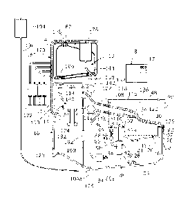
Referring to Figures 1 and 2, a perfusion system according to an embodiment of
the
invention generally comprises a sling 10 on which an organ can be supported, a
fluid
reservoir 12, an oxygenator 14, and a perfusion circuit 16 arranged to
circulate fluid
between the reservoir, the organ, and the oxygenator during perfusion. A
controller 18
is arranged to control the functioning of the system as will be described in
more detail
below.
The sling 10 is of moulded plastics or other suitable material and designed to
be
compliant so as to enable non-traumatic support of the organ whilst providing
a degree
of shock absorption during transport. The sling 10 has a perforated base 19
through
which fluids leaking from the organ can flow out, and side walls 20 extending
upwards from the base 19, and a rim 22 extending around the top of the side
walls 20.
A fluid sump 24 which, where the organ is a liver, forms an ascites sump, is
located
beneath the sling 10, and comprises a concave base 26 that tapers downwards to
a
drainage hole 28, which is formed through its lowest point. The sump 24 is
arranged
to catch fluid leaking through the base 19 of the sling. The sump 24 also
comprises
side walls 30 that extend upwards from the base 26, around the side walls 20
of the
sling, and have a flange 32 around their top which supports the rim 22 of the
sling 10.
A removable cover 34, which is of moulded plastics, fits over the top of the
sling 10
and has a rim 36 around its lower edge which fits against the rim 22 of the
sling.
The sling 10 is supported within an organ container 40 which has the ascites
sump 24
and a bile sump 42 supported in its base 44, and in this embodiment formed
integrally
CA 02855337 2014-05-09
WO 2013/068751 PCT/GB2012/052781
8
with it. The organ container 40 has side walls 46 extending upwards from its
base 44
and a removable cover 48. The bile sump 42 is about twice as deep as the
ascites
sump 24 and generally narrow and tubular in shape, and extends downwards from
the
base 44 of the container 40 with its rim 52 level with the rim 32 of the
ascites sump 24
and the rim 22 of the sling.
The bile sump 42 is formed in two parts, an upper part 42a and a lower part
42b, both
of which are integral with the base 44 of the organ container. The lower part
42b has a
bile inlet port 54 formed in its side, towards its upper end 56, and a bile
overflow
port 58 formed in its upper end. A bile outlet port 60 is formed in the base
44 of the
organ container close to the top of the bile sump, with an upper connector 60a
for
connection via a cannula to the liver, and a lower connector 60b for
connection to a
bile measurement system 62. The bile measurement system 62 is arranged to
measure
the volume of bile secreted by the liver before allowing it to flow into the
bile
sump 42.
As can best be seen in Figure 2, the bile measurement system 62 comprises a
bile
receiving duct 64 having its upper end connected to the lower connector 60b,
and its
lower end connected to a T-piece connector 66, a bile outlet duct 68 having
its upper
end connected to the connector 66 and its lower end connected to the bile sump
inlet
port 54, and an overflow duct 70 having its lower end connected to the
connector 66
and its upper end connected to a further port 69 formed in the base 44 of the
container. An overflow pipe 72 connects the top of the further port 69 to the
bile
overflow port 58 in the top of the lower part 42b of the sump. A liquid level
sensor 74
is arranged to measure the level of fluid in the overflow duct 70 and to
output a signal
indicative of the fluid level to the controller 18. In this embodiment the
liquid level
sensor 74 is arranged to detect when the liquid level in the overflow duct 70
reaches a
predetermined height, and send a signal indicative of this to the controller
18. A flow
control valve, which in this embodiment comprises a pinch valve 76, in the
bile outlet
duct 68 is switchable between a closed state in which it closes the outlet
duct 68 so
that bile can build up on the measurement system 62 and an open state in which
it
allows bile to drain from the measurement system 62 into the bile sump 42. The
controller 18 is arranged to control the flow control valve 76.
CA 02855337 2014-05-09
WO 2013/068751 PCT/GB2012/052781
9
The controller 18 is arranged to measure the rate at which bile is secreted by
the liver
by closing the pinch valve 76 so that bile builds up in the outlet duct 68,
and then in
the bile receiving duct 64 and overflow duct 70. When the level sensor 74
detects that
the bile has reached the predetermined level, it is arranged to send a signal
to the
controller 18 which responds by opening the pinch valve 76, for example for a
predetermined period, to allow the bile to drain out of the measurement system
into
the sump, and then closes it again so that bile can start to collect in the
measurement
system again. The controller 18 is also arranged to record in memory the times
at
which the bile reaches the predetermined level, and therefore the times at
which the
measurement system is filled. This information, together with the known volume
of
the system when it is filled to the predetermined level, allows the rate at
which bile
secreted over time to be monitored. For example the controller 18 may be
arranged to
calculate a flow rate each time the valve 76 is opened from the known volume
of the
system and the time interval between the valve opening and the previous valve
opening. That flow rate can be displayed on the GUI 17, being updated each
time a
new calculation of flow rate is recorded. Alternatively the controller 18 may
be
arranged to store this flow rate information in memory, so that flow rate data
for the
whole perfusion process can be stored and then output or displayed via the GUI
17. As
a further alternative, the controller may not perform any calculation but may
generate
an output which varies with the flow rate, and the GUI may be arranged to
respond to
the output by generating a display, such as a line graph, which is indicative
of the
flow rate, for example by having appropriately marked axes. It will be
appreciated
that, for organs other than the liver, this measurement system can be arranged
to
measure other fluids leaking from, or excreted by, the organ during perfusion,
and to
record and display the measured volume. For example the organ may be a kidney
and
the fluid may be urine.
Referring back to Figure 1, an ascites duct 80 is connected at one end to the
drainage
hole 28 in the bottom of the ascites sump 26 and at the other end to an
ascites return
port 82 in the top of the fluid reservoir 12. The ascites duct 80 has a
central
portion 80a that is the lowest part of the duct 80, being below the level of
the ascites
sump 26, as well as below the level of the reservoir 12. An ascites pump 84 is
provided in the central portion 80a of the ascites duct 80 to pump ascites
from the
sump 26 back up into the reservoir 12. An ascites measurement tube 86 extends
vertically upwards from the central portion 80a of the ascites duct, adjacent
to, and
CA 02855337 2014-05-09
WO 2013/068751 PCT/GB2012/052781
upstream of, the pump 84, and has a fluid level sensor 88 in it. This level
sensor 88 is
arranged to detect, and output a signal, when fluid in the measurement tube 86
reaches
a predetermined level that is below the base 19 of the sling 10, and in this
embodiment
above the drainage port 28 in the ascites sump. The fluid level sensor 88 is
connected
5 to the controller 18 which receives the signals from it, and can
therefore detect when
the level of ascites in the sump reaches a predetermined level. In response to
this the
controller 18 is arranged to activate the ascites pump 84, for example for a
predetermined time, to reduce the level of ascites in the sump 26. The speed
of the
pump 84 may be variable and the controller 18 may be arranged to control the
speed
10 of the pump, or the duty ratio of the pump, or the average speed of the
pump, on the
basis of the measured fluid level. In other embodiments the ascites level
sensor can be
located within the sump 26. Indeed any suitable system for measuring the
volume of
accumulated ascites can be used as feedback to control the operation of the
pump 84.
For example a pressure sensor located close to the pump 84 could be used to
measure
accumulated ascites volume. In still other embodiments the ascites pump 84 can
simply be arranged to operate for fixed periods with no measurement of ascites
volume.
In a modification to this embodiment, there is a further ascites level sensor
in addition
to the sensor 88, so that the sensors can detect when the ascites level
reaches upper
and lower levels. The controller 18 is arranged to start the ascites pump 84
when the
ascites is detected as reaching the upper level, and to step the ascites pump
84 when
the ascites level drops to the lower level. The controller is then arranged to
record the
timing of each time the pump is turned on, and this provides an indication of
the total
volume of ascites and the flow rate of ascites during perfusion. This
information can
be stored and displayed on the GUI 17 in the same way as the bile
measurements. The
speed of the pump 84 may be variable and the controller 18 may be arranged to
control the speed of the pump, or the duty ratio of the pump, or the average
speed of
the pump, on the basis of the measured fluid level. It will be appreciated
that, for
other organs, this measurement system can be used to measure the total volume
or
flow rate of other fluids leaking from, or excreted by, the organ during
perfusion. This
measurement can also be provided with only one ascites level sensor as shown
in
Figure 1, for example if the pump 84 is arranged to operate until it has
pumped all of
the ascites that is upstream of the pump 84, which can be assumed to be a
fixed
volume.
CA 02855337 2014-05-09
WO 2013/068751 PCT/GB2012/052781
11
The perfusion circuit 16 further comprises a first fluid supply duct 100,
which when
used for perfusion of a liver forms a portal duct, a second fluid supply duct
102,
which when used for perfusion of a liver forms a hepatic artery duct, and a
fluid
removal duct 104, which when used for perfusion of a liver forms an inferior
vena
cava (IVC) duct. The system and its operation will now be described for
perfusion of a
liver, but it will be appreciated that it can equally be used for other
organs, in
particular single-inflow single-outflow organs such as the kidney, small bowel
or
pancreas if arranged as per the alternative configuration of Fig. 6 . The
portal duct 100
has one end connected to an outlet port 106 in the fluid reservoir and the
other end
attached to a portal vein connector 108. The portal duct 100 extends through a
port 110 in the side wall 46 of the organ container 40 so that the portal vein
connector 108 is located inside the container. A flow control valve 112, in
the form of
a pinch valve, having a variable degree of opening, is provided in the portal
duct 100
and is connected to the controller 18. The controller 18 is arranged to vary
the degree
of opening of the pinch valve 112 so as to control the rate of flow of fluid
from the
reservoir 12 to the portal vein of a liver. A portal flow sensor 113 is
provided in the
portal duct 100 and is arranged to output a signal indicative of the flow rate
of fluid in
the portal duct 100. The output of the flow sensor 113 is connected to the
controller 18 which can therefore monitor the flow rate in the portal duct.
The
controller 18 is also arranged to determine from the flow sensor 113 signal
when the
flow of fluid from the reservoir ceases due to the reservoir being empty. In
response
to detection of an empty reservoir the controller 18 is arranged to close the
flow
control valve 112 so as to prevent air from reaching the organ and to enable
replenishment of the perfusion fluid volume within the reservoir. The flow
sensor in
this embodiment is also arranged to act as a bubble detector, arranged to
output a
signal indicative of the presence of air bubbles in the fluid in the portal
duct 100. The
controller 18 is arranged to close the flow control valve 112 on detection of
bubbles in
the same way as if it detects a completely empty reservoir on the basis of
fluid flow.
The hepatic artery duct 102 has one end connected to a first outlet port 114
of the
oxygenator 14 and the other end attached to a hepatic artery connector 116.
The
hepatic artery duct 102 extends through a port 118 in the side wall 46 of the
organ
container 40 so that the hepatic artery connector 116 is located inside the
container.
The IVC duct 104 has one end attached to an IVC connector 120, which is
located
inside the container 40, and extends out through a port 122 in the base 44 of
the organ
container 40, having its other end connected to an inlet port 124 of the
oxygenator 14.
CA 02855337 2014-05-09
WO 2013/068751 PCT/GB2012/052781
12
A pump 123 is provided in the IVC duct 104 having its inlet connected by a
part of the
IVC duct 104 to the IVC connector 120, and its outlet connected to the inlet
port 124
of the oxygenator 14. The pump 123 is arranged to pump fluid from the IVC duct
104
into the oxygenator 124. The pump 123 is a variable speed pump and is
connected to,
and controlled by, the controller 18. An IVC flow sensor 125 is arranged to
measure
the rate of fluid flow rate in the IVC duct 104 and is arranged to output a
signal
indicative of the flow rate of fluid in the vena cava duct 104. The output of
the flow
sensor 125 is connected to the controller 18 which can therefore monitor the
flow rate
in the IVC duct 104.
Each of the connectors 108, 116, 120 is a quick-release connector arranged to
allow
the duct to which it is attached to be connected, either via a cannula to the
appropriate
vein or artery of the liver, or to a surrogate organ 126 which is arranged to
complete
the perfusion circuit prior to connection of the real organ. The surrogate
organ 126
comprises two inlet ducts 128, 130 for connection to the portal duct 100 and
the
hepatic artery duct 102, and one outlet duct 132 for connection to the IVC
duct 104. In
this embodiment the surrogate organ is in the form of a simple Y-piece
connector 134
which connects the two inlet ducts 128, 130 to the outlet duct 132 so that,
when it is
connected into the circuit, fluid can flow through it from the portal duct 100
and the
hepatic artery duct 102 to the IVC duct 104.
Each of the portal duct 100, the hepatic artery duct 102 and the IVC duct 104
has a
pressure sensor 136, 137, 138 in it, arranged to measure the pressure of fluid
in the
duct 100, 102, 104. Each of these pressure sensors 136, 137, 138 is arranged
to
measure pressure at a point close to the respective connector 108, 116, 120,
and to
output a signal indicative of the pressure at that point. In this embodiment,
each of the
ducts 100, 102, 104 is split into two sections and each of the pressure
sensors 136, 137, 138 is located in a moulded plastics sensor body which also
serves
to connect the two sections of the duct together. The sensors 136, 137, 138
are each
located just outside the wall 46 or the base 44 of the organ container 40. In
each case
the duct between the pressure sensor 136, 137, 138 and the connector 108, 116,
120 is
of substantially constant cross section, so the pressures sensed by the
sensors 136, 137, 138 are approximately equal to the pressure of fluid flowing
into
and out of the surrogate organ, or the actual organ when that is connected
into the
circuit.
CA 02855337 2014-05-09
WO 2013/068751 PCT/GB2012/052781
13
The oxygenator 14 has a second outlet port 140 which is connected by a
pressure
control duct 142 to a pressure control port 144 in the fluid reservoir 12. A
flow
control valve, in the form of a pinch valve 146, having a variable degree of
opening, is
provided in the pressure control duct 142 and is connected to the controller
18 so that
the controller can vary the degree of opening of the pinch valve 146 thereby
to control
the return flow of fluid from the oxygenator 14 to the reservoir 12. This,
together with
the speed of the pump 123, is controlled by the controller 18 to control the
pressure of
fluid flowing to the organ through the hepatic artery duct 102, as well as the
pressure
of the fluid in the vena cava duct 104 flowing away form the organ.
Referring to Figure 3, the oxygenator 14, which is shown schematically,
comprises a
through duct 150 arranged to carry fluid from the inlet port 124 to the two
outlet
ports 114, 140. An oxygen chamber 152 has an inlet port 154 for connection to
an
oxygen supply and an air supply, and an outlet or vent port 156 for venting
the oxygen
and air from the oxygen chamber. A vent 158 is connected at its lower end to
the
through duct 150 and extends upward so that its upper end is approximately
level with
the top of the reservoir 12. This vent 158 is closable, and is arranged to be
opened
during filling of the fluid circuit to vent air from the oxygenator, but is
closed during
perfusion. A permeable membrane 160 between the oxygen chamber 152 and the
through duct 150 allows oxygen in the oxygen chamber 152 to oxygenate fluid,
which
may be blood, in the through duct 150, and allows air in the oxygen chamber
152 to
carry away CO2 from the fluid. A water chamber or duct 162 is also connected
to a
water inlet port 164 and a water outlet port 166, and is separated from the
through
duct 150 by a thermally conductive wall 168. This provides a heat exchanger
which
allows water, or another suitable thermal control fluid, to be circulated
through the
oxygenator 14 to control the temperature of the perfusion fluid. A heater 167,
such as
a Peltier heater, is provided to heat water entering the oxygenator via the
water inlet
port 164, and a thermometer 169a is provided to measure the temperature of the
perfusate flowing out of the oxygenator into the hepatic artery duct 102. A
further
thermometer 169b is arranged to measure the temperature of the water that is
supplied
to the heat exchanger. The heater 167 and the thermometers 169a, 169b are
connected
to the controller 18 which is arranged to measure and monitor the temperature
of the
perfusate supplied to the organ and the water supplied to the heat exchanger,
and
control the heater 167 so as to maintain the perfusate temperature at a
desired level,
for example within a target temperature range.
CA 02855337 2014-05-09
WO 2013/068751 PCT/GB2012/052781
14
It will be appreciated that other devices can be used for adding oxygen to,
and
extracting carbon dioxide from, the perfusate. For example a bubbler can be
used,
instead of the type of oxygenator shown in Figure 3, which bubbles the
concentrated
oxygen through the perfusate. Also, instead of one device which brings a gas
into
contact with the perfusate and in which the oxygen and carbon dioxide content
of the
gas are controlled, the system can include separate devices one for each gas.
Referring to Figure 3a the flow sensor 113 in the portal duct 100 is, as
described
above, also arranged to act as a bubble detector. In this embodiment the flow
sensor 113 comprises a housing 300 arranged to be clipped around the conduit,
in this
case the portal duct 100. Two ultrasound transducers 302, 304 are supported in
the
housing 300 and arranged so that they are located on one side of the conduit.
A
reflector 306 is supported in the housing 300 and arranged to be located on
the
opposite side of the conduit from the transducers 302, 304. The transducers
302, 304
are offset from each other along the conduit in the direction of fluid flow,
and angled
so that when each of them transmits an ultrasound signal it will be reflected
from the
reflector 304 onto the other transducer, such that it can be detected. Each
transducer 302, 304 is arranged to emit a series of pulses of ultrasound, and
the timing
of the pulses is controlled so that the two transducers 302, 304 emit pulses
alternately,
with the non-emitting transducer being arranged to detect the emitted pulse
after it has
been reflected from the reflector 306. The time taken for ultrasound to travel
in each
direction between the two transducers is measured, using the emission and
detection
times, and the detector 145 is arranged to determine the difference between
the
transmission times in the two directions and from that difference to calculate
the flow
rate of fluid in the conduit 102. If gas bubbles are present in the perfusate
these reflect
ultrasound back to the transducer that transmitted it and, in some cases,
reflect the
ultrasound on to the other non-transmitting transducer so that they arrive at
a different
time from those reflected from the reflector 304, and generally at much
smaller
amplitudes. Therefore the bubble detector 145 is arranged to analyse the
detection
signals from both of the transducers 302, 304 and determine from their timing
and
amplitude when bubbles are present in the perfusate. The signals from the
ultrasound
detector can be processed locally in a processor forming part of the bubble
detector,
so that the processor in the bubble detector sends a simple signal to the
controller 18
indicative of the presence of gas bubbles in the perfusate, or the detector
signals can
CA 02855337 2014-05-09
WO 2013/068751 PCT/GB2012/052781
be input directly to the controller 18 which can be arranged to analyse them
to detect
the presence of the gas bubbles itself.
In response to the detection of gas bubbles the controller 18 may be arranged
to output
5 a warning signal to the GUI which can be arranged to provide a visual or
audible
warning on receipt of the warning signal. In addition, the controller is
arranged to stop
the flow of perfusate into the organ via the portal duct if it determines that
gas
bubbles are present in the perfusate. Specifically in this case, in response
to the
detection of bubbles in the portal duct 100, the controller 18 is arranged to
close the
10 pinch valve 112. It is also arranged to fully open the pinch valve 146
for a fixed time
period, to enable replenishment of the volume within the reservoir. Following
this
time delay it is arranged to re-open the pinch valve 112, and to re-set the
valve 146 so
as to achieve the desirable arterial pressure.
15 In other embodiments, the system may include a further bubble detector
in the hepatic
artery duct or the IVC duct. In this case the controller 18 is arranged, when
gas
bubbles are detected, to stop the pump 123 to stop the flow of fluid through
the organ
as well as to provide the warning. This enables a user to take precautionary
measures,
such as allowing the gas bubbles to escape from the perfusate, or even to
disconnect
the organ and flush the gas bubbles form the fluid circuit, before re-starting
perfusion.
In other embodiments, other types of bubble detector can be used. For example
an
ultrasound bubble detector can be used that is not combined with a flow rate
sensor,
and includes only a single transducer. In that case the flow rate sensor can
be provided
separately, and can be of a different form other than an ultrasound sensor.
Referring back to Figure 1, a nutrient control circuit 170 comprises a set of
syringes 172, in this case four, each containing a respective nutrient, and a
nutrient
feed duct 174 which has one end connected to a separate fluid reservoir 176
and the
other end connected to a nutrient inlet port 178 in the top of the main fluid
reservoir 12. Each of the syringes 172 is connected to the nutrient feed duct
174 by a
respective nutrient input duct 180. A nutrient pump 182 is arranged in the
nutrient
feed duct 174 to pump fluid through the nutrient feed duct from the nutrient
feed
reservoir 176 into the main reservoir 12 via the nutrient inlet port 178. The
pump 182
CA 02855337 2014-05-09
WO 2013/068751 PCT/GB2012/052781
16
and the syringes 172 are controlled by the controller 18 so that the rate at
which each
of the nutrients is fed into the reservoir 12 is controlled.
A small diameter fluid analysis duct 190 has one end connected to the IVC duct
104,
upstream of the pump 123, and in this case downstream of the IVC flow sensor
125,
and the other end connected to the pressure control duct 142, upstream of the
pressure
control valve 146, so that fluid can flow through the fluid analysis duct 190
from the
pressure control duct 142 to the IVC duct 104, bypassing the organ. A
measurement
system, in this case in the form of a blood gas analyser (BGA) 192 is arranged
to
measure various parameters of the fluid flowing through the fluid analysis
duct 190.
In this embodiment the BGA 192 is arranged to measure the oxygen content and
the
carbon dioxide content of the fluid flowing through it. Other parameters,
including
any one or more of temperature, pH, base excess, potassium, glucose,
haematocrit and
oxygen saturation can also be measured and monitored. The BGA 192 is connected
to
the controller 18 and arranged to output signals each of which is indicative
of the
value of one of the parameters it measures, and the controller 18 is arranged
to receive
those signals so that the parameters can be monitored by the controller 18.
The signals
therefore include an oxygen level signal and a CO2 level signal in this
embodiment.
A priming bag or reservoir 194 is supported at a level which is above the top
of the
reservoir 12, and connected by a priming duct 196 to the perfusion circuit at
a priming
point which is in the vena cava duct 104 at its lowest point 104a. This is
also the
lowest point of the perfusion circuit 16, which allows the whole circuit 16 to
be filled
from the bottom, as will be described in more detail below.
Referring to Figure 4, the oxygen supply to the oxygenator inlet 154 is
provided by an
oxygen concentrator 200. This comprises a pair of zeolite towers 202, 204, an
air
inlet 206 arranged to receive gas in the form of air at atmospheric pressure,
a
compressor 208 arranged in the inlet to compress the incoming air, and a two
way
switch valve 210 operable to control the flow of incoming air into the zeolite
towers 202, 204. Each of the towers 202, 204 has an outlet 212, 214 and these
are
connected together to form a single outlet from the oxygen concentrator which
in turn
is connected to the inlet 154 of the oxygenator. In use, as the compressed air
flows
through the zeolite towers 202, 204, the zeolite extracts nitrogen from the
air which
increases the concentration of oxygen in the gas. The nitrogen leaves the
towers via
CA 02855337 2014-05-09
WO 2013/068751 PCT/GB2012/052781
17
vents 216, and the gas leaving the concentrator 200, which comprises
concentrated
oxygen as well as some nitrogen and traces of other gases, is fed to the
oxygenator
inlet 154. A proportional valve 224 in the outlet from the oxygen concentrator
is
arranged to control the flow rate of gas, and hence oxygen, from the oxygen
concentrator 200 to the oxygenator 14. The proportional valve 224 is connected
to,
and controlled by, the controller 18 so that the controller can control the
flow rate of
oxygen into the oxygenator 14. The air supply to the oxygenator inlet 154 is
provided
by a further compressor 220 which has an inlet 222 arranged to receive air at
atmospheric pressure. A further proportional valve 226 in the outlet from the
compressor 220 is connected to and controlled by the controller 18, so that
the
controller can control the flow rate of air from the compressor 220 to the
oxygenator,
and hence the rate of extraction of carbon dioxide.
In a modification to the arrangement of Figure 4, the second compressor 220 is
omitted and the output from the first compressor 208 is connected both to the
oxygen
concentrator 200 and through a separate air duct via the second proportional
valve 226
to the oxygenator gas inlet. The single compressor 208 therefore provides the
pressure
for the oxygen and air supplies, the flow rates of which are controlled
independently
by their respective flow control valves 224, 226.
Referring to Figure 5, when the system is in operation for perfusing a liver,
the
surrogate organ 126 is removed, and the liver 250 to be perfused is placed in
the
sling 10. The portal vein, hepatic artery, inferior vena cava (IVC), and bile
duct of the
liver are cannulated, and the cannulae connected to the portal vein connector
108, the
hepatic artery connector 116, the vena cava connector 120, and the bile outlet
port 60
respectively.
Referring back to Figure 1, during perfusion, when the system is operating in
a
perfusion mode, perfusate fluid flow through the liver is controlled by the
controller 18 which is arranged to controlling the pressure in the hepatic
artery
duct 102 and the IVC duct 104 to maintain them at approximately constant
pressures,
allowing the liver to regulate the flow rate of fluid through itself. To do
this, the
controller 18 is arranged to monitor the pressure in the hepatic artery duct
102 by
monitoring the output signal from the pressure sensor 137 and the pressure in
the IVC
duct 104 by monitoring the output of the pressure sensor 138, and to control
the
CA 02855337 2014-05-09
WO 2013/068751 PCT/GB2012/052781
18
perfusion pump 123 and the pinch valve 146 in the pressure control duct 142 so
as to
maintain the measured pressures, i.e. the pressure sensor output signals, at
respective
set levels, or within respective ranges.
The oxygen level in the perfusate fluid is also controlled by the controller
18 during
perfusion. While most of the oxygenated perfusate from the oxygenator outlet
114
flows through the hepatic artery duct 102, a small proportion of it is
diverted through
the fluid analysis duct 190 and through the BGA 192. The BGA 192 detects the
level
of oxygen in the perfusate, which is monitored by the controller 18. The
controller 18
is arranged to control the pressure and flow rate of oxygen supplied by the
oxygen
concentrator 200 to the oxygenator by controlling the pump 208 and the two-way
valve 210 of the oxygen concentrator 200, so as to control the rate at which
perfusate
is oxygenated in the oxygenator 100. The controller 18 is arranged to keep the
oxygen
level of the blood at a predetermined level or within a predetermined range.
The
controller 18 has a memory in which a target level or range of the oxygen
content can
be stored and the controller is arranged to compare the measured level with
the stored
level to determine how the oxygen level needs to be controlled. The stored
target level
can be selected and altered by means of a user input which in this case is in
the form
of a graphic user interface (GUI) 17 connected to the controller 18. The GUI
17 is also
arranged to display various information including the values of various
operating
parameters of the system. These can include oxygen level in the perfusion
fluid,
carbon dioxide level in the perfusion fluid, temperature of the perfusion
fluid, the
level of any nutrient in the perfusion fluid, such as glucose.
The carbon dioxide (CO2) level in the perfusate is also monitored and
controlled by
the controller 18 during perfusion in a similar way to the oxygen level, with
the
controller 18 continuously using the CO2 level signal from the BGA 192 to
measure
the CO2 level in the perfusate, comparing it with target levels stored in
memory in the
controller 18, and controlling the air flow control valve 226 to control the
flow rate of
air into the oxygenator 16. The target CO2 level can also be set and adjusted
by a user
by means of the user input 17.
The temperature of the perfusate supplied to the organ is monitored and
controlled by
the controller 18 which is arranged, during perfusion, to monitor the signal
from the
perfusate thermometer 169a and the water thermometer 169b and control the
water
CA 02855337 2014-05-09
WO 2013/068751 PCT/GB2012/052781
19
heater 167 to control the temperature of water flowing in the heat exchanger,
and
optionally also the flow rate of water flowing through the heat exchanger,
thereby to
maintain the perfusate temperature within a target temperature range. This
target
range is stored in memory in the controller 18 and can be set and adjusted by
means of
the user input 17.
The level of each of the monitored nutrients in the perfusate is also
monitored and
controlled by the controller 18 during perfusion in a similar way to the
oxygen level,
with the controller 18 using the nutrient level signal from the BGA 192 to
measure the
nutrient level in the perfusate, comparing it with target levels stored in
memory in the
controller 18, and controlling the appropriate syringe 172 to add the nutrient
if the
nutrient level falls below a predetermined level. The addition of nutrients
will
generally be intermittent, so syringe 172 can be controlled simply to add a
predetermined amount of the nutrient if the nutrient level in the perfusate
falls below
the target lower level. Alternatively, or in addition, the speed of the
nutrient pump 182
can be variable and can be controlled by the controller to vary and control
the rate at
which nutrients are added into the perfusate. One of the nutrients which can
be
detected by the BGA 192 and controlled in this way is glucose. However one or
more
other nutrients can also be controlled in the same way,
The controller 18 is also arranged to monitor the signal from the bubble
detector 113
during perfusion and, if it detects the presence of gas bubbles in the
perfusate, or more
than a minimum bubble content in the perfusate, the controller 18 is arranged
to close
the pinch valve 112 as described above. The controller 18 can also be arranged
to
display a warning on the GUI 17 if bubbles are detected.
The surrogate organ 126 is already connected into the circuit as part of the
disposable
set, as is the oxygenator 14, and the pump 123. The perfusion circuit is then
filled
with perfusate. To achieve this, the flow control valves 112, 146 in the
portal duct 100
and pressure control duct are opened A perfusion bag 194 containing perfusate
is
connected to the upper end of the priming duct 196. The priming bag 194 is
then
raised to a level that is higher than top of the fluid reservoir 12. This
causes perfusate
fluid from the priming bag to flow into the perfusion circuit at the priming
point 104a
in the vena cava duct 104, and flow upwards through the whole perfusion
circuit from
that point. As the fluid level in the perfusion circuit rises, this fills the
vena cava
CA 02855337 2014-05-09
WO 2013/068751 PCT/GB2012/052781
duct 104, the surrogate organ 126, the hepatic artery duct 102 and the portal
duct 100,
the through duct 150 of the oxygenator, and the pressure control duct 142, and
the
reservoir 12, with the ports 82, 178 in the top of the reservoir being used to
vent air
out of the system as it fills. The pump head can be independently moved and
tapped
5 relative to is driving motor to enable removal of any gas trapped within
the pump head
during filling
When the perfusion circuit 16 has been filled, the ascites duct is connected
to the
ascites return port 82 in the reservoir and the nutrient feed duct 174 is
connected to
10 the nutrient feed port 178 in the reservoir, and the vent 158 from the
oxygenator 14 is
closed. The system is then switched on, for example by a user inputting a
start
command using the GUI 17 and starts to run and the controller 18 is arranged
to
control the system as follows. When the system starts to run, both the
pressure control
valve 146 and the flow control valve 112 in the portal vein duct are opened.
Initially,
15 therefore, the pump 123 pumps fluid through the hepatic artery duct 102,
through the
portal vein duct 100, through the surrogate organ 126, and through the IVC
duct 104,
also ensuring constant circulation of the perfusion fluid within the reservoir
12. The
controller 18 is arranged initially to control the pump 123 to operate at a
constant
speed and to monitor the pressures in the hepatic artery duct 102 and the IVC
duct 104
20 and compare them. Since the surrogate organ 126 is present, the pressure
drop across
it is low, in particular significantly lower than what it would be if a real
organ were
connected into the circuit, and this enables the controller 18 to detect the
presence of
the surrogate organ from the outputs from the difference between the pressures
measured by the pressure sensors 136, 138.
In a modification to this embodiment, just one of the two measured pressures
can be
used to detect the presence of the surrogate organ 126. For example the
surrogate
organ may be determined as being present (or the real organ as being absent)
provided
the pressure in the hepatic artery duct remains below a predetermined value.
In
another alternative modification, the measured fluid flow rate at at least one
point in
the circuit, for example in the fluid removal duct 104 as measured by the flow
sensor 125, or in the second fluid supply duct 102, can be used, either on its
own or in
combination with data defining the speed of the pump 123, to determine whether
the
organ is present in the circuit. This is because flow rates will be slower
generally, and
more specifically will be slower for any given pump speed, when the organ is
present
CA 02855337 2014-05-09
WO 2013/068751 PCT/GB2012/052781
21
than when it is not. This is because the organ provides a greater resistance
to fluid
flow, which can be measured by measuring the fluid flow rate.
While the surrogate organ is present, and in particular while the controller
18 detects
that the surrogate organ is present, the controller 18 operates in a
preparation mode it
which it is preparing the system for connection of the real organ. In this
mode, the
controller 18 is arranged to control the pump 123 so that it pumps fluid
through the
oxygenator at a constant flow rate, and monitor and adjust the various
parameters of
the fluid, as described above, so as to bring them within target ranges
suitable for
perfusion of a real organ. The target ranges for each of the parameters may be
entered
into the system by a user via the GUI 17, or may be set as a default value.
The bubble
content of the perfusate can also be considered as one of the parameters that
is
monitored by the controller using the bubble detector 145. When the system is
first
started up it is possible that some gas bubbles are present in the perfusate.
The
controller 18 is arranged to monitor for their presence and to check whether
the
bubble content is within a predetermined target range, which is typically
defined
solely by a maximum acceptable value, which may be zero. When the perfusate
parameters have reached the target values, the system is ready for connection
of the
real organ. The controller 18 may be arranged to detect the reaching of all
target
ranges or values, and to provide an indication, via the GUI 17, that the
system is
ready.
To enable connection of the real organ, the pump 123 is stopped. The GUI 17
allows a
user demand to be input to the controller 18 to stop the pump 123. When this
demand
is received by the controller, the controller is arranged to stop the pump 123
so that
circulation of the perfusate stops. The surrogate organ 126 is then
disconnected from
the circuit, and the organ 250 connected into the circuit as shown in Figure
5. The
controller is arranged, when it receives a 'start' demand from a user, input
via the
GUI 17, to start the pump 123 at a constant rate again, and again to monitor
the
pressures in the hepatic artery duct 102 and the IVC duct 104 and compare
them.
Now, as the real organ 250 provides a significant resistance to perfusate
flow, a
pressure differential will quickly build up across the organ 250. Specifically
the
pressure in the hepatic artery duct 102 increases as perfusate is pumped into
it, and the
pressure in the IVC duct 104 decreases as perfusate is pumped away from it.
When the
controller detects that the difference between the pressures in those two
ducts reaches
CA 02855337 2014-05-09
WO 2013/068751 PCT/GB2012/052781
22
a predetermined level, this provides an indication that the real organ 250 is
connected
into the circuit and the controller switches to a perfusion mode. In the
perfusion mode
the controller 18 is arranged to control the pressure in the hepatic artery
duct 102 and
the IVC duct 104, by controlling the speed of the pump 123 and the degree of
opening
of the pressure control valve 146 as described above, to maintain them within
pre-
determined target pressure ranges. As mentioned above, the presence of the
real organ
can be detected by detecting simply when the pressure in the hepatic artery
duct 102
reaches a predetermined level.
With the real organ 250 present, the controller 18 is arranged to start to
measure the
volume of bile using the bile measurement system 62 as described above. It is
also
arranged to start draining ascites from the sump 26, and measuring the volume
of that
ascites, as described above. The controller is also arranged to record the
total number
times that the bile measurement system valve 76 is opened and the total number
of
times that the ascites pump 84 is activated to measure the total volume of
bile and the
total volume of ascites that are produced by the liver during perfusion. It is
also
arranged to measure the time between each pair of subsequent operations of the
valve 76, and each pair of subsequent operations of the pump 84, and to
calculate for
each pair of operations, an associated flow rate of bile, and an associated
flow rate of
ascites, from the liver.
It will be appreciated that, if an organ other than the liver is connected
into the
system, the bile measurement system and the ascites measurement system can
each be
used to measure different fluids as produced by that organ. For example they
can be
used to measure urine from a kidney. Also in another embodiment of the system,
a
measurement system which is the same as the bile measurement system 62
described
above is included in the ascites duct 80 upstream of the pump 84 to give a
more
accurate measurement of ascites.
In a still further embodiment, the bile measurement system 62 is provided
without the
rest of the perfusion system described above, and can then be connected to an
organ,
such as a liver, during surgery, to measure the volume or flow rate of fluid
produced
by the organ during surgery.
CA 02855337 2014-05-09
WO 2013/068751 PCT/GB2012/052781
23
Referring to Figure 6, the system of Figure 1 can be modified for perfusion of
a
pancreas, or other organ with only one vein and one artery that need
connection to the
perfusion circuit. The only significant modification is that the downstream
end of the
first fluid supply duct 100 is not connected to the organ, but instead is
connected to
the fluid removal duct 104 just upstream of the pump 123. The other two ducts
are
connected to the organ in the same way as for the liver: the second fluid
supply
duct 102 is connected to the organ to supply perfusion fluid to the organ, and
the fluid
removal duct 104 is connected to the organ to carry perfusion fluid from the
organ.
When the organ is not present, the circuit can be completed using a surrogate
organ 126' which in this case is a simple length of conduit having an inlet
end and an
outlet end, each of which has a connector on it so that they can be connected
to the
second connector 116 and the third connector 120 respectively. Operation of
the
system in this configuration is the same as that described above with
reference to
Figure 1, and will not be described again in detail, except that fluid flow
from the
reservoir 12 through the first duct 100 simply replaces fluid that flows
through the
pressure relief duct 142 back to the reservoir. For the pancreas the bile sump
and
measurement system is not used, whilst any fluid leaked by the organ can still
be
collected and re-circulated using fluid sump 24.
Referring to Figures 7a, 7b, and 7c, in one embodiment the whole of the system
of
Figure 1, or Figure 6, is mounted on a support stand 700 which is stowable
within a
transport trolley 702. The trolley 702 has a flat substantially rectangular
base 704
supported on four wheels or castors 705, and four side walls 706 each
extending
upwards from the base and defining a storage volume within the walls. The
stand 700
comprises a vertical side wall 708, a shelf 710 projecting horizontally from
the bottom
edge of the side wall, towards one end of the side wall, and a rectangular
support
panel 712 which is inclined against the other end of the side wall. The
support
panel 712 is included at about 30 to the vertical, with its upper end
parallel to, and
joined to, the upper edge of the side wall 708 and its lower edge spaced from
the side
wall 708 by a distance equal to the width of the shelf 710. The bottom of the
support
stand 700 is therefore rectangular with one half being formed by the shelf 710
and the
other half being the open lower end of a cavity 713 formed between the
inclined
support panel 712 and the side wall 708. The support stand 700 further
comprises a
top panel 714 which extends horizontally from the top edge of the side wall.
The top
panel 714 and the bottom of the support stand are of equal size and both
arranged to
CA 02855337 2014-05-09
WO 2013/068751 PCT/GB2012/052781
24
fit inside the storage volume within the trolley. The GUI 17 is mounted in the
top
panel 714 of the support stand, and can be raised for use as shown in Figure
7a or
lowered for storage as shown in Figure 7b. The system can further comprise a
detachable hand-held display 720 which can be arranged to communicate
wirelessly
with the controller 18 and arranged to display the same information as the GUI
17 and
to include a further user input to enable a user to input the same data as can
be input
via the GUI 17.
The support stand 700 is mounted within the trolley 702 on a lifting mechanism
(not
shown) which allows the support stand 700 to be moved between a stored
position, or
transit configuration, as shown in Figure 7b, in which the top panel 714 is
flush with
the top of the trolley walls, and a raised position, or surgery configuration,
as shown
in Figures 7a and 7c, in which the bottom of the support stand 700 is level
with the
top of the trolley walls. As shown in Figures 7a and 7c, one or more oxygen
bottles 722 and a battery 724 can be stored within the transport trolley,
supported on
its base 704, and located so that they are within the cavity 713 inside the
support
stand 700 when the support stand is in the lowered position.
Referring to Figures 8a, 8b, and 8c, in a further embodiment the transport is
similar to
that of Figures 7a, 7b and 7c, except that the support stand 800 is not
connected to the
trolley 802 but simply rests on the wheeled base 804 when the system is in the
transit
configuration as shown in Figure 8b. Also the support stand includes a base
panel 810
which forms whole of the lower end of the support stand, with a vertical wall
808
extending upwards from the base panel 810 parallel to its ends and about half
way
along it. The base panel 810 therefore forms the shelf on one side of the
vertical
wall 808, and on the other side forms a base below a cavity between the
support
panel 812 and the central wall, on which the oxygen bottle or other items can
be
located. The support panel 812 has its lower edge along one end of the base
panel 810,
and is inclined against the vertical wall 808. A cover comprises side walls
806 and a
top panel 814, and is arranged to fit over the support stand 800 with its
lower edge
resting on the trolley 804 in the transit configuration. A seal is provided
between the
cover and the base to seal the transfusion system inside. To use the
transfusion
system, the cover is simply lifted off the base 804, the cover 806, 814 is
replaced on
the base, and the support stand 800 is then rested on the top panel 814 of the
cover as
shown in Figure 8c.
CA 02855337 2014-05-09
WO 2013/068751 PCT/GB2012/052781
Referring to Figures 9a, 9b, 9c and 9d, in a transport system according to a
further
embodiment of the invention, the support stand 900 is similar to that of
Figure 7a, but
the trolley 902 is of a clam-shell design, comprising a wheeled base 904 and
two
cover sections 906a, 906b each of which is hinged to the base 904 along a
respective
5 side of the base. Each of the cover sections 906a, 906b comprises a side
panel 930, the
bottom edge of which is hinged to the base 904, and two end portions 932 and a
top
portion 914. When the cover is closed as shown in Figure 9a, the side panels
930 are
substantially vertical defining a cavity between them, and the to portions 914
extend
over the top of the cavity to meet each other and the end portions 932 at each
end of
10 the cover extend across the side of the cavity to meet each other. The
cavity is
therefore sealed between the two cover sections 906a, 906b and the support
stand can
be contained inside the cover. To remove the transfusion system from the
cover, the
two cover sections 906a, 906b are opened and the support stand 900 which
supports
the transfusion system is simply lifted out of the cover, and can be place,
for example,
15 on a table for use.
Referring to Figure 10, a transport system according to a further embodiment
of the
invention comprises a support stand 1000, a wheeled trolley 1002, and a cover
1006.
The trolley 1002 is formed from a frame structure 1002a and a plastic
20 moulding 1002b. The moulding 1002b rests on part of the frame structure
1002a to
form the base 1004 of the trolley, and part 1002c of the frame structure forms
a handle
for pushing the trolley which can be folded for easy stowing of the trolley.
The
support stand 1000 is arranged to rest on the base 1004 of the trolley, and
comprises a
base panel 1010 one half of which forms a shelf 1011 and the other half of
which
25 supports a support tower 1013, one face 1012 of which supports the
perfusion
circuit 16, the reservoir 12, the GUI 17, the pump 123, and the syringes 172.
The
cover 1006 comprises side walls and a top panel 1014, and is arranged to fit
over the
support stand 1000, and seal against its base 1010, to cover and protect the
perfusion
system. For transportation the support stand 1000 is placed on the base of the
trolley 1002, and the cover 1006 is place over it. When the perfusion system
is to be
used, the cover 1006 is lifted off, and the support stand 1000 with the
perfusion
system mounted on it is lifted off the trolley and placed on a table or
similar support.