Note: Descriptions are shown in the official language in which they were submitted.
PATENT APPLICATION
TISSUE TREATMENT SYSTEMS AND METHODS HAVING A POROUS
SUBSTRATE WITH A COMPRESSED REGION AND AN EXPANDED REGION
BACKGROUND
1.
[0001]
2. Field
[0002] This specification relates generally to tissue treatment systems and
more
particularly, but without limitation, to a reduced pressure tissue treatment
system having a
porous substrate with a compressed region and an expanded region.
3. Description of Related Art
[0003] Clinical studies and practice have shown that providing a reduced
pressure in
proximity to a tissue site augments and accelerates the growth of new tissue
at the tissue site.
The applications of this phenomenon are numerous, but one particular
application of reduced
pressure involves treating wounds. This treatment (frequently referred to in
the medical
community as "negative pressure wound therapy," "reduced pressure therapy," or
"vacuum
therapy") provides a number of benefits, including migration of epithelial and
subcutaneous
tissues, improved blood flow, and micro-deformation of tissue at the wound
site. .Together
these benefits result in increased development of granulation tissue and
faster healing times.
Typically, reduced pressure is applied by a reduced pressure source to tissue
through a porous
pad or other manifold device. The porous pad contains cells or pores that are
capable of
distributing reduced pressure to the tissue and channeling fluids that are
drawn from the tissue.
1
CA 2855972 2018-12-10
CA 02855972 2014-05-14
WO 2013/074829
PCT/US2012/065342
The porous pad often is incorporated into a dressing having other components
that facilitate
treatment.
SUMMARY
[0004] The problems presented by existing reduced pressure treatment systems
are
solved by the systems and methods of the illustrative embodiments described
herein. In one
illustrative embodiment, a system for treating a tissue site of a patient is
provided. The system
includes a dressing filler adapted to be positioned at the tissue site. The
dressing filler is
comprised of a porous substrate having at least one compressed region and at
least one
expanded region. The compressed region of the porous substrate is held in a
compressed state
by a first coating capable of dissolving in the presence of a fluid, and the
expanded region of
the porous substrate is held in an expanded state by a second coating.
[0005] In another embodiment, a system for treating a tissue site of a patient
includes
a dressing filler adapted to be positioned at the tissue site. The dressing
filler is comprised of a
porous foam having a textured wound-facing surface, the wound-facing surface
having at least
one compressed region in which the foam is held in a compressed state by a
first coating. 'the
wound-facing surface includes at least one relaxed region in which the foam is
neither
compressed nor expanded but rather is in a relaxed state. The porous foam
includes at least
one expanded region positioned above the at least one relaxed region. The
expanded region is
held in an expanded state by a second coating.
[0006] In yet another embodiment, a system for treating a tissue site of a
patient
includes a dressing filler adapted to be positioned at the tissue site. The
dressing filler is
comprised of a porous substrate having at least one compressed region held in
a compressed
state by a coating capable of being removed in the presence of a fluid. The
system further
includes a cover adapted for positioning over the dressing filler to create a
sealed space
beneath the cover and a reduced pressure source configured for fluid
communication with the
sealed space.
[0007] Other objects, features, and advantages of the illustrative embodiments
will
become apparent with reference to the drawings and detailed description that
follow.
CA 02855972 2014-05-14
WO 2013/074829 PCT/US2012/065342
BRIEF DESCRIPTION OF THE DRAWINGS
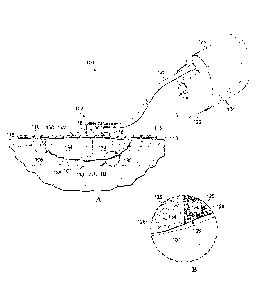
[0(08] FIGS. 1A and 1B illustrate a partially cross-sectional, perspective
view of a
tissue treatment system according to an illustrative embodiment, the tissue
treatment system
having a dressing filler;
[0009] FIG. 2 illustrates a front view of the dressing filler of the tissue
treatment
system of FIG. 1A, the dressing filler having a plurality of compressed
regions in a
compressed state and a plurality of expanded regions in an expanded state;
[0010] FIG. 3 illustrates a front view of the dressing filler of FIG. 2, both
the
compressed regions and the expanded regions being illustrated in a relaxed
state;
[0011] FIG. 4 illustrates a schematic front view of a dressing filler
according to an
illustrative embodiment, the dressing filler having a compressed region in a
compressed state
and an expanded region in an expanded state;
[0012] FIG. 5 illustrates a schematic front view of the dressing filler of
FIG. 4, the
compressed region and the expanded region being illustrated in a relaxed
state;
[0013] FIG. 6 illustrates a front perspective view of a rack having a
plurality of pins
for insertion into a porous substrate to form compressed regions or expanded
regions within
the porous substrate;
[0014] FIG. 7 illustrates the rack of FIG. 6 with the pins of the rack
inserted into the
porous substrate prior to the compressed region being compressed and prior to
the expanded
region being expanded;
[0015] FIG. 8 illustrates the rack of FIG. 6 with the pins of the rack
inserted into the
porous substrate, the porous substrate being illustrated subsequent to the
compressed region
being compressed and subsequent to the expanded regions being expanded;
[0016] FIG. 9 illustrates a front view of a dressing filler according to an
illustrative
embodiment, the dressing filler have a plurality of compressed regions in a
compressed state
and a plurality of expanded regions in an expanded state;
[0017] FIG. 10 illustrates a front view of the dressing filler of FIG. 9, the
compressed
and expanded regions being illustrated in a relaxed state;
3
CA 02855972 2014-05-14
WO 2013/074829
PCT/US2012/065342
[0018] FIG. 11 illustrates a front view of a dressing filler according to an
illustrative
embodiment, the dressing filler have a compressed region in a compressed
state; and
[0019] FIG. 12 illustrates a front view of the dressing filler of FIG. 11, the
compressed region being illustrated in a relaxed state.
4
CA 02855972 2014-05-14
WO 2013/074829 PCT/US2012/065342
DETAILED DESCRIPTION OF ILLUSTRATIVE EMBODIMENTS
[0020] In the following detailed description of several illustrative
embodiments,
reference is made to the accompanying drawings that fotin a part hereof, and
in which is
shown by way of illustration specific preferred embodiments in which the
subject matter of
this specification may be practiced. These embodiments are described in
sufficient detail to
enable those skilled in the art to practice the disclosed subject matter, and
it is understood that
other embodiments may be utilized and that logical, structural, mechanical,
electrical, and
chemical changes may be made without departing from the scope of this
specification. To
avoid detail not necessary to enable those skilled in the art to practice the
embodiments
described herein, the description may omit certain information known to those
skilled in the
art. The following detailed description is, therefore, not to be taken in a
limiting sense, and the
scope of the illustrative embodiments are defined only by the appended claims.
Unless
otherwise indicated, as used herein, "or" does not require mutual exclusivity.
[0021] The tei ___ "reduced pressure" as used herein generally refers to a
pressure less
than the ambient pressure at a tissue site that is being subjected to
treatment. In most cases,
this reduced pressure will be less than the atmospheric pressure at which the
patient is located.
Alternatively, the reduced pressure may be less than a hydrostatic pressure
associated with
tissue at the tissue site. Although the terms "vacuum" and "negative pressure"
may be used to
describe the pressure applied to the tissue site, the actual pressure
reduction applied to the
tissue site may be significantly less than the pressure reduction notinally
associated with a
complete vacuum. Reduced pressure may initially generate fluid flow in the
area of the tissue
site. As the hydrostatic pressure around the tissue site approaches the
desired reduced
pressure, the flow may subside, and the reduced pressure is then maintained.
Unless otherwise
indicated, values of pressure stated herein are gauge pressures. Similarly,
references to
increases in reduced pressure typically refer to a decrease in absolute
pressure, while decreases
in reduced pressure typically refer to an increase in absolute pressure.
[0022] The tissue treatment systems and methods described herein improve the
treatment of a tissue site by providing a porous substrate that is used in
conjunction with
reduced pressure tissue treatment. The porous substrate includes a wound-
facing surface that
CA 02855972 2014-05-14
WO 2013/074829 PCT/US2012/065342
is capable of contacting the tissue site and creating microstrain at the
tissue site. The
microstrain is created by the force applied to the tissue site by the wound-
facing surface of the
porous substrate. When the porous substrate is an open-celled foam, the force
is transmitted to
the tissue site by the cell walls or struts of the foam. The application of
force to the porous
substrate produces a force distribution that provides forces to the tissue
site at any point that is
contacted by the porous substrate. This force distribution therefore results
in a particular
microstrain distribution, which of course will vary based on the porosity and
other
characteristics of the porous substrate, as well as how the porous substrate
is positioned at the
tissue site. Since microstrain at the tissue site assists in the development
of new granulation
tissue, it is beneficial to vary the distribution of force and microstrain
during treatment such
that a more even development of granulation tissue is obtained.
[0023] Referring to FIGS. lA and 1B, an illustrative embodiment of a reduced
pressure tissue treatment system 100 for treating a tissue site 101 on a
patient includes a
dressing 102 placed proximate to the tissue site 101 and a therapy unit 104
fluidly coupled to
the dressing 102. As used herein, the term "tissue site" may refer to a wound,
such as a
wound, or defect located on or within any tissue, including but not limited
to, bone tissue,
adipose tissue, muscle tissue, neural tissue, dermal tissue, vascular tissue,
connective tissue,
cartilage, tendons, or ligaments. The term "tissue site" may further refer to
areas of any tissue
that are not necessarily wounded or defective, but are instead areas in which
it is desired to add
or promote the growth of additional tissue. For example, reduced pressure
tissue treatment
may be used in certain tissue areas to grow additional tissue that may be
harvested and
transplanted to another tissue location.
[0024] The dressing 102 is configured to promote the growth of new tissue at
the
tissue site 101 and includes a dressing filler 106 positioned adjacent to or,
in some
embodiments, in contact with the tissue site 101. The dressing 102 may further
include a cover
110 or drape positioned over the dressing filler 106 to secure the dressing
filler 106 at the
tissue site 101 and to seal a space that is located beneath the cover and that
is at least partially
occupied by the dressing filler 106. In one embodiment, the cover 110 extends
beyond a
perimeter of the tissue site 101 and is placed either in contact with or
otherwise in proximity to
6
CA 02855972 2014-05-14
WO 2013/074829 PCT/US2012/065342
a patient's epidermis 113 to create a fluid seal between the cover 110 and the
epidermis 113.
The cover 110 may include an adhesive 115 or bonding agent to secure the cover
110 to the
epidermis 113. In one embodiment, the adhesive 115 may be used to create a
seal between the
cover 110 and the epidermis 113 to prevent leakage of reduced pressure from
the tissue site
101. In another embodiment, a seal layer (not shown) such as, for example, a
hydrogel or
other material may be disposed between the cover 110 and the epidermis 113 to
augment or
substitute for the sealing properties of the adhesive 115. As used herein,
"fluid seal" means a
seal adequate to maintain reduced pressure at a desired site given the
particular reduced
pressure source involved and the particular treatment desired. In one
embodiment, the cover
110 and the bonding characteristics of the cover 110 provide sealing
sufficient to prevent
leakage greater than 0.5 L/min at 125 mmIIg reduced pressure.
[0025] The dressing 102 further may include a reduced pressure adapter or
interface
116 fluidly coupled to the space beneath the cover 110. In one embodiment, the
interface 116
may be positioned adjacent to or coupled to the cover 110 to provide fluid
access to the
dressing filler 106 and the tissue site 101. The cover 110 includes an
aperture 118 for
providing fluid access to the interface 116. A conduit 120 fluidly couples the
therapy unit 104
and the interface 116. The interface 116 is capable of delivering reduced
pressure to the tissue
site 101.
[0026] In one embodiment, the therapy unit 104 includes a fluid containment
member
122 in fluid communication with a reduced pressure source 124. In the
embodiment illustrated
in FIG. 1, the fluid containment member 122 is a collection canister that
includes a chamber
for collecting fluids from the tissue site 101. The fluid containment member
122 alternatively
could be an absorbent material or any other container, device, or material
that is capable of
collecting fluid.
[0027] The conduit 120 may be a multi-lumen tube that is capable of providing
one or
more conduits to deliver reduced pressure to the dressing 102 and one or more
conduits to
sense the amount of pressure at the tissue site 101. Liquids or exudates
communicated from
the dressing filler 106 through the conduit 120 are removed from the conduit
120 and retained
within the collection canister 122.
7
[0028] Referring still to FIG. 1A, the reduced pressure source 124 may he an
electrically-driven vacuum pump. In another implementation, the reduced
pressure source 124
instead may be a manually-actuated or manually-charged pump that does not
require electrical
power. In one embodiment, the reduced pressure source 124 may be one or more
piezoelectric-actuated micropumps that may be positioned remotely from the
dressing 102, or
at the dressing beneath or adjacent to the cover 110. The reduced pressure
source 124 instead
may he any other type of pump, or alternatively a wall suction port or air
delivery port such as
those available in hospitals and other medical facilities. The reduced
pressure source 124 may
be housed within or used in conjunction with the therapy unit 104, which may
also contain
sensors, processing units, alarm indicators, memory, databases, software,
display units, and
user interfaces that further facilitate the application of reduced pressure
treatment to the
tissue site 101. In one example, pressure-detection sensors (not shown) may be
disposed at or
near the reduced pressure source 124. The pressure-detection sensors may
receive pressure
data from the interface 116 via lumens in the conduit 120 that are dedicated
to delivering
reduced pressure data to the pressure-detection sensors. The pressure-
detection sensors may
communicate with a processing unit that monitors and controls the reduced
pressure that is
delivered by the reduced pressure source 124.
[0029] In the embodiment illustrated in FIGS. IA and 1B, the dressing filler
106
comprises a porous substrate that may be in one embodiment a porous foam. More
specifically, the porous substrate may be an open-celled foam such as the open-
celled,
reticulated polyurethane foam sold under the name GRANUFOAMO by Kinetic
Concepts,
Inc. of San Antonio, Texas. Alternatively, a non-reticulated foam or a foam
comprised of
biocompatible materials other than polyurethane may be used. In one
embodiment, a
bioabsorbable foam or other porous substrate may be employed. Examples of
other materials
that may he suitable porous foams or porous substrates include those formed
from acrylics,
acrylates, thermoplastic elastomers (for example, styrene ethylene butene
styrene (SEBS) and
other block copolymers), polyether block polyamide (PEBAX), silicone
elastomers, poly
caprolactam, poly lactic acid, and polyolefins, such as polythene and
polypropylene. Still
8
CA 2855972 2018-12-10
CA 02855972 2014-05-14
WO 2013/074829 PCT/US2012/065342
other biocompatible materials may be used if capable of being formed or
otherwise made into
a porous substrate as described herein.
[0030] The porous substrate preferably includes a plurality of openings or
other flow
channels that facilitate movement of fluids and distribution of reduced
pressure in the sealed
space beneath the cover. In an embodiment employing a foam such as the
reticulated,
polyurethane foam, the flow channels are provided by openings 125 or cells
within the foam.
The foam further may include cell walls or struts 126 that form a framework
for the openings
125 (see FIG. 1B). The struts 126 press upon the tissue site 101 when a force
is applied to the
dressing filler 106, thereby creating distribution of forces and microstrain
at the tissue site 101
that depends on the local positioning of the struts 126.
[0031] Referring still to FIGS. 1A and 1B, but also to FIGS. 2 and 3, the
dressing
filler 106 includes a plurality of first, or compressed regions 130 and a
plurality of second, or
expanded regions 134 arranged in vertically-oriented layers that extend from a
wound-facing
surface 138 of the porous dressing filler 106 to an opposing surface 142. The
compressed
regions 130 illustrated in FIGS. 1A, 1B, and 2 are in a compressed state,
while the same
compressed regions 130 illustrated in FIG. 3 are in a relaxed state. Similarly
the expanded
regions 134 illustrated in FIGS. 1A, 1B, and 2 are in an expanded state, while
the same
expanded regions 134 illustrated in FIG. 3 are in a relaxed state. Referring
more specifically to
FIG. 1B, the effect of compressing or expanding the dressing filler 106 is
depicted. When the
compressed regions 130 are in the compressed state and the expanded regions
134 are in the
expanded state (as depicted in FIG. 1B), the density of struts per unit volume
in the
compressed regions 130 is greater than the density of struts 126 per unit
volume in the
expanded regions 134. When the compressed regions 130 and the expanded regions
134 are in
the relaxed state (as depicted in FIG. 3), the density of struts per unit
volume in the
compressed regions 130 is approximately equal to the density of struts 126 per
unit volume in
the expanded regions 134.
[0032] While the phrase "compressed region" has been used to describe certain
regions of the dressing filler 106 that are in some cases compressed, the
phrase also is capable
of describing a region that has assumed a relaxed state from a compressed
state. In some
9
CA 02855972 2014-05-14
WO 2013/074829 PCT/US2012/065342
situations, due to the compression of the dressing filler 106 during reduced
pressure tissue
treatment, as is described more fully herein, certain compressed regions may
actually
experience expansion in certain directions when compared to the original
compressed state.
Similarly, the "expanded region" is capable of existing in multiple states, at
least two of which
are the expanded state and the relaxed state. Again, it is conceivable during
the application of
reduced pressure to the dressing filler 106 that certain expanded regions may
undergo some
compression beneath the cover 110.
[0033] In general, the description of the dressing filler 106 as having a
compressed
state or an expanded state is meant to describe the forces acting on the foam.
When a region of
the dressing filler 106 is in the compressed state, the forces acting on the
region, which are
represented by arrows 150 in FIG. 2, are directed in an outward direction such
that the region,
if not constrained, would expand in at least one direction. When a region of
the dressing filler
106 is in the expanded state, the forces acting on the region, which are
represented by arrows
154 in FIG. 2, are directed in an inward direction such that the region, if
not constrained,
would contract in at least one direction. When a region of the dressing filler
106 is in the
relaxed state, no significant forces are acting on the region.
[0034] Referring still to FIGS. 1A, 1B, and 2, the compressed regions 130 are
held or
secured in the compressed state by a first coating. Similarly, the expanded
regions 134 are
held or secured in the expanded state by a second coating. In one embodiment,
the first and
second coatings are the same coating. The first and second coatings may be any
material that
is capable of securing the dressing filler 106 in the compressed state or the
expanded state and
that is then capable of being removed such that the dressing filler 106 can
change to a relaxed
state. In one embodiment, the first and second coatings may be capable of
dissolving in the
presence of a fluid. Dissolvable coatings allow the coating to be removed as
exudate from the
tissue site 101 enters the dressing filler 106. Alternatively, a liquid such
as a saline solution, or
any other biocompatible solvent or liquid, may be delivered to the dressing
filler 106 when it is
desired to remove the first and second coatings. Examples of coatings that may
be used
include without limitation starches or other sugars, polyvinyl alcohol,
polyvinyl pyrrolidone,
carboxy methyl cellulose (CMC) and its salts and esters, alginates, gums such
as guar and
CA 02855972 2014-05-14
WO 2013/074829 PCT/US2012/065342
xanthan, or other polymers. Water soluble salts or effervescing mixes (such as
tartaric acid
and carbonate or bicarbonate salts) may be incorporated into water insoluble
polymers. When
exposed to water the water soluble salts are leached from the polymer, or in
the case of the
effervescent mixes gas is released ¨ in both cases creating porosity in the
carrier polymer, and
weakening it, allowing the physical form of the dressing filler to change. The
time period over
which the coatings described herein may release will vary depending on the
desired treatment
regimen. In one embodiment, the coating may be configured to release as
quickly as within a
few hours (e.g. 4 hours) or as long as a day or more.
[0035] The coatings may be positioned on the exterior surfaces of the dressing
filler
106 or alternatively may be used to coat both exterior surfaces and the inner
passages of the
dressing filler 106. In one embodiment, it may be desirable to uniformly coat
the dressing
filler 106 both externally and internally. Delivery of the coating to the
dressing filler 106 may
be accomplished by dipping the dressing filler 106 in the coating, or by
spraying, brushing,
rolling, or otherwise applying the coating. In one embodiment, the coating may
be applied to a
unitary dressing filler 106 that includes regions that have been compressed
and expanded.
Alternatively, the coating may be applied to separate pieces of compressed or
expanded
material that are then assembled to build the dressing filler 106. The
attachment of multiple
pieces to build the dressing filler 106 may be accomplished by bonding,
mechanical fastening,
welding, or any other attachment means.
[0036] Referring to FIG. 3, as the coating is removed, both compressed and
expanded
regions are released and move toward a relaxed state as shown in FIG. 3. In
the relaxed state,
assuming the same type of dressing filler is used in both the compressed and
expanded regions,
the density of openings and cell struts per unit volume in the compressed
regions will be
approximately equal to the density of openings and cell struts per unit volume
in the expanded
regions. By compressing a particular compressed region the same approximate
amount as a
corresponding expanded region is expanded, the expansion of a compressed
region to the
relaxed state may be offset by the contraction of an expanded region to the
relaxed state. In
other words, when a dressing having expanded and compressed regions moves to a
relaxed
state, the dressing undergoes a net-zero volume change.
11
CA 02855972 2014-05-14
WO 2013/074829 PCT/US2012/065342
[0037] As an alternative to the coating, a removable sheath or other covering
may be
placed around the dressing filler 106 when the various regions have been
charged to their
appropriate compressed or expanded states. The sheath may be a dissolvable or
bioabsorbable
substance such as a woven or non-woven fabric, polyvinyl pyrrolidone, carboxy
methyl
cellulose (CMC) and its salts and esters, alginates, gums such as guar and
xanthan, polyvinyl
alcohol, polycaprolactam, poly lactic acid and their copolymers or blends, or
other polymers or
materials. In one embodiment, if a sheath or other covering is used, the
sheath may surround
the dressing filler 106 completely, or may partially surround a portion of the
dressing filler
106. If a dissolvable or bioabsorbable sheath is employed, the particular
material used and its
thickness may be chosen to provide release based on a desired time period. For
example, a
sheath that will dissolve within 12 hours may be chosen to provide treatment
for up to 12 hours
with the regions of dressing filler 106 in the compressed and expanded states.
In this particular
example, following dissolution of the sheath at or around 12 hours allows the
regions of the
dressing filler 106 to change to a relaxed state, thereby changing the
microstrain profile at the
tissue site 101. In another embodiment, the sheath may instead be manually
removable from
the dressing filler 106 by a care giver or the patient when the change in
microstrain profile is
desired.
[0038] Referring to FIGS. 4 and 5, a dressing filler 406 according to an
illustrative
embodiment comprises a porous substrate that is similar to the porous
substrate described
above with reference to FIGS. 1-3. Dressing filler 406 further includes a
compressed region
430 and an expanded region 434 similar to those previously described. The
dressing filler 406
also includes nodes 412 which may be projections to transmit forces to a
tissue site and thus
generate microstrain. Nodes 412 may also be representative of the struts of
dressing filler 106.
Visualization of the nodes 412 illustrated in FIGS. 4 and 5 allows an
understanding of how the
expansion of the compressed region 430 and the contraction of the expanded
region 434
benefit tissue site treatment and wound healing. In FIG. 4, nodes 412 have a
first position
indicated by nodes 412a. As the coating securing the compressed and expanded
regions 430,
434 is removed, the compressed region 430 expands and the expanded region 434
contracts,
which moves the nodes 412 to a second position indicated by nodes 412b. As
illustrated in
CA 02855972 2014-05-14
WO 2013/074829 PCT/US2012/065342
FIG. 5, the repositioning of the nodes 412 during the expansion and
contraction of certain
regions of the dressing filler 406 allow a spatial redistribution of the point
loads applied by the
nodes 412 to the tissue site. This in turn creates a different microstrain
profile (i.e. the
distribution of microstrain) at the tissue site, thereby aiding in the even
development of
granulation tissue and preventing the adhesion of new tissue growth to the
dressing filler 406.
[0039] Referring to FIGS. 6-8, a dressing filler 606 similar to dressing
fillers 106, 406
is illustrated prior to being placed into a compressed or expanded state. To
prepare the
dressing filler 606 for use in tissue treatment, the dressing filler 606 is
compressed or expanded
in sections or regions. In FIG. 6, a first region, or compressed region 610
and a second region,
or expanded region 614 are both illustrated in a relaxed state. A plurality of
racks 622 each
having a plurality of pins 626 are provided to provide the necessary
compression or expansion
of the dressing filler 606. The racks 622 are slidably positioned on one or
more stringers 630
that provide support for the racks 622 and constrain the racks to movements
along one axis. In
other embodiments, racks may be provided that allow movement, and thus
compression or
expansion of the dressing filler, along multiple axes.
[0040] As more specifically illustrated in FIGS. 6 and 7, the dressing filler
is
positioned on the pins 626 of the racks 622 such that the pins 626 of at least
two of the racks
penetrate each region 610, 614. To compress or expand each region 610, 614,
the racks 622
are slidably moved along the stringers 630 in the directions illustrated by
arrows 640, 644 in
FIG. 7. To move the compressed region 610 of the dressing filler 606 to a
compressed state,
the two racks 622 associated with the compressed region 610 are moved toward
one another as
indicated by arrows 640. To move the expanded region 614 of the dressing
filler 606 to an
expanded state, the two racks 622 associated with the expanded region 614 are
moved away
from one another as indicated by arrows 644. The movement of the racks 622 may
be
performed manually, or may instead be hydraulically, pneumatically,
electrically, or
mechanically driven.
[0041] Following the movement of the racks 622 in the directions indicated by
arrows
640, 644 the compressed region 610 is in the compressed state and the expanded
region 614 is
in the expanded sate (see FIG. 8). The regions 610, 614 of the dressing filler
606 may be held
13
CA 02855972 2014-05-14
WO 2013/074829 PCT/US2012/065342
or secured in the compressed and expanded states by applying a removable
coating or sheath to
the dressing filler 606 as previously described herein. After the dressing
filler 606 has been
locked by the coating or sheath, the pins 626 of the racks 622 may be removed
from the
dressing filler 606.
[0042] FIGS. 6-8 provide only one example of how the various regions of the
dressing fillers described herein may be compressed or expanded. Other devices
that penetrate
or contact the regions of the dressing filler may be used to compress or
expand the dressing
filler. While the examples described above provide a method for compressing
and expanding
different portions of a unitary piece of foam or porous substrate, the various
regions that
require compression or expansion may instead by compressed or expanded
independently of
one another and then joined together to form the dressing tiller. For example,
in one
embodiment, two pieces of a porous foam in a relaxed state may be provided.
One of the
pieces of porous foam may be compressed and secured in the compressed state.
The other
piece of porous foam may be expanded and secured in the expanded state.
Following the steps
of compressing or expanding each piece of foam individually, the two pieces of
foam may be
adhesively bonded, thermally coupled, mechanically attached, ultrasonically
welded, or
otherwise connected to form a unitary dressing filler.
[0043] Referring to FIGS. 9 and 10, a dressing filler 906 according to an
illustrative
embodiment is capable of being used with a reduced pressure treatment system
similar to
reduced pressure treatment system 100 of FIG. 1. The dressing filler 906
comprises a porous
substrate that may be in one embodiment a porous foam. More specifically, the
porous
substrate may be an open-celled foam such as the open-celled, reticulated
polyurethane foam
sold under the name GRANUFOAMO by Kinetic Concepts, Inc. of San Antonio,
Texas.
Alternatively, a non-reticulated foam or a foam comprised of biocompatible
materials other
than polyurethane may be used. In one embodiment, a bioabsorbable foam or
other porous
substrate may be employed. Examples of other materials that may be suitable
porous foams or
porous substrates include those fondled from acrylics, acrylates,
thermoplastic elastomers (for
example, styrene ethylene butene styrene (SEBS) and other block copolymers),
polyether
block polyamide (PEBAX), silicone elastomers, poly caprolactam, poly lactic
acid, and
14
CA 02855972 2014-05-14
WO 2013/074829 PCT/US2012/065342
polyolefins, such as polythene and polypropylene. Still other biocompatible
materials may be
used if capable of being formed or otherwise made into a porous substrate as
described herein.
[0044] The porous substrate preferably includes a plurality of openings or
other flow
channels that facilitate movement of fluids and distribution of reduced
pressure in the sealed
space beneath the cover. In an embodiment employing a foam such as the
reticulated,
polyurethane foam, the flow channels are provided by openings or cells within
the foam
similar to those described previously with reference to FIGS. 1A and 1B. The
foam further
may include cell walls or struts that form a framework for the openings as
previously
described.
[0045] The dressing filler 906 includes a plurality of first, or compressed
regions 930
and a plurality of second, or expanded regions 934 arranged in horizontally-
oriented layers
located between a wound-facing surface 938 of the porous dressing filler 906
and an opposing
surface 942. The compressed regions 930 illustrated in FIG. 9 are in a
compressed state, while
the same compressed regions 930 illustrated in FIG. 10 are in a relaxed state.
Similarly the
expanded regions 934 illustrated in FIG. 9 are in an expanded state, while the
same expanded
regions 934 illustrated in FIG. 10 are in a relaxed state.
[0046] The forces acting on the compressed region 930 in the compressed state
are
represented by arrows 950 in FIG. 9. These particular forces are directed in
an outward
direction such that the compressed region 930 in the compressed state, if not
constrained,
would expand in at least one direction. The forces acting on the expanded
region 934 in the
expanded state are represented by arrows 954 in FIG. 9. These forces are
directed in an inward
direction such that the expanded region 934 in the expanded state, if not
constrained, would
contract in at least one direction. When the compressed regions 930 or the
expanded regions
934 of the dressing filler 906 are in the relaxed state (see FIG. 10), no
significant forces are
acting on the regions.
[0047] Referring still to FIG. 9, the compressed regions 930 are held or
secured in the
compressed state by a first coating. Similarly, the expanded regions 934 are
held or secured in
the expanded state by a second coating. In one embodiment, the first and
second coatings are
the same coating. The first and second coatings may be any material that is
capable of
CA 02855972 2014-05-14
WO 2013/074829
PCT/US2012/065342
securing the dressing filler 906 in the compressed state or the expanded state
and that is then
capable of being removed such that the dressing filler 906 may change to a
relaxed state. In
one embodiment, the first and second coatings may be capable of dissolving in
the presence of
a fluid. Dissolvable coatings allow the coating to be removed as exudate from
a tissue site
enters the dressing filler 906. Alternatively, a liquid such as a saline
solution, or any other
biocompatible solvent or liquid, may be delivered to the dressing filler 906
when it is desired
to remove the first and second coatings. Examples of coatings that may be used
include
without limitation starches or other sugars, polyvinyl alcohol, polyvinyl
pyffolidone, carboxy
methyl cellulose (CMC) and its salts and esters, alginates, gums such as guar
and xanthan, or
other polymers. Water soluble salts or effervescing mixes (such as tartaric
acid and carbonate
or bicarbonate salts) may be incorporated into water insoluble polymers. When
exposed to
water the water soluble salts are leached from the polymer, or in the case of
the effervescent
mixes gas is released ¨ in both cases creating porosity in the carrier
polymer, and weakening it,
allowing the physical form of the dressing filler to change. The time period
over which the
coatings described herein may release will vary depending on the desired
treatment regimen.
In one embodiment, the coating may be configured to release as quickly as
within a few hours
(e.g. 4 hours) or as long as a day or more.
[0048] The coatings may be positioned on the exterior surfaces of the dressing
filler
906 or alternatively may he used to coat both exterior surfaces and the inner
passages of the
dressing filler 906. In one embodiment, it may be desirable to unifomily coat
the dressing
filler 906 both externally and internally. Delivery of the coating to the
dressing filler 906 may
be accomplished by dipping the dressing filler 906 within the coating, or by
spraying,
brushing, rolling, or otherwise applying the coating. In one embodiment, the
coating may be
applied to a unitary dressing filler 906 that includes regions that have been
compressed and
expanded. Alternatively, the coating may be applied to separate pieces of
compressed or
expanded material that is then assembled to build the dressing filler 906. The
attachment of
multiple pieces to build the dressing filler 906 may be accomplished by
bonding, mechanical
fastening, welding, or any other attachment means.
16
CA 02855972 2014-05-14
WO 2013/074829 PCT/US2012/065342
[0049] Prior to removal of the coating, the wound-facing surface 938 of the
dressing
filler 906 is irregular or non-planar, with indentations 962 created by the
compression of the
compressed regions 930. The wound-facing surface 938 further includes
protrusions 966
created by the expansion of the expanded regions 934. The location of the
indentations 962
and protrusions 966 along the wound-facing surface may form a regular grid-
type or other
pattern, or may instead be more random in nature. While the compressed regions
930 are
located adjacent to and incorporate the wound-facing surface 938, the
compressed regions 930
could instead be spaced apart from the wound-facing surface 938 and located
closer to
opposing surface 942. Similarly, while the expanded regions 934 are spaced
apart from the
wound-facing surface 938 and are located closer to the opposing surface 942,
the expanded
regions 934 could instead be positioned adjacent to and may even incorporate
the wound-
facing surface 938.
[0050] Referring to FIG. 10, as the coating is removed, both the compressed
and
expanded regions are released and move toward a relaxed state as shown in FIG.
10. In the
relaxed state, assuming the same type of dressing filler is used in both the
compressed and
expanded regions, the density of openings and cell struts per unit volume in
the compressed
regions will be approximately equal to the density of openings and cell struts
per unit volume
in the expanded regions. The height of the expanded regions 934 typically
decreases as the
expanded regions 934 move to the relaxed state, while the height of the
compressed regions
930 typically increases as the compressed regions 930 move to the relaxed
state. The change
in height of the compressed and expanded regions results in a change in the
profile of the
wound-facing surface 938. While the compressed regions 930 had previously been
associated
with indentations 962 (see FIG. 9), the expansion of the compressed regions
930 to the relaxed
state results in the formation of protrusions 970 (see FIG. 10). The expanded
regions 934,
which had previously been associated with protrusions 966 (see FIG. 9),
contract to the relaxed
state such that indentations 974 are foimed (see FIG. 10). This reversal of
protrusions and
indentations on the wound-facing surface 938 changes the interface of the
dressing filler 906
with a tissue site and therefore is capable of changing the distribution of
loads and microstrain
at the tissue site during treatment.
17
CA 02855972 2014-05-14
WO 2013/074829
PCT/US2012/065342
[0051] As an alternative to the coating, a removable sheath or other covering
may be
placed around the dressing filler 906 when the various regions have been
charged to their
respective compressed or expanded states. The sheath may be a dissolvable or
bioabsorbable
substance such as a woven or non-woven fabric, polyvinyl pyrrolidone, carboxy
methyl
cellulose (CMC) and its salts and esters, alginates, gums such as guar and
xanthan, polyvinyl
alcohol, polycaprolactam, poly lactic acid and their copolymers or blends, or
other polymers or
materials. In one embodiment, if a sheath or other covering is used, the
sheath may surround
the dressing filler 906 completely, or may partially surround a portion of the
dressing filler
906. If a dissolvable or bioabsorbable sheath is employed, the particular
material used and its
thickness may be chosen to provide release based on a desired time period. In
another
embodiment, the sheath may instead be manually removable from the dressing
filler 906 by a
care giver or the patient when the change in microstrain profile is desired.
[0052] Referring to FIGS. 11 and 12, a dressing filler 1106 according to an
illustrative embodiment is capable of being used with a reduced pressure
treatment system
similar to reduced pressure treatment system 100 of FIG. 1. The dressing
filler 1106
comprises a porous substrate that may be in one embodiment a porous foam. More
specifically, the porous substrate may be an open-celled foam such as the open-
celled,
reticulated polyurethane foam sold under the name GRANUFOAM by Kinetic
Concepts,
Inc. of San Antonio, Texas. Alternatively, a non-reticulated foam or a foam
comprised of
biocompatible materials other than polyurethane may be used. In one
embodiment, a
bioabsorbable foam or other porous substrate may be employed. Examples of
other materials
that may be suitable porous foams or porous substrates include those fonned
from acrylics,
acrylates, thermoplastic elastomers (for example, styrene ethylene butene
styrene (SEBS) and
other block copolymers), polyether block polyamide (PEBAX), silicone
elastomers, poly
caprolactam, poly lactic acid, and polyolefins, such as polythene and
polypropylene. Still
other biocompatible materials may be used if capable of being formed or
otherwise made into
a porous substrate as described herein.
[0053] The porous substrate preferably includes a plurality of openings or
other flow
channels that facilitate movement of fluids and distribution of reduced
pressure in the sealed
18
CA 02855972 2014-05-14
WO 2013/074829 PCT/US2012/065342
space beneath the cover. In an embodiment employing a foam such as the
reticulated,
polyurethane foam, the flow channels are provided by openings or cells within
the foam
similar to those described previously with reference to FIGS. 1A and 1B. The
foam further
may include cell walls or struts that form a framework for the openings as
previously
described.
[0054] The dressing filler 1106 includes one or more compressed regions 1130
arranged in horizontally-oriented layers located between a wound-facing
surface 1138 of the
porous dressing filler 1106 and an opposing surface 1142. The compressed
region 1130
illustrated in FIG. 11 is in a compressed state, while the same compressed
region 1130
illustrated in FIG. 12 is in a relaxed state. The forces acting on the
compressed region 1130 in
the compressed state are represented by arrows 1150 in FIG. 11. These
particular forces are
directed in an outward direction such that the compressed region 1130 in the
compressed state,
if not constrained, would expand in at least one direction. When the
compressed region 1130
of the dressing filler 1106 is in the relaxed state (see FIG. 12), no
significant forces act upon
the compressed region 1130.
[0055] Referring still to FIG. 11, the compressed region 1130 is held or
secured in the
compressed state by a coating. The coating may be any material that is capable
of securing the
dressing filler 1106 in the compressed state and that is then capable of being
removed such that
the dressing filler 1106 may change to a relaxed state. In one embodiment, the
coating may be
capable of dissolving in the presence of a fluid. Dissolvable coatings allow
the coating to be
removed as exudate from a tissue site enters the dressing filler 1106.
Alternatively, a liquid
such as a saline solution, or any other biocompatible solvent or liquid, may
be delivered to the
dressing filler 1106 when it is desired to remove the coating. Examples of
coatings that may
be used include without limitation starches or other sugars, polyvinyl
alcohol, polyvinyl
pyrrolidone, carboxy methyl cellulose (CMC) and its salts and esters,
alginates, gums such as
guar and xanthan, or other polymers. Water soluble salts or effervescing mixes
(such as
tartaric acid and carbonate or bicarbonate salts) may be incorporated into
water insoluble
polymers. When exposed to water the water soluble salts are leached from the
polymer, or in
the case of the effervescent mixes gas is released ¨ in both cases creating
porosity in the carrier
19
CA 02855972 2014-05-14
WO 2013/074829
PCT/US2012/065342
polymer, and weakening it, allowing the physical form of the dressing filler
to change. The
time period over which the coatings described herein may release will vary
depending on the
desired treatment regimen. In one embodiment, the coating may be configured to
release as
quickly as within a few hours (e.g. 4 hours) or as long as a day or more.
[0056] The coatings may be positioned on the exterior surfaces of the dressing
filler
1106 or alternatively may be used to coat both exterior surfaces and the inner
passages of the
dressing filler 1106. In one embodiment, it may be desirable to uniformly coat
the dressing
filler 1106 both externally and internally. Delivery of the coating to the
dressing filler 1106
may be accomplished by dipping the dressing filler 1106 within the coating, or
by spraying,
brushing, rolling, or otherwise applying the coating. In one embodiment, the
coating may be
applied to a unitary dressing filler 1106 that includes regions that have been
compressed.
Alternatively, the coating may be applied to separate pieces of compressed
material that is then
assembled to build the dressing filler 1106. The attachment of multiple pieces
to build the
dressing filler 1106 may be accomplished by bonding, mechanical fastening,
welding, or any
other attachment means.
[0057] Referring to FIG. 12, as the coating is removed, the compressed region
is
released and moves toward a relaxed state as shown in FIG. 12. In the relaxed
state, assuming
the same type of dressing filler is used in the compressed and the rest of the
dressing filler
1106, the density of openings and cell struts per unit volume in the
compressed regions will be
approximately equal to the density of openings and cell struts per unit volume
in the remainder
of the dressing filler 1106. The height of the compressed region 1130
typically increases as
the compressed region 1130 moves to the relaxed state.
[0058] Unlike dressing filler 106 in which there may be zero net volume gain,
the
presence of the compressed region 1130 with no expanded region in the dressing
filler 1106
results in a positive net volume gam when the compressed region 1130 moves to
the relaxed
state. When the dressing filler 1106 is sealed beneath a cover similar to
cover 110 during
tissue treatment, the change in height of the compressed region, and thus the
increase in
volume of the dressing filler 1106, results in an increase in the force
applied to the tissue site.
This increase in force could be used to ensure patient comfort by gradually
increasing the force
CA 02855972 2014-05-14
WO 2013/074829 PCT/US2012/065342
applied to the tissue site as treatment begins. Alternatively, the increase in
force may allow a
wider range of treatment regimens that promote new tissue growth.
[0059] As an alternative to the coating, a removable sheath or other covering
may be
placed around the dressing filler 1106 when the compressed region has been
charged to its
compressed state. The sheath may be a dissolvable or bioabsorbable substance
such as a
woven Or non-woven fabric, polyvinyl pyrrolidone, carboxy methyl cellulose
(CMC) and its
salts and esters, alginates, gums such as guar and xanthan, polyvinyl alcohol,
polycaprolactam,
poly lactic acid and their copolymers or blends, or other polymers or
materials. In one
embodiment, if a sheath or other covering is used, the sheath may surround the
dressing filler
1106 completely, or may partially surround a portion of the dressing filler
1106. If a
dissolvable or bioabsorbable sheath is employed, the particular material used
and its thickness
may be chosen to provide release based on a desired time period. In another
embodiment, the
sheath may instead be manually removed from the dressing filler 1106 by a care
giver or the
patient when the change in treatment force is desired.
[0060] Each of the dressing fillers 106, 406, 606, 906, 1106 described herein
may be
used with the reduced pressure treatment system 100 of HG. 1 or similar
reduced pressure
treatment systems to encourage new tissue growth at a tissue site. During
reduced pressure
treatment, as reduced pressure is applied to the sealed space beneath the
cover, the cover
presses on the dressing filler, thereby urging the dressing filler toward the
tissue site. Some of
the structural components (e.g. struts 126 or nodes 412) of the dressing
filler contact the tissue
site, and these structural components provide point loads to the tissue site,
thereby creating a
particular distribution of microstrain across the tissue site. When the
coating associated with
the dressing filler is removed, the various regions of the dressing filler
that have been
expanded or compressed change state, which prompts movement of the dressing
filler and the
associated structural components. In the case of dressing fillers 106, 406,
606, and 906, this
movement causes a spatial redistribution of the point loads applied by the
structural
components to the tissue site. This in turn creates a different microstrain
profile (i.e. the
distribution of microstrain) at the tissue site and helps prevent adhesion of
new tissue growth
to the dressing filler. In the case of dressing fillers 906, 1106, the
movement of the dressing
21
CA 02855972 2014-05-14
WO 2013/074829 PCT/US2012/065342
filler is capable of changing the magnitude of the point loads applied to the
tissue site. This
also creates a different microstrain profile which is beneficial to wound
healing.
[0061] The systems and methods described herein allow modification of the
microstrain experienced by a tissue site without changing the dressing at the
tissue site. In
some cases, the microstrain modification involves simply a redistribution of
the microstrain
profile, while in other cases, the amplitude of the microstrain may be
increased or decreased.
The dressings described herein each incorporate nodes, struts, or other strain-
inducing
structures that include at least one compressed region that is selectively
held in a compressed
state or expanded region that is selectively held in an expanded state. When
released, the
compressed or expanded regions move to a relaxed state, which changes the
distribution or
amount of microstrain applied to the tissue site. Each of the dressings
described herein is
therefore believed to improve the treatment of a tissue site using reduced
pressure tissue
treatment, since changing the microstrain profile during reduced pressure
treatment will result
in more even formation of granulation tissue and will prevent adhesion of new
tissue to the
dressing.
[0062] While many of the systems described herein have been illustrated in use
with
tissue sites or wounds that are at or near the epidennis of a patient, the
systems and methods
may similarly be used to treat subcutaneous tissue sites, tunnel wounds, or
other undeimined
areas of tissue.
[0063] While a number of discrete embodiments have been described, aspects of
each
embodiment may not be specific to only that embodiment and it is specifically
contemplated
that features of embodiments may be combined with features of other
embodiments. While the
subject matter of this specification is shown in only a few of its forms, it
is susceptible to
various changes and modifications without departing from the scope thereof.
[0064] Variations on the embodiments described herein comprising both a
compressed and expanded region may be provided utilizing only an expanded or
only a
compressed region. For example, the embodiment of FIG. 1 may be modified to
utilize only
vertical compressed or expanded regions, but not regions of both types.
77
CA 02855972 2014-05-14
WO 2013/074829 PCT/US2012/065342
[0065] Where reference is made to a dimension, part, or region being
horizontal this
is with reference to a plane substantially parallel with the wound-facing
surface of the
dressing. Similarly, vertical is referenced to this plane and so describes a
plane running
substantially perpendicular to the wound-facing surface. Where reference is
made to one part
being 'above' or 'below' another part, this is made with reference to the
wound-facing surface
being the 'bottom' of the dressing.
[0066] The coatings securing the compressed and expanded regions may be
different
such that the first and second coatings may be removed at a different time.
For example, the
coatings may be of different materials, or configured to dissolve over
differing periods of time.
23