Note: Descriptions are shown in the official language in which they were submitted.
CA 02856063 2014-05-15
WO 2013/075248
PCT/CA2012/050847
DEVICES AND METHODS FOR PRODUCING PLANAR POLYMERIC
MATERIALS USING MICROFLUIDICS
CROSS-REFERENCE TO RELATED APPLICATION
This application claims priority to U.S. Provisional Application No.
61/563,506 titled "DEVICE AND METHODS FOR DIGITAL PRINTING" and
filed on November 23, 2011, the entire contents of which are incorporated
herein by reference, and to U.S. Provisional Application No. 61/623,445 titled
"DEVICES AND METHODS FOR PRODUCING CONTROLLED
HETEROGENEITY IN PLANAR MATERIALS USING MICROFLUIDICS" and
filed on April 12, 2012, the entire contents of which are incorporated herein
by
reference.
BACKGROUND
The present disclosure relates to devices and methods for forming
heterogeneous materials using microfluidics.
Materials with a spatially non-uniform composition that is closely linked
to their function are common in nature and often possess a hierarchical
architecture with length scales ranging from hundreds of nanometers to
several millimeters. Currently available strategies for creating materials
with
an organized microscale composition mimic nature's ability in two ways: by
initially preparing building blocks and subsequently assembling them along
fluid interfaces and by replica molding. These strategies necessitate a
sequence of processing steps and often lack spatiotemporal control. To date,
the controlled formation of heterogeneous soft materials has been limited to
1
CA 02856063 2014-05-15
particles (e.g., encapsulated and Janus particles) and coded fibers.
SUMMARY
Methods and devices are disclosed for providing the controlled
formation of planar homogeneous or heterogeneous polymeric materials
using microfluidic devices. In one embodiment, a planar array of microfluidic
channels is employed to produce a flowing liquid sheet having heterogeneous
structure by spatially and temporally controlling dispensing of polymer liquid
from selected microchannels. The resulting liquid sheet is polymerized to
produce a planar heterogeneous material that may be continuously drawn
and/or fed from the plurality of microfluidic channels. The liquid may include
a
payload that may be selectively incorporated into the polymeric structure. In
some embodiments, the local material composition is controllable, thereby
allowing control over local and bulk material properties, such as the
permeability and the elasticity, and of creating materials with directionally
dependent properties.
Accordingly, in one aspect, there is provided a microfluidic device
comprising:
a substantially planar array of microfluidic channels, wherein inlets of
said microfluidic channels are connectable to one or more liquid polymer
dispensing devices for delivering polymer solution at a controlled rate;
a substantially planar channel having:
an inlet in fluid communication with outlets of said microfluidic
channels,
a length such that the polymer solution emerges from an outlet
2
CA 02856063 2014-05-15
WO 2013/075248
PCT/CA2012/050847
of the planar channel as a substantially planar liquid sheet; and
a polymerization reservoir in fluid communication with the outlet of the
planar channel for receiving the planar liquid sheet into an additional
liquid,
such that the planar liquid sheet is polymerizable into a substantially planar
polymeric material within the additional liquid.
In another embodiment, there is provided a method of forming a planar
polymeric material using a microfluidic device, the microfluidic device
comprising:
a substantially planar array of microfluidic channels, wherein inlets of
said microfluidic channels are connected to one or more liquid polymer
dispensing devices for delivering at least one polymer solution at a
controlled
rate;
a substantially planar channel having:
an inlet in fluid communication with outlets of said microfluidic
channels,
a length such that the polymer solution emerges from an outlet
of the planar channel as a substantially planar liquid sheet; and
a polymerization reservoir in fluid communication with the outlet of the
planar channel, wherein the polymerization reservoir contains an additional
liquid;
the method comprising:
controlling the one or more liquid polymer dispensing devices to
dispense the polymer solution into the microfluidic channels at a controlled
rate; and
polymerizing the planar liquid sheet as it emerges from the
3
CA 02856063 2014-05-15
WO 2013/075248
PCT/CA2012/050847
output of the planar channel into the additional liquid, thereby forming a
substantially planar polymeric material.
A further understanding of the functional and advantageous aspects of
the disclosure can be realized by reference to the following detailed
description and drawings.
BRIEF DESCRIPTION OF THE DRAWINGS
Embodiments will now be described, by way of example only, with
reference to the drawings, in which:
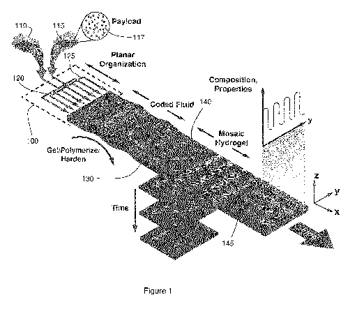
Figure 1 schematically illustrates a method for the formation of a
planar heterogeneous hydrogel using a flow-focusing microfluidic device.
Figure 2 illustrates a system for the continuous formation of hydrogel
sheets, showing (a) an example apparatus consisting of a microfluidic device
with inlets for a base biopolymer solution and focusing fluid; (b) an
illustration
of the fluidic exit portion of the microfluidic device, (c) a photograph of
fluidic
exit portion of the microfluidic device; (d) a graph demonstrating control
over
planar soft material thickness by varying drum rotation speed UP, with base
biopolymer flow rate QB = 160 pl/min, =90 1/min, =120 pl/min; (e) and (f)
SEM images showing the pore structure of planar biopolymer of
homogeneous composition: (e) 2%w.t. alginate, (f) 1%w.t. pectin-1%w.t.
alginate; (g) an illustration of the microfluidic device for the formation of
planar heterogeneous materials; (h) a photograph of multilayered microfluidic
device with on-chip reservoirs for the supply of biopolymers 1-7 into a base
biopolymer; (i) a schematic of valve actuation and pressurization of on-chip
reservoirs (scale bars 1 mm (b, c), 2 j.tm (e, f), 5 mm (g, h)); (j) bilayer
4
CA 02856063 2014-05-15
WO 2013/075248
PCT/CA2012/050847
structured formed from device with stacked microfluidic arrays (inset shows
rolled bilayer).
Figure 3 demonstrates dynamically encoding spots and information in
planar hydrogels, showing (a) an illustration of encoding information by
dynamically incorporating spots of a secondary (fluorescently labeled)
biopolymer into a base biopolymer and subsequently decoding the contained
information; (b) a hydrogel sheet with an array of void areas as imaged by
confocal fluorescence (top) and scanning electron microscopy (bottom); (c)
confocal fluorescence image illustrating dimensions and shape of spots
created by incorporating a secondary biopolymer with a payload of
fluorescently labeled microspheres at conditions P = 3.5 kPa, QB = 160
pl/min, UP = 12 mm/s, tv = 50 ms (insets represent the xy-plane (center
location of sheet); d) confocal fluorescence image (x-z plane) of
cardiomyocytes incorporated within a planar biomaterial (top and bottom
sheet boundaries indicated by dashed lines); e) confocal fluorescence image
of spot with incorporated fibroblasts at a cell density of 10 million cells/mL
(40x, Day 5); f) 5x magnification confocal scan of fibroblasts spot shown in
(e) (40x, Day 5); g) wide-field fluorescence image and corresponding
distributions of 100 pM 40kDa FITC-dextran loaded in 2%w.t. alginate and
incorporated into the same base material (images captured at times 0 and 3
hrs.); h) diffusivity of 4 kDa, 10 kDa, and 40 kDa dextran in 2%w.t. alginate
(dark gray), 1%w.t. pectin-1%w.t. alginate (light gray); i) line camera
intensity
scan (top) and fluorescence image (bottom) of encoded letters; j)
fluorescence image of pattern formed with 10 million/mL cardiomyocytes in
1.2%w.t. alginate and in 0.08%w.t. collagen type I from rat tail (Day 0)
5
CA 02856063 2014-05-15
WO 2013/075248
PCT/CA2012/050847
(approximately 25,000 cells were incorporated, operating conditions: P = 3.5
kPa, QB = 160 pl/min, Up = 12 mm/s, valve 65 ms open; k) fluorescence line
scan of binary code (top) and schematic of valve actuation with white
sections corresponding to valve open (bottom) (n = 7 binary characters); I)
sample fluorescence line scan of the UN charter in ASCII code (n=1, 2, ...
1047 binary characters including space); scale bars are 500 pm (b), 150 pm
(c), 200 pm (d), 50 pm (e), 10 pm (f), 100 pm (g) and 2mm (i-k).
Figures 4a to 4h provide images of mosaic hydrogels with various
tessellations, where two to three distinct material compositions are
illustrated
(insets represent schematic of desired patterns), including two parallel
stripes (a), squares (b), alternating wave patterns (c), axially connected
spots (d), and multiple parallel stripes (e-h) (continuous inlet gas pressures
ranging from 2-14 kPa were used, with valve opening times between 50 ms
and infinity (for continuous stripe patterns)), where g) provides SEM image
of striped heterogeneous material and (h) provides wide-field fluorescence
image of two parallel stripes containing 10 million cells/mL of
cardiomyocytes (light gray) and fibroblasts (dark gray). Figure 4i shows
millimeter-scale 3D organization of mosaic hydrogel sheets with tessellations
corresponding to (f). Figure 4j plots the modulus of elasticity of
homogeneous and mosaic hydrogels with CaCl2 concentrations of 50, 100,
and 150 mM: 2%w.t. alginate (dark gray), 1%w.t. pectin-1%w.t. alginate
(lighter gray), 2%w.t. alginate with patterns of 1%w.t. pectin-1%w.t. alginate
(light gray) as illustrated in (d), and (white) in (f). Figure 4k illustrates
single
(top) and multiple (middle and bottom) cell incorporation into a base planar
material (top and bottom figures are fibroblasts (dark gray) and endothelial
6
CA 02856063 2014-05-15
WO 2013/075248
PCT/CA2012/050847
cells (light gray) at a cell density of 10 million cells/mL; middle figure
consists
of fibroblasts (dark gray) and cardiomyocytes (light gray) at a cell density
of
2 million cells/mL; images were captured on Day 0); bottom figure consists
of fibroblasts (dark gray) and endothelial cells (light gray). Figure 41 shows
combinations of multiple cell types incorporated along with 6-bit barcoding of
a planar material; scale bars 500 pm (a-h, k, l), 1mm (i).
Figure 5 shows (a) three microfluidic masters used in the fabrication of
an example multilayer device, and (b) an illustration of the layers of a
multilayered microfluidic device composed of 10 PDMS layers.
Figure 6 plots the dependence of material thickness on the flow rate of
the focusing stream OF, and for QB = 120p1/min, demonstrating control over
soft material thickness.
Figure 7 plots the time dependence of pressure in on-chip reservoirs,
for a valve activation pattern of : (a) 0.15 ms open ¨ 2 s close, and (b) 0.25
ms open ¨2s close (input pressure 7kPa; insets represent magnified view of
pressure evolution during valve actuation).
Figure 8 shows (a) a graph showing statistics of cell distribution within
a single pattern (n=5), according to the image shown in (b); scale bar 200 pm.
Figure 9 shows line camera intensity measurements of the UN
Charter, Chapter 1, Article1, "The purposes of the United Nations".
Figure 10 is an illustration of the shear stress profile within a
microfluidic channel.
Figure 11 plots results from studies of survival of fibroblasts when
incorporated into a patterned hydrogel sheet (n=5).
Figure 12 plots the modulus of elasticity for a homogeneous soft
7
CA 02856063 2014-05-15
WO 2013/075248
PCT/CA2012/050847
material, composed of 2% w.t. alginate that was produced in the free-
extrusion (dark grey) and pulled-extrusion modes (light gray).
Figure 13 is a confocal fluorescence image of cardiomyocyte
attachment within a patterned hydrogel sheet (40x, Day 5); scale bar 10 m.
Figures 14 (a) to (h) illustrate several example embodiments of in-
plane and vertical assembly of planar sheets, including (a) a rolled sheet
forming a multilayered cylindrical structure, (b) an overlapping layered
cylindrical structure formed by collecting a planar strip on a roller at an
oblique
angle, (c) an overlapping layered structure with overlapping layers in two
lateral directions, (d) a vertically stacked structure formed from layers with
patterned holes; and (e) to (h) show experimental realizations of the
embodiment shown in Figures 14 (b) and (c).
Figures 15(a) and (b) show an example implementation of a device for
forming a wide homogeneous hydrogel sheet without the use of flow-focusing
streams, showing (a) a rendered schematic of microfluidic device containing a
single layer for the extrusion of a planar homogeneous soft material sheet
that
is 3 cm in width, and (b) a close-up photograph obtained during a running
experiment where a homogeneous 3 cm wide soft material sheet is produced
and collected onto the rotating drum, without the use of flow-focusing streams
(the device illustrated in (a) was used). Scale bars are 1 cm (a), and 3 cm
(b).
Figures 16(a) and (b) show an example implementation of a device for
forming a planar biopolymeric material, including (a) a rendering of the
example device placed within the liquid filled reservoir and (b) a photograph
of
an experimental implementation of the device placed within the liquid filled
reservoir.
8
CA 02856063 2014-05-15
WO 2013/075248
PCT/CA2012/050847
DETAILED DESCRIPTION
Various embodiments and aspects of the disclosure will be described
with reference to details discussed below. The following description and
drawings are illustrative of the disclosure and are not to be construed as
limiting the disclosure. Numerous specific details are described to provide a
thorough understanding of various embodiments of the present disclosure.
However, in certain instances, well-known or conventional details are not
described in order to provide a concise discussion of embodiments of the
present disclosure. It should be understood that the order of the steps of the
methods disclosed herein is immaterial so long as the methods remain
operable. Moreover, two or more steps may be conducted simultaneously or
in a different order than recited herein unless otherwise specified.
As used herein, the terms, "comprises" and "comprising" are to be
construed as being inclusive and open ended, and not exclusive. Specifically,
when used in the specification and claims, the terms, "comprises" and
"comprising" and variations thereof mean the specified features, steps or
components are included. These terms are not to be interpreted to exclude
the presence of other features, steps or components.
As used herein, the term "exemplary" means "serving as an example,
instance, or illustration," and should not be construed as preferred or
advantageous over other configurations disclosed herein.
As used herein, the terms "about" and "approximately", when used in
conjunction with ranges of dimensions of particles, compositions of mixtures
or other physical properties or characteristics, are meant to cover slight
variations that may exist in the upper and lower limits of the ranges of
9
CA 02856063 2014-05-15
dimensions so as to not exclude embodiments where on average most of the
dimensions are satisfied but where statistically dimensions may exist outside
this region. It is not the intention to exclude embodiments such as these from
the present disclosure.
As used herein, the phrase "microfluidic" refers to a device, or a fluidic
component of a device, that is configured for containing, flowing, processing,
or otherwise manipulating of volumes of liquid in the sub-picoliter to sub-
milliliter range. In some example embodiments, the maximal cross-sectional
dimension of a microfluidic feature, such as a microfluidic channel, may be
less than 1 mm, less than 500 microns, less than 100 microns, less than 50
microns, or less than 25 microns.
As used herein, the term "biopolymer" is understood to encompass
naturally occurring polymers, as well as synthetic modifications or
derivatives
thereof. Such biopolymers include, without limitation, hyaluronic acid,
collagen, recombinant collagen, cellulose, elastin, alginates, chondroitin
sulfate, chitosan, chitin, keratin, silk, blends thereof as well as physical
and
chemical modifications of thereof.
As used herein, the phrase "polymer solution" refers to a solution
containing a polymerizable substance. Similarly, the phrase "biopolymer
solution" refers to a solution containing a substance that is polymerizable
into
a biopolymer.
Embodiments of the present disclosure provide a microfluidic approach
for the controlled formation of planar polymeric materials. A planar array of
microfluidic channels is employed to produce a flowing liquid sheet, which
may be formed with a heterogeneous structure by spatially and temporally
CA 02856063 2014-05-15
WO 2013/075248
PCT/CA2012/050847
controlling dispensing of a polymer solution from the microchannels. The
resulting liquid sheet is solidified to produce a planar heterogeneous
material
that may be continuously drawn and/or fed from the plurality of microfluidic
channels. The ability to dynamically control the local material composition
also
provides an effective means of altering local and bulk material properties,
such as the permeability and the elasticity, and of creating materials with
directionally dependent properties.
Figure 1 schematically illustrates an example microfluidic method for
producing a planar heterogeneous material from two or more biopolymer
solution sources. Microfluidic device (schematically shown at 100) dynamically
incorporates at least one secondary biopolymer solution 115 within a layer
130 formed with base biopolymer solution 110, based on the controlled
dispending of secondary biopolymer solution 115 from microfluidic channels.
The base biopolymer solution 110 and secondary biopolymer solution 120
biopolymer solution solidify into a planar material of controlled
heterogeneity
upon exit of microfluidic device 100.
As will be further described below, base biopolymer solution 110 and
secondary biopolymer solution 115 flow within microfluidic device 100 through
base microfluidic array 120 and secondary microfluidic array 125 respectively,
with the microfluidic channels of the arrays arranged such that near an output
of microfluidic device 100, the outputs of the microfluidic channels forming
secondary microfluidic array 125 (containing secondary biopolymer solution
115) are spatially interleaved with outputs of the microfluidic channels
forming
base microfluidic array 120 (containing base biopolymer solution 110), as
shown in the Figure.
11
CA 02856063 2014-05-15
WO 2013/075248
PCT/CA2012/050847
In the present example embodiment, the dispensing of secondary
biopolymer solution 115 from secondary microfluidic array 125 is controllable
on a per-microfluidic channel basis by per-channel dispensing or metering
mechanisms or devices (not shown in Figure 1, but shown, for example, in
Figure 3a). Accordingly, the flow of secondary biopolymer solution 115 from a
given microfluidic channel of secondary array 125 may be actuated on or off,
thereby controlling the relative contribution of secondary biopolymer solution
to the composition of the combined fluidic layer formed by the microfluidic
channels in the array.
At the fluid exit of microfluidic device 100, the microfluidic array 120
and secondary microfluidic arrays 125 may be sandwiched between upper
and lower planar flows of focusing fluid (not shown in Figure 1, but shown,
for
example, in Figures 2 and 3) to focus the fluid layer formed by the biopolymer
solution emerging from base 120 and secondary 125 microfluidic channels.
Such a flow focusing embodiment enables the spatial localization of the fluid
streams emerging from the microfluidic device, with the ability to spatially
localize the fluid stream layer into a planar fluidic sheet having a thickness
that is controllable (by controlling the flow rate and/or other properties of
the
flow focusing fluid) and may be substantially thinner than a thickness (or
height) of the microfluidic channel apertures. Furthermore, the focusing fluid
serves to confine the extruded material and eliminate unwanted flow
instabilities at the device exit. In other embodiments, the flow focusing
fluid is
not provided, as described in further detail below.
In some embodiments, the density of the flow focusing fluid may be
selected such that its density is substantially equal to that of the
biopolymer
12
CA 02856063 2014-05-15
WO 2013/075248
PCT/CA2012/050847
solution extruded from the microchannels, such that the complex fluid remains
neutrally buoyant during the formation process, contributing to the structural
stability of the fluid network generated. For example, this may be achieved by
the addition of glycerol.
Upon exiting microfluidic device 100, the spatial organization of the
secondary biopolymer streams within the base layer is retained via a
solidifying process to form a substantially planar solid material 130 with
controlled heterogeneity. The fluid streams are emitted by the microchannels
in the form of a three-dimensional array of complex fluid, which flow into an
enclosed liquid filled horizontal reservoir. A narrow extrusion section is
designed with the same cross-sectional area as that of the multilayered device
exit region, for reducing flow instabilities during the extrusion process and
thereby acting as a flow focusing geometry. The horizontal reservoir may be
integrated with the microfluidic device (the substrate material of which would
need to be thick enough to store the required volumes), or may be provided
as an external reservoir that is interfaced with the microfluidic device.
Although the preceding example embodiments, and many of the
embodiments and examples below, refer to the formation of planar
biopolymeric materials from the controlled microfluidic dispensing of
biopolymer solutions, it is to be understood that the scope of the present
disclosure is not intended to be limited to materials formed from biopolymers.
In other embodiments, solidifying process may be a polymerization process,
gelation process, emulsification process, or other hardening process such that
the planar sheet that emerges from microfluidic device is transformed into a
planar material that is solid, physically resilient or in a substantially non-
13
CA 02856063 2014-05-15
WO 2013/075248
PCT/CA2012/050847
flowing state. It is to be understood that the term "solid", as used herein,
includes soft materials such as hydrogels. The thickness of the planar
heterogeneous material emerging from the device may be controlled, for
example, by varying the flow rate of the base fluid and the extrusion speed.
Other suitable solidification methods include other forms of
polymerization, including physical and chemical crosslinking. In some
embodiments, the polymerization may be achieved by photopolymerization. In
other embodiments, the polymerization may be achieved via free radical
polymerization. For example, solidification may be achieved using a polymer
such as polyethylene glycol diacrylate (PEGDA) with a commercially available
photoinitiator Irgacure 2595, or methacrylic alginate that is able to
polymerize
with both an ionic and a photo crosslinking reaction. Additionally or
alternatively, thermally induced polymerization may be employed as a
solidification method. For example, a solidified material may be obtained by
thermally induced gelation of Matrigel and collagen, and mixtures of these
with synthetic or natural hydrogels. Example hardening materials that may be
employed include polymers such as PLGA, PLA, and mixtures thereof, and
hydrogels including interpenetrating polymer networks (IPNs) and other types
of gelation (for example, shear-induced gelation of micelles).
It is to be understood that the polymer solution need not contain
biopolymeric monomers, precursors, or other biomolecular species that form
biopolymers. In some embodiments, polymerization may be performed such
that the planar polymeric material is formed from a polymeric material other
than a biopolymer.
One example process for solidifying the base and secondary polymer
14
CA 02856063 2014-05-15
WO 2013/075248
PCT/CA2012/050847
solution is a cross-linking process. For example, in some embodiments, the
flow-focusing liquid, and/or the liquid into which the heterogeneous sheet
emerges (e.g. the liquid within the horizontal reservoir), may contain a cross-
linking species (such as an ionic species), and the base and secondary
polymer solutions may include monomers or polymers that are cross-linked in
the presence of the cross-linking species, such that cross-linking of the base
and secondary fluid layer is initiated at or near the output of microfluidic
device 100 where the base and secondary fluidic layer contacts the flow
focusing liquid. Accordingly, the solidification of polymer solution or fluid
streams forms a planar material with a spatial heterogeneity that is
determined by the controlled dispensing of the secondary polymer.
As shown in the examples below, planar homogeneous and
heterogeneous materials according to various embodiments have been
produced with thickness ranging from approximately 100 m to approximately
700 m. It is to be understood that this thickness range is merely provided
within the context of an example embodiment, and the in other embodiments,
the thickness may be less than 100 m, or in excess of 700 m , depending
on the choice of materials and the configuration of the microfluidic device.
For
example, in some embodiments, thin sheets having a thickness down to
approximately 50 m, or below approximately 50 m, may be realized.
Figures 2g, 2h, 5a and 5b provide an example 10-layer microfluidic 400
device that may be employed for performing selected embodiments of the
present disclosure. In one embodiment, the microfluidic device layers are
individually molded and vertically attached using a partial curing process13,
resulting in a 10-layer device able to withstand pressures up to 600kPa. It is
CA 02856063 2014-05-15
WO 2013/075248
PCT/CA2012/050847
to be understood that this device is provided merely as an example, and that a
wide variety of alternative device configurations are possible without
departing
from the scope of the present disclosure.
Referring first to Figures 5a and 5b, the microfluidic structure of the
individual layers is shown. Layer 6 includes base microfluidic channels 420
forming the base microfluidic array and secondary microfluidic channels 430
forming the secondary microfluidic array, such that the base microfluidic
channels 420 and the secondary microfluidic channels 430 distribute the base
and secondary biopolymer solutions, and have their output apertures
interleaved in a planar array near the output aperture 410 of the device. As
shown in the Figure, the device includes a planar output channel 440 prior to
the device output aperture. The length of planar output channel is configured
such that the biopolymer solution emerges from an outlet of the planar output
channel as a substantially planar liquid sheet.
Planar output channels 464 and 440 serve to distribute the flow
focusing solution and the biomaterial sheet solution, respectively. In other
words, planar output channels 464 and 440 merge the microfluidic channels
into a fluid sheet (of either biomaterial (440), or flow focusing solution
(464), or
both; it is noted that in some embodiments, flow focusing layers are not
included). For both flow focusing and biomaterial layers, multiple
microfluidic
channels are provided that encounter planar output channel prior to flowing
out into the liquid reservoir, thus forming a single continuous sheet, as
opposed to forming fibers.
In some embodiments, substantial polymerization of the liquid sheet is
not initiated prior to the liquid sheet exiting the planar output channel
(into the
16
CA 02856063 2014-05-15
WO 2013/075248
PCT/CA2012/050847
reservoir). For example, the device may be configured such that the reaction
occurs due to contact between the polymer liquid sheet and one or both of the
flow focusing solution and the additional solution residing in the reservoir,
such that polymerization reaction is diffusion-based at and beyond the device
exit.
In other embodiments, the polymerization reaction may be initiated
within the planar output channel, prior to the liquid sheet exiting into the
reservoir. For example, a pair of liquid sheets of flow focusing liquid may be
formed above and below the liquid sheet of polymer solution within the planar
output channel for initiating the polymerization of the liquid sheet. This may
be
achieved with a device in which the outlets of both the polymer distribution
microfluidic array and the flow focusing arrays are in fluid communication
with
the inlet of the planar output channel, with the outlets of the polymer
distribution array provided between the respective outlets of the two flow
focusing arrays. Accordingly, the polymerization reaction could be made to
occur within the planar output channel due to the contact between the flow
focusing liquid sheets and the polymer liquid sheet.
In other example embodiments in which polymerization is initiated
within the planar output channel, the polymerization reaction may be initiated
by another mechanism, such as photopolymerization. In such an example
embodiment, at least a portion of the device may be transparent to an incident
photopolymerization light beam, in order to facilitate the photopolymerization
reaction within the planar output channel. According to one embodiment,
during operation of the device, secondary microfluidic channels 430 are
interfaced (through vertical fluidic access ports, not shown in Figure 5) with
17
CA 02856063 2014-05-15
WO 2013/075248
PCT/CA2012/050847
per-channel liquid dispensing or metering devices, such that the dispensing of
the secondary biopolymer solution is controllable on a per-channel basis. In
the present example embodiment, base microfluidic channels are interfaced
with a single external base biopolymer solution dispensing device, which may
be brought in fluidic communication with base microfluidic channels 420
through a network of base biopolymer solution distribution channels provided
in layer 1. Accordingly, layer 1 is an optional initial distribution layer for
evenly
distributing the base layer from an initial microfluidic channel 450, through
a
series of branching points 452 to a plurality of microfluidic channels 454.
In other embodiments, base biopolymer solution microfluidic channels
420 may also be interfaced with per-channel liquid dispensing or metering
devices, such that the dispensing of the base biopolymer solution is also
controllable on a per-channel basis. In another embodiment, each microfluidic
channel may be selectively connected to a source of base fluid and secondary
fluid, such that either base fluid or secondary fluid may be selectively
introduced into a given microfluidic channel.
In one embodiment, the device may include a single microfluidic array
for dispensing one or more polymer solutions, as opposed to two separate
arrays of microfluidic channels, as described in the preceding embodiments.
For example, in one embodiment, the device may include a single
microfluidic array, where the inlet of each microfluidic channel in the array
is
connected, or connectable, to a common dispensing device for controllably
dispensing a common polymer solution to all channels in the array. This
embodiment provides a device that can be employed to produce a
homogeneous planar polymeric material, such as a planar hydrogel sheet.
18
CA 02856063 2014-05-15
WO 2013/075248
PCT/CA2012/050847
In another embodiment, a device for forming a substantially
homogeneous planar polymeric material need not include an array of
microfluidic inlets coupled to a planar output channel, and may instead
include
a single input channel that is in fluid communication with a planar output
channel. In one example implementation, a device for forming a substantially
homogeneous planar polymeric material may include a single input that is
connected to a planar output channel by a structure similar in configuration
to
the flow focusing distribution layer that is shown in Figure 5a, where a
single
inlet channel is connected to a planar output channel by a transition section
462 that is configured to produce a uniform planar fluidic output due to
controlled fluidic resistance. In another embodiment, a single inlet channel
may be connected directly to a planar output channel, where the planar output
channel has a length sufficient for forming a substantially homogeneous liquid
sheet prior to polymerization.
In another embodiment, each microfluidic channel in the array may be
connected, or connectable, to a unique dispensing device, such that the
dispensing of polymer solutions may be controlled on a per-channel basis for
forming a planar polymeric material with controlled heterogeneity in
composition.
In yet another embodiment, two or more of the microfluidic channels
may be connected, or connectable, to a common dispensing device for
dispensing a common polymer solution to a subset of the microfluidic
channels in the array, and each remaining microfluidic channel in the array
may be connected, or connectable, to a unique dispensing device for per-
channel dispensing of one or more additional polymer solutions. One example
19
CA 02856063 2014-05-15
WO 2013/075248
PCT/CA2012/050847
of such an embodiment is an array of microfluidic channels where even or odd
microfluidic channels are connected, or connectable, to a common dispensing
device (e.g. for dispensing a base polymer solution), while each remaining
microfluidic channel is connected, or connectable, to a unique dispensing
device (e.g. for dispensing a secondary polymer solution).
In some embodiments, more than one type of polymer fluid may be
selectively introduced into a given microfluidic channel, in order to provide
increased diversity and control over the composition of the planar
heterogeneous material.
Referring again to Figure 5, and as described above, in some
embodiments, one or more flow focusing layers may be incorporated above
and below layer 6. These layers are shown as layers 2-5 and 7-10 in Figure
5b. Each flow focusing layer includes an input channel 460, which is directed
through a fluidic resistive and distribution zone 462 in order to evenly
spread
the fluid over the width of flow focusing output channel 465. Upon sandwiching
layer 6 between two flow focusing layers, the base and secondary fluid sheet
emerging in planar output channel 440 is contacted, and focused, by flow
focusing fluid in flow focusing output channels 465 (shown, for example, in
Figure 2b). Figures 2g illustrates a microfluidic device 400 assembled based
on the layers shown in Figure 5, showing the various locations for providing
the focusing fluid, base biopolymer solution, and secondary biopolymer
solution.
The secondary polymer solution within the microfluidic channels may
be dispensed and/or metered by any suitable liquid dispensing device. One or
more components of the dispensing device may be incorporated on or within
CA 02856063 2014-05-15
WO 2013/075248
PCT/CA2012/050847
the microfluidic device. Suitable liquid dispensing devices and mechanisms
include syringe pumps, peristaltic pumps, electronic or robotic pipettors, and
valves with associate pressure devices. For example, in some embodiments,
one or more reservoirs for the polymer solution may be included on the
microfluidic device, and connected through valves to an external pressure
regulation device for controlling the pressure in the head space above the
reservoir.
An example of such an embodiment is shown in Figure 2h, which
provides an image of an example device 500 that includes integrated
reservoirs 510 for providing and dispensing the secondary fluid on a per-
channel basis. As shown in Figure 2i, the pressure in the head space of each
integrated reservoir is varied relative to atmospheric pressure by an external
pressure regulation device and controllable valves. Accordingly, a single
pressure regulation device (e.g. a pump) may be employed to establish a
dispensing pressure level that is above atmospheric pressure, where the
pressure applied to each reservoir headspace may be switched between the
dispensing pressure level and atmospheric pressure by actuating valves (such
as solenoid valves). In one example, the dispensing devices have a response
time on a millisecond timescale, such as 10 milliseconds or less.
Referring now to Figure 2a, an example embodiment is shown in which
the solidified planar heterogeneous material 310 emerging from an example
microfluidic device 300 is collected and drawn by a rotating drum 310, which
rotates with tangential surface velocity U. The drum, or an alternative
extrusion device, may be located at a suitable distance from the output of the
microfluidic device so that the planar heterogeneous material (or
21
CA 02856063 2014-05-15
WO 2013/075248
PCT/CA2012/050847
homogeneous material, as described herein in alternative embodiments) is
sufficiently strong or solid to be collected. The collected planar
heterogeneous
material may be further subdivided into individual sections, for example, for
conducting separate assays or conducting cultures under different conditions.
As shown in the Figure, flow focusing liquid 340 is delivered to the
microfluidic device 300 by gear pump 345, and base biopolymer solution 330
is delivered to microfluidic device 300 via an external syringe pump (the
mechanisms and reservoirs for the delivery of secondary fluid are omitted in
this figure for simplicity). The apparatus may also include an optical
monitoring device 350, such as an imaging camera or microscope, which may
also be employed to provide feedback for use in controlling the dispensing of
liquids or other aspects of the process, based on the optical measurements.
As noted above, the planar polymeric material emerging from the
microfluidic device 300 may be received in a liquid filled reservoir prior to
being further processed (e.g. wound onto a drum and/or segmented into
pieces). In one example implementation, the emerging planar material is
passed through three sections: a device section where the microfluidic device
sits, an extrusion section 360, and a collecting section 370.
Regardless of the polymerization process employed in this
embodiment, the initially extruded fluid sheet may be first flow-focused in a
narrow extrusion section, as this flow-focusing geometry minimizes formation
of vortices at the microfluidic device exit, resulting in smooth fluid-
interface
between the biopolymer solution and the liquid filled reservoir, and thereby
producing soft material sheet of uniform thickness.
In one example embodiment in which an ion-based polymerization
22
CA 02856063 2014-05-15
WO 2013/075248
PCT/CA2012/050847
process is employed to solidify the sheet, the liquid in external reservoir
360
may contain Ca2+ ions to further cross-linking the extruded soft material
sheet.
In other example embodiments involving photo or thermal polymerization, the
reservoir may contain a water-glycerol solution, with the glycerol serving
strictly to balance the overall fluid density such that the extruded soft
material
sheet remains buoyant within external reservoir 360.
In the present example embodiment, third section 370 of the reservoir
includes rotating drum 320 onto which the planar heterogeneous material
sheet is wound. Rotating drum 320 is immersed (fully or partially) in liquid
reservoir 370 in order to minimize surface tension effects at the air-liquid
interface which would have detrimental effects on the general material sheet
structure. In some embodiments, the base and secondary fluids contain
hydrogel-forming precursors, such as certain biopolymers, that are solidified
through a cross-linking process involving the diffusive transport of ions from
the flow focusing fluid. As shown below, the planar hydrogel's properties
(e.g.
elasticity, diffusivity of different molecular payloads) can be tailored by
controlling its microscale composition. However, it is to be understood that
any
solidification process may be employed to solidify the liquid sheet emerging
from the microfluidic device, and that the scope of the present disclosure is
not
intended to be limited to the formation of planar heterogeneous hydrogel
materials.
In some embodiments, secondary biopolymer solution 115 and base
biopolymer solution 110 differ by composition such that they exhibit different
structural, chemical, biochemical, mechanical, optical, elastic, or other
properties. For example, secondary biopolymer solution 115 and base
23
CA 02856063 2014-05-15
WO 2013/075248
PCT/CA2012/050847
biopolymer solution 110 may differ only by the addition of a chromophore or a
fluorophore.
In other embodiments, any polymer solution forming the planar
biopolymeric material may include a payload. As further described below, in
some embodiments, molecular, solid, particulate, liquid, and/or gaseous
constituents may be provided within a polymer solution for incorporation into
the planar polymeric material. In some embodiments, the payload may be a
suspension and/or a solution.
Such embodiments may provide artificial biological systems, culture
media and/or supports, reagent storage and/or delivery vehicles (e.g.
microarrays), MALDI targets, and separation media (e.g. planar separation
devices for chromatography or electrophoretic separation), with optional
internal identification and/or quality control or calibration elements, among
other selected example applications. The payload may include reagents for
performing ligand-receptor assays, such as beads coated with antibodies for
performing immunoassays, nucleic acids, aptamers, or other suitable binding
and/or recognition species. Such reagents may be coated onto beads, which
are provided as a payload.
In other embodiments, the payload may include biological molecules,
cells, and/or tissues, for example, but not limited to DNA, RNA, biological
molecules, proteins, growth factors, cytokines, tissues, pieces of tissues and
organs.
In other example embodiments, the payload may include
biodegradable beads or bubbles, optionally covered with reagents, affinity
molecules or functional groups (specific or non-specific), or other bioactive
24
CA 02856063 2014-05-15
WO 2013/075248
PCT/CA2012/050847
molecules. Such an embodiment may be employed to produce a scaffold for
cells to aggregate, where the scaffold, or a portion thereof, is biodegradable
in
situ, such that the scaffold degrades while aggregated cells produce an
extracellular matrix.
Other example payloads for incorporation into any of the polymer
solutions employed in the device include medicaments and flavor compounds,
which may be employed in an embodiment where the polymer forming the
planar polymeric material is edible and/or non-toxic. For example, the
medicaments or flavor compounds may be provided in an encapsulated form
to facilitate time-release and/or timed activity. The flavor compound may be
selected such that the resulting planar polymeric material is suitable as a
confectionary, or as another related item such as a breath odor control item.
Other examples of payloads include fragrances and antiperspirants.
In other embodiments, the control over the spatial and temporal
dispensing of secondary biopolymer solution 115 may be utilized to produce
planar heterogeneous materials with pre-selected spatial concentration
gradients of diffusing or binding molecules, which may be employed for
directionally dependent mechanical and transport properties to be realized.
In addition to polymers and/or biopolymers, large diffusing or binding
molecules, such as synthetic polymers, soluble factors, drugs, proteins and
polysaccharides, can be controllably incorporated during the formation stage
of the material. A variety of different molecules such as soluble and
insoluble
factors and drugs can be incorporated in the soft material with exquisite
spatial control. Examples include polysaccharides and proteins using
dextran, albumin, polylysine, and streptavidin.
CA 02856063 2014-05-15
WO 2013/075248
PCT/CA2012/050847
The payload may include cells, thereby enabling cells to be co-
localized and/or co-cultured within the same material substrate. In some
embodiments, the planar heterogeneous material may be a soft substrate
such as a hydrogel for maintaining cell viability, and base biopolymer
solution 110 and/or secondary biopolymer solution 115 may include media
or reagents suitable for cell culture and/or cell assays. Depending on the
choice of liquid/fluid constituents (e.g. polymers and payloads),
tessellations
and other microenvironmental conditions, the planar heterogeneous
material may either display time-constant or dynamically changing
characteristics. The programmable microscale composition of the planar
heterogeneous material allows local and/or bulk properties to be controlled
and tailored. As such, a wide variety of structures and applications may be
realized according to variations of the embodiments disclosed herein.
In other embodiments, single and/or multiple cell types may be
incorporated as a payload within the polymer solution. In tissue engineering
applications, it is beneficial to authentically represent the physiological
environmental milieu of a particular tissue or organ. Resembling the structure
and function of tissues and organs requires multiple cell types and ECM
molecules to be co-localized in two or three dimensional patterns at length
scales that exceed several millimeters. Currently available cell patterning
methods allow one to either incorporate multiple cell types in microparticles
and subsequently organize them in one or two directions, or achieve co-
localization along one direction within a fiber, but do not yet provide
dynamic
control over the matrix composition and the incorporation of multiple cell
types
in two or more directions.
26
CA 02856063 2014-05-15
WO 2013/075248
PCT/CA2012/050847
The ability to pattern multiple cell types in close geometrical proximity
offers the potential of systematically exploring cell-cell interactions via
secreted factors as well as the interrogation of heterotypic and homeotypic
cell interactions. For example, Figure 41 illustrates how the incorporation of
different cell types can be combined with the ability to record the associated
experimental parameters in the form of a barcode that can be tracked
throughout the duration of cell culture.
Depending on its composition, the flow focusing liquid may or may not
be incorporated into the solidified planar heterogeneous material. In some
embodiments, a portion of the flow focusing liquid may be retained on or
within the solidified planar heterogeneous material. For example, in the
example embodiment described above, a cross-linking species is provided by
the flow focusing liquid, and this cross-linking species forms a component of
the solidified planar heterogeneous material. For example, in other
embodiments, the flow focusing liquid is discarded after the output of the
device.
In one example embodiment, the flow focusing liquid is itself solidified
and forms layers of the solidified planar heterogeneous material. For example,
the flow focusing fluid may include a constituent, such as a monomer, that can
be hardened upon exit of the microfluidic device. The solidified flow focusing
material may thus form an external solid coating around the internal planar
heterogeneous sheet. According, in such an embodiment, the secondary
polymer or biopolymer solution need not be solidified, and may be replaced by
a composition that is incorporated as a solid, liquid, or gas. For example, in
one embodiment, the secondary biopolymer or polymer solution may be
27
CA 02856063 2014-05-15
WO 2013/075248
PCT/CA2012/050847
replaced with a secondary liquid that maintains a liquid state after being
locally dispensed, with the solidified base biopolymer or polymer solution and
the solidified flow focusing layer locally encapsulating liquid droplets in
the
heterogeneous material. Such liquid droplets may form suitable volumes for
performing chemical assays and/or culturing cells.
In another embodiment, as shown in the examples below, the
secondary biopolymer or polymer may be replaced with a secondary liquid
having a non-solidifying composition, such as a composition similar to or
equal to that of the flow focusing liquid, such that the secondary liquid does
not solidify upon exit from the microfluidic device. The resulting structure,
having a Swiss-cheese-like topology with a network of holes formed
therethrough, may be suitable for providing internal perfusion of cells with
the
planar material is formed into a three-dimensional multilayer structure.
In another example embodiment, the secondary biopolymer or polymer
solution may solidify upon exit of the microfluidic device, but the
composition
of the secondary biopolymer or polymer solution (or a payload of the
secondary biopolymer or polymer solution) may be selected such that its solid
form, or a portion thereof, may be selectively removed without disturbing the
structural integrity of the solid structural backbone formed from the
solidified
base material. For example, the solidified secondary material may be
selectively removed by dissolving or etching in a suitable solvent.
Referring again to Figure 1, an illustration is provided of how the
selective control of the dispensing of secondary biopolymer solution 115 may
be employed to produce planar heterogeneous materials with coded
information 140 or tessellated structures 145. For example, spatiotemporal
28
CA 02856063 2014-05-15
WO 2013/075248
PCT/CA2012/050847
control may be achieved by incorporating a code (such as a binary code),
wherein encoded bit has a volume on a nanoliter scale. Tessellated structures
produced in this manner can exhibit directionally dependent properties, and
therefore may allowed the local storage or the timed release of an embedded
colloidal or biomolecular payload. Furthermore, tessellations of different
hardenable materials may produce mosaic patterns with variable and
controllable stiffness and/or diffusivity patterns. In some embodiments,
features or bits may be encoded with a density of up to approximately 1 bit or
spot per 200 m in the flow direction (with the lateral direction density
dictated
by the relative spacing of the microfluidic channels).
A variety of patterns can be created within such continuously extruded
planar heterogeneous materials. Selected example embodiments are
demonstrated in examples below and through confocal and fluorescence
microscopy by incorporating fluorescence microbeads into soft material. The
size and shape of the secondary liquid component incorporated within the
base liquid is dependent on the dispensing pressure Pi, base liquid flow rate
QM, extrusion velocity Up, and dispensing actuation time. As shown below,
confocal images of the planar sheet cross-section illustrate the shape of the
material formed and can show that the patterned secondary liquid can
spatially replace or displace the base liquid.
Although selected embodiments of the present disclosure describe
methods of producing planar heterogeneous materials with coded information,
tessellations, and/or spatial patterns, it is to be understood that in other
embodiments, the planar heterogeneous material need not include
geometrically repeating structures, information, or patterns. Some of the
29
CA 02856063 2014-05-15
WO 2013/075248
PCT/CA2012/050847
preceding embodiments, and the examples below, describe microfluidic
devices in which one layer is provided for generating a planar heterogeneous
material based on the dispensing and subsequently solidifying of a base
biopolymer or polymer solution and a secondary biopolymer or polymer
solution, wherein the dispensing of at least the secondary biopolymer or
polymer solution is controllable on a per-microfluidic channel basis. However,
it is to be understood that the present disclosure is not limited to devices
having a single dispensing layer, and that in some embodiments, the
microfluidic device may include two or more layers for producing planar
heterogeneous sheets from base and secondary biopolymer or biopolymer
solutions. A suitable number of flow focusing layers may be provided between
each layer having base microfluidic channels and secondary microfluidic
channels. For example, in one embodiment, a multi-layer device may be
employed to produce multiple planar heterogeneous materials at the same
time (i.e. in parallel).
Alternatively, the additional layer may be provided without including an
additional flow focusing layer. For example, in one embodiment, a device may
include a first array of microfluidic channels and an additional array of
microfluidic channels in a stacked relationship, without an intervening flow
focusing layer. Figure 2j shows a planar biopolymeric material formed
according to such an embodiment, in which a bilayer structure is formed from
two adjacent arrays of microfluidic channels, each array dispensing a single
biopolymer solution into a common planar outlet channel. The inset to Figure
2j shows the bilayer planar biopolymeric material rolled into a multilayer
configuration. According to selected embodiments, three-dimensional
CA 02856063 2014-05-15
WO 2013/075248
PCT/CA2012/050847
structures may be formed by layering planar heterogeneous materials formed
according to the methods described herein. Figure 14 illustrates several
example embodiments of in-plane and vertical assembly of planar sheets to
form layered structures.
In Figure 14(a), a multilayered cylindrical structure 500 is formed by
rolling a planar strip along a cylindrical axis. The strip may be rolled
around a
cylindrical object, such as rotating drum. The resulting three-dimensional
structure may be employed to simulate a biological organ, such as a blood
vessel, or another organ containing a lumen. The planar strip may be coated
on one or both sides with a material suitable for adhering the adjacent layers
of the formed structure, such as an adhesive or an appropriate surface
functionalization or surface chemistry, which may be provided according to a
chemistry implementation such as peptide chemistries, photochemistries and
bioconjugation schemes. An adhesive or other desired coating may be
applied to the planar strip during the extrusion process, for example, by
incorporating the adhesive or other coating material within the flow focusing
liquid, or within a liquid in the extrusion reservoir.
Figure 14(b) illustrates another example embodiment, in which a planar
strip 510 is layered as it is collected on a cylindrical support 510, thereby
providing a composite layered structure 520 that has a cylindrical cross
section, and extends longitudinally along an axis of the cylindrical support.
This may be achieved, for example, by aligning the planar sheet at an oblique
angle relative to the axis of the cylindrical support or roller.
A planar overlapping sheet embodiment is shown in Figure 14(c), in
which a plurality of planar strips are overlapped along two directions to form
a
31
CA 02856063 2014-05-15
WO 2013/075248
PCT/CA2012/050847
structure that is extendable in two dimensions. As shown in the Figure,
adjacent planar strips 530 and 535 are overlapped to create an extended
structure along one dimension, and a second layer of overlapping strips 540 is
provided to extend the structure along a second direction. Such an
embodiment may be employed to create planar structures based on
hydrogels, or other biocompatible materials, for applications in artificial
skin
and wound dressings.
Figures 14(e) to 14(h) show experimental realizations of the
embodiments described above and shown in Figures 14(b) and 14(c). As an
illustration, a rotating capillary tube (22-690-943, Fisher Scientific,
Canada)
was manually translated to collect a continuously extruded hydrogel sheet
with 50% overlap in the sheet surface area. The overlap ensures the
tubular architecture to be retained upon the removal of the capillary tube.
Homogeneous and heterogeneous hydrogel tubes with inner diameters of
approximately 1.5 mm and lengths of up to several centimeters were
produced.
In Figure 14(d), a porous vertically stacked structure is formed by
vertically stacking planar layers 550 and 555, where planar layers 550 and
555 are created with holes 560 formed therein (for example, according to the
embodiments described herein). In one embodiment, the successive layers
may be aligned such that the holes in adjacent layers are spatially aligned.
In
other embodiments, the adjacent layers may be randomly aligned, or aligned
with an offset in order to provide increased access to internal surfaces of
the
structure.
Such three-dimensional embodiments may be employed for
32
CA 02856063 2014-05-15
WO 2013/075248
PCT/CA2012/050847
applications involving automated 3D cell culture, combinations of cell culture
and assays (e.g. bead assays), the scalable formation of 3D simulated tissues
at physiologically relevant (organ) scales, 3D cell culture for multiple cell
types, staining and imaging applications, bioreactors, photobioreactors, and
artificial leaves via incorporation of microorganisms.
In some embodiments, as demonstrated in the examples below,
different secondary biopolymer or polymer solutions may be provided to
individual microfluidic channels of the device. Accordingly, such embodiments
provide the ability to control both the heterogeneity in terms of time, space,
and composition. In some embodiments, some of the secondary microfluidic
channels may be provided for one or more payloads (e.g. cells or assay
reagents), and other secondary microfluidic channels may provide
chromophores, fluorophores, or other species for providing identification
features (such as barcodes). Although the examples provided below disclose
planar heterogeneous materials having widths on a millimeter to centimeter
scale, it is to be understood that the width of the materials is not
inherently
limited, and may be increased by adding more microfluidic channels in the
microfluidic array. Accordingly, the present embodiments may be adapted or
scaled to provide wide planar materials, for example, with widths on a
centimeter (or wider) scale, with arbitrary lengths due to the continuous
nature
of the extrusion process (the length scales with the extrusion time). For
example, although the example embodiments have demonstrated widths up
to approximately 3 cm, widths of on the scale of tens of centimeters (or more)
may be attainable by modifying the device design.
In some embodiments, uniform thickness in the lateral direction may be
33
CA 02856063 2014-05-15
WO 2013/075248
PCT/CA2012/050847
achieved by configuring the channels such that the flow resistances of
individual feed channels are substantially uniform. In other embodiments, the
flow resistances could be chosen to be non-uniform and thereby produce
sheets with a thickness gradient in a direction normal to flow.
As described further below, embodiments of the disclosure may be
adapted for cell storage, transport, assays, identification and culture. In
embodiments in which on-chip reservoirs are employed, a small dead volume
(for example, less than approximately 15 1_ may be selected to prevent cell
settling. The demonstrated throughput of 160p1/min may be suitable for
applications in high-throughput screening. Furthermore, the extrusion process
is compatible with sterile conditions, and may be implemented within an
incubator. Additionally, in some embodiments, the planar heterogeneous
materials described herein do not require a base substrate or physical
template, and may be compatible with a variety of gelation chemistries
(temperature, UV, ion exchange) and extracellular matrix constituents.
In one example embodiment, present embodiments may be adapted to
provide a platform for continuous and automated soft material formation, cell
incorporation, culture (and cell co-culture), staining and imaging. Such an
embodiment could provide screening for more than 5,000 culture conditions
(cell type(s), matrix material, soluble factors, dissolved gasses) in
continuously defined and cultured biomaterial per day.
According to another embodiment, one or more diffusion barriers may
be incorporated (for example, to obtain directed molecular transport of
soluble
factors, drugs, etc within the sheet) if the secondary and primary polymers
have distinctly different diffusivities. Furthermore, the sheets may display
34
CA 02856063 2014-05-15
WO 2013/075248
PCT/CA2012/050847
stimulus responsive properties in response to environmental changes, such
as pH changes. Accordingly, such properties may be employed to obtain a
desired release-characteristic for drug delivery applications.
As shown in the examples provided herein, various secondary
biopolymers may be incorporated within a planar, unsupported hydrogel
sheet, at a sub-millimeter spatial resolution with a minimum feature size of
approximately 100 pm or less. In other embodiments, the minimum feature
size may be as small as approximately 50 pm. It is to be understood that the
spatial resolution may depend on device and material parameters that include
the feature sizes of the microfluidic channels, and the liquid viscosity. For
example, smaller microfluidic channel feature sizes, and a correspondingly
higher spatial resolution of the formed planar heterogeneous materials, may
be achieved using a microfluidic device formed in silicon. The ability to
precisely control the incorporation of the secondary hydrogel in the lateral
direction, and in time, allows for the controlled generation various
dispensing
patterns or arrangements in the x, y-plane.
The following examples are presented to enable those skilled in the art
to understand and to practice embodiments of the present disclosure. They
should not be considered as a limitation on the scope of the present
embodiments, but merely as being illustrative and representative thereof.
EXAMPLES
Example 1: Materials and Sample Preparation
Materials
Alginate (alginic acid sodium salt) and calcium chloride were
purchased from Sigma-Aldrich (St. Louis, MO, US). The alginate sample
CA 02856063 2014-05-15
WO 2013/075248
PCT/CA2012/050847
contained 2%w.t. alginate in a solution of 60% v/v glycerol in DI water. The
pectin-alginate solution was obtained by incorporating 1%w.t. pectin (Sigma-
Aldrich) into an aqueous solution containing 1%w.t. alginate and 65% v/v
glycerol. The crosslinking solutions consisted of 50mM, 100mM, and 150mM
CaCl2 in DI water containing 65%, 63%, and 61% v/v of glycerol respectively.
The density of all solutions was 1.168g/mL.
Two types of fluorescence microbeads were used either for
continuously projecting wide-field fluorescence images of the formed
hydrogels from an upright fluorescence microscopic setup (Nikon Eclipse
E600, Nikon, Japan) onto a line camera (LC1-USB, Thorlabs, Newton, NJ,
USA) or for off-line characterization using laser-scanning confocal
microscopy (Olympus IX81 Inverted Microscope with FluoView FV1000,
Olympus, Pennsylvania, USA). Specifically, microspheres with mean
diameter of 1 mm with excitation/emission of 505/515nm and 535/575nm
were purchased (F8852 and F8819, Invitrogen, Canada). Microbeads were
added to the biopolymer solutions at a ratio of 1:600, followed by 20min
sonication (B5510-MT, Branson Ultrasonics, Danbury, Connecticut, USA) to
minimize aggregation.
Neonatal Rat Heart Isolation
Neonatal Sprague¨Dawley rats (1-2 day old) were euthanized according
to the procedure approved by the University of Toronto Committee on Animal
Care. The cells from the heart ventricles were isolated by treating with
trypsin
overnight (4 C, 6120 U/mL in Hanks's balanced salt solution, HBSS) followed
by serial collagenase digestion (220 U/mL in HBSS)43. The supernatant from
five collagenase digests of the tissues was centrifuged at 750 rpm (RCF = 94 x
36
CA 02856063 2014-05-15
WO 2013/075248
PCT/CA2012/050847
g) for 4 minutes, resuspended in culture medium, and pre-plated into T75
flasks
(Falcon) for 1 h intervals to separate the adherent cells (non-myocyte) from
the
non-adherent cells (enriched cardiomyocyte).
Primary cardiac fibroblasts were obtained by cultivating for up to 7 days
the cells adhered to the T75 flask during the pre-plating. Culture medium for
both cardiomyocyte and fibroblast consisted of Dulbecco's modified Eagle's
medium (DMEM) with 4.5 g/L glucose, 4 mM L-glutamine, 10% certified fetal
bovine serum (FBS), 100 U/mL penicillin, 100 pg/mL streptomycin and 10 mM
4-2- hydroxyethy1-1-piperazineethanesulphonic acid buffer (HEPES) (Gibco,
Invitrogen, Canada). Human umbilical vein endothelial cells (HUVEC) were
purchased from Lonza, Canada.
Cell Patterning
Cells were suspended in a 1:1 ratio of cell suspension solution and
RGDS (arg-gly-asp-ser) peptide-functionalized alginate solution. The cell
suspension solution consisted of 12.3%v/v DI water, 1.2%v/v glucose solution
(0.3g/mL), 7.7%v/v 10x Medium 199 (Sigma-Aldrich, Canada), 1.1%v/v NaOH
solution (1N), 2.0%v/v NaHCO3 solution (0.075g/mL), 0.8%v/v HEPES
(Invitrogen, Canada), 19.1%v/v MatrigelTM, and 55.9%v/v collagen type I from
rat tail (3.66mg/mL, BD Biosciences, Canada).
The peptide functionalized alginate solution consisted of 1.5%w.t.
RGDS-alginate and 0.08%w.t. collagen type I from rat tail. Peptide-
functionalized alginate was obtained following a previously described
procedure. Briefly, RGDS peptide (American Peptide 44-0-14) was
conjugated to alginate using carbodiimide chemistry with N-
hydroxysulfosuccinimide ester (sulfo-NHS) stabilizer (Pierce, Fisher 24510).
37
CA 02856063 2014-05-15
WO 2013/075248
PCT/CA2012/050847
The resulted solution was purified by dialysis, dried by lyophilize, and
stored
at -20 C until use.
Cell Tracking
CellTrackerTm Red CMPTX (034552, Molecular Probes, Invitrogen,
Canada) was used for fibroblasts and CellTrackerTm Green for
cardiomyocytes (C2925, Molecular Probes). A 10mM concentration of
CellTrackerTm dyes in DMSO was further diluted in serum-free culture medium
(DMEM) to create a working concentration of 10 M. The cells were incubated
in 1mL of dye solution for 30 min at 37QC in 5% 002. Following the incubation
step, the dye-cell suspension was centrifuged and the pellet was washed two
times with DMEM.
Immunofluorescence Staining
Some cell samples were fixed in 4% paraformaldehyde in PBS at room
temperature for 15 minutes followed by incubation in mouse anti-vimentin
(Sigma, 1:100 dilution) overnight at 4 C. Samples were then incubated with
anti-mouse Alexa 488 (Sigma, 1:100) at room temperature for 1 hour and
imaged with confocal microscope (Olympus FV5-PSU confocal with IX70
microscope, Canada).
In other experiments, cell samples were fixed in 4% Paraformaldehyde
in PBS at room temperature for 15 minutes followed by incubation in mouse
anti-troponin T(Sigma, 1:100) overnight at 4 C. Samples were then incubated
with anti-mouse TRITC (Sigma, 1:100) at room temperature for 1 hour, and
imaged with confocal microscope (Olympus FV5-PSU confocal with IX70
microscope, Canada).
Tensile Testing
38
CA 02856063 2014-05-15
WO 2013/075248
PCT/CA2012/050847
Tensile tests on planar samples of the cross-linked gel were
performed in order to determine the stiffness for different microscale
compositions. The modulus of elasticity of 1- 2% alginate hydrogels were
determined at different crosslinking concentrations (50, 100, 150 mM) using
an Instron tensile tester.
Samples were cut to lengths of approximately 20 mm and fixed with a
cyanoacrylate adhesive (Krazyglue Advanced Formula, Elmer's Products,
Columbus, OH, USA) to cardboard strips, which were vertically clamped
between tensile grips for testing. A ramp of 0.1 mm/s was applied using a
1000g load cell until failure.
Bulk elastic moduli were calculated from the obtained stress-strain
curves in the linear-elastic region, for both samples produced by free
extrusion and wheel-extrusion. All samples measurements were obtained
and averaged from n = 5. Free extrusion produced mechanically weaker
planar soft material compared to wheel-extrusion, potentially due to the
alignment of the alginate molecules under tension in the latter case,
resulting
in mechanically more robust planar materials. In addition, increasing the
concentration of CaCl2 in the crosslinking bath from 50mM, 100mM, and
150mM generally increased the gel stiffness.
Sample Preparation for Scanning Electron Microscopy
Hydrogel samples were fixed in 2% glutaraldehyde in a 0.05M sodium
cacodylate buffer at pH 7.4 for lhr at room temperature, followed by gradual
replacement of the liquid phase with 100% ethanol. Dehydration of the
samples was achieved with liquid CO2 at 10 C in a critical point dryer.
Samples were subsequently heated to 31 C with a pressure increase to
39
CA 02856063 2014-05-15
WO 2013/075248
PCT/CA2012/050847
7.2MPa, transitioning the 002 to supercritical fluid conditions. Lowering the
pressure from the supercritical state allowed a direct transition into the gas
phase without causing any unwanted liquid-gas phase transitions. The
dehydrated sample was then transferred into a vacuum and vapour-deposited
with a thin film of gold to render the outer surface of the substrate
electrically
conductive.
Example 2: Microfluidic Device Fabrication
The microfluidic device consisted of 10 vertically stacked and bonded
PDMS layers that were individually obtained by moulding from different
masters. Figure 5 represents a rendered view of the microfluidic device
design and its various components.
Masters with 15011m tall features were defined by spin coating negative
photoresist SU8-2050 (MicroChem Corp, Newton, MA, USA) onto clean glass
substrate. The final feature height was achieved by two spin coating steps at
1600rpm (30s with a 5s linear ramp to 1600rpm), producing a 7511m thick
resist layer in each step. This two-step procedure ensured thickness
uniformity across the entire master. After the first spin coating step, the
substrate was postbaked for 6min at a temperature of 65 C, followed by
15min at 95 C. Following the second spin coating step, the substrate was
baked for 10min at 65 C and 35 min at 95 C.
Features with minimum width of 230 pm at the device exit section were
patterned by soft lithography with 24mW/cm2UV intensity and 9s exposure
time (total energy of 220 mJ). The exposed substrate was baked for 30 s at
65 C and 20 min at 95 C, left to cool to room temperature, and developed
under constant shaking for 12 min with 5U8 developer (MicroChem Corp).
CA 02856063 2014-05-15
WO 2013/075248
PCT/CA2012/050847
Figure 5 illustrates a rendered exploded view of the various layers that are
part of the final microfluidic device.
Individual layers of the microfluidic device were defined by spin
coating44 poly(dimethylsiloxane) (1:10 ratio of curing agent to monomer)
(PDMS, Sylgard 184 Silicone Elastomer Kit, Dow Corning, Midland, MI, USA).
Spin coating PDMS at 450 rpm for 30 s resulted in layers with uniform
thickness of 400 7 m. The multilayer device was obtained by sequentially
aligning and bonding individual layers that were previously partially cured
for
8 min at 80 C, producing a final multilayer device composed of 10 layers.
Microfluidic channels within different layers were connected by
manually puncturing through holes using a 19 gauge blunt needle. Other
means of forming holes, including laser ablation or molding holes, may
alternatively be employed.
On-chip reservoirs were obtained from 3m1 BD syringe barrels cut in
half, resulting in a total fluid storage volume of 1.5m1. The section of the
barrel containing the female Luer lock connector was used for easy
connection to the computer-controlled solenoid valves using male Luer lock
connectors (Upchurch Scientific, Oak Harbor, Washington, USA). These
reservoirs were implemented onto the microfluidic device by first fixing with
epoxy and subsequently pouring a 1cm thick uncured PDMS layer over the
final device, preventing the reservoirs from delaminating. The completed
multilayer microfluidic device was further cured for 8 hrs at 80 C. Devices
consistently withstood inlet pressures up to 600 kPa without any delamination.
Example 3: Formation of Planar heterogeneous Hydrogels
Planar heterogeneous hydrogels (thickness 8 = 150 ¨ 350 m, width
41
CA 02856063 2014-05-15
WO 2013/075248
PCT/CA2012/050847
-3mm) were formed using a multilayer microfluidic device along with the
experimental setup shown in Figure 2. As noted above, device layers were
individually molded and vertically attached using a partial curing process,13
resulting in a 10-layer-device that was able to withstand pressures of up to
600kPa (Figure 5). The center layer (indicated as layer #6 in Figure 5)
carried
to the device exit via a set of parallel microchannels a time-varying content
of
biopolymer solutions. Additional layers located above and below delivered
the crosslinker at the device exit (Figures 2a-c).
The produced biopolymer sheet flowed into a liquid-filled reservoir
containing flow focusing liquid (see Figure 2a, and description provided
above). To reduce the unwanted effect of flow instabilities at the device exit
and to ensure a uniform and controlled sheet thickness, 8, two co-flowing
fluids were delivered from above and below the soft biopolymer sheet in a
flow-focusing configuration. The focusing fluids carried cross-linking ions
and
induced gelation of the sheet. In the present example, a 2%w.t. alginate
solution was used, which is a biopolymer with well-known biocompatibility16
and ionic crosslinking mechanism.17 To increase the fluid viscosity and
render the produced biopolymer sheet neutrally buoyant with respect to the
focusing fluids, glycerol was added to both the biopolymer and focusing
streams, with the latter containing CaCl2 as the crosslinker.
The focusing fluids were continuously supplied by an annular gear
pump (mzr-2921, HNP Mikrosysteme, Parchim, Germany) at a rate of 8
mL/min. At a location approximately 50 mm downstream of the device exit,
the sheet was manually attached to a collection drum (21.3 mm in diameter)
that rotated at a constant tangential velocity, Up (Figure 2a).
42
CA 02856063 2014-05-15
WO 2013/075248
PCT/CA2012/050847
Planar hydrogel sheets with well-defined thicknesses were also
produced without additional pulling via the collection drum, by relying
exclusively on the shear imposed by the focusing fluid. Thickness of samples
produced at QB = 120p1/min and OF = 2-10m1/min was characterized.
Thicknesses ranging from 170-700 pm were obtained and measured by
optical microscopy of the cross section (Figure 6).
Computer-controlled solenoid valves (The Lee Company, Connecticut,
US) (Figure 2g-i and Figure 7) initiated the outflow of secondary biopolymers
from one of the seven on-chip reservoirs during a time period tVat which the
head pressure was raised from the atmospheric pressure level P1 to P2. A
biopolymer spot was then predictably incorporated within the hydrogel sheet
and cross-talk between different reservoirs was prevented. Accordingly,
continuous gas pressure, ranging from 0.3-2 psi, was supplied to 7 solenoid
valves which open/close to pressurize/depressurize their respective on-chip
reservoir, with valve actuation response time of 10ms.
Characterization of the dynamic behaviour of the computer-actuated
solenoid valves (model LHLA0521111H, The Lee Company, Westbrook, CT,
USA) was achieved using piezoresistive pressure transducers (pressure
range: 0-30psi, time resolution: 1ms, model HSCDIP030PGAA5, Honeywell,
Morristown, New Jersey, USA). On-chip measurements obtained in the
reservoirs during valve actuation in terms of a voltage were converted to a
pressure reading using a calibration curve. Two actuation cycles were
considered: 0.15 s open and 2s closed, and 0.25 s open and 2s closed.
The measured pressures and valve actuation times were found to be in good
agreement with the programmed input parameters.
43
CA 02856063 2014-05-15
WO 2013/075248
PCT/CA2012/050847
After having drawn an initial amount of the sheet around the drum, the
flow focusing pump was stopped, and the hydrogel sheet continuously exited
the device and was collected by the drum (some focusing fluid remained in
contact with the fluid sheet departing from the output of the microfluidic
device, such that delivery of CaCl2 ions was maintained for initiating the
solidification of the planar material via crosslinking). Although the shear
stress exerted by the focusing fluid alone was sufficient to consistently form
hydrogel sheets (as demonstrated in Figure 6), the rotating drum was
employed for this purpose, as this configuration allowed the continuous
formation, image-based inspection, and collection of mosaic hydrogels.
The sheet thickness 8 was dynamically controlled by varying the flow
rate of the base biopolymer, QB, using a syringe pump (model PHD,
Harvard Apparatus, Massachusetts, US) and by varying UP (Figure 2d).
To elucidate the effects of material formation procedure on elasticity,
the moduli of elasticity of soft material samples produced in the free-
extrusion
and pulled-extrusion modes were measured. The employed base
biopolymers and their pore sizes (Figures 2e,f) along with the increased
alignment of the polymer fibers due to the axial stress imposed by the pulling
drum affected the elastic moduli of the produced sheets (see Figure 12). In
general, samples obtained in the pulled-extrusion mode exhibited increased
moduli of elasticity as compared to those formed in the free-extrusion mode.
Without intending to be limited by theory, it was hypothesized that this
difference was caused by a promoted alignment of the alginate molecules
during the pulling process.
Example 4: Spatiotemporal Control and Payload Incorporation
44
CA 02856063 2014-05-15
WO 2013/075248
PCT/CA2012/050847
The experimental apparatus 300 shown in Figure 3a was used to
assess the spatiotemporal control of the process. A computer interface
provided control over each reservoir through the solenoid valves described
above, allowing individual localized spots of the secondary biopolymer to be
dispensed on demand.
In one experiment, the secondary biopolymer was substituted with a
viscosity-matched aqueous solution having a composition identical to the
focusing fluids (i.e., it contained 50mM CaCl2). A planar soft material sheet
with an array of void areas was obtained, shown in Figure 3b, by periodically
dispensing the viscosity-matched aqueous solution from each secondary
microchannel, at the following experimental conditions: Up = lOmm/s, QB =
200p1/min, inlet pressure P= 3.5kPa, and tv= 65ms.
In a second experiment, the extent to which the incorporated
biopolymer replaced the base biopolymer was investigated by first employing
fluorescently labelled microspheres as the payload. Confocal microscopic
scans were performed and found the smallest ellipsoidal spot (lengths of
semi-principal axes: 100pm [w], 130pm [L], 130pm [8]) that completely
replaced the base hydrogel across the entire sheet, as shown in Figure 3c.
The spot was produced with the conditions Up = 12 mm/s, QB = 160 pl/min,
inlet pressure P = 3.5kPa, and tV= 50ms.
In a third experiment, viable cells were selected as the payload. The
secondary biopolymer was modified to improve cell viability and functionality,
since alginate alone is insufficient to promote cell proliferation,
attachment,
and migration [21, 22]. A payload of neonatal rat cardiomyocytes at a density
of 10 million cells/mL was suspended in a peptide functionalized hydrogel
CA 02856063 2014-05-15
WO 2013/075248
PCT/CA2012/050847
solution (as described above). Upon day 3, beating of cardiomyocyte clusters
was observed. Preliminary cell attachment was also observed and is
illustrated in Figure 13.
The homogeneous distribution of cells within the hydrogel sheet was
assessed by z-stack confocal scans of five spots containing cardiomyocytes
pre-labelled with CellTrackerTm Green (Molecular Probes, Invitrogen, Canada)
and incorporated at a density of 10 million cells/mL. Z-stack scans were
collected with a 3011m step size to prevent cells from being counted twice.
For
each spot sample, three slices located in the middle, top, and bottom, were
Confocal microscopic scans revealed a uniform distribution of
cardiomyocytes across the hydrogel sheet, shown in Figure 3(d), in a
configuration that is not attainable in a single step using conventional top-
The fact that 8 is at least tenfold greater than the average size of the
46
CA 02856063 2014-05-15
WO 2013/075248
PCT/CA2012/050847
In another cell-based experiment, fibroblasts were suspended in the
same secondary biopolymer as the cardiomyocytes and were incorporated as
patterned spots at a cell density of 10 million cells/mL. Confocal
fluorescence
images of the patterned spots obtained on Day 5 revealed the cells ability to
attach onto the hydrogel matrix, as shown in Figures 3e and f. Patterned
sheets were fixed and immunostained following the protocol described above.
In a fourth experiment, the secondary biopolymer consisted of alginate
containing fluorescently labeled diffusible molecules (concentration 100pM,
molecular weights 4kDa and 40kDa FITCdextran, and 10kDa rhodamine-
dextran, Sigma-Aldrich, Missouri, US). Spots of the secondary biopolymer (-4
nL in volume) were incorporated in either 2%w.t. alginate (I) or in 1%w.t.
pectin-1%w.t. alginate (II) and the diffusive release of the fluorescent
marker
was followed in time-sequences of fluorescence micrographs. Figure 3g
shows two fluorescence images that were taken from such a sequence for a
spot with a 40kDa FITC-dextran payload, the first one right after gelation and
the second one 3hrs later.
The diffusion coefficient for molecular transport of dextran (4kDa,
10kDa, and 40kDa) through two different hydrogel matrices that were
composed of either 2%w.t. alginate or 1%w.t. pectin-1%w.t. alginate were
calculated by curve fitting the time-lapsed experimental data with the
analytical solution for one dimensional diffusive transport into a semi-
infinite
domain9:
I(x, t) = loerfc (_
vxD t) =
The resulting diffusivity, plotted in Figure 3h, was obtained for a best fit
using the least mean squares method (LMS). The LMS value is defined as
47
CA 02856063 2014-05-15
WO 2013/075248
PCT/CA2012/050847
the sum of the residuals squared, S = q, where the difference between
the experimental intensity value and the value predicted by the model is
ri = lexmi ¨ t).
For all considered molecular payloads, a higher diffusivity was
consistently obtained for hydrogel (I) as compared to hydrogel (II), an effect
that was attributed to the larger average pore size of hydrogel (I) that was
confirmed by scanning electron microscope (SEM) images shown in Figures
2e and f. As expected, the diffusivity in both base hydrogels decreased as the
molecular weight of the payload increased.
Example 5: Encoding of Information
The ability to incorporate isolated spots of a secondary biopolymer into
unsupported solidified sheets allows information to be encoded in a compact
manner. In the present experiment, illustrated in Figure 3a, the secondary
biopolymer alginate contained fluorescent microspheres as the payload. At a
location downstream of the device exit, the encoded information was
continuously projected onto a line camera using a fluorescence imaging
configuration.
The precise spatial and temporal control over the pattern formation
(-80 pm, 10ms) enables the use of these material patterns as means of
encoding high density information within the extruded planar material. A
Labview interface was designed to program the actuation of 7 valves to
write, in the form of patterns, analog as well as digital information.
As a demonstration of this capability, the word 'TORONTO' was
patterned onto the extruded template material in approximately 14s, with QM
= 160 pl/min, Pi = 0.5 psi, Up= 12 mm/s. Valves were actuated at a pressure
48
CA 02856063 2014-05-15
WO 2013/075248
PCT/CA2012/050847
of P2 = 3.5 kPa with opening and closing times of 75 ms and 1000 ms,
respectively. The velocity of the drum was UP = 12 mm/s. Each letter was
represented by 7-20 individual spots and occupied an area of approximately
6.25 mm2.
Fluorescence image and line camera intensity measurements of the
encoded word were obtained by labeling matrix solution with Nile red
microbeads, with the results shown in Figure 3h. Ultimately, the short valve
response time of 10ms and the small achievable spot size of approximately
8011m offer higher information density, which can be achieved by encoding
words and texts in a digital fashion, using 7 digit binary numbers that can be
converted into standard ASCII codes and converted back into text. This was
achieved by assigning the 7 valves to 7 digits in the standard ASCII binary
code. The information to be patterned can be programmed into the valve
actuation Labview interface and the resulting encoded planar material is
continuously read by a line camera as it is being extruded. A Matlab code was
designed to read the intensity information recorded from the line camera,
convert it into binary numbers, and subsequently translate back into a text.
As
an illustration, the word 'TORONTO' was encoded within approximately 7.5s.
Similarly, cardiomyocytes as a payload were pre-labelled
(CellTrackerTm Green, Molecular Probes, Invitrogen, Canada) and
incorporated in multiple spots that represented the letters "T" and "0"
(Figure
3i). The base biopolymer was 2%w.t. alginate and the secondary biopolymer
was a suspension of 10 million cells/mL in the same peptide-functionalized
hydrogel as described previously for cardiomyocytes.
The density of the encoded information was increased 19 fold by
49
CA 02856063 2014-05-15
WO 2013/075248
PCT/CA2012/050847
employing the 7-bit American Standard Code for Information Interchange
(ASCII) where each of the seven solenoid valves was assigned to one bit.
The intensity values recorded from the formed hydrogel sheet were
interpreted by a custom computer program, translated back into text and
validated against the original text. In ASCII format, "TORONTO" was
incorporated within a 37.5 mm long hydrogel sheet during approximately 7.5
s at QB =160 pl/min and UP = 8.25 mm/s (Figure 3j).
To demonstrate the ability of consistently writing and reading
information, article 1, chapter 1 of the UN Charter (165 words and 1,047
characters including spaces) was encoded in the same format (see Figure
3k and Figure 9). A 5.6 m long sheet was produced within 18.8 min and
subsequently validated the encoded information with 100% accuracy.
Example 6: Geometric Control over Mosaic Hydrogel Properties
The ability to dynamically control the local material composition
provides an effective means of altering local and bulk material properties,
such as the permeability and the elasticity, and of creating soft materials
with
directionally dependent properties. In the present example, mosaic hydrogels
were formed and characterized, using confocal and wide-field fluorescence
microscopy, with a variety of tessellations including square tiles (Figure
4b),
stripes of variable width (Figure 4c), axially interconnected spots (Figure
4d)
and sections of uniformly wide stripes (Figs. 4a, e-h).
In another experiment, the obtained mosaic hydrogel sheets were
vertically stacked to produce 3D assemblies with a defined non-isotropic
composition. To illustrate this approach, five hydrogel sheets with patterns
corresponding to the ones shown in Figure 4f were stacked such that
CA 02856063 2014-05-15
WO 2013/075248
PCT/CA2012/050847
individual patterns were rotated by 90 degrees in between neighboring layers.
In order to increase contrast between the two biopolymer solutions during a
confocal fluorescence scan, only one of the biopolymers contained a payload
of fluorescence microspheres. The resulting 3D structure had dimensions 5
mm [x] x 5 mm [y] x 1.5 mm [z] (Figure 4i).
In cases where the base hydrogel and the secondary biopolymer were
chemically distinct from each other (i.e., they did not differ by the presence
or
absence of a payload only), the effect of the different tessellations on the
bulk
elastic modulus was studied. Homogeneous and mosaic alginate sheets were
formed via cross-linking with three CaCl2 concentrations, 50, 100, 150 mM,
and tensile tests were conducted (Custom 840LE2 tensile tester, Test
Resources Inc., Minnesota, USA) (Figure 4j, see above for procedure). Two
homogeneous hydrogel samples with compositions (I) and (II) (see Example
4) were prepared, along with mosaic hydrogels with the tessellations shown in
Figs. 4d and 4f.
Figure 4j summarizes the elastic moduli that were obtained for the
different crosslinker concentrations. The values obtained for mosaic hydrogels
fall in between the ones corresponding to homogeneous samples. A
comparison between the two mosaic hydrogels suggests that axially aligned
tessellations (Figure 4f) resulted in higher elastic moduli than laterally
aligned
ones (Figure 4d).
All samples exhibited an increase in the bulk elastic modulus when the
crosslinker concentration increased from 50 mM to 100 mM. As the
concentration of CaCl2 increased, the crosslinking rate increased
proportionally. As a result, a mosaic hydrogel with a locally increased
stiffness
51
CA 02856063 2014-05-15
WO 2013/075248
PCT/CA2012/050847
in proximity of the sheet surface was formed, limiting the diffusion of CaC12
into the hydrogel and thereby creating weaker internal polymer networks.
Without intending to be limited by theory, it was believed that the decrease
in
elastic modulus that was observed at 150mM was associated with this effect.
Example 7: Planar Co-Localization of Single and Multiple Cell Types
In the present example, experiments were conducted to demonstrate
continuous two-directional patterning of cardiomyocytes, endothelial cells and
fibroblasts, major components of the native heart29'30, at a resolution of -
400
pm and at length scales of several millimeters. The cellular payloads
suspended in the biopolymer streams are exposed to shear levels less than 2
dyne/cm2 while passing through the microfluidic device (Figure 10), which is
within physiological ranges31-33 and well below shear stresses of 167-200,000
dyne/cm2 commonly associated with directprinting26 and ink-jet printing
strategieS.34-36
Neonatal rat fibroblasts were incorporated at a concentration of 10
million cells/mL and the conditions Up=12mm/s, QB=160p1/min, inlet pressure
P=3.5kPa, and tv=65ms (Figure 4k, top panel). Cell survival within 15 days of
culture was investigated using a Live/Dead viability/cytotoxicity kit for
mammalian cells (L3224, Invitrogen, Canada), resulting in 88.7% viability on
Day 15 (Figure 11). The co-localization of two cell types (cardiomyocytes or
endothelial cells with fibroblasts) within separate tessellations within a
mosaic
hydrogel was illustrated by patterning parallel stripes (Figure 4h) or islands
(Figure 4k, center and bottom figures).
Figure 41 illustrates how the incorporation of different cell types can be
combined with the ability to record the associated experimental parameters in
52
CA 02856063 2014-05-15
WO 2013/075248
PCT/CA2012/050847
the form of a barcode that can be tracked throughout the duration of cell
culture. Patterns consisting of cardiomyocytes (light gray) and fibroblasts
(dark gray) were co-localized using four on-chip reservoirs. The remaining
three reservoirs were dedicated to a 6 bit computer-readable code where a
2%w.t. alginate with 5% v/v fluorescence microspheres (P8819, Invitrogen,
Canada) was used as the secondary biopolymer.
Example 8: Shear Stress During Cell Patterning
Shear stresses during the flow of cell suspension into the microfluidic
device were calculated to ensure that the shear stress experienced by the
cells did not exceed physiological levels. Given the employed microfluidic
channel geometry and experimental conditions, the inlet pressures (wells) of
2-4 kPa and a viscosity of the (uncrosslinked) biopolymers of approximately
0.05 Pa.s, the shear stress is linearly distributed between the location of
the
channel center (zero) and its maximum value of 13-26 dyne/cm2 at the wall
(Poiseuille flow).
The cells suspended within the biopolymer are therefore subjected to
shear stresses less than 2 dyne/cm2 (as shown in Figure 10), a level well
within physiological conditions. Endothelial cells, e.g., experience 15-20
dyne/cm2 in undisturbed regions of the vascular system, can be transiently
exposed to 40-50 dyne/cm2 in areas of disturbed flow45'46and exhibit reduced
adhesion above 100 dyne/cm2 47.
The calcium chloride concentrations of 50-100mM that were used for
cross-linking of hydrogel sheets are consistent with conditions previously
employed for cell encapsulation and are not detrimental to ce11s5-7.
53
CA 02856063 2014-05-15
WO 2013/075248
PCT/CA2012/050847
Example 9: Formation of Wide Hydrogel Sheets Without the Use of Flow-
Focusing Streams
The present example illustrates the following ability to form planar
hydrogel sheets having a width significantly exceed that described in the
preceding examples. Figure 15(a) is a rendered schematic of the microfluidic
device containing a single layer for the extrusion of a planar homogeneous
soft material sheet, where the width of the device aperture 610 is 3 cm. An on-
chip reservoir 620 can be used to supply the desired biopolymer solution
while minimizing dead volume. Figure 15(b) is a photograph obtained during a
running experiment, where a homogeneous, 3 cm wide, planar soft material
sheet 630 is produced and collected onto rotating drum 640. Unlike some of
the aforementioned embodiments, the present example device was fabricated
and operated without the use of flow-focusing streams. Furthermore, the
present example device was implemented using a single biopolymer solution
for forming a substantially homogeneous biopolymeric sheet.
Example 10: Experimental Implementation of Apparatus for Forming
Planar Biopolymeric Material
The experimental implementation of the apparatus is described as
follows. Figure 16(a) is a rendered illustration of an example device placed
within a liquid filled reservoir for the formation of a planar biopolymer
sheet.
The rendered example device is supplied with a base biopolymer solution
which flow rate Q is controlled by a syringe pump, up to seven same or
distinct secondary biopolymer solutions which are distributed using gas
pressure through seven on-chip reservoirs, and a flow focusing solution that
is
supplied using a gear pump. Upon exit of the planar biopolymer solution into
54
CA 02856063 2014-05-15
WO 2013/075248
PCT/CA2012/050847
the liquid filled reservoir, a diffusion-based reaction triggers the
solidification
of the biopolymer fluid into a biopolymer sheet which is subsequently
collected onto a drum that rotates at velocity Up. Figure 16(b) is a
photograph
of the rendered experimental embodiment illustrated in Figure 16(a).
The specific embodiments described above have been shown by way
of example, and it should be understood that these embodiments may be
susceptible to various modifications and alternative forms. It should be
further
understood that the claims are not intended to be limited to the particular
forms disclosed, but rather to cover all modifications, equivalents, and
alternatives falling within the spirit and scope of this disclosure.
CA 02856063 2014-05-15
WO 2013/075248
PCT/CA2012/050847
REFERENCES
1. Bong, K. W., Bong, K. T., Pregibon, D. C. & Doyle, P. S. Hydrodynamic
focusing lithography. Angewandte Chemie-International Edition 49, 87-90
(2010).
2. Dendukuri, D., Pregibon, D. C., Collins, J., Hatton, T. A. & Doyle, P. S.
Continuous- flow lithography for high-throughput microparticle synthesis.
Nature Materials 5, 365-369 (2006).
3. Glotzer, S. C. & Solomon, M. J. Anisotropy of building blocks and their
assembly into complex structures. Nature Materials 6, 557-562 (2007).
4. Kim, J. W., Larsen, R. J. & Weitz, D. A. Synthesis of nonspherical
colloidal
particles with anisotropic properties. Journal of the American Chemical
Society 128, 14374- 14377 (2006).
5. Walther, A. & Muller, A. H. E. Janus particles. Soft Matter 4, 663-668
(2008).
6. Whitesides, G. M. & Grzybowski, B. Self-assembly at all scales. Science
295, 2418-2421 (2002).
7. Chung, S. E., Park, W., Shin, S., Lee, S. A. & Kwon, S. Guided and
fluidic self-assembly of microstructures using railed microfluidic
channels. Nature Materials 7, 581-587 (2008).
8. Bowden, N., Terfort, A., Carbeck, J. & Whitesides, G. M. Self-assembly of
mesoscale objects into ordered two-dimensional arrays. Science 276, 233-
235 (1997).
9. Kang, E. et al. Digitally tunable physicochemical coding of material
composition and topography in continuous microfibres. Nature Materials
10, 877-883 (2011).
10. Choi, N. W. et al. Microfluidic scaffolds for tissue engineering. Nature
56
CA 02856063 2014-05-15
WO 2013/075248
PCT/CA2012/050847
Materials 6, 908-915 (2007).
11. Tan, W. & Desai, T. A. Microscale multilayer cocultures for biomimetic
blood
vessels. J. Biomed. Mater. Res. Part A 72A, 146-160 (2005).
12. Ladet, S., David, L. & Domard, A. Multi-membrane hydrogels. Nature 452,
76-U76 (2008).
13. Unger, M. A., Chou, H. P., Thorsen, T., Scherer, A. & Quake, S. R.
Monolithic microfabricated valves and pumps by multilayer soft
lithography. Science 288, 113- 116 (2000).
14. Knight, J. B., Vishwanath, A., Brody, J. P. & Austin, R. H. Hydrodynamic
focusing on a silicon chip: Mixing nanoliters in microseconds. Physical
Review Letters 80, 3863-3866 (1998).
15. Anna, S. L., Bontoux, N. & Stone, H. A. Formation of dispersions
using "flow focusing" in microchannels. Applied Physics Letters 82,
364-366 (2003).
16. Augst, A. D., Kong, H. J. & Mooney, D. J. Alginate hydrogels as
biomaterials. Macromolecular Bioscience 6, 623-633 (2006).
17. Fang, Y. et al. Binding behavior of calcium to polyuronates: Comparison
of pectin with alginate. Carbohydrate Polymers 72, 334-341 (2008).
18. Chen, C. H., Shah, R. K., Abate, A. R. & Weitz, D. A. Janus particles
templated from double emulsion droplets generated using microfluidics.
Langmuir25, 4320-4323 (2009).
19. Nie, Z. H., Li, W., Seo, M., Xu, S. Q. & Kumacheva, E. Janus and ternary
particles generated by microfluidic synthesis: Design, synthesis, and self-
assembly. Journal of the American Chemical Society 128, 9408-9412
(2006).
57
CA 02856063 2014-05-15
WO 2013/075248
PCT/CA2012/050847
20. Derda, R. et al. Multizone paper platform for 3D cell cultures. Plos One 6
(2011).
21. Nagakura, T. et al. Effect of viscous injectable pure alginate sol on
cultured
fibroblasts. Plastic and Reconstructive Surgery 116, 831-838 (2005).
22. Pokrywczynska, M., Drewa, T., Jundzill, A. & Lysik, J. Alginate is not
a good material for growth of rapidly proliferating cells.
Transplantation Proceedings 40, 1664-1667 (2008).
23. Radisic, M. et al. Oxygen gradients correlate with cell density and cell
viability in engineered cardiac tissue. Biotechnol Bioeng 93, 332-343
(2006).
24. Kuo, C. K. & Ma, P. X. lonically crosslinked alginate hydrogels as
scaffolds
for tissue engineering: Part 1. Structure, gelation rate and mechanical
properties. Biomaterials 22, 511-521 (2001).
25. Du, Y., Lo, E., Ali, S. & Khademhosseini, A. Directed assembly of cell-
laden
microgels for fabrication of 3D tissue constructs. Proc Nat/ Acad Sci U S A
105, 9522- 9527 (2008).
26. Bruzewicz, D. A., McGuigan, A. P. & Whitesides, G. M. Fabrication of a
modular tissue construct in a microfluidic chip. Lab chip 8, 663-671
(2008).
27. Qi, H. et al. Patterned differentiation of individual embryoid bodies in
spatially organized 3D hybrid microgels. Adv. Mater. 22, 5276-5281
(2010).
28. Fernandez, J. G. & Khademhosseini, A. Micro-masonry: construction of 3D
structures by microscale self-assembly. Adv. Mater. 22, 2538-2541 (2010).
58
CA 02856063 2014-05-15
WO 2013/075248
PCT/CA2012/050847
29. Naito, H. etal. Optimizing engineered heart tissue for therapeutic
applications as surrogate heart muscle. Circulation 114, 172-78 (2006).
30. Brown, M. A., lyer, R. K. & Radisic, M. Pulsatile perfusion bioreactor for
cardiac tissue engineering. Biotechnol Prog 24, 907-920 (2008).
31. Davies, P. F. Flow-mediated endothelial mechanotransduction.
Physiological Reviews 75, 519-560 (1995).
32. Dewey, C. F., Bussolari, S. R., Gimbrone, M. A. & Davies, P. F. The
dynamic response of vascular endothelial cells to fluid shear stress. J.
Biomech. Eng.-Trans. ASME 103, 177-185 (1981).
33. Born, C., Zhang, Z., Alrubeai, M. & Thomas, C. R. Estimation of
disruption of animal-cells by laminar shear-stress. Biotechnology and
Bioengineering 40, 1004- 1010 (1992).
34. Nair, K. et al. Characterization of cell viability during bioprinting
processes. Biotechnology Journal 4, 1168-1177 (2009).
35. Saunders, R. E., Gough, J. E. & Derby, B. Delivery of human fibroblast
cells by piezoelectric drop-on-demand inkjet printing. Biomaterials 29, 193-
203 (2008).
36. Lee, W. et al. On-demand three-dimensional freeform fabrication of multi-
layered hydrogel scaffold with fluidic channels. Biotechnology and
Bioengineering 105, 1178- 1186 (2010).
37. Pregibon, D. C., Toner, M. & Doyle, P. S. Multifunctional encoded
particles
for high- throughput biomolecule analysis. Science 315, 1393-1396 (2007).
38. Chapin, S. C., Pregibon, D. C. & Doyle, P. S. High-throughput flow
alignment of barcoded hydrogel microparticles. Lab on a chip 9, 3100-
3109 (2009).
59
CA 02856063 2014-05-15
WO 2013/075248
PCT/CA2012/050847
39. Lee, H., Kim, J., Kim, H. & Kwon, S. Colour-barcoded magnetic
microparticles for multiplexed bioassays. Nature Materials 9, 745-749
(2010).
40. Carrier, R. L. et al. Control of the structure and metabolism of
engineered cardiac muscle by direct perfusion of culture medium.
Abstracts of Papers American Chemical Society 219, 76 (2000).
41. Carrier, R. L. et al. Perfusion improves tissue architecture of
engineered cardiac muscle. Tissue Engineering 8, 175-188 (2002).
42. Casey, T. M. & Arthur, P. G. Hibernation in noncontracting
mammalian cardiomyocytes. Circulation 102, 3124-3129
(2000).
43. lyer, R. K., Chui, J. & Radisic, M. Spatiotemporal tracking of cells in
tissue- engineered cardiac organoids. Journal of Tissue Engineering and
Regenerative Medicine 3, 196-207 (2009).
44. Unger, M.A., Chou, H.P., Thorsen, T., Scherer, A. & Quake, S.R.
Monolithic microfabricated valves and pumps by multilayer soft
lithography. Science 288, 113-116 (2000).
45. Dewey, C.F., Bussolari, S.R., Gimbrone, M.A. & Davies, P.F. THE
DYNAMIC-RESPONSE OF VASCULAR ENDOTHELIAL-CELLS TO
FLUID SHEAR-STRESS. Journal of Biomechanical Engineering-
Transactions of the Asme 103, 177-185 (1981).
46. Davies, P.F. FLOW-MEDIATED ENDOTHELIAL
MECHANOTRANSDUCTION. Physiological Reviews 75, 519-560 (1995).
CA 02856063 2014-05-15
WO 2013/075248
PCT/CA2012/050847
47. Young, E.W.K., Wheeler, A.R. & Simmons, C.A. Matrix-dependent
adhesion of vascular and valvular endothelial cells in microfluidic
channels. Lab on a Chip 7, 1759-1766 (2007).
48. Plouffe, B.D., Brown, M.A., lyer, R.K., Radisic, M. & Murthy, S.K.
Controlled capture and release of cardiac fibroblasts using peptide-
functionalized alginate gels in microfluidic channels. Lab on a Chip 9,
1507-1510 (2009).
49. Li, X., etal. Culture of neural stem cells in calcium alginate beads.
Biotechnology Progress 22, 1683-1689 (2006).
50 7. Kang, E., etal. Digitally tunable physicochemical coding of material
composition and topography in continuous microfibres. Nat Mater advance
online publication(2011).
51 8. lyer, R.K., Chui, J. & Radisic, M. Spatiotemporal tracking of cells in
tissue-engineered cardiac organoids. Journal of Tissue Engineering and
Regenerative Medicine 3, 196-207 (2009).
52 9. Bird, R.B., Stewart, W.E. & Lightfoot, E.N. Transport Phenomena,
(Wiley, New York, 2007).
61