Note: Descriptions are shown in the official language in which they were submitted.
CA 02856075 2014-05-15
WO 2013/074851
PCT/US2012/065371
PCT Application
Fundus Camera
Yeou-Yen Cheng and Jay Wei
Related Applications
[0001] This application claims priority to U.S. Provisional Application No.
61/561,266, filed
on November 18, 2011, and to U.S. Nonprovisional Application No. 13/678,488,
filed on
November 15, 2012, which are herein incorporated by reference in their
entirety.
Background
1. Field of Invention
[0002] Embodiments of the invention relate generally to an ophthalmic
photographing
apparatus.
2. Description of Related Art
[0003] In a conventional fundus camera, a focus index, such as a split-bar
pattern, is
generated from a focus index projection system using a light source with
wavelength in the
range of dark red or near infrared. The focus index projection is then
branched into a fundus
illumination path through a beam splitter or a flipping mirror (shutter).
Another way of
branching the focus index projection into the fundus illumination path is
through the
projection of the focus index on to a retractable stick mirror which is
conjugate to the fundus
of a subject's eye (Ef). The split-bar pattern is then re-imaged at the fundus
(Ef) of the eye
under examination after the illumination beam passes through the ocular lens
and the eye.
The image of the fundus (Ef), usually obtained with Near Infra Red (NIR) for
observation or
1
CA 02856075 2014-05-15
WO 2013/074851
PCT/US2012/065371
alignment purpose, superimposed with the focus index, can then be captured by
a sensor
located at the end of the imaging path. The operator then judges the degree of
focus by
looking at the alignment of the two halves of the split bar image. When the
focus setting is
correct, the two halves of the split bar image become aligned; otherwise, the
two halves are
misaligned, depending on the direction and amount of defocus. After the
operator adjusted
the focus and triggered image acquisition, a control system of the fundus
camera turns off the
NIR light sources for both the fundus and the focus index illumination and
retracts the stick
mirror out of the main illumination path before turning on the flash light
(white) to capture a
color fundus image.
[0004] During the focus adjustment of the conventional fundus camera, the
entire focus index
projection unit, including the light source, mask, condenser lens, bi-prism,
slit, folding
mirror, projection lens, and the solenoid retractable stick mirror, are moved
along the optical
axis to synchronize the movement of the focusing lens, which is usually
located after an
imaging aperture stop, through a mechanical linkage, such as a gear system.
This
conventional approach requires a large space to accommodate the movement of
the entire
focus index projection unit, the focusing lens in the imaging path, and the
mechanical
linkage, and, therefore, is not suitable for a low-cost compact system design.
[0005] In an attempt to solve this problem, a simplified focus index
projection system was
previously disclosed where the slit and the bi-prism were attached to a
transparent plate to
deflect the light from the fundus illumination light source, the whole
assembly can be flipped
in-and-out and moved longitudinally during focusing. However, sharing the
light source of
the fundus illumination with that of the focus index illumination would result
in
unsatisfactory visibility of the focus index observed by the operator.
[0006] Another method was also disclosed to enhance the visibility of the
focus index by
passing the focus index illumination light through the central opening of a
crystalline lens
2
CA 02856075 2014-05-15
WO 2013/074851
PCT/US2012/065371
diaphragm. However, the opening hole at the central blocking disk of the
crystalline lens
diaphragm results in leakage or ghost light.
[0007] A method to avoid the leakage from the central opening of the
crystalline lens
diaphragm was previously disclosed. In this method, the focus index
illuminating light
source, green Light Emitting Diode (LED), was mounted on the mechanical arm
holding the
focus index optical assembly. Since the arm and the focus index together need
to be flipped
in and out at a rapid rate during each switching between the observation mode
and the image
capturing mode, the light source would inevitably experience shock and
vibration, and this
method would result in reliability issues. Also, the visible light, such as
the green LED
disclosed, is not suitable for non-mydriatic application as the patient's
pupil size can be
sensitive to the visible light generated by the green LED.
[0008] Therefore, there is a need for systems and methods to generate focus
index of a
fundus camera with good visibility, reliability, and enhanced user-
friendliness that can be
suitable in a compact design.
Summary
[0009] In accordance with some embodiments, an ophthalmic imaging apparatus is
provided.
An ophthalmic imaging apparatus for capturing images of an eye according to
some
embodiments includes a fundus illumination system, the fundus illumination
system includes
a spatially interlaced light source array of one or more wavelength bands and
a focus index
illumination light source where the focus index illumination light source is
mounted on a
non-moving part of the ophthalmic imaging apparatus, a focus index optical
assembly, and a
fundus imaging system.
Brief Description of the Drawings
3
CA 02856075 2014-05-15
WO 2013/074851
PCT/US2012/065371
[0 0 1 0] FIG. 1 is a cross sectional view of a compact fundus camera in
accordance with some
embodiments of the present invention.
[0011] FIGs. 2a and 2b show an exemplary crystalline lens diaphragm with small
prism
mirror attached at a central light blocking disk.
[0012] FIGs. 2c and 2d show another exemplary crystalline lens diaphragm with
small prism
mirror attached at a central light blocking disk.
[0013] FIGs. 3a, 3b, 3c, 3d, 3e, and 3f show an example of an interlaced white
and NIR ring
LED arrays.
[0014] FIGs. 4a and 4b show an example of a focus index optical assembly with
multiple
fixation targets.
[0015] FIG. 5 shows the lens slider mounted with different compensation lenses
according to
some embodiments of the present invention.
Detailed Description
[0016] Embodiments of the present invention are described herein with
reference to the
exemplary drawings. In the drawings, elements having the same element
designation have
the same or similar functions.
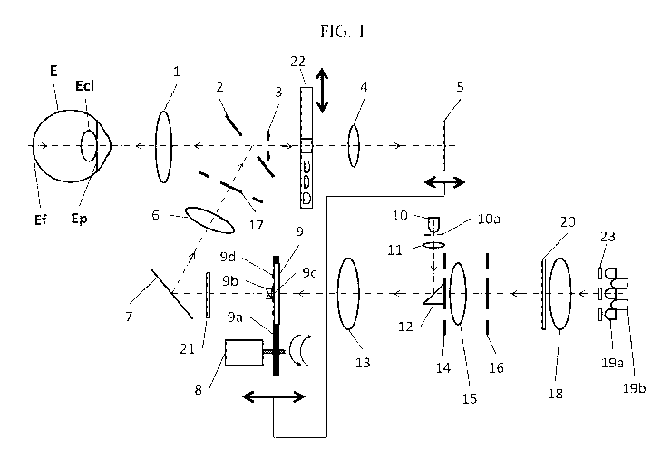
[0017] FIG.1 shows a cross sectional view of a compact fundus camera in
accordance with
some embodiments of the present invention. As shown in Fig. 1, some
embodiments of the
fundus camera include an ocular lens 1; hole mirror 2; aperture stop 3; relay
lens system 4; a
sensor 5; a relay lens 6; a mirror 7; a solenoid 8; an optical assembly 9 with
housing structure
9a, bi-prism 9b, focus index 9c, and fixation targets 9d; a focus index
illuminating light
source 10; a field stop 10a; a relay lens 11; a small folding mirror 12; a
second relay lens 13;
a crystalline lens diaphragm 14; relay lens 15; a ring aperture plate 16; an
aperture 17; a
condenser lens 18; LED ring arrays 19a and 19b; a diffuser plate 20; black dot
plate 21; a lens
4
CA 02856075 2014-05-15
WO 2013/074851
PCT/US2012/065371
slider 22; and filters 23. As shown in Fig. 1, light from light source 10 can
be directed by
folding mirror 12 through second relay lens 13, optical assembly 9, black dot
plate 21, mirror
7, lens 6, aperture 17, hole mirror 2, and ocular lens 1 to the eye. Further,
light from LED
ring arrays 19a and 19b can be directed through lenses 18, 15, 13, 6, and 1 to
the eye. Light
from the eye can be directed through lens slider 22 and lens 4 onto sensor 5.
Although lenses
1, 4, 6, 13, 15, and 18 shown in the FIG.1 are all illustrated as a single-
element lens, some or
all of them can be multi-element lenses. This system is further described
below.
[0018] The focus index illuminating light source 10, which can be a NIR LED,
is mounted on
a fixed part. Such fixed part, for example, can be a lens housing mounted on a
base structure
of the apparatus. In some embodiments, this fixed part is kept further away
from the movable
focus index optical assembly 9 to minimize the vibration or shock energy by-
product
generated from the rapid in-and-out retraction motion of the focus index
optical assembly 9
during each switching cycle between an observation mode and an image
acquisition mode.
The reliability of the focus index illumination can be improved by reducing
the vibration and
shock by-product. Such embodiments have a further advantage of removing the
focus index
illuminating light source 10 from the focus index optical assembly 9 so that
additional space
becomes available between the relay lens 13 and the black dot plate 21 for
wider range of
focus adjustment.
[0019] A black dot plate 21 is commonly used in a fundus camera setup to
eliminate surface
reflection of the ocular lens 1. In some embodiments, a field stop 10a is
attached in front of
the light source 10, such as a NIR LED, as shown in FIG. 1, so that it is re-
imaged by the
relay lens 11 to a position near the front focal plane of the second relay
lens 13 of the fundus
illumination path. The second relay lens 13 re-images the crystalline lens
diaphragm 14 to a
surface close to the back surface of the crystalline lens (Ed) of the eye (E),
with relay lens 6,
and ocular lens 1. In this arrangement, the relay lens 13 also serves as the
collimating lens for
5
CA 02856075 2014-05-15
WO 2013/074851
PCT/US2012/065371
the focus index illuminating light beam generated from the light source 10. A
small folding
mirror 12, such as a prism mirror, can be attached onto and hidden from ring
arrays 19a and
19b behind the central disk 28 of the crystalline lens diaphragm 14 to
minimize interference
with the fundus illumination beam when passing through the ring opening of the
diaphragm
14. The construction of the prism mirror on the crystalline lens diaphragm 14
is described in
details below and is shown in embodiments of diaphragm 14 shown in FIGs. 2a,
2b, 2c, and
2d.
[0020] After being reflected by the small folding mirror 12, the optical axis
of the focus
index illuminating beam coincides with that of the lens 13 and the focus index
9c. In some
embodiments, the size of the folding mirror 12 and the position of the
combination of the
light source 10 and the field stop 10a can be adjusted to minimize stray light
from the focus
index illumination beam by minimizing the beam size illuminating the central
part 44 of the
focus index optical assembly 9.
[0021] FIGs. 2a, 2b, 2c, and 2d show examples of crystalline lens diaphragms
14 in
accordance to some embodiments of the present invention. FIGs. 2a and 2b
illustrate a
diaphragm constructed from material that is capable of light
blocking/absorption. As is
shown in FIG. 2a, diagram 14 is formed of a central disk 28 with supporting
structures 30.
FIG. 2b illustrates a cross section along the A-A direction illustrated in
FIG. 2a.
[0022] FIGs. 2c and 2d show another example diaphragm 14 constructed by
depositing a thin
layer of light blocking material 30 onto a translucent material 29. FIG. 2d
illustrates a cross
section along the A-A direction illustrated in FIG. 2c.
[0023] Returning to FIG. 1, in some embodiments, in the fundus observation
mode,
illumination can be achieved by turning on the NIR LED ring array 19b of a
dual band
interlaced LED ring arrays 19a and 19b and the focus index illuminating light
source 10.
According to some embodiments, the spatially interlaced dual-band LED ring
array can be
6
CA 02856075 2014-05-15
WO 2013/074851
PCT/US2012/065371
arranged on a single Printed Circuit board (PCB) or separated into multiple
layers with
supporting structure holding the two interlaced LED ring arrays 19a and 19b
together.
[0024] An example of the interlaced ring arrays is shown in FIGs. 3e and 3f.
FIGs. 3e and 3f
show a ring array which constitutes of two layers of multiple LEDs. As shown
in FIG. 3f,
LED ring array 19a includes LEDs 39 mounted on printed circuit board 32. LED
ring array
19b includes LEDs 38 mounted on printed circuit board 34. LEDs 38 are arranged
to insert
through holes 36 on printed circuit board 32. As shown in FIG 3e, then, a ring
of LEDs 39
and LEDs 38 are formed. The LEDs 39 can also be arranged in a ring array,
evenly spaced
apart, and interlaced spatially with LEDs 38 so that each NIR LED can
illuminate the
condenser lens 18 through the open holes between adjacent white LEDs of the
first layer. As
can be appreciated by a person of ordinary skill in the art, the order of the
LED layers and the
combination of the visible band and the NIR band can be alternatively arranged
within the
spirit of the subject invention.
[0025] FIGs. 3a and 3b illustrate LED ring 19a. As shown in FIG. 3a, LEDs 39
are arranged
in a ring on a printed circuit board 32. Openings 36 in printed circuit board
32 are
interspersed between LEDs 39. FIG. 3b illustrates a cross section along the A-
A direction of
LED ring 19a.
[0026] An example composite of these 2 layers is shown in FIG. 3e. One of the
advantages
of this approach is the elimination of the need of a dichroic filter to
combine the light beams
of the different wavelength bands from each of the two separated LED ring
arrays 19a and
19b. In some embodiment, the NIR light generated from the array 19b is focused
on the ring
aperture plate 16 through the opening 36 of a mount, such as the PCB of the
white LED
arrays 19a as shown in FIG. 3a, a condenser lens 18 and a diffuser plate 20
which makes the
illumination more uniform across the fundus (Ef) image plane. The ring
aperture plate 16 is
conjugate with a position between the pupil (Ep) and the cornea of the eye
through the relay
7
CA 02856075 2014-05-15
WO 2013/074851
PCT/US2012/065371
lenses 15, 13, and 6, the hole mirror 2, and the ocular lens 1. The
crystalline lens diaphragm
14 is conjugate with the back surface of the crystalline lens (Ed) through
relay lenses 13 and
6, and the ocular lens 1. Also, in this exemplary optical setup, the cornea
ring aperture 17 is
conjugate with the cornea.
[0027] FIGs. 4a and 4b shows an exemplary drawing of a portion of optical
assembly 9.
FIG. 4a provides a planar view and FIG. 4b provides a cross-sectional view of
optical
assembly 9. As shown in Figure 4a, optical assembly 9 includes a covering of
thin light-
blocking material 44 on a translucent plate 42 to form focus index 9c and
fixation targets 9d.
Focus index 9c can be a slit opening surrounded by a light-blocking central
disk. Multiple
fixation targets 9d can be black dots or small openings of any useful shape
and size. These
fixation targets can be used to stabilize the eye during examination by
drawing the patient's
attention to any one of these fixation target(s). Note that this method
provides passive
fixation in a sense that the target illumination is shared with the fundus
illumination light
source and does not need any additional fixation light source for each
fixation position as in
the case of the conventional fundus cameras and further save the cost and
power of the
system. The longitudinal position of these fixation targets 9d relative to
that of the focus
index 9c can be adjusted to compensate for the field curvature and the index
of refraction of
the bi-prism 9b so that images of both the fixation targets and the focus
index are at focus
together at the fundus (Ef). In some embodiments, as shown in Fig. 4b, the bi-
prism 9b is
attached on top of the focus index 9c to deflect the incident beam into two
opposite
directions. FIG. 4a is a top view of the exemplary optical assembly 9 with the
patterns for
the focus index and the fixation targets. FIG. 4b shows the side view of the
translucent plate
of FIG. 4a showing the bi-prism 9b attached at the top of the focus index 9c.
The focus index
optics assembly 9 can be held in position by a mechanical housing structure 9a
(FIG. 1)
fastened on the shaft of the solenoid 8 so that the focus index optics
assembly 9 can be
8
CA 02856075 2014-05-15
WO 2013/074851
PCT/US2012/065371
flipped in-and-out of the fundus illumination path when the operator switches
between the
observation mode and the image capturing mode.
[0028] As shown in FIG. 1, when the operator adjusts the focus of the fundus
camera system,
the combination of the focus index optical assembly 9 and the solenoid 8
mounted on a
translation stage (not shown) can be moved longitudinally together with the
movement of the
sensor 5. The movement can be at different rates facilitated by a CAM wheels
structure, a
gear system or other commonly used methods to control mechanical movement.
These
embodiments described in FIG. 1 eliminate the need for a focusing lens since
the sensor 5 can
be used as part of the focus adjustment.
[0029] Since the focus index 9c is conjugate with the fundus (Ef), the split-
bar pattern is
superimposed onto the fundus image captured by the sensor 5 through the ocular
lens 1, the
central opening of the hole mirror 2, the aperture stop 3, the lens slider 22,
and the relay lens
system 4. The split-bar pattern can then be displayed on a display device so
that the operator
can observe and adjust for focusing.
[0030] The sensor 5 in some embodiments is a dual-band sensor which can
capture both
color and NIR images. An example of this type of sensor can be constructed by
removing the
IR cut filter of a typical solid-state sensor, such as a color CMOS or a CCD
sensor; where the
silicon material is sensitive to visible wavelength band and NIR wavelength
band up to
around 1,000 nm. This approach has the advantage of using only one sensor for
both the
observation mode (using NIR light) and the image capturing mode (using visible
light).
Removing the IR cut filter has the advantage of allowing the sensor to capture
the dark red
spectrum of the white LED illumination which penetrates deeper into the
choroid area of the
eye; on the other hand, it can blur the color image slightly due to chromatic
aberration. In
some embodiments, this disadvantage is overcome here by attaching small IR cut
filters 23
used for typical cell phone cameras in front of each white LED 19a.
9
CA 02856075 2014-05-15
WO 2013/074851
PCT/US2012/065371
[0031] According to some embodiments, a lens selection module, such as the
lens slider 22,
can be used to achieve adequate focus range for different eye conditions
during image
acquisition. As shown in FIG. 5a, slider 22 may be a light blocking material
42 with multiple
transparent areas. As shown in FIG. 5a, slider 22 includes a first position
44, a second
position 45, a third position 46, and a fourth position 47. As shown in FIG.
1, slider can be
positioned to allow light to pass through one of positions 44, 45, 46, and 47
to arrive at sensor
5. FIG. 5a is a planar view of slider 22 while FIG. 5b is a cross sectional
view along A-A.
[0032] Slider 22 can be utilized for fundus imaging of patients with a wide
range of
refractive error. For example, for patient with minor refractive error, the
operator can move
lens slider 22 to first position 44, which can be an open hole, as shown in
FIGs. 5a and 5b.
For patients with severe myopia, the lens slider can be moved to a second
position 45 for
diopter compensation with a weak negative lens. For patients with severe
presbyopia or
hyperopia, the slider can be moved to third position 46 with a weak positive
lens during
image acquisition. The fourth position 47, with a strong positive lens, in the
exemplary lens
slider 22 can be used for imaging the anterior area of the eye. In some
embodiments, the
operator can image the anterior area of the eye using the system in FIG. 1 by
positioning the
whole system from its nominal working distance to a distance around two times
the nominal
value and adjusting for a proper focus. The number of compensation lenses and
the ordering
positions of the exemplary slider 22 can vary based on the clinical needs and
can be
understood by a person of ordinary skill in the art.
[0033] To capture a fundus image using the system in FIG. 1, an operator will
first align the
system to a patient's eye under examination by positioning the system so that
lens 1 is about
2" to 5" away from the cornea of the eye and adjust the system laterally (X-Y)
so the image
of the pupil of the patient's eye is centered in the NIR video image on the
display device (not
shown) that displays the image captured by sensor 5 during the observation
mode. In the
CA 02856075 2014-05-15
WO 2013/074851
PCT/US2012/065371
observation mode, both the focus index illuminating light source (NIR) 10 and
the LED ring
array (NIR) 19b are turned on to illuminate the focus index and the fundus
during the
observation mode. Then, the operator can move the system toward the patient's
eye until the
image of a working distance indicator (not shown), e.g. a dual luminous spots,
commonly
used in conventional fundus camera, becomes sharp. Now, the image of the
fundus and the
focus index 9c are shown on the display with the correct working distance. The
operator can
then instruct the patient to look at one of the fixation target(s) 9d for
focusing.
[0034] When the two halves of the split-bar pattern of the focusing index are
aligned, the
imaging mode can be triggered to acquire the fundus image. The imaging mode
can be
triggered by a commonly used user's input, such as a button press on a
joystick control, a
mouse click, a foot rest. When the imaging mode is triggered, the focus index
optics
assembly 9 will flip away quickly from the light path as described above. The
focus index
illuminating light source 10 and the LED ring array (NIR) 19b will also be
turned off and the
white LED arrays 19a will then flash the fundus for capturing a fundus image
by the image
sensor 5.
[0035] The above examples are provided in order to demonstrate and further
illustrate certain
embodiments and aspects of the present invention and are not to be construed
as limiting the
scope thereof. In the description above, reference is made primarily to the
eye as the object.
This has to be understood as merely a way to help the description and not as a
restriction of
the application of the present invention. As such, where the term "eye" is
used, a more
general transparent and scattering object or organ may be sought instead.
Although various
embodiments that incorporate the teachings of the present invention have been
illustrated and
described in detail herein, a person of ordinary skill in the art can readily
device other various
embodiments that incorporate the teachings of this subject invention.
11