Note: Descriptions are shown in the official language in which they were submitted.
CA 02856314 2014-05-16
WO 2013/085924 PCT/US2012/067801
MODULATING FUNCTION OF NEURAL STRUCTURES NEAR THE EAR
Inventor: Mark Klingler Borsody
RELATED APPLICATIONS
[0001] This application claims the benefit of U.S. Provisional Application
No.
61/676,631 filed on 7/27/2012 entitled "Apparatus and means of use for
modulating the
function of neural structures within and near the middle ear," U.S.
Provisional Application
No. 61/624,958 filed on 4/16/2012 entitled "Apparatus and means of use for
modulating the
function of neural structures within and near the middle ear," U.S.
Provisional Application
No. 61/633,371 filed on 2/10/2012 entitled "Apparatus and means of use for
modulating the
function of neural structures within and near the middle ear," and U.S.
Provisional
Application No. 61/630,150 filed on 12/6/2011 entitled "Apparatus and means of
use for
modulating the function of neural structures within and near the middle ear,"
the entire
disclosures of which are hereby incorporated by reference herein in their
entireties for all
purposes.
BACKGROUND OF THE INVENTION
Field of the invention
[0002] The invention relates to apparatuses and methods for treatment of
conditions
caused directly or indirectly by functions of the vasculature. More
specifically, the invention
relates to apparatuses and methods for treatment of conditions related to the
cranial
vasculature, and even more specifically to modulating the function of
particular neural
structures in the vicinity of the ear for treatment of stroke and other
conditions.
Description of the Related Art
[0003] Stroke is the most common cause of physical disability and the third
most
common cause of death in the United States. Nearly 900,000 cases of stroke
occur each year
in the United States, costing $69 billion in healthcare costs. Worldwide,
there are nearly 15
million cases of stroke annually; the cost of healthcare services and lost
productivity on such
a scale is incalculable.
[0004] Most cases of stroke are caused by loss of blood flow to the brain
because of
occlusion of a cerebral artery or carotid artery. Artery occlusion commonly
results from (1) a
blood clot that is carried by the blood flow into an artery in which it
becomes lodged or (2)
1
CA 02856314 2014-05-16
WO 2013/085924 PCT/US2012/067801
by formation of a blood clot upon an area of atherosclerotic plaque inside the
artery. Loss of
blood flow by either mechanism, or by any of several less-common mechanisms,
deprives
areas of the brain fed by the artery of nutrients and oxygen, leading to cell
death and tissue
necrosis.
[0005] The emergency treatment of stroke is limited. Only one drug, the
thrombolytic
tissue plasminogen activator (tPA; Alteplase), has been approved for the
treatment of acute
stroke in the United States. Alteplase acts to dissolve blood clots such as
those that occlude
cerebral and carotid arteries, causing stroke. As a result, Alteplase can also
cause severe
intracranial hemorrhage, which is its most serious complication. In order to
reduce the
chance of intracranial hemorrhage, Alteplase is subject to numerous
restrictions that
ultimately limit its use to only about 5% of all ischemic stroke patients.
[0006] In addition to Alteplase, endovascular techniques employing intra-
arterial
catheters are used to treat acute stroke. Endovascular techniques, based
largely on retrieval
of the blood clot from the cerebral or carotid artery or else local
administration of
thrombolytic drugs directly onto the blood clot, are costly and dangerous, and
their use is
limited to large hospitals that have highly-trained endovascular physicians on
staff.
Accordingly, only several thousand stroke patients are treated with
endovascular techniques
each year in the United States.
[0007] A possible treatment of stroke currently under development is
electrical
stimulation of the sphenopalatine ganglion. This potential treatment involves
placement of a
metal rod through the roof of the mouth (hard palate) into the vidian canal,
which leads to the
sphenopalatine ganglion. This device and method has a number of drawbacks.
First,
placement of the rod requires specialized training and equipment that will
restrict its use to
the largest and best-equipped hospitals. By inserting the rod through the
mouth into the
vidian canal, there is a risk of introducing dangerous oral bacteria into the
bones of the face.
In addition, the blind insertion of the rod into the confines of the vidian
canal (which not only
leads to the sphenopalatine ganglion but also contains the vidian artery and
nerve) risks
inducing bleeding or nerve injury. Stroke patients also commonly have
difficulty swallowing
as part of their neurological injury. Procedures implanting foreign bodies in
the mouth, as
required by this method, may lead to aspiration in patients who have airways
already
compromised by the neurological injury from stroke. Finally, this device and
method only
stimulates the sphenopalatine ganglion and its immediate connections, which in
animals has a
small effect on blood flow to the brain compared to stimulation of the nerve
trunk.
2
CA 02856314 2014-05-16
WO 2013/085924 PCT/US2012/067801
Furthermore, this device under development is only applied to one of the two
sphenopalatine
ganglia, neglecting the potentially additive effect of stimulating both
ganglia.
[0008] Because of the magnitude of the disease and the limited treatments
for it, a
significant unmet medical need exists in acute stroke. Thus, there is a need
for a solution that
solves the problems with current acute stroke treatments noted above, and
that: (a) does not
require highly-trained endovascular physicians or specialized training for
use; (b) does not
risk intracranial hemorrhage, aspiration injury, bleeding and nerve injury, or
facial bone
infection; and (c) is non- or minimally-invasive.
SUMMARY OF THE INVENTION
[0009] Disclosed herein is a medical device and method-of-use that solves
the above
problems and that improves blood flow to the brain by causing dilation of the
cerebral and
carotid arteries using the body's own regulation of that vascular bed. The
invention is an
apparatus and method for modulating function of neural structures for
treatment of stroke and
other conditions. In one embodiment, the apparatus is a stimulator that causes
dilation
(relaxation) of the cerebral arteries. The cerebral and carotid arteries are
innervated by
nerves originating in the brainstem ("cranial nerves"), one of which ¨ the
facial nerve (also
known as the 7th cranial nerve) ¨ acts or else contains or is associated with
components that
act to regulate those arteries. Stimulation of the facial nerve system in
ischemic stroke
patients may then cause dilation of the arteries supplying the brain and the
head, allowing for
blood flow to circumvent an obstruction and reach previously deprived brain
tissue.
However, stimulation of the facial nerve in hemorrhagic stroke patients may
fail to dilate the
arteries of the brain and/or head, or else cause constriction of the arteries
supplying the brain
and/or head, beneficially reducing the likelihood of additional hemorrhage
from the site of
arterial rupture. The apparatus and method may be used to modify the function
of numerous
additional neural structures, including the entry region of the facial nerve
into the internal
auditory canal / internal acoustic meatus, the geniculate ganglion, the
tympanic plexus,
paratympanic organ(s), the intermediate nerve (of Wrisberg), the
pterygopalatine /
sphenopalatine nerves and ganglion, the petrosal nerves, the ethmoidal nerves,
the palatine
nerves, the vidian nerve, the sensory and motor fibers of any of the
aforementioned
structures, fibers of passage through the aforementioned structures, the
communicating
branches and connections of the aforementioned structures, and the
communicating branches
and connections between the aforementioned structures and the ophthalmic,
trigeminal,
glossopharyngeal, cervical, or vagal nerves.
3
CA 02856314 2014-05-16
WO 2013/085924 PCT/US2012/067801
[0010] We have discovered in preclinical / animal studies of subarachnoid
hemorrhage and
intracerebral hemorrhage that hematomas from these hemorrhages, once stable in
size, do not
enlarge after stimulation of the facial nerve using stimulation parameters
that are otherwise
effective at increasing cerebral blood flow in ischemic stroke. In ischemic
stroke, we have
demonstrated that facial nerve stimulation improves blood flow to the brain
and also
increases blood flow to the tissues of the head outside of the skull. In
contrast, in
hemorrhagic stroke, stimulation of the facial nerve does not appear to
significantly increase
blood flow to the brain, and it dramatically reduces blood flow to the tissues
of the head
outside of the skull. Thus, most of the embodiments of the device we describe
herein are
intended to be applied to a stroke patient without knowing if the patient has
an ischemic or
hemorrhagic stroke.
[0011] This property of the facial nerve, i.e., to withhold dilating cranial
arteries and
increasing cranial blood flow in the condition of hemorrhagic stroke, may
reflect sensitivity
to blood products, elevated intracranial pressure, or other properties of the
hemorrhagic
stroke. This property of the facial nerve may in part be mediated by
additional neural
structures including but not limited to sensory branches of the ophthalmic,
trigeminal,
glossopharyngeal, cervical, and vagus nerves, or by the circumventricular
organs of the brain.
[0012] Thus, the invention may have different effects depending on the type
of stroke a
patient is experiencing. Furthermore, in some methods of use, the invention
may serve to
diagnose, or support the diagnosis, of stroke subtype (i.e., ischemic stroke
versus
hemorrhagic stroke) by virtue of the different blood flow responses induced by
facial nerve
stimulation in the different subtypes of stroke.
[0013] In one embodiment, the apparatus approaches the facial nerve and its
branches as
they pass through and near to the ear in a non-invasive manner. The apparatus
can be used in
the emergency treatment of acute stroke or can be employed for chronic use in
the long-term
maintenance of blood flow to the brain, e.g., in people with atherosclerotic
disease of the
cerebral vasculature in whom blood flow to parts of the brain is chronically
compromised, or
in patients with certain kinds of dementia. In comparison to the above-
described
sphenopalatine ganglion stimulator device under development that is inserted
into the roof of
the mouth, the invention described herein may stimulate the entire facial
nerve, which
activates the sphenopalatine ganglion as well as several other nerves, nerve
branches, and
ganglia, and which has a larger and/or more widespread effect on blood flow to
the brain.
[0014] The apparatus is generally comprised of one or more electrically-
conductive
elements, such as one or more electrodes or electrically-conductive wires
that, when provided
4
CA 02856314 2014-05-16
WO 2013/085924
PCT/US2012/067801
electrical current, generates stimulation energy, such as energy in the form
of one or more
electromagnetic (EM) fields. The stimulation energy might also take the form
of heat,
ultrasound, radio frequency, microwave, infrared, ultraviolet, and electrical
energy. In an
embodiment where the stimulation energy takes the electromagnetic (EM) form,
the EM
field(s) are formed, shaped, distorted, or otherwise generated in a manner to
activate the
facial nerve system. In some embodiments, the electrically-conductive element
is shaped
substantially as a coil. In some embodiments, electrically-conductive elements
are placed on
both sides of the head and/or neck. In some embodiments, the electrically-
conductive
element is positioned on the head in such a manner as to orient the focus of
the EM field or
to summate multiple EM fields on a part of the facial nerve. In some
embodiments, the
orientation of the element is based on one or more anatomical structures of
the head or neck.
The apparatus can also include an energy regulating housing that can contain
or house at least
a part of the electrically-conductive element(s). Where there is more than one
electrically
conductive element, there may be separate housings for each element or a
single housing for
all elements. The housing electrically insulates the electrically-conductive
element and / or
dissipates heat in a desirable manner.
[0015] The
apparatus also comprises a stimulus generator in electrical communication /
direct connection with the electrically-conductive element(s) for supplying
stimulus energy to
the electrically-conductive element(s) for stimulating a neural system, such
as one or more
components of the facial nerve system in the vicinity of the ear. In some
embodiments where
the electrically-conductive element takes the form of an array of electrically
conductive wires
the stimulus generator is attached to these arrays that deliver stimulus
energy, whereas in
other embodiments the stimulus generator also serves as the arrays of
electrically-conductive
wire. The apparatus also includes a power source in electrical communication
with the
stimulus generator for providing power to the stimulus generator to supply the
electrical
current to the electrically-conductive element (e.g., arrays of electrically-
conductive wire).
In some embodiments, the stimulus generator is regulated, programmed, or
directed by a
stimulus controller. In some embodiments, the stimulus controller is guided,
directed,
programmed, or informed by a variety of sensors. As used herein, the term
"stimulator" refers
to the overall apparatus and its components.
CA 02856314 2014-05-16
WO 2013/085924 PCT/US2012/067801
BRIEF DESCRIPTION OF THE DRAWINGS
[0016] These and other features, aspects, and advantages of the present
invention will
become better understood with regard to the following description and
accompanying
drawings where:
[0017] Figure lA depicts an exploded view of an electromagnetic (EM) coil
composed
of an energy-regulating housing, a repeating loop of metal wire (coil), and a
housing cap,
illustrating the assembly of these components into the closed EM coil.
[0018] Figure 1B depicts a cross section of the ear including external,
middle, and inner
ear structures and surrounding structures, and the Figure illustrates the
positioning of an EM
coil using positioning components (e.g., an ear plug) so that the stimulus
energy (e.g., EM
field) is directed at a part of the facial nerve system, in this example the
geniculate ganglion.
[0019] Figure 2 depicts two EM coils in a figure-8 coil design held against
the side of the
head by a head strap so that the EM coils are positioned on opposite sides of
the head by an
ear plug attached to the EM coil housing, for the purpose of focusing the EM
field on a part
of the facial nerve system (in this example the geniculate ganglion).
[0020] Figure 3 depicts the figure-8 coil design shown in Figure 2, in
which the two EM
coils are kept in an adjustable housing that allows for separation and
angulation of the EM
coils according to head size and shape.
[0021] Figure 4 depicts a coolant cartridge shaped to accept a figure-8
coil into a groove
on one surface, thereby leaving only a single face of the figure-8 coil
exposed for application
to the side of the head of a subject.
[0022] Figure 5 depicts the two coils of a figure-8 coil oriented
vertically so as to
position the lower coil substantially over the internal carotid artery and the
upper coil over
the intracranial arteries, for the purpose of measuring blood flow in the
intracranial arteries.
[0023] Figure 6 depicts an assembly of two EM coils applied to one side of
the head for
unilateral stimulation of the facial nerve system in which a small EM coil is
placed in the ear
canal and a large EM coil is placed on the side of the head, and in which the
two EM coils
generate an EM field focused by the small EM coil in the ear canal so as to
target a portion of
the facial nerve system, in this example the geniculate ganglion.
[0024] Figure 7 depicts an assembly of two EM coils in which one EM coil is
applied to
each side of the head and in which the two EM coils operate in a coordinated
manner so as to
create an EM field crossing the diameter of the head that stimulates the
facial nerve system or
components thereof in a bilateral manner.
6
CA 02856314 2014-05-16
WO 2013/085924 PCT/US2012/067801
[0025] Figure 8A depicts use of two asymmetric EM coils applied to the side
of the head
in which a small EM coil serves to condense an EM field generated by a large
EM coil placed
on the opposite side of the head.
[0026] Figure 8B depicts use of two asymmetric EM coils in which an EM coil
placed in
the ear canal serves to condense an EM field generated by an EM coil placed on
the opposite
site of the head.
[0027] Figure 9 depicts two pairs of EM coils applied bilaterally to the
head, in which
each pair is composed of an internal EM coil placed into the ear canal and an
external EM
coil placed against the side of the head, and in which the two pairs of EM
coils operate in a
coordinated manner so as to create an EM field across the diameter of the head
that
stimulates the facial nerve system in a bilateral manner.
[0028] Figure 10A depicts a single EM coil equipped with a laterally-placed
ferromagnetic rod, in which the ferromagnetic rod extends into the ear canal.
[0029] Figure 10B depicts a single EM coil equipped with a ferromagnetic
cone, in
which the vertex of the ferromagnetic cone extends into the ear canal.
[0030] Figure 11A depicts an assembly of three EM coils that are
substantially round in
design equipped with a centrally-placed ferromagnetic rod.
[0031] Figure 11B depicts an assembly of four EM coils that exhibit
convergence at their
common central point equipped with a centrally-placed ferromagnetic rod.
[0032] Figure 11C depicts an assembly of four EM coils in which two of the
EM coils
are perpendicularly out-of-plane with the EM coils that are applied to the
surface of a subject,
for the purpose of blocking certain portions of the EM field.
[0033] Figure 11D shows an embodiment of the deviced in which a plurality
of EM coils
are arranged such that two EM coils arranged as a figure-8 coil are used to
activate the facial
nerve system.
[0034] Figure 12 depicts an apparatus for bilateral stimulation of the
facial nerve in
which a pair of EM coils is attached to each side of a head strap, and in
which one of the EM
coils of each pair is small enough to fit into the ear canal so that the
apparatus is positioned in
a manner to direct the EM fields at the desired part of the facial nerve
system.
[0035] Figure 13 depicts an apparatus for unilateral stimulation in which
the stimulation
controller is connected to several sensor devices placed on or between the
stimulator, coolant
cartridge, and subject.
[0036] Figure 14 depicts a method-of-use for an apparatus in which
activation of the
apparatus requires a local metal detection function and attachment of coolant
cartridges as
7
CA 02856314 2014-05-16
WO 2013/085924 PCT/US2012/067801
safety measures, and in which activation of a stimulus generator is directed
by a stimulus
controller.
[0037] Figure 15 depicts a method-of-use for an apparatus in which the
ongoing delivery
of electrical energy from a stimulus generator to the EM coils is limited by a
blood flow
sensor and a timer.
[0038] The skilled artisan will understand that the drawings are for
illustration purposes
only. The drawings are not intended to limit the scope of the present
teachings in any way.
DETAILED DESCRIPTION OF THE INVENTION
Neural Structure Modulation Apparatus
[0039] The purpose of stimulation of the facial nerve system by some
embodiments of
the apparatus is to modulate the cranial blood flow. Cranial blood flow
generally includes
blood flow to the brain and blood flow to other, non-brain tissues of the head
and neck.
Modulation of blood flow, such as cranial blood flow, includes increasing,
decreasing,
redistributing, connecting, or disconnecting various subdivisions thereof, or
otherwise
changing blood flow, such as to the cerebral, carotid, and/or extracerebral
arteries, including
but not limited to the arteries of the brain, brainstem, meninges, face,
scalp, head and neck
soft tissues, ears, and eyes of a mammalian subject. As used herein, the term
"mammalian
subject", "subject", or "patient" refers to any mammal, including humans. The
term "facial
nerve system" as used herein includes, but is not limited to, the facial
nerve, the entry region
of the facial nerve into the internal auditory canal / internal acoustic
meatus, the geniculate
ganglion, the tympanic plexus, paratympanic organ(s), the intermediate nerve
(of Wrisberg),
the pterygopalatine / sphenopalatine nerves and ganglion, the petrosal nerves,
the ethmoidal
nerves, the palatine nerves, the vidian nerve, the sensory and motor fibers of
any of the
aforementioned structures, fibers of passage through the aforementioned
structures, the
communicating branches and connections of the aforementioned structures, and
the
communicating branches and connections between the aforementioned structures
and the
ophthalmic, trigeminal, glossopharyngeal, cervical, or vagal nerves. These
components of
the facial nerve system are in the vicinity of, in proximity to, or are
proximate to the ear.
[0040] In some embodiments, the apparatus stimulates the facial nerve
system in order to
increase blood flow to the brain of the subject for treatment of an ischemic
stroke, to enhance
delivery of a blood-borne pharmacologic agent to treat a condition of the
subject, or to dilate
arteries for the purpose of allowing passage of an endovascular catheter. In
other
8
CA 02856314 2014-05-16
WO 2013/085924 PCT/US2012/067801
embodiments, blood flow to the brain or other parts of the head is decreased
by stimulation.
As used herein, the term "stroke" refers to any type of stroke, and the phrase
"stroke caused
by atherosclerotic disease" refers specifically to stroke caused by
atherosclerotic disease
involving the cerebral arteries, which includes about 20% of all stroke. As
used herein, the
term "condition" refers to any condition for which increase (or reduction in
some instances)
of blood flow provides treatment or some alleviation of the pathophysiology,
signs, or
symptoms.
[0041] Other disease of abnormal blood flow to the brain may similarly
benefit from
facial nerve system stimulation. For example, regular facial nerve stimulation
may improve
blood flow in conditions of dementia or head trauma. As another example,
facial nerve
stimulation may reduce blood flow in cases of brain tumor, headache, or other
hyperemic
diseases. Additionally, stimulation of the facial nerve system may offer
benefit in disorders
of cerebral excitability, such as epilepsy and seizure disorders.
Alternatively, stimulation of
the facial nerve system may be used to treat disorders of the eye or ear,
including those
related to blood flow or pressure in those structures. In some embodiments
used for these
conditions, the orientation of various portions of the apparatus may be
adjusted and/or the
stimulation parameters may be adjusted to achieve the desired effect or
benefit. In some
embodiments used for these conditions, the orientation of the apparatus and
the stimulation
parameters employed are the same as those used for stroke.
[0042] As mentioned above, in some cases, stimulation of the facial nerve
in hemorrhagic
stroke patients may not dilate the arteries of the brain and/or head, but may
cause constriction
of the arteries supplying the brain and/or head, reducing the likelihood of
additional
hemorrhage from the site of arterial rupture. Preclinical / animal studies of
subarachnoid
hemorrhage and intracerebral hemorrhage performed in association with the
invention have
shown that hematomas from these hemorrhages, once stable in size, do not
enlarge after
stimulation of the facial nerve using stimulation parameters that are
otherwise effective at
increasing cerebral blood flow in ischemic stroke.
[0043] In ischemic stroke, it has been demonstrated that facial nerve
stimulation
performed in association with the invention improves blood flow to the brain
and also
increases blood flow to the tissues of the head outside of the skull. In
contrast, in
hemorrhagic stroke, stimulation of the facial nerve does not appear to
significantly increase
blood flow to the brain, and it reduces blood flow to the tissues of the head
outside of the
skull. Thus, most of the embodiments of the device described herein are
intended to be
applied to a stroke subject without knowing if the subject has an ischemic or
hemorrhagic
9
CA 02856314 2014-05-16
WO 2013/085924 PCT/US2012/067801
stroke (undifferentiated' stroke). In this use, the device may provide
benefits to the subject,
or it may only provide benefit to those subjects with ischemic stroke while
doing no harm to
those subjects with hemorrhagic stroke.
[0044] This property of the facial nerve, i.e., to withhold dilating
cranial arteries and
increasing cranial blood flow, in the condition of hemorrhagic stroke may
reflect sensitivity
to blood products, elevated intracranial pressure, or other properties of the
hemorrhagic
stroke. This property of the facial nerve may in part be mediated by
additional neural
structures including but not limited to sensory branches of the ophthalmic,
trigeminal,
glossopharyngeal, cervical, or vagus nerves, or by the circumventricular
organs of the brain.
[0045] FIG. lA depicts an embodiment of the device in which an electrode or
electrically-conductive element, in this case repeating loops of metal wire
132, is housed in
an energy-regulating housing 136 so as to form an electromagnetic (EM) coil
100. Placement
of the repeating loops metal wire 132 inside the hollow lumen of the energy-
regulating
housing 136 allows for the egress of wires connecting to the stimulus
generator 134. The
repeating loops of metal wire 132 are closed inside the hollow lumen of the
energy-regulating
housing 136 by a housing cap 130.
[0046] In some embodiments of the apparatus, the energy-regulating housing
is
composed of two or more materials with different heat conductivities. The
energy-regulating
housing that touches, faces, or approximates the patient is composed of a
material that has a
low thermal conductivity whereas all other sides (including the side facing
away from the
patient) has a high thermal conductivity. The general purpose of this is to
direct heat flow
from the electrically-conductive elements away from the patient.
[0047] FIG. 1B illustrates various components of the ear, including the
pinna 309, ear
canal 306, middle ear space 310, and auditory canal 308. As used herein, the
term "ear"
refers to any portion of the ear, including the external, middle, and inner
ear, unless otherwise
specified, to the general region of the head where those structures are found,
or to the region
of the temporal bone housing auditory, vestibular, and other neural
structures. In some
embodiments, the apparatus stimulates components of the facial nerve system
that pass
through, have a portion or branch within, or contribute to a structure within
the middle ear
310. Furthermore, in some embodiments, the apparatus stimulates components of
the facial
nerve system that are immediately outside the middle ear 310. As used herein,
the term
"limited facial nerve system" includes the nerves and neural structures listed
above, but not
including the sphenopalatine nerves and ganglion, the petrosal nerves and
communicating
branches thereof, the ethmoidal nerves and communicating branches thereof, the
palatine
CA 02856314 2014-05-16
WO 2013/085924 PCT/US2012/067801
nerves including nasopalatine nerves, the vidian nerve and communicating
branches thereof,
and communicating branches between any of the aforementioned structures and
the
trigeminal nerve system. The apparatus is described throughout as one that is
used in
association with the ear, but the apparatus can also be positioned elsewhere
on the head or
other location that permits the apparatus to stimulate the facial nerve
system. Similarly, the
apparatus can also be used for stimulation of other neural systems. As used
herein, the term
"neural system" refers to any nervous tissue in the body of a subject
[0048] FIG. 1B depicts an embodiment of the device in which the EM coil 100
is placed
against the surface of the head near the ear. Throughout this description, the
term "EM coil"
is used, but this can also refer to other types of electrodes or electrically
conductive elements
in the embodiments described. The EM coil 100 is held against the surface of
the head by an
ear grip 122 that reaches behind the pinna 309 of the external ear (though
other ear grip
designs or mechanisms for ear or head attachment can also be used), and is
positioned by an
ear plug 120 that is inserted into the ear canal 306 and a positioning
component 126 that is
interposed between the ear plug 120 and the EM coil 100 (though other
mechanisms for
positioning can also be used). The ear plug 120 and positioning component 126
serve to
orient the EM coil 100 such that the EM field 101 generated by the EM coil 100
is directed at
the facial nerve 300 or at a part of the facial nerve system such as the
geniculate ganglion
302.
[0049] Continuing with FIG. 1B, in some embodiments, the positioning
component 126
is formed of a substantially rigid material. In some embodiments, the
positioning component
126 is formed from a substantially flexible material. In some embodiments, the
shape of the
positioning component 126 can be adjusted to alter the orientation of the EM
field 101
generated by the EM coil 100.
[0050] In some embodiments, the apparatus is applied to one side of the
head, as shown
in Fig. 1B. In other embodiments, the apparatus is applied to both sides of
the head, similar
in appearance to ear muffs or ear plugs.
[0051] FIG. 1B depicts a power source 500 connected by cables to a stimulus
generator
510 that provides, gates, directs, shapes, or otherwise delivers the
electrical energy to the EM
coil 100 in a manner that allows for generation of the EM field 101. The power
source may
also be in electrical communication with the stimulus generator with cables as
shown or
wirelessly. This is true for all embodiments of the power source and stimulus
generator
described herein. Similarly, the stimulus generator 510 may be in electrical
communication
with the EM coil 100 by wires or wirelessly, which is true for all embodiments
described
11
CA 02856314 2014-05-16
WO 2013/085924 PCT/US2012/067801
herein. The power source 500 provides the electrical current to the stimulus
generator 510,
which powers the EM coil 100.
[0052] As shown in FIG. 1B, in some embodiments, the apparatus further
includes a
stimulation controller 520 attached to the stimulus generator 510 for
adjusting, defining,
modulating, or otherwise determining the electrical current applied to the EM
coil 100 or
controlling one or more settings of the stimulus generator 510. The stimulus
controller 520
can include a user interface by which the operator of the apparatus can
provide instructions
to, or otherwise interact with, the apparatus. The stimulus controller 520 can
allow the
operator to control the strength, frequency, duration of application, and/or
other parameters
of the stimulus energy. For example, the stimulus controller 520 can include
particular
controls (e.g., knobs, digital settings, etc) for increasing or decreasing the
strength of the
electrical current and controlling various other factors or parameters in the
operation of the
apparatus. Where the apparatus is connectable to a computer or other
machinery, the
operator may also be able to interact with and control the apparatus via the
interface of the
computer, including tracking the subject's vital signs, responses to the
stimulus energy over
time, machinery performance, and so forth.
[0053] The stimulus controller 520 can further be used to adjust the
stimulus energy for
various purposes. For example, the stimulus energy can be adjusted based on
one or more
physiological or pathophysiological responses of the subject to the stimulus
energy (e.g.,
carotid artery blood flow; cerebral artery blood flow; blood flow to the
central nervous
system; facial nerve electrical potentials; skin / scalp galvanic responses;
skin / scalp blood
flow; ear temperature; pupilometry; intraocular pressure; blood flow to the
eye; bioelectric
potentials; electroencephalogram waveforms; electrophysiological testing of
the auditory or
vestibular systems; taste sensation; audition; lacrimation; nasal drainage;
nasal
congestion; salivation; sound sensitivity; face, head, or hand movements or
electromyographic potentials; speech production or arrest; sensation of body
movement;
eye movements; cranial blood flow; direct or indirect activity of a nerve; and
severity of
neurological dysfunction of the subject). For example, if the subject exhibits
certain eye
movements, the operator can observe this and respond to this by changing the
stimulus
energy or certain other parameters associated with the stimulus energy. As
another example,
the stimulus energy can be adjusted to increase or otherwise control blood
flow to the brain
of the subject as either the direct treatment of a disease process or else to
facilitate the
delivery of blood-borne pharmacologic agents as the treatment of a disease
process. As
another example, the time since the onset of stroke symptoms may inform the
stimulus
12
CA 02856314 2014-05-16
WO 2013/085924 PCT/US2012/067801
controller 520 to allow the stimulus generator 510 to deliver stimulation
energy of certain
characteristics or duration. As another example, signal provided to the
stimulus controller
520 representing physiological or pathophysiological responses of the subject
may direct the
stimulus controller 520 to adjust the shape of a positioning component 126. In
some
embodiments, the operator is replaced by a servocontrol or automatic control
mechanism that
can detect one or more physiological or pathophysiological responses of the
subject to the
stimulus energy.
[0054] The apparatus of FIG. 1B can be a chronic / repeated treatment
device or can be
an acute / single treatment device. For acute treatment of a condition, the
apparatus of FIG.
1B can be placed on the subject's ear, and can deliver stimulus energy as
desired by a
physician or other operator. The stimulus generator 510 can be attached to a
stimulus
controller 520 that allows a physician or other operator to control when the
stimulus energy is
delivered, the intensity of the stimulus energy, etc. For chronic treatment or
prevention of a
condition, the apparatus of FIG. 1B can be worn as a chronic treatment device
that is worn
continuously or regularly by the subject. It can be worn all the time, at
certain times of day,
or whenever prescribed. As one example in which chronic treatment is useful,
atherosclerotic
disease of the cerebral arteries narrows cerebral arteries, which may
chronically impair blood
flow to parts of the brain, thereby causing among other symptoms recurring
near-strokes /
transient ischemic attacks as blood flow becomes intermittently compromised.
In order to
overcome the narrowing in the cerebral arteries caused by atherosclerosis or
other
malformations, repeated stimulation of the facial nerve system provided by a
chronic
treatment device can be used to maintain dilation of the arteries, thereby
preventing stroke
caused by atherosclerotic disease. The apparatus of FIG. 1B can thus
chronically stimulate or
modulate one or more components of the facial nerve system in the vicinity of
the ear to treat
stroke or other conditions.
[0055] In some embodiments, the EM coil 100 is between 2 cm and 8 cm in
diameter. In
some embodiments, the EM coil 100 is a hollow "doughnut" shape. In some
embodiments
that include repeating loops of wire, the more central loops of wire are
progressively raised
off the plane of the largest loop so as to form a substantially cone-like
shape wherein the apex
of the cone is inserted into the ear canal.
[0056] In some embodiments, the ear plug 120 is formed from a sound-
dampening
material. In some embodiments, the ear plug 120 and /or positioning component
126 are
formed from heat-adsorbent or heat-resistant materials. In some embodiments,
the position
of the ear plug 120 and / or positioning component 126 relative to the EM coil
100 offsets the
13
CA 02856314 2014-05-16
WO 2013/085924 PCT/US2012/067801
EM coil 100 from a position immediately over the ear canal for the purpose of
directing,
focusing, or otherwise changing the use of the EM field 101.
[0057] Placement of the stimulator against the external ear may be
performed, guided, or
assisted with various accessory devices such as the positioning component 126
of FIG. 1B
that are of different or customized geometric shape (such as wedges) for the
purpose of
orienting the EM coil 100 in a manner that effectively stimulates the facial
nerve system or
select components of the facial nerve system. Such accessory devices may
relate to facial or
cranial anatomy, external ear anatomy, and / or ear canal anatomy. In some
embodiments,
such accessory devices may be adjusted or formed based on neuroimaging of the
facial nerve
system, the location of select components of the facial nerve system such as
the geniculate
ganglion, the bony structures that house the facial nerve system, and / or
other nearby
structures. In some embodiments, such accessory devices may be adjusted based
on the
length, width, height, circumference, or other external measures of the head
and/or neck of
the subject. In some embodiments, such accessory devices may be adjusted based
on
feedback from a sensor device.
[0058] FIG. 2 depicts an embodiment in which multiple electrodes or
electrically-
conductive elements, in this case EM coils, are arranged in a geometric shape
and applied to
the surface of the head in the vicinity of the ear for the purpose of
stimulating the facial nerve
system or a portion of the facial nerve system. In this embodiment, two
circular EM coils are
placed side-by-side so as to form a figure-8 coil 150, and an ear plug 120 is
placed upon the
contact surface 160 of the figure-8 coil. The position of the ear plug 120 on
the surface of the
figure-8 coil 150 is determined so that the juncture of the two coils of the
figure-8 coil 150 is
placed over the ear canal 306 when the contact surface 160 of the figure-8
coil 150 is placed
in position on a subject 700. In some embodiments, the housing for the figure-
8 coil 150 is
made of a substantially flexible material that allows adjustment of the
separation and/or
angulation of the two EM coils. In other embodiments, the EM coils (as shown
in element
100 of FIG. 1B) of the figure-8 coil 150 are contained in separate housings.
In some
embodiments, a positioning component is placed or can be placed between the
ear plug 120
and the figure-8 coil 150. The housing for the figure-8 coil 150 can also be
an energy-
regulating housing capable of electrically insulating the electrically-
conductive element and
dissipating heat from the apparatus in a desired manner.
[0059] FIG. 2 illustrates the relationship between the two EM coils and
underlying
structures including the middle ear space 310, facial nerve 300, geniculate
ganglion 302,
branches of the geniculate ganglion (including but not limited to the greater,
lesser, and
14
CA 02856314 2014-05-16
WO 2013/085924 PCT/US2012/067801
external petrosal nerves 322), trigeminal nerve 320, and intracranial arteries
380 (including
cerebral, meningeal, and other arteries).
[0060] Continuing from FIG. 2, then, FIG. 3 depicts the manner in which the
two EM
coils of the figure-8 coil 150 can be angulated and/or separated to
accommodate different
head sizes and shapes so as to allow for the intersection of the two EM
fields, producing a
summation or focusing of stimulation intensity at the desired region of the
facial nerve
system. FIG. 3 illustrates the shape of the skull 330 in a subject with a thin
head 342 and in a
subject with a round head 344. The skull 330 from each head has been opened
with an axial
cut in these images and the brain removed to show the base of the skull,
including the
foramen magnum 334 through which the spinal cord enters the skull and fuses
with the
brainstem, and the temporal ridge 332 / petrous portion of temporal bone that
contains in it
the ear canal 306, middle ear space, and inner ear structures. When applied to
the thin head
342, the figure-8 coil 150 as described above will assume a shape in which the
two EM coils
are closely spaced and / or obtusely angled. In comparison, when applied to
the round head
344 as described above, the figure-8 coil 150 will assume a shape in which the
two EM coils
are more widely separated and / or acutely angled. By adjusting the separation
and/or
angulation between the two EM coils of the figure-8 coil 150, the EM fields
101 generated by
the opposing edges of the two EM coils will focus on the desired target part
of the facial
nerve 301. In some embodiments, the angulation and/or separation between the
two EM
coils are in part determined by a mechanical apparatus contained within the
substantially
flexible housing of the figure-8 coil 150 that restricts movement of the two
EM coils. In
some embodiments, the mechanical apparatus that restricts the angulation and /
or separation
of the EM coils 100 is external to the housings for the EM coils and connects
the separate
housings together in the general form of a figure-8 coil 150. In other
embodiments, the
angulation and/or separation of the two EM coils are in part determined by
additional
anatomical landmarks of the head and/or neck.
[0061] FIG. 4 depicts an apparatus for reducing the temperature of an EM
coil, here
represented by an embodiment of a figure-8 coil 150. In this apparatus, a
case, such as
coolant cartridge 140, composed of an external container form-fitted to the
figure-8 coil 150
by means of a groove 142 on one surface is placed upon the figure-8 coil 150.
Because in
this embodiment the figure-8 coil 150 has a hollow central region within each
of its two EM
coils, the coolant cartridge 140 is shaped so as to have two plugs 144 that
fill the hollow
concentric regions of the two EM coils when the figure-8 coil 150 is placed
into the groove
142 of the coolant cartridge 140. This general configuration allows for
maximum surface-to-
CA 02856314 2014-05-16
WO 2013/085924 PCT/US2012/067801
surface contact between the figure-8 coil 150 and the coolant cartridge 140,
thereby
maximizing heat transfer from the figure-8 coil 150 to the coolant cartridge
140 while not
obstructing the contact surface 160 of the figure-8 coil 150 that is to be
directly or otherwise
closely applied to the external surface of a subject 700 such as the vicinity
of the ear. The
coolant cartridge 140 can include a number of components (e.g., materials,
compounds, or
devices) that absorb or dissipate heat. In some embodiments, the coolant
cartridge 140
contains a phase-change material. In some embodiments, the phase-change
material is a
hydrated salt. In some embodiments, the coolant cartridge 140 or its contents
undergo an
irreversible change during heat absorption. In some embodiments, the coolant
cartridge 140
contains a component that allows for electrical current to flow through the EM
coil when the
component combines with, connects to, or contacts an aspect of the EM coil,
acting as a 'fail
safe' switch to ensure connection of the coolant cartridge 140 and EM coil. In
some
embodiments, the 'fail safe' switch is destroyed after a single combination,
connection, or
contact between the coolant cartridge 140 and the EM coil. In some
embodiments, the
coolant cartridge is a separate structure from the rest of the apparatus and
in other
embodiments it is integrated with the apparatus. The energy regulating housing
can also
include a coolant cartridge or components for modulating the temperature of
the apparatus.
[0062] FIG. 5 demonstrates an apparatus intended for stimulation of the
facial nerve,
facial nerve system, or part thereof, which is also designed for the purpose
of measuring
blood flow in the vicinity of the EM coils. In some embodiments, the EM coils
of the
apparatus are also employed for the purpose of measuring blood flow. In some
embodiments,
two EM coils 100 arranged as a figure-8 coil 150 are placed over the ear by
means of
attachment to a head strap 124 that orients the two coils in a substantially
vertical manner. In
some embodiments, this position is also determined by an ear plug or similar
device attached
to the subject side / contact surface of the figure-8 coil 150 that uses the
ear or part thereof as
an anatomical landmark. When the EM coils 100 are not receiving electrical
current for the
purpose of generating an EM field to stimulate neural structures, they are
controlled
independently by a sensor device (not shown) that delivers, or instructs the
stimulus
generator to deliver, electrical current to the lower of the two EM coils 100
(in this example,
Coil 2) so as to generate an EM field, thereby providing a magnetic label to
blood as it passes
through the underlying extracranial carotid artery 384. The sensor device is
able to receive
and interpret an electrical current provided by the upper of the two EM coils
100 (in this
example, Coil 1) that reflects the release or decay of energy from the
magnetized blood, or
that detects the magnetic property of the blood, in the intracranial carotid
artery 384 and/or
16
CA 02856314 2014-05-16
WO 2013/085924 PCT/US2012/067801
cerebral arteries 386, thereby reflecting movement of labeled blood from the
extracranial
carotid artery 384 into the intracranial carotid artery 382 and/or cerebral
arteries 386. The
sensor device is capable of interpreting the electrical current provided by
the upper of the two
EM coils 100 to provide a blood flow measure of either absolute or relative
units. In some
embodiments, the sensor device is capable of separating multiple blood flow
signals in a
manner based on the depth-of-origin of the release or decay of magnetic energy
of the blood
and/or based on the magnitude of the release or decay of magnetic energy of
blood. In some
embodiments, the plurality of EM coils 100 is oriented to magnetically-label
blood in the
intracranial carotid artery 382 and detect signal in the intracranial carotid
artery 382 and/or
cerebral arteries 386. In some embodiments, a single EM coil 100 serves to
both
magnetically label blood and detect the magnetic label of blood.
[0063] In some embodiments, intermittent measurement of blood flow is
accomplishedby
the creation of a uniform magnetic field by an EM coil that is disturbed,
disrupted, or
otherwise changed by the movement of the blood. In other embodiments,
measurement of
blood flow may be accomplished by ultrasound, infrared, electrical, optical,
microwave,
acoustic, mechanical, or other electromagnetic measurements.
[0064] Because EM fields can cause heating of metal, or the movement of
metal, it may
be useful to have a metal detection function as part of the apparatus. In some
embodiments
(not shown), one or more EM coils used primarily to deliver stimulus energy
are employed in
a secondary manner to detect metal between the EM coil(s) and the subject, or
placed on or
implanted in the subject. In these embodiments, one of the EM coils receives
and alternating
electrical current from the stimulus generator or power source, creating eddy
currents in any
external metal near the coil. The second EM coil then acts as a magnetometer
to detect the
eddy current created by the external metal. In some embodiments, the apparatus
is equipped
with a separate metal detector device.
[0065] Multiple EM coils can be assembled into an array for the purpose of
combining,
shaping, or distorting the EM field in a desirable manner. In some
embodiments, the
plurality of EM coils is arranged on a single side of the head. In some
embodiments, the
plurality of EM coils is arranged on both sides of the head in either a
symmetric or
asymmetric manner. In some embodiments, one or more of the plurality of EM
coils is
placed in the mouth or under the chin.
[0066] FIG. 6 depicts an assembly of a plurality of EM coils on one side of
the head. In
this embodiment, one EM coil is placed in the ear canal 306, serving as an
internal EM coil
103. The other EM coil is placed against the side of the head, serving as an
external EM coil
17
CA 02856314 2014-05-16
WO 2013/085924 PCT/US2012/067801
104. Electrical current is then delivered to the two EM coils in a manner that
is coordinated
so as to generate an EM field 101. In some embodiments, the external EM coil
104 serves to
provide the majority of the stimulus energy necessary for EM field 101
generation whereas
the internal EM coil 103 acts to focus or orient the EM field 101. In some
embodiments, the
position of the internal EM coil 103 in the ear canal is intended to direct
the EM field 101 to
a part or portion of the facial nerve on its pre-ganglionic 341 segment or its
post-ganglionic
340 segment, wherein the ganglion referred to by the term "ganglionic" is the
geniculate
ganglion. In some embodiments, the position of the internal EM coil 103 in the
ear canal is
intended to direct, orient, or guide the EM field 101 at the geniculate
ganglion 302 and/or
anatomical projections of the geniculate ganglion such as but not limited to
the greater,
lesser, and external petrosal nerves 322.
[0067] FIG. 7 depicts a plurality of EM coils arranged on both sides of the
head for the
purpose of stimulating the facial nerve system bilaterally. In some
embodiments, a pair of
EM coils 100 is placed over the ear or in the vicinity of the ear in a
symmetric manner.
Coordinated use of the two EM coils 100 then creates a single EM field 101
that is formed in
a substantially linear manner between the two EM coils 100 and that is
positioned in such a
manner as to stimulate the facial nerve system. In some embodiments,
electrical current from
a single stimulus generator is alternated between the sets of EM coils on each
side of the head
by a switch located at the output source of the stimulus generator.
[0068] In some embodiments, a portion of the facial nerve system such as
the geniculate
ganglion 302 is centered in the EM field 101. In some embodiments, one or more
parts of the
facial nerve between the brainstem and the geniculate ganglion (preganglionic
facial nerve
340) are centered in the EM field 101. In some embodiments, one or more parts
of the facial
nerve between the geniculate ganglion and the stylomastoid foramen
(postganlionic facial
nerve 341) are centered in the EM field 101. In some embodiments, one or more
of the
plurality of EM coils receive direct electrical current.
[0069] FIGs. 8A-B depict an array of more than one EM coil arranged on both
sides of
the head so as to stimulate one or both facial nerve systems. The EM coils are
of different
sizes, shapes, and/or positions. The purpose of such arrays is to create an EM
field 101 that
is concentrated or powerful at the site of only one facial nerve system or
part thereof In
some embodiments, electrical current is then delivered to the two EM coils in
a manner that
generate an EM field 101 between the two coils that is substantially
asymmetric. As shown
in FIG. 8A, in some embodiments, a small external EM coil 151 is placed
against the side of
the head on one side of the head, whereas a large external EM coil 152 is
placed against the
18
CA 02856314 2014-05-16
WO 2013/085924 PCT/US2012/067801
other side of the head. As shown in FIG. 8B, in some embodiments, one EM coil
is
sufficiently sized and shaped to allow for its insertion into the ear canal
(internal EM coil
103). In some embodiments, the EM coils are of different sizes and/or shapes.
[0070] FIG. 9 depicts an array of coils (e.g., a plurality or multiple
coils) arranged
symmetrically on both sides of the head so as to stimulate one or both facial
nerve systems in
which the EM coils are of different sizes, shapes, and/or positions. The
purpose of such
arrays is to create an EM field 101 that is of maximal strength at positions
of the target
segment of the facial nerve system such as the geniculate ganglion 302,
preganglionic facial
nerve 340, and/or postganglionic facial nerve 341. In some embodiments, the EM
coils are
divided into pairs, in which a pair of EM coils is placed on each side of the
head. In some
embodiments, one EM coil of each pair of EM coils is an internal EM coil 103
that is placed
in the ear canal, and the other EM coil in the pair of EM coils is an external
EM coil 104
placed against the side of the head. In some embodiments, the physical or
spatial relationship
of the internal EM coil 103 and external EM coil 104 is fixed by a
substantially rigid housing.
In some embodiments, the two pairs of EM coils on each side of the head are
connected to a
single stimulus generator that discharges electrical energy into each EM coil
of a pair of EM
coils, or into each pair of EM coils, in a coordinated manner that can be
simultaneous or
alternating, and that may be directed by a stimulus controller.
[0071] FIG. 10 depicts designs of the apparatus that involve a component
composed of
ferromagnetic metal or other EM-conducting material for the purpose of
extending, shaping,
directing, distorting, or otherwise changing the EM field generated by the EM
coils 100. As
shown in FIG. 10A, in some embodiments, a ferromagnetic bar 600 is placed on
or attached
to the face of an EM coil 100 such that magnetic energy is advanced through
the
ferromagnetic bar 600 to the distal end 610 of the ferromagnetic bar 600. The
distal end 610
of the ferromagnetic bar 600 is placed in the ear canal near to the tympanic
membrane / ear
drum 307, thereby facilitating the delivery of the EM field 101 to the facial
nerve 300 or a
target part of the facial nerve system. In some embodiments, the ferromagnetic
bar 600 has a
180-degree bend within the EM coil 100 so that the two ends of the
ferromagnetic bar 600 are
brought into proximity in an elongated "horseshoe" structure (not shown), and
then the two
ends of the ferromagnetic bar 600 are placed in proximity to the tympanic
membrane / ear
drum (not shown). In some embodiments, the ferromagnetic bar 600 has a 180-
degree bend
at its distal end 610 to create the elongated "horseshoe" structure, and then
the bend of the
ferromagnetic bar 600 is placed in proximity to the tympanic membrane / ear
drum (not
shown). In some embodiments, the proximal end of the ferromagnetic bar or
other
19
CA 02856314 2014-05-16
WO 2013/085924 PCT/US2012/067801
significantly linear ferromagnetic structure is placed in the central region
of the EM coil 100,
whereas in other embodiments it is placed on or around the body of the EM coil
100. In
some embodiments, as shown in FIG. 10B, the ferromagnetic metal or other EM
conducting
material is shaped as a cone 620 with its base on the circumference or within
the inner space
of the EM coil 100, and its vertex 630 placed in the ear canal. In some
embodiments, the
ferromagnetic bar is composed of Permalloy or Mu-metal. In some embodiments,
the EM
field 100 carried by the ferromagnetic bar is focused or amplified by
placement of
ferromagnetic material in the facial nerve canal, fallopian aqueduct, or
middle ear space. In
some embodiments, the EM-conducting material is a gel that surrounds the EM
coil(s) that
protrudes or extends into the ear canal.
[0072] As shown in FIG. 11A-D, in some embodiments, a plurality of EM coils
are
employed in a manner to selectively stimulate part of the facial nerve system
to the exclusion
of other parts of the facial nerve system. In some embodiments, the EM coils
generate EM
fields that are directed at different parts of the facial nerve system. The
coils can be
angulated and separated in a constrained manner based on head size and shape
so that the
individual EM fields are reliably fixed on the different desired components of
the facial nerve
system. The EM fields generated by the multiple coils may be identical and
synchronous or
of different characteristics and/or asynchronous. In other embodiments, the EM
coils may be
arranged in a manner to eliminate, reduce, or counteract portions of the EM
field that are not
directed at the specific part of the facial nerve system targeted for
stimulation.
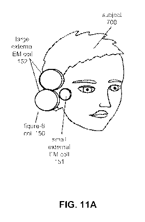
[0073] FIG. 11A shows an embodiment of the invention in which a plurality
of EM coils
generate EM fields that are directed at different parts of the facial nerve
system. Here,
multiple parts of the facial nerve system are stimulated for the purpose of
inducing desired
effects from one part of the facial nerve system while blocking undesired
effects of another
part of the facial nerve system. In this specific embodiment, a small external
EM coil 151 is
placed anterior to the ear, over the region of the parotid gland for the
purpose of inhibiting
action potentials carried by the external motor branches of the facial nerve,
thereby reducing
facial muscle movements. A pair of larger external EM coils 152 formed as a
figure-8 coil
150 is then placed in a manner to direct its EM field at the geniculate
ganglion or other part
of the facial nerve system that is proximal to the brain from the post-
ganglionic segment of
the facial nerve system; stimulation from this component of the embodiment of
the device
acts to increase blood flow to the brain or cranium. In some embodiments, one
or more EM
coils receive alternating electrical current from the stimulus generator while
other EM coils
receive direct electrical current. In some embodiments, one or more EM coils
generate
CA 02856314 2014-05-16
WO 2013/085924 PCT/US2012/067801
pulses of magnetic field while other EM coils generate a constant magnetic
field. In some
embodiments, the apparatus also contains electrodes that can be applied to the
subject for the
purpose of delivering electrical current.
[0074] In some embodiments, the EM field or fields will be generated in a
manner that
directs action potential conduction in the facial nerve system to progress in
a preferred
direction, and/or that blocks propagation of the action potential in other
directions, by
selective direction of electrical current flow 170 through the EM coils. As
shown in FIG.
11B, in some embodiments, action potentials are preferentially propagated away
from the
brain. An array of two EM coils arranged as a figure-8 coil 150 and placed
over the right ear
of the subject 700 conducts electrical current through the lower EM coil in a
clockwise
direction while conducting electrical current through the upper EM coil in a
counterclockwise
direction. The EM field pulse is then generated in a manner that
preferentially conducts
action potentials from the target portion of the facial nerve system (here,
the geniculate
ganglion) away from the brain. In other embodiments of the invention, action
potentials are
preferentially propagated toward the brain. As shown in FIG. 11C, an array of
two EM coils
arranged as a figure-8 coil 150 and placed over the right ear of the subject
700 conducts
electrical current through the lower EM coil in a counterclockwise direction
while conducting
electrical current through the upper EM coil in a clockwise direction. The EM
field pulse is
then generated in a manner that preferentially conducts action potentials from
the target
portion of the facial nerve system (here, the geniculate ganglion) toward the
brain. In some
embodiments of the invention, multiple parts of the facial nerve system are
stimulated in such
a manner for the purpose of colliding and neutralizing action potential
propagation in certain
parts of the facial nerve system. In some embodiments, a constant EM field is
generated for
the purpose of blocking local action potential conduction.
[0075] FIG. 11D shows an embodiment of the invention in which a plurality
of EM coils
are arranged such that two EM coils arranged as a figure-8 coil are used to
activate the facial
nerve system or portion thereof (stimulating EM coils 110) whereas another
pair of EM coils
arranged perpendicular to the figure-8 coil along its long axis and its
bisecting axis are used
to reduce, neutralize, or counteract unwanted portions of the EM field of the
figure-8 coil
(blocking EM coils 112). In some embodiments, the blocking EM coils 112 are
removed or
separated from the contact surface 160 of the stimulating EM coils 110.
[0076] FIG. 12 depicts an apparatus for bilateral stimulation of the facial
nerve system
and/or target component of the facial nerve system. In this embodiment, a pair
of EM coils is
arranged on either side of the head near the ear and is supplied with stimulus
energy from a
21
CA 02856314 2014-05-16
WO 2013/085924 PCT/US2012/067801
stimulus generator by one or more cables 530. Each pair of EM coils is
composed of (i) an
external EM coil 104 shaped as a ring with a hollow center that is intended
for placement
against the side of the head over the external ear and (ii) an internal EM
coil 103 that is sized
and shaped so as to fit into the ear canal. In this example, the two EM coils
of the pair of EM
coils are contained in a single housing to maintain a precise spatial
relationship with each
other. A cap 540 composed of or containing a sound-dampening and / or heat-
reflecting
material or substance is placed over the internal EM coil 103 so as to provide
the contact
surface for the apparatus on the subject. In some embodiments, the cap 540 may
be sterile.
[0077] Continuing with FIG. 12, the surface opposite the contact surface
160 of the
apparatus may, in some embodiments, be designed to receive or connect to a
coolant
cartridge 140. In some embodiments, the coolant cartridge 140 may fill a
central hole in the
external EM coil 104 and provide a contact surface for the internal EM coil
103. A coolant
cartridge 140 is then attached to the two EM coils that form a pair of EM
coils with the
following results: the external / non-subject side of the external EM coil 104
is covered by
the coolant cartridge 140; the hollow center of the external EM coil 104 is
filled by an
extension of the coolant cartridge 140; the external / non-subject side of the
internal EM coil
103 is in apposition to a projection of the coolant cartridge 140.
[0078] In other embodiments, the cap 540 is positioned on a head strap 124
in a manner
that orients the generated EM field in a certain direction (not shown). In
other embodiments,
the cap is associated with positioning components or accessory devices that
orient the
generated EM field in a certain direction (not shown). In some embodiments,
the cap 540 is
composed of ferromagnetic material that distorts or modifies an electric or
magnetic field in a
desirable manner. In some embodiments, the cap 540, EM coils, or housing of
the EM coils
include one or more fiducial markers that indicate the expected direction or
position of the
EM field (not shown). In some embodiments, the cap 540 may incorporate aspects
of a
speculum for visualization of the tympanic membrane / ear drum.
[0079] In some embodiments, the coolant cartridge 140 connects to the EM
coil in a
manner that allows for electrical current to flow through the EM coil. In some
embodiments,
a connector or other component of the coolant cartridge 140 is irreversibly
inactivated or
destroyed by connection to the EM coil, thereby preventing reuse of the
coolant cartridge
140. In some embodiments, electrical current flow through the EM coil serves
to inactivate,
destroy, or otherwise render as inoperable the connection between the coolant
cartridge 140
and the EM coil.
22
CA 02856314 2014-05-16
WO 2013/085924 PCT/US2012/067801
[0080] As shown in FIG. 13, in some embodiments the EM coil 100 or assembly
of more
than one EM coil is supplied with electrical current through one or more
cables 530
connecting to a stimulus generator 510. In some embodiments, the stimulus
generator is
supplied by or contains a power source 500 and is affected, directed,
modulated, or instructed
by a stimulus controller 520. In some embodiments, the stimulus generator 510
provides
feedback to the stimulus controller 520 that affects the function of the
stimulus controller
520.
[0081] In some embodiments, the stimulus controller 520 is affected,
directed,
modulated, or instructed by input or information it receives from one or more
sensor devices
520. In some embodiments, a sensor device 520 is equipped with one or more
sensors that
can include physiological sensors 558, temperature sensors 552, blood flow
sensors 554,
contact sensors 556, and other sensors. In some embodiments, a sensor is
directed at or
placed on the EM coil 100, cable 530, coolant cartridge 140, or subject 700,
or is directed at
or placed between a combination of the EM coil 100, cable 530, coolant
cartridge 140, and
subject 700. Signal or information provided directly or indirectly to the
stimulation
controller 520 by sensors may, in some embodiments, change or instruct the
function of the
stimulus generator 510, EM coil 100, or a positioning component (as described
for FIG. 1B),
or else prompt an operator of the apparatus to modify the function of these
parts of the
apparatus.
[0082] Additional embodiments of the device may be adapted for use on
different regions
of the body. For example, an embodiment of the device may be adapted for
stimulating the
ganglia of the heart, lungs, major blood vessels, gut, or other organs. In
such embodiments,
the stimulation elements may be arranged as large coils placed on the ventral
and/or dorsal
aspects of the thorax or abdomen. As another example, an embodiment of the
device may be
adapted for stimulation of the cranial nerves coursing through the neck. In
that embodiment,
the stimulation elements may be arranged as a necklace with groupings of
elements on one or
both sides of the neck surface. In some embodiments, the group of elements
focus
stimulation energy at a target located deep to the anterior / carotid triangle
of the neck. In
other embodiments, the group of elements focus stimulation energy at a target
located deep to
the posterior / occipital triangle of the neck. In some embodiments, the
target is the vagus
nerve, the accessory nerve, the glossopharyngeal nerve, the hypoglossal nerve,
a laryngeal
nerve, the ansa cervicalis, a portion of the brachial plexus, or the ganglia
of these neural
structures. In other embodiments, the target is the carotid bulb or sinus.
23
CA 02856314 2014-05-16
WO 2013/085924 PCT/US2012/067801
[0083] Another embodiment of the device may be adapted for stimulation of
the neural
structures near to, or derived from, the spine. In some embodiments,
stimulation elements
are arranged as chains longitudinally placed alongside the spine on the
posterior surface of
the neck or on the back. In some embodiments, the target for stimulation
includes the
phrenic nerve, the spinal sympathetic chain, an occipital nerve, a portion of
the brachial
plexus, or the ganglia of these neural structures.
[0084] In some embodiments, different regions of the body are stimulated in
conjunction
with stimulation of the facial nerve.
Neural Structure Modulation Methods
[0085] Referring now to FIG. 14, there is shown a flow diagram providing a
method for
neural structure modulation, according to an embodiment of the invention. It
should be
understood that these steps are illustrative only. Different methods of the
invention may
perform the illustrated steps in different orders, omit certain steps, and/or
perform additional
steps not shown in FIG. 14 (the same is true for the other Figures). The
method can start and
end at various points in the process, and often the method is a continuous
process with
multiple steps occurring simultaneously, so the Figures provide only examples
of one
ordering of method steps. In addition, the method can be performed using any
of the
apparatuses described herein or other apparatuses capable of performing the
steps provided
below.
[0086] As shown in FIG. 14, the method includes an initial step for
application of the
apparatus to the body, such as for placement 1000 of the apparatus on the
head. In some
embodiments, placement 1000 of apparatus on the head involves the unilateral
application of
an apparatus. In other embodiments, placement 1000 of apparatus on head
involves
application of an apparatus to both sides of the head or application of one
apparatus to each
side of the head. In some embodiments, for safety purposes, the apparatus can
detect 1004
whether a condition exists that would interfere with stimulation of the neural
system of the
subject with the apparatus. For example, the apparatus can detect 1004 whether
a material,
such as a metal, is on or inside of the subject near to the stimulation
components of the
apparatus before use. Presence of a metal might provide an unsafe condition
since the
apparatus will be generating an EM field. Thus, the detection 1004 can notify
the user that
the metal is present such that the metal can be removed before an EM field is
generated. In
these embodiments, once the apparatus is applied to the head and is in a
suitable position for
use, the apparatus is employed in a manner that allows the apparatus (e.g.,
the electrically-
24
CA 02856314 2014-05-16
WO 2013/085924 PCT/US2012/067801
conductive element(s), such as the EM coils) to detect 1004 the local presence
of metal. In
another embodiment, the detection is a question-and-answer review with, and
observation
and examination of, a subject performed by the user of the apparatus.
In response to the detection of the conditions (such as detection of a metal),
the apparatus can
perform an adjustment to remove or alleviate this condition. In one
embodiment, the
detection of metal will cause the method to end 1200 and the apparatus may
shut down or be
unwilling to operate until the metal is no longer detected. In other
embodiments of the
method, the apparatus will cause an alarm to sound that informs an operator of
the local
presence of metal on the subject. In this method, failure to detect 1004 the
local presence of
metal can be required, but is not necessarily sufficient, for activation of
the apparatus (i.e.,
activation can require other steps in some embodiments, such as attachment of
coolant
cartridges to the EM coils 1008). In another embodiment, the adjustment
performed is
removal of the metal by a user or informing of the subject that the metal must
be removed or
that the procedure cannot be performed. In a further embodiment, the condition
detected is
that the apparatus requires cooling, and the apparatus absorbs or dissipates
heat. For
example, the apparatus may have a cooling cartridge to absorb or dissipate
heat, or the user
may attach such a cooling cartridge. In another embodiment, the method
includes providing
a functional nerve block for application, such as to a pure somatic motor or a
somatic sensory
component of the neural system, thereby improving tolerability of the
simulation by the
subject. This can be provided to the subject by the apparatus or by a user of
the apparatus.
For example, the apparatus can deliver a pharmacologic agent, or the user can
provide the
agent (e.g., topically or as an injection). Additionally, the nerve block may
be an electrical
current applied by an electrode of the stimulator apparatus or the nerve block
is an
electromagnetic field created by one of a plurality of electrically-conductive
wire arrays of
the stimulator apparatus.
[0087] With this detection 1004 step achieved, the stimulus controller can
be
programmed (if necessary) and activated 1014 in embodiments that include such
a stimulus
controller. In these embodiments, the stimulus controller directs or allows
the activation
1018 of the stimulus generator, which then delivers stimulus energy (e.g.,
electrical current)
to the electrically-conductive element (e.g., an EM coil). Thus, stimulation
energy can be
administered to the subject by the apparatus (e.g., controlled by the operator
of the
apparatus) for a period of time to generate an electromagnetic field to
stimulate the neural
system of the subject. In one embodiment, electrical current is delivered 1020
to a first set of
EM coils 1020 and delivered 1024 to a second set of EM coils. In some
embodiments, the
CA 02856314 2014-05-16
WO 2013/085924 PCT/US2012/067801
first set of EM coils is composed of EM coils placed on one side of the head
and the second
set of EM coils is composed of EM coils placed on the other side of the head.
In some
embodiments, the first set of EM coils are EM coils placed in the ear canal
and the second set
of EM coils are external EM coils placed on the side of the head. In some
embodiments, the
delivery 1020 of electrical current to the first set of EM coils is initiated
before the delivery
1024 of electrical current to the second set of EM coils. In other
embodiments, the delivery
1020 of electrical current to the first set of EM coils is of a different
voltage, current,
duration, and / or waveform than is the delivery 1024 of electrical current to
the second set of
EM coils and the electrical current is delivered to the two sets of EM coils
simultaneously.
[0088] The stimulation energy can be delivered 1024 as electromagnetic
pulses. In one
example, these pulses are of biphasic shape and of 100-450 microseconds in
duration and
0.5-2.0 Tesla field strength at a surface of the apparatus. In another
example, the
electromagnetic pulses are delivered at 5-20 Hertz frequency in a continuous
manner for less
than 5 minutes. The stimulation can also be delivered intermittently. In one
example, the
duration, intensity, frequency, waveform, or other parameter of the
administered stimulation
energy as a function of blood flow, electroencephalography potentials,
intracranial pressure, a
duration of the condition of the subject, or other physiological or
pathophysiological
parameter.
[0089] Continuing with FIG. 14, following delivery of the stimulus energy
to the
electrically-conductive element(s) (e.g., delivery 1020, 1024 of electrical
current to the EM
coils), the need for further stimulation is determined by a component of the
apparatus, such as
the stimulus generator. The apparatus thus assesses whether continued
stimulation is needed
to treat the subject after the period of stimulation time has elapsed. In some
embodiments, a
minimum stimulation time must be achieved 1028 before termination of the
stimulation. If
the minimum stimulation time is not achieved, the stimulus generator is
activated 1018 and
the delivery 1020, 1024 of electrical current to the EM coils repeats. If the
minimum
stimulation time is achieved or it is otherwise determined that continued
stimulation is not
needed, the stimulation process ends 1200 and stimulation is discontinued. As
another
example, the determination of whether continued stimulation is needed is based
on delivery
of a certain number of stimulus pulses. In a further example, the
determination is based upon
feedback from the subject or feedback from a sensor associated with the
apparatus
[0090] The method also includes providing power via a power source for
supplying the
stimulus energy to the electrically-conductive elements, such as the EM coils
(the power may
also be provided automatically, as the device may be constantly connected to
or in
26
CA 02856314 2014-05-16
WO 2013/085924 PCT/US2012/067801
communication with the power source). The power can be provided via wires
connecting the
power source to the device. In some methods, supplying stimulus energy to the
EM coil or
coils modulates blood flow to the brain of the subject or enhances delivery of
a blood-borne
pharmacologic agent to treat stroke or another condition of the subject. In
some methods,
supplying stimulus energy to the EM coil or coils modulates
electroencephalographic activity
(e.g., of the cerebral cortex) to treat or prevent seizures or another
condition of neural
excitability / inactivity of the subject. The modulation can be performed in
the condition or
expectation of epilepsy and seizure disorders as either the direct treatment
of a disease
process or else to prevent onset of the disease process.
[0091] If any adjustments are needed or desired regarding the stimulus energy,
the method
can include adjusting the stimulus energy. For example, the adjustments can be
made based
on physiological or pathophysiological responses of the subject to the
stimulus energy. The
method can continue with supplying and adjusting as needed until the method is
done. When
the method is done (i.e., the treatment is successful and complete), the
apparatus can be
removed from the subject. If the apparatus has a detachable cap, the method
can include
detaching the cap and attaching a new cap. If the apparatus has a detachable
coolant
cartridge, the method can include detaching the coolant cartridge and
attaching a new coolant
cartridge.
[0092] Referring now to FIG. 15, there is shown a method of facial nerve
system stimulation,
according to an embodiment of the invention, in which sensor feedback
regulates the use of
the apparatus. In this embodiment, placement 1100 of the apparatus on the body
(e.g., on the
head) is followed by attachment or application of a blood flow sensor to the
subject. Then a
baseline blood flow measurement is obtained 1132 and a threshold defining
blood flow
increase is set 1134. In some embodiments, other or additional sensors can
also be applied to
regulate use of the apparatus. In some embodiments, these sensors may detect
properties of
the apparatus or of the subject including but not limited to the following:
carotid artery blood
flow; cerebral artery blood flow; blood flow to the central nervous system;
facial nerve
electrical potentials; skin / scalp galvanic responses; skin / scalp blood
flow; ear
temperature; pupilometry; intraocular pressure; blood flow to the eye;
bioelectric
potentials; electroencephalogram waveforms; electrophysiological testing of
the auditory or
vestibular systems; taste sensation; audition; lacrimation; nasal drainage;
nasal
congestion; salivation; sound sensitivity; face, head, or hand movements or
electromyographic potentials; speech production or arrest; sensation of body
movement;
eye movements; cranial blood flow; direct or indirect activity of a nerve; and
severity of
27
CA 02856314 2014-05-16
WO 2013/085924 PCT/US2012/067801
neurological dysfunction of the subject. In some embodiments 1114, the
stimulus controller
is entirely preprogrammed and cannot be adjusted or programmed by a user of
the apparatus
(although in other embodiments it can be programmable). In some embodiments,
the
stimulus controller offers a minimum of stimulation options that can be
selected by a user.
In some embodiments, the stimulation options offered by the stimulus
controller 1114 are
adjusted, altered, restricted, or enhanced by feedback, information, or
signals provided to the
stimulus controller 1114 by sensors and /or sensor devices to the apparatus.
[0093] Continuing with FIG. 15, the stimulus generator is activated 1118 and
delivers
electrical current to the electrically-conductive element(s) (e.g., delivers
1120 electrical
current to a first set of EM coils). In some embodiments, as a subsequent or
simultaneous
step, the stimulus generator delivers energy, such as electrical current, to
an electrically
conductive element such as a second set of EM coils 1124. Steps 1120 and 1124
in this
method may be sequential or simultaneous, and / or may be distinct in terms of
the quantity,
polarity, duration, waveform, or other parameter of the electrical current. In
some
embodiments, the first set of EM coils is composed of EM coils placed on one
side of the
head and the second set of EM coils is composed of EM coils placed on the
other side of the
head. In some embodiments, the first set of EM coils are EM coils placed in
the ear canal
and the second set of EM coils are external EM coils placed on the side of the
head. In some
embodiments, the first set of EM coils may be placed in the vicinity of the
ears and the
second set may be placed elsewhere on the head (e.g., inside the mouth or
under the chin).
[0094] Upon delivery 1120, 1124 of electrical current to the EM coils, the
stimulus controller
then determines if a minimum stimulation time has not been achieved 1130 for
blood flow
assessment, and if the minimum stimulation time has not been achieved then the
steps
described in 1118, 1120, 1124 are repeated. Once the stimulus controller
determines that a
sufficient duration since the time of activation has been achieved to perform
a blood flow
measurement, it inactivates the stimulus energy delivery 1132 and activates
1134 the blood
flow sensor. The purpose of this step is to determine if the stimulation of
the facial nerve
system or portion of the facial nerve system has achieved the desired result,
namely, to
achieve 1140 a threshold for increase of blood flow.
[0095] In some embodiments, measurement of blood flow is achieved by use of
two or more
EM coils, one or more of which serve to magnetize nearby blood while the
others serve to
detect the release or decay of energy from the magnetized blood once it has
moved in a
cranial or rostral direction, as described in FIG. 5. Returning to FIG. 15, in
some
embodiments, means of measuring blood flow in the head, neck, brain, or other
anatomical
28
CA 02856314 2014-05-16
WO 2013/085924 PCT/US2012/067801
structure 1130 other than magnetization of blood are employed for determining
of blood flow
measurement to achieve threshold for increase 1140. If the threshold for blood
flow increase
is achieved 1140, the process ends 1210. If the threshold for blood flow
increase is not
achieved 1140, the stimulus generator is activated 1118 for delivery of
stimulus energy and
additional electrical current is delivered 1120, 1124 to the EM coils, and the
process repeats.
[0096] The method can also include monitoring one or more physiological or
pathophysiological responses of the subject over a period of time. In this
method, if it is
determined that an adjustment is needed, the method can include adjusting the
intensity,
frequency, pattern, etc. of the stimulus energy supplied to the EM coils. The
adjustment can
be made based on the one or more physiological or pathophysiological responses
of the
subject, the decision of a user, or based on other factors. In some methods,
the adjustment
can occur automatically without requiring any action by a physician, operator,
or other user
to make the adjustment. In other methods, the physician, operator, or other
user can have
access to the monitored responses of the subject, and can control the
adjustment based on the
monitoring. The method can include continued supplying of stimulus energy to
the EM coils
with periodic monitoring and adjusting as needed over a period of time to
treat the subject, or
monitoring can be continuous until a sensor signal is received or a threshold
is crossed that
causes reactivation of the apparatus leading to the delivery of additional
stimulation energy.
In this manner, if stimulation is needed or desired of one or more components
of the facial
nerve system to dilate vessels and treat stroke and/or prevent stroke
recurrence, the device
can provide such stimulation. This can be done automatically or under the
control of a
physician, operator, or other user using the device.
[0097] In some methods of use, repeating stimulation between intervals of non-
stimulation is
desirable and expected. In some methods of use, the interval between periods
of stimulation
is defined by the blood flow response. In other methods of use, the interval
between periods
of stimulation is predetermined. In some methods of use, the stimulation
parameters
employed in second and subsequent stimulations is differently set than the
initial stimulation
parameters. In some embodiments, the stimulation parameters used in the
initial or
subsequent stimulations are set so as to induce long-term or other types of
neural
potentiation. In some embodiments, long-term or other types of neural
potentiation involve
the geniculate ganglion, sphenopalatine ganglion, brainstem, or other neuron
groups.
[0098] In some methods-of-use, the sensor device detects and / or interprets
electroencephalographic potentials, and informs the activity of the stimulus
controller based
on that information.
29
CA 02856314 2014-05-16
WO 2013/085924 PCT/US2012/067801
[0099] In some methods-of-use, the sensor device measures and / or interprets
intracranial
pressure, and informs the activity of the stimulus controller based on that
information.
[0100] In some methods-of-use, failure of the apparatus to achieve the
desired result will
be followed by filling the ear canal with electrically-conductive materials,
gels, or solutions,
and / or anesthetics and / or pharmacological substances. Once filling of the
ear canal is
complete, stimulation of the facial nerve system with the apparatus may be
attempted again.
In some embodiments, the electrically-conductive material, gel, solution, or
anesthetics and /
or pharmacological substances placed into the ear canal surrounds or otherwise
encompasses
the EM coils of the apparatus.
[0101] In some methods-of-use, failure of the apparatus to achieve the
desired result will
be followed by advancement of a cannula into the middle ear space. In this
situation, a hole
is punctured in the ear drum using a sharpened distal end of a cannula. The
cannula is
equipped so as to allow injection of electrically-conductive materials, gels,
solutions, or
anesthetics and / or pharmacological substances. Once injection into the
middle ear space is
complete, stimulation of the facial nerve system with the apparatus may be
attempted again.
Puncture of the ear drum in order to obtain access to the middle ear space may
be facilitated
by a stereotaxic device that positions the distal end of the cannula, or by
fiber optic
visualization. Alternatively, puncture of the ear drum may be accomplished by
means of a
pressure-sensitive distal end of the cannula or by electrical conductivity
changes at the distal
end of the cannula.
[0102] In some methods-of-use, conduction block of extracranial / distal
facial nerve is
applied prior to, or during, stimulation of the facial nerve system. In some
methods-of-use,
the conduction block is accomplished by a local injection of a pharmacological
substance
into the face or head. In some methods-of-use, the conduction block is
accomplished by
application of an electrical current across or in the vicinity of the facial
nerve trunk external
to the middle ear. In some methods-of-use, the conduction block is
accomplished by
generation of a constant EM field in the vicinity of the facial nerve trunk
external to the
middle ear.
[0103] In some methods-of-use, stimulation of the facial nerve system with
EM fields in
frequencies of 5-20 Hertz may be optimal for inducing the desired effect. In
some methods-
of-use, stimulation patterns involving on-off periods may be optimal for
inducing the desired
effect. In some methods-of-use, stimulation strengths of 0.5-2.0 Tesla (8-32
kT/s at coil
surface) may be optimal for inducing the desired effect. In some methods-of-
use, stimulation
waveform width of 100-450 microseconds may be optimal for inducing the desired
effect. In
CA 02856314 2014-05-16
WO 2013/085924 PCT/US2012/067801
some methods-of-use, stimulation waveform shapes that are substantially
bipolar may be
optimal for inducing the desired effect. In some methods-of-use, stimulation
may be
maintained for 0.5 to 5 minutes in duration. In some methods-of-use, the
stimulation
parameters, direction of electrical current, the orientation of the EM coils,
and / or the
configuration of the EM coils may be changed depending upon the condition of
the patient.
[0104] In some methods, use of the apparatus may be appropriate in
disorders of
cerebrovascular circulation (stroke, chronic cerebrovascular atherosclerosis),
head trauma,
dementia, headache disorders, or other neurological conditions. In some
methods, use of the
stimulator may be appropriate prior to procedures that involve the cerebral
and carotid
arteries, such as endovascular clot retrieval during stroke, endovascular coil
and stent
placement in aneurysmal subarachnoid hemorrhage, diagnostic angiography, or
surgical
carotid endarterectomy. In some methods, use of the device may interrupt
seizure activity,
reduce the likelihood of developing a seizure, or prevent the development of
epilepsy. In
some methods, use of the apparatus may modulate intracranial pressure. In some
embodiments, use of the stimulator may modulate inflammatory and immune
reactions within
or of the head, neck, and/or elsewhere in the body.
[0105] While the present teachings are described in conjunction with
various
embodiments and methods, it is not intended that the present teachings be
limited to such
embodiments. On the contrary, the present teachings encompass various
alternatives,
modifications, and equivalents, as will be appreciated by those of skill in
the art. Most of the
words used in this specification have the meaning that would be attributed to
those words by
one skilled in the art. Words specifically defined in the specification have
the meaning
provided in the context of the present teachings as a whole, and as are
typically understood
by those skilled in the art. In the event that a conflict arises between an
art-understood
definition of a word or phrase and a definition of the word or phrase as
specifically taught in
this specification, the specification shall control. It must be noted that, as
used in the
specification and the appended claims, the singular forms "a," "an," and "the"
include plural
referents unless the context clearly dictates otherwise.
31