Note: Descriptions are shown in the official language in which they were submitted.
CA 02857730 2014-07-23
METHODS AND APPARATUS TO FORM OPHTHALMIC DEVICES
INCORPORATING FLUORESCENCE DETECTORS
BACKGROUND OF THE INVENTION
1. Field of the Invention
The present invention relates to ophthalmic devices that have fluorescence
detectors upon or within them. The ophthalmic devices may be used for
analyzing an
analyte in fluids in an ophthalmic environment.
2. Discussion of the Related Art
Traditionally, an ophthalmic device, such as a contact lens, an intraocular
lens, or a
punctal plug, included a biocompatible device with a corrective, cosmetic, or
therapeutic
quality. A contact lens, for example, may provide one or more of vision
correcting
functionality, cosmetic enhancement, and/or therapeutic effects. Each function
is provided
by a physical characteristic of the lens. A design incorporating a refractive
quality into a
lens may provide a vision corrective function. Pigmentation incorporated into
the lens
may provide a cosmetic enhancement. An active agent incorporated into a lens
may
provide a therapeutic functionality. Such physical characteristics are
accomplished without
the lens entering into an energized state. An ophthalmic device has
traditionally been a
passive device.
Novel ophthalmic devices based on energized ophthalmic inserts have recently
been described. These devices may use the energization function to power
active optical
components.
In a related field, recently developed glucose detecting devices that are
implanted
into ophthalmic tissue have been described. The devices and future
alternatives may utilize
Forster resonance energy transfer ( FRET) probes to produce a resonance signal
that is
sensitive to the concentration of glucose near the probe. Exemplary devices
may include
implants placed into relatively transparent tissue in the eye to probe for
glucose
concentration in Interstitial Fluid (ISF) in the ophthalmic environment.
Some of these devices are probed by hand held fluorescence detectors. Contact
lenses, which incorporate fluorescence detector arrangements, may provide
convenient
1
CA 02857730 2014-07-23
and improved means for detecting glucose concentrations. Therefore, it may be
useful to
design ophthalmic devices to which may incorporate energized fluorescence
detectors to
analyze glucose levels.
SUMMARY OF THE INVENTION
The ophthalmic devices incorporating fluorescence detectors in accordance with
the present invention overcome the disadvantages associated with the prior art
as briefly
described above. Accordingly, the present invention includes an encapsulated
media insert
that comprises a fluorescence analyzer component.
In some exemplary embodiments, an ophthalmic lens media insert device may be
formed which comprises an annular media insert comprising a sealed component
cavity
within the annular media insert. The annular media insert may be formed from a
front
annular insert piece and a rear annular insert piece which are joined
together. The rear
annular insert piece may equivalently be referred to as a back annular insert
piece. In
some exemplary embodiments, the front annular insert piece may be sealed to
the rear
annular insert piece. The sealing may occur in various manners and locations
and in some
exemplary embodiments both an inner annular seal location and an outer annular
seal
location may be formed and sealed. When an annular media insert is sealed in
such a
manner, the region between the various seals and the annular front insert
piece and the
annular back insert piece may be a cavity in which various components may be
located.
In some exemplary embodiments, an energy source may be a component that may
be located in the sealed component cavity within the annular media insert. In
other
exemplary embodiments, an energy source may be located externally or in part
externally
to the sealed component cavity, but have the ability to make electrical
connection to
components within the sealed cavity.
In some exemplary embodiments the ophthalmic lens media insert device may also
comprise interconnects that may be at least partially located within the
sealed component
cavity.
In some exemplary embodiments, an electronic circuit may be located within the
sealed component cavity.
In some exemplary embodiments, a tab of material may protrude from the annular
insert piece from either the front annular insert piece, the back or rear
annular insert piece
2
CA 02857730 2014-07-23
or as a connected feature to either of these. In some exemplary embodiments,
the tab may
support a fluorescence analysis component along at least portions of the
surface of the
annular insert piece. The surface portions may be located externally to the
sealed
component cavity in some exemplary embodiments or it may be located within a
sealed
component cavity that may be located within the tab region or may be connected
to the tab
region. The fluorescence analysis component may connect to a portion of the
interconnects and may be in electrical communication with electronic circuits
within the
annular media insert device.
In some exemplary embodiments, a tab region may be formed on the front annular
surface and may also be formed on the rear or back annular insert piece. The
fluorescence
analysis component may be supported upon the tab region of the rear annular
insert piece.
In other exemplary embodiments, the fluorescence analysis component may be
supported
upon the tab region of the front annular insert piece. In still other
exemplary
embodiments, the fluorescence analysis component may be located in proximity
to either
1 5 or both of the front annular insert piece and the rear annular insert
piece, but may not be
directly affixed to either piece. In some exemplary embodiments, there may a
tab region
formed on only one of the front annular insert piece or the rear annular
insert piece.
In some exemplary embodiments, an ophthalmic contact lens device may be
formed utilizing the annular media insert device. In some exemplary
embodiments, a
media insert device consistent with the previous descriptions may be
encapsulated or
shrouded within a skirt of material that may be consistent with interaction
with human
tissue in an ophthalmic environment. In some exemplary embodiments, the skirt
may
comprise a hydrogel material.
A fluorescence probe may be located proximate the tissue of a human eye
environment and may therefore have a fixed location and orientation in that
environment.
It may be important for an ophthalmic contact lens device to maintain an
orientation so
that a fluorescence analysis component may correctly interact with a
fluorescence probe.
The skirt of material that enshrouds or encapsulates the media insert may
include
structural features that may be useful in orienting the contact lens. These
devices which
may be referred to as stabilization features or zones may keep the ophthalmic
device in a
correct orientation through their interaction with the ocular environment
including, for
example, the eyelids of a user.
3
CA 02857730 2014-07-23
= =
The various exemplary fluorescence detection embodiments may include
electronic components to control functions, perform analysis functions, retain
data and
similar such functions as well as communicate data to electronic components,
receivers or
transceivers located outside the fluorescence detection device. In some
exemplary
embodiments, some or all of the electronic components may be in a stacked
integrated
component form.
In some exemplary embodiments, an ocular fluid analysis system may comprise an
ophthalmic device. The ophthalmic device may comprise energization elements or
batteries where the elements are a portion of the ophthalmic device. The
ophthalmic
device may in some exemplary embodiments be suitable to be worn by a user
while in
contact with ocular fluid of the user's eye. A fluorescence analysis system
may be in
electrical communication with the energy source or sources. The fluorescence
analysis
system may be configured operatively to measure a fluorescence signal in the
ocular
environment.
1 5 In some exemplary embodiments, a fluorescence analysis probe may be
located
within ocular tissue of the user in such a manner that the portion of the
ophthalmic device
comprising the fluorescence analysis system may interact therewith. The
fluorescence
analysis probe may interact with at least a first compound that is located in
the interstitial
tissue spaces that surround the fluorescence analysis probe. The fluorescence
analysis
probe may interact with the first compound in such a manner that the
interaction causes
the probe to emit a characteristic signal based at least in part upon the
concentration of the
first compound that it interacts with. The emission of the characteristic
signal may be
predicated, triggered and/or supported by interaction with an excitation
signal that it may
receive from the fluorescence analytical system. In some exemplary
embodiments, a
light-based excitation signal may be emanated by the fluorescence analytical
system under
control of electronic circuitry within the device. The light-based excitation
signal may be
absorbed within the fluorescence analysis probe and the probe may subsequently
emit the
characteristic signal which may be a fluorescence signal. The electronic
circuitry may
comprise a processor that may form a portion or part of the ophthalmic device.
The
processor may be capable of executing a program, and the processor may be
capable of
storing values or data related to the fluorescence signal observed by the
fluorescence
analysis system. In some exemplary embodiments, the processor May cause or be
configured to emanate or transmit or otherwise output a signal. In some
exemplary
4
CA 02857730 2014-07-23
embodiments, the transmission may be caused or initiated based on receiving a
transmission of a signal that is sent from external to the device. The output
signal may
encode in various fashions the stored values into a transmittable signal.
In still some other exemplary embodiments, the processor, which may be capable
of executing a program, may evaluate a detected fluorescence signal against a
preprogrammed threshold value. In yet other exemplary embodiments, the
threshold value
may be received from a transmission from an external source. The threshold
value may
relate to one or more ocular fluid properties that may be indicated by the
fluorescence
signal characteristics. In some exemplary embodiments, when the evaluated
detected
fluorescence signal is outside the limit relating to the threshold value the
processor or
electronic circuitry may cause the outputting of a signal. The signal may be
received by
an external transceiver or receiver and cause an alarm or other action to be
precipitated
external to the ophthalmic device in some exemplary embodiments.
The ophthalmic device that incorporates fluorescence detectors may be utilized
in
various manners. In some exemplary embodiments, an ophthalmic device
comprising a
fluorescence analytical system may be provided to a user. The user may, for
example,
wear the ophthalmic device as a contact lens device. In some exemplary
embodiments,
there may be a reception of a data value external to the ophthalmic device of
a
transmission that originates from the ophthalmic contact lens. In other words,
the
ophthalmic device may be configured for one or two way communication with
external
devices.
DESCRIPTION OF THE DRAWINGS
The foregoing and other features and advantages of the invention will be
apparent
from the following, more particular description of preferred embodiments of
the invention,
as illustrated in the accompanying drawings.
Figs. lA ¨ 1B illustrate an exemplary embodiment of a media insert for an
energized
ophthalmic device and an exemplary embodiment of an energized ophthalmic
device.
Figs. 2A ¨ 2B illustrate an exemplary annular shaped insert and a cross
sectional
representation at an indicated location.
5
CA 02857730 2014-07-23
Fig. 3 illustrates an exemplary embodiment of a media insert with a
fluorescence sensor
located in a peripheral location in electrical communication with an energized
electronic
circuit element.
Figs. 4A ¨ 4B illustrate an exemplary embodiment of an ophthalmic contact lens
with a
media insert that comprises a fluorescence sensor. Other features of an
exemplary contact
lens are also depicted.
Figs 5A- 5B illustrate an exemplary ophthalmic environment with an exemplary
fluorescence probe for an analyte. A cone sponding overlay of an ophthalmic
lens in the
ophthalmic environment is also depicted.
Fig. 6 illustrates an exemplary ophthalmic lens in an ophthalmic environment
comprising
a fluorescence probe for an analyte transferring information wirelessly to a
receiving unit.
Fig. 7 demonstrates an exemplary stacked die implementation of fluorescence
detection
elements incorporated within ophthalmic devices.
Fig. 8 demonstrates a processor that may be used to implement exemplary
embodiments of
1 5 the present invention.
DETAILED DESCRIPTION OF THE PREFERRED EMBODIMENTS
The present invention relates to an ophthalmic device having sensing elements
capable of stimulating a fluorescence probe and sensing the fluorescent
emissions from the
probe. In the following sections detailed descriptions of exemplary
embodiments of the
invention are given. The description of both preferred and alternative
embodiments are
exemplary embodiments only, and it is understood that to those skilled in the
art that
variations, modifications and alterations may be apparent. It is therefore to
be understood
that the exemplary embodiments do not limit the scope of the underlying
invention.
Glossary
In the description and claims directed to the present invention, various terms
may be
used for which the following definitions will apply:
6
CA 02857730 2014-07-23
"Electro-wetting on Dielectric" or "EWOD": as used herein refers to a class of
devices or a class of portions of devices where a combination of immiscible
fluids or
liquids, a surface region with defined surface free energy and an electro-
potential field are
present. Typically, the electro-potential field will alter the surface free
energy of the surface
region, which may alter the interaction of the immiscible fluids with the
surface region.
"Energized": as used herein refers to the state of being able to supply
electrical
current to or to have electrical energy stored within.
"Energy": as used herein refers to the capacity of a physical system to do
work.
Many uses within the present invention may relate to the capacity being able
to perform
electrical actions in doing work.
"Energy Source": as used herein refers to a device or layer that is capable of
supplying energy or placing a logical or electrical device in an energized
state.
"Energy Harvester": as used herein refers to a device capable of extracting
energy
from the environment and converting it to electrical energy.
"Fluorescent probe": as used herein refers to a chemical probe that interacts
with its
environment to alter a fluorescence property that the probe manifests.
"Fluorescence based analysis": as used herein refers to the use of a
fluorescence
property in performing a chemical based analysis.
"Fluorophore": as used herein refers to a fluorescent chemical compound that
may
be bound or combined with other chemical compounds that can re-emit light upon
light
excitation.
"Functionalized": as used herein refers to making a layer or device able to
perform a
function including for example, energization, activation, or control.
"Leakage": as used herein refers to unwanted loss of energy.
"Lens" or "Ophthalmic Device": as used herein refers to any device that
resides in
or on the eye. These devices may provide optical correction, may be cosmetic,
or may
provide functionality unrelated to the eye. For example, the term lens may
refer to a
contact lens, intraocular lens, overlay lens, ocular insert, optical insert,
or other similar
device through which vision is corrected or modified, or through which eye
physiology is
cosmetically enhanced (e.g. iris color) without impeding vision.
Alternatively, the lens
may provide non-optic functions, for example, monitoring glucose or
administrating
medicine. In some embodiments, the preferred lenses of the present invention
are soft
7
CA 02857730 2014-07-23
contact lenses are made from silicone elastomers or hydrogels, which include,
for
example, silicone hydrogels, and fluorohydrogels.
"Lens-forming mixture" or "Reactive Mixture" or "Reactive Monomer Mixture"
(RMM): as used herein refers to a monomer or prepolymer material that may be
cured and
crosslinked or crosslinked to form an ophthalmic lens. Various embodiments may
include
lens-forming mixtures with one or more additives, for example, UV blockers,
tints,
photoinitiators or catalysts, and other additives one might desire in an
ophthalmic lenses
such as, contact lenses or intraocular lenses.
"Lens-forming Surface": as used herein refers to a surface that is used to
mold a
lens. In some exemplary embodiments, any such surface may have an optical
quality
surface finish, which indicates that it is sufficiently smooth and formed so
that a lens
surface fashioned by the polymerization of a lens forming material in contact
with the
molding surface is optically acceptable. Further, in some embodiments, the
lens-forming
surface may have a geometry that is necessary to impart to the lens surface
the desired
optical characteristics, including spherical, aspherical and cylinder power,
wave front
aberration correction, corneal topography correction and the like as well as
any
combinations thereof.
"Lithium Ion Cell": as used herein refers to an electrochemical cell where
Lithium
ions move through the cell to generate electrical energy. This electrochemical
cell, typically
called a battery, may be reenergized or recharged in its typical forms.
"Media Insert": as used herein refers to an encapsulated insert that will be
included
in an energized ophthalmic device. The energization elements and circuitry may
be
incorporated in the media insert. The media insert defines the primary purpose
of the
energized ophthalmic device. For example, in embodiments where the energized
ophthalmic device allows the user to adjust the optic power, the media insert
may include
energization elements that control a liquid meniscus portion in the optical
zone.
Alternatively, a media insert may be annular so that the optical zone is void
of material. In
such embodiments, the energized function of the lens may not be optic quality,
but may be,
for example, monitoring glucose or administering medicine.
"Mold": as used herein refers to a rigid or semi-rigid object that may be used
to
form lenses from uncured formulations. Some preferred molds include two mold
parts
forming a front curve mold part and a back curve mold part.
"Operating Mode": as used herein refers to a high current draw state where the
8
CA 02857730 2014-07-23
current over a circuit allows the device to perform its primary energized
function.
"Optical Zone": as used herein refers to an area of an ophthalmic lens through
which a wearer of the ophthalmic lens sees.
"Power": as used herein refers to work done or energy transferred per unit of
time.
"Rechargeable" or "Re-energizable": as used herein refers to a capability of
being
restored to a state with higher capacity to do work. Many uses within the
present invention
may relate to the capability of being restored with the ability to flow
electrical current at a
certain rate and for a certain, reestablished period.
"Reenergize" or "Recharge": as used herein refers to restoring to a state with
higher
capacity to do work. Many uses within this invention may relate to restoring a
device to the
capability to flow electrical current at a certain rate and for a certain,
reestablished period.
"Released from a Mold": as used herein refers to a lens that is either
completely
separated from the mold, or is only loosely attached so that it may be removed
with mild
agitation or pushed off with a swab.
"Stacked": as used herein means to place at least two component layers in
proximity to each other such that at least a portion of one surface of one of
the layers
contacts a first surface of a second layer. In some exemplary embodiments, a
film, whether
for adhesion or other functions may reside between the two layers that are in
contact with
each other through said film.
"Stacked Integrated Component Devices" or "SIC Devices": as used herein refers
to the products of packaging technologies that assemble thin layers of
substrates that may
include electrical and electromechanical devices into operative-integrated
devices by
means of stacking at least a portion of each layer upon each other. The layers
may
comprise component devices of various types, materials, shapes, and sizes.
Furthermore,
the layers may be made of various device production technologies to fit and
assume
various contours.
"Storage Mode": as used herein refers to a state of a system comprising
electronic
components where a power source is supplying or is required to supply a
minimal designed
load current. This term is not interchangeable with standby mode.
"Substrate Insert": as used herein refers to a formable or rigid substrate
capable of
supporting an energy source within an ophthalmic lens. In some exemplary
embodiments,
the substrate insert also supports one or more components.
9
CA 02857730 2014-07-23
Energized Ophthalmic Device
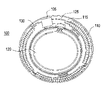
Referring to Fig. 1A, an exemplary embodiment of a media insert 100 for an
energized ophthalmic device and a corresponding energized ophthalmic device
150 (Fig.
1B) are illustrated. The media insert 100 may comprise an optical zone 120
that may or
may not be functional to provide vision correction. Where the energized
function of the
ophthalmic device is unrelated to vision, the optical zone 120 of the media
insert may be
void of material. In some exemplary embodiments, the media insert may include
a portion
not in the optical zone 120 comprising a substrate 115 incorporated with
energization
elements 110 (power source) and electronic components 105 (load).
In some exemplary embodiments, a power source 110, for example, a battery, and
a load 105, for example, a semiconductor die, may be attached to the substrate
115.
Conductive traces 125 and 130 may electrically interconnect the electronic
components
105 and the energization elements 110. The media insert may be fully
encapsulated to
protect and contain the energization elements 110, traces 125, and electronic
components
105. In some exemplary embodiments, the encapsulating material may be semi-
permeable,
for example, to prevent specific substances, such as water, from entering the
media insert
and to allow specific substances, such as ambient gasses or the byproducts of
reactions
within energization elements, to penetrate or escape from the media insert.
In some exemplary embodiments, as depicted in Fig. 1B, the media insert 100
may
be included in an ophthalmic device 150, which may comprise a polymeric
biocompatible
material. The ophthalmic device 150 may include a rigid center, soft skirt
design wherein
the central rigid optical element comprises the media insert 100. In some
specific
embodiments, the media insert 100 may be in direct contact with the atmosphere
and the
corneal surface on respective anterior and posterior surfaces, or
alternatively, the media
insert 100 may be encapsulated in the ophthalmic device 150. The periphery 155
of the
ophthalmic device or lens 150 may be a soft skirt material, including, for
example, a
hydrogel material. The infrastructure of the media insert 100 and the
ophthalmic device
150 may provide an environment for numerous embodiments involving fluid sample
processing with fluorescence based analysis elements.
Referring to Fig. 2A ¨ 2B, a depiction of an exemplary multi-piece insert 200
in
annular form is illustrated in both plan view, Fig. 2A, and cross section
view, Fig. 2B. The
insert 200 is an annular insert with a ring of material around a central
optical zone that is
devoid of material. The annular insert 200 comprises an exterior extent as
shown by item
CA 02857730 2014-07-23
. .
220 and an internal annulus edge at item 230. Included in the insert 200 may
be found
energization elements, interconnect features of various types and electronic
circuit
elements.
A dashed line at 290 represents a cross sectional direction. In the detail of
Fig. 2B,
shown as 290, is a cross section along the direction of the dashed line. The
cross section
reveals that the insert 200 may be a combination of a front insert piece 291
and a rear
insert piece 292. Various means of joining and sealing these two pieces along
the various
surfaces of the annulus may be defined, and an exemplary sealing design is
depicted. Also
shown in an encapsulated location may be an integrated circuit element 293
connected to
interconnection elements. In some exemplary embodiments, the rear insert piece
292 may
have a gap in the region of an integrated circuit 293. In these exemplary
embodiments,
integrated circuit 293 may include a sensor that may function in an improved
fashion if it
may sense emanations without the perturbing aspects of an insert piece.
Fluorescence Based Probe Elements for Analyte Analysis
Various types of analytes may be detected and analyzed using fluorescence
based
analysis techniques. A subset of these techniques may involve the direct
fluorescence
emission from the analyte itself. A more generic set of techniques relate to
fluorescence
probes that have constituents that bind to analyte molecules and in so binding
alter a
fluorescence signature. For example, in Forster Resonance Energy Transfer
(FRET),
probes are configured with a combination of two fluorophores that may be
chemically
attached to interacting proteins. The distance of the fluorophores from each
other can
affect the efficiency of a fluorescence signal emanating therefrom. One of the
fluorophores may absorb an excitation irradiation signal and can resonantly
transfer the
excitation to electronic states in the other fluorophore. The binding of
analytes to the
attached interacting proteins may disturb the geometry and cause a change in
the
fluorescent emission from the pair of fluorophores. Binding sites may be
genetically
programmed into the interacting proteins, and for example, a binding site,
which is
sensitive to glucose, may be programmed. In some cases, the resulting site may
be less
sensitive or non-sensitive to other constituents in interstitial fluids of a
desired sample.
The binding of an analyte to the FRET probes may yield a fluorescence signal
that
is sensitive to glucose concentrations. In some exemplary embodiments, the
FRET based
probes may be sensitive to as little as a 10 uM concentration of glucose and
may be
11
CA 02857730 2014-07-23
=
sensitive to concentrations up to hundreds of micromolar. Various FRET probes
may be
genetically designed and formed. The resulting probes may be configured into
structures
that may assist analysis of interstitial fluids of a subject. In some
exemplary embodiments,
the probes may be placed within a matrix of material that is permeable to the
interstitial
fluids and their components, for example, the FRET probes may be assembled
into
hydrogel structures.
In some exemplary embodiments, these hydrogel probes may be included into the
hydrogel based processing of ophthalmic contact lenses in such a manner that
they may
reside in a hydrogel encapsulation that is immersed in tear fluid when worn
upon the eye.
In other exemplary embodiments, the probe may be inserted in the ocular
tissues just
above the sclera. A hydrogel matrix comprising fluorescence emitting analyte
sensitive
probes may be placed in various locations that are in contact with bodily
fluids containing
an analyte.
In the examples provided, the fluorescence probes may be in contact with
interstitial fluid of the ocular region near the sclera. In these cases, where
the probes are
invasively embedded, a sensing device may provide a radiation signal incident
upon the
fluorescence probe from a location external to the eye such as from an
ophthalmic lens or
a hand held device held in proximity to the eye.
In other exemplary embodiments, the probe may be embedded within an
ophthalmic lens in proximity to a fluorescence-sensing device that is also
embedded
within the ophthalmic lens. In some exemplary embodiments, a hydrogel skirt
may
encapsulate both an ophthalmic insert with a fluorescence detector as well as
a FRET
based analyte probe.
Ophthalmic Insert Devices and Ophthalmic Devices with Fluorescence Detectors
Referring to Fig. 3, an ophthalmic insert is demonstrated including components
that may form an exemplary fluorescence based analytical system. The
demonstrated
ophthalmic insert is shown in an exemplary annular form having an internal
border of 335
and an external border of 320. In addition to energization elements 330,
control circuitry
310, and interconnect features 360 there may be a fluorescence analytical
system 350,
which in certain exemplary embodiments may be positioned on a flap 340. The
flap 340
may be connected to the insert 300 or be an integral, monolithic extension
thereof. The
flap 340 may properly position the fluorescence analytical system 350 when an
12
CA 02857730 2014-07-23
=
ophthalmic device comprising a fluorescence is detector is worn. The flap 340
may allow
the analytical system 350 to overlap with portions of the user's eye away from
the optic
zone. The fluorescence based analytical system 350 may be capable of
determining an
analyte, in terms of its presence or its concentration, in a fluid sample. As
a non-limiting
example, the fluorophores may include Fluorescein, Tetramethylrhodamine, or
other
derivatives of Rhodamine and Fluorescein. It may be obvious to those skilled
in the art
that any fluorescence emitting analyte probe, which may include fluorophore
combinations for FRET or other fluorescence-based analysis may be consistent
with the
art herein.
For a fluorescence analysis, a probe may be irradiated with an excitation
light
source. This light source may be located within the body of the analytical
system 350. In
some exemplary embodiments, the light source may comprise a solid-state device
or
devices such as a light emitting diode. In an alternative exemplary
embodiment, an InGaN
based blue laser diode may irradiate at a frequency corresponding to a
wavelength of 442
nm for example. Nanoscopic light sources as individual or array sources may be
formed
from metallic cavities with shaped emission features such as bowties or
crosses. In other
exemplary embodiments, light emitting diodes may emit a range of frequencies
at
corresponding wavelengths that approximate 440 nm, for example. As well, the
emission
sources may be supplemented with a band pass filtering device in some
embodiments.
Other optical elements may be used to diffuse the light source from the solid-
state
device as it leaves the insert device. These elements may be molded into the
ophthalmic
insert body itself. In other exemplary embodiments, elements such as fiber
optic filaments
may be attached to the insert device to function as a diffuse emitter. There
may be
numerous means to provide irradiation to a fluorescence probe from an
ophthalmic insert
device 300 of the type demonstrated in Fig. 3.
A fluorescence signal may also be detected within the fluorescence based
analytical system 350. A solid-state detector element may be configured to
detect light in a
band around 525 nm as an example. The solid-state element may be coated in
such a
manner to pass only a band of frequencies that is not present in the light
sources that have
been described. In other exemplary embodiments, the light sources may have a
duty cycle
and a detector element's signal may only be recorded during periods when the
light source
is in an off state. When the duty cycle is used, detectors with wide band
detection ability
may be advantageous.
13
CA 02857730 2014-07-23
An electronic control bus of interconnects shown schematically as item 360 may
provide the signals to the light source or sources and return signals from the
detectors. The
powered electronic component, item 310 may provide the signals and power
aspects. The
exemplary embodiment of Fig. 3, illustrates a battery power source 330 to the
electronic
circuitry 310. In other exemplary embodiments, energization may also be
provided to the
electronic circuitry by the coupling of energy through wireless manners such
as
radiofrequency transfer or photoelectric transfer.
Referring to Figs.4A ¨ 4B, an ophthalmic lens in the form of a contact lens
with a
protruding tab 411 is illustrated which has incorporated a fluorescence
detector. In Fig.
4B, item 410 represents a top view of an ophthalmic lens. It includes an
ophthalmic lens
skirt 470. In some exemplary embodiments, the skirt 470 may be formed of the
various
hydrogel compounds consistent with the formation of contact lenses. In other
exemplary
embodiments, certain hydrogel compounds may be preferred for their properties
relating
to the various fluorescence probes that have been discussed.
A cross section is demonstrated by the dashed line 430. At Fig. 4A, a cross
sectional depiction along dashed line 430 may be found. The insert device 300
described
in Fig. 3 may be located within the ophthalmic lens and have the annular cross
sectional
representation 432 as depicted at 430. The lens is depicted with a printed
iris pattern 440
that may be demonstrated on the annular pieces as item 431. Beneath the
printed pattern
may be insert components as described with reference to Fig. 3. The
fluorescence
detection element may be located at 433. The ophthalmic lens 410 may also have
other
features important to the function of a lens with a fluorescence detector. At
450 and 460,
stabilization features may be included in the body of the formed ophthalmic
device. These
features may allow the lens 400 to be oriented in a preferred configuration
when worn by a
user. In exemplary embodiments where a fluorescence based analyte probe is
embedded in
a user's eye, the stabilization features 450 and 460 may be useful to aid in a
good overlap
between the embedded analyte probe and the fluorescence analysis element 480.
In other
exemplary embodiments where the analyte probe may be included in the hydrogel
body
470 of the lens, the stabilization features 450 and 460 may be useful in
orienting the
analysis element and probe into a preferred region of the eye with tear fluid.
Referring to Figs. 5A and 5B, the use of an ophthalmic device incorporating
fluorescence detectors in a user's eye 510 is illustrated in concert with a
sub-tissue
embedded analyte probe 520. As depicted, when the ophthalmic device 530 is
placed upon
14
CA 02857730 2014-07-23
a users eye and assumes an orientation that may be guided by features on the
ophthalmic
device it may locate a fluorescence based analysis element 540 in an overlap
with the
analyte probe 520. It may be useful to scale the size of the analysis element
540 to be
larger than that of the probe 520 to allow for some flexibility to alignment
of the two
features.
Referring to Fig. 6, the transfer of information from an ophthalmic device
incorporating fluorescence detectors to an external data reception device is
depicted. The
ophthalmic lens 610 may be worn upon a user's eye for a period long enough to
allow the
embedded analysis element to equilibrate with its surroundings and to begin
performing
analysis for an analyte. Either as the data is collected or in other exemplary
embodiments
after the data has been collected, a data transfer protocol may be initiated.
A wireless
signal, represented by arrow 630, may be emitted from the ophthalmic lens 610
to
communicate with an external reception device 650. A receiving element 640 may
include
an antenna for radiofrequency transmissions or other transducers to transform
light-based
signals, sound based signals or other wireless forms of communication into
received
information at the reception device 650.
In some exemplary embodiments, the powered operation of an ophthalmic lens
incorporating fluorescence detectors may function well with monolithic
components
included in the ophthalmic insert device. In other exemplary embodiments, such
as that
depicted in Fig. 7, a stacked integrated component may be useful to perform
the various
functions including power management 715, communications 745, control
functions 750
and the transmission of light and reception of fluorescence signal 710.
In exemplary embodiments of this type, as depicted in Fig. 7, the media insert
may
include numerous layers of different types that are encapsulated into forms
consistent with
the ophthalmic environment that they will occupy. In some exemplary
embodiments, these
inserts with stacked integrated component layers may assume the entire annular
shape of
the insert. Alternatively, in some exemplary embodiments, the media insert may
be an
annulus whereas the stacked integrated component may occupy just a portion of
the
volume within the entire shape.
Continuing with the example of Fig. 7, a stacked integrated component media
insert may assume numerous functional aspects. As shown in Fig. 7, there may
be thin
film batteries used to provide energization. In some exemplary embodiments,
these thin
film batteries may comprise one or more of the layers that are stacked upon
each other, in
CA 02857730 2014-07-23
this case layers 730 may represent the battery layers, with multiple
components in the
layers.
As may be seen in nearly all of the layers, there may be interconnections that
are
made between two layers that are stacked upon each other. In the state of the
art there may
-- be numerous manners to make these interconnections; however, as
demonstrated the
interconnection may be made through solder ball interconnections between the
layers. In
some cases only these connections may be required, however in other cases the
solder
balls may contact other interconnection elements, as for example with a
component having
through layer vias.
In other layers of the stacked integrated component media insert, a layer
dedicated
to interconnection of various components in the interconnect layers may be
found, as for
example, layer 725. This layer may comprise vias and routing lines that pass
signals from
various components to others. For example, layer 725 may provide the various
battery
elements connections to a power management unit 720, which includes supply 740
and
-- battery charger 765, that may be present in the technology layer components
of layer 715.
As well, the interconnection layer 725 may make connections between components
in the
technology layer and components outside the technology layer; as may exist for
example
in the integrated passive device component 760 shown as item 755. There may be
numerous manners for routing of electrical signals that may be supported by
the presence
-- of dedicated interconnect layers.
There may be numerous layers identified as technology layers in a given
application; however, in this exemplary embodiment there is a single layer
715. These
features represent a diversity of technology options that may be included in
media inserts.
In some exemplary embodiments, the layer may include CMOS, BiCMOS, Bipolar, or
-- memory based technologies. Alternatively, the layer may include different
technology
families within a same overall family; as for example layer 715 may include
electronic
elements produced using a 0.5 micron CMOS technology and also may include
elements
produced using a 20 nanometer CMOS technology. It may be apparent that many
other
combinations of various electronic technology types would be consistent within
the art
-- described herein.
In some exemplary embodiments, the media insert may include locations for
electrical interconnections to components outside the media insert. In other
exemplary
16
CA 02857730 2014-07-23
embodiments; however, the media insert may also include interconnection to
external
components in a wireless manner. In such cases, the use of antennas may
provide
exemplary manners of wireless communication. In some such exemplary
embodiments, a
layer may exist, as shown as item 735, where such an exemplary antenna may be
supported in the layer. In many cases, such an antenna layer may be located on
the top or
bottom of the stacked integrated component device within the media insert.
In some of the exemplary embodiments discussed herein, the battery elements
may
be included as elements in at least one of the stacked layers themselves. It
may be noted as
well that other exemplary embodiments may be possible where the battery
elements are
located externally to the stacked integrated component layers. Still further
diversity in
exemplary embodiments may derive from the fact that a separate battery or
other
energization component may also exist within the media insert, or
alternatively these
separate energization components may also be located externally to the media
insert.
At item 710, a fluorescence detection element may be attached to a stacked
integrated component. The fluorescence detection component may be attached as
a portion
of a layer in some exemplary embodiments. In other exemplary embodiments, the
entire
fluorescence detection element may also comprise a similarly shaped component
as the
other stacked components. The various diversity of types of fluorescence based
analysis
elements that have been discussed herein may be consistent with a stacked
integrated
component device, where other features such as light sources and light sensors
either are a
portion of a layer or alternatively attached to a stacked integrated
component.
Control Systems for Ophthalmic Devices with Integrated Fluorescence based
Analysis
Components
Referring now to Fig. 8 a controller is illustrated that may be used in
accordance
with some exemplary embodiments of the present invention. The controller
includes one
or more processors 810, which may include one or more processor components
coupled to
a communication device 820.
The processors 810 may be coupled to a communication device configured to
communicate energy via a communication channel. The communication device may
be
used to communicate electronically with components within the ophthalmic
insert within
the ophthalmic device. The communication device 820 may also be used to
communicate,
17
CA 02857730 2014-07-23
for example, with one or more controller apparatus or manufacturing equipment
components during the production of ophthalmic devices incorporating
fluorescence based
analysis elements.
The processor 810 may also be in communication with a storage device 830. The
storage device 830 may comprise any appropriate information storage device,
including
combinations of magnetic storage devices (e.g., magnetic tape and hard disk
drives),
optical storage devices, and/or semiconductor memory devices such as Random
Access
Memory (RAM) devices and Read Only Memory (ROM) devices.
The storage device 830 may store a program 840 for controlling the processor
810.
The processor 810 performs instructions of a software program 840, and thereby
operates
in accordance with the present invention. For example, the processor 810 may
receive
information descriptive of media insert placement, component placement, and
the like.
The storage device 830 may also store ophthalmic related data in one or more
databases
850 and 870. The database may include customized specific control sequences
for
controlling the function of a fluorescence-based analytical system. The
database may also
include parameters and controlling algorithms for the control of fluorescence-
based
analysis components that may reside in the ophthalmic device as well as data
that result
from their action. In some exemplary embodiments, that data may be ultimately
communicated to a reception device externally located to the ophthalmic
device.
It is important to note that all of the components described herein may be
sized and
configured for use in ophthalmic devices or applications. In addition, the
components are
preferably biocompatible or encapsulated in biocompatible materials.
Although shown and described in what is believed to be the most practical and
preferred embodiments, it is apparent that departures from specific designs
and methods
described and shown will suggest themselves to those skilled in the art and
may be used
without departing from the spirit and scope of the invention. The present
invention is not
restricted to the particular constructions described and illustrated, but
should be
constructed to cohere with all modifications that may fall within the scope of
the appended
claims.
18