Note: Descriptions are shown in the official language in which they were submitted.
CA 02858389 2014-06-06
WO 2013/086636
PCT/CA2012/050899
1
SOLUBLE IGF RECEPTOR Fc FUSION PROTEINS AND
USES THEREOF
RELATED APPLICATIONS
[0001] This
application claims priority to U.S. Provisional Application No.
61/576,034, filed December 15, 2011, the entire contents of which are hereby
incorporated by reference.
TECHNICAL FIELD
[0002] The
present invention relates to novel soluble IGF receptor Fc fusion
proteins and compositions and methods of use thereof for treating cancer and
metastasis.
BACKGROUND OF THE INVENTION
[0003] The
receptor for the type I insulin like growth factor (IGF-IR) plays a
critical role in progression of malignant disease. Increased expression of IGF-
IR
and/or its ligands has been documented in many human malignancies and high
plasma IGF-I levels were identified as a potential risk factor for
malignancies
such as breast, prostate and colon carcinomas (Samani et al., 2007, Endocr
Rev, 28: 20-47). Recent data have shown that the IGF axis promotes tumor
invasion and metastasis through several mechanisms, and it has been identified
as a determinant of metastasis to several organ sites, particularly the lymph
nodes and the liver (Long et al., 1998, Exp Cell Res, 238: 116-121; Wei, et
al.,
2006, Ann Surg Oncol, 13: 668-676; Samani et al., 2007, Endocr Rev, 28: 20-
47; Reinmuth et al., 2002, Clin Cancer Res, 8: 3259-3269). The IGF receptor
can affect metastasis by regulating tumor cell survival and proliferation in
secondary sites and also by promoting angiogenesis and lymphangiogenesis
either through direct action on the endothelial cells or by transcriptional
regulation of vascular endothelial growth factors (VEGF) A and C (reviewed in
Li, S. et al., In: Liver metastasis:Biology and Clinical Management 2011;
Brodt
P., Editor: 233-72)).
CA 02858389 2014-06-06
WO 2013/086636
PCT/CA2012/050899
2
[0004] The IGF-
IR ligands include three structurally homologous peptides
IGF-I, IGF-II and insulin, but the receptor binds IGF-I with the highest
affinity.
The major site of endocrine production for IGF-I and IGF-II is the liver
(Werner
& Le Roith, 2000, Cell Mol Life Sci 57: 932-942), but autocrine/paracrine IGF-
I
production has been documented in extra-hepatic sites such as heart, muscle,
fat, spleen and kidney. The physiological activities and bioavailability of
IGF-I
and IGF-II are modulated through their association with 6 secreted, high-
affinity
binding proteins (IGFBP1-6).
[0005] IGF-IR
has been validated as a target for anti-cancer therapy in
various tumor types. A number of IGF-IR inhibitors are in clinical or
preclinical
development (see, for example, Zha, J. and Lackner, M.R., Clinical Cancer
Research 2010; 16: 2512-7; Gualberto, A. and Pollak, M., Oncogene 2009; 28:
3009-21; and Li, S. et al., In: Liver metastasis: Biology and Clinical
Management 2011; Brodt P., Editor: 233-72). However, targeting the IGF-I
system in vivo poses several challenges: First, due to the high degree of
homology between the IGF-I and insulin receptors, drugs that target the IGF
axis may also affect the insulin receptor/insulin axis with undesirable
effects on
glucose and lipid metabolism. Hyperglycemia has, in fact, been observed as
one of the undesirable effects of anti-IGF-IR therapy (Karp, D.D. et al., J.
Thorac. Oncol. 2009; 4: 1397-403; Bruchim, I., et al., Expert Opinion on
Therapeutic Targets 2009; 13: 1179-92; Sachdev, D. and Yee, D., Mol. Cancer
Ther. 2007; 6: 1-12; Rodon, J. et al., Mol. Cancer Ther. 2008; 7: 2575-88).
Moreover, inhibition of IGF-I signaling may result in altered serum growth
hormone levels leading to insulin insensitivity and could potentially cause a
reduction in pancreatic insulin production and diabetes (Zha, J. and Lackner,
M.R., Clinical Cancer Research 2010; 16: 2512-7). Second, the use of
antibody-based therapy may result in ADCC reactions leading to hematological
toxicity as observed in some trials (Reidy, D.L., et al., Journal of Clinical
Oncology; 28: 4240-6; Zha, J. and Lackner, M.R., Clinical Cancer Research
2010; 16: 2512-7). Furthermore, some tumors also express isoform A of the
insulin receptor (IR-A) that can bind IGF-II with high affinity and this may
provide an alternate survival mechanism for cancer cells whose IGF-IR has
CA 02858389 2014-06-06
WO 2013/086636
PCT/CA2012/050899
3
been neutralized by antibody treatment or kinase inhibitors (Zha, J. and
Lackner, M.R., Clinical Cancer Research 2010; 16: 2512-7).
[0006] The use
of soluble receptors (decoys) to antagonize the activity of
soluble ligands for treatment of malignant disease has been taught as a
potential therapeutic treatment and has become an accepted form of therapy for
some conditions. Decoy receptors can inhibit the biological activity of the
cognate, membrane-bound receptors by binding and decreasing ligand
bioavailability for the latter receptor (Rudge, et al., 2007, Proc Natl Acad
Sci
USA, 104: 18363-18370). Current examples include a soluble TNF receptor
(Enbrel) that is in routine clinical use for the treatment of inflammatory
conditions (Richard-Miceli, C. and Dougados, M., BioDrugs 2001; 15: 251-9), as
well as a VEGF¨Trap (Aflibercept) that is in clinical trials for the treatment
of
cancer and other conditions (Rudge, J.S. et al., Cold Spring Harbor Symposia
on Quantitative Biology 2005; 70: 411-8). These reagents are advantageous
over antibody-based therapy because they are highly specific, bind to the
ligand
with high affinity, and bypass some of the undesirable effects of reagents
with
off-target activity.
[0007] Thus, a
soluble IGF-I receptor could potentially overcome some of the
shortcomings of current IGF-targeting drugs, such as, for example, cross-
reaction with the insulin system, ADCC-related hematological toxicity, and the
compensatory effects of insulin receptor isoform A (IR-A).
[0008] It would
be highly desirable therefore to be provided with a soluble
IGF-1 receptor for treatment of angiogenic-associated disorders and malignant
disease, including cancer and metastasis.
SUMMARY OF THE INVENTION
[0009] In
accordance with a broad aspect of the invention, there are
provided fusion proteins comprising an Fc portion of an antibody and a soluble
IGF-IR protein. The Fc portion may be derived from, for example, a human IgG
antibody, such as an IgG1 or IgG2 antibody.
CA 02858389 2014-06-06
WO 2013/086636
PCT/CA2012/050899
4
[0010] In an aspect, fusion proteins provided herein bind specifically to
IGF-1
and IGF-2. In some embodiments, fusion proteins bind to IGF-1 and IGF-2 with
at least about the same affinity. In some embodiments, the affinity of the
fusion
proteins for insulin is at least about 1000-fold lower than for IGF-1 or IGF-
2. In
some embodiments the fusion proteins do not bind detectably to insulin.
[0011] In some embodiments, the Fc portion of a fusion protein of the
invention comprises a modified Fc portion. In one embodiment, a fusion protein
comprises an Fc domain modified to remove one or more Cys residues, e.g., to
replace one or more Cys residues with Ser residues. In another embodiment, a
fusion protein comprises an Fc domain modified to replace an 11 aa linker with
a longer, more flexible linker, e.g., a 22aa or a 37aa flexible GS linker. In
an
embodiment, a fusion protein comprises an Fc domain modified both to remove
one or more Cys residues, e.g., to replace one or more Cys residues with Ser
residues, and to replace an 11 aa linker with a longer, more flexible linker,
e.g.,
a 22aa or a 37aa flexible GS linker. In some embodiments, fusion proteins
having modified Fc domains do not produce HMW species or produce a
reduced amount of HMW species compared to unmodified Fc domains.
[0012] In some embodiments, a soluble IGF-IR protein comprises or
consists
of the extracellular domain of IGF-IR having the amino acid sequence of SEQ
ID NO: 1 or 6, or a biologically active fragment or analog thereof. In other
embodiments, a soluble IGF-IR protein comprises or consists of the amino acid
sequence of the extracellular domain of full-length IGF-IR having the amino
acid
sequence of SEQ ID NO: 4, or a biologically active fragment or analog thereof.
A soluble IGF-IR protein may form the tetrameric structure of SEQ ID NO: 1, 4,
or 6.
[0013] In some embodiments, a fusion protein comprises or consists of the
sequence set forth in SEQ ID NO: 8 (Fc-sIGFIR, IgG1) or SEQ ID NO: 10 (Fc-
sIGFIR, IgG2), or a biologically active fragment or analog thereof. The
biologically active fragment or analog of the fusion protein may have, for
example, at least 70%, at least 80%, at least 90%, at least 95%, or at least
98%
CA 02858389 2014-06-06
WO 2013/086636
PCT/CA2012/050899
sequence identity to the fusion protein. The biologically active fragment or
analog may also retain the binding specificity of the fusion protein.
[0014] In some
embodiments, a fusion protein comprises or consists of the
sequence set forth in SEQ ID NO: 12 (5IGF1R-hFc-IgG1 Mod#1), SEQ ID NO:
14 (5IGF1R-hFc-IgG1 Mod#2), SEQ ID NO: 16 (5IGF1R-hFc-IgG1 Mod#3),
SEQ ID NO: 18 (5IGF1R-hFc-IgG1 Mod#4), or a biologically active fragment or
analog thereof. The biologically active fragment or analog of the fusion
protein
may have, for example, at least 70%, at least 80%, at least 90%, at least 95%,
or at least 98% sequence identity to the fusion protein. The biologically
active
fragment or analog may also retain the binding specificity of the fusion
protein.
[0015] Nucleic
acids encoding the fusion proteins or biologically active
fragments or analogs thereof are also provided. For example, the fusion
proteins or biologically active fragments or analogs thereof may be encoded by
a nucleic acid having the sequence set forth in SEQ ID NO: 5, 7, or 9, or a
degenerate variant thereof. In an embodiment, fusion proteins are encoded by
a nucleic acid having the sequence set forth in SEQ ID NO: 11, 13, 15, or 17,
or
a degenerate variant thereof. In an embodiment, nucleic acids having at least
70%, at least 80%, at least 90%, at least 95%, or at least 98% sequence
identity to the sequence set forth in SEQ ID NO: 5, 7, 9, 11, 13, 15, or 17
are
provided herein. Vectors comprising nucleic acids described herein are also
provided.
[0016] In other
aspects, pharmaceutical compositions comprising fusion
proteins or biologically active fragments or analogs thereof, and a
pharmaceutically acceptable carrier, are provided.
[0017] In yet
other aspects, there are provided uses of fusion proteins or
biologically active fragments or analogs thereof, or compositions thereof, for
treating an angiogenic associated disorder or a malignant disease, such as
cancer or metastasis, in a subject. For
example, fusion proteins or
compositions of the invention may be used to treat tumor metastasis,
colorectal
carcinoma, lung carcinoma, breast cancer, liver cancer, bladder cancer, lung
cancer, pancreatic cancer, multiple myeloma, glioblastoma multiforme, or liver
CA 02858389 2014-06-06
WO 2013/086636
PCT/CA2012/050899
6
metastasis. Methods
of inhibiting angiogenesis in a subject having an
angiogenic associated disorder, such as tumor metastasis, colorectal
carcinoma, lung carcinoma, breast cancer, liver cancer, bladder cancer, lung
cancer, pancreatic cancer, multiple myeloma, glioblastoma multiforme, or liver
metastasis, are also provided herein. Methods and compositions for preventing
or treating cancer or tumor metastasis are provided herein as well.
[0018] In
further aspects, there are provided methods of inhibiting
angiogenesis in a subject having an angiogenic associated disorder comprising
administering to said subject an autologous cell, e.g., a dendritic cell, a
hepatocyte, or a stromal cell, genetically modified to express fusion proteins
or
biologically active fragments or analog thereofs. The autologous cell may be,
e.g., a stromal cell, e.g., a bone marrow derived mesenchymal stromal cell.
[0019] In a
still further aspect, the methods provided herein further comprise
administering a fusion protein or biologically active fragment or analog
thereof,
or compositions thereof, in combination with another angiogenesis inhibitor
and/or in combination with one or more other anti-cancer agents. The two or
more agents may be administered concomitantly or sequentially.
[0020] In yet
another aspect, fusion proteins or biologically active fragments
or analogs, or compositions thereof, are administered via injection, e.g.,
intravenous or intraperitoneal injection. In another aspect, fusion proteins
or
biologically active fragments or analogs, or compositions thereof, are
administered orally.
[0021] In an
embodiment, there is provided herein a fusion protein
comprising an Fc portion of an antibody and a soluble IGF-IR protein. In one
embodiment, the fusion protein comprises an antibody, which is a human IgG
antibody. In an embodiment, the antibody is an IgG1 or an IgG2 antibody. In
an embodiment, the fusion protein binds specifically to IGF-1 and IGF-2. In
one
embodiment, the fusion protein binds to IGF-1 and IGF-2 with at least about
the
same affinity. In another embodiment, the affinity of the fusion protein for
IGF-2
is higher than the affinity of the fusion protein for IGF-1. In yet another
embodiment, the affinity of the fusion protein for insulin is at least about
1000-
CA 02858389 2014-06-06
WO 2013/086636
PCT/CA2012/050899
7
fold lower than the fusion protein's affinity for IGF-1 or IGF-2. In an
embodiment, the fusion protein does not bind detectably to insulin.
[0022] In one
embodiment, a fusion protein comprises a soluble IGF-IR
protein comprising the extracellular domain of IGF-IR having the amino acid
sequence of SEQ ID NO: 1 or 6, or a biologically active fragment or analog
thereof. In an embodiment, a soluble IGF-IR protein forms the tetrameric
structure of SEQ ID NO: 1 or 6. In another embodiment, a soluble IGF-IR
protein consists of SEQ ID NO: 1 or 6 or a biologically active fragment or
analog
thereof. In yet another embodiment, a soluble IGF-IR protein comprises the
extracellular domain of IGF-IR having the amino acid sequence of SEQ ID NO:
4, or a biologically active fragment or analog thereof.
[0023] In one
embodiment, a fusion protein comprises an Fc portion of an
antibody and a soluble IGF-IR protein, wherein the soluble IGF-IR protein
consists of SEQ ID NO: 1 or 6 or a biologically active fragment or analog
thereof.
[0024] In an
embodiment, a fusion protein comprises the sequence set forth
in SEQ ID NO: 8 or SEQ ID NO: 10. In another embodiment, a fusion protein
comprises the sequence set forth in SEQ ID NO: 12, 14, 16 or 18. In yet
another embodiment, there is provided herein a fusion protein consisting of
the
sequence set forth in SEQ ID NO: 8, 10, 12, 14, 16 or 18. In a further
embodiment, there is provided herein a fusion protein comprising the amino
acid sequence encoded by the nucleic acid set forth in SEQ ID NO: 7, 9, 11,
13,
15 or 17, or a degenerate variant thereof. In a still further embodiment,
there is
provided herein a fusion protein consisting of the amino acid sequence encoded
by the nucleic acid set forth in SEQ ID NO: 7, 9, 11, 13, 15 or 17, or a
degenerate variant thereof.
BRIEF DESCRIPTION OF THE DRAWINGS
[0025] Having
thus generally described the nature of the invention, reference
will now be made to the accompanying drawings, showing by way of illustration,
a preferred embodiment thereof, and in which:
CA 02858389 2014-06-06
WO 2013/086636
PCT/CA2012/050899
8
[0026] Figure 1
shows subcloning of CHO pools of stably-transduced cell
lines to identify best producers of sIGF1R (Trap D) and Fc-sIGF1R (Trap E).
Three subclones of CHO cell pools were isolated: CHO-Cum2-CR5-IGF1R-9-
33-1-6; CHO-Cum2-CR5-IGF1R-10-48-2-5, and CHO-Cum2-CR5-IGF1R-hFc-
16-13-1-6. For each subclone, 600,000 cells/ml were cultured for 2 days at 37
C
and 7 days at 300 C. Samples analyzed by denaturing, non-reducing SDS-
PAGE, 12p1/lane, Novex0 Tris-Glycine 10% TG 1.5. The lanes shown are as
follows: 1: IGF1R-9-33-1-6 pool; 2: IGF1R-9-33-1-6 clone #5, 3: IGF1R-9-33-1-
6 clone #6, 4: IGF1R-9-33-1-6 clone #10, 5: IGF1R-10-48-2-5 pool; 6: IGF1R-
10-48-2-5 clone #5, 7: IGF1R-10-48-2-5 clone #8, 8: IGF1R-10-48-2-5 clone
#12, 9: IGF1R-hFc-16-13-1-6 pool; 10: IGF1R-hFc-16-13-1-6 clone #4, 11:
IGF1R-hFc-16-13-1-6 clone #5, 12: IGF1R-hFc-16-13-1-6 clone #7.
[0027] Figure 2
shows purification of sIGF1R (Trap D) using a calcium
hydroxyapatite (CHT) column followed by gel filtration. For the hydroxyapatite
column, 170 ml of 400-fold concentrated & diafiltrated sIGF1R was loaded onto
25 ml of CHT column. Samples were analyzed by denaturing, non-reducing
SDS-PAGE, Novex0 Tris-Glycine 10% TG 1.5. SDS-PAGE is shown in (A).
Samples in lanes 1-9 are from the CHT column and in lanes 10-17 are from the
gel filtration column, runs # 3 to 4 as indicated. The lanes shown are as
follows:
1: Feed (non-concentrated), 5pg/lane, 2: Permeate; 3: Feed (concentrated); 4:
Flow-through, 0 to 115 ml, 5: Flow-through + chase; 6: Pool A2-A7, 15% 61, 7:
Pool A3-A5, 15% 61, 8: Pool A10-61, 20% 61, 9: Pool 133-137, 100% 62 (CIF);
10: High Molecular Weight markers (details are shown in part 13 of the
figure);
11: Run#3 A6 (5pg), 12: Run#3 A7 (5pg), 13: Run#3 A10 (out of range); 14:
Run#4 A6 (5pg), 15: Run#4 A7 (5 pg), 16: Run#4 All (out of range); 17:
Purified IGF1R-CHT-GF, 2.6pg. Molecular weight markers are shown in detail
in (13). Letters and numbers (A2-A7, 61, A3-A5, A10-61, 133-137, 62) refer to
fractions collected from columns; letters and numbers indicate position of
tube
on rack of fraction collector.
[0028] Figure 3
shows purification of Fc(IgG1)-5IGF1R (Trap E) using a
calcium hydroxyapatite (CHT) column followed by gel filtration. Samples were
analyzed by denaturing, non-reducing SDS-PAGE, Novex0 Tris-Glycine 10%
CA 02858389 2014-06-06
WO 2013/086636
PCT/CA2012/050899
9
TG 1.5. SDS-PAGE is shown in (A). Samples in lanes 1-5 are from the CHT
column and in lanes 6-15 are from the gel filtration column. The lanes shown
are as follows: 1: A9-Al2, 2: 61-136, 67-C1, 4: Cl 0-D3, 5: E5-E8, 6: Feed
(5p1),
7: Feed (2p1), 8: A9-A10, 9: All- Al2, 10: 61-133, 11: 66, 12: 68-139, 13: 610-
611, 14: 612-C1, and 15: Purified IGF1R-CHT-hFc-GF, 2.6 pg. The Red arrow
indicates the expected position of the Fc-sIGFIR tetramer, HMW: High
molecular weight markers. Molecular weight markers are shown in detail in (6).
Letters and numbers (A9-Al2, B1-136, 67-C1, C10-D3, E5-E8, etc.) refer to
fractions collected from columns; letters and numbers indicate position of
tube
on rack of fraction collector.
[0029] Figure 4
shows purification of Fc(IgG1)-5IGF1R (Traps F and G)
using protein A chromatography. Samples were analyzed by denaturing, non-
reducing SDS-PAGE, Novex Tris-Glycine 4-20% TG 1.5. SDS-PAGE is
shown in (A). Samples in lanes 1 to 4 are from purification of Trap F (eluted
at
pH 4), lane 1: 2 pi; lane 2: 1 pi; lane 3: 0.5 pi; lane 4: 0.25 p1/lane; lane
HMW:
High molecular weight markers. Samples in lanes 5 to 8 are from purification
of
Trap G (eluted at pH 3.5), lane 5: 1 pi; lane 6: 0.5 pi; lane 7: 0.25 pi; lane
8:
0.125 p1/lane. Samples in lanes 9 to 14 show IgG2 (purchased from Sigma),
lane 9:3 pg, lane 10:2 pg, lane 11: 1 pg, lane 12: 0.5 pg, lane 13: 0.25 pg,
lane
14: 0.125 pg. The Red arrow indicates the expected position of the Fc-sIGFIR
tetramer, the Black arrows indicate high molecular weight (HMVV) species.
[0030] Figure 5
shows purification of endotoxin-free Fc(IgG1)-sIGF1R (Traps
H and 1) using protein A chromatography. Samples were analyzed by
denaturing, non-reducing SDS-PAGE, Novex Tris-Glycine 4-20% TG 1.5.
SDS-PAGE is shown in (A). 14 p1/lane was loaded. Samples in lanes 5 to 8 are
from purification of Trap H (eluted at pH 4). Samples in lanes 9 to 12 are
from
purification of Trap I (eluted at pH 3.5). Lane 1: Feed; lane X: nothing
loaded;
lane 2: Flow-through (F.T.), lane 3: Al-A2, lane 4: A3-A4, lane 5: A6-A7, lane
6:
A8-A10, lane 7: All-Al2; lane 8: 61-132, lane 9: 63-134, lane 10: 65-136, lane
11: 67-1310, lane 12: B11-612. The Red arrow indicates the expected position
of the Fc-sIGFIR tetramer. Letters and numbers (Al-A2, A3-A4, A6-A7, A8-Al 0,
CA 02858389 2014-06-06
WO 2013/086636
PCT/CA2012/050899
A11-Al2, B1-132, etc.) refer to fractions collected from columns; letters and
numbers indicate position of tube on rack of fraction collector.
[0031] Figure 6
shows a schematic representation of vectors used to make
Trap proteins of the invention. The sIGF1R sequence was inserted into the
pMPG-0R5 vector as shown in (A), and the sIGF1R sequence fused to either
the human IgG1 Fc or IgG2 Fc was inserted into the pMPG-0R5 vector as
shown in (B) and (C), respectively. These vectors were used for transient or
stable expression of Trap proteins in CHO cells.
[0032] Figure 7
shows a comparison of the most predominant glycopeptides
of sIGF1R and sIGF1R-hFc by mass spectrometry. In (A), relative percentage
refers to the types of sugars attached at each glycosylation site; sites 4, 5,
7, 8,
12, 15 and 16 are glycosylation sites in the peptides; solid bars represent
sIGF-
IR (Trap D), cross-hatched bars represent sIGF-IR-hFc (Trap E), and the colors
indicate the nature of the glycosylation, as indicated in the legend shown in
(B).
[0033] Figure 8
shows that Traps D and E inhibit tumor cell proliferation in
response to hIGF-I equally. (A) shows a plot of OD vs. time where 10 ng/mL
IGF-I was used; (B) shows a plot of OD vs. time where 50 ng/mL IGF-I was
used. = indicates IGFI, = indicates sIGF-IR (Trap D) + IGFI, A indicates sIGF-
IR-hFc (Trap E) + IGFI and **** indicates p <0.001 at all time points tested.
[0034] Figure 9
shows a dose-dependent increase in anoikis (detachment-
induced apoptosis) in the presence of Trap D. FBS: Fetal Bovine Serum; SF:
Serum-free; "IGF-I: Trap D" is the molar ratio of IGF-I to Trap D, which is
2:1,
1:1 or 1:2 as indicated; * indicates p<0.05, ** indicates p<0.01, and ****
indicates p<0.001.
[0035] Figure
10 shows a dose-dependent increase in anoikis (detachment-
induced apoptosis) in the presence of Traps D and E and a comparison
between Traps D and E. FBS: Fetal Bovine Serum; SF: Serum-free; Ratios are
IGF-1:sIGFIR molar ratios (2:1, 1:1 or 1:2 as indicated); * indicates p<0.05,
**
indicates p<0.01, and **** indicates p<0.001. The data illustrate superior
performance of Trap E (Fc-sIGFIR).
CA 02858389 2014-06-06
WO 2013/086636
PCT/CA2012/050899
11
[0036] Figure
11 shows increased anoikis in the presence of the IGF-Traps
E, F and G, illustrating the effect of protein A purification. FBS: Fetal
Bovine
Serum; SF: Serum-free; Molar ratios of IGF-I:Trap protein are as indicated;
****
indicates p<0.001.
[0037] Figure
12 shows reduced anchorage-independent growth in the
presence of Traps D and E, and a comparison between Traps D and E. In (A) it
is shown that the number of colonies was significantly reduced in the presence
of the Traps; *indicates p< 0.05; p was < 0.01 under all conditions tested.
Colors indicate the proteins tested, as indicated in the legend shown in (B).
The
data illustrate superior performance of the Fc fusion protein.
[0038] Figure
13 shows a time course analysis indicating the effect of Traps
D and E on tumor cell invasion and a comparison between Traps D and E. Blue
line (*) represents baseline (no IGF-I), Pink line (=) indicates invasion with
IGF-
I; Green line (A) indicates Trap ID, and Red line (.) indicates Trap E.
[0039] Figure
14 shows in (A), the effect of Traps D, E, F and G on tumor
cell invasion at 48 hours; **** indicates p<0.0005. (B) shows a time course
analysis for the effect of Traps D, E, F and G on tumor cell invasion: blue
line
(*) is IGF-I, green line (=) is baseline (no IGF-I), light brown line (*) is
Trap ID,
dark green line (A) is Trap E, red line (=) is Trap F, light blue line (.) is
Trap G.
[0040] Figure
15 shows in (A), the effect of Traps E, H and I on tumor cell
invasion at 48 hours, illustrating a comparison of Trap E before and after
protein
A purification; **** indicates p< 0.001. (B) shows a time course analysis for
the
effect of Traps E, H and I on tumor cell invasion: blue line (*) is IGF-I,
pink line
(=) is Trap E, green line (=) is Trap H, red line (=) is Trap I; and orange
line (=)
is baseline (no IGF-I).
[0041] Figure
16 shows curve fitting for a multi-cycle SPR titration. There is
shown a representative analysis of experimental data (solid colored lines) to
the
"1:1 kinetic" model (global fit, dashed black lines) for hIGF-I (0 ¨ 66 nM, 2-
fold
dilution series) binding to amine-coupled Trap B (9500 RU).
CA 02858389 2014-06-06
WO 2013/086636
PCT/CA2012/050899
12
[0042] Figure
17 shows curve fitting for a single-cycle SPR titration. There is
shown a representative analysis of experimental data (solid colored lines, 0 ¨
530 nM, 2-fold dilution series) to the "1:1 titration" model (local fits,
dashed
black lines) for mIGF-I (green), hIGF-I (red) and hIGF-II (blue) binding to
amine-
coupled Trap E (6400 RU).
[0043] Figure
18 shows a pharmacokinetic analysis of Traps D and E,
indicating a greater than 2-fold increase in the half-life of Fc-sIGF1R (Trap
E)
compared to sIGF1R (Trap D). Trap D is shown in (A); Trap E is shown in (13),
red circles represent observed values; and the blue line shows predicted
values.
[0044] Figure
19 shows a pharmacokinetic analysis of Traps D, E, H and I,
indicating inferior in vivo performance of Protein A- purified Fc-sIGFIR
enriched
for HMW species. Trap D is shown in (A); Trap E is shown in (13), Trap H (pH
4.0) is shown in (C), and Trap I (pH 3.5) is shown in (D). Red circles
represent
observed values, and the blue line shows predicted values.
[0045] Figure
20 shows reduced tumor volume in mice inoculated with colon
carcinoma MC-38 cells and treated with IGF-Trap H. Representative H&E
stained formalin fixed paraffin embedded sections of livers derived from colon
carcinoma MC-38-injected mice 19 days post tumor injection are shown. Top
row: livers from mice not treated with IGF-Trap H (Non-treated); bottom row:
livers from mice treated with IGF-Trap H (Trap-treated); L indicates liver; T
indicates tumor; Mag-x20-50, inset ¨ x400. The far right panel in the top row
shows an expanded view (X400) of the indicated metastasis.
[0046] Figure 21 shows reduced IGF-IR phosphorylation in
micrometastases. C57BL6 female mice were injected intrasplenically with 105
GFP-tagged H-59 cells followed by injection of 5mg/kg IGF-Trap H (Trap-
treated) or vehicle only (Non-treated) on days 1 and 3 post tumor injection (3
mice per group). Mice were sacrificed on day 6, livers removed and snap frozen
and 10 pM cryostat sections prepared and immunostained with a rabbit
polyclonal anti-mouse pIGF1R antibody followed by a goat anti-rabbit Alexa
Fluor 647 (far-red) antibody. Sections were washed and mounted with the
CA 02858389 2014-06-06
WO 2013/086636
PCT/CA2012/050899
13
GOLD anti-fade reagent and analyzed with a Carl Zeiss LSM 510 Meta,
confocal microscope. In (A), there are shown representative merged confocal
images, as follows: A. sections from non-treated mice; B. sections from Trap-
treated mice; Green fluorescent protein (GFP) is shown in green; DAPI staining
is shown in blue; pIGF1R is shown in white; Images were taken at Mag. X200.
In (B), there is shown the calculated means of percent of pIGF-IR+ green
fluorescent tumor cells in each group (Non-treated, or Trap- treated at 5
mg/Kg,
as indicated); P<0.001.
[0047] Figure
22 shows increased tumor cell apoptosis in IGF-Trap H treated
mice. Liver cryostat sections were obtained as described above for Fig. 21.
Sections were incubated first with a rabbit polyclonal anti-mouse cleaved
caspase-3 antibody (ab4501-Abcam) and then with a goat anti-rabbit Alexa
Fluor 647 antibody. In (A), representative merged confocal images are shown,
as follows: a. sections from non-treated mice (Non-treated); b. sections from
Trap-treated mice (IGF-Trap -treated); Green fluorescent protein (GFP) is
shown in green; DAPI staining is shown in blue; Cleaved Caspase 3+ cells are
shown in red; Images were taken at Mag. X200. In (B), there is shown the
calculated means of percent of cleaved-caspase 3+ green fluorescent tumor
cells in each group (Non-treated, or Trap- treated at 5 mg/Kg, as indicated);
P<0.001.
[0048] Figure
23 shows decreased tumor cell proliferation in IGF-Trap H
treated mice. Liver cryostat sections were obtained as described above for
Fig.
21. Sections were incubated first with a rabbit polyclonal anti-mouse Ki67
antibody and then with a goat anti-rabbit Alexa Fluor 647 antibody. The
percentage of GFP + tumor cells that were Ki67 positive (a marker of
proliferation) was calculated. In (A), representative merged confocal images
are
shown, as follows: left panel: sections from non-treated mice (Non-treated);
right panel: sections from Trap-treated mice (IGF-Trap -treated); Green
fluorescent protein (GFP) is shown in green; Ki67 positive cells are shown in
red; Images were taken at Mag. X200. In (B), there is shown the calculated
means of percent of Ki67 + green fluorescent tumor cells in each group (Non-
treated, or Trap ¨treated at 5 mg/Kg, as indicated); p=0.0012.
CA 02858389 2014-06-06
WO 2013/086636
PCT/CA2012/050899
14
[0049] Figure
24 shows decreased vessel count (angiogenesis) in IGF-Trap
H injected mice. Liver cryostat sections were obtained as described above for
Fig. 21. Sections were incubated first with a rat monoclonal anti-mouse 0D31
antibody and then with a goat anti-rat Alexa Fluor 568 (orange-red) antibody.
The number of CD31+ endothelial cells within tumor micrometastases per field
(20X objective) was counted in 16 sections per treatment group and the mean
number was calculated. In (A), representative merged confocal images are
shown, as follows: A. sections from non-treated mice (Non-treated); B.
sections
from Trap-treated mice (IGF-Trap -treated); Green fluorescent protein (GFP) is
shown in green; DAPI staining is shown in blue; CD31+ cells are shown in red;
Images were taken at Mag. X200. In (B), there is shown the calculated means
of CD31+ cells per field in each group (Non-treated, or IGF-Trap ¨treated at 5
mg/Kg, as indicated); p=0.0057.
[0050] Figure
25 shows tumor growth reduction and increase in animal
survival in an orthotopic murine mammary carcinoma (4T1) model. Balb/c
female mice were injected into the mammary fatpad (MFP) with 105 mouse
mammary carcinoma 4T1 cells. Four hours and 3 days later the treatment group
received an i.v. injection of 10mg/kg of IGF-Trap H followed by 2 injections
of 5
mg/kg on days 6 and 10 post tumor inoculation (indicated by arrows in part
(A)).Tumors were measured three times weekly using a caliper and the tumor
volumes calculated using the formula 1/2(length x width2). In (A), there is
shown a graph of Tumor volume (mrn3) vs. Days post tumor inoculation for mice
non-treated (Control) or treated with IGF-Trap (IGF-Trap), as indicated. In
(B),
there is shown a plot of mouse survival vs. Days post tumor inoculation for
control or IGF-Trap treated, as indicated; p<0.01 using both Mantel-Cox and
Gehan-Breslow-Wilcoxon tests.
[0051] Figure
26 shows tumor growth inhibition in IGF-Trap-treated mice
orthotopically implanted with human breast cancer cells. One million MD-MBA-
231 human breast cancer cells were orthotopically implanted with Matrigel in
the mammary fatpads of nu/nu mice. Tumors were measured three times
weekly using a caliper and the tumor volumes calculated using the formula
1/2(length x width2). When tumors were established (50-100mm3) (day 11,
CA 02858389 2014-06-06
WO 2013/086636
PCT/CA2012/050899
indicated by an arrow in part (A)), the animals were randomized and treated
with 5mg/kg of IGF-Trap H or vehicle (i.v.) twice weekly up to day 33. Mice in
the control group were all moribund by day 44 (indicated by a dashed line in
part (A)). In (A), there is shown a graph of Tumor volume (mm3) vs. Days post
tumor inoculation for non-treated mice (Control) or mice treated with IGF-Trap
(IGF-Trap treated), as indicated. In (B), longitudinal bioluminescence imaging
is shown; this was used to monitor tumors. The color scale for bioluminescence
is shown at the left side of panel (B), and mice at the indicated day post
tumor
inoculation are shown; left panel shows non-treated mice and right panel shows
Trap-treated mice. The bioluminescence was quantitated and is shown in (C)
for control (Non-treated; black line) and Trap-treated (red line) mice. The
unit of
measurement p/sec/cm2/sr stands for photons per second per cm2/steradian.
[0052] Figure
27 shows molecular models serving as templates for the
design of modified sIGF1R-ed-Fc constructs. Crystal structures for IR-ed, and
for Fc complexes with FcgRIII-ed were retrieved from PDB (codes given in
parentheses). The image on the left side shows that 22aa flexible linkers
(white
lines) utilized in the constructs Mod#2 and Mod#3 are sufficiently long to
allow
intra-molecular pairing of Fc fragments (cyan/green ribbons) and further allow
binding to the FcgRIII-ed (surface rendering). The image on the right side
illustrates the same concept for the 27aa linkers of the Mod#4 modified
variant
protein that uses a hinge-truncated version of the Fc.
[0053] Figure
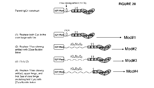
28 shows schematic depictions of the designed sIGF1R-ed-Fc
modified variant proteins. On the basis of sequence modeling of Insulin growth
hormone fused to human IgG Fc fragment, we designed and generated 4 new
constructs with different modifications in the junction of the sIGF1R and IgG1
sequences. The modifications are as follows: (1): Both cysteines in the core
hinge were substituted with serines (referred to as 5IGF1R-hFc-IgG1-Mod#1),
(2): The 11aa-cloning artifact was replaced with a 22aa-flexible linker
(referred
to as 5IGF1R-hFc-IgG1-Mod#2), (3): A combination of 1 & 2 (referred to as
5IGF1R-hFc-IgG1-Mod#3), and (4): The 11aa-cloning artifact, upper hinge, and
first 3aa of core hinge (including first Cysteine) were replaced with a 27aa-
flexible linker (referred to as sIGF1R-hFc-IgG1-Mod#4).
CA 02858389 2014-06-06
WO 2013/086636
PCT/CA2012/050899
16
[0054] Figure
29 shows SDS-PAGE analysis of fusion proteins. Five pg
(lanes 1 to 6) and 10pg (lanes 8 to 13) of each parental and modified sIGF1R-
hFc-IgG1 protein were separated with SDS-PAGE under denaturing and non-
reducing conditions. Lanes 1 & 8: sIGF1R-hFc-IgG1 (parent construct, Trap H)
purified by Hydroxyapatite chromatography follow with gel filtration; lanes 2
& 9:
sIGF1R-hFc-IgG1 (parent construct, Trap H) purified by protein A; Lanes 3 &
10: sIGF1R-hFc-IgG-Mod#1 purified by protein A; Lanes 4 & 11: sIGF1R-hFc-
IgG1-Mod#2 purified by protein A; Lanes 5 & 12: sIGF1R-hFc-IgG1-Mod#3
purified by protein A; Lanes 6 & 13: sIGF1R-hFc-IgG1-Mod#4 purified by
protein A; Lane 7: Hi-Mark Unstained HMW protein standard (InVitrogen), Lane
14: Precision Plus Protein TM Unstained Standards (BioRad).
[0055] Figure
30 shows Western blot analysis of designed modified sIGF1R-
hFc-IgG1 proteins expressed in cells. Twenty ml of supernatant of CHO-BRI-
rcTA-IGF1R-hFc-IgG1-Mod#1 (lanes 2, 7 & 12), Mod#2 (lanes 3, 8 & 13),
Mod#3 (lanes 4, 9 & 14) and Mod#4 (lanes 5, 10 & 15) were separated on SDS-
PAGE under denaturating and non-reducing conditions. The membrane blot
was probed with anti-a chain (lanes 1-5), anti-8 chain (lanes 6-10) or anti-Fc
(lanes 11-15) antibodies. Lanes 1, 6 & 11: Ez-Run Prestained Rec protein
ladder (Fisher). It is noted that p+Fc is about 80-90kID, Fc-Fp-Fa is about
210-
220kD (monomer); and Fc-Fp-Fa+a-F8-FFc is about 420-440kD (homodimer).
[0056] Figure
31 shows Western blot analysis of fusion proteins. Non-
purified or purified parental fusion protein (Trap H) or purified modified
sIGF1R-
hFc-IgG1 were the subject of SDS-PAGE under denaturing and non-reducing
(lanes 1-7 & 9-15) or reducing (lanes 16-22) conditions. Membranes were
probed with anti-a (lanes 1-7) and anti-Fc antibodies (lanes 9-22). The lanes
shown are as follows: lanes 1, 9 & 16: supernatant of non-purified parental
sIGF1R-hFc-IgG1, lanes 2, 10 & 17: parental construct purified by
Hydroxyapatite chromatography followed by gel filtration; lanes 3, 11 & 18:
parental construct purified by protein A; lanes 4, 12 & 19: purified sIGF1R-
hFc-
IgG1-Mod#1, lanes 5, 13 & 20: purified sIGF1R-hFc-IgG1-Mod#2, lanes 6, 14 &
21: purified IGF1R-hFc-IgG1-Mod#3, lanes7, 15 & 22: purified IGF1R-hFc-
IgG1-Mod#4, lane 8: EZ-Run* Prestained Rec Protein Ladder (Fisher).
CA 02858389 2014-06-06
WO 2013/086636
PCT/CA2012/050899
17
[0057] Figure
32 shows stability testing for 9 sub-clones of CHO-Cum2-CR5-
sIGF1R-hFc-IgG1 (non modified (parent) trap protein). Nine sub-clones of CHO-
Cum2-CR5-sIGF1R-hFc-IgG1 were kept in culture for 2 months. At time zero, 1
month and 2 months, 7 ml of 1.5X 10⁶ cells/ml of each sub-clone in Power-CHO
medium was cultured in presence of cumate for 1 day at 37 C and 7 days at
30 C. 14 ml of supernatant of each was loaded on SDS-PAGE under
denaturing, non-reducing conditions.
[0058] Figure 33 shows representative single-cycle surface plasmon
resonance (SPR) for the indicated ligands (hIGF-1, hIGF-2, mIGF-1, h-insulin,
maltose binding protein (MEP); 3-fold serial dilutions) binding to the
indicated
amine-coupled sIGF1R-hFc-IgG1 proteins (Mod#1, Mod#2, Mod#3, Mod#4,
Trap H, 25 pL/min x 5 min association + 1-10 min dissociation).
[0059] Figure
34 shows representative multi-cycle SPR for the indicated
ligands (hIGF-1, hIGF-2, mIGF-1, h-insulin, and control MEP; 3-fold serial
dilutions) binding to the indicated amine-coupled sIGF1R-hFc-IgG1 proteins
(Mod#3, Mod#4, Trap H, 25 pL/min x5 min association + 10 min dissociation).
[0060] Figure
35 shows representative multi-cycle SPR for the indicated
ligands (hIGF-1, hIGF-2, 2-fold serial dilutions) binding to the indicated
amine-
coupled sIGF1R-hFc-IgG1 proteins (Mod#3, Mod#4, Trap H, 25 pL/min x 5 min
association + 10 min dissociation).
DETAILED DESCRIPTION
[0061] The
present invention provides novel soluble IGF receptor Fc fusion
proteins (Fc-sIGFR) and compositions and methods of use thereof for treating
angiogenic-associated disorders and malignant disease, including cancer and
metastasis.
[0062] We have
previously described a 933 amino acid soluble form of the
IGF-IR that exhibits a potent anti-tumorigenic/anti-metastatic activity
against
three different tumor types as well as anti-angiogenic properties (Wang, N.,
et
CA 02858389 2014-06-06
WO 2013/086636
PCT/CA2012/050899
18
al., Mol. Ther. 2009; 17: 1241-9; WO 2010/012088). Here, we report a novel
recombinant fusion protein including the 933 amino acid soluble form of IGF-IR
and the Fc portion of a human IgG antibody (Fc-sIGF-IR fusion protein).
[0063] We
report also the finding that the Fc-sIGF-IR fusion proteins
described herein may bind, in some cases, with high specificity and affinity
to
both IGF-1 and IGF-2. In some cases, the affinity of the sIGFIR-Fc fusion for
IGF-2 may be unexpectedly about the same as its affinity for IGF-1. In some
cases, the sIGFIR-Fc fusion may unexpectedly have higher affinity for IGF-2
than IGF-1. In some cases, the affinity of the sIGFIR-Fc fusion for IGF-1 is
also
increased compared to the affinity of the soluble sIGF-IR alone. Thus, we
report
the finding that Fc-sIGF-IR fusion proteins may, in some embodiments, bind
with high affinity and with at least about the same affinity to both IGF-1 and
IGF-
2, in contrast to reports in the literature that IGF-IR binds IGF-2 with about
6-10
fold lower affinity than it binds IGF-1 (see, for example, Surinya et al JBC,
2008,
283: 5355-5363; Forbes, B.E., et al., Eur. J. Biochem. 2002; 269: 961-8; and
Jansson, M., et al., J. Biol. Chem. 1997; 272: 8189-97). In some embodiments,
however, Fc-sIGF-IR fusion proteins bind with high affinity to IGF-1 and, as
expected based on reports in the literature, bind to IGF-2 with an affinity
approx.
6-7 fold lower than affinity for IGF-1.
[0064] In
addition, we report herein that Fc-sIGF-IR fusion proteins bind, in
some embodiments, with unexpectedly high specificity to IGF-1 and IGF-2 as
compared to insulin. As reported herein, sIGFIR-Fc fusion's binding affinity,
as
determined using surface plasmon resonance, is about 1-2000 fold lower for
insulin than for the IGF-1 and IGF-2 ligands.
[0065] The Fc-
sIGF-IR proteins provided herein also have an in vivo stability
(half-life) in mice of between 35 and 48 hours, which would be expected to
provide a half-life in humans that is amply sufficient for therapeutic
applications.
[0066] It is
further reported herein that the Fc-sIGF-IR proteins show
enhanced potency in vitro, compared to the sIGF-IR protein, in assays for anti-
cancer effects, and this in vitro activity was improved with purification.
Although
an increase in stability in vivo is expected with addition of the Fc portion,
it was
CA 02858389 2014-06-06
WO 2013/086636
PCT/CA2012/050899
19
not expected that this would lead also to increased activity in vitro in anti-
cancer
assays.
[0067] The Fc-
sIGF-IR proteins of the invention may therefore present
significant therapeutic advantages compared to the sIGF-IR protein alone.
Unexpectedly, the Fc portion increased the affinity of the protein for ligand
(i.e.,
IGF-1 and IGF-2). Not only is the binding affinity of Fc-sIGF-IR for IGF-2
significantly higher than expected in some embodiments (e.g., similar to or
higher than binding affinity to IGF-1, in some embodiments), but in addition
the
binding affinity of Fc-sIGF-IR for IGF-1 is in some cases about 2-fold higher
than that of native sIGFIR alone. Without wishing to be bound by theory, it is
believed that the high affinity of Fc-sIGF-IR protein to both ligands (IGF-1
and
IGF-2) in some embodiments will provide significant therapeutic benefit. For
example, it has been reported that tumors can develop resistance to
monoclonal antibodies against IGFIR by increasing expression of IGF-1, IGF-2
and IR-A (see, for example, BioCentury, The Bernstein Report on BioBusiness,
April 11, 2011, page A5). Similarly, if an agent binds and inhibits only one
of
IGF-1 and IGF-2, then tumors can develop resistance. Higher binding
specificity
would also be expected to increase therapeutic benefit by limiting off-target
effects. Finally, the high specificity of binding of some Fc-sIGF-IR proteins
to
ligand (IGF-1/2) compared to insulin may eliminate or reduce many of the
unwanted side effects of other agents (e.g., antibodies, kinase inhibitors),
such
as undesirable effects on glucose and lipid metabolism through interaction
with
insulin. Further, fusion proteins having modified Fc domains may present
further advantages, as discussed herein.
[0068] As used
herein, the term "angiogenesis" means the proliferation of
new blood vessels that penetrate into tissues or organs or into cancerous
growths. Under normal physiological conditions, humans or animals undergo
angiogenesis only in very restricted situations. For example, angiogenesis is
normally observed in wound healing, fetal and embryonic development and
formation of the corpus luteum, endometrium and placenta.
CA 02858389 2014-06-06
WO 2013/086636
PCT/CA2012/050899
[0069]
Pathological angiogenesis occurs in a number of disease states, for
example, tumor metastasis and abnormal growth by endothelial cells, and
supports the pathological damages seen in these conditions. The diverse
pathological disease states in which abnormal angiogenesis is present have
been grouped together as "angiogenic dependent" or "angiogenic associated"
disorders.
[0070]
Angiogenesis is tightly regulated by both positive and negative
signals. Angiogenic stimulators, such as fibroblast growth factor (FGF) and
vascular endothelial growth factor (VEGF), are potent mitogens for endothelial
cell proliferation and strong chemoattractants for endothelial cell migration.
These positive regulators can promote neovascularization to sustain the
expansion of both primary and metastatic tumors. Among the negative
regulators described to date, angiostatin ranks as one of the most effective
endogenous inhibitors of angiogenesis.
[0071] The
receptor for the type 1 insulin-like growth factor (IGF-IR) has
been identified as a target for anti-cancer therapy. IGF-IR is a
heterotetrameric
receptor tyrosine kinase (RTK) consisting of two 130-135 kDa a and two 90-95
kDa p chains, with several a-a and a-13 disulfide bridges. It is synthesized
as a
polypeptide chain of 1367 amino acids that is glycosylated and proteolytically
cleaved into a- and p- subunits that dimerize to form a tetramer. The ligand
binding domain is on the extracellulac a subunit, while the p subunit consists
of
an extracellular portion linked to the gi subunit through disulfide bonds, a
transmembrane domain and a cytoplasmic portion with a kinase domain and
several critical tyrosines and serine involved in transmission of ligand-
induced
signals (Samani et al., 2004, Cancer Research, 64: 3380-3385).
[0072] The
ability of cancer cells to detach from the primary tumor and
establish metastases in secondary organ sites remains the greatest challenge
to the management of malignant disease. The liver is a major site of
metastasis
for some of the most prevalent human malignancies, particularly carcinomas of
the upper and lower gastrointestinal (GI) tract. IGF-IR expression and
function
are critical for liver metastases formation in different tumor types. Tumor
cells
CA 02858389 2014-06-06
WO 2013/086636
PCT/CA2012/050899
21
engineered to express a soluble form of IGF-IR (sIGFIR) lost the ability to
metastasize to the liver (Samani etal., 2004, Cancer Res, 64: 3380-3385).
[0073] An
effective strategy for blocking the action of cellular receptor
tyrosine kinases (RTKs) is the use of soluble variants of these receptors that
can bind and reduce ligand bioavailability to the cognate receptor in a highly
specific manner (Kong & Crystal, 1998, J Natl Cancer lnst, 90: 273-286; Tseng
et al., 2002, Surgery, 132: 857-865; Trieu et al., 2004, Cancer Res, 64: 3271-
3275). One example for successful application of this strategy is the
production
of the VEGFR1NEGFR2-Fc decoy receptor (the VEGF Trap) that is currently in
clinical trials as a new type of anti-angiogenic, anti-cancer drug (Rudge et
al.,
2005, Cold Spring Herb Symp Quant Biol, 70: 411-418).
[0074] Such
soluble variants of cellular receptor tyrosine kinases that bind
and reduce ligand bioavailability to the cognate receptor in a highly specific
manner are referred to herein as "decoy" receptors or "Trap" proteins (because
they "trap" the ligand). The terms "decoy receptor", "Trap protein" (or simply
"Trap") and "soluble receptor" are used interchangeably herein.
[0075] U.S.
patent No. 6,084,085 discloses the use of soluble IGF-IR
proteins for inducing apoptosis and inhibiting tumorigenesis. The soluble IGF-
IR
proteins disclosed in U.S. patent No. 6,084,085 comprise up to about 800
amino acids of the N-terminus of IGF-IR, such that the C-terminus
transmembrane domain is completely deleted or is present to the extent that
the
protein comprising a portion of the transmembrane domain is not able to be
anchored in the cell membrane. U.S. patent No. 6,084,085 disclosed the
preferred use of a protein comprising the N-terminal 486 amino acids of IGF-IR
without a signal peptide (amino acids 1 to 486), or comprising 516 amino acids
with a signal peptide (amino acids -30 to 486). The proteins disclosed in U.S.
patent No. 6,084,085 do not include the regions of the IGF-IR required for
dimerization and multimerization.
[0076]
International patent application No. WO/2010/012088 describes a 933
amino acid soluble form of the IGF-IR that exhibits a potent anti-
tumorigenic/anti-metastatic activity against three different tumor types, both
in a
CA 02858389 2014-06-06
WO 2013/086636
PCT/CA2012/050899
22
gene therapy setting and when injected directly into mice (see also Wang, N.,
et
al., Mol. Ther. 2009; 17: 1241-9). This 933 amino acid soluble form of the IGF-
IR is referred to herein as soluble IGF-IR, sIGFIR, sIGF-IR, sIGFIR933 or
sIGFR, these terms are used interchangeably throughout. It was shown
previously that sIGFR forms a complex with circulating mouse IGF-I, that bone
marrow stromal cells producing a soluble IGF-I receptor inhibit the
development
of experimental hepatic metastases and associated angiogenesis and
apoptosis, and that liver metastasis is reduced in sIGFIR injected mice. These
experiments represented the first demonstration that administration of a
purified
sIGFR reduced metastasis and induced apoptosis of tumor cells.
[0077] However,
it should be noted that in studies described previously, the
treatment was prophylactic only, as sIGFIR was injected before tumor cell
injection. In contrast, we report herein for the first time a therapeutic use
of
fusion proteins of the invention. As reported herein, fusion proteins of the
invention, e.g., Fc-sIGFIR proteins, can be used therapeutically to treat
tumors.
For the first time, fusion proteins injected after tumor cell injection are
shown to
have a therapeutic effect.
[0078] We also
report herein for the first time that a fusion protein including a
soluble IGF-IR receptor and the Fc portion of a human IgG antibody has high
binding specifity for ligand (e.g., IGF-1, IGF-2) compared to insulin, and
therefore has significant potential therapeutic advantages compared to soluble
IGF-IR receptor alone.
[0079] In
addition, we report herein for the first time novel Fc fusion proteins
having modified Fc domains. In order to avoid production of undesirable high
molecular weight species (HMVV) of Fc fusion proteins, novel Fc-modified
fusion
proteins (also referred to herein as variant proteins) were designed and
produced. For example, in some modified Fc domains, cysteines in the hinge
region of the Fc were replaced with serine residues. In other modified Fc
domains, an Ilea linker was replaced with a 22aa flexible (GS) linker. In some
modified Fc domains, both of these approaches (mutation of Fc hinge Cys
residues, and utilization of a longer flexible linker) were combined. In
further
CA 02858389 2014-06-06
WO 2013/086636
PCT/CA2012/050899
23
modified Fc domains, the Fc hinge region was truncated to retain only the
lower
Cys residue and the length of the flexible linker was increased to 27aa. As
reported herein, these novel Fc domains reduce HMW species in fusion
proteins of the invention. Further, in some embodiments, modified Fc linkers
and fusion proteins may have the advantage of being sufficiently long and
flexible to allow not only binding to the FcRn receptor for improved
pharmacokinetic properties (half-life), but also to allow simultaneous binding
of
the Fc portions to the FcRylIl receptor ectodomain that may confer other
beneficial properties (e.g., complement function). Our results indicate that
hinge
Cys residues are involved in promoting inter-molecular oligomerization, and
that
in some cases, a longer linker promotes intra-molecular dimerization, which
may protect a Fc fragment from proteolytic degradation. In some embodiments,
Fc fusion proteins of the inventions have some or all of these advantages.
[0080] Thus, in
some embodiments there are provided herein fusion proteins
including a soluble IGF-IR receptor and the Fc portion of a human IgG
antibody,
wherein the Fc portion is modified. For example, the Fc portion may be
modified to remove one or more Cys residues, e.g., to replace one or more Cys
residues with Ser residues, and/or to replace an 11 aa linker with a longer,
more
flexible linker, e.g., a 22aa or a 37aa flexible GS linker. In an embodiment,
fusion proteins having a modified Fc portion do not produce HMW species or
produce reduced HMW species compared to fusion proteins having an
unmodified Fc portion.
[0081]
Accordingly, there are provided herein Fc-sIGF-IR fusion proteins
having anti-tumorigenic, anti-metastatic and/or anti-angiogenic properties.
[0082] Soluble
IGF-IR receptor is referred to herein as sIGFIR, sIGF-IR,
soluble IGFIR, soluble IGF-IR, sIGFR, or sIGFIR933 and these terms are used
interchangeably. The fusion protein including the soluble IGF-IR receptor is
referred to herein as Fc-sIGFIR, Fc-sIGF-IR, soluble Fc-IGFIR, soluble Fc-IGF-
IR, Fc-sIGFR, sIGFIR-Fc, sIGFR-Fc, Fc-sIGFIR933, etc.; these terms are used
interchangeably herein.
CA 02858389 2014-06-06
WO 2013/086636
PCT/CA2012/050899
24
[0083] In some
embodiments, the term "about the same" as in, e.g., "about
the same binding affinity", refers to two values that are approximately the
same
within the limits of error of experimental measurement or determination. For
example, two values which are about 5%, about 10%, about 15%, about 20%,
about 25%, or about 30% apart from each other, after correcting for standard
error, are considered to be "about the same". Two values that are "about the
same" may also be referred to as "similar" herein, as in, e.g., two proteins
having similar binding affinity. In one embodiment, "about the same" or
"similar"
binding affinity refers to binding affinities where one affinity is not more
than 2-
or 3-fold greater than the other. In another embodiment, a difference in
binding
affinity of at least about 6-fold or at least about 10-fold means that the two
binding affinities are not "about the same" or "similar".
[0084] The term
"genetically-engineered stromal cell" or "transgenic stromal
cells" as used herein is intended to mean a stromal cell into which an
exogenous gene has been introduced by retroviral infection or other means well
known to those of ordinary skill in the art. The term "genetically-engineered"
may also be intended to mean transfected, transformed, transgenic, infected,
or
transduced. Other autologous cells may also be genetically-engineered or
transgenic, e.g., dendritic cells or hepatocytes may also be used in methods
and compositions of the invention.
[0085] The term
"ex vivo gene therapy" is intended to mean the in vitro
transfection or retroviral infection of cells, e.g., stromal cells, to form
transfected
cells, e.g., transfected stromal cells, prior to implantation into a mammal.
[0086] The
expression "transduction of bone marrow stromal cells" refers to
the process of transferring nucleic acid into a cell using a DNA or RNA virus.
A
RNA virus (i.e., a retrovirus) for transferring a nucleic acid into a cell is
referred
to herein as a transducing chimeric retrovirus. Exogenous genetic material
contained within the retrovirus is incorporated into the genome of the
transduced bone marrow stromal cell. A bone marrow stromal cell that has been
transduced with a chimeric DNA virus (e.g., an adenovirus carrying a cDNA
encoding a therapeutic agent), will not have the exogenous genetic material
CA 02858389 2014-06-06
WO 2013/086636
PCT/CA2012/050899
incorporated into its genome but will be capable of expressing the exogenous
genetic material that is retained extrachromosomally within the cell.
[0087] The term
"stromal cells" as used herein is intended to mean marrow-
derived fibroblast-like cells defined by their ability to adhere and
proliferate in
tissue-culture treated petri dishes with or without other cells and/or
elements
found in loose connective tissue, including but not limited to, endothelial
cells,
pericytes, macrophages, monocytes, plasma cells, mast cells, adipocytes, etc.
Other cell types, e.g., dendritic cells, hepatocytes, may also be used in
methods
and compositions of the invention, and are intended to be encompassed herein.
The term "autologous cells" is used herein to refer to such cells and
includes,
for example, stromal cells, dendritic cells, and hepatocytes.
[0088] The use
of autologous cells that have a regenerative capacity and
can be genetically engineered to produce effective concentrations of the
desired
protein is a promising therapeutic strategy (Buckley, 2000, Nat Med, 6: 623-
624;
Cavazzana-Calvo et al., 2000, Science, 288: 669-672; Dobson, 2000, Bmj, 320:
1225; Stephenson, 2000, Jama, 283: 589-590). Bone marrow derived
mesenchymal stromal cells (BMSC) have been used to this end and have
several advantages as delivery vehicles: they are abundant and available in
humans of all age groups, can be harvested with minimal morbidity and
discomfort, have a proliferative capacity, can be genetically engineered with
reasonable efficiency and are easy to re-implant in the donor without "toxic"
conditioning regimen such as radiotherapy, chemotherapy or
immunosuppression. BMSCs have been validated as an efficient autologous
cellular vehicle for the secretion of various beneficial proteins in vivo in
both
immunodeficient and immunocompetent hosts and could become an effective
tool for protein delivery in clinical practice (Stagg & Galipeau, 2007, Handb
Exp
Pharmacol, 45-66). Thus, BMSCs autologous cells can be used as vehicles for
the secretion of Fc-sIGFIR933. Any other vehicle for expressing protein known
in the art is also encompassed herein, and thus BMSCs represent one
embodiment of the present invention, which is not restricted to BMSCs.
CA 02858389 2014-06-06
WO 2013/086636
PCT/CA2012/050899
26
[0089] We have
previously shown that genetically altered stromal cells
produced and secreted high levels of the soluble receptor that were detectable
in the serum for up to several weeks post implantation (W010/012088). In mice
implanted with these cells, but not with control stromal cells, marked
reductions
in the number of hepatic metastases were seen following the injection of
murine
colorectal carcinoma MC-38 (up to 82% reduced) and lung carcinoma H-59 (up
to 95%) cells, as well as human colorectal carcinoma KM12SM cells (up to
64%) that were inoculated into athymic nude mice. These results identified
sIGFIR as a potent anti-angiogenic agent and also as a therapeutic, anti-
metastatic agent.
[0090] Also
encompassed within the scope of the present invention are Fc-
sIGFIR933 variations and fragments, including biologically active fragments,
and biologically active analogs involving amino acid deletions, additions
and/or
substitutions. "Biologically active fragment" includes fragments of Fc-
sIGFIR933
that maintain essentially the same biological activity of the Fc-sIGFIR933
from
which the fragment is derived. "Biologically active analogs" includes
variations
of Fc-sIGFIR933 region(s) that do not materially alter the biological activity
(i.e.,
anti-angiogenic or anti-metastatic activity or binding specificity) of the Fc-
sIGFIR933 from which the analog is derived. Included within the scope of the
invention are changes made to the Fc-sIGFIR933 and Fc-sIGFIR933
fragment(s) that increase anti-angiogenic activity and/or anti-metastatic
activity
and/or binding specificity.
[0091] In one
embodiment, an Fc-sIGFIR fusion protein of the invention
includes a biologically active fragment of sIGFIR, which retains the ability
to
form a-ct and a-13 disulfide bridges. Particularly, a biologically active
fragment of
sIGFIR may comprise a- and p- subunits that dimerize to form a tetramer. In
another embodiment, the invention encompasses a Fc-sIGFIR fusion protein
comprising a biologically active fragment of sIGFIR which retains the
disulfide
bonds in the extracellular domain of the native (wild-type) receptor and/or
mimics the 3D conformation of the native (wild-type) receptor. In another
embodiment, a biologically active fragment of Fc-sIGFIR retains high affinity
ligand binding specificity. In a
further embodiment, a biologically active
CA 02858389 2014-06-06
WO 2013/086636
PCT/CA2012/050899
27
fragment of Fc-sIGFIR retains binding specificity for IGF-1 and/or IGF-2 as
compare to insulin. For example, in an embodiment, a biologfically active
fragment of Fc-sIGFIR binds IGF-1 and/or IGF-2 with an affinity at least about
100-fold or at least about 1000-fold higher than its affinity for binding
insulin.
[0092] Some
embodiments include analogs that incorporate modifications to
the sIGFIR933 region(s) and/or fragment(s). The resulting sequences differ
from
the wild-type sequence of sIGFIR933 by one or more conservative amino acid
substitutions or by one or more non-conservative amino acid substitutions,
deletions or insertions, wherein the substitutions, deletions or insertions do
not
abolish the biological activity of the wild-type sequence. Conservative
substitutions typically include the substitution of one amino acid for another
with
similar characteristics, e.g., substitutions within the following groups:
valine,
glycine, glycine, alanine, valine, isoleucine, leucine, aspartic acid,
glutamic acid;
asparagine, glutamine; serine, threonine, lysine, arginine, and phenylalanine,
tyrosine. Other conservative amino acid substitutions are known in the art and
are included herein. Non-conservative substitutions, such as replacing a basic
amino acid with a hydrophobic one, are also well-known in the art.
[0093] Other
analogs within the invention are those with modifications which
increase protein or peptide stability; such analogs may contain, for example,
one or more non-peptide bonds (which replace the peptide bonds) in the protein
or peptide sequence. Also included are analogs that include residues other
than
naturally occurring L-amino acids, e.g., D-amino acids or non-naturally
occurring or synthetic amino acids, e.g., p or y amino acids.
[0094] Fc-sIGFR
fusion proteins having a variety of configurations are also
included. For example, the N-terminus of sIGFIR may be linked by a
polypeptide bond to the C-terminus of the immunoglobulin heavy chain constant
region. Alternatively, the C-terminus of sIGFIR may be linked by a polypeptide
bond to the N-terminus of the immunoglobulin heavy chain constant region.
[0095] As used
herein, the term "immunoglobulin heavy chain constant
region" is used interchangeably with the terms "Fc", "Fc region" and "Fc
domain" and is understood to mean the carboxyl-terminal portion of an
CA 02858389 2014-06-06
WO 2013/086636
PCT/CA2012/050899
28
immunoglobulin heavy chain constant region, or an analog or portion thereof
capable of binding an Fc receptor. As is known, each immunoglobulin heavy
chain constant region comprises four or five domains. The domains are named
sequentially as follows: CH1-hinge-0H2-0H3(--0H4). 0H4 is present in IgM,
which has no hinge region. The immunoglobulin heavy chain constant region
useful in the fusion proteins of the invention may comprise an immunoglobulin
hinge region, a 0H2 domain and a 0H3 domain. As used herein, the term
immunoglobulin "hinge region" is understood to mean an entire immunoglobulin
hinge region or at least a portion of the immunoglobulin hinge region
sufficient
to form one or more disulfide bonds with a second immunoglobulin hinge
region.
[0096] As used herein, in some embodiments "Fc" includes modified Fc
domains, e.g., Fc domains which are modified to remove one or more Cys
residues, e.g., to replace one or more Cys residues with Ser residues, and/or
to
replace an 11 aa linker with a longer, more flexible linker, e.g., a 22aa or a
37aa
flexible GS linker. In an embodiment, fusion proteins having modified Fc
domains do not produce HMW species or produce a reduced amount of HMW
species compared to fusion proteins having unmodified Fc domains.
[0097] It is contemplated that suitable immunoglobulin heavy chain
constant
regions may be derived from antibodies belonging to each of the
immunoglobulin classes referred to as IgA, IgD, IgE, IgG, and IgM, however,
immunoglobulin heavy chain constant regions from the IgG class are preferred.
Furthermore, it is contemplated that immunoglobulin heavy chain constant
regions may be derived from any of the IgG antibody subclasses referred to in
the art as IgG1, IgG2, IgG3, and IgG4. In one embodiment, an Fc region is
derived from IgG1. In another embodiment, an Fc region is derived from IgG2.
[0098] lmmunoglobulin heavy chain constant region domains have cross-
homology among the immunoglobulin classes. For example, the CH2 domain of
IgG is homologous to the CH2 domain of IgA and IgD, and to the CH3 domain
of IgM and IgE. Preferred immunoglobulin heavy chain constant regions include
protein domains corresponding to a CH2 region and a CH3 region of IgG, or
CA 02858389 2014-06-06
WO 2013/086636
PCT/CA2012/050899
29
functional portions or derivatives thereof. The choice of particular
immunoglobulin heavy chain constant region sequences from certain
immunoglobulin classes and subclasses to achieve a particular result is
considered to be within the level of skill in the art. The Fc regions of the
present
invention may include the constant region such as, for example, an IgG-Fc, IgG-
CH, an Fc or CH domain from another Ig class, i.e., IgM, IgA, IgE, IgD or a
light
chain constant domain. Truncations and amino acid variants or substitutions of
these domains may also be included.
[0099] A
variety of nucleic acid sequences encoding Fc fusion proteins may
also be used to make the Fc-sIGFR fusion proteins of the invention. For
example, the nucleic acid sequences may encode in a 5' to 3' direction, either
the immunoglobulin heavy chain constant region and the sIGFR polypeptide, or
the sIGFR polypeptide and the immunoglobulin heavy chain constant region.
Furthermore, the nucleic acid sequences optionally may also include a "leader"
or "signal" sequence based upon, for example, an immunoglobulin light chain
sequence fused directly to a hinge region of the immunoglobulin heavy chain
constant region. In a particular embodiment, when the Fc region is based upon
IgG sequences, the Fc region encodes in a 5' to 3' direction, at least an
immunoglobulin hinge region (i.e., a hinge region containing at least one
cysteine amino acid capable of forming a disulfide bond with a second
immunoglobulin hinge region sequence), an immunoglobulin 0H2 domain and a
0H3 domain. Furthermore, a nucleic acid sequence encoding the Fc-sIGFR
fusion proteins may also be integrated within a replicable expression vector
that
may express the Fc fusion protein in, for example, a host cell.
[00100] In one embodiment, the immunoglobulin heavy chain constant region
component of the Fc-sIGFIR fusion proteins is non-immunogenic or is weakly
immunogenic in the subject. The Fc region is considered non- or weakly
immunogenic if the immunoglobulin heavy chain constant region fails to
generate a detectable antibody response directed against the immunoglobulin
heavy chain constant region. Accordingly, the immunoglobulin heavy chain
constant region should be derived from immunoglobulins present, or based on
amino acid sequences corresponding to immunoglobulins present in the same
CA 02858389 2014-06-06
WO 2013/086636
PCT/CA2012/050899
species as the intended recipient of the fusion protein. In some embodiments,
human immunoglobulin constant heavy region sequences are used for the Fc-
sIGFIR fusion protein, which is to be administered to a human. Nucleotide and
amino acid sequences of human Fc IgG are known in the art and are disclosed,
for example, in Ellison et al., Nucleic Acids Res. 10:4071-4079 (1982).
[00101] The Fc-sIGFR fusion proteins of the invention may be made using
conventional methodologies known in the art. For example, Fc-sIGFIR fusion
constructs may be generated at the DNA level using recombinant DNA
techniques, and the resulting DNAs integrated into expression vectors, and
expressed to produce the Fc-sIGFIR fusion proteins of the invention. As used
herein, the term "vector" is understood to mean any nucleic acid comprising a
nucleotide sequence competent to be incorporated into a host cell and to be
recombined with and integrated into the host cell genome, or to replicate
autonomously as an episome. Such vectors include linear nucleic acids,
plasmids, phagemids, cosmids, RNA vectors, viral vectors and the like. Non-
limiting examples of a viral vector include a retrovirus, an adenovirus and an
adeno-associated virus. As used herein, the term "gene expression" or
"expression" of an Fc-sIGFIR" fusion protein, is understood to mean the
transcription of a DNA sequence, translation of the mRNA transcript, and
secretion of an Fc fusion protein product. As an alternative to fusion of
proteins
by genetic engineering techniques, chemical conjugation using conventional
chemical cross-linkers may be used to fuse protein moieties.
[00102] In an embodiment, Fc-sIGFIR fusion proteins of the invention
comprise an amino acid sequence comprising the sequence set forth in SEQ ID
NO: 8, 10, 12, 14, 16, or 18, and/or are encoded by a nucleic acid comprising
the sequence set forth in SEQ ID NO: 5, 7, 9, 11, 13, 15, or 17. In one
embodiment, the Fc region is an IgG1 Fc. In another embodiment, the Fc
region is an IgG2 Fc. lntron sequences, e.g., introns in the Fc regions, may
or
may not be included in fusion proteins. Linker sequences between the sIGFIR
and the Fc may or may not be included.
CA 02858389 2014-06-06
WO 2013/086636
PCT/CA2012/050899
31
[00103] In other embodiments, Fc-sIGFIR fusion proteins of the invention
consist of the amino acid sequence set forth in SEQ ID NO: 8 or 10. In other
embodiments, Fc-sIGFIR fusion proteins of the invention consist of the amino
acid sequence set forth in SEQ ID NO: 12, 14, 16, or 18.
[00104] In one aspect, there is provided herein a therapeutic approach for the
prevention and/or treatment of angiogenic dependent or angiogenic associated
disorders and/or metastatic disease, e.g. hepatic metastases, based on the
sustained in vivo delivery of soluble Fc-IGFR fusion protein.
[00105] In an embodiment, compositions comprising the Fc-sIGFIR933 fusion
protein described herein, or a biologically active fragment or analog thereof,
which are useful to treat angiogenic-dependent or angiogenic-associated
disorders and/or metastasis are provided herein. Such compositions may also
include a pharmaceutically acceptable carrier, adjuvant or vehicle.
[00106] In an aspect, the compositions and methods of the invention are used
to inhibit angiogenesis in a subject in need thereof, e.g. in a subject having
an
angiogenic dependent or angiogenic associated disorder. In one aspect, the
angiogenic associated disorder is tumor metastasis, colorectal carcinoma, lung
carcinoma or hepatic cancer or hepatic metastases. In another aspect, the
compositions and methods of the invention are used to treat metastasis in a
subject in need thereof.
[00107] The present invention includes methods of treating an angiogenic-
dependent or angiogenic-associated disorder with an effective amount of a Fc-
sIGFIR fusion protein or composition thereof. The present invention also
includes methods of treating metastatic disease with an effective amount of a
Fc-sIGFIR fusion protein or composition thereof.
[00108] Angiogenic dependent and/or angiogenic associated disorders
include, but are not limited to, solid tumors, blood born tumors such as
leukemias; tumor metastasis; benign tumors, for example, hemangiomas,
acoustic acuromas, neurofibromas, trachomas, and pyogenic granulomas;
rheumatoid arthritis; psoriasis; ocular angiogenic diseases, for example,
CA 02858389 2014-06-06
WO 2013/086636
PCT/CA2012/050899
32
diabetic retinopathy, retinopathy of prematurity, macular degeneration,
corneal
graft rejection, neovascular glaucoma, retrolental fibroplasia, rubeosis,
Osier-
Webber Syndrome; myocardial angiogenesis, plaque neovascularization,
telangiectasia, hemophiliac joints; angiofibroma, and wound granulation. The
compositions of the present invention are useful in treatment of disease of
excessive or abnormal stimulation of endothelial cells. These disorders
include,
but are not limited to, intestinal adhesions, atherosclerosis, scleroderma,
and
hypertrophic scars, i.e., keloids. The compositions can also be used as birth
control agents by preventing vascularization required for embryo implantation.
[00109] Additional embodiments include methods of treating a malignant
tumor or a metastasis in a mammal. These methods can include selecting a
mammal in need of treatment for a malignant tumor or metastasis; and
administering to the mammal a therapeutically effective amount of a Fc-sIGF-IR
fusion protein or composition thereof. In some aspects, the animal is human.
In
some aspects, the fusion protein has the sequence set forth in SEQ ID NO: 8,
10, 12, 14, 16, or 18, or is a biologically active fragment or analog thereof.
[00110] Non-limiting examples of treatable diseases include melanoma, non-
small cell lung cancer, glioma, hepatocellular (liver) carcinoma, thyroid
tumor,
gastric (stomach) cancer, prostrate cancer, breast cancer, ovarian cancer,
bladder cancer, lung cancer, glioblastoma, endometrial cancer, kidney cancer,
colon cancer, pancreatic cancer, Ewing sarcoma, osteosarcoma, pancreatic
carcinoma and epidermoid carcinoma. In an aspect, there are provided
methods of treating colon cancer, breast cancer, liver metastasis,
glioblastoma
multiforme, and/or multiple myeloma comprising administering a Fc-sIGFIR
fusion protein or composition thereof to a subject. In another aspect, there
are
provided methods of treating breast, liver, bladder, lung and/or pancreatic
cancer.
[00111] The compositions and methods of the present invention may be used
in combination with other compositions, methods and/or procedures for the
treatment of angiogenic-dependent or angiogenic-associated disorders and/or
metastasis. For example, a tumor may be treated conventionally with surgery,
CA 02858389 2014-06-06
WO 2013/086636
PCT/CA2012/050899
33
radiation, chemotherapy, or targeted (biological) therapy (e.g., monoclonal
antibody, TKI, etc.), and then compositions comprising a Fc-sIGFIR933 fusion
protein as disclosed herein may be subsequently administered to the patient to
extend the dormancy of micrometastases and to stabilize any residual primary
tumor.
[00112] The
present invention also provides pharmaceutical (i.e., therapeutic)
compositions comprising Fc-sIGFIR, or a biologically active fragment or analog
thereof, optionally in combination with at least one additional active
compound,
and/or any pharmaceutically acceptable carrier, adjuvant or vehicle.
"Additional
active compounds" encompasses, but is not limited to, an agent or agents such
as an immunosuppressant or an anti-cancer agent.
[00113] Non-limiting examples of anti-cancer agents which may be used in
combination with compositions and methods of the invention include targeted
cancer therapies and treatments, which interfere with specific mechanisms
involved in carcinogenesis and tumour growth. Non-limiting examples of
targeted cancer therapies include therapies that inhibit tyrosine kinase
associated targets (such as Iressa0, Tarceva0 and Gleevec0), inhibitors of
extracellular receptor binding sites for hormones, cytokines, and growth
factors
(Herceptin0, Erbitux0), proteasome inhibitors (Velcade0) and stimulators of
apoptosis (Genasense0). Such targeted therapies can be achieved, for
example, via small molecules, monoclonal antibodies, antisense, siRNA,
aptamers, gene therapy and/or cancer vaccines.
[00114] Non-limiting examples of anti-cancer treatments and procedures
which may be used in combination with compositions and methods of the
invention include surgery, radiology, chemotherapy, or a targeted cancer
treatment. More specifically, the targeted cancer treatment is selected from
the
group consisting of small molecules, monoclonal antibodies, cancer vaccines,
antisense, siRNA, aptamers and gene therapy. A subject may also receive a
combination of treatments, procedures or therapeutic regimens. Any other
treatment, procedure or therapeutic regimen known in the art can be used in
the
CA 02858389 2014-06-06
WO 2013/086636
PCT/CA2012/050899
34
methods described herein, alone or in combination with other treatments or
therapeutic regimens.
[00115] The term "pharmaceutically acceptable carrier, adjuvant or vehicle"
refers to a carrier, adjuvant or vehicle that may be administered to a
subject,
incorporated into a composition of the present invention, and which does not
destroy the pharmacological activity thereof. Pharmaceutically acceptable
carriers, adjuvants and vehicles that may be used in the pharmaceutical
compositions of the present invention include, but are not limited to, the
following: ion exchangers, alumina, aluminum stearate, lecithin, self-
emulsifying
drug delivery systems ("SEDDS"), surfactants used in pharmaceutical dosage
forms such as Tweens or other similar polymeric delivery matrices, serum
proteins such as human serum albumin, buffer substances such as phosphates,
glycine, sorbic acid, potassium sorbate, partial glyceride mixtures of
saturated
vegetable fatty acids, water, salts or electrolytes such as protamine sulfate,
disodium hydrogen phosphate, potassium hydrogen phosphate, sodium
chloride, zinc salts, colloidal silica, magnesium trisilicate, polyvinyl
pyrrolidone,
cellulose-based substances, polyethylene glycol, sodium
carboxymethylcellulose, polyacrylates, waxes, polyethylene-polyoxypropylene-
block polymers, polyethylene glycol and wool fat. Cyclodextrins such as a-, 1-
and y-cyclodextrin, or chemically modified derivatives such as
hydroxyalkylcyclodextrins, including 2- and 3-hydroxypropyl-p-cyclodextrins,
or
other solubilized derivatives may also be used to enhance delivery of the
compositions of the present invention.
[00116] The compositions of the present invention may contain other
therapeutic agents as described herein and may be formulated, for example, by
employing conventional solid or liquid vehicles or diluents, as well as
pharmaceutical additives of a type appropriate to the mode of desired
administration (for example, excipients, binders, preservatives, stabilizers,
flavors, etc.) according to techniques such as those well known in the art of
pharmaceutical formulation.
CA 02858389 2014-06-06
WO 2013/086636
PCT/CA2012/050899
[00117] The compositions of the present invention may be administered by
any suitable means, for example, orally, such as in the form of tablets,
capsules, granules or powders; sublingually; buccally, parenterally, such as
by
subcutaneous, intravenous, intramuscular, intraperitoneal or intrastemal
injection or infusion techniques (e.g., as sterile injectable aqueous or non-
aqueous solutions or suspensions); nasally such as by inhalation spray;
topically, such as in the form of a cream or ointment; or rectally such as in
the
form of suppositories; in dosage unit formulations containing non-toxic,
pharmaceutically acceptable vehicles or diluents. The present compositions
may, for example, be administered in a form suitable for immediate release or
extended release. Immediate release or extended release may be achieved by
the use of suitable pharmaceutical compositions, or, particularly in the case
of
extended release, by the use of devices such as subcutaneous implants or
osmotic pumps.
[00118] Exemplary compositions for oral administration include suspensions
which may contain, for example, microcrystalline cellulose for imparting bulk,
alginic acid or sodium alginate as a suspending agent, methylcellulose as a
viscosity enhancer, and sweeteners or flavoring agents such as those known in
the art; and immediate release tablets which may contain, for example,
microcrystalline cellulose, dicalcium phosphate, starch, magnesium stearate
and/or lactose and/or other excipients, binders, extenders, disintegrants,
diluents and lubricants such as those known in the art. The present compounds
may also be delivered through the oral cavity by sublingual and/or buccal
administration. Molded tablets, compressed tablets or freeze-dried tablets are
exemplary forms which may be used. Exemplary compositions include those
formulating the present compositions with fast dissolving diluents such as
mannitol, lactose, sucrose and/or cyclodextrins. Also included in such
formulations may be high molecular weight excipients such as celluloses
(avicel) or polyethylene glycols (PEG). Such formulations may also include an
excipient to aid mucosal adhesion such as hydroxy propyl cellulose (HPC),
hydroxy propyl methyl cellulose (HPMC), sodium carboxy methyl cellulose
(SCMC), maleic anhydride copolymer (e.g., Gantrez), and agents to control
CA 02858389 2014-06-06
WO 2013/086636
PCT/CA2012/050899
36
release such as polyacrylic copolymer (e.g., Carbopol 934). Lubricants,
glidants, flavors, coloring agents and stabilizers may also be added for ease
of
fabrication and use.
[00119] The effective amount of a compound of the present invention may be
determined by one of ordinary skill in the art, and includes exemplary dosage
amounts for an adult human of from about 0.1 to 500 mg/kg of body weight of
active compound per day, which may be administered in a single dose or in the
form of individual divided doses, such as from 1 to 5 times per day. It will
be
understood that the specific dose level and frequency of dosage for any
particular subject may be varied and will depend upon a variety of factors
including the activity of the specific compound employed, the metabolic
stability
and length of action of that compound, the species, age, body weight, general
health, sex and diet of the subject, the mode and time of administration, rate
of
excretion and clearance, drug combination, and severity of the particular
condition. Preferred subjects for treatment include animals, most preferably
mammalian species such as humans, and domestic animals such as dogs, cats
and the like, subject to angiogenic dependent or angiogenic associated
disorders.
[00120] The compositions of the present invention may be employed alone or
in combination with other suitable therapeutic agents useful in the treatment
of
angiogenic dependent or angiogenic associated disorders, such as
angiogenesis inhibitors other than those of the present invention.
[00121] The present invention will be more readily understood by referring to
the following examples which are given to illustrate the invention rather than
to
limit its scope.
EXAMPLES
[00122] Table I shows purified sIGFIR and Fc-sIGFIR Trap proteins, which
were prepared and tested as described in the Examples.
Table I. Description of purified Trap proteins.
CA 02858389 2014-06-06
WO 2013/086636
PCT/CA2012/050899
37
Trap Description
protein
A His-tagged Human- (h-) sIGF1R purified from 293 cells1
His-tagged h-sIGF1R purified from 293 cells1
His-tagged h-sIGF1R purified from 293 cells1
h-sIGF1R, purified from CHO cells by calcium hydroxyapatite
(CHT) column followed by gel filtration (GF)
h-sIGF1R-Fc, purified from CHO cells by CHT and GF2 (SEQ ID
NO: 8)
h-sIGF1R-Fc, purified from CHO cells using protein A, pH 4.0
elution2 (SEQ ID NO: 8)
h-sIGF1R-Fc, purified from CHO cells using protein A, pH 3.5
elution2(SEQ ID NO: 8)
h-sIGF1R-Fc, purified from CHO cells using protein A, pH 4.0
elution, endotoxin-free2(SEQ ID NO: 8)
h-sIGF1R-Fc, purified from CHO cells using protein A, pH 3.5
elution, endotoxin-free2(SEQ ID NO: 8)
Mod#1 Modified Trap H protein, in which the cysteines in the hinge
region
of the Fc are replaced with serine residues (see Fig. 28; SEQ ID
NO: 12)
Mod#2 Modified Trap H protein, in which the Ilea linker is replaced
with a
22aa flexible (GS) linker (see Fig. 28; SEQ ID NO: 14)
Mod#3 Modified Trap H protein, in which the cysteines in the hinge
region
of the Fc are replaced with serine residues, and the 11 aa linker is
replaced with a 22aa flexible (GS) linker (see Fig. 28; SEQ ID NO:
16)
Mod#4 Modified Trap H protein, in which the Fc hinge region is
truncated
to retain only the lower Cys residue, and the length of the flexible
linker is increased to 27aa (see Fig. 28; SEQ ID NO: 18)
'Traps A-C are different batches of the same trap protein.
2 Traps E, F, G, H and I are the same trap protein (SEQ ID NO: 8), produced
using different purification conditions.
Mod#1, Mod#2, Mod#3, and Mod#4 are modified sIGF1R-hFc-IgG1
proteins (also referred to herein as h-sIGF1R-Fc and h-sIGF1R-Fc IgG1
proteins), created by modifying a parent sIGF1R-hFc-IgG1 protein (Trap H,
SEQ ID NO: 8, encoded by the DNA sequence set forth in SEQ ID NO: 7), as
described in Table I and in Figs. 27 and 28. The four modified proteins are
CA 02858389 2014-06-06
WO 2013/086636
PCT/CA2012/050899
38
encoded by the DNA sequences set forth in SEQ ID NOs: 11, 13, 15 and 17,
respectively.
The sequence for an exemplary sIGF1R-hFc-IgG2 protein is set forth in
SEQ ID NO: 10, which is encoded by the DNA sequence set forth in SEQ ID
NO: 9.
Example 1. Production and purification of Trap proteins.
[00123] We first developed and optimized a purification method for His-tagged
sIGFIR. Thirteen liters of 293 cells expressing sIGF1R were produced and
concentrated. The His-tagged sIGF1R was purified from the concentrated stock
using IMAC-chromatography. The purified protein was used as control for
developing an affinity chromatography purification protocol using insulin for
sIGF1R capture. After unsuccessful attempts to capture sIGF1R on insulin
columns, a new 2-step purification method was developed: a capture step on a
hydroxyapatite column followed by gel filtration. Purified protein was
obtained
("Traps A, B, C", Table I) for testing. After developing the method with His-
tagged sIGFIR produced in 293 cells, it was validated using tag-free sIGFIR
that
was produced from pooled CHO cells expressing sIGFIR and Fc-sIGFIR (i.e.,
without and with Fc, respectively) as described below.
[00124] For purification of sIGFIR from a CHO cell pool, two independent
lentivirus vectors expressing sIGF1R were generated by transient transfection
of 293-PacLV cells and by producer pools as described and detailed elsewhere
(Gaillet, B. et al., Biotechnol. Bioeng., 106: 203-15). The CHO cell lines
were
transduced up to 6 times with lentiviruses harboring the sIGFIR gene. The CHO
pools of stable cell lines were subcloned to isolate the best producer clone
(Fig.
1). Production was scaled up, CHO supernatants were concentrated, and
sIGFIR was purified using hydroxyapatite columns followed by gel filtration
(as
noted above). Purified sIGFIR ("Trap D") was obtained for testing;
representative results are shown in Fig. 2.
[00125] For purification of Fc-sIGFIR from a CHO pool, two independent
lentivirus vectors expressing Fc-sIGFIR (Fc of human IgG1) were generated by
CA 02858389 2014-06-06
WO 2013/086636
PCT/CA2012/050899
39
transient transfection of 293-PacLV cells and by producer clones as described
and detailed elsewhere (Gaillet, B. et al., Biotechnol. Bioeng., 106: 203-15).
The
CHO cell lines were subsequently transduced up to six times with lentivirus
vectors harboring the Fc-sIGFIR gene. The pools of stably transduced CHO cell
lines were subcloned to select the best producer clones (Fig. 1). Large-scale
production of Fc-sIGFIR was then initiated, CHO supernatants harvested and
concentrated, and Fc-sIGFIR purified using hydroxyapatite columns followed by
gel filtration. Purified Fc-sIGFIR ("Trap E") was obtained for testing;
representative results are shown in Fig. 3.
[00126] A fraction of Fc-sIGF1R was also purified using protein A
chromatography. High molecular weight (HMVV) species were detected in the
crude and purified preparations, but elution at low pH (4.0 ¨ 4.5) partially
reduced the HMW protein fraction in the preparations (Figs. 4 & 5). It is
noted
that by using a pH step elution of IGF1R-hFc bound to protein A, approximately
half of the high molecular weigh (HMVV) species could be removed.
[00127] Purified Fc-sIGF1R was eluted at pH 4.0 ("Trap F") and pH 3.5 ("Trap
G") for testing; representative results are shown in Fig. 4. For purified Trap
F
(pH 4), the Bio-Rad DC Protein micro-assay indicated 2.7 mg/ml (2.27m1 total);
Gel scanning results showed 3 to 3.2 mg/ml with a purity of 100%.
[00128] Endotoxin-free batches of these Fc-sIGF1R preparations were also
produced and eluted at pH 4.0 ("Trap H") and pH 3.5 ("Trap 1") for additional
in
vivo studies; representative results are shown in Fig. 5. For endotoxin-free
Trap
H (pH 4.0) and Trap I (pH 3.5) in Fig. 5, 304 ml of production CHO-cum2-CR5-
1GF1R-hFc-(lgG1)-16-13-1-6#7 was loaded into mabSelect SuRe 2.08 ml,
10.75 cm H, lot #10029791. Sanitization was in 0.5M NaOH, All to A14, Pump
690, F2, F8 ON; A15 and column, 1h30 with 0.5M NaOH + overnight with 0.1M
NaOH, binding buffer was 20mM sodium phosphate pH 7; and elution was with
sodium citrate 0.1M, pH 4.5, 4, 3.5 & 2.5. (Letters and numbers, such as Al to
A15, B1 to B15, Cl, D1, El, and so on, refer to fractions collected from
columns; letters and numbers indicate position of tube on rack of fraction
collector. Two types of fraction collectors were used; for small tubes, the
CA 02858389 2014-06-06
WO 2013/086636
PCT/CA2012/050899
positions were Al to A15, B1 to B15 and so on, and for large tubes the
positions were Al to Al2, B1 to B12, and so on).
[00129] We also generated an alternative Fc-sIGFIR fusion protein using the
Fc region of human IgG2. The production of HMW species with this fusion
protein could be reduced due to increased stability in the hinge region,
thereby
eliminating concerns regarding potential secondary effects of HMW species.
[00130] It will
be appreciated that stable CHO lines capable of industry grade
production of Trap proteins can also be produced using standard methods
known in the art.
Example 2. Analytical assays for quality control of Trap proteins.
[00131] For
characterization of Trap proteins, analytical assays to determine,
for example, purity, integrity, aggregation and glycosylation of the proteins,
were
developed. Both sIGFIR and Fc-sIGFIR proteins appeared to be significantly
pure, except for the presence of HMW species in the Fc-sIGFIR preparations,
based on gel scanning (See Fig. 2, for which gel scanning indicated purity of
95
to 97% for sIGFR for lane 17; and Fig. 3, for which gel scanning indicated
purity
of 94% for Fc(IgG1)-5IGF1R for lane 15). No aggregation of either protein was
observed after several months of storage at 4 C or -70 C.
[00132] Glycosylation patterns in the two proteins were analyzed by mass
spectrometry (Fig. 7). The analysis showed that sIGFIR ("Trap D") and Fc-
sIGFIR ("Trap E") have 19 and 20 potential N-linked sites, respectively. Each
site is decorated with a variety of glycans differing in size and degree of
sialilation. The glycoform distribution varies between sites, but small, bi-
antennary glycoforms are most common at most sites. Glycoform distribution
and degree of sialilation, but not glycan type, were found to differ between
sIGFIR and Fc-sIGFIR. Overall, Fc-sIGF1R was found to contain more complex
(larger), less sialilated glycans than sIGFIR.
Example 3. Functional in vitro assays for sIGFR and Fc-sIGFR proteins.
CA 02858389 2014-06-06
WO 2013/086636
PCT/CA2012/050899
41
[00133] In order
to select the most sensitive and functional in vitro assays for
tesing the decoy proteins of the invention, we first used 4 different in vitro
assays to measure the effect of purified Trap proteins on tumor cell
properties
relevant to malignant progression and metastasis (Table l). Namely, we
measured the ability of the Trap proteins to block tumor proliferation, cell
survival, anchorage independent growth, and invasion in the presence of IGF-I.
For all experiments, we used highly metastatic Lewis lung carcinoma subline H-
59 cells. After the initial screening, we selected the anoikis and invasion
assays
for complete analyses of all Trap proteins because of they are: (i) semi-
automated, (ii) less subject to user-dependent variability, (iii) have
superior
reproducibility, and (iv) are considered better in vitro correlates of the
metastatic
potential of tumor cells. The results of all functional in vitro assays are
summarized below.
[00134] Proliferation was measured using the colorimetric (344,5-
Dimethylthiazol-2-y1)-2,5-diphenyltetrazolium bromide (MTT) assay. After
preliminary analyses to optimize the assay conditions, the cells were serum
starved overnight and then incubated with 10 or 50 ng/ml IGF-I in the presence
or absence of purified Traps D and E at a concentration calculated to deliver
an
IGF-I:Trap molar ratio of 1:1. The results (Fig. 8) showed a complete
inhibition
of cell proliferation in the presence of either 10 ng/ml or 50 ng/ml IGF-I
(p<0.001
at all time points).
[00135] Cell survival was analyzed using the anoikis (detachment-induced
apoptosis) assay, as previously described (Bumier, J.V., et al., Oncogene, 30:
3766-83, 2011]). Briefly, tumor cells (2.5x10⁵ / well) were plated in 24-well
plates that were pre-coated with 10 mg/ml PolyHEMA (Sigma) to prevent their
attachment; they were then incubated at 37 C for 48 hr in the presence of
serum or serum-free medium containing IGF-I, with or without Traps D and E.
At the end of the incubation period, apoptosis was analyzed using the In Vivo
Cell Death Detection-RED staining kit (Roche Canada) as per the
manufacturer's instructions. Results of this analysis clearly identified IGF-I
as a
survival factor in this assay and showed that dose-dependent increases in
anoikis (i.e., blockage of pro-survival/anti-apoptotic effect of IGF-I) with
Trap E
CA 02858389 2014-06-06
WO 2013/086636
PCT/CA2012/050899
42
were more significant compared to Trap D (Fig. 9; p< 0.05). Furthermore,
subsequent Trap purification using protein A columns (i.e., Traps F and G)
improved somewhat their ability to block the anti-apoptotic effect of IGF-I,
especially at the lower Trap: IGF-I ratio of 1:2 (Fig. 10; p< 0.05).
[00136] Anchorage independent tumor cell growth was measured using the
semi-solid agar clonogenicity assay, as previously described (Brodt, P. et
al., J.
Biol. Chem. 2001; 276: 33608-15). Briefly, tumor cells in RPM! medium
containing the indicated concentration of FCS, with or without IGF-I, were
mixed
with a 0.8% agarose solution (at 1:1 ratio) and plated onto 35 mm culture
dishes
(2x10⁴ cells/dish) on a solidified I% agarose layer. To the overlay, RPM!
medium containing the same concentration of FCS was added and the plates
incubated at 37 C for 14 days, at which time the cells were fixed and colonies
exceeding 80 pM in diameter scored using a microscope equipped with an
ocular grid. Results of this assay (Fig. 11) showed that Trap D and Trap E
significantly reduced the ability of the tumor cells to form colonies in semi-
solid
agar (p<0.01 at all conditions) and there was only a minor difference in the
activities of the two Traps under these assay conditions (p<0.05 only in the
presence of 1% FCS).
[00137] Tumor cell invasion was measured using a real-time, electrical-
impedance-based technique using the new, automated xCELLligenceTM system
(Roche). The xCELLigenceTM instrument measures changes in electrical
impedance at an electrode/cell interphase, as a population of (malignant)
cells
invades a Matrigel layer and migrates to a lower chamber of a Boyden-chamber
system. The impedance is displayed as a dimensionless parameter termed cell-
index (or cellular unit), which is directly proportional to the total area of
tissue-
culture well that is covered by cells, as described and demonstrated by others
(Ungefroren, H. et al., Int. J. Oncol., 38:797-805; Rahim, S. and Uren, A., J.
Vis.
Exp., 50: 1-4, 2011). Tumor H-59 cells (in the upper chamber) were plated in
wells (5x10⁴ cells/well) that were pre-coated with the extracellular matrix
mixture
MatrigelTM (BD Biosciences) at a concentration pre-determined to allow optimal
invasion. They were then placed on top of a lower chamber containing 50 ng/ml
IGF-I to which the indicated IGF-Traps were added (or not) at different
CA 02858389 2014-06-06
WO 2013/086636
PCT/CA2012/050899
43
(approximate) IGF: Trap molar ratios. When the inhibitory effects of Trap D
(5IGF1R) and Trap E (Fc-5IGF1R) on cell invasion were compared at a
Trap:IGF-I molar ratio of 1:1 (Fig 13), they demonstrated an apparent
increased
activity for Trap E (p<0.05 at 36 hr). Following protein A purification
without (i.e.
Traps F and G, Fig. 12) or with (Traps H and I; Fig. 13) endotoxin removal,
the
enhanced activity of all these preparations indicated that the significant
inhibition seen was not related to non-specific effects of endotoxin. It
should be
noted that in the invasion assays, fractions eluted at pH 3.5 (i.e. enriched
for
high molecular weight species, see Fig. 5 and 6) appeared more active than
those depleted of high molecular weight species (p<0.01), suggesting that the
high molecular weight proteins retained an IGF-I "trapping" ability.
[00138] It can
be seen from the results presented here that, surprisingly, the
Fc-sIGFR protein demonstrated increased potency in vitro in these anti-cancer
assays compared to the sIGFR protein, and this enhanced potency of the Fc-
sIGFR protein was improved with purification.
Example 4. Binding specificity and affinity of sIGFR vs. Fc-sIGFR.
[00139] Binding
between purified Trap receptors ("A" to "I") and IGF-IR
ligands (mIGF-1, hIGF-1, hIGF-2, and human insulin) was measured using
label-free, real-time Surface Plasmon Resonance (SPR). Experiments were
performed at 25 C using BIACORETM 3000 instrumentation (GE Healthcare
Bio-Sciences AB, Uppsala, Sweden) as described by others (Forbes, B.E., et
al., Eur. J. Biochem. 2002; 269: 961-8; Jansson, M., et al., J. Biol. Chem.
1997;
272: 8189-97; Surinya, K.H., et al., J. Biol. Chem. 2008; 283: 5355-63).
Initially,
the ligands were immobilized (-125 RU, Biacore Amine Coupling Kit) to
dextran-coated sensor chips and the receptors were titrated over reference
(i.e.,
no ligand) and ligand surfaces in tandem. In reciprocal experiments, the
ligands
were titrated over immobilized Trap surfaces (-8000 RU). Mass transport-
independent data were double-referenced (Myszka DG. Improving biosensor
analysis. J Mol Recognit 1999; 12: 279-84) and were representative of
duplicate
injections acquired from two independent trials. For the multi-cycle
titrations,
equilibrium dissociation constants (KD) were determined by global fitting of
the
CA 02858389 2014-06-06
WO 2013/086636
PCT/CA2012/050899
44
data to a "1:1 kinetic" model (BlAevaluation v4.1 software) or the "steady-
state
affinity" model (for human insulin titrations only). For the single-cycle
titrations,
KD values were determined by local fitting of the data to a "1:1 titration"
model
(Karlsson, R., et al., Anal. Biochem. 2006; 349: 136-47).
[00140] His-
tagged sIGFIR variants ("Traps A, B, C") were initially tested and
used to standardize assay conditions for Trap binding to amine-coupled ligand
surfaces in preparation for subsequent analysis of Trap proteins. Over
nanomolar titration ranges, Trap B exhibited the best overall activity and its
binding to immobilized mouse or human IGF-I was significant compared to little
or no response with human insulin (specificity control; micromolar affinity)
or
maltose-binding protein (negative control; no affinity). In reciprocal
experiments,
hIGF-I was titrated and bound to HEK293-purified Trap B and CHO-purified
Trap D and E surfaces with nanomolar affinity, whereas human insulin bound
with weaker, micromolar affinity in all cases (Table II). On average, Traps B,
D,
and E exhibited similar association and dissociation rate constants (k, -2.6 x
105 M-1s-1 and kd -2 x 10⁻³ s-1, respectively) in these multi-cycle trials.
The
results confirmed that Traps D and E could specifically bind hIGF-I ligand
with
high affinity; the interaction of Trap E with hIGF-I was modestly stronger
compared to Trap D.
Table II. Results of initial SPR screening using multi-cycle analyses (n=4).
Shown are calculated equilibrium dissociation constants (KD +1- SE) for
binding
between Trap proteins and ligands. The results clearly demonstrate Trap
specificity for hIGF-I compared to insulin. The Fc-sIGF-IR fusion showed an
affinity for hIGF-1 approximately 1000-2000 times higher than its affinity for
human insulin.
Trap hIGF-1 Human insulin
B (control) 8 +1- 0.1 nM 10 +1- 2 M
13+!- 0.2 nM 16+!- 4 p,M
6+!- 0.2 nM 14+!- 8 M
CA 02858389 2014-06-06
WO 2013/086636 PCT/CA2012/050899
[00141] Protein A-purified Traps F and G (with D as control) were flowed over
ligand-immobilized surfaces and exhibited low nanomolar affinities for hIGF-I
as
well as mIGF-I and hIGF-II (Table III). It was also noted in these multi-cycle
trials that Trap F had a slower dissociation rate constant (kd -4.3 x 10⁻⁴ s-
1)
compared to Trap D (kd -8 x 10⁻⁴ s-1), and Trap G was even slower to
dissociate
(kd -1.5 x 10⁻⁴ s-1) compared to Trap F. Finally, endotoxin-free versions of
Traps F and G (i.e. Traps H and I, respectively, with E as control) were
immobilized for SPR analysis. While Traps E (Fig. 14), H, and I shared similar
association and dissociation kinetics in these single-cycle trials, the
nanomolar
KD values estimated for Trap I were quite different than those of Traps E and
H
(Table III). This finding was likely due to the increased sample complexity
(i.e.
HMW species) of the Trap I preparation, and the very low amount of the desired
species in the Trap I preparation (see Fig. 5, lanes 9 to 12; the red arrow
indicates the desired species).
[00142] In general, it is noted that Traps I and E were contaminated by high
molecular weight species. It is believed that this contamination accounts for
the
differences seen between Traps I and E and Trap H (e.g., in Table III, and
elsewhere), and for much of the variability in the results reported herein. In
an
embodiment, therefore, Trap H represents the preferred preparation.
Table III. Affinity of Traps D, E, H and I for IGF-IR ligands.
Shown are the calculated equilibrium dissociation constants (KD +1- SE) for
binding between purified Traps and IGF-IR ligands. ***n=4 in multi-cycle SPR.
Trap EnIGF-1 hIGF-1 h1GF-2
=
10 ¨/- 0.1 0,1 11 +I- 0.1 RAI +/- 0.1
E (contuol) 14¨..- O. LIM 4 0.1 E.A1 26 0.9 IIINI
IS¨..- O. 6.71,1 10+- 0.5 n:41 ¨/-
71 +/- 3 BM 53 -HI-2 uM 127-1-56 nN1
[00143] In summary, the SPR results successfully demonstrated binding
between the purified Trap proteins and IGF-IR ligands using two different
CA 02858389 2014-06-06
WO 2013/086636
PCT/CA2012/050899
46
coupling orientations. Despite variable constructs and purification protocols
employed to generate different Trap preparations (i.e. "A" ¨ "I"; see Table!),
the
traps exhibited mostly similar association and dissociation kinetics. However,
the protein A- purified preparations containing the enriched, native
tetrameric
protein (e.g., Traps F and H, eluted at pH 4.0) generated better quality SPR
fits
as compared to preparations containing a higher relative proportion of the
high
molecular weight species (e.g., Traps G and 1 eluted at pH 3.5).
[00144] Overall, the affinity constants for Traps A ¨ I were in agreement with
similar published SPR data in which ligand binding to immobilized hIGF-IR have
been reported: for example, Forbes et al. (Forbes, B.E., et al., Eur. J.
Biochem.
2002; 269: 961-8) reported KD (hIGF-1 4 hIGF-IR) = 4.5 nM and KD (hIGF-II 4
hIGF-IR) = 23 nM, Jansson et al. (Jansson, M., et al., J. Biol. Chem. 1997;
272:
8189-97) reported KD (hIGF-1 4 hIGF-IR) = 3.5 nM and KD (hIGF-II 4 hIGF-
IR) = 20 nM. However, surprisingly Traps E, F, H and I demonstrated similar
binding affinities for both the IGF-1 and IGF-2 ligands, or in some cases,
even
higher affinity for IGF-2 than IGF-1. In addition, in some cases the affinity
of the
Trap Fc-fusion proteins for IGF-1 was higher than that of the soluble sIGFIR
alone. It is noted that Trap E did not show similar affinities for both
ligands as
Traps H and F did; this is likely due to the purification protocol used.
Example 5. In vitro stability and pharmacokinetic properties of SIGFR vs.
Fc-sIGFR.
[00145] As indicated above, no aggregation of either protein was observed
after several months of storage at 4 C or -70 C. However, we noted that
functional activity of these proteins was optimal within the first 3-6 months
of
storage at -70 C. This may explain the reduced half life of Traps D and E
observed in latter analyses (e.g., after 9 month storage, see Table V)
compared
to earlier ones (e.g., after 3 months storage, see Table IV).
Table IV. Final pharmacokinetic parameters for Traps D and E.
(N/A: not applicable)
CA 02858389 2014-06-06
WO 2013/086636 PCT/CA2012/050899
47
Final
Units Trap D Trap E
Parameters
Corr_XY N/A -0.9955 -0.9819
Tmax hr 0.0830 0.0830
Cmax pg/mL 55.4600 28.5830
CO pg/mL 65.6995 35.5274
Tlast hr 240.0000 240.0000
Clast pg/mL 0.0020 0.0050
he
AUCall 405.8128 98.0906
pg/mL
he
AUCINFobs 405.8649 98.2187
_
pg/mL
MRTINF_obs
hr 21.8848 47.5156
(Half-Life)
Table V. Final pharmacokinetic parameters for Traps D, E, I, and H.
(N/A: not applicable)
Final Parameters Units Trap D Trap E Trap I Trap H
Corr_XY N/A -0.99 -0.98 -0.96 -1.00
Tmax hr 0.08 0.08 0.08 0.08
Cmax lag/mL 50.83 28.58 23.98 67.14
CO lag/mL 57.05 35.53 28.21 77.26
Tlast hr 240 312 288 312.0
Clast lag/mL o o o o
AUCall hr*pg/mL 452.38 107.37 74.86 541.45
AUCINF_obs hr*pg/mL 452.40 107.42 74.89 541.49
MRTINF_obs
hr 20.88 39.94 10.92 35.15
(Half-Life)
[00146] Mice were injected intravenously with 10 mg/kg of each of the tested
Trap proteins. The mice were divided into several groups of 3 mice each and
CA 02858389 2014-06-06
WO 2013/086636
PCT/CA2012/050899
48
blood was collected from alternate groups beginning at 5 minutes post
injection
and continuing at 0.33, 1, 3 ,6, 12, 16 and 24 hr and daily thereafter for up
to 14
days. Plasma was prepared and soluble IGF-IR levels analyzed using ELISA
(R&D Systems). Data for each group of mice bled at the same interval were
pooled.
[00147] The results (Fig. 15) showed a superior in vivo stability for CHO cell
-
produced Trap proteins D and E as compared to His-tagged Trap protein (293
cell-produced Trap A). They also showed distinct clearance and in vivo
stability
profiles for Traps D and E. Pharmacokinetic analysis subsequently performed
on these data showed a greater than 2-fold difference in half-life, with Trap
E
showing superior stability in vivo (47.5 hr as compared to Trap D at 21.8 hr,
Table IV). These data confirmed that the addition of the Fc-IgGi fragment
increased the in vivo stability of the Trap proteins. When endotoxin-free,
protein
A-purified, Fc-sIGFIR proteins (Traps H and I) were then analyzed in a similar
manner, we found that the fraction eluted at pH 4.0 (Trap H, high molecular
weight species depleted) had superior pharmacokinetics performance (3.5-fold
increase in half life, Table V) to that eluted at pH 3.5 (Trap I, high
molecular
weight species-enriched) (Fig. 16).
[00148] These results show that the addition of the Fc fragment to the soluble
IGF-IR significantly improved both binding affinity and the pharmacokinetic
properties of the Trap protein. In vitro, Fc-sIGFIR had increased activity as
compared to native sIGF-IR and the activity of Fc-sIGFIR was increased
following protein A purification. Protein A purification was not effective in
separating the single tetrameric Trap protein from high molecular weight
species. However, elution at pH 4.0 was effective in reducing their relative
proportion in the preparations. Finally, while the presence of high molecular
weight species did not markedly affect the IGF-trapping activity of the
proteins
in vitro (with a possible slight advantage to high molecular weight proteins),
they
had a markedly reduced pharmacokinetic profile, with half-life values of 10 hr
constituting the lowest observed for all Trap proteins tested.
Example 6. Reduction of liver metastases in a mouse model.
CA 02858389 2014-06-06
WO 2013/086636 PCT/CA2012/050899
49
[00149] Mice were injected with 5X10⁴ lung carcinoma H-59 or colon
carcinoma MC-38 cells via the intrasplenic/portal route to generate
experimental
liver metastases. On the following day they received the first i.v. injection
of
5mg/kg Trap H (or vehicle for control) followed by a second injection of the
same dose on day 5. Mice were euthanized and metastases enumerated and
sized on day 18 post tumor injection.
[00150] Results are shown in the table below and in Fig. 20. Trap H reduced
the number and size of hepatic metastases for H-59 and MC-38 tumors,
compared to vehicle alone, in the mouse model.
Experimental Mice with No. of Size of
group hepatic metastases/liver metastases
metastases (mean(range)) (mm)
(incidence)
Tumor H-59
7/7
Vehicle only 46 (12-80) 0.91
Tumor H-59 Trap
H- 5mg/kg 6/7 22* (0-53) 0.41
Tumor MC-38
Vehicle only 6/6 34 (7-119) 0.65
Tumor MC-38
Trap H- 5mg/kg 3/5 10* (0-31) 0.79
*p<0.05 as compared to control (Mann-Whitney test)
Shown in Fig. 20 are representative H&E stained formalin fixed paraffin
embedded sections of livers derived from MC-38 colon carcinoma-injected mice
19 days post tumor injection.
Example 7. An IGF-Trap protein inhibit IGF-IR signaling in tumor cells in
vivo.
[00151] 057BL6 female mice were injected intrasplenically with 105 GFP-
tagged H-59 cells followed by injection of 5mg/kg IGF-Trap or vehicle only
(untreated) on days 1 and 3 post tumor injection (3 mice per group). The mice
CA 02858389 2014-06-06
WO 2013/086636
PCT/CA2012/050899
were sacrificed on day 6, livers removed and snap frozen and 10 pM cryostat
sections prepared and immunostained with a rabbit polyclonal anti-mouse
pIGF1R antibody (ab39398-Abcam, Cambridge, MA) diluted 1:100 followed by
a goat anti-rabbit Alexa Fluor 647 (far-red) antibody (Molecular Probes
lnvitrogen, Eugene, OR) diluted 1:200. Incubations were each for 1 h at room
temperature in a humidified chamber in the presence of DAPI (1:2000). The
sections were washed and mounted with the GOLD anti-fade reagent
(Invitrogen) and analyzed with a Carl Zeiss LSM 510 Meta, confocal microscope
(Carl Zeiss Canada Ltd, Toronto, ON, Canada) equipped with a Zen image
analysis station. For each treatment group, 12-16 sections were analysed and
the percentage of GFP+ tumor cells that were pIGFIR positive was calculated.
Representative merged confocal images are shown in Fig. 21A, and the
calculated means of percent of pIGF-IR+ green fluorescent tumor cells in each
group is shown in Fig. 21B. The results show that as a consequence of
treatment with the IGF-Trap, activation and signalling of IGF-I receptors on
the
tumor cells were significantly reduced.
Example 8. An IGF-Trap increases tumor cell apoptosis in vivo.
[00152] Liver cryostat sections were obtained as described above in Example
7. The sections were incubated first with a rabbit polyclonal anti-mouse
cleaved
caspase 3 antibody (ab4501-Abcam) diluted 1:100 and then with a goat anti-
rabbit Alexa Fluor 647 antibody (Molecular Probes) diluted 1:200. Incubation
and processing of the sections were as described in Example 7. For each
treatment group 11-14 sections were analysed and the percentage of GFP+
tumor cells that were cleaved caspase 3 positive (an indicator of apoptosis)
was
calculated. Representative merged confocal images are shown in Fig. 22A, and
the calculated means of percent of cleaved-caspase 3+ green fluorescent tumor
cells in each group is shown in Fig. 22B. The results show that treatment with
the IGF-Trap caused a significant increase in the proportion of tumor cells
undergoing apoptosis.
Example 9. An IGF-Trap inhibits tumor cell proliferation in vivo.
CA 02858389 2014-06-06
WO 2013/086636
PCT/CA2012/050899
51
[00153] Liver cryostat sections were obtained as described above in Example
7. Sections were incubated first with a rabbit polyclonal anti-mouse Ki67
antibody (ab15580 - Abcam) diluted 1:100 and then with a goat anti-rabbit
Alexa
Fluor 647 antibody (Molecular Probes) diluted 1:200. Incubation and processing
of the sections were as described in Example 7. For each treatment group 14
sections were analysed and the percentage of GFP + tumor cells that were Ki67
positive (a marker of proliferation) was calculated. Representative merged
confocal images are shown in Fig. 23A and the calculated means of percent of
Ki67 + green fluorescent tumor cells in each group is shown in Fig. 23B. The
results show that tumor cell proliferation was significantly reduced in IGF-
Trap
treated mice.
Example 10. An IGF-Trap blocks angiogenesis in vivo.
[00154] Liver cryostat sections were obtained as described above in Example
7. Sections were incubated first with a rat monoclonal anti-mouse CD31
antibody (Clone MEC 13.3, from BD Biosciences, San Jose, CA) diluted 1:100
and then with a goat anti-rat Alexa Fluor 568 (orange-red) antibody (Molecular
Probes, lnvitrogen) diluted 1:200. (Fig. 24). The number of CD31+ endothelial
cells within tumor micrometastases (Fig. 24A) per field (20X objective) was
counted in 16 sections per treatment group and the mean number calculated.
Representative merged confocal images are shown in Fig. 24A and the
calculated means of CD31+ cells per field in each group is shown in Fig. 24B.
The results show that tumor-associated angiogenesis was significantly reduced
in IGF-Trap treated mice.
Example 11. Tumor growth arrest in mice injected with murine mammary
carcinoma 4T1 cells.
[00155] Balb/c female mice were injected into the mammary fatpad (MFP)
with 105 mouse mammary carcinoma 4T1 cells (Tabaries, S. et al., Oncogene
30(11):1318-28, 2011) Four hours and 3 days later the treatment group
received an i.v. injection of 10mg/kg of the IGF-Trap followed by 2 injections
of
mg/kg on days 6 and 10 post tumor inoculation (Fig. 25A-arrows).Tumors
were measured three times weekly using a caliper and the tumor volumes
CA 02858389 2014-06-06
WO 2013/086636
PCT/CA2012/050899
52
calculated using the formula 1/2(length x width2). In all non-treated mice
tumors
grew rapidly resulting in death of all mice by day 14 post tumor injection
(Fig. 25
A, B) with macroscopic liver metastases. In the treatment group, tumors did
not
significantly progress while IGF-Trap was administered. Tumor growth was
seen only after cessation of treatment (day 14 onward, Fig. 25A). Mice
survived
up to 35 days post tumor injection (Fig. 25B) (p < 0.01 using both Mantel-Cox
and Gehan-Breslow-Wilcoxon Tests).
Example 12. Growth arrest and rearession in nude mice injected with
human breast carcinoma MDA-MB-231 cells.
[00156] One million MD-MBA-231 human breast cancer cells (Mourskaia,
A.A. et al., Oncogene, 28(7): 1005-15, 2009) were orthotopically implanted
with
Matrigel in the mammary fatpads of nu/nu mice. Tumors were measured three
times weekly using a caliper and the tumor volumes calculated using the
formula 1/2(length x width2). When tumors were established (50-100mm3) (Fig.
26A-day 11-arrow), the animals were randomized and treated with 5mg/kg of
IGF-Trap or vehicle (i.v.) twice weekly up to day 33. Mice in the control
group
were all moribund by day 44 (Fig. 26A-dashed line). In the IGF-Trap group,
growth of all tumors was arrested during treatment. In some animals, tumors
began to progress 20 days after administration of the last treatment (Day 55).
All treated mice survived at least until day 70 (study still ongoing).
Complete
regression (cure) was seen in 1/5 mice and tumor stabilization (growth arrest)
was seen in 1/5 mice. In all the mice, tumors were also monitored using
longitudinal bioluminescence imaging showing an increase in bioluminescence
signal intensity in the control group and a marked reduction in signal in the
IGF-
Trap treated group over time (Fig. 26B).
[00157] Based
upon efficacy in cell-based assays, high-affinity ligand binding
to both the IGF-1 and IGF-2 ligands, in vivo stability, and efficacy in mouse
tumor models, the Fc-Trap proteins described herein are attractive therapeutic
candidates for the treatment and/or prevention of cancer, metastasis and/or
angiogenesis-associated disorders.
CA 02858389 2014-06-06
WO 2013/086636
PCT/CA2012/050899
53
Example 13. Rational desicm of sIGF1R-ed-Fc variants for eliminatind
hiqh-molecular-weiciht (HMW) species.
[00158] As shown above, fused forms of sIGF1R to Fc IgG1 or IgG2 (sIGF1R-
hFc-IgG1 or sIGF1R-hFc-IgG2, respectively) expressed in CHO cells displayed
about 50% of disulphide linked high molecular weight species (HMW). Under
reducing conditions these HMW could be separated into non-disulphide linked
sIGF1R-hFc.
[00159] In order to address the HMW heterogeneity of the original sIGF1R-Fc
fusions having an 11 amino acid (aa) linker between the sIGF1R ectodomain
(sIGF1R-ed) and the IgG-Fc fragment, we explored several possibilities. Using
the crystal structure of the homologous insulin receptor ectodomain (IR-ed),
we
inferred that the distance between the C-termini of the sIGF1R-ed dimer should
be about 120 A (Fig. 27). Hence, given the geometrical constraints imposed by
the sIGF1R-ed dimer, we hypothesized that it is unlikely that in the original
11aa-linked construct the intra-molecular pairing of two Fc moieties can
occur.
The unpaired Fc chains may become available to open-ended inter-molecular
associations, particularly enhanced by the presence of the available cysteine
residues in the hinge region of the Fc, thereby explaining the observed HMW
ladder.
[00160] To test this idea, and to design modified sIGF1R-hFc-IgG1 variant
proteins, we first replaced the cysteines in the hinge region of the Fc with
serine
residues (variant Mod#1, see Fig. 27). As an alternative, in order to promote
intra-molecular Fc dimerization by increasing the length of the linker, we
effectively replaced the 11aa linker with a 22aa flexible (GS) linker, as
incorporated in the modified variant protein called Mod#2 (Figs. 27, 28). Both
of
these approaches (mutation of Fc hinge Cys residues, and utilization of a
longer
flexible linker) were combined into a third modified protein, the Mod#3
variant
(Fig. 28). Finally, we attempted to reduce the HMW disulfide-linked species by
truncating the Fc hinge region to retain only the lower Cys residue and
accordingly further increasing the length of the flexible linker to 27aa
(Mod#4,
Figs. 27, 28). In addition to the intended reduction of HMW species, the
CA 02858389 2014-06-06
WO 2013/086636
PCT/CA2012/050899
54
designed longer linkers (22aa in Mod#2 and Mod#3, and 27aa in Mod#4) are
intended to be sufficiently long and flexible to allow not only binding to the
FcRn
receptor for improved pharmacokinetic properties (half-life), but also to
allow
simultaneous binding of the Fc portions to the FcRylIl receptor ectodomain
that
may confer other beneficial properties (e.g., complement function).
[00161] Materials and Methods for this and the following Examples are as
follows:
[00162] Generation of pMPG-CR5 vectors expressing four modified
sIGF1R-hFc-IgG1 sequences. To generate a pMPG-0R5 vector expressing
the four modified sIGF1R-hFc-IgG1 sequences, different subcloning steps were
required for each construct. Briefly, the Smal site of PUC19 was removed by
Smal-Ndel digestion to accept subsequence subcloning. In the next step, the
full length of sIGF1R-hFc-IgG1 was cloned into the BamHI site of the modified
PUC19. The 542nt Smal fragment, which contains the junction of sIGF1R and
hFc was removed from the sIGF1R-hFc-IgG1 sequence. This modified PUC19-
sIGF1R-hFc-IgG1 vector with Smal deleted fragment was used as backbone for
further subcloning. Four
modified fragments of sIGF1R-hFc-IgG1 were
synthesized by Genescript. These fragments were inserted in the Smal site of
the modified PUC19-sIGF1R-hFc-IgG1 with the Smal fragment deletion. Finally,
the full length of 4 modified sIGF1R-hFc-IgG1 was excised with BamHI
digestion and sub-cloned into a pMPG-0R5 expression vector to generate
pMPG-0R5- sIGF1R-hFc-IgG1-Mod#1, pMPG-0R5- sIGF1R-hFc-IgG1-Mod#2,
pMPG-0R5- sIGF1R-hFc-IgG1-Mod#3, and pMPG-0R5- sIGF1R-hFc-IgG1-
Mod#4.
[00163] Transient expression of the 4 modified sIGF1R-hFc-IgG1
proteins in a CHO-BRI-rcTA-55E3 cell line. CHO-BRI-rcTA cells were
transfected with each of the plasmids encoding the 4 modified sIGF1R-hFc-
IgG1 proteins (5IGF1R-hFc-IgG1-Mod#1, Mod#2, Mod#3 & Mod#4) using
PElpro. Five days after transfection, the expression level of the 4 modified
sIGF1R-hFc-IgG1 proteins and formation of high molecular weight species were
analyzed by SDS-Polyacrylamide Gel Electrophoresis (SDS-PAGE) and
CA 02858389 2014-06-06
WO 2013/086636
PCT/CA2012/050899
Western Blotting. 200 ml of each supernatant were purified using protein A
columns.
[00164] SDS¨PAGE and Western blotting. To evaluate the gel migration
patterns of the 4 modified sIGF1R-hFc-IgG1 proteins, and the purified proteins
(5 and 10 pg of each) were separated by 4-12% SDS-PAGE. To compare the
intensity or absence of HMW species, a sample of parent sIGF1R-hFc-IgG1
purified by Hydroxyapatite chromatography followed by gel filtration and a
sample of parent sIGF1R-hFc-IgG1 purified by protein A were used as controls
(Fig.29). Twenty pl of CHO-BRI-rcTA-sIGF1R-hFc-IgG1-Mod#1, Mod#2, Mod#3
and Mod#4 supernatants were separated by 4-12% SDS-PAGE and transferred
onto a membrane. For immunoblotting, the primary antibodies rabbit polyclonal
anti-a IGF1R chain (SC-7952 Santa-cruz 1/600) or rabbit polyclonal anti-a
IGF1R chain (SC-
9038, Santa-cruz, 1/400) were used. Cy5-anti-Rabbit
(Jackson, 1/100) was used as a secondary antibody. The IgG1-Fc portion of
the sIGF1R-hFc-IgG1 fusion protein was detected with Cy5-goat-anti-Human
IgG (H+L, Jackson, 1/400) (Fig. 30).
[00165] After purification of the 4 modified sIGF1R-hFc-IgG1 proteins using
protein A, 200ng of each protein was subjected to SDS-PAGE followed by
Western Blotting. The sIGF1R-a chain and Fc portions of the fusion proteins
were detected as described in the previous paragraph. Purified and non-
purified
parent constructs were used as controls (Fig. 31). In addition, a set of
samples
was run under reducing conditions (300mM of DTT).
[00166] Generation of four industry-grade, CHO-Cum2-sIGF1R-hFc-IgG1
cell lines producing the parent protein. Four pools of industry grade stable
cell lines expressing IGF1R-hFc-IgG1 were generated by transfection of a
CHO-Cum2-L72 cell line (Mu!lick, A. et al., BMC Biotechnol. 6 :43, 2006) with
pMPG-CR5-IGF1R-hFc-IgG1 vector. Cells were kept under hygromycin
selection for 3 weeks. The CHO-Cum2 pools of stable cell lines were subcloned
to isolate the best producer clone. The subclones with higher expression
levels
were kept for 2 months in culture for stability testing. The subclone with
highest
stability and productivity was scaled up, CHO supernatants were concentrated,
CA 02858389 2014-06-06
WO 2013/086636
PCT/CA2012/050899
56
and sIGFIR-hFc-IgG1 was purified using a protein A purification method (Fig.
32).
Example 14. Engineering and Testing 4 modified sIGF1R-hFc-IgG1
proteins suggested by sequence modeling.
[00167] Presence of HMW species at > 1% is not recommended for
manufacturing of recombinant proteins. As discussed above, unfortunately
about half of the original sIGF1R-hFc-IgG1 and sIGF1R-hFc-IgG2 fusion
proteins in the parent preparations were present as HMW species. Our success
in removing these HMW species by adding a step of elution at pH 4.5 following
protein A chromatography was partial and was not scalable because only a
small fraction of protein was eluted at this pH.
[00168] To prevent or at least reduce the formation of HMW species, four
modified sIGF1R-hFc-IgG1 proteins with different modifications in the junction
of the sIGF1R and IgG1 sequences were constructed, as described above.
There is one Smal restriction site at 3' of sIGF1R sequence and another one at
5' end of hFc-IgG1 sequence. The presence of these two sites gave us the
opportunity to modify this region by swapping any newly synthesized Smal
fragment with the original sequence. As a first step, the sequence of PUC-19
was modified to accommodate sub-cloning subsequences of full length sIGF1R-
hFc-IgG1 and swapping the original sequence with the synthesized (modified)
Smal fragment. Finally
the full length modified sequences were sub-cloned
into a pMPG-0R5 expression vector.
[00169] In the supernatants of CHO-BRI-rCTA-55E3 cells (also referred to
herein as CHO-BRI-rCTA cells, for brevity) transiently transfected with the 4
modified sIGF1R-hFc-IgG1 proteins, the proteins could not be detected by
SDS-PAGE. To have enough material for SDS-PAGE analysis, 200m1 of each
supernatant were then purified with protein A. The level of HMW species in
parental sIGF1R-hFc-IgG1 purified by Hydroxyapatite chromatography followed
by gel filtration and parent sIGF1R-hFc-IgG1 purified by protein A, was
compared with the 4 modified sIGF1R-hFc-IgG1 proteins (Fig. 29). Formation of
HMW species was completely absent in modified proteins Mod#1 and Mod#3,
CA 02858389 2014-06-06
WO 2013/086636
PCT/CA2012/050899
57
in which both cysteines in the core hinge were replaced with serines. HMW
species were still present in sIGF1R-hFc-IgG1-Mod#2 and Mod#4 proteins, but
their level of production was lower than in the parental form of the proteins
(5IGF1R-hFc-IgG1). However, two low molecular weight (LMW) bands with MW
about 80-90kDa and 210-220kDa were found in gels of Mod#1 and Mod#3, and
to a lesser extent in gels of Mod#4. In addition to these LMW bands, a protein
of
about 30kD was also detected in the SDS-PAGE profile for Mod#1.
[00170] Western Blots on supernatants containing the 4 modified sIGF1R-
hFc-IgG1 constructs were performed using antibodies against the a and p
chains of IGF-IR and the Fc portion of the fusion proteins (Fig. 30). No HMW
bands were detectable in supernatants containing Mod#1 and Mod#3. The level
of HMW bands in Mod#4-containing supernatants was lower than in
supernatants containing the parent form of the fusion protein. The anti-p and
anti-Fc antibodies also detected some LMW species. On the basis of the
Western blot results, the band of approximately 80-90kD appears to be of a
single p chain fused to Fc and the band of 210-220 kDa is probably a monomer
form of sIGF1R-hFc-IgG1 (Fc+p+a chain). The intensity of these LMW forms in
the supernatant of CHO cells was approximately half of the tetramer+Fc protein
as assessed by Western Blot.
[00171] To determine the abundance of these bands in the purified protein
fractions and compare it to levels obtained with the parental construct, non-
purified and purified parental sIGF1R-hFc-IgG1 and the 4 modified sIGF1R-
hFc-IgG1 proteins were analyzed by Western Blotting using anti-a subunit and
anti-Fc antibodies (Fig. 31). Under non-reducing conditions, the parental
sIGF1R-hFc-IgG1, non-purified or purified fractions showed a similar pattern
and no LMW species were detected. However, under non-reducing conditions
when anti-Fc antibody was used, LMW bands were detectable in purified
preparations of the modified sIGF1R-hFc-IgG1 proteins (Mod#1 and Mod#3).
The mechanism for formation of these LMW species is not clear. Perhaps
replacing both Cys residues in the core hinge with Ser (as in Mod#1 and
Mod#3) renders the remaining disulphide bonds of sIGF1R-hFc more sensitive
to reduction in the cell culture medium. Interestingly, for Mod#4 where one
CA 02858389 2014-06-06
WO 2013/086636
PCT/CA2012/050899
58
cysteine (i.e., one disulphide bond) is retained, the concentration of LMW
species was reduced (relative to Mod#1 and Mod#3) but some HMW species
appeared in SDS-PAGE gels and Western blots. Notably however, the levels of
LMW bands significantly decreased following fractionation of protein A
columns,
suggesting that they have different binding dynamics (e.g., affinity) to
protein A
and could likely be eliminated by protein A purification.
[00172] Under
fully reducing conditions, when all disulphide bonds are
reduced, HMW species should appear as two bands, one at 130-140kDa
corresponding to the full length a-chain (not detectable with anti-Fc
antibody)
and another at 80-90kDa corresponding to the p subun.it-Fc fusion protein.
However, a band of 210-220 kDa (corresponding to a sIGF1R-hFc-IgG1
monomer) was detectable in the gels. This finding suggested that disulphide
bonds formed between the a-chain and the 8-Fc fusion protein of each
monomer were more resistant to reduction by DDT at 300mM than the
disulphide bonds between a-chains of two separate monomers. A low MW band
of approximately 30kDa was also detected in the non-purified protein fraction
and in Mod#1 and probably represents a truncated form of the Fc--fusion
protein.
[00173] Although a rational design was employed in constructing all 4
modified proteins, results indicated that only 2 of the new constructs
produced
proteins that did not form HMW Species. For example, in the case of modified
protein Mod#2, in which a longer linker was introduced but the hinge Cys
residues were not substituted, HMW species could still be observed, albeit at
a
lower level than in the parent protein. This finding suggests that, while some
intra-molecular Fc dimers may have been established in the Mod#2 variant (as
postulated), there was still a significant level of Fc protein available for
inter-
molecular association. On the other hand, the fact that the Cys-Ser
substitutions
in the hinge domain of Fc resulted in complete elimination of HMW species in
modified proteins Mod#1 and Mod#3, together with the finding of an
intermediate level of HMW species in Mod#4 that retained only one of the two
hinge Cys (Figs. 29, 30, 31) indicates that hinge Cys residues are indeed
involved in promoting inter-molecular oligomerization, as predicted by our
CA 02858389 2014-06-06
WO 2013/086636
PCT/CA2012/050899
59
molecular modelling. Interestingly, the 30 kDa protein originating from the Fc
fragment was seen only in Mod#1 and not in Mod#3. This may indicate that the
intra-molecular dimerization that occurs in the Mod#3 protein due to its
longer
linker protects the Fc fragment from proteolytic degradation. Proteolytic
cleavage appears to have occurred more readily in the Mod#1 protein where
the Fc fragment is unpaired both intra-molecularly due to the short linker and
inter-molecularly due to the absence of hinge cysteines.
[00174] In summary, these results suggest that the modified protein Mod#3
may be the most suitable candidate for scaled-up production of a protein which
is a single band, which is desirable for development as a therapeutic.
Example 15. Generation of industry grade four modified CHO-Cum2-
sIGF1R-hFc-IgG1 cell lines.
[00175] Four
pools of industry-grade stable cell lines expressing the modified
sIGF1R-hFc-IgG1-Mod#1, Mod#2, Mod#3 & Mod#4 proteins were generated in
the CHO-BRI-rcTA-55E3 cells. Transfected cells were kept under hygromycin
selection for 2-3 weeks. The level of production of each of the modified
sIGF1R-
hFc-IgG1 proteins was measured in supernatants of cells cultured in presence
of 1 pg/ml cumate (cum) (for induction of protein production). After 8 days in
culture, protein concentrations in the conditioned media were 21, 17, 20 & 31
pg/ml for modified sIGF1R-hFc-IgG1 Mod#1, Mod#2, Mod#3 & Mod#4,
respectively. Subcloning of these producing cell pools and selection of high
producer clones is expected to result in increases of 3-5 fold in production
levels of the selected proteins.
Example 16. Determination of binding affinity for modified sIGF1R-hFc-
IgG1 proteins using surface plasmon resonance.
[00176] As discussed above, modified fusion proteins Mod#1 and Mod#3
produced one major band at the expected MW for the sIGF1R-hFc-IgG1 protein
and no detectable production of HMW species. In order to determine whether
the binding affinity (and therefore biological activity) of these modified
proteins
was unchanged as compared to the parent protein, all four modified proteins
CA 02858389 2014-06-06
WO 2013/086636 PCT/CA2012/050899
(M0d#1 , Mod#2, Mod#3 and Mod#4) were amine-coupled to surface plasmon
resonance (SPR) sensors and rapid, single-cycle screening was used to
compare the profiles of the 4 modified proteins (Fig. 33). These results
showed
that the 4 modified proteins (Mod #1, Mod#2, Mod#3 and Mod#4) had similar
binding affinities to ligands, and that their binding affinities were also
highly
similar to those of Trap H (the parent trap protein, used as a positive
control).
Specific, dose-dependent binding responses were strongest with hIGF-1 in all
cases (Table II), weaker for other ligands (hIGF-2, mIGF-1, human insulin),
and
no binding responses were observed with maltose binding protein (MBP,
negative control).
Table II. Equilibrium dissociation constants (KD +1- standard error) for IGF1R
ligands binding to immobilized sIGF1R-hFc-IgG1 proteins. Experimental data
(5-point single-cycle SPR titrations, n = 2) was fit to the "1:1 Titration"
model in
the BlAevaluation software.
Purified hIGF-1 hIGF-2 mIGF-1 h-insulin
TRAP protein KD (nM +/- SE) KD (nM +/- SE) KD (nM +/- SE)
KD (nM +/- SE)
Mod#1 24 +/- 1 195 +/- 56 252 +/-21 6375 +/- 176
Mod#2 17 +/- 1 97 +/- 9 172 +/- 11 5362 +/- 222
Mod#3 19 +/- 1 169 +/-29 894 +/- 89 29902 +/- 1694
Mod#4 18 +/- 1 126 +/- 12 557 +/- 62 21695 +/- 1205
Trap H (parent
11 +/- 1 98 +/- 8 540 +/- 53 15424 +/- 508
protein)
[00177] Based upon SDS-PAGE analysis of the four modified proteins and
the results of the rapid, single-cycle screening, we selected the Mod#3 and
Mod#4 proteins for more extensive multi-cycle testing (Figs. 34, 35; Table
III).
Consistent with results seen for Trap H, the binding affinity of hIGF-1 to
Mod#3
and Mod#4 was highest (-6 nM, Table III), weaker binding was observed with
hIGF-2 (-37 nM) and mIGF-1 (-150 nM), while binding affinity to human insulin
(-7 uM) was about 100-fold lower than that to hIGF-I.
CA 02858389 2014-06-06
WO 2013/086636 PCT/CA2012/050899
61
Table Ill. Equilibrium dissociation constants (KD +1- standard error) for
IGF1R
ligands binding to immobilized sIGF1R-hFc-IgG1 proteins. Experimental data
(10-point (hIGF-1 and hIGF-2) or 5-point (mIGF-1 and h-insulin) multi-cycle
SPR titrations, n = 2) was fit to the "1:1 Kinetic" model in the BlAevaluation
software.
Purified hIGF-1 hIGF-2 mIGF-1 h-insulin
TRAP proteins KD (nM +/- SE) KD (nM +/- SE) KD (nM +/- SE)
KD (nM +/- SE)
Mod#3 6.2 +/- 0.1 42 +/- 1 206 +/- 72 7575 +/- 987
Mod#4 6.5 +/-0.1 37 +1-i 162 +/-38 7692 +/-1201
Trap H (parent
5.7 +/- 0.1 32 +/- 1 74 +/- 15 5050 +/-
676
protein)
[00178] All references and documents referred to herein are hereby
incorporated by reference in their entirety.
[00179] While the invention has been described in connection with specific
embodiments thereof, it will be understood that it is capable of further
modifications and this application is intended to cover any variations, uses,
or
adaptations of the invention following, in general, the principles of the
invention
and including such departures from the present disclosure as come within
known or customary practice within the art to which the invention pertains and
as may be applied to the essential features hereinbefore set forth, and as
follows in the scope of the appended claims.