Note: Descriptions are shown in the official language in which they were submitted.
1
TITLE
SAMPLE ANALYSIS SYSTEM
[0001j
FIELD
[0002] The present application relates generally to sample analysis systems
and, in particular, to an integrated sample-to-answer analysis system for
detection of
biological materials in a sample.
BACKGROUND
[0003] Molecular testing is a test carried out at the molecular level for
detection of biological materials, such as DNA, RNA and/or proteins, in a test
sample.
Molecular testing is beginning to emerge as a gold standard due to its speed,
sensitivity and specificity. For example, molecular assays were found to be
75%
more sensitive than conventional cultures when identifying enteroviruses in
cerebrospinal fluid and are now considered the gold standard for this
diagnostic
(Leland et al., Clin. Microbiol Rev. 2007, 20:49-78)
[0004] Microarrays are most prevalent in research laboratories as tools for
profiling gene expression levels because thousands of probes can interrogate a
single
sample. Microarrays have not been widely adopted by clinical laboratories in
molecular testing because of their operational complexity and cost (often
hundreds of
dollars per test) The high cost of microarray tests are due to three
fundamental
limitations: (1) the multi-step manufacturing process that often relies on
photolithography (2) the device assembly, which frequently consist of glass or
silicon
substrates, and sometimes contains complex microfluidic designs to execute
long
sequence of steps, and/or (3) the labor associated with running these high
complexity
tests. Therefore, there exists a need for developing more cost effective
methods and
devices for performing molecular tests using microarray technology.
CA 2858608 2018-01-30
CA 02858608 2014-06-06
WO 2012/078863
PCT11JS2011/063937
2
SUMMARY
[0005] One aspect of the present application relates to a disposable reaction
cassette for a sample analysis device. The disposable reaction cassette
comprises a
plurality of containers and a flow strip. Each container has an open top end
and a
closed bottom end. At least one of the plurality of containers is pre-packaged
with a
reagent needed for a sample analysis procedure and is sealed with a removable
or
pierceable cover at the top end of the container. The flow strip comprises a
plurality
of ports and one or more reaction chambers connected to one or more ports.
Each
reaction chamber comprises a microarray. The plurality of ports interact with
the
sample analysis device via one or more fluid communication devices to
establish fluid
communication between the plurality of ports and the sample analysis device.
[0006] Another aspect of the present application relates to a flow strip. The
flow strip comprises a plurality of ports and a plurality of reaction
chambers. Each
port comprises a pierceable septum or a dome valve for establishing fluid
communication with a sample purification device. Each reaction chamber
contains a
microarray and is connected to a port.
[0007] Another aspect of the present application relates to a flow control
manifold. The flow control manifold comprises a manifold body, a plurality of
fluid
supply ports that are formed on the manifold body and are adapted to be
connected to
a fluid supply device, a plurality of plunger channels formed within the
manifold
body, and a plurality of plungers that are movable along the length of the
plunger
channels. Each plunder channel has a plunger channel inlet at one end and a
plunger
channel outlet at another end. Each plunger comprises a seal that seals
against the
interior wall of the plunger channel in which the plunger is located. The
plungers
enter the plunger channels from the plunger channel inlets. Each of the
plurality of
fluid supply ports is connected to a plunger channel at a location in the
proximity of
the plunger channel inlet of the plunger channel.
100081 Another aspect of the present application relates to a flow-control
selector. The flow-control selector comprises a selector channel having a
plurality of
outlet ports, and a linear motion actuator comprising an elongated shaft and a
motor
that controls the linear movement of the shaft. The elongated shaft has a
proximal
CA 02858608 2014-06-06
WO 2012/078863
PCT/US2011/063937
3
end, a distal end, and an enclosed fluid communication channel within the
shaft. The
fluid communication channel extends from a first opening at the proximal end
of the
shaft to a second opening at the distal end of the shaft. The first opening is
adapted to
be connected to a fluid source, and the second opening is flanked by two seals
on the
shaft such that when the shaft is placed in the selector channel, the two
seals seal
against the interior wall of the selector channel and form a fluid
communication
passage between the two seals. A fluid communication is established between
the
fluid source and an outlet port of the flow-control selector when the fluid
communication passage is formed between the second opening and the outlet
port.
[0009] Another aspect of the present application relates to an integrated
sample analysis system. The system comprises (1) a sample preparation/analysis
module comprising a sample purification device comprising a monolith that
binds
specifically to nucleic acids, and a sample analysis device comprising a
microarray
enclosed in a reaction chamber having a hydrophilic interior surface; (2) a
temperature control module comprising a thermocycler comprising a thermally
conductive temperature-control bladder, the bladder being configured such
that, upon
receiving the temperature-control substance, the bladder expands to abut an
exterior
surface of the reaction chamber to enable thermal exchange between the
temperature-
control substance and the internal volume of the reaction chamber; and (3) an
imaging
device positioned to capture an image of the microarray in the reaction
chamber.
BRIEF DESCRIPTION OF DRAWINGS
[0010] For the purposes of this disclosure, unless otherwise indicated,
identical reference numerals used in different figures refer to the same
component.
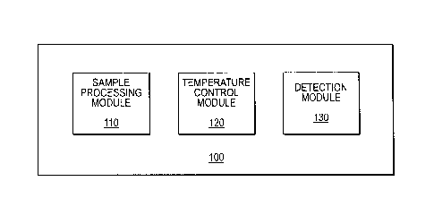
[0011] Figure 1 is a diagram of the sample detection system of the present
invention.
[0012] Figure 2 is a diagram showing a sample preparation system of the
present application.
[0013] Figure 3 shows an embodiment of a complete sample detection system
with the disposable cassette.
[0014] Figure 4 shows another embodiment of the disposable cassette of the
present invention.
CA 02858608 2014-06-06
WO 2012/078863
PCT/US2011/063937
4
[0015] Figure 5 shows a three-dimensional view of the flow strip portion of a
flow strip cassette.
[0016] Figure 6 shows the effect of air flow rates on the CT values of DNA
amplification.
[0017] Figure 7A shows a linear 8-way selector. Figure 7B is a close-up
view of the a-ring seal structure at the distal end of the selector plunger.
[0018] Figure 8 shows a 8-channel manifold that interacts with the 8-way
selector and a 8-sample disposable cassette.
[0019] Figure 9 shows an automated sample analysis system highlighting the
components needed for sample extraction.
[0020] Figure 10 shows the front and back views of a flow strip with a multi-
array flow cell.
[0021] Figure 11 shows an embodiment of the reagent layout in a 2mL, 96
deep-well reagent plate for MRSA extraction and on-slide PCR.
[0022] Figures 12A-12C show several embodiments of the optic design in the
sample analysis system of the present application.
[0023] Figure 13 shows the array image following TruTip processing of live
MRSA, on-chip PCR, on chip washing, and image acquisition on a sample analysis
system.
DETAILED DESCRIPTION
[0024] The following detailed description is presented to enable any person
skilled in the art to make and use the invention. For purposes of explanation,
specific
nomenclature is set forth to provide a thorough understanding of the present
application. However, it will be apparent to one skilled in the art that these
specific
details are not required to practice the invention. Description of specific
embodiments
and applications is provided only as representative examples. This description
is an
exemplification of the principles of the invention and is not intended to
limit the
invention to the particular embodiments illustrated.
[0025] This description is intended to be read in connection with the
accompanying drawings, which are considered part of the entire written
description of
this invention. The drawing figures are not necessarily to scale and certain
features of
CA 02858608 2014-06-06
WO 2012/078863
PCT/US2011/063937
the invention may be shown exaggerated in scale or in somewhat schematic form
in
the interest of clarity and conciseness. In the description, relative terms
such as
"front," "back" "up," "down," "top" and "bottom," as well as derivatives
thereof,
should be construed to refer to the orientation as then described or as shown
in the
5 drawing figure under discussion. These relative terms are for convenience
of
description and normally are not intended to require a particular orientation.
Terms
concerning attachments, coupling and the like, such as "connected" and
"attached,"
refer to a relationship wherein structures are secured or attached to one
another either
directly or indirectly through intervening structures, as well as both movable
or rigid
attachments or relationships, unless expressly described otherwise.
[0026] As used herein, the term "sample" includes biological samples such as
cell samples, bacterial samples, virus samples, samples of other
microorganisms,
samples obtained from a mammalian subject, preferably a human subject, such as
tissue samples, cell culture samples, stool samples, and biological fluid
samples (e.g.,
blood, plasma, serum, saliva, urine, cerebral or spinal fluid, lymph liquid
and nipple
aspirate), environmental samples, such as air samples, water samples, dust
samples
and soil samples.
[0027] The term "monolith," "monolith adsorbent" or "monolithic adsorbent
material," as used in the embodiments described hereinafter, refers to a
porous, three-
dimensional adsorbent material having a continuous interconnected pore
structure in a
single piece. A monolith is prepared, for example, by casting, sintering or
polymerizing precursors into a mold of a desired shape. The tem' "monolith" is
meant
to be distinguished from two or more filters that are placed next to each
other or
pressed against each other. The term "monolith adsorbent" or "monolithic
adsorbent
material" is meant to be distinguished from a collection of individual
adsorbent
particles packed into a bed formation or embedded into a porous matrix, in
which the
end product comprises individual adsorbent particles. The term "monolith
adsorbent"
or "monolithic adsorbent material" is also meant to be distinguished from a
collection
of adsorbent fibers or fibers coated with an adsorbent, such as filter papers
or filter
papers coated with an adsorbent.
[0028] The term "specifically bind to" or "specific binding," as used in the
embodiments described hereinafter, refers to the binding of the adsorbent to
an
CA 02858608 2014-06-06
WO 2012/078863
PCT/US2011/063937
6
analyte (e.g., nucleic acids) with a specificity that is sufficient to
differentiate the
analyte from other components (e.g., proteins) or contaminants in a sample. In
one
embodiment, the term "specific binding" refers to the binding of the adsorbent
to an
analyte in a sample with a binding affinity that is at least 10-fold higher
than the
.. binding affinity between the adsorbent and other components in the sample.
A person
of ordinary skill in the art understands that stringency of the binding of the
analyte to
the monolith and elution from the monolith can be controlled by binding and
elution
buffer formulations. For example, elution stringencies for nucleic acids can
be
controlled by salt concentrations using KC1 or NaCI. Nucleic acids, with their
higher
negative charge, are more resistant to elution than proteins. Temperature, pH,
and
mild detergent are other treatments that could be used for selective binding
and
elution. Thermal consistency of the binding and elution may be maintained with
a
heat block, water bath, infrared heating, and/or heated air directed at or in
the
solution. The manipulation of the binding buffer is preferable since the
impact of the
modified elution buffer on the downstream analyzer would need to be evaluated.
[0029] The term "nucleic acid," as used in the embodiments described
hereinafter, refers to individual nucleic acids and polymeric chains of
nucleic acids,
including DNA and RNA, whether naturally occurring or artificially synthesized
(including analogs thereof), or modifications thereof, especially those
modifications
.. known to occur in nature, having any length. Examples of nucleic acid
lengths that
are in accord with the present invention include, without limitation, lengths
suitable
for PCR products (e.g., about 50 to 700 base pairs (bp)) and human genomic DNA
(e.g., on an order from about kilobase pairs (Kb) to gigabase pairs (Gb)).
Thus, it will
be appreciated that the term "nucleic acid" encompasses single nucleic acids
as well
as stretches of nucleotides, nucleosides, natural or artificial, and
combinations thereof,
in small fragments, e.g., expressed sequence tags or genetic fragments, as
well as
larger chains as exemplified by genomic material including individual genes
and even
whole chromosomes. The term "nucleic acid" also encompasses peptide nucleic
acid
(PNA) and locked nucleic acid (LNA) oligomers.
100301 The term "hydrophilic surface" as used herein, refers to a surface that
would form a contact angle of 45 or smaller with a drop of pure water resting
on such
a surface. The term "hydrophobic surface" as used herein, refers to a surface
that
7
would form a contact angle greater than 45 with a drop of pure water resting
on such
a surface. Contact angles can be measured using a contact angle goniometer.
[0031] The term "pierceable seal" or "pierceable cover" as used herein, refers
to a seal or cover that is pierceable by a liquid communication device, such
as a
pipette tip, during normal operation of the sample analysis system of the
present
application. Examples of a pierceable seal or cover include, but are not
limited to,
membranes, films, rubber (e.g., silicone) mats with slits or foils that are
attached to
the opening of a tube or container with heat sealing, an adhesive, or
crimping. The
pierceable seal or cover allows packaging of liquid reagents in the cassette
of the
present invention. It also allows for packaging of lyophilized reagents with
sufficient
moisture barriers to protect the lyophilized reagents from liquid reagents in
the same
cassette.
Integrated Sample-To-Answer Sample Analysis System
[0032] One aspect of the instant application relates to an integrated sample-
to-answer sample analysis system 100 for the detection of a biomolecule, such
as
DNA, RNA or protein. In certain embodiments, the system 100 comprise a sample
processing module 110, a temperature control module 120 and a detection module
130 (Figure 1).
[0033] The sample processing module 110 prepares a sample for analysis.
Such preparation typically involves purification or isolation of the molecules
of
interest, such as DNA, RNA or protein, from the original sample using a sample
purification device. In some embodiments, the sample purification device is a
pipette
tip containing a filter that binds specifcally to the molecules of interest.
Examples of
such filters are described in more details in U.S. Patent No. 7,785,869 and
U.S. Patent
Application No. 12/213,942.
[0034] Figure 2 shows an embodiment of a sample purification device 200
that comprises a housing 210 and a sample filter 220. The housing 210 defines
a
sample passage way 212 between a first opening 214 and a second opening 216.
The
shape and size of the housing 210 are not particularly limited. In this
embodiment,
the preferred housing configuration is substantially cylindrical so that the
flow vectors
during operation are substantially straight. In the embodiment shown in Figure
2, the
CA 2858608 2018-01-30
8
housing 210 has a pipette tip geometry, i.e., the first opening 214 has a
diameter that
is greater than the diameter of said second opening 216, and the first opening
214 is
dimensioned to fit onto the tip of a pipette. The sample filter 220 is placed
in the
close proximity of the second opening 216 so that samples are filtered
immediately
after being taken into the housing 210 through the second opening 216. In one
embodiment, the sample filter 220 is contiguous with the second opening 216.
In
another embodiment, the sample filter 220 is separated from the second opening
216
by a distance of 1-20 mm. In some embodiments, the monolith sample filter is a
glass
frit with a average pore size of 20-200 micron. In another embodiment, the
sample
.. filter 220 is a monolith filter with two sections having different
porosities: a first
section 221 at the proximity of the second opening 216 and a second section
222 that
is separated from the second opening 216 by the first section 221. In one
embodiment, the first section has an average pore size of 40-200 micron,
preferably
40-60 micron, and the second section has an average pore size of 1-40 micron,
.. preferably 1-20 micron.
100351 In another embodiment, the sample prosessing module 110 comprises
an affinity column filed with a medium that binds specifcally to the molecules
of
interest. The sample prosessing module 110 may further comprise a fluid
handling
device, such as an automatic pippette or a pump to transport liquid samples.
The
.. prosessed sample, which is enriched for the molecules of interest, is then
transported
to a reaction chamber and is subjected to an amplification reaction or a
binding
reaction for the detection of a molecule of intersest in the sample. In some
embodiments, the reaction chamber contains a microarray and is located within
a flow
cell (also refered to as a "biochip"), as described in U.S. Patent Application
Nos.
12/149,865 and 12/840,826. Briefly, the flow cell contains a microarray formed
on a
planar substrate and a reaction chamber formed around the microarray.
100361 The microarray can be a polynucleotide array or a protein/peptide
array. In one embodiment, the microarray is formed using the printing gel
spots
method described in e.g., US Patent Nos, 5,741,700, 5,770,721, 5,981,734,
6,656,725
and US Patent Application Nos. 10/068,474, 11/425,667 and 60/793,176.
CA 2858608 2018-01-30
9
The planar substrate can be glass or plastic (films and injection molded) in
black,
white, clear, or other colors.
[0037] The reaction chamber has a plurality of interior surfaces including a
bottom surface on which the microarray is formed and a top surface that faces
the
bottom surface and is generally parallel to the bottom surface. At least one
of the
plurality of interior surfaces is a hydrophilic surface that facilitate the
complete filling
of the reaction chamber. In one embodiment, the top surface of the reaction
chamber
is a hydrophilic surface. In some embodiments, the flow cell further comprises
a
piereceable and re-sealable septum, such as a dome valve for loading a liquid
sample
into the reaction chamber and a sample channel connecting the one-way valve to
the
reaction chamber. In other embodiments, the reaction chamber is connected to a
waste chamber or an absorbent via a waste channel.
[0038] In some other embodiments, the sample processing module 110 further
comprises a cell lysis chamber having a plurality of cell lysis beads and a
magnetic
stirrer. Cell lysis is achieved by rotating the magnetic stirrer inside the
cell lysis
chamber in the presence of the cell lysis beads. The rotation of the magnetic
stirrer
can be caused by creating a rotating magnetic field around the magnetic
stirrer. The
cell lysis beads can be any particle-like or bead-like material that has a
hardness
greater than the hardness of the cells to be lysed. The cell lysis beads may
be made of
plastic, glass, ceramics, or any other non-magnetic materials, such as non-
magnetic
metal beads. In certain embodiments, the cell lysis beads are rotationally
symmetric
to one axis (e.g., spherical, rounded, oval, elliptic, egg-shaped, and droplet-
shaped
particles). In other embodiments, the cell lysis beads have polyhedron shapes.
In
other embodiments, the cell lysis beads are irregular shaped particles. In yet
other
embodiments, the cell lysis beads are particles with protrusions. The magnetic
stirrer
can be a bar-shaped, cross-shaped, V-shaped, triangular, rectangular, rod or
disc-
shaped stir element, among others. In some embodiments, the magnetic stirring
element has a rectangular shape. In some embodiments, the magnetic stirrer has
a
two-pronged tuning fork shape. In some embodiments, the magnetic stirrer has a
V-
like shape. In some embodiments, the magnetic stirrer has a trapezoidal shape.
In
certain embodiments, the longest dimension of the stir element is slightly
smaller than
the diameter of the container (e.g. about 75-95% of the diameter of the
container). In
CA 2858608 2018-01-30
10
certain embodiments, the magnetic stirrer is coated with a chemically inert
material,
such as polymer, glass, or ceramic (e.g., porcelain). In certain embodiments,
the
polymer is a biocompatible polymer such as PTFE and parylene. A more detailed
description of the magnatic lysis method is described in Application No.
12/886,201.
[0039] In some embodiments, the sample prosessing module 110 comprises a
disposable cassette that comprises (1) a plurality of containers, each having
an open
top end and a closed bottom end; (2) a flow strip comprising a plurality of
ports that
interact with the sample analysis device via one or more fluid communication
devices
to establish fluid communication between the cassette and the sample analysis
device;
and (3) a plurality of reaction chambers, each reaction chamber is connected
to a port
on the flow strip. At least one of the reagent containers is pre-packaged with
a
reagent needed for a sample analysis procedure and is sealed with a pierceable
cover
at the top end of the container. In some embodiments, the cassette comprises a
combination of one or more containers with a lyopholized reagent prepackaged
therein and one or more containers with a liquid reagent prepackaged therein.
In
some embodiments, the cassette further comprises one or more containers with a
plurality of cell lysis beads and a magnetic stirrer prepackageed therein. In
other
embodiments, the cassette further comprises one or more containers with an
absorbent
prepackaged therein.
[0040] As used herein, the term "fluid communication device," refers to any
device or component of the system that is capable of establishing a fluid
connection
between two locations. Examples of fluid communication device include, but are
not
limited to, tubes, tubings, columns, channels, pipette tips and combinations
thereof.
[0041] . In some other embodiments, the flow strip further comprised one or
more pin valves to control fluid flow within the flow strip, e.g., from a
reaction
chamber to a waste chamber.
[0042] In other embodiments, the disposable cassette further comprises one or
more sample purification devices. In one embodiment, the one or more sample
purification devices, such as TruTips, are used as the fluid communication
devices to
establish fluid communication between the cassette and the sample analysis
device.
CA 2858608 2018-01-30
CA 02858608 2014-06-06
WO 2012/078863
PCT/US2011/063937
11
[0043] As used herein, the term "sample purification device," refers to any
devices capable of purifying, isolating or enriching a target molecule.
Examples of
sample purification device include, but are not limited to, filters, affinity
filters,
affinity columns, chromatograph columns, and filter tips such as TruTips. In
one
embodiment, the sample purification device is a pipette tip comprising a
monolith
filter that binds specifically to nucleic acids.
[0044] In other embodiments, each port in the disposable cassette contains a
connector for establishing fluid communication with a fluid communication
device.
Such a connector may comprise a pierceable septum or a dome valve.
[0045] In another embodiment, the flow strip further comprises an absorbent
that absorbs waste reagents from reaction chambers. In one embodiment, the
absorbent is in fluid communication with one or more reaction chambers via one
or
more pin valves. The absorbent can be any material capable of retention of a
large
quantity of liquid. In one embodiment, the absorbent is made of an aggregate
of
fibers. In another embodiment, the absorbent is a nonwoven fabric produced in
a
through-air bonding process. The constituent fibers of the nonwoven fabric can
be
hydrophilic synthetic fibers, natural cellulose fibers of pulp or the like, or
regenerated
cellulose fibers. The fibers may be coated or infiltrated with a surfactant or
a
hydrophilic oil to improve liquid absorbance. Not limited to the through-air
bonding
process, the nonwoven fabric for use herein may be produced in any other
process
such as a spun-bonding process, an air laying process, a spun-lacing process,
etc. In
another embodiments, the absorbent is a cellulose paper.
[0046] In another embodiments, the disposable cassette further comprises a
mixing tower connected to the flow strip via one of the plurality of ports.
[0047] In some embodiments, the plurality of containers are arranged in the
form of a 96-well plate. The plate may contain one or more containers having a
lyopholized reagent pre-packaged therein, one or more containers having a
liquid
reagent pre-packaged therein, and optionally, one or more containers having an
absorbent pre-packaged therein. The plate may further comprise one or more
containers pre-packaged with a plurality of lysis beads and a magnetic
stirrer. The
volume of the wells may vary depending on the amounts of the reagents needed.
The
wells may have the same volume or different volumes. In certain embodiments,
the
CA 02858608 2014-06-06
WO 2012/078863
PCT/US2011/063937
12
wells have volumes in the ranges of 50 L to 5000 L, 50 L to 500 L, 500 L
to
2500 L, and 1000 L to 5000 L. In one embodiment, the wells have a uniform
volume of about 2200 L.
[0048] The disposable cassette is connected to the sample analysis system 100
via one or more fluid communication devices and a flow-control manifold on the
sample analysis system 100. The flow control manifold comprises a manifold
body, a
plurality of fluid supply ports that are formed on the manifold body and are
adapted to
be connected to a fluid supply device, a plurality of plunger channels formed
within
the manifold body, and a plurality of plungers that are movable along the
length of the
plunger channels. Each plunder channel has a plunger channel inlet at one end
and a
plunger channel outlet at another end. Each plunger comprises a seal that
seals
against the interior wall of the plunger channel in which the plunger is
located. The
plungers enter the plunger channels from the plunger channel inlets. Each of
the
plurality of fluid supply ports is connected to a plunger channel and is
located in the
proximity of the plunger channel inlet of the plunger channel. The plunger
channel
outlets contain adaptors that connect to a one or more sample purification
devices,
such as TruTips.
[0049] In some embodiments, the flow control manifold further comprises a
channel selector for directing fluid flow to a desired fluid control channel
through a
fluid supply port. In one embodiment, the channel selector comprises a rotary
valve.
In another embodiment, the channel selector comprises a selector channel
having a
plurality of outlet ports and a linear motion actuator. The plurality of
outlet ports
connect to a corresponding fluid supply port on the flow-control manifold. The
linear
motion actuator comprises a motor and an elongated shaft having a proximal
end, a
distal end, and an enclosed fluid communication channel within the shaft. The
fluid
communication channel extends from one or more openings at the proximal end of
the
shaft to one or more openings at the distal end of the shaft. The one or more
openings
at the proximal end of the shaft are adapted to be connected to a fluid supply
device.
The one or more openings at the distal end of the shaft are flanked by two
seals, such
as o-rings. When the shaft extends into the selector channel, the two seals
seal against
the interior wall of the selector channel and foun a fluid communication
passage
within the selector channel. Fluid communication between the fluid supply
device
13
and an outlet port of the channel selector is established when the shaft is
placed in the
selector channel in such a position that the fluid communication passage is
formed
between the one or more openings at the distal end of the shaft and the outlet
port of
the channel selector. In one embodiment, the selector channel has a vent that
prevents
pressure change in the selector channel when the shaft moves within the
selector
channel. For example, such a vent would allow the shaft to move forward within
the
selector channel without experiencing back pressure.
[0050] The temperature control module 120 controls the temperture during the
amplification or binding reactions. In certain embodiments, the temperature
control
.. module comprises a device with a flexible temperature control surface, as
described in
U.S. patent nos. 7,955,840 and 7,955,841. In certain embodiments, the device
comprises a first heater for heating a temperature-controls ubstance to a
first
temperature; a second heater for heating said temperature-control substance to
a
second temperature; a pump located in between and connected in series with
said first
heater and said second heater; and a bladder unit comprising a pair of
bladders. Each
bladder is coupled to a bladder support and is connected to said first and
second
heaters via different ports. The pair of bladders are inflatable with the
temperature-
control substance that controls the temperature of the pair of bladders. The
pair of
bladders are positioned in a substantially opposing arrangement with a space
in
between such that both bladders, when inflated, are capable of contacting a
reaction
chamber placed in the space. During a PCR reaction, the pump introduces the
temperature-control substance into the pair of bladders at the first
temperature and the
second temperature alternatively with a regular interval to enable the PCR.
[0051] In other embodiments, the device comprises a bladder assembly
comprising: a first temperature-control bladder configured to receive a
temperature-
control fluid from a first inlet channel and expel the temperature-control
fluid from a
first outlet channel, a second temperature-control bladder configured to
receive the
temperature-control fluid from a second inlet channel and expel the
temperature-
control fluid from a second outlet channel, a first heat exchanger that
maintains the
temperature-control fluid at a first temperature and is connected to both the
first and
second inlet channels via a first two-way valve and a first three-way
connector, a
CA 2858608 2018-01-30
CA 02858608 2014-06-06
WO 2012/078863
PCT/US2011/063937
14
second heat exchanger that maintains the temperature-control fluid at a second
temperature and is connected to both the first and second inlet channels via
the first
two-way valve and the first three-way connector, and a pump located between
the
bladder assembly and the heat exchangers. The pump is connected to the first
and
second outlet channels via a three-way connector and is connected to either
the first
heat exchanger or the second heat exchanger via a second two-way valve. The
first
and second temperature-control bladder each comprises a flexible, heat
conductive
surface that comes in contact with at least a portion of an exterior surface
of a reaction
chamber after receiving the temperature-control fluid.
[0052] The detection module 130 detects the presence of a reaction product.
In certain embodiments, the detection module 130 comprises an optical
subsystem
designed to capture images of the microarray in the reaction chamber. In
certain
embodiments, the optical subsystem is specifically designed for low-level
fluorescence detection on microarrays. The optical subsystem uses confocal or
quasi-
confocal laser scanners that acquire the microarray image pixel by pixel in
the process
of interrogating the object plane with a tightly focused laser beam. The laser
scanners
offer the advantages of spatially uniform sensitivity, wide dynamic range, and
efficient rejection of the out-of-focus stray light.
[0053] In other embodiments, the optical subsystem uses imaging devices
with flood illumination, in which all the microarray elements (features) are
illuminated simultaneously, and a multi-element light detector, such as a CCD
camera, acquires the image of microarray either all at once or in a sequence
of a few
partial frames that are subsequently stitched together. Compared to laser
scanners,
CCD-based imaging devices have simpler designs and lower cost. CCD-based
imaging systems are an attractive option for both stand-alone and built-in
readers in
cost-sensitive applications relying on microarrays of moderate complexity
(i.e.,
having a few hundred or fewer array elements). Commercial instruments
typically
use cooled CCD cameras and employ expensive custom-designed objective lenses
with an enhanced light-collection capability that helps to balance, to some
extent, the
low efficiency of the excitation scheme.
[0054] In other embodiments, the optical subsystem contains an imaging
device that uses a non-cooled CCD camera. Although non-cooled cameras
typically
CA 02858608 2014-06-06
WO 2012/078863
PCT/US2011/063937
have a noticeably higher dark current as compared to the cooled models, the
optical
subsystem could provide the required sensitivity without using exposures in
excess of
a few seconds by (1) increasing the excitation intensity, or (2) employing an
objective
lens with high light collection efficiency; or (3) using the above two
approaches in
5 combination. The light source can be a conventional light source, such as
a metal
halide or mercury bulb, a laser-based system, or a high-intensity LED.
[0055] In some embodiments, an integrated sample analysis system
comprises:(1) a sample preparation/analysis module comprising a sample
purification
device having a monolith that binds specifically to nucleic acids; and a
sample
10 analysis device comprising a microarray enclosed in a reaction chamber
having a
hydrophilic interior surface; (2) a temperature control module comprising a
thermocycler having a thermally conductive temperature-control bladder that,
upon
receiving a temperature-control substance, expands to abut an exterior surface
of the
reaction chamber to enable thermal exchange between the temperature-control
15 substance and the internal volume of the reaction chamber; and (3) an
imaging device
capable of capturing an image of the microarray in the reaction chamber. In
one
embodiment, the sample analysis/preparation module further comprises a cell
lysis
chamber containing a plurality of cell lysis beads and a magnetic stirrer.
EXAMPLES
Example 1: Prototype Sample Analysis System
[0056] A sample-to-answer sample analysis system is developed by
integrating the following technologies: magnetic lysing, TruTip purification,
bladder
thermocycling, PCR-Microarray Biochip amplification, LED microarray
illumination,
and gel element microarray imaging into a point-of-care molecular instrument
with a
disposable cassette.
[0057] The magnetic lysing technology involves an external rotating magnet
that vigorously mixes and homogenizes tissue/cells in a sample solution with
beads
using a miniature rotating magnetic stir bar that is placed in close proximity
to the
external magnet. This approach has the virtue of not requiring a mechanical or
electrical interface to the consumable device. Using this method at a 1:1
ratio of
sample to beads in a total volume of 1 mL, lysis of 104 cfu/mL of gram
positive S.
pyogenese was achieved in 30 seconds in a tube, located several cm from the
external
CA 02858608 2014-06-06
WO 2012/078863
PCT/US2011/063937
16
magnet. This approach resulted in a 2.5 cycle improvement compared with bead
vortexing when analyzed by qPCR.
[0058] The TruTipTm nucleic acid purification device (see Figure 2) consists
of a porous monolith. The monolith is a rigid and thick glass matrix, which
enables
easy insertion into a pipette tip with a low manufacturing burden in a form
factor that
is easily amenable for automating extraction protocols. The protocol, which
can
require as few as 4 min, consists of pipetting back and forth through the
monolith to
bind, wash, air dry, and elute. Cycling back and forth across the porous
monolith
improves recovery. The monolith is designed to have a large porosity to reduce
the
back pressure across the monolith when processing viscous samples such as
nasopharangeal aspirate (NPA). Nucleic acid purification of M.TB, Vaccinia,
VEE,
B. anthracis, Y. pestis, Influenza A/B, S. pyogenes, C. pneumoniae, and MRSA
has
been demonstrated on sample types such as NPA, Nasopharyngeal swabs (NPS),
blood, soil, sputum and urine. Comparisons of the qPCR results obtained using
TruTip operated by a Rainin Electronic Pipettor and a standard Qiagen kit
indicated
that both methods exhibited the same efficiency and recovery in an extensive
study.
The TruTip, however, was 5-times faster, accommodated a larger sample volume,
and
did not require centrifugation.
[0059] A study was performed on the TruTip-epMotion system using FluA
(H3N2) and FluB spiked into five different Flu-Negative NPA samples, obtained
from
Wadsworth Center, State of NY Dept of Health, with varying viscosity (low to
high
mucus content). FluA was reproducibly detected (100%) at 10 gc tL1. FluB was
reproducibly detected (100%) at 102 gc El, with 10 gc 4-1 approaching the
detection limit of the real time RT-PCR assay.
[0060] The purified nucleic acids were then loaded into the microarray
chamber of a PCR-microarray biochip. The PCR-microarray biochip designs allow
PCR amplification in the microarray chamber. The biochip may also have a waste
chamber to allow washing while maintaining a closed amplicon system. The waste
chamber and the microarray chamber are separated by a microfluidic stop or a
pin
valve, which confines the reaction mix to the microarray chamber during
thermocycling. Unlike others, the method of the present invention does not
require
special hydrophobic coatings or treatments. Rather, it has been demonstrated
that a
CA 02858608 2014-06-06
WO 2012/078863
PCT/US2011/063937
17
design based on geometry and materials can confine the liquid reagents in the
microarray chamber until an additional reagent such as a wash solution is
added.
[0061] The PCR-Microarray Biochips, described above, can be used for on-
chip PCR and post-hybridization washing. The PCR-Microarray Biochip may
include
a fluidic channel layer in double-sided tape, and the use of a hydrophilic
cover film to
allow uniform and predictable biochip filling. These biochips may include a
pierceable check valve (e.g., Minivalve DS052). This component will ensure a
closed
amplicon device. Alternatives include the addition of a backseal (permit
liquid to
flow through the check valve without piercing it) and the use of luer-
activated valves
.. (only permit flow when engaged). Plastic pin valves that use 2.4mm o-rings
are an
alternative or additional approach to the "valve-less" strategy in which the
reaction
chamber is isolated from the waste chamber. These valves withstand
thermocyling
and are low-cost to manufacture.
[0062] Liquids flow unidirectionally into but not out of the disposable PCR-
Microarray Biochip as a means of ensuring a closed amplicon workflow. In some
embodiments, a mixing chamber is included to keep the workflow for reactions
such
as Allele Specific Primer Extension (APEX). In one embodiment, the mixing
chamber is an extended pin valve, so that following PCR, APEX buffer and
enzymes
could be added to the PCR-Microarray Biochip while simultaneously allowing the
pin
valve to move up the column, creating space for the mixture. In this example
the
downstream valve would be closed, and the check valve at the inlet would
prevent
liquid from exiting the biochip. Air could also be introduced to further
enhance
mixing, or movement of the pin valve back and forth could assist in mixing.
[0063] The microarray consists of gel elements, which have a sterically-
favorable spacing of immobilized molecules throughout an aqueous volume of a
hemispherical porous hydrophilic polymer. Probes are suspended in a pre-
polymer
solution, patterned on a surface, and co-polymerized by photopolymerization to
create
a "gel drop" array. Probes are therefore immobilized to the substrate. The net
result
of this polymeric structure is increased hybridization kinetics, higher
probe
.. immobilization capacity, and up to 100-fold increased detection sensitivity
compared
with surface-immobilized 2D planar arrays. These features enable low-cost
optical
instrumentation, rapid hybridization, and the ability to do attachment
chemistry in a
CA 02858608 2014-06-06
WO 2012/078863
PCT/US2011/063937
18
bulk polymeric phase, which reduces the manufacturing burden, and thus cost
per
device. Additionally, the co-polymerization methodology can be implemented on
native plastics, which eliminates the need for high-priced glass substrates.
[0064] The PCR reaction was performed using a speciallydesigned bladder
thermal cycling device in which thermally-controlled recirculating flow
expands a
bladder pair to make intimate contact with the PCR-microarray biochips. As a
demonstration of implementing the bladder thermal cycler with coupled PCR and
microarray hybridization, one ng of S. pyogenes genomic DNA was mixed with PCR
master mix and loaded into two PCR-microarray biochips. The thermal cycling
protocol took less than 26 minutes (44 cycles of 5 sec at 85 C and 30 sec at
50 C),
and hybridization was less than 15 minutes, compared to 3 to 4 hours on a
conventional slide block thermal cycler. Despite the use of a thick (1mm)
glass
substrate, rapid PCR amplification was achieved for the following 3 reasons:
(1) Fast ramp times (-10 C/s), as opposed to prolonged cooling of a large
metal block, was possible by the use of fluidic switching.
(2) Tight intimate contact of the bladder pair with the biochip substrates
resulted in high thermal conductivity. Poor contact between the heater and the
reaction vessel with conventional methods is typically responsible for
substantial
thermal inefficiencies. (3) The
recirculating flow convectively heats and cools
.. the reaction chamber. Convection is typically the most effective heat
transfer mode.
[0065] The amplified signals are detected by an imaging device, which
consists of a single LED and a non-cooled CCD camera.
[0066] Pre-packaged reagents for molecular diagnostics instruments reduces
the complexity of the device. Thus, Akonni has developed a disposable cassette
300
that can be inserted into the sample analysis system 100 through a retractable
carriage
112 (Figure 3). The cassette 300 comprises a strip of pierceable reagent
container
310, one or more reaction chambers 320, and a flow strip 330 that controls
fluid flow
from a sample purification device 340, such as a TruTip, to the reaction
chambers
320. The reaction chambers 320 may be formed within a PCR microarray biochip
350. The reagents may contain reagents for lysis, purification and PCR
amplification.
The lids 312 of the tubes are made of pierceable foil that could be attached
with heat
sealing, an adhesive, or crimping a metal cover around a glass or plastic
vial. The foil
CA 02858608 2014-06-06
WO 2012/078863
PCT/US2011/063937
19
may also be attached to a plastic tube such as a PCR tube. The cassette 300
allows
ease of packaging lyophilized reagents with sufficient moisture barriers to
protect
them from liquid reagents. A pipette tip can pierce the foil and remove the
reagents
from the tube and transport nucleic acid and/or liquids from one tube to
another. In
this embodiment, the flow strip cassette includes a disposable TruTip 340 that
engages a pipette head on the instrument for the purification protocol,
reagent
rehydration, and PCR-microarray biochip filling. In one embodiment, only
nucleic
acid, adsorbed to the monolith, is transported from one tube to the next, thus
liquids
remain in their respective tubes, reducing the risk of sample contamination.
Rehydrated mastermix with purified sample is then introduced via the TruTip
into the
PCR-microarray biochip, which is subsequently inserted between a bladder pair
for
thermocycling. A pierceable check valve confines the amplicon to a closed
system,
but allows a wash solution to flow across the array for subsequent imaging. In
other
embodiments, the TruTip 340 is designed to contain a filter that binds
specifically to a
target molecule of interest, such as a protein, a peptide, a DNA, an RNA or
other
biomolecules. Figure 4 shows a cassette 300 with a sample port 314 and pin
valves
316 that control the fluid flow within the biochip 350.
[0067] Figure 5 shows the flow strip 330 portion of a cassette 300. In this
embodiment, the flow strip 330 comprises a sample port 314 to receive the
TruTip
340, and pin valves 316 that control the liquid flow from reaction chambers
320 to
waste chamber 360. In some other embodiment, the flow strip 330 further
comprises
one or more magnetic lysing or mixing towers (not shown)
[0068] The containers 310 in the cassette 300 can be plastic tubes, glass
vials
or wells in a plate (e.g., 96 deep-well plate). Miniature linear actuators
with an
integrated positional-feedback potentiometer may be used for repeatedly
dispensing
and withdrawing from the bottom of 2 mL tubes (11 mm diameter) and glass
lyophilization vials. In one embodiment, the monolith is placed towards the
top of the
pipette tip, increasing the volume below the monolith. This increases the
volume that
does not make contact with the monolith, which may be useful for pipetting
reagents
such as the PCR buffer into the flow strip. Contact of the PCR buffer with the
monolith may introduce unwanted air into the PCR buffer, causing bubbles. With
this
embodiment a single pipette tip could be used for all steps. Another
embodiment is to
CA 02858608 2014-06-06
WO 2012/078863
PCT/US2011/063937
use multiple tips for multiple pipetting steps. In one embodiment, disposable
pierceable check valves (e.g., Minivalve) are press-fit under a screw cap with
an
access hole as a means of introducing sample and providing access for the
TruTip
without releasing aerosols during magnetic rotation. Hydrophobic-coated lysing
5 beads are a means to minimize DNA adsorption, and thus eliminate the need
for a
sample transfer step to a separate chaotrophe tube. Alternative TruTip designs
include various porosity sizes (1 to 100 micron), different thickness (0.1 to
10 mm),
stacks of different porosity monoliths (1 to 10), single monolith with
sections of
different porosities and/or conventional approaches (e.g., bead vortexing,
stepper
10 motors, multiple pipette tips). To reduce the PCR multiplexing
complexity, multiple
chambers may be used to split the PCR Mastermix/sample reagents into multiple
reservoirs. This may be useful for simultaneous sample processing of both
bacteria
and viruses.
Example 2: Multiway Selector Design
15 [0069] This example will consider the testing and design process of
a device
used to select between eight different ports on an eight -port manifold,
allowing air to
flow through only a single port at a time. This device is referred to as an
eight -way
selector, which is used to dry pipette tips on an automated liquid handling
system.
This system uses eight pipette tips to simultaneously complete eight separate
sample
20 preparations. In one embodiment, an eight-way selector is designed in
order to allow
airflow from a common air source to dry a matrix within these pipette tips.
A. Testing on flow rate
[0070] Prior to integration of the 8-way selector to the 8-port manifold,
testing
was conducted to determine the effect of air flow rate on the cross threshold
(CT)
values during the DNA extraction and amplification processes used. Briefly,
the
system was connected to a flow meter to measure flow. Five different new flow
rates
were tested for their effects on the CT values during the DNA extraction and
amplification processes. A previously-used manual flow rate was included in
the test
as the control flow rate, which resulted in a control CT value of around 23.5.
As
shown in Figure 6, all the tested flow rates resulted in CT values that are
lower than
the control CT value. Based on the results of Figure 6, it appears that 5
liters per
CA 02858608 2014-06-06
WO 2012/078863
PCT/US2011/063937
21
minute is the most desirable flow rate for the 8-way selector because it
resulted in the
lowest CT value.
B. Eight-way selector design
[0071] Several designs may be used for the eight-way selectors. First, the
selective access to each port on the eight -port flow strip may be controlled
by an
eight-way rotary valve, which is commercially available but expensive.
[0072] Alternatively, a linear actuator can be used to control access of air
to
each of the eight-ports through the TruTips for additional drying or in the
flow strip
for drying the microarray. As shown in Figures 7A and 7B. The linear actuator
700
contains a motor 750 and a shaft 710 having a proximate end 720 and a distal
end
730. The shaft 710 comprises two 0-rings 732 and 734 at the distal end 730.
The
shaft 710 has a channel that is connected to an air supply on the proximal end
720 and
one or more air outlet 712 at the distal end 730. The air outlet 712 is
located between
the two 0-rings 732 and 734. The shaft 710 travels in a selector channel 760
that is
connected to eight outlet ports 770. The selector channel 760 has a vent 780
at the
distal end to prevent pressure built-up in the channel. As shown in Figure 7B,
the
two 0-rings 732 and 734 seal against the interior wall of the selector channel
760 to
form a fluid communication passage 790. Air travelling down the hollow length
of
the shaft 710 and exiting at the air outlet 712 would be trapped between the
two 0-
rings 732 and 734, and could only escape through a single port 770 on the
manifold at
any time. It is possible, however, to adjust the distance between the two 0-
rings 732
and 734 so that air may escape through two or more ports 770 at the same time.
Similarly, multiple 0-rings may be used to foul' multiple fluid communication
passages, thus allowing air flow to multiple ports at the same time.
[0073] Figure 8 shows an eight-channel manifold 800 having eight fluid
supply ports 810, eight plunger channel inlet 820, eight plunger channels 830
and
eight plunger channel outlet ports 840, which connect to pipette tip ports
(i.e., TruTip
ports) (not shown). The fluid supply ports 810, which connect to the
corresponding
eight-way selector valve ports 770, are placed towards the end of the plunger
channels
830 so as to allow plungers (not shown), which enters the plunger channel 830
through the plunger channel inlet 820, to travel the vast majority of the
length without
changing the pipette flow dynamics of aspirating and dispensing fluids. When
it is
CA 02858608 2014-06-06
WO 2012/078863
PCT/US2011/063937
22
time for the air drying step, the plungers can be pulled back so that air can
travel from
the eight-way selector described in Figures 7A and 7B through the fluid supply
ports
810 into the plunger channels 830 and out the plunger channel outlet port 840.
In one
embodiment, only a single plunger channel 830 will be open to airflow at any
one
time. This air will be forced to flow into the pipette tips, as a plunger in
the manifold
will be behind the fluid supply port 810, preventing air from escaping out of
the
plunger channel inlet 820.
[0074] Another design is to allow all eight pipette tips to be exposed to the
common air source at the same time. This design would eliminate the need for
selecting a single port for airflow.
Example 3: Automated Multi-sample Detection System
[0075] Figure 9 shows an automated sample-to-answer system 900 that is
able to perfolin sample extractions, on-slide PCR, and array imaging for eight
samples simultaneously.
A. Sample Purification/extraction
[0076] There are three main sub-systems of the system 900 that relate to
sample purification and extraction. These sub-systems include tip holder 910,
plate
holder 920, and plunger system 930. The tip holder 1100 secures the TruTips
(not
shown) to the system 900 and holds them stationary in the X- Y plane. However,
the
tip holder 910 is connected to an actuator which allows control of the TruTips
in the Z
plane. It's also conceivable that the TruTips are moved in all directions
(i.e., not
stationary). The plate holder 920 secures a 2mL 96 deep well plate 921 which
is used
as a reservoir for all reagents and samples needed for an end-to-end run. The
plate
holder 920 moves the deep well plate 921 in the X-Y plane allowing for the
TruTips
to move from column to column on the deep well plate 921. Finally, the plunger
system 930, which is connected to a stepper motor 940, controls the volume in
which
the TruTip can aspirate and dispense.
[00771 Multiple sample extractions have been perfoimed on system 900 using
genomic Methicillin-resistant Staphylococcus aureus DNA (gMRSA) and live MRSA
in two mediums ¨ water and nasal pharyngeal aspirate (NPA). Automated
extractions
on the system 900 rely on the 2 mL deep-well plates 1201 to contain all
necessary
reagents, e.g., lysis buffer, wash buffer, and elution buffer (see, e.g.,
Figure 11). The
CA 02858608 2014-06-06
WO 2012/078863
PCT/US2011/063937
23
TruTips are inserted into each column of the plate 921 and the reagents are
toggled
through the tips for sample purification and extraction to occur. The first
column of
the plate contains the sample along with lysis buffer ¨ this mixture (500-1000
III)
flows through the tips for 5-20 cycles depending on the medium in which the
sample
is in. In one embodiment, 15 cycles are used for samples in water and 20 for
samples
in NPA. This is then followed by a wash step that requires toggling the wash
buffer
(500 ItL) for 10 cycles. Next, the matrix within the TruTip is air dried and
finally the
elution step occurs where the elution buffer (501.tL) is toggled through the
tips for
another 10 cycles ¨ DNA is recovered in this buffer.
[0078] Throughout the testing effort it had been determined that incorporating
a unidirectional forced air system helps dry the TruTip matrix allowing for
better
recovery of DNA, even when compared to traditional manual extractions. Air
drying
follows the wash step and is required to properly dry the matrix ¨ each tip is
dried
separately for 1 minute. Residual wash buffer can interfere with recovery and
inhibit
polymerase chain reaction (PCR). A comparison of manual vs. automated
extractions
of 250 uL of 100 pg/ L gMRSA in H20 showed that the manual extractions average
a CT of 23.73 while the automated extractions average 22.38 ¨ 1.5 cycles
lower. The
air drying component was applied to all further extractions.
[0079] Once testing on genomic MRSA was completed, live whole cells were
used. Live MRSA was grown in-house and suspended in saline solution for a
final
concentration of 0.5 McFarland. An initial lysis step was required for these
cells and
was performed manually; however, this can be included in the automated system.
The lysis was done with a magnetic lysing, described earlier, using 50 grams
of
Ceroglass 100-200 micron ceramic beads and 2504 of the live MRSA cells. The
cells were lysed at 100% speed for two minutes and then placed into the 1st
column of
the 2mL deep well plate. Cells were also heat killed at 100 C for 15 minutes
prior to
use to prevent any possible infection of users. This experiment followed the
same
protocol as the gMRSA in H20 and did not require additional ethanol. The
average
CT was 22.88, which is equivalent to the 100 pg/uL sample that was run as a
positive
control..
[0080] Sample purification was also tested on live MRSA cells spiked in NPA
¨ used to represent a clinical sample. This sample required a manual lysis
step to
CA 02858608 2014-06-06
WO 2012/078863
PCT/US2011/063937
24
homogenize the NPA and lyse the MRSA cells. For this sample, lysis was
performed
on 250 [IL of 0.5 McFarland MRSA (heat killed) mixed with 2504 of NPA. Once
lysing treatment was complete, the sample was added to the lysis and binding
buffer
with an additional 250 L of 95% ethanol (total volume of 1000p1). The sample
was
toggled on the sample analysis system through the TruTip for 20 cycles which
was
then followed by the wash, air dry, and elution steps. Eight samples were
extracted
on the system 1000 and the real-time results show a CT average of 23.84 which
is
equivalent to the 100pg/p1 sample that was run as a positive control.
B. On-slide PCR
[0081] All extractions performed on the system 900 were used to complete
on-slide PCR using the bladder thermal cycler and obtain sample-to-answer
results.
The system 900 embodiment has the ability to perform on-slide PCR for eight
samples at a time using a microarray and bladder thermal cycler. The bladder
thermal
cycler has five main components: a hot reservoir, a cold reservoir, a pump,
one or
more valves, and a bladder or a bladder pair. The basic mechanism behind the
bladder thermal cycler is to circulate two different temperatures of liquid
through the
bladder for rapid thermal cycling. Both the hot and cold reservoir must
initially be
brought up to temperature before thermal cycling can begin. The pumps force
the
fluid through the path and rely on selection valves to direct the proper
temperature
fluid to enter the bladder. The bladder or bladder pair, once filled with
liquid, expand
around the inserted multi-chamber flow cell encasing it and transferring the
proper
temperature.
[0082] As shown in Figure 10, the multi-chamber flow cell 1000 has eight
independent microarrays 1010 that are enclosed in the reaction chambers 1020,
which
allow the PCR mixture to interact with the array 1010. The multi-chamber flow
cell
1000 is secured to a flow strip 1100 by a housing 1110 that encases dome
valves
1120, pin valves 1130, and an absorbent 1140. The housing 1110 directs the PCR
mixture that is pipetted in from the 2mL 96 deep well plate to the flow cell
1000
through these dome valves 1120, which also act as a seal during thermal
cycling
preventing any leakage. The pin valves 1130 are controlled by a linear
actuator that
enables them to be opened and closed. In an open position, the pin valves 1130
allow
CA 02858608 2014-06-06
WO 2012/078863
PCT/US2011/063937
liquid flow during the wash steps. In a closed position, the pin valves 1130
help trap
the PCR mixture in reaction chamber 1010 of the flow cell 1000 during thermal
cycling. The absorbent 1140 attached to the housing 1110 collects all wash
buffers
once passed through the flow cell 1000.
5 [0083] The on-chip PCR portion of a sample-to-answer test begins with
the
warm-up of the bladder thermal cycler. This warm-up step is used to bring both
the
hot and cold reservoir up to the required temperatures of 88 C and 51 C
respectively.
During this warm-up step, the PCR buffer is placed in the same 2mL 96 deep
well
plate used during sample extraction. On-chip PCR requires the uses of 4
columns:
10 PCR mastermix, 1xSSPE, Water, and Acetone. Figure 11 shows the reagent
layout
of a representative plate. Fifty microliters of the PCR buffer is introduced
to all 8 of
the housing ports, which is connected to the 8 chamber flow cell, using the
automated
system. Once all 8 chambers are filled, the pin valves are closed and the flow
cell is
inserted into the bladder and thermal cycling initiates. The thermal cycling
15 parameters are an initial 88 C for 2 minutes followed by 40 cycles of 88
C for 45
seconds and 51 C for 90 seconds. There is a final cool down step of 51 C for 5
minutes. Once thermal cycling is complete, the automated system removes the
flow
strip from the bladder and hybridization occurs at room temperature.
Hybridization
occurs for 2 hours and then the 3 different washes flow into the flow strip
and into the
20 flow cell array chambers at 501.tL aliquots, of 1xSSPE,water and acetone,
sequentially. Acetone is an optional reagent for drying the microarray,
C. Imaging/analysis
[0084] The system 900 has an integrated imaging system that is able to
capture the fluorescence of all 8 microarrays individually. The imager is
mounted on
25 a moving platfoim that controls its location on the X-Y plane and has
the ability to
move in the Z plane for focusing. After the completion of on-chip PCR and
washing,
the arrays are imaged and analyzed. Analysis was completed using MCI Software
and an Akonni MRSA analysis workbook. The MCI software uses a fixed circle
method to determine the intensity of each probe present on the array. Each
array has
4 identical quadrants (i.e., each probe is present on the array 4 times). Once
intensities are determined, the highest and lowest values are removed and the
median
is taken from the other two probes. This median determines the overall
intensity of
CA 02858608 2014-06-06
WO 2012/078863
PCT/US2011/063937
26
the probe. In order to determine if the signal is considered positive or
negative, two
factors are used: the d1N20 Ratio and the Sigma Ratio. The dN20 spots, a
mixture of
random 20 mer nonsense probes included in the microarray, are used to measure
"biological noise" due to effects such as poor washing, cross-hybridization,
and/or
excess DNA in the sample. Its measured intensity is determined the same way as
signal spots. The overall intensity of each probe is subsequently divided by
the
overall intensity of the dN20 signals. If this ratio is above 1 then the
signal is
considered to be detectable. Sigma is also used to determine if the signal is
above
threshold. Sigma is the standard deviation of the background (region where
spots are
not located) in the image. Each probe is divided by three times sigma to
calculate the
spot signal-to-noise ratio. The ratio to determine whether or not the spot is
considered
a detection event is to divide by the greater value (dN20 or 3xSigma ratio).
This
approach was used for the analysis described.
[0085] Figures 12A-12C show embodiments of oblique angle illumination for
microarray imaging schemes. Figure 12A shows the general concept of oblique
angle illumination for microarray imaging. The system's optical train
comprises two
separate channels 1210 and 1220. Channel 1220 is used for fluorescence
excitation
and channel 1210 is used for imaging the array. Figure 12B is an embodiment of
the
illumination optical train that includes a mirror to divert the illumination
source at a
90 degree angle to allow a significant portion of the illumination optics to
be parallel
to the microarray substrate. Figure 12C is an embodiment of the collection
light
optical train that includes a mirror to divert the collection light at a 90
degree angle to
allow a significant portion of the detection optics to be parallel to the
microarray
substrate.
[0086] As shown in Figures 12B and 12C, the optical train includes high-
quality off-the-shelf imaging optics (an objective lens 1230 and a matching
video lens
1240) available from Leica Microsystems (Bannockburn, IL), a compact low-noise
monochrome 1/3" CCD camera 1250 (Allied Vision Technologies Canada Inc.,
Burnaby, BC), and a 530 nm high-intensity LED (Philips Lumileds Lighting
Company, San Jose, CA) as a fluorescence excitation source 1260. In contrast
to the
commonly-used fluorescence microscopy epi-illumination scheme, in which the
objective is used for both illuminating and imaging the object, this design
eliminates
CA 02858608 2014-06-06
WO 2012/078863
PCT/US2011/063937
27
the background due to both the excitation light back scattered in the
objective and the
possible optics autofluorescence. Also, oblique illumination at a 45
incidence angle
helps to direct the major portion of the excitation light reflected from the
microarray
substrate away from the objective lens. This design is facilitated by the long
working
distance (39 mm) and a relatively high light collecting efficiency (NA =
0.234) of the
Planapo 2x objective lens developed by Leica for their high-end line of stereo
microscopes. Since the objective is infinity-corrected, the array surface of
the slide
should be positioned at the front focal plane of the lens. The emission filter
1255
(part # FF01-593/40-25, Semrock, Rochester, NY) is located in the infinity
space
between the objective and video lens and two-component beam expander
comprising
a plano-concave lens 1265 and an achromatic doublet 1270 (part ## LC1582-A and
AC254-100-A-ML, respectively; Thorlabs, Newton, NJ). The beam expander (not
shown) reduces the magnification factor of the entire lens system to 0.75x.
With the
current CCD sensor having 1/3" format and a 7.4 gm pixel size, this
magnification
adjustment allows imaging arrays of up to 12x18 gel elements with a spatial
resolution (limited by the CCD array pixel size) of about 10 gm. The
fluorescence
excitation channel implements the Kohler illumination scheme for a projection
system, which ensures unifolin (within 3%) illumination of the object plane
despite
the complex structure of light emitting region of the LED (part # M530L1
available
from Thorlabs). The bandpass clean-up filter (part # FF01-525/45-25, Semrock)
placed between the collector and condenser lenses cuts off the long-wavelength
wing
of the LED emission spectrum that overlaps with the fluorescence band of Cy3.
100871 Figure 13 shows a representative real-time PCR results following
automated TruTip processing, using the system described herein, of live MRSA
samples in water with a pre-conditioning step of magnetic lysing. Additional
automated processing steps included subsequent filling of the microarray flow
cell
chamber with eluent and PCR Mastermix via a dome valve in the flow strip
housing,
closing the flow strip pin valves, insertion of the flow cell between the
bladders of the
thermal cycler, removal of the flow cell following theimal cycling, opening
the pin
valves, washing, drying with acetone, and imaging with the optical train shown
in
Figures 12A-12C. Six different probes were tested. Figure 13 shows an example
of
CA 02858608 2014-06-06
WO 2012/078863 PCT/US2011/063937
28
the resultant image at an exposure time of 0.5s. All five samples were
detected with
all probes using MCI software.
[0088] Another experiment included a test for the presence of MRSA across
eight samples of live MRSA in NPA. Subsequent processing for all eight samples
were performed as described above. Real-time PCR results of the automated
processing on the system described herein are shown in Table 1 All MRSA was
properly detected in all 8 samples using the image analysis algorithm
described
above.
Table 1: Detection of live MRSA in NPA
Probe ID Sample ID
NHT-1 NHT-2 NHT-3 NHT-4 NI-IT-5 NHT-6 NHT-7 NHT-8
MecA 29 Detected Detected Detected Detected Detected Detected
Detected Detected
Staph
Detected Detected Detected Detected Detected Detected Detected Detected
Aureus 31
SCCmecA 35 Detected Detected Detected Detected Detected Detected Detected
Detected
SCCmecA 36 , Detected Detected Detected Detected Detected Detected Detected
Detected
SCCmecA 37 Detected Detected Detected Detected Detected Detected Detected
Detected
M13 90 Detected Detected Detected Detected Detected Detected
Detected Detected
[0089] The above description is for the purpose of teaching the person of
ordinary skill in the art how to practice the present invention, and it is not
intended to
detail all those obvious modifications and variations of it which will become
apparent
to the skilled worker upon reading the description. It is intended, however,
that all
such obvious modifications and variations be included within the scope of the
present
invention, which is defined by the following claims. The claims are intended
to cover
the components and steps in any sequence which is effective to meet the
objectives
there intended, unless the context specifically indicates the contrary.