Note: Descriptions are shown in the official language in which they were submitted.
CA 02861027 2014-07-11
1
DESCRIPTION
COLLAGEN STRUCTURE, AND METHOD FOR PRODUCING COLLAGEN
STRUCTURE
Technical Field
[0001] The present disclosure relates to a collagen structure comprising
collagen
fibers and a method of producing the collagen structure.
Background Art
[0002] Collagen is a principal protein that constitutes skins, tendons,
bones and the
like of for example, fish, pigs and cows. Since collagen is highly homologous
among
animals, it has a low antigenicity and is excellent in its biocompatibility
and
histocompatibility. Thus, collagen has excellent properties as a medical
material. As
artificial materials and the like that are capable of stably providing an
implant tissue and
avoiding immunorejection in the case of some sort of abnormality in a
biological tissue,
various members utilizing collagen as a raw material have been developed.
[0003] For example, there has been disclosed a cell-invasive medical
material in
which modified collagen having a helix content of 0 to 80% is bound to or
coated on a
carrier made of a synthetic resin or the like (Patent Literature 1). Although
collagen has
excellent tissue affinity, it is degraded by collagenase in vivo. In this cell-
invasive
medical material, in order to avoid such degradation, collagen whose
properties for
remaining in the body are improved by a cross-linking treatment is used. It is
described
that, when implanted into a living body or coated on a wound surface, the cell-
invasive
medical material according to Patent Literature 1 shows resistance to
catabolic enzymes
in the body, retains necessary mechanical strength for a certain period of
time, has good
affinity to cells and tissues, and is likely to allow proliferating cells to
readily migrate into
the inside.
[0004] There has been also disclosed a technology of using, as an
artificial skin, a
cross-linked collagen sponge which is obtained by adjusting the pH of a
diluted collagen
CA 02861027 2014-07-11
2
solution with acetic acid, adding glutaraldehyde thereto and then freeze-
drying the
resulting solution (Patent Literature 2). A collagen sponge implanted into an
affected
part such as a bum is known to provide numerous pores suitable for fibroblast
proliferation because of its porous structure, help the fibroblast
proliferation and thereby
facilitate the healing of the affected part; however, in the preparation of
conventional
collagen sponges, the step of foaming a collagen solution is complex. In
Patent
Literature 2, it is described that a collagen sponge can be prepared without
foaming a
collagen solution.
[0005] In addition, there has been disclosed a collagen sponge comprising
a
microporous collagen hydrogel (Patent Literature 3). The invention of Patent
Literature
3 is characterized in that a collagen sponge prepared in advance is
impregnated with an
aqueous solution of a hydrophilic organic solvent and then dried by a freeze-
drying
treatment. Collagen sponges can be used as an artificial skin, a wound-
covering
material or the like; however, conventional collagen sponges are stored being
immersed
in a solution and this is likely to cause deterioration of collagen. On the
other hand,
collagen sponges undergo contraction when they are stored in a dry state. The
invention
of Patent Literature 3 was made in view of these points. In examples thereof,
a porcine
tendon-derived atelocollagen having a concentration of 0.3% was homogenized on
ice,
frozen in a square molding frame and then freeze-dried under vacuum and
further
heat-dried under vacuum to be cross-linked, followed by immersion in a
glutaraldehyde
solution for further cross-linking. It is described that, by impregnating the
collagen
sponge prepared in this manner with an aqueous solution of a hydrophilic
organic solvent
and subsequently freeze-drying it at a temperature of -80 C or lower where
contraction
hardly occurs in general, the cracking of the resulting dry article can be
reduced.
[0006] Further, there has been also disclosed a technology of producing a
collagen
structure by molding a collagen solution into a tubular or sheet form while
concentrating
the collagen solution (Patent Literature 4). In this technology, a circular
collagen
CA 02861027 2014-07-11
3
structure is formed by bringing a collagen solution into contact with a
thickening agent
such as polyethylene glycol via a permeable member so as to concentrate the
collagen
solution to a collagen concentration of 50 to 100 mg/ml and subsequently
molding the
concentrated solution into a circular form.
[0007] Still further, there has been disclosed a collagen gel comprising
collagen
fibers that are cross-linked by bringing a collagen solution not subjected to
fiber
formation into contact with an aqueous salt solution having buffering capacity
and a
cross-linking agent simultaneously (Patent Literature 5). Collagen gels are
effective as
cell carriers, medical materials and the like; however, they have poor thermal
stability
and their gel strength may not be satisfactory. In a conventional cross-
linking method
where a collagen gel is brought into contact with a protein cross-linking
agent, although
cross-linking takes place on the surfaces of collagen fibers, since the cross-
linking agent
does not infiltrate into the central part of the gel, the thermal stability of
the gel is not
sufficiently improved. According to Patent Literature 5, by allowing cross-
linking
reaction to take place between fibers in the middle of collagen fiber
formation, the
mechanical strength and thermal stability of the resulting collagen gel can be
improved
by the cross-linking and fiber formation.
[0008] Yet still further, there has been disclosed a collagen material
comprising a
laminate in which a collagen ultra-fine fibrous nonwoven fabric-like
multilayer structure
is sandwiched between non-fibrous collagen layers (Patent Literature 6). The
invention
of Patent Literature 6 was made in view of such problems that medical
materials in which
collagen is combined with a synthetic polymer material such as nylon may cause
granulation, inflammation and/or the like that is attributed to the synthetic
polymer
material; and that cross-linked collagens using glutaraldehyde or epoxy pose a
problem of
toxicity caused by the cross-linking agent.
[0009] Furthermore, there has been disclosed a collagen implant having a
density of
about 0.01 to 0.3 g/cm3 (Patent Literature 7). This collagen implant is
produced by:
CA 02861027 2014-07-11
4
adding an alkali to an acidic aqueous solution of atelocollagen to allow
collagen to be
precipitated; preparing a dispersion by dissolving the resulting precipitates;
casting the
dispersion at a desired thickness; flash-freezing the thus casted dispersion
to form a
collagen matrix; and then compressing the collagen matrix to a thickness of
about 1 to 20
mm. It is described that at least 80% of pores of this collagen implant have a
diameter
of 35 to 282 gm.
[0010] Moreover, there have been disclosed methods of producing a high-
density
cultured tissue which comprise performing circulation culture of a cell
culture solution
containing collagen and animal cells so as to allow the collagen and animal
cells to be
accumulated at a high density (Patent Literatures 8 and 9). According to these
methods
disclosed in Patent Literatures 8 and 9, an artificial tissue in which
collagen and animal
cells are accumulated at a high density can be quickly produced with simple
operations.
Citation List
Patent Literature
[0011] Patent Literature 1: Examined Japanese Patent Application
Publication No.
H06-022579
Patent Literature 2: Japanese Patent No. 4681214
Patent Literature 3: Examined Japanese Patent Application Publication No.
H07-000100
Patent Literature 4: Japanese Patent No. 3221690
Patent Literature 5: Japanese Patent No. 4064435
Patent Literature 6: Japanese Patent No. 4251665
Patent Literature 7: Japanese Patent No. 2820209
Patent Literature 8: Japanese Patent No. 4671365
Patent Literature 9: Unexamined Japanese Patent Application Kokai Publication
No. 2010-172247
Summary of Invention
CA 02861027 2014-07-11
Technical Problem
[0012] In vivo, collagen exists extracellularly in a fibrous form and
constitutes a
variety of tissues at high concentrations of 25% in skin, 32% in tendons, 16%
in cartilage,
23% in bone and 18% in dentin, per unit wet weight. In vivo collagen has a
structure in
5 which three polypeptide chains are twisted together into a triple helix
and forms
tropocollagens having a length of about 300 nm and a thickness about 1.5 nm,
which
associate with each other in a slightly staggered manner to form a thick and
long fiber
called "collagen microfibril". The bone matrix and cartilage matrix are
constituted by
the collagen microfibrils. Further, a plurality of the above-described
collagen
.. microfibrils associate with each other to form a large and strong fiber
called "collagen
fiber". Collagen fibers have a thickness of several micrometers to several
tens of
micrometers and constitute the skin dermis, tendons and the like. In this
manner,
collagen molecules form collagen fibers suitable for tissues through
association, thereby
exerting a wide variety of functions.
[0013] However, those collagen materials that are disclosed in the above-
described
Patent Literatures 1 to 3, 6 and 7 are all prepared using a collagen solution
having a
collagen concentration lower than the in vivo collagen concentration;
therefore, in the
resulting products, the collagen concentration is low or thick and long
collagen fibers are
not formed, so that these collagen materials cannot be tissue-equivalent
materials. For
instance, in Example 1 of Patent Literature 1, while stirring 0.3-w/v%
atelocollagen
solution, 03-w/v% denature ate locollagen solution is added thereto, and the
resulting
solution is subsequently subjected to rapid freezing and freeze-drying. In
this collagen
solution, since collagen molecules are discretely dissolved, no thick and long
collagen
fiber is formed, so that the dry article obtained by freeze-drying this
collagen solution is
not constituted by collagen fibers.
[0014] Further, in Example 3 of Patent Literature 2, glutaraldehyde is
added to a
solution having a collagen concentration of 3 mg/ml to a final glutaraldehyde
CA 02861027 2014-07-11
6
concentration of 0.05 mM; 50 g of the resulting glutaraldehyde-containing
diluted
collagen solution is poured into a stainless-steel frame for freeze-drying (11
cm x 8.5
cm); the stainless-steel frame is cooled to -40 C to freeze the collagen foam
solution; and
the thus frozen collagen foam solution is then freeze-dried under reduced
pressure (0.01
mmHg) at 30 C for 24 hours. Since collagen molecules are discretely dissolved
in the
collagen foam solution, similarly to Patent Literature 1, it is believed that
no thick and
long collagen fiber is formed.
[0015] Moreover, in Example 1 of Patent Literature 3, porcine tendon-
derived
atelocollagen having a concentration of 0.3% and pH of 3.0 is homogenized on
ice and
then frozen in a square frame, followed by freeze-drying under vacuum;
therefore,
similarly to Patent Literature 1, no thick and long collagen fiber is formed.
[0016] Furthermore, in Example 1 of Patent Literature 6, 1-wt% collagen
solution
is poured into a Petri dish to form a collagen solution layer, which is frozen
at -20 C for
24 hours, freeze-dried at -80 C for 24 hours and then compressed to form a non-
fibrous
collagen layer. This non-fibrous collagen layer is also not constituted by
collagen fibers.
Here, in Patent Literature 7 as well, in order to produce a collagen matrix, a
collagen
solution is vacuum-suctioned at -20 C for 24 hours and then dried for about 8
hours
under vacuum so as to remove the remaining water content. Since collagen
molecules
are discretely dissolved in this collagen solution, no thick and long collagen
fiber is
formed, so that the resulting collagen matrix is also not constituted by
collagen fibers.
[0017] Meanwhile, since collagen is swollen with a small amount of
water, it is not
easy to produce dry collagen. Not only that, when dry collagen is obtained by
freeze-drying a collagen solution, since the processing time is long and very
large drying
energy is required, it is also difficult to mold the resulting collagen into a
desired shape.
Therefore, it is desired to develop a production method which is capable of
easily
producing a collagen structure that is an artificial material having a high
collagen
concentration and can be molded into a thick article other than a film or a
sheet.
CA 02861027 2014-07-11
7
[0018] Furthermore, those products that are disclosed in the above-
described Patent
Literatures 4 and 5 are both hydrates. Native collagen retaining a triple-
helical structure
has excellent moisture-retaining property and shows excellent cell adhesion
activity;
however, collagen dissolved in a solution has a low thermal denaturation
temperature and
.. is thus denatured even at normal temperature, so that it must be stored
under refrigeration.
Since these products of Patent Literatures 4 and 5 are both hydrates, they
have poor
thermal stability and are thus likely to be denatured by bacterial
contamination or the like.
In addition, since these products have a water content of 90 (w/w)% or higher,
storage
and transportation of these products are expensive. Therefore, it is desired
to develop a
collagen structure that has excellent biocompatibility and thermal stability
as well as a
low water content.
[0019] When an artificial medical material such as an artificial tissue
or an artificial
bone is used in regenerative medicine, the regenerative medicine material is
applied to a
defective site of dermis, bone, joint cartilage, tendon or the like to
maintain a space where
cells can migrate to promote regeneration. In order to allow such regeneration
to take
place smoothly, it is required that the medical material has excellent
biocompatibility and
is capable of maintaining cells and that the cells are able to moderately
proliferate. The
above-described cell-invasive medical material disclosed in Patent Literature
1 uses a
synthetic resin such as polyester, polyurethane or vinyl chloride as a
carrier; however, if
the cell-invasive medical material could be constituted only by biological
materials,
inflammation and the like that are caused by the synthetic resin would be
avoidable.
Moreover, the above-described methods disclosed in Patent Literatures 8 and 9
are
excellent in that they are capable of culturing animal cells in three
dimensions; however,
considering the convenience in storage and transportation, it is desired to
develop a dry
collagen structure.
[0020] In view of the above-described circumstances, an object of the
present
disclosure is to provide a collage structure which has a low water content and
can be used
CA 02861027 2014-07-11
8
in a wide range of medical applications and the like.
[0021] Another object of the present disclosure is to provide a method
by which a
collagen structure can be easily produced.
Solution to Problem
[0022] The present inventors discovered that: when collagen fibers are
generated by
adding a neutral buffer to an acidic collagen solution and the resulting
solution is gently
stirred, association of collagen molecules is facilitated, so that thick and
long collagen
fibers are precipitated; by filtering this solution, crude collagen fibers
having a collagen
fiber concentration of 12 to 50 (w/v)% can be obtained; the crude collagen
fibers, after
being separated and molded into a prescribed shape, can be dried by freeze-
drying or the
like; the crude collagen fibers can also be dehydrated efficiently by
dispersing them in a
hydrophilic organic solvent; and a collagen structure can be produced by
molding the
separated collagen fibers into a prescribed shape and then air-drying the
resultant, thereby
completing the present disclosure.
[0023] That is, the present disclosure provides collagen structure, which
is
constituted by collagen fibers of 1 to 5 gm in average diameter; and has a
water content
of 0 to 15 (w/w)% and a collagen density of 50 to 800 mg/cm3.
[0024] The present disclosure also provides the collagen structure
described above
which further comprises at least one factor selected from the group consisting
of cell
chemotactic factors, growth factors, cell proliferation factors, blood
coagulation factors
and anticoagulant factors.
[0025] Further, the present disclosure provides the collagen structure
described
above which is used as an artificial medical material, a member for disease
treatment, a
cosmetic material or a cell culture material.
[0026] Still further, the present disclosure provides a method of producing
a
collagen structure, which comprises the steps of:
generating collagen fibers by neutralizing an acidic collagen solution;
CA 02861027 2014-07-11
9
forming crude collagen fibers having a collagen concentration of 12 to 50
(w/v)% by
separating the collagen fibers from the solution containing the collagen
fibers;
molding the crude collagen fibers into a prescribed shape; and
drying a molded article obtained in the molding step.
[0027] Yet still further, the present disclosure provides the above-
described method
of producing a collagen structure, the method being characterized by further
comprising
the steps of, following the step of forming the crude collagen fibers: after
dispersing the
crude collagen fibers in a hydrophilic organic solvent, separating the
collagen fibers from
the hydrophilic organic solvent and dehydrating the thus separated collagen
fibers; and
molding the thus dehydrated collagen fibers.
[0028] Yet still further, the present disclosure provides the above-
described method
of producing a collagen structure, the method being characterized by further
comprising
the steps of, following the step of dehydrating the collagen fibers:
subjecting the
dehydrated collagen fibers to a cross-linking treatment and/or a chemical
treatment; and
drying the thus treated collagen fibers.
Advantageous Effects of Invention
[0029] According to the present disclosure, a collagen structure is
prepared by
drying crude collagen fibers having a collagen concentration of 12 to 50
(w/v)% in a
prescribed shape; therefore, the collagen structure is equivalent to an in
vivo collagen
tissue. In addition, since the collagen structure is prepared using collagen
fibers formed
by association of plural collagen molecules as raw material, the collagen
structure has
excellent mechanical strength as well.
[0030] The collagen structure of the present disclosure has a water
content of 0 to
15 (w/w)%; therefore, it has excellent thermal stability and is thus capable
of efficiently
avoiding deterioration caused by bacteria and the like.
[0031] According to the collagen structure production method of the
present
disclosure, drying can be performed by air-drying; therefore, in addition to a
sheet-form
CA 02861027 2014-07-11
article, a three-dimensional article can also be easily produced.
Brief Description of Drawings
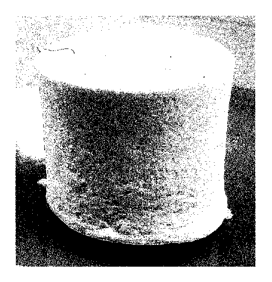
[0032] FIG. 1 is an image showing the sheet-form collagen structure
produced in
Example 1;
5 FIG. 2 is a stereoscopic micrograph showing the crude collagen fibers
formed in
Example 1;
FIG. 3 is a scanning electron micrograph (SEM) showing the surface of the
collagen structure prepared in Example 1;
FIG. 4 is a scanning electron micrograph (SEM) showing a cross-section of the
10 .. collagen structure prepared in Example 1;
FIG. 5 is a graph showing the results of measuring the denaturation
temperature of
the collagen structure prepared in Example 1 and that of the collagen solution
used in the
preparation of the collagen structure, using a differential scanning
calorimeter (DSC) at a
heating rate of 2 C/minute;
FIG. 6 is an image taken by a fluorescence microscope after swelling the
collagen
structure obtained in Example 1 with DMEM/10% FBS, inoculating the collagen
structure with Human Foreskin Fibroblast (HFF) cells at a cell density of 1.0
x 104
cells/cm2 and then, 20 hours later, staining the cells with calcein AM;
FIG. 7 is an image showing the block-form collagen structure prepared in
Example
2;
FIG. 8 is a scanning electron micrograph (SEM) showing the dry material
produced in Comparative Example 1 by drying a collagen gel prepared from a
collagen
solution having a collagen concentration of 0.2 (w/v)%;
FIG. 9 is an image taken by a fluorescence microscope after acclimating the
collagen gel obtained in Comparative Example 1 with DMEM/10% FBS, inoculating
the
collagen gel with FITT cells at a cell density of 1.0 x 10⁴ cells/cm2 and
then, 20 hours
later, staining the cells with calcein AM; and
CA 02861027 2014-07-11
11
FIG. 10. is a scanning electron micrograph showing the collagen sponge that
was
produced in Comparative Example 3 by freeze-drying 1 (w/v)% collagen solution.
Description of Embodiments
[0033] The first embodiment of the present disclosure is a collagen
structure, which
.. is composed of collagen fibers of 1 to 5 gm in average diameter; and having
a water
content of 0 to 15 (w/w)% and a collagen density of 50 to 800 mg/em3. Further,
the
second embodiment of the present disclosure is the collagen structure
described above
which is used as an artificial medical material, a member for disease
treatment, a
cosmetic material or a cell culture material. The present disclosure will now
be
described in detail.
[0034] (1) Collagen structure
The term "collagen" used herein refers to a protein constituting dermis,
ligaments,
tendons, bones, cartilages and the like. A molecule in which three peptide
chains of
collagen protein are twisted together into a triple helix is called "collagen
molecule". In
the present disclosure, the term "collagen fiber" refers to an assembly of
collagen
microfibrils and the term "collagen microfibril" refers to an assembly of
plural collagen
molecules.
[0035] Conventionally, type Ito type XXIX collagens are known, and the
collagen
used in the present disclosure may be any of these collagens or a newly
discovered
.. collagen. The majority of collagens contained in a living body are
insoluble in water
and, in the present disclosure, those collagens that are capable of forming
collagen fibers
can be widely used. For example, a "solubilized collagen", which is obtained
by
solubilizing collagen contained in a raw material such as skin or bone of an
animal by an
addition of an enzyme such as protease, can be used. It is noted here that
biological
materials such as animal skins and bones may also contain a trace amount of
"soluble
collagen" that is soluble in a neutral salt solution and/or an acidic
solution, such soluble
collagen can be used also in the present disclosure. The constituent amino
acids in the
CA 02861027 2014-07-11
12
above-described "solubilized collagen" and "soluble collagen" may also be
modified in
performing a chemical treatment.
[0036] Further, the collagen molecules constituting the collagen fiber
may also be
collagen derivatives. In the present disclosure, the term "collagen
derivative" means the
above-described collagen molecule whose constituent amino acid(s) is/are
modified with
other functional group. Examples of such "collagen derivative" include
acylated
collagens and esterified collagens. As the acylated collagens, for example,
succinylated
collagens, phthalated collagens and maleylated collagens can be mentioned.
Examples
of "collagen derivative" also include acylated collagens such as succinylated
collagens,
phthalated collagens and maleylated collagens, which are obtained by adjusting
an
atelocollagen solution extracted by an enzyme treatment to have a pH of 9 to
12 and then
adding thereto an acid anhydride such as succinic anhydride, phthalic
anhydride or
maleic anhydride. Further, examples of the esterified collagens include, in
addition to
those solubilized collagens that are esterified, insoluble collagens that are
esterified and
then solubilized by an enzyme reaction or the like.
[0037] In the present disclosure, the term "collagen structure" refers to
a solid
material having a prescribed shape. Therefore, the term "collagen structure"
does not
encompass any fluid such as powder or granule. Examples of the prescribed
shape
include film-forms, sheet-forms, and block-forms such as those of a cylinder,
a cone, a
polygonal column and a sphere. The prescribed shape may be any shape as long
as it
can be maintained, or it may be an amorphous shape as well. Here, the term
"film-form" refers to the form of a thin film having a thickness of less than
200 gm and
the term "sheet-form" refers to the form of a film having a thickness of not
less than 200
gm. Further, the term "block-form" refers to an aggregate of planar
material having a
thickness in the vertical direction.
[0038] The collagen structure of the present disclosure comprises
collagen fibers
having an average diameter of 1 to 5 gm in a dry state. As described above, in
a
CA 02861027 2014-07-11
13
collagen solution, collagen molecules having a triple-helical structure are
discretely
dissolved; therefore, when such a collagen solution is molded into a film form
by
air-drying or the like, a film is formed by the collagen molecules and
assemblies thereof.
Since the collagen molecules and assemblies thereof are thin and short and the
gaps
.. between the collagen molecules and between the assemblies are thus small,
cells cannot
pass through the gaps. Even if cells were cultured on such a film, the cells
would be
localized on the film surface, not being able to migrate into the film. In
addition, since
the film is constituted by thin and short collagen molecules and the like, the
mechanical
strength of the film is low. However, in the present disclosure, since a
collagen
structure is constituted by thick collagen fibers of 1 to 5 fun in average
diameter that are
obtained by further association of collagen microfibrils formed by association
of collagen
molecules having a triple-helical structure, the gaps between the collagen
fibers are large,
so that cells can freely pass therethrough. Thus, when the collagen structure
of the
present disclosure is loaded to a living body, cells migrate into the collagen
structure.
Besides, the fiber structure of such collagen is similar to that of collagen
found in the
connective tissues of a living body such as tendons and ligaments. Therefore,
the
mechanical strength of the collagen itself can be maintained at a high level.
[0039] The collagen fibers constituting the collagen structure of the
present
disclosure have, in a dry state, an average diameter of I to 5 vim, more
preferably 2 to 3
gm. In this range, a collagen structure having excellent cell infiltration
property can be
obtained. In the collagen fibers that are formed by association of collagen
molecules,
when the average diameter of the collagen fibers is 1 to 5 pm, the average
fiber length is
generally 1 to 10 mm as long as the collagen fibers are not subjected to
physical cutting
or any other treatment after the formation. It is noted here that, in the
present disclosure,
the average diameter and the average fiber length of the above-described
collagen fibers
are defined as the values that are measured for a collagen structure in a dry
state, that is,
in a state of having a water content of 0 to 15 (w/w)%, by the respective
methods
CA 02861027 2014-07-11
14
described below in the section of Examples.
[0040] The collagen structure of the present disclosure has a water
content of 0 to
15 (w/w)%, more preferably 0 to 10 (w/w)%. Since the collagen structure is a
dry
material having a low water content, it has excellent thermal stability and is
capable of
avoiding deterioration caused by bacterial contamination and the like. In
addition, the
collagen structure is different from powder and the like in that it is a
molded article in the
form of a film, sheet, block or the like; therefore, by molding the collagen
structure into
the shape of a defective part of a living body, the collagen structure can be
easily attached
or loaded to the living body. It is noted here that, in the present
disclosure, the water
content is defined as the value measured by the method described below in the
section of
Examples.
[0041] In the collagen structure of the present disclosure, when the
water content is
0 to 15 (w/w)%, the collagen density is 50 to 800 mg/cm3, more preferably 110
to 600
mg/cm3, particularly preferably 120 to 400 mg/cm3. Collagen exists in an
insoluble
form in vivo, forming connective tissues at high concentration of 25 (w/v)% in
the skin
tissue and 32 (w/v)% in the tendon tissue. In order to extract collagen from
an animal
tissue, collagen is required to be solubilized but the resulting solubilized
collagen is
highly viscous. Therefore, it is difficult to prepare a highly concentrated
collagen
solution and there has been thus no high-density collagen structure. However,
according to the present disclosure, a collagen structure having a collagen
density of 50 to
800 mg/cm3, which is equivalent to the in vivo collagen density, can be
provided, and this
collagen structure can be used as a tissue-equivalent material. It is noted
here that, in the
present disclosure, the collagen density is defined as the value measured by
the method
described below in the section of Examples.
[0042] The collagen structure of the present disclosure has a porosity of
20 to 90%,
more preferably 30 to 80%, particularly preferably 40 to 70%. Since the
collagen
structure is porous, it is quickly swollen when immersed in a solvent. It is
noted here
CA 02861027 2014-07-11
that, in the present disclosure, the porosity is defined as the value measured
by the
method described below in the section of Examples.
[0043] The collagen structure of the present disclosure is a porous
structure which
comprises collagen fibers and has an average pore size of 1 to 50 prn, more
preferably 5
5 .. to 30 urn. The collagen structure of the present disclosure is
constituted in such a
manner that the above-described collagen fibers are folded and overlapped as
in a
nonwoven fabric. Accordingly, the pores serve as communicating pores that can
be in
communication with other pores. Therefore, cells entering the pores can
migrate into
the inside of the collagen structure through the communicating pores. It is
noted here
10 .. that, in the present disclosure, the "average pore size" is defined as
the value measured by
the method described below in the section of Examples.
[0044] The collagen structure of the present disclosure may also
comprise at least
one factor selected from the group consisting of cell chemotactic factors,
growth factors,
cell proliferation factors, blood coagulation factors and anticoagulant
factors. By adding
15 .. these components, the collagen structure can be imparted with efficacies
such as wound
healing, inhibition of tumor cell proliferation, immunoregulation,
osteogenesis,
hematopoietic regulation, hemostasis and anticoagulation.
[0045] Examples of the chemotactic factors include cytokines such as
erythropoietin and interleukin 1 (IL-1); and chemokines such as interleukin 8
(IL-8),
.. NAP-2 and MIP-2.
[0046] Further, examples of the growth factors include epidermal growth
factors
(EGFs), insulin-like growth factor (IGFs), transforming growth factors (TGFs),
nerve
growth factors (NGFs) and platelet-derived growth factors (PDGFs).
[0047] Examples of the proliferation factors include brain-derived
neurotrophic
factors (BDNFs), vascular endothelial growth factors (VEGFs), granulocyte
colony-stimulating factors (G-CSFs), granulocyte-macrophage colony-stimulating
factors
(GM-CSFs), erythropoietin (EPO), thrombopoietin (TP0), basic fibroblast growth
factors
CA 02861027 2014-07-11
16
(bFGF and FGF2) and hepatocyte growth factors (HGFs).
[0048] Further, examples of the coagulation factors include
fibrinogen/fibrin
(Factor I), prothrombin/thrombin (Factor II) and tissue factors (Factor III,
thromboplastin), and examples of the anticoagulant factors include heparin and
antithrombin HI.
[0049] These additives may be bound to the collagen structure by
impregnation or
the like, or may be bound to the collagen structure via a bonding means, and
the additives
can be selected as appropriate in accordance with the intended use. For
example, the
collagen structure, which is impregnated with a solution containing the above-
described
component(s) to allow the component(s) to adsorb to the collagen structure and
subsequently dried, can be used as a member of a drug delivery system or the
like
because it slowly releases the above-described component(s) upon being loaded
to a
wound.
[0050] Examples of the binding means include polypeptide chains of
collagen-binding domains, such as the collagen-binding domain of von
Willebrand factor
and that of collagenase. By binding a polypeptide chain of a collagen-binding
domain
to the above-described components in advance, the components can be stably
bound to
collagen fibers via the binding means.
[0051] The collagen structure of the present disclosure may also be
formed by
performing cross-linking within each collagen fiber or between collagen
fibers. Since
collagen is a biological constituent, it is degraded in vivo by collagenase or
the like.
Accordingly, in cases where the collagen structure is used as a bone material
or the like at
a site or in an application where biodegradation is desired to be avoided, a
cross-linked
structure is introduced. By introducing a cross-linked structure,
biodegradation is
inhibited, so that the mechanical strength can be improved. Such a cross-
linked
structure may be introduced only to the surface of the collagen structure, or
may be
introduced to the inside of the collagen structure as well.
CA 02861027 2014-07-11
17
[0052] The collagen structure of the present disclosure is molded into a
film form, a
sheet form or a block form. The block-form may be a columnar-form, a spherical
form
or a cone-form, or the collagen structure may be molded into an arbitrary
shape.
Particularly, the collagen structure may also be molded into a specific shape
of a
biological tissue. Examples of the specific shape include biological shapes of
a crescent
constituting a knee joint, a tympanic membrane, a finger, a nose, an ear and
the like; and
those shapes of certain cartilages. By subcutaneously embedding the collagen
structure
of the present disclosure or by filling a bone fracture site with the collagen
structure as an
artificial bone, the neighboring cells are allowed to proliferate and, by
applying the
.. collagen structure of the present disclosure as an artificial skin to form
a boundary
between inside and outside the body, invasion of bacteria and the like can be
inhibited
and the regenerative function can be facilitated. It is noted here the
collagen structure of
the present disclosure may also comprise other layer(s) laminated thereon.
A conventional collagen sponge may be compressed into the form of a sheet
having a high collagen density. However, since such a collagen sponge is not
constituted by collagen fibers, it cannot secure such a strength that can be
provided by
collagen fibers. The collagen structure of the present disclosure is formed in
prescribed
shape without any compression processing; therefore, it has excellent cell
infiltration
property and is capable of maintaining a strength provided by the collagen
fibers even
when it is used in a hydrated state.
[0053] (2) Application
The collagen structure of the present disclosure can be used as an artificial
medical
material, a member for disease treatment, a cosmetic material, a cell culture
materials or
the like.
[0054] As an artificial medical material, the collagen structure of the
present
disclosure is capable of adapting to a defective part of dermis, bone, joint
cartilage,
tendon, ligament, blood vessel or the like so as to facilitate the maintenance
of space,
CA 02861027 2014-07-11
18
introduction of cells and the like. Such an artificial medical material can be
applied to
regenerative medicine. Further, the collagen structure that is in a film form
and
impregnated with a hemostatic agent can be coated over a bleeding site to be
used as a
hemostatic material.
[0055] As a member for disease treatment, the collagen structure of the
present
disclosure can be used in the treatment of, for example, eye injury, severe
burn,
skin-grafted site, decubitus ulcer, diabetic ulcer, surgical incision wound or
keloid-forming wound.
[0056] As a cosmetic material, the collagen structure of the present
disclosure can
be used as a pack material by cutting the film-form or sheet-form collagen
structure into a
face shape and impregnating it with a cosmetic lotion or the like.
[0057] As a cell culture material, by using the collagen structure of the
present
disclosure as a three-dimensional cell culture medium, cells can be
subcultured. Further,
since the collagen structure of the present disclosure has excellent cell
infiltration and cell
immobilization properties, it can be also used as, for example, a substrate
for drug
permeability test. Examples of subject cells to which the collagen structure
of the
present disclosure can be applied include ES cells and iPS cells.
[0058] Further, as an application of artificial medical material, the
collagen
structure of the present disclosure can be used as a carrier of a drug
delivery system.
When the collagen structure bound with various components is applied or loaded
to a
living body, the collagen structure releases the drug components with time,
functioning as
a drug delivery system.
[0059] (3) Method of Producing Collagen structure
The method of producing the above-described collagen structure is not
particularly
restricted. However, the collagen structure can be produced by performing the
steps of:
generating collagen fibers by neutralizing an acidic collagen solution;
forming crude
collagen fibers having a collagen concentration of 12 to 50 (w/v)% by
separating the
CA 02861027 2014-07-11
19
collagen fibers from a solution containing the collagen fiber; molding the
crude collagen
fibers into a prescribed shape; and drying a molded article obtained in the
molding step.
The collagen structure can also be produced by further performing the steps
of, following
the step of forming the crude collagen fibers: after dispersing the crude
collagen fibers in
a hydrophilic organic solvent, separating the collagen fibers from the
hydrophilic organic
solvent and dehydrating the thus separated collagen fibers; and molding and
drying the
thus dehydrated collagen fibers. Moreover, a cross-linked collagen structure
can be
produced by further performing the steps of, following the step of dehydrating
the
collagen fibers: subjecting the dehydrated collagen fibers to a cross-linking
treatment
and/or a chemical treatment; and drying the thus treated collagen fibers.
[0060] The collagen to be used in the present disclosure can be
collected from a
skin of an animal such as cow, pig, bird or fish or other collagen-containing
tissue. In
general collagen is contained in a large amount in animal connective tissues;
however,
when extracted by a heat treatment, collagen is thermally denatured and its
unique
triple-helical structure is broken, causing the collagen to be in a gelatinous
state. In the
present disclosure, a collagen having a triple-helical structure is used. As a
method of
extracting such a collagen, for example, a solubilization method in which a
material such
as animal bone or skin is subjected to an acid treatment and/or an enzyme
treatment can
be employed. Preferred examples of the material from which the collagen is
extracted
include dermis and tendons of cow, pig, chicken, ostrich, horse, fish and the
like. It is
preferred to use a tissue of a young animal, such as an embryo-derived tissue,
since the
yield is improved.
[0061] For preparation of a collagen solution to be treated with an
enzyme, for
example, a tissue obtained by grinding and defatting the dermal layer of a
bovine skin can
be used. After suspending this tissue in distilled water to a final collagen
concentration
of 0.5 to 5 (w/v)%, the pH of the resulting suspension is adjusted to 3.0 by
adding thereto
hydrochloric acid. Then, acid protease is added in an amount of one-hundredth
of the
CA 02861027 2014-07-11
collagen weight to perform a solubilization treatment at 25 C for 72 hours.
After
terminating the enzyme reaction, the thus obtained enzyme-solubilized collagen
solution
is subjected to salt precipitation, and the recovered salt precipitates are
then dispersed in
distilled water to a collagen concentration of 1 to 5 (w/v)% and uniformly
dissolved with
5 an addition of hydrochloric acid, thereby a collagen solution can be
obtained.
[0062] The pH of the above-described acidic collagen solution is
preferably 1.0 to
6.0, more preferably 3.0 to 4Ø When the pH is higher than this range, it may
be
difficult to form collagen fibers.
[0063] In the present disclosure, the above-described acidic collagen
solution is
10 neutralized. Regardless of whether the collagen solution is prepared by
an enzyme
treatment or an acid treatment, the collagen solution is acidic for dissolving
collagen
molecules therein. An alkaline or neutral buffer is added to such acidic
collagen
solution. As an alkaline solution, for example, a sodium hydroxide solution or
a
potassium hydroxide solution can be used. Further, as the neutral buffer, a
buffer which
15 shows buffering action in the vicinity of pH 7.0, such as a phosphate
buffer which
comprises phosphoric acid and sodium phosphate and has a pH of 7.0 to 9.5, a
HEPES
(244-(2-hydroxyethyl)-1-piperazinyllethanesulfonic acid) buffer (pH: 6.8 to
8.2), a
citrate-phosphate buffer (pH: 2.6 to 7.0), a 50 mM Tris buffer (pH: 7.4) or a
50 mM
phosphoric acid (pH: 7.4), can be widely used. It is noted here that "neutral"
pl4 may be
20 any pH of 6.0 to 9Ø
[0064] The above-described alkaline solution and neutral buffer may also
contain
other salt and the like in such an amount that does not change the pH.
Examples of such
a salt include sodium chloride and potassium chloride. When the collagen
solution is
made isotonic to human body fluid by an addition of such a salt, collagen
fibers in which
collagen molecules are staggered by 67 nm in the same manner as in vivo
collagen can be
formed. Therefore, it is preferred that the salt be added in such an amount
that allows
the osmotic pressure of the collagen solution after the neutralization
treatment to be
CA 02861027 2014-07-11
21
isotonic to human body fluid.
[0065] In the present disclosure, the collagen solution after the
neutralization
treatment has a collagen concentration of 0.01 to 5 (w/v)%, more preferably
0.1 to 5
(w/v)%, particularly preferably 0.3 to 5 (w/v)%. When the collagen
concentration is
lower than 0.01 (w/v)%, the subsequent concentration process is not easily
carried out.
Meanwhile, since collagen is highly viscous, it is difficult to prepare a
collagen solution
having a concentration of higher than 5 (w/v)%.
[0066] In the present disclosure, after the above-described
neutralization treatment,
the resulting collagen solution is left to stand in a temperature range of 4
to 45 C, more
preferably 30 to 37 C. In this temperature range, the collagen molecules
dissolved in
the collagen solution are allowed to associate with each other in the solution
by the
neutralization treatment, thereby forming a collagen gel.
[0067] In the present disclosure, the resulting collagen gel is
subsequently stirred
gently. By this gentle stirring, association of the collagen molecules
constituting the
collagen gel is facilitated and the moisture contained between the fibers is
released while
the structure of the collagen fibers is maintained, so that thick and long
collagen fibers are
precipitated in the solution. Therefore, the stirring may be performed at any
level as
long as association of the collagen molecules can be facilitated. When the
collagen
solution is vigorously stirred, the generated collagen fibers are physically
broken into thin
and short collagen fibers. The collagen fibers precipitated in the solution by
gentle
stirring have an average diameter of 1 to 100 gm and a length of 1 to 10 mm.
It is noted
here that, in the present disclosure, the average diameter and the average
fiber length of
collagen fibers precipitated out of a collagen solution are defined as the
values of average
diameter and average length that are measured for 20 fibers randomly selected
from those
fibers observed in a stereoscopic micrograph, respectively.
[0068] By filtering or centrifuging this solution in which the collagen
fibers are
precipitated, the collagen fibers can be separated and recovered. In the
present
CA 02861027 2014-07-11
22
disclosure, the collagen fibers that are separated from the collagen solution
are referred to
as "crude collagen fibers". Accordingly, the crude collagen fibers comprise
collagen
fibers and water as main components. When the concentration of the collagen
fibers
contained in the crude collagen fibers is less than 12 (w/v)%, by again
performing
centrifugation, filtration or the like, the crude collagen fibers are further
concentrated to a
collagen concentration of 12 to 50 (w/v)%, more preferably 15 to 40 (w/v)%,
particularly
preferably 18 to 30 (w/v)%.
[0069] In order to separate the crude collagen fibers having the above-
described
concentration by filtration, it is preferred to use a filter paper having a
pore size of 1 gm
to 1 mm, more preferably 10 gin to 100 gm. As long as the pore size is in the
above-described range, a large amount of collagen fibers can be efficiently
processed.
[0070] Meanwhile, crude collagen fibers can also be separated by
centrifuging the
above-described collagen solution. For example, the collagen solution is
centrifuged at
10,000 to 20,000 rpm for 10 minutes to 1 hour. Here, in order to adjust the
collagen
concentration to the above-described range, centrifugation can be performed a
plurality of
times.
[0071] In the present disclosure, the thus recovered crude collagen
fibers are
molded into a prescribed shape. As for the shape of the molded crude collagen
fibers,
the crude collagen fibers can be molded into a film form, a sheet form or a
variety of
three-dimensional configurations. In cases where the collagen structure is
used for
filling a tissue, it may be molded into a shape that conforms to the part to
be filled in the
subject body.
For example, in cases where a collagen solution in which collagen fibers are
precipitated is filtered to separate crude collagen fibers, by arranging a
filter paper on a
porous filter paper mount formed in the middle part of a funnel and then
filtering the
collagen solution through the filter paper, the crude collagen fibers can be
deposited in a
sheet or block form on the filter paper. Alternatively, using the filter paper
mount
CA 02861027 2014-07-11
23
deformed into a prescribed shape in advance as a mold, the crude collagen
fibers may be
deposited on the filter paper mount and molded into a prescribed shape. The
above-described methods are examples of embodiment where the step of forming
crude
collagen fibers and the molding step are performed continuously. Also, the
crude
collagen fibers deposited on the filter paper may be molded by being filled
into a mold
having a prescribed shape.
[0072] The above-described molding methods can be applied in the same
manner
also in those cases where crude collagen fibers are formed by centrifugation.
For
example, using a centrifuge tube as a mold at the time of performing
centrifugation, crude
collagen fibers can be centrifuged and molded into a prescribed shape at the
same time.
This method is another example of embodiment where the step of forming crude
collagen
fibers and the molding step are performed continuously. Here, after the
centrifugation
process, the crude collagen fibers may also be molded by being filled into a
mold having
a prescribed shape.
[0073] Subsequently, the molded crude collagen fibers are dried. In the
production method of the present disclosure, since crude collagen fibers
having a
collagen concentration of 12 to 50 (w/v) /0 are molded, a collagen structure
can be
produced by dehydrating and drying the molded crude collagen fibers by freeze-
drying,
air-drying, hot-air drying, vacuum suction and/or the like. Here, a collagen
structure
having the shape of a cylinder, a column or the like may be obtained in
advance and this
may be further shaped by scraping or the like. As for the extent of the
drying, the
molded crude collagen fibers are dried to a water content of 0 to 15 (w/w)%.
This is
because the molded crude collagen fibers have superior storage stability as
compared to
the collagen solution.
[0074] The collagen structure of the present disclosure may also be
subjected to
compression molding after the above-described drying step. The collagen fibers
constituting the collagen structure of the present disclosure have an average
diameter of 1
CA 02861027 2014-07-11
24
to 5 j.tm and a length of I to 10 mm. Such thick and long collagen fibers are
deposited
in a nonwoven fabric-like form and the cell infiltration property and the
strength are
thereby maintained; therefore, even when the collagen structure of the present
disclosure
is subjected to compression molding, the collagen concentration can be
increased without
reduction in the cell infiltration property or the strength. Such compression
molding
may also be performed in a step other than those performed after the drying,
for example,
at the time of molding the crude collagen fibers into a prescribed shape.
[0075] In the present disclosure, prior to the separation of crude
collagen fibers, a
hydrophilic organic solvent solution dispersing crude collagen fibers may be
prepared by
adding a hydrophilic organic solvent to the crude collagen fibers in an amount
of 3 to
2,000 parts by mass, preferably 5 to 1,000 parts by mass, more preferably 10
to 100 parts
by mass, particularly preferably 10 to 30 parts by mass, and then the crude
collagen fibers
may be separated by filtration and dehydrated. The crude collagen fibers used
in the
present disclosure has a collagen concentration of 12 to 50 (w/v)%, the
concentration is
higher than that of a conventional collagen solution. By dispersing the crude
collagen
fibers in a high-concentration hydrophilic organic solvent such as 100%
ethanol, highly
hydrophilic crude collagen fibers can be efficiently dehydrated. Such a
hydrophilic
organic solvent solution dispersing crude collagen fibers has a higher
fluidity than the
collagen solution, so that the filtration efficiency of solution as well as
the drying
efficiency of the molded crude collagen fibers can be both improved. Since the
occurrence of clogging is inhibited during the filtration operation, a thick
block-form
collagen structure can be produced.
[0076] Dehydration of collagen by ethanol or the like is conventionally
known and
it has been a common practice to dehydrate collagen while gradually increasing
the
alcohol concentration. However, in the present disclosure, since the crude
collagen
fibers have a high collagen concentration of 12 to 50 (w/v)%, for example,
even when
ethanol is used, 100% ethanol can be used. Therefore, dehydration by a
hydrophilic
CA 02861027 2014-07-11
organic solvent can be performed simply and efficiently.
[0077] The hydrophilic organic solvent for dispersing crude collagen
fibers may be
any carbon-containing solvent as long as it is miscible with water, and
examples thereof
include alcohols, ketones, ethers, esters and polar aprotic solvents. Examples
of the
5 alcohols include monohydric alcohols having 1 to 6 carbon atoms, such as
methanol,
ethanol, isopropanol and t-butanol; and polyhydric alcohols such as ethylene
glycol and
propylene glycol. Examples of the ketones include acetone and methyl ethyl
ketone.
Further, examples of the ethers include glycol ethers such as diethyl ether,
methyl ethyl
ether, ethylene glycol monomethyl ether and diethylene glycol monobutyl ether;
and
10 cyclic ethers such as tetrahydrofuran and dioxane. Moreover, examples of
the esters
include ethyl acetate and ethyl lactate, and examples of the polar aprotic
solvents include
dimethyl sulfoxide (DMSO), dimethylformamide (DMF) and pyridine. 'Thereamong,
examples of preferred solvents that are miscible with water at an arbitrary
ratio include
acetone, methanol, ethanol, isopropanol, acetonitrile, tetrahydrofuran,
dimethyl sulfoxide
15 and dimethylformamide. Among these preferred solvents, ethanol, acetone,
diethyl
ether, or a mixed solution thereof can be suitably used.
Here, the temperature of the hydrophilic organic solvent to be used is
preferably
not higher than 15 C. This is because collagen fibers are not denatured at
such a
temperature and the triple-helical structure of the collagen molecules can
thus be
20 maintained.
[0078] By filtration or the like of the hydrophilic organic solvent
solution
dispersing crude collagen fibers, the crude collagen fibers can be isolated
from the
hydrophilic organic solvent solution and, consequently, the crude collagen
fibers can be
dehydrated. At the time of filtering the hydrophilic organic solvent solution
containing
25 crude collagen fibers, by arranging a filter paper on a porous-filter-
paper-mounting-part
formed in the middle of a funnel and then filtering the above-described
hydrophilic
organic solvent solution dispersing the crude collagen fibers through the
filter paper, the
CA 02861027 2014-07-11
26
crude collagen fibers are deposited in a sheet form on the filter paper. By
this,
dehydration and molding of the crude collagen fibers can be performed
continuously.
Also, by increasing the amount of deposition, the crude collagen fibers can be
molded
into a block form. It is noted here that the dehydrated crude collagen fibers
can also be
molded using a prescribed mold.
[0079] Collagen is highly hydrophilic and thus not readily dried.
Particularly, it is
not easy to dry a three-dimensional collagen. However, in the present
disclosure, since
crude collagen fibers having the above-described collagen concentration are
dehydrated
using a hydrophilic organic solvent, a collagen structure which has a high
collagen
density and is capable of retaining a three-dimensional shape can be produced.
[0080] After being molded, the crude collagen fibers can be dried also by
freeze-drying or air-drying, although the drying method is variable depending
on the
shape and the size thereof. Air-drying is inexpensive and it can inhibit
thermal
denaturation of the collagen fibers.
[0081] The collagen structure of the present disclosure may further
comprise a
cross-linked structure. By introducing a cross-linked structure, decomposition
of the
collagen structure after it is embedded in a living body can be inhibited. The
cross-linking method can be selected as appropriate in accordance with the
intended use.
For example, a cross-linked structure can be introduced by bringing the
collagen fibers or
collagen structure into contact with an aldehyde such as formaldehyde or
glutaraldehyde,
xylose, glucose, mannose, galactose or the like. Alternatively, the collagen
structure can
be cross-linked by adding thereto a carbodiimide-based, epoxide-based and/or
imidazole-based cross-linking agent(s). Further, the collagen structure can
also be
cross-linked by irradiating it with ultraviolet ray, 7-ray, electron beam or
the like. It is
noted here that, when collagen is naturally dried, a cross-linked structure is
partially
formed in some cases.
[0082] Regardless of the presence or absence of cross-linking, the
collagen
CA 02861027 2014-07-11
27
structure of the present disclosure may be bound with at least one factor
selected from the
group consisting of cell chemotactic factors, growth factors, cell
proliferation factors,
blood coagulation factors and anticoagulant factors. Such factor(s) may be
bound by
chemical bonding or by physical bonding such as adsorption or deposition.
The step of binding the factor(s) can be performed in any of the steps for
producing
a collagen structure. For example, in any one of the steps prior to the step
of drying the
crude collagen fibers, at least one factor selected from the group consisting
of cell
chemotactic factors, growth factors, cell proliferation factors, blood
coagulation factors
and anticoagulant factors can be bound to the collagen fibers. The step of
binding the
factor(s) can be selected as appropriate in accordance with the chemical
properties and
the like of the component(s) to be added. For example, a collagen structure
can be
produced by: adding the above-described factor(s) to crude collagen fibers;
uniformly
stirring the resulting mixture to physically bind the factor(s) to the crude
collagen fibers;
molding the crude collagen fibers into a prescribed shape; and then drying the
resultant.
Alternatively, a collagen structure can be produced by: dispersing crude
collagen fibers in
a hydrophilic organic solvent; filtering the solvent to dehydrate the crude
collagen fibers;
mixing the thus dehydrated crude collagen fibers with above-described
component(s);
and then drying the resulting mixture.
Further, after producing a collagen structure having a water content of 0 to
15
(w/w)%, the collagen structure may be impregnated with an aqueous solution of
the
above-described factor(s) and then dried again to a water content of 0 to 15
(w/w)%.
[0083] In order to chemically bind the above-described factor(s) to the
collagen
structure, the factor(s) which a collagen-binding means is formed in advance
may be used.
Examples of such a binding means include the polypeptide chain of the collagen-
binding
domain of von Willebrand factor and that of the collagen-binding domain of
collagenase.
For example, by binding a polypeptide chain of a collagen-binding domain to
the
above-described factor(s) and then impregnating the collagen structure with a
solution of
CA 02861027 2014-07-11
28
the factor(s) having such a binding means, the factor(s) is/are bound via the
binding
means. An amino acid sequence of a collagen-binding domain is capable of
specifically
bind to collagen in the same manner as a collagenase which is an enzyme whose
substrate
is collagen.
[0084] The collagen structure of the present disclosure is characterized in
that it has
a high collagen density and is molded into a desired shape. As the shape, a
film form, a
sheet form, a block form or the like can be selected in accordance with the
intended use.
Thin-layer molded articles such as film-form and sheet-form molded articles,
as well as
collagen sponges and tubular collagen structures have been available; however,
there has
been no block-form collagen structure having a high collagen concentration.
This is
because it is difficult to improve the collagen concentration prior to drying.
In the
present disclosure, particularly by dispersing crude collagen fibers in a
hydrophilic
organic solvent to dehydrate the crude collagen fibers, a large deposit of the
crude
collagen fibers can be simply formed and easily dried by air-drying or the
like. Further,
by filling the large deposit into a mold having a prescribed shape, it can be
molded and a
collagen structure having a complex shape can be produced.
Examples
[0085] The present disclosure will now be concretely described by way of
examples thereof; however, the present disclosure is not restricted thereto by
any means.
[0086] (Example 1)
(1) Preparation of Collagen structure
A tissue, which was prepared by grinding the dermal layer of a porcine skin
using
a meat grinder or the like and then defatting and sufficiently washing the
resultant, was
used as a raw material. In a solubilized aqueous solution in which pepsin and
acetic
acid were mixed at final concentrations of 5 mg/ml and 50 mM, respectively,
the raw
material was suspended to a final collagen concentration of 4.5 (w/v)%, and
the resulting
suspension was subjected to an overnight solubilization treatment at 4 C. To
the
CA 02861027 2014-07-11
29
resulting enzyme-solubilized collagen solution obtained in the above-described
manner,
sodium chloride was added to a fmal concentration of 5 (w/v)% to perform salt
precipitation, and the thus formed precipitates were recovered by
centrifugation. The
recovered salt precipitates were dispersed in distilled water to a collagen
concentration of
3 (w/v)% and then uniformly dissolved by adjusting the pH to 3.0 with an
addition of
hydrochloric acid, thereby preparing a collagen solution.
To 2.5 ml of this collagen solution (temperature: 4 C), 47.5 ml of
phosphate-buffered saline (pH: 7.5, temperature: 4 C) was added, and the
resultant was
left to stand at 37 C for 24 hours.
By this process, collagen molecules were allowed to associate with each other
to
form a gelatinous material and, when this gelatinous material was gently
stirred, the
association was facilitated, so that collagen fibers were formed and dispersed
in the
resulting solution. The dispersed fibers were filtered out by pouring the
solution onto a
nylon mesh having a pore size of 80 gm, thereby recovering crude collagen
fibers on the
mesh. The thus obtained crude collagen fibers had a collagen concentration of
20
(w/v)%.
Then, the collagen fibers recovered on the mesh were freeze-dried to obtain a
0.2
mm-thick sheet-form collagen structure. The outer appearance of this collagen
structure
is shown in FIG. I.
(2) Water Content
The water content of the above-described collagen structure was measured to be
9.4 (w/w)% by the following method.
(i) Method of Measuring Water Content
The mass of the collagen structure (wl) is measured. Then, after heating the
collagen structure at 120 C for 2 hours to evaporate water, the mass of the
resulting
collagen structure (w2) is measured. The change in the mass before and after
the
heating (WI - w2) is determined as the amount of water, and the water content
is defmed
CA 02861027 2014-07-11
as the percentage (%) of this amount of water with respect to the mass of the
collagen
structure (wl).
(3) Average Diameter and Average Fiber Length of Crude Collagen Fibers
The crude collagen fibers recovered on the nylon mesh were observed under a
5 stereoscopic microscope. FIG. 2 is a stereoscopic micrograph thereof.
Under the
stereoscopic microscope, 20 crude collagen fibers were randomly selected,
their
diameters and lengths were measured, and the average values of the 20 fibers
were
calculated. The 20 crude collagen fibers had an average diameter of 1.15 gm
and an
average length of 4.09 mm. It is noted here that the shortest fiber length was
1.9 mm
10 and the longest fiber length was 8.75 mm.
[0087] (4) Scanning Electron Micrograph (Surface)
The surface fiber structure of the thus obtained collagen structure was
observed
under a scanning electron microscope (SEM). The result thereof is shown in
FIG. 3.
[0088] (5) Average Diameter and Pore Size of Collagen Fibers Constituting
15 Collagen structure
For the above-described collagen structure, the average fiber diameter and the
average pore size were measured in a dry state by the following methods. The
results
thereof are shown in Table 1. The collagen structure had an average pore size
of 18.47
pin, meaning that the collagen structure had sufficient spaces for allowing
cells of 5 to 7
20 pm in diameter to infiltrate.
(i) Average Diameter of Collagen Fibers
From the collagen fibers observed under a scanning electron microscope (SEM).
20 fibers are randomly selected, and their diameters are measured. The average
of the
diameters measured for the 20 fibers is calculated as the average fiber
diameter.
25 (ii) Average Pore Size of Collagen Fibers
From the fibers observed under a scanning electron microscope (SEM), the
diameters of randomly selected 20 fiber pores are measured. The average size
of the 20
CA 02861027 2014-07-11
31
pores is calculated as the average pore size.
[0089] (6) Scanning Electron Micrograph (Cross-section)
The fiber structure of a cross-section of the collagen structure was observed
under
a scanning electron microscope (SEM). The result thereof is shown in FIG. 4.
[0090] (7) Denaturation Temperature
The denaturation temperature of the thus obtained collagen structure and that
of the
collagen solution used as a control were measured using a differential
scanning
calorimeter (DSC) at a heating rate of 2 C/minute. The results thereof are
shown in FIG.
5 and Table 2. The collagen structure was observed to have a peak of
denaturation
temperature at 115.03 C, while the collagen solution had a denaturation
temperature of
42.75 C. Therefore, it was revealed that the collagen structure had superior
thermal
stability as compared to the collagen solution.
[0091] (8) Collagen Density and Porosity
The collagen density and the porosity were measured by the following methods.
As a result, the collagen density was found to be 200 mg,/cm3 and the porosity
was found
to be 40.9%.
(i) Method of Measuring Collagen Density
The collagen structure is cut precisely into a size of 1-cm square to prepare
a test
piece. The thickness of this test piece is precisely measured using a
thickness gauge so
as to calculate the volume. Then, the test piece is dissolved in 5 ml of 5mM
acetic acid
solution and the collagen concentration is measured by a microburet method.
From the
volume and collagen concentration of the test piece, the amount of collagen
per unit
volume is calculated as the collagen density.
(ii) Porosity
The porosity is measured by mercury intrusion porosimetry using Pascal 140 and
440 (manufactured by Carlo-Erba Instruments, Ltd.).
[0092] (8) Cell Infiltration Property
CA 02861027 2014-07-11
32
The thus obtained collagen structure was swollen with DMEM/10% FBS and
inoculated with HFF cells at a cell density of 1.0 x 10⁴ cells/cm2. Then, 20
hours later,
the cells were stained with calcein AM and observed under a fluorescence
microscope.
The result thereof is shown in FIG. 6.
[0093] (Example 2)
(1) Preparation of Collagen structure
A tissue, which was prepared by grinding the dermal layer of a porcine skin
using
a meat grinder or the like and then defatting and sufficiently washing the
resultant, was
used as a raw material. In a solubilized aqueous solution in which pepsin and
acetic
acid were mixed at final concentrations of 5 mg/ml and 50 mM, respectively,
the raw
material was suspended to a final collagen concentration of 4.5 (w/v)%, and
the resulting
suspension was subjected to an overnight solubilization treatment at 4 C. To
the
resulting enzyme-solubilized collagen solution obtained in the above-described
manner,
sodium chloride was added to a final concentration of 5 (w/v)% to perform salt
precipitation, and the thus formed precipitates were recovered by
centrifugation. The
recovered salt precipitates were dispersed in distilled water to a collagen
concentration of
3 (w/v)% and then uniformly dissolved by adjusting the pH to 3.0 with an
addition of
hydrochloric acid, thereby preparing a collagen solution. To 5 ml of this
collagen
solution (temperature: 4 C), 95 ml of phosphate-buffered saline (pH: 7.5,
temperature:
4 C) was added, and the resultant was left to stand at 37 C for 24 hours. By
this process,
collagen molecules were allowed to associate with each other to form a
gelatinous
material
When this gelatinous material was gently stirred, the association was
facilitated to
form collagen fibers, which were dispersed and precipitated in the resulting
solution.
The precipitated fibers were recovered by 20-minute centrifugation at 17,500
rpm to
obtain crude collagen fibers. The thus obtained crude collagen fibers had a
collagen
concentration of 20 (w/v)%.
CA 02861027 2014-07-11
33
Thereafter, 0.75 g of the thus obtained crude collagen fibers was added to 10
g of
20 C ethanol and dispersed by gently stirring the resulting mixture for 10
minutes. The
resulting dispersion was filtered to separate the crude collagen fibers. The
thus
recovered crude collagen fibers were filled into a columnar mold of 10 mm in
diameter
and 10 mm in height and then air-dried at room temperature to obtain a
collagen structure.
The thus obtained collagen structure is shown in FIG. 7.
[0094] (2) Water Content, Collagen Density, Porosity, and Average
Diameter of
Collagen Fibers
For the thus obtained collagen structure, the water content, the collagen
density,
the porosity, and the average diameter of collagen fibers were measured in the
same
manner as in Example 1. As a result, it was found that this collagen structure
had a
water content of 6.7 (w/w)%, a collagen density of 127 mg/cm' and a porosity
of 76.6%.
Further, the average diameter of the collagen fibers was 1.59 pm.
[0095] (Comparative Example 1)
(1) Preparation of Freeze-Dried Gel Material
The collagen solution obtained in Example I was diluted to a concentration of
0.4
(w/v)% by adding thereto distilled water and then mixed with an equivolume of
2x
concentrated phosphate-buffered saline (pH: 7.5) at 4 C. The resulting mixture
was
gently poured on a cell culture plate and this plate was left to stand at 37 C
for 24 hours
to produce a gelatinous material.
The thus obtained gelatinous material was freeze-dried as it was, without
isolating
collagen fibers therefrom.
[0096] (2) Water Content and Collagen Density
The water content and the collagen density were measured in the same manner as
in Example 1. As a result, it was found that this freeze-dried material had a
water
content of 10 to 15 (w/w)% and a collagen density of 2.0 mg/cm3.
[0097] (3) Average Diameter and Pore Size of Collagen Fibers
CA 02861027 2014-07-11
34
The average diameter and pore size of the collagen fibers constituting the
freeze-dried material were measured in the same manner as in Example I. The
collagen
fibers constituting this film were found to have an average diameter of 0.17
gm. It is
noted here that the fiber length could not be measured since the collagen
fibers were in
contact with each other. The results are shown in Table 1.
[0098] (4) Scanning Electron Micrograph of Gelatinous Material
The gelatinous material before being freeze-dried was observed under a
scanning
electron microscope (SEM). The result thereof is shown in FIG. 8.
[0099] (5) Cell Infiltration Property
The gelatinous material before being freeze-dried was acclimated with
DMEM/10`)/0 FBS and inoculated with T-IFF cells at a cell density of 1.0 x 10⁴
cells/cm2
in the same manner as in Example 1. Then 20 hours later, the cells were
stained with
calcein AM and observed under a fluorescence microscope. The result thereof is
shown
in FIG. 9. In FIG. 6 of Example 1, a condition where the cells were three-
dimensionally
arranged was observed with both in-focus cells and out-of-focus cells existing
at the same
time; however, in FIG. 9, since the cells were in focus, it was observed that
the cells
existed two-dimensionally in a single plane.
[0100] (Comparative Example 2)
The collagen solution obtained in Example 1 was adjusted to have a collagen
concentration of 0.075 (w/v)% by adding thereto fivefold concentrated
phosphate-buffered saline (pH: 7.5), and the resultant was stirred overnight
at 37 C rather
intensely (600 rpm) to form collagen fibers. The resulting collagen fiber-
containing
solution was stirred using a homogenizer to physically cut the collagen fibers
or to inhibit
binding of collagen molecules in the longitudinal direction. The collagen
assembly
contained in this solution had an average diameter of 1.13 pm and a length of
213 gm.
Then, the collagen assembly was recovered by 20-minute centrifugation at
17,500
rpm and centrifugation was repeated until a collagen concentration of 30
(w/v)% was
35
attained. After dispersing the thus obtained precipitates in 20 times volume
of ethanol,
the resulting dispersion was filtered through a nylon mesh having a pore size
of 80 gm to
recover crude collagen assembly on the mesh. The thus obtained precipitates
did not
form a sheet and were in the form of powder.
[0101] (Comparative Example 3)
The collagen solution obtained in Example 1 was diluted to a concentration of
0.8
(w/v)% by adding thereto distilled water and then freeze-dried as it was
without
neutralization, thereby preparing a collagen sponge. The thus obtained
collagen sponge
was porous and film-like structure and was not constituted by collagen fibers.
FIG. 10
.. shows an electron micrograph of the collagen sponge. It is noted here that
the collagen
sponge had a collagen density of 8 mg/cm3.
[0102] [Table 1]
Example 1 Comparative Example 1
Average fiber diameter 2.53 pm 0.17 gm
Average pore size 18.47 gm 1.15 tun
[0103] [Table 2]
Example 1
Collagen solution Collagen structure
Denaturation
42.75 C 115.03 C
temperature
Amount of
48.13 m.I/mg 35.55 mJ/mg
denaturation heat
Industrial Applicability
[0104] The collagen structure according to the present disclosure is a dry
material
having a high collagen density. The collagen structure according to the
present
disclosure is useful since it also has a high thermal stability and can thus
be used as a
tissue-equivalent material in regenerative medicine and the like.
CA 2861027 2019-07-26