Note: Descriptions are shown in the official language in which they were submitted.
CA 02861139 2015-12-09
,
- 1 -
ADJUSTING LASER ENERGY IN ACCORDANCE WITH OPTICAL DENSITY
TECHNICAL FIELD
The present disclosure relates generally to surgical systems, and more
particularly to adjusting laser energy in accordance with optical density.
BACKGROUND
The cornea is normally a clear outer layer of the eye. Cloudiness of the
cornea is a loss of transparency of all or a portion of the cornea. The
cloudiness
may be caused by any of a number of conditions, such as chemical burns,
surgery,
trauma, poor nutrition, or disease. The cloudiness reduces the amount of light
entering the eye, which may impair vision.
BRIEF SUMMARY
In certain embodiments, a device comprises a laser device and a control
computer. The laser device directs a laser beam having laser energy through an
outer portion of an eye towards a target portion of the eye. The control
computer
receives an optical density measurement of the outer portion, determines the
laser
energy according to the optical density measurement, and instructs the laser
device
to direct the laser beam with the laser energy through the outer portion of
the eye
towards the target portion of the eye.
In certain embodiments, a method includes receiving, at a control computer,
an optical density measurement of an outer portion of an eye. Laser energy of
a
laser beam is determined by the control computer according to the optical
density
measurement and a corneal depth. A laser device is instructed by the control
computer to direct the laser beam with the laser energy through the outer
portion of
the eye towards a target portion of the eye.
In certain embodiments, a device comprises a laser device and a control
computer. The laser device directs a laser beam with laser energy towards a
target
portion of an eye. The control computer instructs the laser device to direct
trial shots
CA 02861139 2015-12-09
- 2 -
towards a trial portion, establishes effects of the trial shots on the trial
portion,
determines the laser energy according to the effects, and instructs the laser
device
to direct the laser beam with the laser energy towards the target portion of
the eye.
In certain embodiments, a method comprises instructing a laser device to
direct trial shots towards a trial portion, establishing effects of the trial
shots on the
trial portion, determining the laser energy according to the effects, and
instructing a
laser device to direct a laser beam with the laser energy towards a target
portion of
the eye.
In further embodiments, a device comprises a device comprising: a laser
device configured to direct a laser beam with laser energy through an outer
portion
of an eye towards a target portion of the eye; and a control computer
configured to:
receive an optical density measurement of the outer portion; determine the
laser
energy according to the optical density measurement and a corneal depth; and
instruct the laser device to direct the laser beam with the laser energy
through the
outer portion of the eye towards the target portion of the eye.
BRIEF DESCRIPTION OF THE DRAWINGS
Exemplary embodiments of the present disclosure will now be described by
way of example in greater detail with reference to the attached figures, in
which:
FIGURE IA illustrates an example of a system that can adjust laser energy
according to optical density values in certain embodiments;
FIGURE IB illustrates an example of a system that can adjust laser energy
according to trial shots in certain embodiments;
FIGURES 2A through 2C illustrate examples of the operation of an image
capture system according to certain embodiments;
FIGURES 3A through 4D illustrate examples of directing trial shots at a
patient cornea according to certain embodiments;
FIGURES 5A and 5B illustrate examples of directing trial shots at a donor
cornea according to certain embodiments;
CA 02861139 2015-12-09
- 2a -
FIGURE 6 illustrates an example of a laser device and a control computer
configured to photodisrupt tissue according to certain embodiments;
FIGURE 7 illustrates an example of a method for adjusting laser energy
according to an optical density measurement in certain embodiments; and
FIGURE 8 illustrates an example of a method for adjusting laser energy
according to trial shots in certain embodiments.
DESCRIPTION OF EXAMPLE EMBODIMENTS
Referring now to the description and drawings, example embodiments of the
disclosed apparatuses, systems, and methods are shown in detail. The
description
and drawings are not intended to be exhaustive or otherwise limit or restrict
the
claims to the specific embodiments shown in the drawings and disclosed in the
description. Although the drawings represent possible embodiments, the
drawings
are not necessarily to scale and certain features may be exaggerated, removed,
or
partially sectioned to better illustrate the embodiments.
FIGURE 1A illustrates an example of a system 10 that can adjust laser
energy according to optical density values in certain embodiments. In certain
embodiments, the system 10 can receive an optical density measurement of the
outer portion of an eye 22, determine the laser energy of a laser beam
according to
the optical density
CA 02861139 2014-07-14
WO 2013/107468
PCT/EP2012/000224
- 3 -
measurement, and instruct a laser device to direct the laser beam with the
laser
energy through the outer portion of the eye 22 to the target portion of the
eye 22.
In the example, the system 10 includes an image capture system 12, a laser
device 15, and a computing system 20. Computing system 20 includes one or more
interfaces (IFs) 24, logic 26, and one or more memories 28. Logic 26 includes
a
control computer 30 and computer code such as a densitometry module 36, a
laser
energy module 38, and a laser control program 34. Memories 28 store the
computer
code, image data 40, and a data structure such as a table 42.
The eye 22 may be an eye of any suitable living organism, such as a human.
lo The eye 22 may comprise different portions. In certain embodiments, a
laser beam
may be directed towards a target portion in order to photodisrupt the tissue
of the
target portion. The laser beam may pass through an outer portion of the eye 22
to
reach the target portion. The outer portion is typically an anterior portion
with
respect to the target portion. A portion may refer to any suitable portion of
the eye
22. In certain embodiments, a portion may refer to a layer of the cornea.
Corneal
layers, from anterior to posterior, include the epithelium, Bowman's layer,
stroma,
Descemet's membrane, and endothelium. For example, the outer portion may be an
outer layer of a cornea, and the target portion may be an inner layer of the
cornea.
In certain embodiments, a portion may refer to a part of the eye. Parts of the
eye,
from anterior to posterior, include the cornea, aqueous humor, lens, vitreous
humor,
and retina. For example, the outer portion may be the cornea and aqueous
humor,
and the target portion may be the crystalline lens.
The image capture system 12 captures an image of the eye 22 from which
measurements of optical density of the eye 22 may be calculated. In certain
embodiments, the image capture system 12 may utilize a slit-scan method, which
may guide light in a linear and/or rotated manner. For example, the image
capture
system 12 may be a Scheimpflug image capture system such as a Scheimpflug slit
camera. In certain embodiments, the image capture system 12 may utilize a
Scheimpflug technique combined with a Placido technique that generates an
image
from concentric rings reflected from the eye 22. In certain embodiments, the
image
capture system 12 may be an optical coherence tomography (OCT) system that
uses
low coherence interferometry to capture an image of the eye 22.
The image data 40 records the image of the eye 22. The image data 40 may
have one or more values for each pixel of the image. Each pixel corresponds to
a
location of the eye, and the values indicate the optical density at the
location.
Examples of images are described in more detail with reference to FIGURE 2.
CA 02861139 2014-07-14
WO 2013/107468
PCT/EP2012/000224
- 4 -
The densitometry module 36 determines an optical density measurement of
the outer portion from the image data 40. The optical density measurement may
include one or more optical density values for one or more locations of the
outer
portion of the eye. Each optical density value indicates an optical density at
a
particular location of the outer portion of the eye.
The optical density measurement may be determined from the image data 40
in any suitable manner. In certain embodiments, the pixel value at a pixel may
be
used to determine the optical density value for the location corresponding to
the
pixel. A calibration table may map pixel values to optical density values
indicated by
lo the pixel values. For example, a calibration table may map pixel
intensity values (0 to
255) to standardized optical density units (ODU) indicated by the intensity
values.
The laser energy module 38 determines the laser pulse energy according to
the optical density measurement. In certain embodiments, the laser energy
module
38 determines the laser energy by accessing a data structure (such as the
table 42)
that maps optical density values to corresponding laser energy adjustment
values. A
laser energy adjustment value that corresponds to an optical density value may
be
an adjustment that can be made to the laser energy in order to compensate for
optical density indicated by the optical density value. For example, an
adjustment
value of Xjoules (3) that corresponds to Y optical density units (ODU)
indicates that
the laser energy should be increased by XJ to compensate for optical density
of Y
ODU. X and Y can have any suitable values. In certain examples, more optical
density may require a larger increase in laser energy, and less optical
density may
require a little or no increase in laser energy. The mappings may be
determined from
experimental data. The laser energy module 38 may identify the appropriate
adjustment value and then adjust the laser energy using the adjustment value.
The laser energy module 38 can use any suitable manner to determine an
initial energy (that can be later adjusted). In certain embodiments, the laser
energy
module 38 determines the initial laser energy according to a corneal depth.
For
example, a table that maps corneal depth and laser energy may be used to
determine the initial laser energy. Then, the initial laser energy can be
adjusted
according to the laser energy adjustment value that compensates for optical
density.
In certain embodiments, the laser energy module 38 determines the laser
energy according to a laser energy formula. In the embodiments, the laser
energy
formula may be a mathematical function with one or more variables, e.g., an
optical
density value and other variables such as a corneal depth and/or a patient
parameter. For example, an optical density value and a corneal depth for a
location
may be input into the function to yield a laser energy value for that
location.
CA 02861139 2014-07-14
WO 2013/107468
PCT/EP2012/000224
- 5 -
The laser energy module 38 sends the laser energy that it calculated to the
laser control program 34. The laser control program 34 instructs controllable
components of the laser device 15 to direct the laser beam with the laser
energy
through the outer portion to the target portion of the eye 22. In certain
embodiments, the laser device 15 can generate pulsed laser radiation (such as
a
laser beam) with the laser energy and ultrashort pulses (such as pico-, femto-
, or
attosecond pulses). The laser device 15 can direct the pulsed laser beam
through an
outer portion of an eye 22 to a target portion of the eye 22 to photodisrupt
tissue of
the target portion.
FIGURE 1B illustrates an example of a system 10 that can adjust laser energy
according to trial shots in certain embodiments. In certain embodiments, the
system
10 can instruct the laser device to direct trial shots towards a trial
portion, establish
effects of the trial shots on the trial portion, determine the laser energy
according to
the effects, and instruct the laser device to direct the laser beam with the
laser
energy towards the target portion of the eye 22.
In the illustrated example, system 10 includes a microscope 13 in place of (or
in addition to) the image capture system 12 and a trial shot module 35 in
place of (or
in addition to) the densitometry module 36. The microscope 13 can be any
suitable
microscope capable of viewing the eye 22 and may be used to determine the
effect
of a trial shot on the cornea of the eye 22.
The trial shot module 35 can instruct the laser device to direct trial shots
towards a trial portion. A trial shot may be a laser pulse directed towards a
trial
portion to determine laser energy. A trial portion may be an inessential
portion of
tissue, such as tissue that is removed from (and may be discarded from) a
patient
cornea or donor cornea. A trial shot may be associated with parameters such as
the
laser energy of the shot, corneal depth of the shot (which may be measured in
the z-
direction as described below), or size and shape of the shot. The parameters
may
have any suitable values. For example, the shot may be rounded or angular. The
trial
shot module 35 can direct the trial shots in any suitable pattern of any
suitable size
and shape. Examples of how trial shots may be directed are described below.
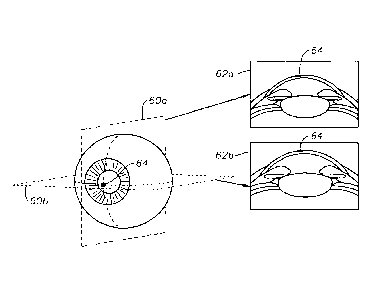
FIGURES 2A through 2C illustrate examples of the operation of an image
capture system according to certain embodiments. FIGURE 2A illustrates an
example
of the edges of planes 50 of an eye that can be imaged by an image capture
system.
FIGURE 28 illustrates an example of a particular plane 52 and an image 54
generated of the plane 52. Image 54 shows cloudiness 56 of the cornea. FIGURE
2C
illustrates an example of images that may be generated by an image capture
system.
The image capture system may generate images 62 (a-b) of planes 60 (a-b) of an
CA 02861139 2014-07-14
WO 2013/107468
PCT/EP2012/000224
- 6 -
eye. For example, image 62a is of plane 60a, and image 62b is of plane 60b.
Images
62 show cloudiness 64 of the cornea.
FIGURES 3A through 4D illustrate examples of directing trial shots at a
patient
cornea according to certain embodiments. In the examples, a patient cornea 150
has
inessential tissue 152, such as a diseased portion that is to be removed and
may be
replaced with a donor cornea. The inessential tissue 152 serves as a trial
portion for
trial shots 154.
FIGURES 3A through 3D illustrate an example of directing a pattern of trial
shots 154a at a patient cornea according to certain embodiments. In the
example,
each trial shot 154a of the pattern has a different laser energy. For example,
a first
trial shot has a first laser energy and a second trial shot has a second laser
energy
different from the first laser energy. In the example, the trial shots 154a of
the
pattern may each be directed to the same corneal depth, that is, the trial
shots 154a
may lay on the same corneal plane.
FIGURES 4A through 4D illustrate another example of directing a pattern of
trial shots 154b at a patient cornea according to certain embodiments. In the
example, each trial shot 154b of the pattern has a different corneal depth
such that
the pattern lies at an angle (greater than zero) to a corneal plane at a
constant
corneal depth. For example, a first trial shot has a first corneal depth and a
second
trial shot has a second corneal depth different from the first corneal depth.
In the
example, the trial shots 154b of the pattern may each have the same laser
energy.
In another example, the energy level of the second trial shot may differ from
the
energy level of the first trial shot to determine the endothelium level with
the
required energy.
FIGURES 5A and 5B illustrate examples of directing trial shots at a donor
cornea according to certain embodiments. In the examples, a donor cornea 160
has
inessential tissue 162, such as an excess portion that is to be removed from
the
portion of the donor cornea 160 to be implanted in a patient. The inessential
tissue
162 serves as a trial portion for trial shots 164.
FIGURE 5A illustrates an example of directing trial shots at a donor cornea in
a
manner similar to that of FIGURES 3A through 3D. In the example, each trial
shot
164a of the pattern has a different laser energy, and may each be directed to
the
same corneal depth.
FIGURE 5B illustrates an example of directing trial shots at a donor cornea in
a
manner similar to that of FIGURES 4A through 4D. In the example, each trial
shot
164b of the pattern has a different corneal depth such that the pattern lies
at an
angle (greater than zero) to a corneal plane of a constant corneal depth. Each
trial
CA 02861139 2014-07-14
WO 2013/107468
PCT/EP2012/000224
- 7 -
shot 164b may have the same laser energy. In another example, the energy level
of
the second trial shot may differ from the energy level of the first trial shot
to
determine the endothelium level with the required energy.
FIGURE 6 illustrates an example of a laser device 15 and a control computer
30 configured to photodisrupt tissue according to certain embodiments. In the
embodiments, the laser device 15 can generate pulsed laser radiation with the
calculated laser energy and ultrashort pulses (such as pico-, femto-, or
attosecond
pulses). The laser device 15 can direct the pulsed laser beam through an outer
portion of an eye to a target portion of the eye to photodisrupt tissue of the
target
portion. The control computer 30 can receive an optical density measurement of
the
outer portion, determine the laser energy according to the optical density
measurement, and instruct the one or more controllable components to direct
the
laser beam with the laser energy through the outer portion to the target
portion.
In certain embodiments, the laser beam may form a corneal element (such as
a corneal flap or corneal cap), which may be removed to allow an excimer laser
to
apply a refractive correction. The corneal element may or may not be replaced
after
the refractive correction. In certain embodiments, the laser beam may form a
lenticule (or lenticle) that may be removed to yield a refractive correction.
In the illustrated example, the computing system 20 includes a control
computer 30 and a memory 28. The memory 28 stores a control program 34. The
laser device 15 includes a laser source 112, a scanner 116, one or more
optical
elements 117, and/or a focusing objective 118 coupled as shown. The laser
device
15 is coupled to a patient adapter 120. The patient adapter 120 includes a
contact
element 124 (which has an abutment face 126 disposed outwardly from a sample)
and a sleeve 128 coupled as shown.
The laser source 112 generates a laser beam 114 with ultrashort pulses. In
this document, an "ultrashort" pulse of light refers to a light pulse that has
a duration
that is less than a nanosecond, such as on the order of a picosecond,
femtosecond,
or attosecond. The focal point of the laser beam 114 may create a laser-
induced
optical breakdown (LIOB) in tissues such as the cornea. The laser beam 114 may
be
precisely focused to allow for precise incisions in the epithelial cell
layers, which may
reduce or avoid unnecessary destruction of other tissue.
Examples of laser source 112 include femtosecond, picosecond, and
attosecond lasers. The laser beam 114 may have any suitable vacuum wavelength,
such as a wavelength in the range of 300 to 1500 nanometers (nm), for example,
a
wavelength in the range of 300 to 650, 650 to 1050, 1050 to 1250, or 1100 to
1500
nm. The laser beam 114 may also have a relatively small focus volume, e.g., 5
CA 02861139 2014-07-14
WO 2013/107468
PCT/EP2012/000224
- 8 -
micrometers (pm) or less in diameter. In certain embodiments, the laser source
112
and/or delivery channel may be in a vacuum or near vacuum.
The scanner 116, optical elements 117, and focusing objective 118 are in the
beam path. The scanner 116 transversely and longitudinally controls the focal
point
of the laser beam 114. "Transverse" refers to a direction at right angles to
the
direction of propagation of the laser beam 114, and "longitudinal" refers to
the
direction of beam propagation. The transverse plane may be designated as the x-
y
plane, and the longitudinal direction may be designated as the z-direction. In
certain
embodiments, the abutment face 126 of the patient interface 120 is on an x-y
plane.
The scanner 116 may transversely direct the laser beam 114 in any suitable
manner. For example, the scanner 116 may include a pair of galvanometrically
actuated scanner mirrors that can be tilted about mutually perpendicular axes.
As
another example, the scanner 116 may include an electro-optical crystal that
can
electro-optically steer the laser beam 114. The scanner 116 may longitudinally
direct
the laser beam 114 in any suitable manner. For example, the scanner 116 may
include a longitudinally adjustable lens, a lens of variable refractive power,
or a
deformable mirror that can control the z-position of the beam focus. The focus
control components of the scanner 116 may be arranged in any suitable manner
along the beam path, e.g., in the same or different modular units.
One (or more) optical elements 117 direct the laser beam 114 towards the
focusing objective 118. An optical element 117 may be any suitable optical
element
that can reflect and/or refract/diffract the laser beam 114. For example, an
optical
element 117 may be an immovable deviating mirror. The focusing objective 118
focuses the laser beam 114 onto the patient adapter 120, and may be separably
coupled to the patient adapter 120. The focusing objective 118 may be any
suitable
optical element, such as an f-theta objective.
Patient adapter 120 interfaces with the cornea of the eye 22. In the example,
the patient adapter 120 has a sleeve 128 coupled to a contact element 124. The
sleeve 128 couples to the focusing objective 118. The contact element 124 is
transparent to the laser beam and has an abutment face 126 that interfaces
with the
cornea and may level a portion of the cornea. In certain embodiments, the
abutment
face 126 is planar and forms a planar area on the cornea. The abutment face
126
may be on an x-y plane, so the planar area is also on an x-y plane. In other
embodiments, the cornea need not have planar area.
The control computer 30 controls controllable components, e.g., the laser
source 112 and scanner 116, in accordance with the control program 34. The
control
program 34 contains computer code that instructs the controllable components
of the
CA 02861139 2014-07-14
WO 2013/107468
PCT/EP2012/000224
- 9 -
laser device 15 to focus the pulsed laser beam with a laser energy calculated
according to optical density of an outer portion of the eye 22.
In certain examples of operation, the scanner 116 may direct the laser beam
114 to form incisions of any suitable geometry. Examples of types of incisions
include
bed incisions and lateral incisions. A bed incision is two-dimensional
incision that is
typically on an x-y plane. The scanner 116 may form a bed incision by focusing
the
laser beam 114 at a constant z-value under the abutment face 126 and moving
the
focus in a pattern in an x-y plane. A lateral incision is an incision that
extends from
under the corneal surface (such as from a bed incision) to the surface. The
scanner
io 116 may form a lateral incision by changing the z-value of the focus of
the laser
beam 114 and optionally changing the x and/or y values.
FIGURE 7 illustrates an example of a method for adjusting laser energy
according to an optical density measurement in certain embodiments. The method
may be performed by a computing system 20. The method begins at step 210,
where the computing system 20 receives an optical density measurement of the
outer portion of an eye 22. In certain embodiments, the outer portion may be
an
outer layer of the cornea. In certain embodiments, the optical density
measurement
may include one or more optical density values for one or more locations of
the outer
portion, where each optical density value indicates the optical density at a
location.
A laser adjustment value is determined according to the optical density
measurement at step 212. In certain embodiments, the laser energy module 38
determines the laser adjustment value. In the embodiments, the laser energy
module 38 may access a data structure (such as table 42) that associates a
number
of optical density values with a number of a laser adjustment values. The
laser
energy module 38 may identify the laser adjustment value for a location
associated
with the optical density value at the location.
Laser energy is determined according to the laser adjustment value at step
214. In certain embodiments, the laser energy module 38 may determine the
laser
energy. In the embodiments, the laser energy module may determine an initial
laser
energy at a location, and then adjust the initial laser energy according to
the laser
adjustment value for the location.
The laser device 15 is instructed to direct the laser beam with the laser
energy
through the outer portion to the target portion at step 216. For example, the
laser
energy module 38 may send instructions to laser device 15 to direct a laser
beam at
a location with the adjusted laser energy determined for the location.
FIGURE 8 illustrates an example of a method for adjusting laser energy
according to trial shots in certain embodiments. The method may be performed
by a
CA 02861139 2014-07-14
WO 2013/107468
PCT/EP2012/000224
- 10 -
computing system 20. The method begins at step 310, where the computing system
20 instructs a laser device to direct trial shots towards a trial portion. In
certain
embodiments, the trial portion may be inessential tissue of a donor or
patient.
Effects of the trial shots are established at step 312. In certain
embodiments,
a microscope 13 may be used to identify a trial shot with a satisfactory
effect. A
satisfactory effect may be one of one or more effects that satisfy one or more
requirements (such as the best effect). For example, a satisfactory effect of
a trial
shot may be creating a cut in the tissue without damaging the tissue.
Laser energy is determined according to the effects at step 314. In certain
embodiments, the laser energy module 38 may determine the laser energy. In the
embodiments, the laser energy module 38 may identify a trial shot with a
satisfactory
effect and determine the laser energy to be that of the identified trial shot.
In certain
embodiments, the laser energy module 38 may be able to interpolate and/ or
extrapolate the laser energy from the measured effects. For example, if one
shot
with a lower laser energy did not create a cut, but the next shot with a
higher laser
energy caused too much damage, a laser energy module between the higher and
lower energies may be used.
The laser device 15 is instructed to direct the laser beam with the laser
energy
to a target portion at step 316. For example, the laser energy module 38 may
send
instructions to laser device 15 to direct a laser beam towards the target
portion with
the laser energy.
A component of the systems and apparatuses disclosed herein may include an
interface, logic, memory, and/or other suitable element, any of which may
include
hardware and/or software. An interface can receive input, send output, process
the
input and/or output, and/or perform other suitable operations. Logic can
perform the
operations of a component, for example, execute instructions to generate
output
from input. Logic may be encoded in memory and may perform operations when
executed by a computer. Logic may be a processor, such as one or more
computers,
one or more microprocessors, one or more applications, and/or other logic. A
memory can store information and may comprise one or more tangible, computer-
readable, and/or computer-executable storage medium. Examples of memory
include
computer memory (for example, Random Access Memory (RAM) or Read Only
Memory (ROM)), mass storage media (for example, a hard disk), removable
storage
media (for example, a Compact Disk (CD) or a Digital Video Disk (DVD)),
database
and/or network storage (for example, a server), and/or other computer-readable
media.
CA 02861139 2014-07-14
WO 2013/107468
PCT/EP2012/000224
- 11 -
In particular embodiments, operations of the embodiments may be performed
by one or more computer readable media encoded with a computer program,
software, computer executable instructions, and/or instructions capable of
being
executed by a computer. In particular embodiments, the operations may be
performed by one or more computer readable media storing, embodied with,
and/or
encoded with a computer program and/or having a stored and/or an encoded
computer program.
Although this disclosure has been described in terms of certain embodiments,
modifications (such as changes, substitutions, additions, omissions, and/or
other
modifications) of the embodiments will be apparent to those skilled in the
art.
Accordingly, modifications may be made to the embodiments without departing
from
the scope of the invention. For example, modifications may be made to the
systems
and apparatuses disclosed herein. The components of the systems and
apparatuses
may be integrated or separated, and the operations of the systems and
apparatuses
may be performed by more, fewer, or other components. As another example,
modifications may be made to the methods disclosed herein. The methods may
include more, fewer, or other steps, and the steps may be performed in any
suitable
order.
Other modifications are possible without departing from the scope of the
invention. For example, the description illustrates embodiments in particular
practical
applications, yet other applications will be apparent to those skilled in the
art. In
addition, future developments will occur in the arts discussed herein, and the
disclosed systems, apparatuses, and methods will be utilized with such future
developments.
The scope of the invention should not be determined with reference to the
description. In accordance with patent statutes, the description explains and
illustrates the principles and modes of operation of the invention using
exemplary
embodiments. The description enables others skilled in the art to utilize the
systems,
apparatuses, and methods in various embodiments and with various
modifications,
but should not be used to determine the scope of the invention.
The scope of the invention should be determined with reference to the claims
and the full scope of equivalents to which the claims are entitled. All claims
terms
should be given their broadest reasonable constructions and their ordinary
meanings
as understood by those skilled in the art, unless an explicit indication to
the contrary
is made herein. For example, use of the singular articles such as "a," "the,"
etc.
should be read to recite one or more of the indicated elements, unless a claim
recites
an explicit limitation to the contrary. As another example, "each" refers to
each
CA 02861139 2014-07-14
WO 2013/107468
PCT/EP2012/000224
- 12 -
member of a set or each member of a subset of a set, where a set may include
zero,
one, or more than one element. In sum, the invention is capable of
modification, and
the scope of the invention should be determined, not with reference to the
description, but with reference to the claims and their full scope of
equivalents.