Note: Descriptions are shown in the official language in which they were submitted.
81781230
CELLULOSE NANOFIBRILS BEARING A UNIFORM COATING OF
MAGNETIC NANOPARTICLES AND METHODS FOR PREPARING SAME
The present invention relates to cellulose nanofibrils decorated with magnetic
nanoparti-
des as well as a method for the preparation thereof and a material comprising
the nanofi-
brils.
BACKGROUND OF THE INVENTION
Magnetic nanoparticles with large surface to bulk ratio is a growing area of
interest. Con-
sidering the potentially large area of application of magnetic nanoparticles,
as filler materi-
als of various polymer materials, it can easily be understood that their
relatively poor rep-
resentation in comparison to micron-sized filler materials in polymers is an
effect of the
difficulties related to the processing of high-surface area nanoparticles. The
explanation
mainly lies in the fact that large surface areas also brings problems in
achieving uniformly
distributed nanoparticle systems due to the favoured particle-particle
interaction in corn-
parison to particle-polymer/liquid interactions. The result is often severe
agglomeration
and aggregates of nanoparticles. The agglomerates in turn affect many
macroscopic prop-
erties, such as mechanical, optical and magnetic etc. since these properties
on a macro-
scopic scale are affected by the degree of close interaction at the nano scale
level. In order
to exploit the effects of nano-sized magnetic nanoparticles employed as
fillers in organic
matrix materials, the control over dispersion is therefore an unavoidable
prerequisite.
Ferrite-loaded membranes of microfibrillated cellulose have been prepared by
mixing
metal ions to a suspension of bacterial cellulose under N2 atmosphere before
precipitation
by NaOH followed by oxidation in atmospheric air. Ferrite particles were
inclined to ag-
gregate into lumps in the fibrillar network Sourty H.; et al., Chem. Mater.
1998, 10 7),
1755-1757). A magnetic paper made of kenaf has been prepared by precipitation
of mag-
netic nanoparticles in a pulp suspension under anaerobic conditions. Chia C.H.
et at., Am.
Appl. Sci., 2006, 3 3), 1750-1754).
Magnetic membranes with improved and controlled properties are of interest for
purifica-
tion/filtration (Dai Q., et al., Chem Soc Rev, 2010, 39, 4057), magneto-
responsive actua-
tors (Hoare, T. et al., Nano Lett, 2009, 9, 3651. Behrens S., Nanoscale, 2011,
3, 877) as
well as for large scale manufacturing of e.g. magneto-acoustic membranes, anti-
1
CA 2861563 2019-09-13
CA 02861563 2014-07-17
WO 2013/119179 PCT/SE2013/050115
counterfeiting papers, radio-frequency materials and flexible data storage.
The magnetic
nanocomposite membranes and films are classically derived from polymers mixed
with
surface modified functional magnetic nanoparticles (Behrens S., Nanoscale,
2011, 3, 877).
However, the dispersion of the high surface area nanoparticles is more
challenging and
nanoparticle agglomerates tend to form easily. Strength and failure properties
are sensitive
to such agglomerates so that the materials become brittle even at moderate
nanoparticle
loadings. The presence of agglomerates also makes it difficult to predict
magnetic compos-
ite properties as related to intrinsic nanoparticle magnetics due to dipolar
interactions (Ols-
son R.T., et al., Polym Eng Sci, 2011, Article in Press). In addition, the
classical prepara-
tion methods (Behrens S., Nanoscale, 2011, 3, 877) are time consuming and
costly since in
most cases they rely on empirical attempts to find particle surface coatings
for improved
dispersions (Balazs AC., et al., Science, 2006, 314, 1107)
Recent progress in the field of bio-nanotechnologi es has shed light on the
possibilities of-
fered by some naturally occurring nano-building blocks (Eichorn S.J., et al. J
Mater Sci,
2010, 45, 1). At the smallest scales of the wood cell wall organization,
cellulose I microfi-
brils (3-5nm wide) aggregate during wood pulping to form nanofibrils with
dimensions in
the range 5-20 nm in width and up to few micrometers in length. These entities
can be re-
leased from the pulp fiber cell wall by mechanical disintegration (A.F.Turbak,
et al., J Appl
Polym Sci, 1983, 37, 815), which is facilitated by an enzymatic or chemical
pre-treatment
of the pulp fibers (M. Henriksson, et al., Eur Polym J, 2007, 43, 3434 and
Saito T. et al.,
Biomacromolecules, 2007, 8, 2485). Due to their intrinsically high strength
and stiffness
(modulus of crystal exceeding 130GPa (Sakurada I. et al., J Polym Sci, 1962,
57, 651)),
long and slender cellulose nanofibrils (NFC) have interesting potential as
nanoreinforce-
ments in various composite materials. Furthermore, strong interfibril
interactions allows
formation of a variety of nanostructures, from dense nanopapersto ultra-light
aerogels and
foams (Henriksson, M. et al., Eur Polym J, 2007, 43, 3434; Paakko, M. et al.,
Soft Matter,
2008,4, 2492; Sehaqui, H. et al., Soft Matter, 2010, 6, 1824; and Svagan, A.J.
et al., J Ma-
ter Chem, 2010, 20, 6646). Here, the fibrillar interactions and the
corresponding network
structure provide favourable mechanical properties. Large-scale availability,
origin from
renewable resources, and low resource cost are advantages of forest-derived
nano-building
blocks.
2
CA 02861563 2014-07-17
WO 2013/119179 PCT/SE2013/050115
Bacterial cellulose nanofibril networks have been used as a template for
precipitation of
magnetic nanoparticles (R.T. Olsson, et al., Nat Nanotechnol, 2010, 5, 584).
The method
allowed to form cellulose-based magnetic aerogels, as well as dense membranes.
A two-
step method for preparing a magnetic nanoparticle cellulose material, wherein
cobalt ferrite
nanoparticles are evenly/finely distributed arranged on the scaffold of fibres
inside the ma-
terial is disclosed in W02008/121069. The disclosed material is in the form of
a hydrogel
or aerogel and the fibres in the material are physically entangled. However,
the methods
are energy consuming due to the freeze-drying steps of the cellulose network
prior to
nanoparticle precipitation. Furthermore, the versatility for nanostructure
formation is re-
stricted by the characteristics of the network synthesized by the bacteria,
which to some
extent predicted the relative density/frequency of the magnetic nanoparticles
as related to
reactive sites for grafting the inorganic nanoparticles
A common problem with previous methods is to achieve reproducibly coated
nanoparti-
cies, making the combined mechanical and magnetic functionality of "classical"
polymer
matrix nanocomposites difficult to achieve. There is a need within the
technical field of
magnetic nanoparticle cellulose material to be able to tailor the magnetic
properties of the
material.
SUMMARY OF THE INVENTION
It is an object of the present invention to provide cellulose nanofibrils
decorated by mag-
netic nanoparticles, wherein the nanoparticles are unifoimly distributed on
the nanofibril.
Another object of the present invention, is to provide a single-step method
for the prepara-
tion of such cellulose nanofibrils. A further object is to provide a magnetic
material com-
prising the cellulose nanofibrils that is decorated by magnetic magnetic
nanoparticles in a
uniform distribution on the nanofibril.
It has surprisingly been found that cellulose nanofibrils, wherein each
nanofibril is deco-
rated with magnetic nanoparticles that are uniformly distributed on the
nanofibril, can be
obtained by a method comprising the steps of:
a) diluting cellulose nanofibrils in a solvent to obtain a suspension,
3
81781230
b) adding at least one metal salt to the suspension obtained in step (a) in
any atmosphere
that allows oxidation to form metal ion complexes that are physically attached
to the
nanofibrils,
c) precipitating the metal ion complexes by forced hydrolysis to form magnetic
nanoparticles on the cellulosic nanofibrils in the suspension,
d) allowing the suspension in step (c) to react until the metal ion complexes
have been
converted to the magnetic phase.
The method of the present invention is a single-step process for the
preparation of cellulose
nanofibrils decorated by magnetic nanoparticles. This method is inexpensive
and rapid compared
to previously known methods for preparing cellulose material loaded with
nanoparticles.
A further object of the present invention is to provide nanocomposite
membranes composed of an
intermingled network of cellulose nanofibrils decorated with magnetic
nanoparticles. The
decorated cellulose nanofibrils of the present invention can be formed into
large and strong
cellulose nanocomposite membranes by vacuum filtration. Nanofibril
entanglements and
interactions result in high strength and toughness of the nanocomposite
membranes formed.
In one particular embodiment, the present invention provides an individual
cellulose nanofibril
having magnetic nanoparticles attached to the nanofibril along its surface,
wherein the magnetic
nanoparticles are made of transition metal ions or an oxide thereof and are in
the size region
1-200 nm, and wherein the amount of magnetic nanoparticles adhering together
or lying very
close together at < 5nm particle to particle inter-distance, in a collection
composed of 20 or more
magnetic nanoparticles, is less than 30%.
In another particular embodiment, the present invention provides a method for
forming individual
cellulose nanofibrils having magnetic nanoparticles attached to the
nanofibrils along its surface
comprising the steps of:
a) diluting cellulose nanofibrils in a solvent to obtain a
suspension of individual
nanofibrils,
4
CA 2861563 2019-09-13
81781230
b) adding at least one metal salt to the suspension obtained in step (a) in an
oxidizing
atmosphere to form metal ion complexes physically attached to the nanofibrils,
c) precipitating the metal ion complexes by forced hydrolysis by addition of
an
alkaline solution to form magnetic nanoparticles on the cellulosic nanofibrils
in the
suspension,
d) allowing the suspension in step c) to react until the metal ion complexes
have been
converted to magnetic nanoparticles,
to obtain individual cellulose nanofibrils decorated with magnetic
nanoparticles, wherein
the amount of magnetic nanoparticles adhering together or lying very close
together at
< 5nm particle to particle inter-distance, in a collection corn-posed of 20 or
more magnetic
nanoparticles, is less than 30%.
The presented platform for direct inorganic modification of cellulose
nanofibrils allows for
uniform distribution of nanoparticles in fiber composites in absence of
surfactants or particle
surface modifications.
BRIEF DESCRIPTION OF THE DRAWINGS
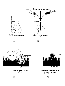
Figure 1 illustrates the method for in-situ preparation of cobalt ferrite
nanoparticles on cellulose
nanofibrils.
Figure 2 shows SEM micrographs of functionalized nanofibrils (30 wt% cobalt
ferrite
nanoparticles) deposited from a dilute aqueous suspension (0.001%), showing
different
morphologies encountered.
4a
CA 2861563 2019-09-13
CA 02861563 2014-07-17
WO 2013/119179 PCT/SE2013/050115
Figure 3 shows number average size distributions of magnetic nanoparticle
(cobalt ferrite)
for in-situ preparation according to the present invention compared to a two-
step process.
Three material compositions are presented in each graph. The percentages refer
to various
average weight fractions of inorganic particles (10 wt%, 30 wt%, 60 wt%) on
the cellulose
nanofibrils.
Figure 4 presents FE-SEM micrographs of the hybrid composite membranes at high
mag-
nification showing different nanoparticle shape after in-situ (a) and separate
(b) precipita-
tion
Figure 5 shows the influence of the fibrils presence on the dispersion of the
magnetic
nanoparticles in the membranes prepared by a method according to the present
invention
(in-situ prepcipitation) (a) and by a metod wherein a solvent with
precipitated metal salts
converted to magnetic phase has been mixed with a suspension of cellulose
nanofibril
(separate precipitation,) (b).
Figure 6 shows XRD spectra of cellulose nanofibril-based hybrid membranes with
30wt%
of cobalt-ferrite magnetic nanoparticles, prepared through a single-step, i.e.
in-situ prep-
cipitation, (a) and two-step, i.e. separate precipitation, (b) process. Peaks
are assigned to
the corresponding constituent as indicated on the graphs.
Figure 7 shows thermograms for samples with different amount of magnetic
nanoparticles
and prepared through the single-step i.e. in-situ prepcipitation, (up) or two-
step, i.e. sepa-
rate precipitation, (down) preparations. TGA was run in the presence of O.
Figure 8 shows representative stress-strain curves for the cellulose
nanofibril-based hybrid
membranes with varying content of magnetic cobalt-ferrite nanoparticles,
prepared through
single-step, i.e. in-situ prepcipitation, (up) and two-step, i.e. separate
precipitation, (down)
process.
Figure 9 shows representative stress-strain curves for membranes obtained
through the sin-
gle-step process, tested at two different levels of relative humidity.
5
CA 02861563 2014-07-17
WO 2013/119179 PCT/SE2013/050115
Figure 10 shows magnetization curves for the cellulose nanofibril-based hybrid
membranes
with varying content of magnetic nanoparticles, prepared through single-step
(up) and two-
step (down) process.
Figure 11 shows magnetization curves for hard(CoFe204) ¨ soft(MnFe204)
composites at
various compositions (a) and comparison of experimental to calculated data for
the 50-50
composite (b). All sample contained 70 wt% cellulose nanofibril ¨ all curves
are normal-
ized to the inorganic mass.
DETAILED DESCRIPTION OF THE INVENTION
A first aspect of the invention is a cellulose nanofibril decorated with
magnetic nanoparti-
cies, wherein the magnetic nanoparticles are uniformly distributed on the
fibril.
Another aspect of the present invention is a magnetic material comprising
cellulose nano-
fibrils decorated with magnetic nanoparticles, wherein the magnetic
nanoparticles are uni-
formly distributed on the fibril.
A distinct advantage with the cellulose nanofibrils of the present invention
is that the mag-
netic properties of the magnetic material comprising cellulose nanofibrils can
be tailored
by mixing cellulose nanofibrils decorated with hard magnetic nanoparticles
with cellulose
nanofibrils decorated with soft magnetic nanoparticles.
For purposes of this invention, the term "cellulose material" is intended to
encompass na-
tive cellulose. Cellulose is found in plants, a few animals and a few bacteria
as microfibrils
2-20 nm in diameter depending on organism source. Cellulose material exists in
nature as
reinforcing phase in plant cell walls, and in other organisms such as bacteria
or tunicate
animals. Cellulose is found in cotton, paper, wood pulp etc. Several different
crystalline
structures of cellulose are known, natural cellulose is denoted cellulose I,
with structures I,
and I. Cellulose produced by bacteria and algae is enriched in I while
cellulose of higher
plants consists mainly of I. Cellulose in regenerated cellulose fibers, such
as rayon and
cellophane, is denoted cellulose II.
6
CA 02861563 2014-07-17
WO 2013/119179 PCT/SE2013/050115
The term "microfibrillated cellulose", abbreviatedNIFC, is used for nanosized
cellulose
fibrils disintegrated from a cellulose material. The starting form of
microfibrillated cellu-
lose (MFC) is typically as a suspension of MFC in liquid, where the solid MFC
content is
less than 10% by volume. It is found in the form of crystalline microfibrils
consisting of
polyglucan molecules in extended chain conformation. The length can be several
microme-
ters and therefore the aspect ratio (ratio of length to diameter) is very
large.
The term "cellulose nanofibrils", also called "nanofibrillated cellulose" and
abbreviated
NFC, is used for fibrillar material extracted from pulp that prior to
mechanical disintegra-
tion, such as in a microfluidizer or homogenizer, has been subjected to
chemical and/or en-
zymatic pre-treatments.
The term "cellulose nanofibril" is intended to encompass a particle with the
smallest di-
mension in the range 5-100 nm. The cellulose nanofibrils are fiber-shaped with
one dimen-
sion (diameter/width/lateral dimension) smaller than the other
(length/longitudinal dimen-
sion). Typically, the aspect ratio (length/width) is above 10. The cellulose
structure in the
particle is cellulose I or cellulose II. The surface of the cellulose
nanofibril may be chemi-
cally modified (ie acetylated, carboxylated, silanised, or modified by other
functional
groups) whereas the interior of the cellulose nanofibril is cellulose I or
celllulose II.
The term "bacterial cellulose" is intended to encompass any type of cellulose
produced via
fermentation or synthesised of a bacteria of the genus, Alacaligenes,
Pseudomonas, Ace-
tobacter, such as Acetobacter xyhnum ( also called Gluconacetobacter
xylinurn), Rhizo-
bium, Agrobacterium, Sarcina, Enterobacter, Achromobacter, and Azotobacter and
in-
eludes materials referred popularly as microfibrillated bacterial cellulose,
reticulated bacte-
rial cellulose, microbial cellulose and the like. In addition prokaryotic
organisms such as
the prokaryotic cyanophycean alga Nostoc are encompassed. Further, the term
"bacterial
cellulose" as used in this invention refers to a product essentially free of
residual bacteria
cells made under agitated culture conditions by a bacterium of the genus
Acetobacter. Bac-
terial celluloses are normally available in a gel produced by the bacteria.
7
CA 02861563 2014-07-17
WO 2013/119179 PCT/SE2013/050115
For purposes of this invention, the term "nanofibrils decorated with magnetic
nanoparti-
cies" is intended to encompass nanofibrils having magnetic nanoparticles
attached to the
nanofibril along its surface.
The term "magnetic cellulose" is intended to encompass a material, referred to
as a mate-
rial consisting of both an inorganic particles fraction/phase with magnetic
properties and an
organic carbon-based phase/fraction. The magnetic cellulose can be
ferromagnetic, fern-
magnetic or superparamagnetic.
The term "magnetic nanoparticles" is intended to encompass nanoparticles made
of transi-
tion metal ions and their oxides, such as ions and oxides selected from Co,
Fe, Ni, Fe2O3,
Fe304, CoFe204, NiFe204, CuFe204, MnFe204, MgFe204, and mixtures of different
transi-
tion metal ions in the same lattice The magnetic nanoparticles may be selected
from fer-
romagnetic, ferrimagnetic and superparamagnetic nanoparticles.
The term "mild oxidation agent" is intended to encompass any type of oxidating
media
which is capable of oxidizing ferrous ions to ferric ions, or any of the
transition metals
mentioned herein to a higher oxidation state, to a sufficient extent that
magnetic particles
can be obtained, for example ferrites. Examples of mild oxidation agents are
selected from
the group comprising metal salts of chlorates, perchlorates, bromates,
nitrates and nitrites,
as well as nitrous acid, such as potassium nitrate (KNO3), potassium chlorate
(KC103), so-
dium chlorate (NaC103), potassium bromate (KBr03), potassium perchlorate
(KC104), are
ammonium nitrate (NH4NO3).
The term "transition metal ions" is intended to encompass metal ions such as
all elements
in the periodic table that can be used to obtain ironoxide based magnetic
nanoparticles.
The term "coordination compounds and d-block elements" is intended to
encompass metal
compounds and/or elements such as manganese, iron, cobalt, zinc etc. d-block
elements are
also referred to as transition metals, the d-block elements in period 4 are
Sc, Ti, V, Cr, Mn,
Fe, Co, Ni, Cu, Zn.
8
CA 02861563 2014-07-17
WO 2013/119179 PCT/SE2013/050115
The term "alkaline solution" is intended to encompass NaOH, KOH, Li0H, NH, and
the
like.
The term "metal ion complex" is intended to encompass coordination complexes
that are
created upon dissolving metal salts in a liquid phase, for example metal oxide
hydroxide
complexes or any metal hydroxide or oxide formed, or combinations thereof
The term "freeze drying" is intended to encompass a method to sublime solid
water (ice) to
gas phase.
The term "metal salt" is intended to encompass salts of metal ions such as
Co2+, Fe2+, Fe3+,
Mn 2+, CL12+, Ni and Mg and the like, in the form of salts, such as FeSO4,
Fe2(SO4)3,
FeCl2, FeCl3, Fe(NO3)2, Fe(NO3)3, Fe(C2H302)2, Fe(C2H302)3, FePO4, MnSO4,
MnC12,
114n(NO3)2, Mn(C2H302)2, CoSO4.,, CoC12, Co(NO3)2, Co(C2H302)2, ZnC12, CuSO4,
NiC12, and their corresponding hydrates, such as CoC12 = 6H20, and FeSO4 =
7H20.
The term "magnetic nanoparticle cellulose material" is intended to encompass a
material
comprising an interconnected fibre network,.
As used herein, the "weight percentage" (wt%) of magnetic or inorganic
nanoparticles, re-
fers to the average weight fraction of inorganic particles on the modified
cellulose nanofi-
brils, of the total weight of the modified cellulose nanofibrils.
The term "magnetic nanoparticles physically attached on the cellulose
material" is in-
tended to encompass magnetic nanoparticles in the size region 1-200 nm.
The term "uniformly distributed" is intended to encompass that the
nanoparticles are
mostly separated, i.e. no agglomerate formation.
The term "agglomerate" is defined herein as a collection of nanoparticles
adhering together
or laying very close together, i.e. < 5nm particle to particle inter-distance
and the collection
of nanoparticles is composed of 20 or more nanoparticles An agglomerate
material (non-
9
CA 02861563 2014-07-17
WO 2013/119179 PCT/SE2013/050115
uniform) would have more than 30% of the nanoparticles lying in above entities
of 20 or
more nanoparticles.
The bacterial cellulose utilized herein may be of any type associated with the
fermentation
product of Acetobacter genus microorganisms, and was previously available from
CPKelco U.S. under the tradename CELLULONv.
In one embodiment of the present invention, the magnetic nanoparticles are
made of transi-
tion metal compounds or an oxide thereof, such as a compound selected from of
Co, Fe,
Ni, Fe2O3, Fe304, CoFe204, NiFe204, CuFe204, MnFe204, and MgFe204. The
magnetic
nanoparticles can be ferromagnetic, ferrimagnetic or superparamagnetic.
Another aspect of the invention, is a method for forming cellulose nanofibrils
decorated
with magnetic nanoparticles that are uniformly distributed on the cellulose
nanofibrils
comprising the steps of:
a) diluting cellulose nanofibrils in a solvent to obtain a suspension,
b) adding at least one metal salt to the suspension obtained in step a) in any
atmos-
phere that can allow oxidation to form metal ion complexes physically attached
to the
nanofibrils,
c) precipitating the metal ion complexes by forced hydrolysis to form magnetic
nanoparticles on the cellulosic nanofibrils in the suspension,
d) allowing the suspension in step c) to react until the metal ion complexes
have been
converted to the magnetic phase.
The method according to the present invention is illustrated in figure 1.
According to the
method of the present invention the magnetic nanoparticles are prepared in
situ with the
suspension of cellulose nanofibrils and are allowed to attach to said
nanofibrils in said sus-
pension.
Preferably, water is used as the solvent for dilution in step a). The dilution
of cellulose
nanofibers may be made in any atmosphere that can allow oxidation, such as
atmospheric
air.
CA 02861563 2014-07-17
WO 2013/119179 PCT/SE2013/050115
The at least one metal salt added to the suspension in step b) may be added in
the form of a
solid metal salt or as a solution of metal ions. When the at least one metal
salt is added in
as a solution of metal ions, said solution should be prepared immediately
before it is added
to the suspension. Preferably, the at least one metal salt is added to the
suspension in step
b) in the form of a solid metal salt.
Preferably, the at least one metal salt added to the suspension in step b) is
a combination of
at least two metal salts selected from salts of the divalent or trivalent
atoms from the d-
block elements in the periodic table, such as Co2+, Fe2+, mn2+, Ni2+, zn2+,
cu2+
and Fe', as
well as hydrates of such salts. Specific examples of suitable metal salts are
FeSO4,
Fe2(504)3, FeCl2, FeCl3, Fe(NO3)2, Fe(NO3)3, Fe(C2H302)2, Fe(C2H302)3, FePO4,
MnSO4,
MnC17, Mn(NO3)2, Mn(C2H302)2, CoSO4,, CoC1/, Co(NO3)2, Co(C2H302)2, NiC12,
ZnC12,
CuSO4 and their hydrates. Preferably, the metal salts are selected from salts
of Co2+, Fe2+,
Mn2+, and Fe3+, as well as hydrates of such salts. More preferably, the metal
salts are se-
lected from FeSO4, CoC12, MnC12, FeC13 and their hydrates.
The combination of metal salts may be added to the suspension in step b) in
the form of
solid metal salts or as a solution of metal ions. When the combination of
metal salts is
added as a solution of metal ions, said solution should be prepared
immediately before it is
added to the suspension. Preferably, the metal salts are added to the
suspension in step b)
in the form of solid metal salts.
Preferably, the at least one metal salt is added to the suspension of
cellulose nanofibrils
under high-shear mixing in step b). The at least one metal salt added to the
suspension in
step b) may be mixed at room temperature with the suspended cellulose
nanofibrils.
In the method according to the present invention step b) is performed in any
atmosphere
that can allow oxidation, such as atmospheric air. The oxidating atmosphere
oxidises the
metal ions in the aqueous suspension to metal ion complex, such as metal oxide-
hydroxide
complexes, metal ion hydroxide complexes or metal ion oxide complexes, that
attach to the
cellulose nanofibrils in the suspension. The mechanisms for the interaction
between the
metal species and the cellulose nanofibrils are understood to rely on the
interaction be-
tween the formed metal ion complexes and the hydroxyl functional groups on the
cel-
11
CA 02861563 2014-07-17
WO 2013/119179
PCT/SE2013/050115
lulose nanofibrils. The metal ion complexes that are attached to the cellulose
nanofibrils
act as precursors and serve as nucleation points for the precipitation of the
magnetic
nanoparticles. Since the metal ion complexes are firmly attached to the
nanofibrils that are
dispersed in the suspension prior to the formation of the magnetic
nanoparticles, associa-
tions and agglomerations of the precipitated magnetic nanoparticles are
prevented.
Preferably, the metal ion complexes formed in the suspension of cellulose
nanofibrils in
step b) are selected from the coordination compounds including divalent or
trivalent atoms
from the d-block elements in the periodic table, such as Co2+, Fe2+, Mn2+, Fe
3+ and their
metal ion oxide-hydroxide complexes, metal ion hydroxide complexes or metal
ion oxide
complexes. The concentration range could be between 0.005 molar - saturated
solution.
The method of the present invention results in uniform distribution of the
magnetic
nanoparticles even at very high inorganic nanoparticle contents, such as at a
content of
more than 80 wt% inorganic nanoparticles.
A further advantage of grafting of the nanoparticles onto the individual
nanofibrils is that
the modified suspension can be diluted to various concentrations and the
average distance
between the nanofibrils could thus be varied during conversion of the
inorganic phase into
solid particles. Further, less tedious cleaning procedures from remaining
counter ions, i.e.
post particle synthesis is required.
Another advantage with the method according to the present invention is that
it permits
complete condensation of the inorganic phase onto the cellulose nanofibrils so
that no par-
tide sediment separates from the suspension of nanofibrils even after long
periods of time,
such as after 2 months; or after exposure to strong magnetic fields, such as
when a 20 cm'
¨ 1.2 T magnet is placed under the suspensions; or ultrasonication, for
example at an en-
ergy of 300 W during 2 min.
A yet further advantage with the cellulose nanofibril decorated with magnetic
nanoparti-
cies according to the present invention is that the magnetic nanoparticles are
strongly at-
tached to the nanofibrils. This has the effect that a material made from the
cellulose nano-
12
CA 02861563 2014-07-17
WO 2013/119179 PCT/SE2013/050115
fibrils according to the present invention is more resistant to fragmentation.
The morphol-
ogy of magnetic nanoparticles attached to the nanofibril is shown in Figure 2.
Preferably, the suspension of nanofibrils with metal ion complexes obtained in
step b) is
heated before the forced hydrolysis in step c). Preferably the suspension of
nanofibrils with
attached metal ion complexes obtained in step b) is heated to at least 70 C,
more preferably
to at least 80 C, even more preferably to at least 90 C. Typically, the
heating rate is any
heating rate between about 0 and about 10 C/min, for example about 2 C/min.
Heating
the suspension in step b) promotes the attachment of the complexes to the
nanofibrils.
Preferably, the forced hydrolysis in step c) is performed by the addition of
an alkaline solu-
tion, such as a solution with a pH >7, or >8, or >9, or >10, or >11, or >12,
or >13, or =14.
Preferably, the alkaline solution in step (c) is chosen from an ammnoium
solution, a solu-
tion of an alkali metal hydroxide, or the like, such as NH3, NaOH, KOH and
Li0H, or a
mixture thereof, providing a pH above 7.
Preferably, the alkaline solution in step c) comprises a dissolved mild
oxidation agent. The
oxidation agent oxidises the metal ions to their preferred state in magnetic
nanoparticles.
Preferably, the mild oxidation agent is selected from the group comprising
metal salts of
nitrates, nitrites, chlorates, perchlorates and bromates, as well as nitrous
acid. More pref-
erably, the mild oxidation agent is selected from metal salts of nitrates and
nitrites. Even
more preferably the mild oxidation agent is KNO3. Preferably, the alkaline
solution in step
c) comprising a dissolved mild oxidation agent has an intitial pH of above 7.
In a specific
embodiment the alkaline solution in step (c) comprises NaOH and KNO3.
The alkaline solution in step (c) is preferably heated before addition to the
suspension.
More preferably, the alkaline solution in step c) is heated to above 50 C, at
latm before
addition to the suspension. If ammonium is used as the alkaline solvent, the
solvent may
have ambient temperature.
The magnetic nanoparticles can be referred to as super-paramagnetic,
paramagnetic, fern-
magnetic or or ferro-magnetic and thus showing such properties.
13
CA 02861563 2014-07-17
WO 2013/119179 PCT/SE2013/050115
The method wherein step (d) proceeds until the agglomerate free and evenly
distributed
ion- complexes that are physically attached on the cellulose nanofibrils are
converted to
magnetic nanoparticles.
In any embodiment of the present invention a polymer may be added after step
d).
In one embodiment, the cellulose nanofibrils of the present invention could be
made from a
cellulose material chosen from a plant, a tree, pulp or cotton. Preferably,
the cellulose nan-
()fibrils are obtained from wood pulp. For example the cellulose nanofibrils
of the present
invention could be obtained by chemical and/or enzymatic pre-treatment of the
wood pulp
followed by mechanical treatment. Preferably, the cellulose nanofibrils of the
present in-
vention are obtained by enzymatic pre-treatment of the wood pulp followed by
mechanical
treatment, such as in a microfluidizer.
With the method of the present invention, each individual cellulose nanofibril
is decorated
with nanoparticles that are uniformly distributed over the nanofibril. Since
the nanoparti-
cies are uniformly distributed along each nanofibril, it is possible to form a
nanocomposite
with highly uniform distribution of the nanoparticles throughout a nanofibril
network. It
could be concluded that an advantage with the method according to the present
invention is
that it produces extremely evenly distributed and physically attached
nanoparticles in a
lightweight cellulosic nanofibril network.
The decorated cellulose nanofibrils of the present invention can be present in
a liquid sus-
pension. In a liquid suspension of cellulose nanofibrils, the cellulose
nanofibrils are well
dispersed in the liquid. This liquid suspension is in liquid form with a
measurable viscos-
ity. The cellulose nanofibrils are not strongly attached to each other. If the
suspension is
diluted, the average distance between the cellulose nanofibrils is increasing.
This is in con-
trast to a hydrogel of cellulose nanofibrils since the strong interaction
between the cellu-
lose nanofibrils in a hydrogel prevents increased average interparticle
distance.
An advantage with the cellulose material prepared from the cellulose
nanofibrils of the
present invention, is that the work to fracture is several times higher than
for nanocompo-
14
CA 02861563 2014-07-17
WO 2013/119179 PCT/SE2013/050115
sites from most other classical engineering polymers. The mechanical
properties of the cel-
lulose material may be varied by controlling the cellulose nanofibril
interaction by water
molecules.
In a network made up of cellulose nanofibrils the nanoparticles are disrupting
the network,
thus reducing interfibril interactions and introducing porosity. The different
mechanical
behaviour between a material made from fibrils prepared according to the one-
step method
of the present invention and a material obtained by the previously known two-
step prepara-
tion method depends on the different micro-structures. In a material made by
nanofibrils
obtained in a two-step method the magnetic nanoparticles are precipitated
separately and
then only mixed with the nanofibrils, resulting in a formation of relatively
large aggregated
regions of sizes up to a few micrometers, of nanoparticles located in pockets
between con-
densed bundles nanofibrils that may allow for sliding between aggregated,
strong, continu-
ous cellulose nanofibril sheets Efficient stress transfer would then be
enhanced along the
interdividing walls between the pockets, making the material less resistant to
rupture.
The dispersion of single domain magnetic nanoparticles can be controlled in
the method of
the present invention by introducing cellulose nanofibrils prior to
precipitation and forma-
tion of nanoparticles. The average distance between the nanofibrils in the
suspensions (re-
gardless of the concentration of metal salts) is on the order of 200 nm, which
is sufficient
to allow for directed condensation of the metal ion onto the surfaces of the
fibril. The na-
noparticles are evenly distributed along the fibril surfaces, as can be seen
from figure 2.
The uniform distribution of particles can be traced back to the relatively
uniform condensa-
tion of metal ion complexes along the cellulose nanofibrils before the
conversion of the
metal ions into its ferromagnetic phase.
The method according to the present invention provides for a new type of
nanoscale build-
ing block that results in the form of individual cellulose nanofibrils
decorated by inorganic
nanoparticles. The method allows complete metal ion condensation of the
inorganic pre-
cursors as magnetic spinel crystals on the fibril.
Crystal growth is influenced by the presence of cellulose nanofibrils in
suspension, leading
to formation of smaller nanoparticles Nanoparticles prepared in presence of
cellulose nan-
CA 02861563 2014-07-17
WO 2013/119179 PCT/SE2013/050115
ofibrils as in the method according to the present invention have
significantly smaller aver-
age sizes as well as more narrow size profiles than particles obtained during
synthesis in
absence of fibrils using the same metal ion concentrations as in a two-step
process, where-
in metal salts are precipitated and converted to magnetic phase before
addition to a suspen-
sion of cellulose nanofibrils, as can be seen in Figure 3. Notably, higher
weight fraction of
precipitated cobalt ferrite also results in larger average particle size, as
can also be seen in
Figure 3.
The size of the magnetic nanoparticles decorating the cellulose nanofibrils of
the present
invention is from about 2 to about 100 nm, or about 2-50 nm, or about 20-40
nm, or about
40-80 nm, measured as the number average diameter.
The concentration of the metal ion solution influences the size of the
nanoparticles ob-
tained by the method. Increasing the concentration of the metal ion solution
from 3 to 45
mM, corresponding to about 10 to about 60 wt%, leads to larger average
particle size and
broader size distributions. Typically, at a metal ion concentration of 12 mM,
the precipi-
tated nanoparticles are of the size 15-20 nm, measured as the number average
diameter. At
a metal ion concentration of 45 mM, the size of the the precipitated
nanoparticles are of the
size 40-80 nm, measured as the number average diameter. Also smaller separated
inorganic
particles with a size of approximately 2-3 nm may simultaneously be present.
Nanoparticles precipitated on the cellulose nanofibrils according to the
present invention
shows a more narrow particle size disitrbution than nanoparticles that are
precipitated be-
fore they are mixed with cellulose fibrils. Further, nanoparticles
precipitated on the cellu-
lose nanofibrils presents a different, more spherical character than the
predominantly cubic
shaped particles obtained for precipitation of nanoparticles in absence of
cellulose fibrils.
See Figure 4 for an example.
The nanoparticle-decorated nanofibrils of the present invention offer several
advantages
over an approach where preformed nanoparticles are simply mixed and dried.
In the cellulose nanofibrils of the present invention the nanoparticles are
evenly distributed
over the nanofibrils, functionalizing them and offer a wide range of
possibilities for further
processing including papermaking processes.
16
CA 02861563 2014-07-17
WO 2013/119179 PCT/SE2013/050115
In one embodiment of the present invention the magnetic nanoparticles are
distributed
along the nanofibril at a particle to particle inter-distance of at about the
same length as the
corresponding particle diameter, and the magnetic nanoparticles are uniformly
distributed
on to the cellulose nanofibril.
In a further embodiment of the present invention the magnetic nanoparticles
are distributed
along the nanofibril at a distance of about 20 nm particle to particle inter-
distance, and the
magnetic nanoparticles are uniformly distributed on to the cellulose
nanofibril.
In a further embodiment of the present invention the magnetic nanoparticles
are distributed
along the nanofibril at a distance, wherin the collection of magnetic
nanoparticles adhering
together or lying very close together, i.e. < 5nm particle to particle inter-
distance, is com-
posed of less than 20 magnetic nanoparticles. Preferably the amount of
magnetic nanopar-
tides adhering together or lying very close together, i.e. < 5nm particle to
particle inter-
distance, in a collection composed of 20 or more magnetic nanoparticles, is
less than 30%.
The inherent properties of cellulose nanofibrils include strong interfibril
interactions, net-
work formation, high aspect ratio and high tensile strength combined with
flexibility in
bending. These features provide high strength and toughness to decorated
nanofibril mem-
branes of high inorganic content, not achievable in classical polymer matrix
nanocompo-
sites.
The cellulose nanofibrils decorated with magnetic nanoparticles are stabilized
in aqueous
suspension, which can be further diluted or alternatively form a gel at lower
water con-
tents.
Water molecules indeed act as plasticizer in the cellulose nanofibril network
by influencing
nanofibril properties and reducing nanofibril interactions. High strength is
nevertheless
preserved due to good stress transfer between the long and slender physically
entangled
nanofibrils. It is possible to vary the mechanical properties by controlling
the cellulose
nanofibril interaction.
17
CA 02861563 2014-07-17
WO 2013/119179 PCT/SE2013/050115
The method for the preparation of the cellulose nanofibrils of the present
invention is envi-
ronmentally benign in that the chemistry is water based. The method is also
unique in its
single¨step preparation characteristics, and is beneficial from the facile up-
scaling poten-
tial and its inexpensive characteristics. Finally, the method and the
cellulose nanofibrils
disclosed herein can be extended to other systems with transition metal oxide
nanoparti-
cies, providing potential for a wide range of extended properties.
The magnetic nanoparticle cellulose nanofibrils can be characterised as the
collection of
nanoparticles adhering together or laying very close together, i.e. < 5nm
particle to particle
inter-distance and an entity is composed of less than 20 nanoparticles.
The magnetic nanoparticle cellulose nanofibrils of the present invention, can
further be
characterised in that the nanofibre diameter is in the range of 1-100 nm,
typically in the
range of 4-20 nm.
The magnetic nanoparticle cellulose nanofibrils can further be characterised
in that the
weight fraction of magnetic nanoparticles on the final magnetic cellulose
nanofibrils is in
the range of from about 1 to about 90 wt%, preferably from about 10 to about
90 wt?/o,
more preferably from about 10 to about 60 wt%.
The method wherein the stoichiometric relation between the metal ion complexes
are in the
rage of 1:1,5 to 1:2,5.
An advantage with the nanoparticle decorated cellulose nanofibrils of the
present invention
is that it provides a magnetic nanoparticle cellulose material that is free
from agglomerates,
wherein the magnetic nanoparticles are extremly uniformly distributed
throughout the
complete material compared with previously known magnetic materials. Further,
the mag-
netic properties of the material can be tailored for each intended use.
Nanocomposites based on magnetic nanoparticles are considered to be among the
most dif-
ficult to produce due to the addition of magnetoelastic interactions such as
exchange iso-
tropic and anisotropic), super exchange, dipole-dipole interactions in
addition to the
chemical interactions such as van der Waals attractions. Eliminating these
forces, which
18
CA 02861563 2014-07-17
WO 2013/119179 PCT/SE2013/050115
cause interactions in agglomerates, results in composites behaving
significantly different
from ferromagnetic composites based on agglomerated nanoparticles. A composite
based
on non-agglomerated nanoparticles have properties such as reflecting the
individual mag-
netic nanoparticles with single domain character.
The direct inorganic modification of cellulose nanofibrils provided for in the
method ac-
cording to the present invention enables a uniform distribution of
nanoparticles in fiber
composites in absence of surfactants or particle surface modifications.
An advantageous effect of the present invention is that cellulose nanofibrils
decorated with
hard magnetic nanoparticles can be mixed with nanofibrils decorated with soft
magnetic
nanoparticles in order to tailor the magnetic properties of the membranes,
which is demon-
strated in Figure 11. In this way the properties of the resulting material can
be cusomized
for each intended purpose. A wide range of magnetic materials based on various
nanostnic-
tures can be envisioned, with the possibility to tune functional and
structural properties.
It is possible to easily prepare magnetic nanocomposite membranes with desired
magnetic
properties, by both acting on the precipitation parameters (Figure 10) and/or
mixing sus-
pensions of various magnetic properties to yield the desired characteristics
(Figure 11).
The magnetization curves correlate with nanoparticle size distributions.
In one aspect the present invention relates to a magnetic suspension
comprising a cellulose
nanofibril decorated with magnetic nanoparticles that are uniformly
distributed along the
nanofibril.
Another aspect of the present invention is a nanocomposite comprising a
magnetic material
according to the present invention. The present invention also relates to a
magnetic mem-
brane, for example a loud-speaker membrane, comprising such nanocomposite.
A further aspect of the present invention is the use of the cellulose
nanofibril according to
the first aspect in a nanocomposite, preferably in a magnetic membrane.
19
CA 02861563 2014-07-17
WO 2013/119179 PCT/SE2013/050115
The magnetic material comprising cellulose nanofibrils decorated with magnetic
nanopar-
tides can be used within the acoustical industry, for example in a loud-
speaker membrane;
magnetic filtration systems; chemical analysis methods; separation methods,
etc.
In traditional loudspeaker the voice coil is bonded to a geometrically shaped
acoustic
membrane, which is suspended in a magnetic field of a bulky permanent magnet.
With a
magnetic membrane according to the present invention the external magnet would
not be
necessary since the magnet instead may constitute an integral part of the
acoustically active
membrane, whereas the coil carrying the signal current and driving the
membrane could be
kept stationary to avoid any moving electrical parts. Thus, the combination of
magnetic
and mechanical functionality of a magnetic membrane according to the present
invention
enables the construction of super-thin loudspeakers. The construction is
possible due to the
advantageous mechanical properties of the magnetic membrane according to the
present
invention, such as high stiffness and strength, in combination with its
ferromagnetic cha-
racteristics from the magnetic nanofibrils.
Applications for the decorated nanofibrils with high surface area and
stability in aqueous
suspension of the present invention are in water purification, catalysis or
biomedical appli-
cations.
Thus one aspect of the present invention is the use of the magnetic material
comprising
cellulose nanofibrils decorated with magnetic nanoparticles in superfine
magnetic fil-
ters/sieves, magnetic filtration set-ups activated by external field,
catalytic support struc-
tures high-sensitivity magnetic membranes, magnetic films with
uniformly/evenly distrib-
uted nanoparticles, microwave absorbers, magnetic foams based on
nanoparticles, support
structure for ferro-fluid based dampeners and template structures for
fabrication of nano-
composites characterized by evenly distributed nanoparticles, i.e. sensitive
electromagnetic
switches, generators, magnetic actuators, magnetic storage media, etc.
The following examples are intended only to further illustrate the invention
and are not in-
tended to limit the scope of the invention which is defined by the claims.
EXAMPLES
CA 02861563 2014-07-17
WO 2013/119179 PCT/SE2013/050115
Extraction of cellulose nanofibrils from wood pulp
Never-dried commercial pulp (Nordic Paper, Sweden) was used as starting
material (hemi-
cellulose and lignin contents of 13.8% and 0.7%, respectively). The indicated
DP was
1200. The cellulose nanofibrils were extracted following a previously reported
procedure
(Henriksson, M. et al., Eur Polym J, 2007, 43, 3434), including enzymatic pre-
treatment in
a water bath at 50 C for 2h with a solution of endoglucanase enzyme (Novozym
476) at
0.25% (0.1 mL enzyme / 40g dry content cellulose). Enzymatic treatment was
followed by
8 passes through a microfluidizer (Microfluidics Ind., USA) to apply
sufficient shear forces
to fibrillate the cellulose fibers down to the nanoscale. A nanofibril
suspension with 1.6
wt.% solid content (gel-like) was obtained.
Example 1. In situ preparation by co-precipitation of cobalt ferrite
nanoparticles on
cellulose nanofibrils
Reagent grade salts were purchased from Sigma-Aldrich and used as delivered:
iron (II)
sulfate heptahydrate, cobalt (II) chloride hexahydrate, potassium nitrate and
sodium hy-
droxide with a purity >97%. The cobalt-ferrite co-precipitation reaction has
been described
and investigated thoroughly in previous studies (Olsson R.T., et al., Nat
Nanotechnol,
2010, 5, 584; and Olsson, R.T., et al., Chem Mater, 2005, 17, 5109).
The nanofibril suspension was diluted to 0.3 wt% in 1.2 L of distilled water
to decrease the
viscosity, and then further subjected to ultrasound (Vibracell, Sonics, USA)
two times 5
minutes in order to improve the dispersion and liberation of the individual
nanofibrils.
Iron sulfate and cobalt chloride were added under high-shear mixing (Ultra-
turrax D125
Basic, IKA, Germany) to the suspension of cellulose nanofibrils (1.2 L)
prepared above.
The stoichiometric ratio of cobalt to iron was 1:2. The co-precipitation was
performed with
different amounts of metal salts in order to vary the final relative amount of
nanoparticles
on the hybrid composites. The targets were 10 wt%, 30 wt% and 60 wt% nominal
loading
of inorganic contents along the fibers, corresponding to 3, 12 and 45 mM of
metal salts,
dissolved into the fibril suspension.
21
CA 02861563 2014-07-17
WO 2013/119179 PCT/SE2013/050115
Separately, sodium hydroxide and potassium nitrate (reagent grade) were
dissolved in 0.4
L of distilled water in ambient air to obtain an alkaline solution. The ratio
[Me2 ]/[01-1-]
and [Me2+]/[KNO3] were kept constant and equal to 1/2 and 1/3, respectively.
The suspension of nanofibrils and the alkaline solution of sodium hydroxide
and potas-
sium nitrate were heated up separately to 90 C under mechanical stirring in an
oil bath
(200 rpm, Memmert, Germany), and the alkaline solution was then quickly poured
into the
metal-cellulose preparation under strong mechanical stirring (500 rpm). The
reaction time
was 6 h at 90 C to ensure complete conversion of the metal oxide-hydroxide
complexes to
the spinel ferrite phase. The modified fibers were rinsed and cleaned from the
metal salt
counter ions with distilled water a minimum of 4 times. The processing route
is repre-
sented in Fig. 1.
Cellulose material
The precipitation of ferrite nanoparticles in the presence of cellulose
nanofibrils ("in-situ")
resulted in complete condensation of the inorganic phase onto the cellulose
crystals. No
particle sediment was present as separated from the suspension of fibers with
grafted inor-
ganic particles even after long periods of time (2 months), or after exposure
to strong mag-
netic fields (a 20 cm' ¨ 1.2 T magnet placed under the suspensions)or
ultrasonication (es-
timated energy 300 W during 2 min). The functionalized nanofibril suspensions
had a solid
content in the range 0.1-0.5 wt.% depending on inorganic content. The
micrographs in Fig.
2 show the morphology of the hybrid nanofibrils from a diluted suspension
after ultrasoni-
cation (estimated energy: 300J/mL ¨ amplitude: 25 m). These cellulose
nanofibrils con-
tained 30 wt% inorganic phase. The resistance against fragmentation shows that
the
nanoparticles are strongly attached to the nanofibrils, enough to withstand
the harsh condi-
tions during ultrasonication.
The presence of ferrite nanoparticles along the individual nanofibrils was
characteristic for
all samples, see Fig.2, independent of fraction of inorganic phase.
Example 2. Separate preparation
Separate nanoparticle samples were made by performing the same reactions as
for the in
situ preparation, i.e. same salt concentrations, procedure and conditions, in
the absence of
22
CA 02861563 2014-07-17
WO 2013/119179 PCT/SE2013/050115
cellulose. Dry content of each sample was estimated by gravimetry oven drying
of 5 ml
aliquot samples at 105 C for 24 h. The separately prepared nanoparticle
suspensions were
then mixed with the different amounts of NFC to prepare membranes with the
same frac-
tions of nanoparticles as for the in-situ modified nanofibrils, i.e. 10,30 and
60 wt% inor-
ganic phase.
Example 3. Reference samples
A 0 wt% inorganic phase reference sample of cellulose was obtained by
subjecting the cel-
lulose nanofibril suspension to the same conditions as in the in-situ
preparation but in ab-
sence of metal salts, the pH being fixed at 10 by the addition of NaOH.
Particle size
The formation of inorganic nanoparticles during the forced hydrolysis reaction
of the metal
ion solution was affected by the presence of the fibrils. Primarily, the
particles prepared in
presence of the cellulose nanofibrils, referred to as "in situ
preparation"(Example 1),
showed significantly smaller average sizes as well as more narrow size
profiles (see Fig. 3)
compared with particles obtained during synthesis in absence of fibrils using
the same
metal ion concentrations, referred to as "separate preparation" (Example 2).
Note that
higher weight fraction of precipitated cobalt ferrite also results in larger
average particle
size. The average particle size is highest for 60 wt% followed by 30 wt% and
10 wt%.
Example 4. Membrane preparation by vacuum filtration
The formation of large cellulose nanopaper sheets were made by vacuum
filtration and fur-
ther drying in a vacuum oven at 93 C (Rapid-Kothen, Frank-PTI, Germany) as
reported by
(Sehaqui et al, Biomacromolecules, 2010, 11, 2195). The magnetic nanofibril
suspensions,
referred to as "in situ preparation" (Example 1), were diluted to 0.2 wt.% and
high-shear
mixed for 10 min, immediately followed by filtration through a 0.65 pm pore
size mem-
brane (Millipore). Enough of material was used to prepare membranes with
thickness in
the range 50-70 pm. The nanoparticle suspensions without cellulose fibers were
mixed
with the corresponding amounts of NFC to form samples referred as "separate
precipita-
tion" (at 10, 30 and 60wt %) (Example 2). The samples are referred for now on
respec-
tively as "separate" and "in-situ". Two 0 wt% reference membranes were
prepared with the
initial untreated NFC, and with the NFC treated as described above (90 C, 6h,
pH of 10).
23
CA 02861563 2014-07-17
WO 2013/119179 PCT/SE2013/050115
The influence of the fibrils presence on the dispersion of the magnetic
nanoparticles in the
membranes prepared in-situ (a) (Example 1) and by "mixing" (b) (Example 2) is
shown in
Figure 5 by SEM micrographs of fractured cross-sections of cellulose
nanofibril-based hy-
brid membranes with 60 wt% (33 vol%) of cobalt-ferrite magnetic nanoparticles.
The membranes prepared by separate synthesis as compared to in-situ
precipitation primar-
ily differ in the distribution of the particles. The separately synthesized
particles formed
aggregates in the membranes, located in pockets of size up to 2.5 1.tm between
the con-
densed bundles of cellulose nanofibrils (Fig. 5b). Thus, only mixing with the
nanofibrils
allows particles to easily associate during the formation of the membranes
(due to mag-
netic dipolar forces), whereas the in-situ prepared particles are more
uniformly dispersed
among the nanofibrils (Fig. 5a).
Thermogravimetric analysis of the magnetic membranes
In order to confirm and obtain actual values of the nanoparticle content, TGA
thermograms
were recorded. Samples from the different membranes were analyzed in a Mettler-
Toledo
thermogravimetric analyzer (TGA/SDTA851) under a 50m1/min 02 flux. The heating
rate
was 10 C/min. After a first ramp to 100 C, the temperature was held for 10 min
to remove
loosely bound residual water in the samples, followed by a second ramp to 120
C and an-
other 10 minutes at this temperature to eliminate all water. The mass at this
point was
taken as the reference dry mass. The analysis was completed by a third ramp up
to 550 C
to ensure complete degradation of the cellulosic material.
Degradation of the cellulose was observed to start around 250 C and to be
completed
above 350 C in the composites (Fig. 7). The mass of cobalt ferrite
nanoparticles is not af-
fected at the temperatures involved. The nanoparticle content is therefore
readily calculated
and reported in Table 1, in good accordance with the targeted concentrations.
Mechanical properties of the membranes
Thin strips (50-70 1.,tm) of the membranes were tested in an Instron 5944
mechanical test-
ing system at 50 %RH and 23 C, with a procedure adapted from the ASTM D882
stan-
dard. The strip width was in the range 4-5 mm and each specimen was accurately
measured
24
CA 02861563 2014-07-17
WO 2013/119179 PCT/SE2013/050115
with a micrometer and a thicknessmeter (Mitutoyo, Japan). The gauge length was
set to
25mm and the cross-head displacement was 10% per min. A minimum of 6 specimens
was
tested for each experimental condition.
Stress-strain curves for the different membranes are plotted in Figure 8,
showing the effect
of nanoparticles on the mechanical properties of the materials. On the graphs
in brackets,
read the following: ("cellulose volume fraction"/"inorganic volume fraction").
This infor-
mation is important since mechanical properties of composites correlate with
volume frac-
tions of the components. Numerical values for the physical mechanical
properties are
summarized in Table 1. A decrease in strength and stiffness was observed with
increasing
nanoparticle content, regardless of processing route. The membranes with 10
wt% sepa-
rately prepared and mixed nanoparticles (93/3 ¨ volume fraction of
cellulose/volume frac-
tion of nanoparticles) were the toughest and strongest, with strength as high
as 260 MPa.
The comparative value was 190 MPa for the single-step "in¨situ" process at
this concentra-
tion (10 wt%, 92/3) At the highest inorganic content, i.e. 60 wt% (50/21), the
Young's
modulus remained in the range of 5 GPa but the strength decreased to about 100
MPa,
which still surpasses most of the polymer/nanoparticle composites prepared by
traditional
processing techniques from engineering polymers (Z. Guo, et al., Compos Sci
Technol,
2008, 68, 1513; and B. Wetzel, et al., ComposSci Technol, 2003, 63, 2055). In
essence, the
nanofibril network provides efficient stress-transfer in the material,
avoiding early fracture
usually encountered with nanoparticle-loaded composites (due to stress
concentration
around aggregates). Similarly sized ferrite nanoparticles in engineering
polymer matrix
resins typically show a reduction on the order of 80 % and 60% for the work-of-
fracture
and strain to failure with the inclusion of around 20 wt% particles (R.T.
Olsson, et al., Po-
lym Eng Sci, 2011, Article in Press). These numbers are only 50% and 5%,
respectively,
for the present membranes prepared with as much as 60 wt% nanoparticles.
Notably, the
work to fracture determined from the area under the stress-strain curve
("Toughness" in
Table 1) of these fibril¨based nanoparticle composites is several times higher
than for
nanocomposites from most classical engineering polymers. Nevertheless, due to
the pres-
ence of nanoparticles, the condensation of the cellulose nanofibril network
into a dense
film was altered. As shown in Table 1, an increase in porosity was observed
when the
amount of nanoparticles increased, explaining the reduction in strength and
stiffness. The
relation between Young's modulus and estimated cellulose volume fraction is
shown to
CA 02861563 2014-07-17
WO 2013/119179 PCT/SE2013/050115
follow closely a rule of mixtures approach, i.e. linear relation passing by
the origin.
Modulus can be estimated from NFC content alone, while nanoparticles do not
contribute
to the stiffness of the network. The orientation distribution of fibrils and
the network struc-
ture needs to be roughly the same at the different compositions for this to
apply.
In Table 1 is presented the physical-mechanical properties measured on the
hybrid mag-
netic membranes with processing route and nanoparticle content provided. In
the table
"strength" denotes ultimate strength, "E," denotes strain to failure, and
"toughness" denotes
work to fracture determined from the area under the stress-strain curve.
Table 1
Prepa- Nanoparticle Physical Mechanical properties
ration content properties
weight volume density porosity E Strength Er Tough-
(%) (%) (g/cmR) (%) (GPa) (1\1Pa) (%) ness
(MJ/m3)
0,0 0,0 1,40 4,0 10,2 214,8 6,1 8,7
10,2 3,1 1,50 4,7 9,5 187,2 6,4 8,1
In-situ
30,9 9,6 1,56 15,5 7,9 160,1 6,9 7,5
59,1 , 21,3 1,77 , 29,2 , 5,2 , 96,0 5,8 , 3,9
0,0 0,0 1,42 3,1 12,0 231,7 5,2 8,0
Separa- 10,4 3,2 1,52 3,6 10,0 259,7 9,3 15,6
te 30,9 10,1 1,61 13,8 7,8 189,4 8,2 10,2
59,0 19,9 1,65 33,8 5,6 109,7 6,3 4,8
Mechanical testing at different humidity conditions
The nature of cellulose is to interact strongly with water and moisture. To
assess this effect
on mechanical properties in the case of the in-situ prepared hybrid magnetic
membranes,
mechanical tests were performed after conditioning at different relative
humidity.
Three different conditionings were evaluated. A first set of specimens was
conditioned for
1 week in a regulated climate room (50 %RH, 23 C) before testing. Another two
sets were
placed in chambers at less than 2 %RH and more than 98 %RH, respectively, for
2 weeks
conditioning before testing.
The density of the materials was obtained from thickness, mass and area
measurements
(from image analysis of black and white photographs). The porosity could be
then calcu-
26
CA 02861563 2014-07-17
WO 2013/119179 PCT/SE2013/050115
lated assuming a cellulose nanofibrils and cobalt-ferrite nanoparticles
density of 1460 and
4900 kg/m3 (Sun, C.C., Int J Pharm, 2008, 346, 93; and Olsson, R.T., et al.,
Chem Mater,
2005, 17, 5109), respectively.
Representative stress-strain curves after conditioning at different relative
humidity are pre-
sented in Figure 9. The trend is not dependent on the amount of nanoparticles
introduced,
with a stiffer and more brittle material in the dry state and a softening and
higher ductility
at higher relative humidity. Water molecules indeed act as plasticizer in the
cellulose nano-
fibril network by influencing nanofibril properties and reducing nanofibril
interactions.
High strength is nevertheless preserved due to good stress transfer between
the long and
slender physically entangled nanofibrils.
Scanning and transmission electron microscopy
Field-emission scanning electron microscopy (FE-SEM, Hitachi S-4300) was used
to ob-
serve fracture cross-sections of the membranes and the individual
functionalized nanofi-
brils. A few nanometer thin (1-3 nm) gold-palladium layer was sputtered
(Cressington
208HR, UK) on the samples to reduce electrical charging of the cellulose
nanofibrils.
The individual functionalized nanofibrils were studied as deposited on a mica
substrate as
derived by a layer¨by¨layer assembly method. A positively charged Polylysine
(Ted Pella,
0.1 wt%) layer was first deposited on the mica substrate by applying a drop of
the polymer
solution 3 min on the surface, followed by rinsing with distilled water and
drying under
gentle N2 flux. The procedure was then repeated with a suspension of
functionalized nano-
fibrils diluted to 0.001 wt%, sonicated for 20 s at 150 W with a 6 mm microtip
(VCX750,
Sonics, USA). The negative charges present at the surface of both cellulose
nanofibrils and
ferrite nanoparticles ensured deposition and attachment to the mica surface.
Transmission electron microscopy was used to determine particle size
distribution. Deco-
rated cellulose nanofibrils from the as-prepared suspension were observed in
TEM after
solvent exchange to ethanol and sonication. These micrographs were also used
for size dis-
tribution determination.
Magnetic characterization of the membranes
27
CA 02861563 2014-07-17
WO 2013/119179 PCT/SE2013/050115
Magnetic characterization was performed in a vibrating sample magnetometer
(VSM, Ox-
ford Instruments, UK). The applied field strength was varied in the range
+500kA/m and
the measurements were performed on thin strips of the prepared membranes
initially de-
magnetized. Data is normalized to the mass of nanoparticles (derived from
weighing of
membranes and results from TGA analysis).
Magnetization curves of the different materials can be found in Figure 10,
where data has
been normalized to the nanoparticle mass. Numerical values for the magnetic
properties
are reported in Table 2. Notably, the in-situ prepared particles have larger
magnetization
and lesser coercivities, in particular for the 10 wt% sample which most likely
is due to a
larger fraction of very small particles below the superparamagnetic limit.
Since magnetic
properties for small particles are rather size dependent, a TEM determination
of size distri-
butions was conducted, reported in Figure 3 and Table 2. The profound
difference between
the in-situ and "separate" processes highlights the influence of spatial
crowding in the
presence of nanofibrils on nanoparticle growth. The in-situ process has a very
distinct me-
dian particle size whereas the "separate" process gives a much broader
distribution. The
presence of the nanofibrils in the suspension might be regarded as a variation
of confined
or template precipitation. This should also contribute to prevent
agglomeration of particles.
Generally, magnetization and coercivity values compares favourably with
literature data
(S.C. Goh, et al., Mater Chem Phys, 2010, 120, 31) in particular since
magnetization val-
ues are below saturation.
Table 2 shows the magnetic properties and nanoparticle sizes for the hybrid
magnetic
membranes depending on processing route and nanoparticle content. Ms is the
magnetiza-
tion at saturation, Hc is the coercivity, Mr is the remanent magnetization.
Table 2
Prepa- Nanopartarticle Average NP Magnetic properties
ration (NP) content size (nm)
weight % volume% TEM Ms Hc Mr
(A=m2/kg) (kA/m) (A=m2/kg)
10,2 3,1 11 77,3 21,2 21,3
In-situ 30,9 9,6 21 74,5 85,5 43,7
59,1 21,3 42 74,3 106,8 47,4
28
CA 02861563 2014-07-17
WO 2013/119179 PCT/SE2013/050115
10,4 3,2 26 70,3 72,0 36,3
Separate 30,9 10,1 40 68,5 107,9 40,1
59,0 19,9 68 73,2 115,2 48,7
Example 5. Tuning of the magnetic properties
Two independent novel batches of magnetic nanofibrils were also prepared
following the
same experimental procedure as above. One batch with "hard" cobalt-ferrite
decorated
nanofibrils with similar characteristics as the material described for the "in
situ" prepara-
tion above, and a second batch with "soft" manganese-ferrite decorated
nanofibrils (MnC12
replaced CoC12 in the experimental route). By simple mixing of the two
suspensions in
controlled proportions prior to membrane formation (the total cellulose amount
is 70 wt%
in all composites), composites with tailored magnetic properties were
fabricated (Figure
11a). Furthermore, high predictability of composites' hysteresis curves could
be achieved
from the "100% hard" and "100% soft" components' curves by using a simple rule
of mix-
ture for the magnetic moment (Figure 11b), given in Eq. (1):
(1) Mcomp(II) = wsoft = Msoft(II) Whard = Mhard(H)
with M, the magnetic moment, H is the applied field strength, and w, the
weight fraction.
The accuracy in the predicted magnetic properties were ca. 0.5 ¨ 3 % in
average over
the full hysteresis loops. The hysteresis loops and magnetic data of the mixed
composition
membranes normalized to the nanoparticle mass) can be found in Fig. 3f and
Table 2. In-
termixing of different hybrid fibrils functionalized with hard (CoFe204) and
soft
ferrite (MnFe204) nanoparticles allowed for tuning of membrane coercive
magnetic prop-
erties two orders of magnitude with great accuracy from 0.4 to 50 kA/m.
Table 3 presents the measured and predicted magnetic properties of mixed
hard/soft nano-
composites.
Table 3
Co/Mn* Mr Ms
(wt%) (Am2 /kg) (Am2 /kg) (kA/m)
29
CA 02861563 2014-07-17
WO 2013/119179 PCT/SE2013/050115
100/0 33.2 73.9 52.5
75/25 26.0/25.6 70.0/70.4 36.0/34.1
50/50 179/18.0 66.7/66.9 14.0/11.2
25/75 9.7/10.3 62.9/63.3 3.1/2.9
0/100 2.7 59.8 0.4
* Proportions of nanofibrils decorated with hard CoFe204 and soft MnFc204,
Mr Remanent magnetization (measured/predicted)
M, Saturation magnetization (measured/predicted)
Coercivity (measured/predicted).
This is a result of additionality of magnetic moments from the hard and soft
phases (P.J.
Wasilewski, Earth Planet Sc Lett, 1973, 20, 67; J.J. Becker, IEEE T Magn,
1982, 18, 1451;
and A.P. Roberts, et al., J Geophys Res, 1995, 100, 17909) suggesting also the
absence of
exchange coupling between the two kinds of nanoparticles (L.H. Bennett et al.,
J Appl
Phys, 2005, 97, 10E502), which would otherwise require the phases to come
within a few
atomic distances from each other.
Therefore it is possible to easily prepare magnetic nanocomposite membranes
with desired
magnetic properties, by both acting on the precipitation parameters (Figure
10) and/or mix-
ing suspensions of various magnetic properties to yield the desired
characteristics (Figure
11).
X-ray diffraction
X¨ray diffraction was performed on a PANalytical X'pert Pro MPD. For all
measurements,
Cu-Ka radiation (k=1.54178A) was used. Due to the strong fluorescence of the
ferrite, a
setup with parabolic mirror and secondary monochromator was used. The analyses
were
done on the unaltered diffraction patterns, i.e. no smoothing or background
correction was
performed. All data collections were performed at ambient temperature (299K).
The X-ray diffraction spectra for the 30 wt% hybrid composite membranes are
shown in
Figure 6. Similar spectra were obtained for all the samples. The hybrid cellu-
lose/nanoparticle samples exhibited the characteristic peaks corresponding to
the magnetic
phase, and at small diffraction angles also the spectra corresponding to
cellulose I. The dif-
fraction patterns do not show any trace of oxide-hydroxide complex phase.
Deeper analysis
of the diffractograms shows no significant variations in the lattice parameter
for the differ-
CA 02861563 2014-07-17
WO 2013/119179 PCT/SE2013/050115
ent samples, in the vicinity of 8.42 A. As expected, the relative amplitude of
the cellulose
and cobalt-ferrite peaks depends on the nanoparticle content of the sample.
Example 6. Procedure for mixing the magnetic nanofibrils with a polymer:
Commercial hydroxyethyl cellulose (HEC) powder is dissolved in water to a 0.1
to 1%
solution. Solution is stirred until the powder is completley dissolved, such
as for 24 h. De-
sired amount of the magnetic nanofibrils aqueous suspension is added to the
HEC solution
and the mixture is stirreduntil dissolution of the particles, such as for 3 h.
The magnetic
nanocomposite membrane is then formed by vacuum-filtration of the mixture,
followed by
drying completely using any conventional method, for example in an oven at 30-
100 C.
Although the present invention has been described in considerable detail with
reference to
certain embodiments, one skilled in the art will appreciate that the present
invention can be
practiced by other than the described embodiments and examples, which have
been pre-
sented for purposes of illustration and not of limitation.
31