Note: Descriptions are shown in the official language in which they were submitted.
81781399
=
COMPOSITION COMPRISING
INACTIVATED STY STRAIN SWINE/OKLAHOMA/1334/2011
This application claims priority to US patent application serial number
13/385,004 filed on January 27, 2012.
BACKGROUND OF THE INVENTION
Influenza C is a common pathogen of humans as most individuals are infected as
children. Influenza C causes a mild respiratory disease similar to the common
cold.
There is one report of influenza C being isolated from pigs in China (Yuanji
and
Desselberger 1984, J. (len. Virol. 65:1857-72). Additionally, this work
demonstrated
that porcine influenza C viruses could infect and transmit between pigs.
Several other
papers have identified antibodies in pigs that react with influenza C,
suggesting that
either pigs are a reservoir for influenza C or that humans very commonly pass
this virus
to pigs (Kimura et at. 1997, Virus Res. 48:71-9; Yamaoka et at. 1991, J Gen
Virol.
72:711-714; Brown et at. 1995, Epidemiol. Infect. 114:511-20; Ohwada et al.
1987,
Microbiol Immunol, 31:1173-80).
SUMMARY OF THE INVENTION
As influenza C has only been reported in one paper, it is rare to find in
pigs. The
virus that has been isolated and characterized is a very unique variant of
influenza C.
The overall percent homology of this virus to known influenza C viruses is
only about
65%, showing that this is a completely new lineage of virus that has never
been described
and is only distantly related to described viruses. This low level of homology
suggests
that vaccines made against typical human influenza C viruses would not protect
against
C/swine/Oklahoma/1334/2011. The finding that "universal" influenza C primer
sets also
failed to detect this virus also speaks to its novelty. Additionally, the
finding of antibody
titers in humans and pigs, as well as the virus' ability to replicate in both
pigs and ferrets,
suggests that this virus is capable of causing disease in pigs and man. We
show that we
can create a vaccine that protects pigs from infection and the scientific
literature would
also support that this vaccine would protect humans.
1
CA 2862328 2018-06-11
81781399
In one aspect, the present invention is a novel influenza C virus with only
low homology to
any influenza C virus previously characterized. Challenge studies show that
the virus can infect
pigs and be transmitted between pigs. Additionally, influenza C is commonly
thought of as a
human pathogen and serological studies have been performed, looking at the
incidence of
antibodies against this virus in both pigs and humans. Approximately 10% of
pigs and 30% of
humans have antibodies to this virus. Additional experimental data show that
the virus can infect
and transmit in ferrets (a surrogate for human infection studies).
In a second aspect, the present invention is a vaccine to this novel influenza
C virus.
In a third aspect, the present invention is the partial genome of this novel
influenza C virus.
In another aspect, the present invention is a method of detection in animals
of this novel
influenza C virus.
In another aspect, there is provided a composition, comprising a veterinarily
acceptable
carrier and an immunologically effective amount of inactivated Sly strain
Swine/Oklahoma/1334/2011, wherein said Swine/Oklahoma/1334/2011 comprises at
least one
nucleic acid molecule encoding eight influenza C polypeptides ( PB2, PB1, P3,
HE, NP, M, NS1
and NS2), wherein: PB2 comprises the amino acid sequence set forth as SEQ ID
NO: 11;
PB1 comprises the amino acid sequence set forth as SEQ ID NO: 12; P3 comprises
the amino acid
sequence set forth as SEQ ID NO: 13; HE comprises the amino acid sequence set
forth as SEQ ID
NO: 14; NP comprises the amino acid sequence set forth as SEQ ID NO: 15; M
comprises the
amino acid sequence set forth as SEQ ID NO: 16; NS1 comprises the amino acid
sequence set
forth as SEQ ID NO: 17; and N52 comprises the amino acid sequence set forth as
SEQ ID
NO: 18.
In another aspect, there is provided use of the composition as described
herein for protecting
a mammal against infection by SIV strain Swine/Oklahoma/1334/2011, wherein
Swine/Oklahoma/1334/2011 comprises at least one nucleic acid molecule encoding
eight
influenza C polypeptides (PB2, PB1, P3, HE, NP, M, NS1 and NS2), wherein: PB2
comprises the
amino acid sequence set forth as SEQ ID NO: 11; PB1 comprises the amino acid
sequence set
forth as SEQ ID NO: 12; P3 comprises the amino acid sequence set forth as SEQ
ID NO: 13;
HE comprises the amino acid sequence set forth as SEQ ID NO: 14; NP comprises
the amino acid
2
Date Recue/Date Received 2020-06-25
81781399
sequence set forth as SEQ ID NO: 15; M comprises the amino acid sequence set
forth as SEQ ID
NO: 16; NS1 comprises the amino acid sequence set forth as SEQ ID NO: 17; and
NS2 comprises
the amino acid sequence set forth as SEQ ID NO: 18.
Brief Description of Drawings
Figure 1 is an electron microscopic photograph of the novel influenza C virus
of the present
invention.
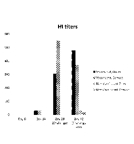
Figure 2 is a graph showing the HI Titer in vaccinated and non-vaccinated
pigs.
Figure 3 is a graph showing real-time PCR threshold values in vaccinated and
non-vaccinated pigs.
Figure 4 is a graph showing tissue culture infection dose of ferrets
challenged with the virus
of the present invention.
2a
Date Recue/Date Received 2020-06-25
CA 02862328 2014-07-22
WO 2013/112995
PCT/US2013/023441
DETAILED DESCRIPTION OF A PREFERRED EMBODIMENT
Isolation of the virus
Samples submitted to Newport Laboratories were nasal swabs from pigs
displaying signs of influenza-like illness. Samples were negative for
influenza A by real
time reverse transcription PCR. An aliquot of the sample was applied to a
confluent
monolayer of Swine Testicle (ST) cells to attempt to grow any viruses that
were present.
After 5 days cytopathic effects were evident, indicating that a virus was
growing in the
swine testicle cells. Samples of cell culture supernatant were analyzed for
influenza A by
QPCR and were negative. Cell culture supernatant was analyzed for the ability
to
hemagglutinate turkey red blood cells (hemagglutination assay). The
hemagglutination
assay was carried out according to the following protocol (Experiment # 1).
Experiment # 1
1.0 Introduction
To quantitate Swine Influenza Virus.
2.0 Materials
= Reservoir
= 96-well microtiter V bottom plates
= Multi-channel pipettor and tips (10-200uL)
= 100-1000 luL pipettor
= 10-100 iLiL pipettor
= Pipette Tips
= Personal Protective Equipment (PPE: lab coat, safety glasses, gloves)
= Biosafety Cabinet
= Cover plate/lid
= Timer
= 50 mL falcon tube
3
CA 02862328 2014-07-22
WO 2013/112995
PCT/US2013/023441
= Centrifuge
3.0 Reagents/Media
= Turkey Red Blood Cells (TRBC) from Lampire
= 1X DPBS
4.0 Media formulations
Turkey Red Blood Cell Solution (0.5%)
= TRBC arrive in Alsevers solution. Centrifuge in 50 mL conical tubes at
2000 rpm for 10 minutes. Aspirate and discard the supernatant.
= Wash the RBC by adding lx DPBS equal to volume of blood started with.
Invert and shake gently. Centrifuge and discard the supernatant. Repeat two
more times.
Store packed cells at 4 C.
= Add 50mL lx DPBS into 50 mL tube. Take out 2504 1X PBS. Add
2501uL of Turkey packed RBC. Mix gently. 5mLs is needed per plate. Store
diluted cells
at 4 C.
5.0 Procedure
Preparation
= Eight samples will run horizontally on a 96 well plate. Figure out how
many plates are needed based on samples to be tested and fill out the
appropriate
paperwork.
= Alternative set-up: Twelve samples can be run vertically on a 96 well
plate.
Test procedure
4
CA 02862328 2014-07-22
WO 2013/112995
PCT/US2013/023441
= Fill all wells of plates with 504 DPBS.
= Put 504 of sample #1 into well Al; sample #2 into Bl, etc.
= With each test set of plates, run a negative control on the last plate
(just
DBPS).
= With 50uL, dilute the plates starting from column 1 through 12 (excluding
negative control row), discarding the remaining 501aL.
= After the plate is diluted, fill all wells with 501u1 of 0.5% TRBC
(invert
tube to mix blood cells, do not vortex). Tap plates gently, cover and incubate
at room
temperature up to 2 hours.
= Alternative set-up: To run twelve samples vertically, put 504 of sample
#1 into well Al; sample #2 into A2.
= With 50 L, dilute the plates starting from row A through row H
(excluding negative control column), discarding the remaining 504.
= After the plate is diluted, fill all wells with 504 of 0.5% TRBC (invert
tube to mix blood cells, do not vortex). Tap plates gently, cover and incubate
at room
temperature up to 2 hours.
6.0 Reading
= Tilt the plates at 30-45 to read. When reading, the last well without a
complete teardrop is the end point. Mark on the corresponding paperwork.
= HA titers are listed on the corresponding paperwork
= Negative control row (or column) should completely tear drop, showing
no hemagglutination.
The unidentified virus had the ability to hemagglutinate crbc's with a titer
of
1280. Many viruses contain a gene encoding a hemagglutinin protein which
enables a
virus to bind cells, including red blood cells. Influenza viruses have the
ability to
hemagglutinate red blood cells.
5
CA 02862328 2014-07-22
WO 2013/112995
PCT/US2013/023441
Experiment # 2
A flask of ST cells infected with the unknown virus was sent to the University
of
Minnesota Veterinary Diagnostic Laboratory for electron microscopy. Images of
the
virus were consistent with the family Orthomyxoviridae (Figure 1). This family
consists
of influenza A, B, C and thogoto virus.
Experiment # 3
PCR was attempted with primers designed to specifically detect either
influenza
A, B or C (Table 1). All PCR reactions were negative. Additionally, a
neuraminidase
activity assay was performed using the neuraminidase substrate
methylumbelliferyl N-
acetylneuraminic acid. The virus did not possess neuraminidase activity (both
influenza
A and B have neuraminidases). An esterase activity assay was next performed
with 4-
nitrophenyl acetate (Sigma Aldrich N8130). The virus exhibited esterase
activity. The
ability to hemagglutinate red blood cells and having an esterase activity is
characteristic
of influenza C and some members of the family Coronaviridae. Based on the
hemagglutinin esterase activity and electron microscopy, the virus was
preliminarily
identified as influenza C although the negative for influenza C by PCR.
Table 1. Primers and probes used for real time reverse transcription PCR for
virus
detection. (IABkQ=Iowa black hole quencher)
Virus Forward Primer Reverse Primer Probe
Swine AGATGAGTCTTCTAACCG TGCAAAAACATCTTCAA 5'-Cy3-TCA GGC CCC CTC
ACiCiTCG GTCTCT(i AAA CiCC GA-3'-lABkQ
influenza A
viruses
Human TCCTCAACTCACTCTTCO CCiCiTGCTCTTGACCAAA 5'-FAM-CCA ATT CCiA
AGCG TTGG GCA GCT GAA ACT GCG
influenza B
GTG-3'-IABkQ
viruses
Human ATTGAGAGCAGGAACGA TCTTAAAGGCCCAGGAA 5'-FAM-
CTG ACG CCCCTCTGGAAAGAGCC
influenza C
ATGCAA-3'-IABkQ
viruses
Experiment #4 - Genome Sequencing
6
CA 02862328 2014-07-22
WO 2013/112995
PCT/US2013/023441
Virus was expanded in cell culture to generate 200mL of cell culture harvest
with
a hemagglutination (HA) titer of 2560. Cell culture fluids were filtered
through a 0.2
micron filter to remove cell debris. The fluids were then centrifuged at
110,000xg for 3
hours to pellet the virus. The virus was then resuspended in lmL of phosphate
buffered
saline and digested with DNase (New England Biolabs M0303S) and RNase (New
England Biolabs M0243S) at 37C for 1 hour. The viral solution was then gently
layered
on top of 35mL of 25% sucrose solution and centrifuged for 3 hours at
110,000xg to
pellet the virus. Viral RNA was extracted from the viral pellet using a
Qiagen[trade]
Viral RNA Mini Kit(Qiagen, Inc., 27220 Turnberry Lane Suite 200 Valencia, CA
91355)according to the manufacturer's instructions. In brief, the viral pellet
was
resuspended in 700 uL of buffer AVL containing carrier RNA and incubated for
10
minutes at room temperature. Next, 560 IA of ethanol was added to the sample
and then
sample was loaded onto a QIAampTm mini column by centrifugation at 6000 x g.
The
column was next washed sequentially with 500 uL each of buffers AW1 and AW2,
dried
.. by centrifugation at 14,000 x g for 2 minutes and then eluted with 60 iaL
of water. cDNA
was then reverse transcribed from the viral RNA using a Promega Reverse
Transcription
Kit (Promega Corp., 2800 Woods Hollow Rd., Madison WI 53711; www-.promega.com)
along with enclosed random primers. The GoScriptTM Reverse Transcription
Systemis a
convenient kit that includes a reverse transcriptase and an optimized set of
reagents
designed for efficient synthesis of first-strand eDNA in preparation for PCR
amplification. The components of the GoScriptTM Reverse Transcription System
can be
used to reverse transcribe RNA templates starting with either total RNA,
poly(A)+
mRNA or synthetic transcript RNA.
The cDNA was next digested for 1 hour at 37C with RNaseH (New England
Biolabs M0297S) to remove RNA from the RNA-cDNA hybrid. The single stranded
cDNA was made double stranded using the Klenow fragment from DNA polymerase
(New England Biolabs M0210S).
Using the BioRuptor0 Sonication System (Diagenode Inc. North America, 376
Lafayette Rd., Suite 102, Sparta, NJ 07871), the double stranded cDNA was
sonicated to
fragment the viral cDNA. The fragmented cDNA was next used to construct a cDNA
library according to the Life Technologies I'm Ion Plus Fragment Library Kit
protocol (
7
CA 02862328 2014-07-22
WO 2013/112995
PCT/US2013/023441
www.lifetechnologies.com) and described by the following: 50 microliters of
the
fragmented cDNA was mixed with 108 microliters nuclease-free water, 40
microliters 5X
End Repair Buffer, and 2 microliters of End Repair Enzyme. The reaction was
incubated
at room temperature for 20 minutes. Following incubation, 360 microliters of
Agencourt0 Ampure0 beads were added to the sample, which was then placed on a
rotator at 8-10 rpm for 10 minutes. The sample was pulse-spun and placed on a
DynaMagTm-2 magnet rack. After the solution cleared, the supernatant was
removed and
discarded. The sample was then twice washed using the subsequent protocol. 500
microliters of freshly made 70% ethanol were added to the sample without
removing it
from the magnet. The tube containing the sample was rotated twice on the
magnet to
move the beads around. After the solution cleared, the ethanol was removed.
This wash
procedure was repeated. Following the second 70% ethanol wash and removal of
the
supernatant, the sample tube was pulse-spun and placed back on the magnetic
rack.
Residual ethanol was aspirated and the sample dried at room temperature for
approximately five minutes. 50 microliters of lx TE (10mM Tris-HC1, 1mM EDTA,
pH
8.0) were added to the cDNA and vortexed, pulse-spun, and placed back on the
magnetic
rack. After the solution cleared, the supernatant containing the eluted cDNA
was
transferred to a new 1.5-mL LoBind Tube[trade].
microliters of 10X Ligase Buffer, 77 microliters Nuclease-Free Water, 50
20 .. microliters of Adapters, and 3 microliters DNA Ligase were mixed with
the 50
microliters of cDNA. The mixture was incubated at room temperature for 30
minutes.
After the solution cleared, the supernatant was removed and discarded. The
sample was
then twice washed using the subsequent protocol: 500 microliters of freshly
made 70%
ethanol were added to the sample without removing it from the magnet. The tube
containing the sample was rotated twice on the magnet to move the beads
around. After
the solution cleared, the ethanol was removed. This wash procedure was
repeated.
Following the second 70% ethanol wash and removal of the supernatant, the
sample tube
was pulse-spun and placed back on the magnetic rack. Residual ethanol was
aspirated
and the sample dried at room temperature for approximately five minutes. 30
microliters
of 1X TE were added to the cDNA and vortexed, pulse-spun, and placed back on
the
8
CA 02862328 2014-07-22
WO 2013/112995
PCT/US2013/023441
magnetic rack. After the solution cleared, the supernatant containing the
eluted cDNA
was transferred to a new 1.5-mL LoBind Tubdtrade].
All 30 microliters of cDNA were loaded onto a 2% agarose gel and subjected to
gel electrophoresis for 80 minutes at 110V. Following gel electrophoresis, the
sample
was size-selected for 180-210 base pairs. The size-selected cDNA library was
gel
purified using the QIAquick[trade] PCR Purification Kit (Qiagen, Inc., 27220
Turnberry
Lane Suite 200 Valencia, CA 91355; www.giagen.com) per the manufacturer's
directions. The excised cDNA fragment of the gel was weighed and 3 volumes of
Buffer
QG to 1 volume gel were added. The mixture was incubated on a rotator at room
temperature until the gel slice was completely dissolved. Once the gel slice
had
dissolved completely, the solution was applied to a QIAquick[trade] spin
column
(Qiagen, Inc., 27220 Tumberry Lane Suite 200 Valencia, CA 91355;
wwvvapiagen.com)
and provided 2 milliliter collection tube and centrifuged at maximum speed
(approximately 16,000 rcf) for 1 minute. The flow-through was discarded and
the
QIAquick[trade] column was placed back in the same tube. The column containing
the
cDNA was washed by adding 750 microliters Buffer PE to the column and
centrifuging it
at maximum speed for 1 minute. The flow-through was discarded and the
QIAquick[trade] column was placed back in the same tube and centrifuged for 4
minutes
at maximum speed. The QIAquick[trade] column was then put in a clean 1.5
milliliter
microcentrifuge tube. 40 microliters of Buffer EB were added to the
QIAquick[trade]
column membrane and allowed to incubate at room temperature for several
minutes
before the column was centrifuged for 1 minute to elute the cDNA.
The eluted size-selected cDNA library was nick-translated and amplified
according to the Life Technologies[trade] Ion Plus Fragment Library Kit (
www.lifetechnologies.com) protocol. 40 microliters of size-selected cDNA were
mixed
with 200 microliters Platinum[trade] PCR SuperMix High Fidelity and 10
microliters of
Library Amplification Primer Mix. 125 microliter aliquots were transferred to
each of 2
PCR tubes and ran on a thermocycler according to the following parameters:
Table 2: PCR Thermocycler Parameters
Stage Step Temperature Time
9
CA 02862328 2014-07-22
WO 2013/112995
PCT/US2013/023441
Holding Nick Translation 72 C 20 min
Holding Denature 95 C 5 min
Cycling (10 cycles) Denature 95 C 15 sec
Anneal 58 C 15 sec
Extend 72 C 1 min
Holding 4 C GC
The samples were pooled in a new 1.5 milliliter microcentrifuge tube and
purified
by adding 375 microliters of Agencourt Ampure beads to the sample and
incubating it
for 10 minutes at room temperature on a rotator. The sample was pulse-spun and
placed
on a DynaMag[trade]-2 magnet rack. After the solution cleared, the supernatant
was
removed and discarded. The sample was then twice washed using the subsequent
protocol. 500 microliters of freshly made 70% ethanol were added to the sample
without
removing it from the magnet. The tube containing the sample was rotated twice
on the
magnet to move the beads around. After the solution cleared, the ethanol was
removed.
This wash procedure was repeated. Following the second 70% ethanol wash and
removal
of the supernatant, the sample tube was pulse-spun and placed back on the
magnetic rack.
Residual ethanol was aspirated and the sample dried at room temperature for
approximately five minutes. 50 microliters of lx TE were added to the cDNA and
vortexed, pulse-spun, and placed back on the magnetic rack. After the solution
cleared,
the supernatant containing the eluted cDNA was transferred to a new 1.5-mL
LoBind
Tube.
The amount of cDNA was quantified using the Ion Library Quantitation Kit
WWW.Il fetechnologies.com) according to the manufacturer's protocol. 5
sequential 10-
fold dilutions were prepared from the E. coli DH1OB Control Library according
to the
following table.
Table 3: Dilutions for E. coli DH1OB Control Library
Standard Control Library Nuclease-free Fold Dilution
Water
CA 02862328 2014-07-22
WO 2013/112995
PCT/US2013/023441
1 5 microliters 45 microliters 0.1
(undiluted)
2 5 microliters Std 1 45 microliters 0.01
3 5 microliters Std 2 45 microliters 0.001
4 5 microliters Std 3 45 microliters 0.0001
5 microliters Std 4 45 microliters 0.00001
A 1:20 dilution of the sample library in Nuclease-free water was initially
prepared. Serial dilutions at 1:2000 and 1:20000 were subsequently prepared as
described
in the following table
5 Table 4: Dilutions for Sample Library
Dilution Library Nuclease-Free Water
1:2000 1 microliter of 1:20 99 microliters
1:20000 5 microliters of 1:2000 45 microliters
As each qPCR reaction was prepared in triplicate, the master mix was prepared
by
mixing 250 microliters of Ion Library TaqMan0 qPCR Mix 2X, 25 microliters of
Ion
Library TaqMan0 Quantitation Assay 20X, and 100 microliters of Nuclease-free
water
per qPCR reaction. For each reaction, 15 microliters of master mix was
pipetted into a
well of the PCR plate. 5 microliters of the diluted control or sample library
were added
to each appropriate well. 5 microliters of nuclease-free water was used as the
no-template
control (NTC). The wells were sealed, briefly centrifuged, and subjected to
the following
real-time PCR conditions.
11
CA 02862328 2014-07-22
WO 2013/112995
PCT/US2013/023441
Table 5: PCR Conditions for qPCR Reaction
Stage Temperature Time
Hold 50 C 2 minutes
Hold 95 C 20 seconds
Cycle (40 cycles) 95 C 3 seconds
60 C 30 seconds
The Template Dilution Factor was calculated using the following equation:
Template Dilution Factor = [(qPCR relative quantity) * (sample library fold
dilution)]/0.32
The sample was diluted accordingly and prepared for sequencing using the Ion
XpressTm Template Kit v2.0 (www.lifetechnologies.com). The Emulsion Oi1TM was
removed from the fridge and mixed. 9 milliliters of Emulsion Oilrm were added
to a
.. IKACR) DT-20 tube. The filled IKAER) tube was placed on ice until ready for
use. The
aqueous PCR mix was generated by combining 582 microliters of Nuclease-free
water,
200 microliters 5x PCR Reagent Mix, 100 microliters of 10x PCR Enzyme Mix, 100
microliters of Ion Sphere[trade] Particles (vortexed for 1 minute before
addition), and 18
microliters of the diluted sample library. The mixture was vortexed for 5
seconds and set
aside while the IKAO DT-20 tube containing the Emulsion Oil was positioned on
the
IKAO Ultra-Turrax0 Tube Drive and locked in place. The adhesive label on the
cap of
the IKAO DT-20 tube was removed to expose the sample loading port. The START
button on the IKAO Ultra-Turrax0 was pushed and the entire volume of the
aqueous
PCR mix was dispensed through the opening in the blue cap of the IKAO DT-20
tube.
After mixing for 5 minutes on the IKAO Ultra-Turrax0 Tube Drive, the emulsion
was
placed on ice for approximately 5 minutes. A wide-bore tip was created by
cutting
approximately 5 mm from a pipette tip to transfer the emulsion. Using an
Eppendorf
Repeater Pipettor fitted with the wide-bore tip, the emulsion was drawn up
and
dispensed in 100 microliter increments to each well of a 96-well PCR plate
until
12
CA 02862328 2014-07-22
WO 2013/112995
PCT/US2013/023441
approximately 90 wells were filled. The 96-well plate was capped and loaded
onto a
thermal cycler according to the following PCR parameters
Table 6: PCR Step Temperature Time
Parameters for
Sequencing Stage
Hold Denature 94 C 6 minutes
Cycle (40 cycles) Denature 94 C 30 seconds
Anneal 58 C 30 seconds
Extend 72 C 90 seconds
Cycle (5 cycles) Denature 94 C 30 seconds
Extend 68 C 6 minutes
Hold 10 C 00
Following the PCR reaction, as much of the contents of the wells as possible
were
transferred using a multi-channel pipette to a multi-channel pipette
reservoir.
Approximately 1.2 milliliters of the emulsion were transferred to each of six
1.5-mL
microcentrifuge tubes. All six microcentrifuge tubes were centrifuged for 2
minutes at
15,500 X g to collect the emulsion. During centrifugation, the Breaking
SolutionTM was
created by mixing 2 milliliters Recovery SolutionTM and 6 milliliters of 1-
butanol. The
Breaking SolutionTM was then vortexed for about 1 minute until a fine white
emulsified
material formed. After centrifugation of the emulsion, the clear top fraction
of oil from
each tube was removed. The 6 tubes containing the white emulsion were each
treated
with one milliliter of the Breaking SolutionTM, vortexed for 30 seconds, and
centrifuged
for 2 minutes at 15,500 X g. After centrifugation, the top organic phase of
each tube was
removed. Each sample tube received 1 mL of Recovery SolutionTM, was vortexed
for 30
seconds, and then centrifuged for 3 minutes at 15,500 X g. The supernatant
from each
tube was removed until only approximately 100 microliters were left. Using the
same
pipette tip, the pellets in all six tubes were resuspended and transferred to
a new 1.5
milliliter microcentrifuge tube. Three of the original tubes were rinsed with
a single 200
microliter aliquot of Recovery SolutionTM. After the third tube was rinsed,
the solution
13
CA 02862328 2014-07-22
WO 2013/112995
PCT/US2013/023441
was transferred to the tube that contained the combined, resuspended pellets.
This
procedure was repeated on the three remaining tubes.
Recovery SolutionTM was added to the combined tube until the total volume was
1.5 milliliters. The tube was then vortexed for 30 seconds and centrifuged at
15,500 X g
for 3 minutes. The supernatant was removed until only about 100 microliters
were left.
The remaining material was resuspended and transferred to a new 1.5 milliliter
microcentrifuge tube. 100 microliters of Wash SolutionTM was added to the
original tube,
rinsed, and then transferred to the new tube containing the sample. The sample
was twice
washed by adding 1 milliliter of Wash SolutionTmTm, vortexing for 30 seconds,
and then
centrifuging the tube for 3 minutes at 15,500 X g. The supernatant was removed
until
only 100 microliters remained. This wash procedure was repeated.
The template-positive Ion SphereTM Particles enrichment was performed in
accordance with the Ion XpressTM Template Kit v2.0 protocol
(www.lifetechnologies.com). The bottle containing the Dynabeads0 MyOneTM
Streptavidin Cl beads was vortexed. 10 microliters of MyOneTM beads were
transferred
to a 1.5 milliliter microcentrifuge tube, washed with 70 microliters of Wash
Solutionrm,
vortexed, and placed on a magnet for 2 minutes. The supernatant was then
discarded. The
MyOneTM beads were resuspended in 10 microliters of new Wash Solution TM and
then
transferred to the sample tube containing the Ion Sphere ParticlesTM (ISPs).
To perform
.. the capture, 100 microliters of Annealing BufferTM were also added to the
sample tube
which was then placed on the rotator for 10 minutes at room temperature. The
sample
tube was centrifuged and placed on the magnet until the solution was clear.
The
supernatant was transferred to a tube labeled "Unbound." The beads were twice
washed
with 200 microliters of Wash SolutionTM, mixed, and placed back on the magnet
before
the supernatant was transferred to the "Unbound" tube. A fresh Melt-Off
SolutionTM
was prepared by combining 200 microliters 1 M NaOH, 16 microliters of 10%
Tween0
20 in molecular grade water, and 1.38 milliliters of molecular grade water. To
elute the
Ion Sphere Particles from the Dynabeads0 MyOne[trade] Streptavidin Cl beads,
400
microliters of the Melt-Off SolutionTM was added to the sample tube, mixed,
and then
placed on the rotator for 7 minutes. The supernatant was removed afterwards
and placed
into the tube labeled "Enriched-1." The "Enriched-1" tube was vortexed and
then spun at
14
CA 02862328 2014-07-22
WO 2013/112995
PCT/US2013/023441
15,500 X g for 4 minutes. All but 100 microliters of supernatant were then
discarded. 1
milliliter of Wash SolutionTM was then applied to the "Enriched-1" tube,
vortexed, and
spun at 15,500 X g for 4 minutes. Once again, all but 100 microliters of
supernatant was
removed. The remaining sample was mixed and the "Enriched-1" tube was placed
back
on the magnet. The supernatant was removed after several minutes and put in a
tube
labeled "Enriched-2."
DNA sequencing was conducted in accordance with the Life Technologies Ion
Sequencing Kit v2.0 user's manual (www. tifete eh n o lo gi es , corn). 50
microliters of the
sample in the "Enriched-2" tube were transferred to a new 0.2 mL PCR tube. 5
microliters of Control Ion Spheres trade] and 150 microliters of Annealing
BufferTM were
added and the solution was mixed and centrifuged for 2 minutes at 15,500 X g.
Supernatant was removed until only 9 microliters remained. 5 microliters of
Sequencing
PrimerTM were added to the sample and placed on a thermal cycler for a single
cycle of 2
minutes at 95 C and then 2 minutes at 37 C. The sample was then removed from
the
thermal cycler, mixed with 1 microliter of Sequencing PolymeraseTM, and
incubated at
room temperature for 5 minutes.
Meanwhile, a new chip was removed from its packaging and placed in the IonTM
centrifuge adaptor/rotor bucket. Using a Rainin SR-L200F pipette tip, 50
microliters of
100% isopropanol were added to the large port of the chip and then aspirated
from the
other port. The chip was washed two times with 50 microliters of Annealing
BufferTM
into the large port on the chip which was then aspirated from the other port.
The "Experiment" tab on the main menu of the PGMTm Sequencer was pressed.
When prompted, the old chip was replaced with the new one. The barcode scanner
was
used to scan the chip barcode on the package. After the barcode was entered,
the -Chip
Check" button was pushed. After Chip Check was complete, the "Next" button was
pressed to proceed to chip calibration. Following calibration, the chip was
removed,
placed back on the Ion centrifuge adaptor/rotor bucket, and washed with 50
microliters of
Annealing Buffer into the large port on the chip, which was then aspirated
from the other
port on the chip. Using the Rainin0 Pipette-Lite LTS-20 pipette with a
Rainin0 SR-
L200F tip, 7 microliters of the sample were deposited to the large port of the
chip. The
CA 02862328 2014-07-22
WO 2013/112995
PCT/US2013/023441
displaced liquid at the other port of the chip was then removed. The Ion
ChipTM was then
transferred to the centrifuge and spun for 4 minutes. Using a fresh RaininER)
SR-L200F
tip, the remainder of the sample was deposited to the loading port of the
chip. The
displaced liquid was removed from the other port and the chip was again
centrifuged for
4 minutes. After the final spin was complete, the "Next" button on the PGM
screen was
pressed and the chip was loaded back onto the Ion Torrent PGMTm machine and
the
sequencing run started.
Sequence reads were assembled using the DNAStar software SeqmanNexGen
(DNASTAR, Inc., 3801 Regent Street, Madison, WI 53705; www.dnastar.com) using
the
de novo assembly option. Sequence assembly identified seven contigs with
greater than
10,000 reads associated each of them. The contigs were trimmed such that they
represent
the complete open reading frames. The trimmed sequences and corresponding
protein
sequences are included in the present application as SEQ ID NOs: 4 ¨ 17.
BLASTP
analysis of the putative translated open reading frames revealed homology to
the seven
.. segments of human influenza C isolates and the isolate was designated
C/swine/Oklahoma/1334/2011. The closest homolog for each segment is shown
below.
The segment noted as encoding the non-structural proteins (NS) is transcribed
into
mRNA and alternatively spliced to yield two different proteins (NS1 and NS2)
as in
Table 7 below. The percentage positive represents percent similarity between
the isolated
influenza C virus and the closest homolog for each segment in the public
databases.
While PB I showed moderate homology to the PB1 of the human influenza C
isolate
C/Johannesburg/1/66 with 85% similarity, all other segments showed lower
homology to
previously sequenced influenza C, with % similarities for the other segments
from 48-
71%. The low overall similarity of C/swine/Oklahoma/1334/2011 with previously
sequenced viruses demonstrates the uniqueness of this virus.
16
CA 02862328 2014-07-22
WO 2013/112995 PCT/US2013/023441
Table 7: BLAST Homologies
ORF (amino acids) Best blast hit (BLASTP)
PB2 762 polymerase 2 [Influenza C virus (C/Ann
Arbor/1/50)]
Identities = 397/762 (52%), Positives = 538/762 (71%), Gaps = 2/762 (0%)
PB1 720 polymerase subunit PB1 [Influenza C virus
(C/Johannesburg/1/66)]
Identities = 512/708 (72%), Positives = 601/708 (85%), Gaps = 2/708 (0%)
P3 710 polymerase 3 [Influenza C virus (C/Ann
Arbor/1/50)]
Identities = 358/722 (50%), Positives = 479/722 (66%), Gaps = 27/722 (4%)
HE ............. 636 hemagglutinin-esterase [Influenza C virus
(C/Catalonia/1318/2009)]
Identities = 306/608 (50%), Positives = 399/608 (66%), Gaps = 19/608 (3%)
NP 552 nucleoprotein [Influenza C virus (C/Ann
Arbor/1/50)]
Identities = 199/504 (39%). Positives = 297/504 (59%), Gaps = 14/504 (3%)
397 unspliced product of M gene [Influenza C virus
(STRAIN C/TAYLOR/1233/47)]
Identities = 145/383 (38%), Positives = 221/383 (58%), Gaps = 12/383 (3%)
NS1 243 nonstructural protein 1 (NS1) [Influenza C virus
(C/Hiroshima/248/2000)]
Identities = 76/228 (33%), Positives = 110/228 (48%), Gaps = 21/228 (9%)
NS2 168 NS2 [Influenza C virus]
Identities = 53/180 (29%), Positives = 87/180 (48%), Gaps = 15/180 (8%)
Serological studies to determine prevalence
Having established C/swine/Oklahoma/1334/2011 as a novel virus with weak
sequence homology to human influenza C, serological studies were performed to
determine incidence of infection for both humans and pigs. Approximately 200
random
swine sera samples submitted to Newport Laboratories from numerous states were
analyzed for antibodies to Ciswine/Oklahoma/1334/2011 using the
hemagglutination
inhibition assay. Approximately 8% of samples were positive in the HI assay
with titers
from 10-160. Similarly, collaborators at St Jude's Children's Hospital in
Memphis, TN,
performed HI assays on a bank of human sera collected from elderly adults (age
65-95)
from Canada. Approximately 28% of the samples had positive HI titers from 10-
80.
Together, these results demonstrate that both humans and pigs are commonly
exposed to
17
CA 02862328 2014-07-22
WO 2013/112995
PCT/US2013/023441
C/swine/Oklahoma/1334/2011. These results also suggest that this virus is
capable of
infecting both humans and pigs
Pig vaccination and challenge experiment
C/swine/Oklahoma/1334/2011 was grown to a high titer (HA=2560) and
inactivated with binary ethyleneimine and then 10% Trigen (an oil in water
adjuvant) was
added to make a killed virus vaccine. 22 pigs that were serologically negative
for
antibodies to C/swine/Oklahoma/1334/2011 were vaccinated on days 0 and 14 with
2mL
of inactivated virus vaccine delivered intra muscularly. Serum samples were
collected on
days 0, 14 and 28 and analyzed for HI titers. 28 pigs were also included as
non-
vaccinated controls. Vaccinated pigs seroconverted by day 28 with an average
HI
titer=433. Non-vaccinated pigs were negative on the HI assay. Table 8 and Fig.
3 shows
the results. Vaccinated pigs showed strong seroconversion. The rule of thumb
for
influenza A is that a HI titer>40 is protective. The antibodies measured in
pig sera in
vaccinated pigs prior to challenge suggests protective antibodies were
present. This data
is consistent with the challenge results
18
Table 8: HI titers of vaccinated and non-vaccinated pigs
Vaccinates, Direct Challenge Vaccinates, Contact Pigs
Non=Vaccinates, Direct Challenge Non=Vaccinates, Cotact Pigs Controls
CJi
day 28 day 42 day 28 day 42 01428 day 42 day 28 day42 day 28
day42
pig ID day14 ((lag) (clay14) piglD cI411 (clay0 (cI4 11) piglD day14 (day0)
(clay14) pigl0 chy 11 (day Oj (day 14) pig 10 clay0 )d40) (clay14)
850 40 320 856 20 320 865 0 0 854 0
0 851 0 0
852 20 160 858 40 640 866 0 0 857 0
0 853 0 0
860 10 320 861 0 20 870 0 0 859 0
0 55 0
863 10 640 8E4 80 1280 873 0 0 861 0
0 E62 0 0 0
868 10 80 872 20 40 874 0 0 869 0 0
891 0 0 0
876 0 160 875 40 320 878 0 0 871 0
0 896 0 0 0
880 20 20 640 8E1 20 1280 320 883 0 0
20 877 0 0 40
886 80 320 640 8E2 80 1280 640 884 0 0
20 879 0 0 40
888 10 640 640 887 10 80 80 889 0 0 40
885 0 0 0
898 80 80 160 890 20 160 160 895 0 0 40
892 0 0 0
899 20 320 320 893 20 640 640 897 0 0 40
894 0 0 0
Average /7 278 480 32 551 368 0 0 32 0 0 16 0 0 0
CA 02862328 2014-07-22
WO 2013/112995 PCT/US2013/023441
On day 28, 11 vaccinated and 11 non-vaccinated pigs were challenged intra-
nasally with 2 mL of 6.2 log10 TCID/mL C/swine/Oklahoma/1334/2011. On day 2
post
challenge 11 vaccinated and 11 non-vaccinated pigs were added to the room to
serve as
contact exposure challenge groups. Temperatures were measured every other day
from
the day of challenge to 14 days post challenge. Table 9 shows the results.
Table 9. Pig temperatures following challenge
Vaccinates, Direct Challenge Vaccinates, Contact Pigs
Day Day Day Day Day Day Day Day Day Day
pig
pig ID 0 2 3 6 8 ID 0 2 3 6 8
102. 102. 103. 103. 104. 102. 104. 104.
850 5 1 4 6 856 5 1 1 4
104. 106. 104. 102. 103. 103. 103.
852 3 105 2 6 858 9 3 6 1
103. 103. 102. 102. 102. 101. 104. 102.
860 5 3 7 4 861 9 8 2 7
101. 102. 102. 103. 103. 102.
863 2 5 103 7 864 1 100 6 6
103. 103. 102. 103. 102. 103. 103. 102.
868 1 9 2 6 872 6 4 5 8
105. 104. 103. 102. 102. 102. 103. 103.
876 7 5 2 8 875 5 9 4 2
105. 104. 103. 104. 103. 104. 104. 104.
Kw 103 2 7 6 7 881 4 103 7 3 1
103. 103. 104. 104. 103. 102. 102. 103. 102. 102.
886 8 9 7 1 8 882 8 9 2 7 7
103. 103. 104. 103. 102. 102. 103.
888 7 7 1 103 6 887 102 8 7 4 103
101. 103. 104. 103. 103. 102. 103. 103. 104.
898 9 1 4 103 3 890 9 2 8 2 4
103. 103. 103. 101. 102. 104. 102. 103. 103. 103.
899 6 8 8 9 6 893 6 7 6 7 6
CA 02862328 2014-07-22
WO 2013/112995 PCT/US2013/023441
103. 103. 103. 103. 103. 103. 102. 103. 103. 103.
Average 3 7 9 2 6 .. 2 5 7 3 6
Non-Vaccinates, Direct Challenge Non-Vaccinates, Contact Pigs
Day Day Day Day Day Day Day Day Day Day
pig
pig ID 0 2 3 6 8 ID 0 2 3 6 8
105. 104. 105. 102. 104. 103. 104. 102.
865 1 6 2 7 854 7 7 2 9
103. 102. 104. 102. 103. 103. 103.
866 4 7 4 1 857 6 6 9 103
104. 103. 102. 104. 101.
870 5 9 104 103 859 3 103 3 9
103. 102. 105. 102. 104. 102. 102.
873 6 9 1 1 867 2 8 103 4
104. 103. 105. 101. 104. 103. 104. 102.
874 6 8 6 8 869 7 1 3 9
102. 102. 102. 103. 103. 103. 103.
102.
878 1 6 4 5 871 3 3 6 3
103. 104. 104. 104. 104. 104. 104. 104. 103. 103.
883 9 2 7 4 4 877 2 1 8 2 3
102. 105. 101. 103. 103. 102. 100. 100. 103.
884 5 3 105 4 8 879 3 9 9 9 9
103. 104. 104. 103. 102. 103. 105. 103. 103.
889 7 2 3 103 1 885 2 4 1 7 3
103. 104. 102. 101. 102. 103. 103. 102. 103.
895 5 3 6 8 8 892 104 9 8 8 6
103. 104. 102. 103. 101. 104. 103. 102.
897 1 8 105 4 103 894 8 6 1 1 3
103. 103. 104. 102. 103. 103. 103. 103. 102. 103.
Average 6 9 4 6 4 7 2 8 6 3
Controls
pig ID Day Day Day Day Day
21
CA 02862328 2014-07-22
WO 2013/112995
PCT/US2013/023441
0 2 3 6 8
103. 103. 102.
851 9 104 3 6
103. 102. 102. 102.
853 1 8 6 7
103. 103. 103. 103.
855 1 1 1 1
103. 102. 102. 101. 102.
862 7 8 7 8 3
103. 104. 102. 103. 103.
891 6 3 7 1 1
103. 103. 102. 103.
896 104 3 3 8 3
103. 103. 103. 102. 102.
Average 6 4 0 7 9
No difference was observed between treatment groups, indicating that the virus
does not cause a fever as influenza typically does.. Similarly, nasal swabs
were collected
every other day from the day of challenge to day 14 post challenge.
Method of Assaying the Presence of the Virus
RNA was prepared from the swabs and the presence of influenza C was assayed by
reverse transcription real time PCR (rt-RT-PCR) using the following primers
and probes
designed based on the genome sequence for C/swine/Oklahoma/1334/2011. The
nucleotide sequences of the primers and probe is included in the present
application as
SEQ ID NOs:1-3, wherein Forward Primer = 5'-GCT GTT TGC AAG TTG ATG GG-3'
(SEQ ID NO:1); Reverse Primer = 5'-TGA AAG CAG GTA ACT CCA AGG-3' (SEQ
ID NO:2); and the Probe = Cy5-labeled-5'-TTC AGG CAA GCA CCC GTA GGA TT-
3'- (SEQ ID NO:3)-IABkQ
22
CA 02862328 2014-07-22
WO 2013/112995 PCT/US2013/023441
The results are shown in Table 10 and Fig. 3. "Ct" stands for cycle threshold.
In
real time PCR, the genetic material (RNA or DNA) is copied by a polymerase by
following cycling the sample/enzyme at various temperatures. Real time PCR
uses a
fluorescent reporter that binds to the produced DNA product. As cycling
progresses and
DNA product accumulates, more fluorescence is produced. At some point the
fluorescence is detected by the PCR machine. A certain level of fluorescence
detection is
called the threshold. The number of cycles required to generate fluorescence
above the
threshold level is referred to as the Ct. Real time PCR can quantify the
amount of
RNA/DNA in the starting sample such that when more DNA/RNA is present prior to
PCR, then fewer cycles/doubling will be required to generate fluorescence
above the
threshold. Consequently, a lower Ct value equates to higher levels of DNA/RNA
in the
original sample.
Table 10: Real time reverse transcription Ct values for
C/swine/Oklahoma/1334/2011 shedding in nasal swabs
Vaccinates, Direct Challenge Vaccinates, Contact
Pigs
pig ID Day 0 Day 2 Day 3 Day 6 Day 8 Day 10
pig ID Day 0 Day 2 Day 3 Day 6 Day 8
850 37.1 37.1 37.1 37.1 856 37.1 37.1
37.1 37.1
852 37.1 37.1 37.1 37.1 858 37.1 37.1
37.1 37.1
860 37.1 37.1 37.1 37.1 861 37.1 37.1
37.1 37.1
863 37.1 37.1 37.1 37.1 864 37.1 37.1
37.1 37.1
868 37.1 37.1 37.1 37.1 872 37.1 37.1
37.1 37.1
876 37.1 37.1 37.1 37.1 875 37.1 37.1
37.1 37.1
880 37.1 37.1 37.1 37.1 33.6 37.1 881 37.1
37.1 37.1 37.1 37.1
886 37.1 37.1 37.1 37.1 37.1 37.1 882 37.1
37.1 37.1 37.1 37.1
888 37.1 37.1 37.1 37.1 37.1 37.1 887 37.1
37.1 37.1 37.1 37.1
898 37.1 37.1 37.1 37.1 37.1 37.1 890 37.1
37.1 37.1 37.1 37.1
899 37.1 37.1 37.1 37.1 37.1 37.1 893 37.1
37.1 37.1 37.1 37.1
Average 37.1 37.1 37.1 37.1 36.4 37.1 37.1 37.1
37.1 37.1 37.1
Non-Vaccinates, Direct Challenge Non-Vaccinates,
Contact Pigs
pig ID Day 0 Day 2 Day 3 Day 6 Day 8 Day 10
pig ID Day 0 Day 2 Day 3 Day 6 Day 8
865 37.1 37.1 34.53 37.1 854 37.1 37.1
37.1 37.1
866 37.1 37.1 37.1 37.1 857 37.1 37.1
37.1 37.1
870 37.1 37.1 37.1 37.1 859 37.1 37.1
37.1 37.1
873 37.1 37.1 37.1 37.1 867 37.1 37.1
37.1 37.1
874 37.1 37.1 35.2 28.6 869 37.1 37.1
37.1 37.1
23
CA 02862328 2014-07-22
WO 2013/112995 PCT/US2013/023441
878 37.1 37.1 37.1 31.32 871 37.1 37.1
37.1 37.1
883 37.1 37.1 35.05 34.85 34.23 37.1 877 37.1
37.1 37.1 37.1 37.1
884 37.1 37.1 37.1 29.65 24.21 29.45 879 37.1
37.1 37.1 37.1 37.1
889 37.1 37.1 34.69 35.54 35.85 29.37 885 37.1
37.1 37.1 37.1 37.1
895 37.1 37.1 35.5 30.26 29 37.1 892 37.1
37.1 37.1 37.1 37.1
897 37.1 37.1 32.8 29.76 28.11 37.1 894 37.1
37.1 37.1 37.1 36.53
Average 37.1 37.1 35.8 33.5 30.3 34.0 37.1 37.1
37.1 37.1 37.0
Controls
pig ID Day 0 Day 2 Day 3 Day 6 Day 8 Day 10
851 37.1 37.1 37.1 37.1
853 37.1 37.1 37.1 37.1
855 37.1 37.1 37.1 37.1
862 37.1 37.1 37.1 37.1 37.1 37.1
891 37.1 37.1 37.1 37.1 37.1 37.1
896 37.1 37.1 37.1 37.1 37.1 37.1
Average 37.1 37.1 37.1 37.1 37.1 37.1
The Ct values in the above chart show viral RNA levels in the nasal swabs.
37.1=negative. Any value less than 37.1 is positive for viral RNA (hence viral
shedding).
This data is also graphed in Figure 3. The non-vaccinated challenge group
viral RNA
shedding numbers are significantly different from the vaccinates. We ran a
Student's 1-
test on the data and the non-vaccinates that were directly challenged shed
virus at higher
levels than all other groups (P<0.001). Consequently, the vaccine protected
pigs. Virus
was only detected in one vaccinated pig following challenge, either direct or
by contact
exposure. In contrast, non-vaccinated pigs began to shed virus on day 3 post
challenge
(directly challenged) or on day 6 post exposure (contact challenge). This
experiment
demonstrates the virus is capable of infecting pigs and can replicate in them
and is shed
to the environment and contact animals. Additionally, pigs vaccinated with a
homologous vaccine are fully protected from infection.
Ferret Challenge
Ferrets are commonly used as a surrogate for humans in influenza research as
human influenza viruses typically replicate well in ferrets. As influenza C in
normally
thought of as a human pathogen, C/swine/Oklahoma/1334/2011 was used to
challenge
ferrets to determine if this virus is a likely human pathogen. Three ferrets
were
24
CA 02862328 2014-07-22
WO 2013/112995
PCT/US2013/023441
challenged intranasally with 6.0 logio tissue culture infectious dose 50 per
ml
(TCID50/mL) of C/swine/Oklahoma/1334/2011. On day 1 post challenge, three
ferrets
were added to the pen to serve as contact animals. Additionally, 3 ferrets
were housed in
a separate pen to serve as aerosol only exposure animals. Table11 and Fig. 4
shows the
results.
CA 02862328 2014-07-22
WO 2013/112995
PCT/US2013/023441
Table 11: TCID50/mL of virus detected in nasal swab washes for ferrets
infected
with C/swine/Oklahoma/1334/2011 by direct inoculation, direct contact or
aerosol
contact
days post inoculation (TCID50/mL)
ferret
id 0 3 5 7 10
direct
55 challenge 3.3 2.3 0.0 0.0
direct
56 challenge 3.9 0.0 0.0 0.0
direct
57 challenge 2.8 0.0 0.0 0.0
direct
mean challenge 0.0 3.3 0.8 0.0 0.0
direct
58 contact 0.0 0.0 2.0 4.5
direct
60 contact 0.0 0.0 0.0 4.2
direct
61 contact 0.0 0.0 2.2 4.1
direct
mean contact 0.0 0.0 0.0 1.4 4.3
aerosol
62 contact 0.0 0.0 0.0 0.0
aerosol
63 contact 0.0 0.0 0.0 0.0
aerosol
65 contact 0.0 0.0 0.0 0.0
26
CA 02862328 2016-04-08
51440-219
aerosol
mean contact 0.0 0.0 0.0 0.0
Virus was detected in nasal swabs by titration of ST cells. Virus was detected
by
day 3 post challenge in intranasally challenged animals and day 6 post
exposure in
contact challenge ferrets. No virus was detected in aerosol exposure ferrets.
This data
demonstrates C/swine/Oklahoma/1334/2011 is capable of infecting ferrets
exposed either
by direct challenge or contact with challenged animals. However, the virus
does not
appear to spread via aerosol transmission. This suggests that
C/swine/Oklahoma/1334/2011 is likely capable of infecting humans.
Unless otherwise defined, all technical and scientific terms used herein have
the
same meaning as commonly understood by one of ordinary skill in the art to
which this
invention belongs. Although methods and materials similar to or equivalent to
those
described herein can be used in the practice or testing of the present
invention, suitable
methods and materials are described below.
The present invention may be embodied in other specific forms without
departing
from the spirit or essential attributes thereof, and it is therefore desired
that the present
embodiment be considered in all respects as illustrative and not restrictive,
reference
being made to the appended claims rather than to the foregoing description to
indicate the
scope of the invention.
27