Note: Descriptions are shown in the official language in which they were submitted.
CA 02862524 2014-07-23
WO 2013/112771 PCT/US2013/023033
CORRELATING BRAIN SIGNAL TO INTENTIONAL AND
UNINTENTIONAL CHANGES IN BRAIN STATE
CROSS REFERENCES
[0001] This application claims priority from United States Provisional
Patent
Application Serial No. 61/590,235 filed January 24, 2012 and United States
Utility Patent
Application Serial No. 13/749,619 filed January 24, 2013; the contents of
which are
incorporated herein in their entirety.
FIELD OF INVENTION
[0002] This invention is directed to methods of analysis to extract and
assess brain
data collected from subject animals, including humans, to detect intentional
brain signals and
unintentional and other unexpected brain signals. These signals are correlated
to higher
cognitive brain functions or unintended, potentially adverse events, such as a
stroke or
seizure, and to translation of those signals into defined trigger events or
tasks. More
particularly the present invention is directed to a physiological data
acquisition from EEG,
EMG, EOG, MEG, ECoG, iEEG, fMRI, LFP or other signals obtained from a
peripheral
channel modulated by the subject's brain activity or modulating brain
activity.
BACKGROUND OF THE INVENTION
[0003] An Electroencephalogram (EEG) is a tool used to measure
electrical activity
produced by the brain. The functional activity of the brain is collected by
electrodes placed
1
CA 02862524 2014-07-23
WO 2013/112771 PCT/US2013/023033
on the scalp. The EEG has traditionally supplied important information about
the brain
function of a patient. Scalp EEG is thought to measure the aggregate of
currents present
post-synapse in the extracellular space resulting from the flow of ions out of
or into dendrites
that have been bound by neurotransmitters. Accordingly, EEG and like
modalities are mainly
used in neurology as a diagnostic tool for epilepsy but the technique can be
used in the study
of other pathologies, including sleep disorders.
[0004] Recent advances in EEG and other signal detection have allowed
for the
automated, real time, detection of sleeping and waking states through the
normalization and
other manipulation of brain activity data. In addition, such applications and
methods can also
be used to automatically access pathological conditions and medication
effects. Related
technology has allowed for the accessing of such data in real time utilizing a
single channel
detector. This in turn has provided the opportunity to further dissect sleep
and waking states,
including clear differentiation between REM and deep sleep states. To aid in
the efficient
collections of such data, head and harness systems have been developed
utilizing single
channels and wireless data transmission. See, e.g., International Patent
Application Number
PCT/US2006/018120; International Patent Application Number PCT/U52009/064632;
International Patent Application Number PCT/U52010/054346; US Patent
Application
Number 8,073,574; and Low, Philip Steven (2007). "A new way to look at sleep:
separation
and convergence". Published Thesis, University of California San Diego
Electronic Theses
and Dissertations (Identified: b6635681), the disclosures of which are herein
incorporated by
reference in their entirety. To date, this technology has been primarily
applied to sleep-
related diagnostic applications, and the impact of pathologies and
medications.
[0005] Development has continued in the area of exoskeletons and related
prostheses that hold the promise to allow paraplegics to walk again and
perform other tasks
that they currently are unable to perform. In addition, such devices may also
be useful with
healthy individuals, such as soldiers in the field, first responders,
construction works etc.
Companies such as Esko Bionics, Parker Hannifan and Argo Medical Technologies
and
consistently advancing such technologies. See, e.g., US Patent Number
8,096,965;
International Patent Application Number W02010101595A1, and US Patent
Application
Number 11/600,291, filed Nov 15, 2006. In addition, other devices are utilized
to allow
2
CA 02862524 2014-07-23
WO 2013/112771 PCT/US2013/023033
severely compromised individuals with ALS, MS and the like to communicate
using voice
synthesizers and the like. Typically, such devices are activated by movement
of a cheek
muscle, eye using Eye Tracker or the like.
[0006] There is therefore a need for non-invasive methods to detect the
intentional
and unintentional communication from subjects, including disabled individuals,
to assess and
potentially respond to or prepare for the physiological implications of these
changes in brain
state.
SUMMARY OF THE INVENTION
[0007] The present invention provides methods to non-invasively detect
the
intentional and unintentional communication from healthy and diseased
subjects, including
disabled individuals with neurological diseases such as ALS, MS and the like,
in the form of
physiological data through, e.g. EEG, EMG, EOG, MEG, ECoG, iEEG, fMRI, LFP and
the
like and to correlate these intentional and unintentional signals to changes
in brain state,
including higher cognitive functions. There is also a need to utilize such
intentional
communication to, e.g., simulate speech or move an artificial prosthesis.
There is further a
need to access unintentional signals from a subject with a pathological
condition, such as
epilepsy (or diseases in the body causing changes in brain activity), to be
correlated with a
pathological condition and to optionally be used to trigger an alarm and/or to
intervene to
alter, suppress or prepare for an unintended event.
[0008] In a preferred method of the present invention, intentional
brain signals
from a subject are detected by attaching at least a single sensor to the
subject, obtaining data
indicative of brain activity, analyzing said data indicative of brain
activity; and correlating
said analyzed data to an intentional higher cognitive function from the
subject. Preferably,
the data is obtained non-invasively by applying at least the single sensor to
the subject, and
more preferably by applying at least a single dry sensor or at least a single
wet sensor.
Further, it is preferred that the data is received from at least a single
channel of EEG, EMG,
EOG, MEG, ECoG, iEEG, fMRI, LFP or a peripheral channel modulated by the
subject's
intention. In an alternative embodiment, the data is received through a multi
channel detector.
Further, it is preferred that the data is communicated and received
wirelessly.
3
CA 02862524 2014-07-23
WO 2013/112771 PCT/US2013/023033
[0009] In another preferred embodiment, the data is analyzed by
normalizing a
spectrogram, including a normalized spectrogram, of the data at least once,
time over
frequency, and normalizing the spectrogram, including a normalized
spectrogram, of the
same data at least once, frequency over time, where both normalizations can be
performed in
either order and can be iterated. In a further preferred embodiment, the data
is analyzed by
computing the spectrogram of the data, normalizing the spectrogram, performing
an
independent or principal component analysis of the normalized spectrogram, and
identifying
clusters. In addition, the analyzing step can also include performing a
temporal
fragmentation analysis, preferred frequency analysis, an iterated (preferably
two times or
more) preferred frequency analysis, and/or spectral fragmentation analysis.
[0010] In an especially preferred embodiment, the methods of the present
invention
further comprise translating the analyzed data to effect a task associated
with the higher
cognitive function, including, but not limited to, intent, speech, memory
recall, thought,
imagination and planning, including but not limited to motion. Importantly,
the task effected
by translating the analyzed data includes simulating speech on a display,
simulating speech
with a voice synthesizer, or movement of an artificial prosthesis, or movement
of an
exoskeleton and the like.
[0011] In yet another preferred embodiment of the methods of the present
invention, brain signals from a subject are correlated with at least one
unintended event by
attaching at least a single sensor to the subject, obtaining data indicative
of brainwave
activity, analyzing said data indicative of brain activity, and correlating
said analyzed data to
at least one unintended event. In a further embodiment, after correlating the
data to an
unintended event, an alarm is triggered. Alternatively, after correlating the
data to an
unintended event, a response can be triggered to ameliorate the effect of the
unintended
event, which can include altering, suppressing or preparing for the unintended
event (which
may also trigger an alarm). This response will be especially impactful where
the unintended
event high fragmentation event, a change in fragmentation of an event, a
surprise, a tremor, a
spasm, an injury or a pathology including but not limited to, an epileptic
seizure, a migraine,
a stroke, a heart attack or an infarction.
[0012] In another preferred embodiment, the methods of the present
invention
4
CA 02862524 2014-07-23
WO 2013/112771 PCT/US2013/023033
detect intentional signals from a subject by attaching at least one detector
capable of detecting
the intentional signal to the subject, obtaining data indicative of detected
activity using EEG,
EMG, EOG, MEG, ECG, ECoG, iEEG, LFP, fMRI or a peripheral channel modulated by
the
intentional signals from a subject, analyzing said data indicative of the
detected activity, and
correlating said analyzed data to an intentional higher cognitive function
from the subject. It
is contemplated that any method, system or information described herein can be
implemented
with respect to any other method, system or information described herein.
[0013] Unless otherwise defined, all terms used herein have the same
meaning as
commonly understood by one of ordinary skill in the art to which this
invention belongs.
Methods and materials are described herein for use of the present invention;
other suitable
methods and materials known in the art can also be used. The materials and
methods, and
examples are illustrative only and not intended to be limiting. All
publications, patent
applications, patents and other references mentioned herein, are incorporated
by reference in
their entirety. In case of conflict, the present specification, including
definitions will control.
[0014] These and other embodiments of the invention will be better
appreciated and
understood when considered in conjunction with the following description and
the
accompanying drawings. It should be understood, however, that the following
description,
while indicating various embodiments of the invention and numerous specific
details thereof,
is given by way of illustration and not of limitation. Many substitutions,
modifications,
additions and/or rearrangements may be made within the scope of the invention
without
departing from the spirit thereof, and the invention includes all such
substitutions,
modifications, additions and/or rearrangements.
BRIEF DESCRIPTION OF THE DRAWINGS
[0015] For the present invention to be clearly understood and readily
practiced, the
present invention will be described in conjunction with the following figures,
wherein like
reference characters designate the same or similar elements, which figures are
incorporated
and constitute a part of the specification.
CA 02862524 2014-07-23
WO 2013/112771 PCT/US2013/023033
[0016] The patent or application file contains at least one drawing
executed in color.
Copies of this patent or patent application publication with color drawing(s)
will be provided
by the Office upon request and payment of the necessary fee.
[0017] Figure 1 is a series of graphs illustrating examples of bring
signal analyses.
The left column shows traditional analyses and the right column shows analyses
using the
methods of the present invention on closed eyes (A), squeeze left hand (B),
squeeze right
hand (C), squeeze left foot (D), and squeeze right foot (E). The final two
graphs (F) illustrate
the traditional analysis in the left column and analyses using the methods of
the present
invention in the right column on the subject in a resting state.
[0018] Figure 2, A-E are data for the same tasks as indicated in Figure 1
A-E
analyzed by the methods of the present invention (absent the resting state of
Figure 1 F). The
green lines indicate the timing of the verbal cue to start the two- or four-
second task, and red
lines indicate the time of the verbal cue to stop the task and relax for ten
seconds.
[0019] Figure 3, A-E are data for the same tasks as indicated in Figure 1
A-E
analyzed using temporal fragmentation with the methods of the present
invention (absent the
resting state of Figure 1 F). The green lines indicate the timing of the
verbal cue to start the
two- or four-second task, and red lines indicate the time of the verbal cue to
stop the task and
relax for ten seconds.
[0020] Figure 4, A-E are data for the same tasks as indicated in Figure 1
A-E
analyzed using one or more normalizations (absent the resting state of Figure
1 F).
Specifically, the graphs plot summed high frequency power in the gamma and
ultra high
gamma (hgamma) range. The green lines indicate the timing of the verbal cue to
start the
two- or four-second task, and red lines indicate the time of the verbal cue to
stop the task and
relax for ten seconds.
[0021] Figure 5, A-E are data for the same tasks as indicated in Figure 1
A-E
analyzed using one or more normalizations (absent the resting state of Figure
1 F).
Specifically, the graphs plot summed alpha frequency power to the summed gamma
frequency power for 5 A-D, while delta frequency was utilized for 5 F (closed
eyes), . The
6
CA 02862524 2014-07-23
WO 2013/112771 PCT/US2013/023033
green lines indicate the timing of the verbal cue to start the two- or four-
second task, and red
lines indicate the time of the verbal cue to stop the task and relax for ten
seconds.
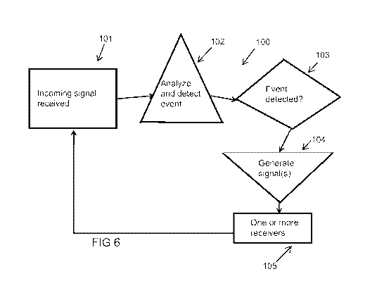
[0022] Figure 6 is a flow chart illustrating the application of the
methods of the
present invention to intentional signals.
[0023] Figure 7 is a flow chart illustrating the application of the
methods of the
present invention to unintentional signals in, e.g., epilepsy.
[0024] Figure 8, data are analyzed using one or more normalizations and
visualizing
and alternating between kicking a football and viewing a bedroom on gamma
frequency
power (A), alpha frequency power (B), summed alpha and gamma frequency power
(C), and
the ratio of summed alpha and ultra high gamma frequency power. The green
lines indicate
the timing of the verbal cue to start, and red lines indicate the time of the
verbal cue to stop
ten second intervals.
[0025] Figure 9 is a screen shot of computer interface using the methods
of the
present invention.
[0026] Figure 10 is a Kolmogorov-Smirnov (KS) two-sample test with 10-
fold, cross-
axes normalized iterative preferred frequency spectrogram from each task as
listed in Figure
1 A-E performed by an immobile ALS subject indicates that more than a single
intended
event can be identified. Each spectrogram is from a distribution different
from that of any
other task (and to no lesser extent in this particular trial left hand
squeezing from right foot
squeezing in this subject).
DETAILED DESCRIPTION
[0027] It is to be understood that the figures and descriptions of the
present invention
have been simplified to illustrate elements that are relevant for a clear
understanding of the
invention, while eliminating, for purpose of clarity, other elements that may
be well known.
The detailed description will be provided herein below with reference to the
attached
drawings.
[0028] A detailed description of one or more embodiments of the invention
is
7
CA 02862524 2014-07-23
WO 2013/112771 PCT/US2013/023033
provided below along with accompanying figures that illustrate the principles
of the
invention. The invention is described in connection with such embodiments, but
the
invention is not limited to any embodiment. The scope of the invention is
limited only by the
claims and the invention encompasses numerous alternatives, modifications and
equivalents.
Numerous specific details are set forth in the following description in order
to provide a
thorough understanding of the invention. These details are provided for the
purpose of
example and the invention may be practiced according to the claims without
some or all of
these specific details. For the purpose of clarity, technical material that is
known in the
technical fields related to the invention has not been described in detail so
that the invention
is not unnecessarily obscured.
[0029] The term "subject" in this application refers to both animals and
humans.
Referring now to Figure 6, wherein is disclosed a flow chart 100 illustrating
a preferred
embodiment of the present methods. Specifically, the subject visualizes and
creates an
intentional brain signal that is received by at least a single sensor 101
according to the present
invention. Preferably, this sensor comprises a single wet electrode or a
single dry electrode.
The incoming signal is relayed to a computational device, such as a computer,
where it is
analyzed 102 to determine if a defined event has occurred 103. Preferably,
this indicative of
brain activity that correlates to a higher cognitive function from the
subject. The computer
then generates a signal 104 that is relayed to one or more receivers 105 on a
peripheral
device. Examples of peripheral devices may include voice synthesizers,
prosthetic devices,
including exoskeletons and the like. As noted in Figure 6, these methods
preferably occur in
real time and provide a continuous response and feedback look between the
intentional
signals of the subject and the translation of those signals into commands
associated with the
peripheral devices controlled by intentions of the subject. In an especially
preferred
embodiment, the data signal from the subject is received from at least a
single channel of
EEG, EMG, EOG, MEG, ECoG, iEEG, fMRI, LFP or a peripheral channel modulated by
the
subject's intention. In a preferred alternative, the data is received through
a multi channel
detector that interfaces wirelessly with the computing device and peripheral
device(s).
[0030] Referring now to Figure 7, wherein is disclosed another flow chart
106
illustrating an alternative preferred embodiment of the present methods.
Specifically, the
subject is monitored for presence of unintended events (e.g., a stroke or a
seizure) via a brain
8
CA 02862524 2014-07-23
WO 2013/112771 PCT/US2013/023033
signal that is received by at least a single sensor 107 according to the
present invention.
Preferably, this sensor comprises a single wet electrode or a single dry
electrode. The
incoming signal is relayed to a computational device, such as a computer,
where it is
analyzed 108 to determine if the defined unintentional event has occurred 109.
There are
numerous examples of potentially harmful, unintentional events, including, but
not limited to,
a surprise, a tremor, a spasm, an injury or a pathology including but not
limited to, an
epileptic seizure, a migraine, a stroke, a heart attack or an infarction.
[0031] The computer then generates a signal 110 that is relayed to one or
more
receivers on a peripheral device. In this instance, the peripheral device is
stimulating device
111 used to ameliorate the effect of an impending seizure in and epileptic
patient by
stimulating the brain region 112 of the subject. As will be appreciated by
those skilled in the
art, the peripheral device 111 may also suppress or not change (or effect) the
subject 112.
For example, the triggering of an alarm to alert the subject or a caregiver.
In an especially
preferred embodiment, the data signal from the subject is received from at
least a single
channel of EEG, EMG, EOG, MEG, ECoG, iEEG, fMRI, LFP or a peripheral channel
modulated by the unintentional occurrence of the unintended event being
monitored. In a
preferred alternative, the data is received through a multi channel detector
that interfaces
wirelessly with the computing device and peripheral device(s).
[0032] In another preferred embodiment of the methods illustrated in both
Figures 6
and 7, the data is analyzed by normalizing a spectrogram, including a
normalized
spectrogram, of the data at least once, time over frequency, and normalizing
the spectrogram,
including a normalized spectrogram, of the same data at least once, frequency
over time,
where both normalizations can be performed in either order and can be
iterated. In a further
preferred embodiment, the data is analyzed by computing the spectrogram of the
data,
normalizing the spectrogram, performing an independent or principal component
analysis of
the normalized spectrogram, and identifying clusters. In addition, the
analyzing step can also
include performing a temporal fragmentation analysis, preferred frequency
analysis, an
iterated (preferably two times or more) preferred frequency analysis, and/or
spectral
fragmentation analysis.
9
CA 02862524 2014-07-23
WO 2013/112771 PCT/US2013/023033
[0033] In a preferred embodiment, detecting brain signals (e.g., high-
frequency
signals detected from the brain) and correlating the detected signals to
higher cognitive
function is disclosed. Examples of higher cognitive functions include, among
others, intent,
speech, memory recall, thought, imagination, and planning, including but not
limited to
motion, and directed task involving imagination and other cognitive processes
and functions.
[0034] In some embodiments, a brain signal of a subject is translated
into speech. For
example, when the subject imagines one or more elements of language, including
but not
limited to a letter, number, word or symbol, a brain signal associated with
this or multiple
elements of language, is detected and this or multiple elements of language is
determined. In
another example, a subject with impaired speech capability is taught to
associate an image,
symbol, word, letter or number with an imaginary movement. When the subject
imagines the
imaginary movement, a brain signal associated with the imaginary movement is
detected and
the word, letter or number associated with imaginary movement is determined.
In an
embodiment, the subject's imaginary movement controls a cursor on a display
which selects
the image, symbol, word, letter or number the subject intends to use. In some
embodiments,
the determined image, symbol, word, letter or number is used with one or more
other
determined images, symbols, words, letters or numbers to construct grammatical
speech. In
some embodiments, the determined image, symbol, word, letter or number is
communicated
using voice synthesizers and/or displayed. In some embodiments, the brain
signal and/or the
output is assigned a non-linguistic value including but not limited to a tone,
series of tones,
micropitch, color, image, electrical stimulation, uni or multidimensional
graphic. In some
embodiments, the brain signal and/or the output is assigned to a brain signal.
In some
embodiments, the brain signal follows and/or precedes and/or occurs
simultaneously with one
or more endogenous and/or exogenous event and/or state including, but not
limited to a
pathological and/or altered event and/or state.
[0035] In some embodiments, a brain signal of a subject is translated to
a movement
of an artificial prosthesis. For example, a detected brain signal associated
with an imaginary
movement is used at least in part to control an artificial prosthesis.
[0036] In some embodiments, one or more physiological recordings,
including but not
limited to EEG, EMG, EOG, MEG, ECoG, iEEG, fMRI, LFP, or a peripheral channel
CA 02862524 2014-07-23
WO 2013/112771 PCT/US2013/023033
modulated by the subject's intention or reading unintentional signals for
unintended events is
used to detect one or more brain signals. For example, a Single-Channel iBrain
EEG
recording is conducted on a high-functioning 70 year old ALS patient
attempting to move one
of four limbs after a verbal cue: the left and right hand and foot. EEG
signals are analyzed
with algorithms, including the SPEARS algorithm, in order to make brain
signals detectable.
Concurrent video recordings may be obtained. During the attempted movements,
the
subject's brain activity demonstrates broad spectrum pulses extending to the
Gamma and
ultra-high Gamma ranges. Such pulses are present in the absence of actual
movement and
absent when the subject was not attempting motion. Activity in the Alpha range
is detected
when the subject closed his eyes. Such high bandwidth biomarkers opens the
possibility to
link intended movements to a library of words and convert them into speech,
providing ALS
sufferers with communication tools utilizing brain signals.
[0037] In some embodiments, clear broad spectrum patterns of activation
across
many frequencies can be detected for actual or imaginary movements as compared
to
different patterns for the resting state. These patterns match the timing of a
subject's actual or
imaginary movements. Traditional spectral analysis does not reveal such
patterns. Analytics
on physiological data may be used to detect brain signals that correlate in
time with a
subject's actual or imaginary movements. In some embodiments, these signals
are transmitted
and parsed in real time to provide additional degrees of freedom for brain
based
communication.
[0038] The methods described herein are disclosed in detail in
International Patent
Application Number PCT/U52006/018120; International Patent Application Number
PCT/U52009/064632; International Patent Application Number PCT/U52010/054346;
US
Patent Application Number 8,073,574; and Low, Philip Steven (2007). "A new way
to look at
sleep: separation and convergence". Published Thesis, University of California
San Diego
Electronic Theses and Dissertations (Identified: b6635681), the disclosures of
which are
herein incorporated by reference in their entirety.
[0039] The present invention utilizes a system and method to obtain and
classify EEG
data in both animals and humans. Obtained EEG signals are low-power frequency
signals and
11
CA 02862524 2014-07-23
WO 2013/112771 PCT/US2013/023033
follow a 1/f distribution, whereby the power in the signal is inversely
related, e.g., inversely
proportional, to the frequency.
[0040] EEG signals have typically been examined in time in series
increments called
epochs. The epochs can be segmented into different sections using a scanning
window, where
the scanning window defines different sections of the time series increment.
The scanning
window can move via a sliding window, where sections of the sliding window
have
overlapping time series sequences. An epoch can alternatively span an entire
time series, for
example.
[0041] In a preferred embodiment of this invention a single channel of
EEG was
sufficient to obtain the data indicative of intentional (or unintentional or
other unexpected)
brain activity.
[0042] Typically, the source data obtained with the methods of the
present invention
is adjusted to increase the dynamic range for power within at least one low
power frequency
range of the frequency spectrum of the source data as compared to a second
higher power
frequency range. A number of adjustment techniques described herein, including
normalization and frequency weighting can be used.
[0043] In an embodiment, electroencephalography source data is normalized
to
increase the low power, higher frequency range data relative to the higher
power, lower
frequency range data or, more generally, to normalize the powers of the
different signal parts.
[0044] After the source data is adjusted, various other processing can be
done. For
example, a visualization of the adjusted source data can be presented.
Further, low power
frequency information can be extracted from the adjusted source data. For
example, low
power frequency information can be extracted from adjusted
electroencephalography source
data. Higher power frequency information can also be extracted from the
adjusted source
data.
12
CA 02862524 2014-07-23
WO 2013/112771 PCT/US2013/023033
[0045] The method described in this or any of the other examples can be a
computer-
implemented method performed via computer-executable instructions in one or
more
computer- readable media. Any of the actions shown can be performed by
software
incorporated within a signal processing system or any other signal data
analyzer system. For
example, The invention can be implemented in numerous ways, including as a
process; an
apparatus; a system; a composition of matter; a computer program product
embodied on a
computer readable storage medium; and/or a processor, such as a processor
configured to
execute instructions stored on and/or provided by a memory coupled to the
processor. In this
regard, Figure 9 is a screen shot of a computer interface utilizing the
methods and outputs of
the present invention. In this specification, these implementations, or any
other form that the
invention may take, may be referred to as techniques. In general, the order of
the steps of
disclosed processes may be altered within the scope of the invention. Unless
stated
otherwise, a component such as a processor or a memory described as being
configured to
perform a task may be implemented as a general component that is temporarily
configured to
perform the task at a given time or a specific component that is manufactured
to perform the
task. As used herein, the term 'processor' refers to one or more devices,
circuits, and/or
processing cores configured to process data, such as computer program
instructions.
[0046] Another embodiment uses multiple normalizations for even further
dynamic
range increase. Normalizations can be performed by normalizing frequency
across time or
time across frequency.
[0047] For example, electroencephalography data with at least one low
power
frequency range can be received. Artifacts in the data can be removed from the
source data.
For example, artifact data can be manually removed from the source data or
automatically
filtered out of source data via a filtering (e.g., DC filtering) or data
smoothing technique. The
source data can also be pretreated with component analysis (e.g., principle or
independent
component analysis). The source data is segmented into one or more epochs;
where each
epoch is a portion of data from the series. For example, the source data can
be segmented into
a plurality of time segments via a variety of separating techniques. Scanning
windows and
sliding windows can be used to separate the source data into time series
increments. The one
or more epochs are normalized for differences in power of the one or more
epochs across
13
CA 02862524 2014-07-23
WO 2013/112771 PCT/US2013/023033
time. For example, the power of each epoch at one or more frequencies can be
normalized
across time to determine appropriate frequency windows for extracting
information. Such
normalization can reveal low power, statistically significant shifts in power
at one or more
frequencies (e.g., Delta, Gamma, Alpha and the like). Any frequency range can
be revealed
and utilized for analysis. Information can be calculated for each of the one
or more epochs
after appropriate frequency windows have been established. Such information
can include
low frequency power (e.g., Delta power), high frequency power (e.g., Gamma
power),
standard deviation, maximum amplitude (e.g., maximum of the absolute value of
peaks) and
the sort. Further calculations can be done on the information calculated for
each of the one or
more epochs creating information such as Gamma power/Delta power, time
derivative of
Delta, time derivative of Gamma power/Delta power and the like. Time
derivatives can be
computed over preceding and successive epochs. After calculating the
information, that
information can then be normalized across the one or more epochs. A variety of
data
normalization techniques can be conducted including z-scoring and other
similar techniques.
[0048] Results of the adjustment of source data to account for
differences in power
over a spectrum of frequencies over time can be presented as one or more
epochs of data. For
example, frequency weighted epochs can be presented as adjusted source data.
[0049] Electroencephalography data for a subject is obtained and input to
segment the
data into one or more epochs. In practice, epochs are of similar (e.g., the
same) length.
Epoch length can be adjusted via a configurable parameter. The one or more
epochs, in turn,
are input to normalize frequency data in the one or more epochs across time,
thereby
frequency weighting the one or more epochs of electroencephalography data. The
one or
more frequency weighted epochs are then input into classifier to classify the
data into states
of intention versus relaxation or non-intention.
[0050] For Example, electroencephalography (EEG) data for a subject is
received.
For example, electroencephalography data, which exhibits lower dynamic range
for power in
at least one low power first frequency range in a frequency spectrum as
compared to a second
frequency range in the frequency spectrum, can be received. The
electroencephalography
data for the subject is segmented into one or more epochs. For example, the
EEG data can be
14
CA 02862524 2014-07-23
WO 2013/112771 PCT/US2013/023033
segmented into one or more epochs via a variety of separating techniques.
Scanning windows
and sliding windows can be used to separate the EEG data into one or more
epochs. The
source data can also be filtered via direct current activity during, prior to,
or after segmenting.
The source data can also be pretreated with component analysis (e.g.,
principle or
independent component analysis). In entire night EEG data the higher
frequencies (e.g.,
Gamma) exhibit lower power than the lower frequencies (e.g., Alpha, Delta,
Theta and the
like) in the whole night EEG data. Frequency power of the one or more epochs
is weighted
across time. For example, the power of each epoch at one or more frequencies
can be
normalized across time to determine appropriate frequency windows for
extracting
information. Such normalization can reveal low power, statistically
significant shifts in power
at one or more frequencies (e.g., Alpha, Delta, Gamma, and the like).
Additionally, each
epoch can be represented by the frequency with the highest relative power over
time to
determine appropriate frequency windows for extracting information.
Alternatively,
component analysis (e.g., principle component analysis (PCA) or independent
component
analysis (ICA)) can be utilized after normalization to further determine
appropriate frequency
windows for extracting information. Any frequency range can be revealed and
utilized for
analysis.
[0051] Information can be calculated for each of the one or more epochs
after
appropriate frequency windows have been established (e.g., after weighting
frequency). Such
information can include low frequency power (e.g., Alpha power), high
frequency power
(e.g., Gamma power), standard deviation, maximum amplitude (e.g., maximum of
the
absolute value of peaks) and the sort. Further calculations can be done on the
information
calculated for each of the one or more epochs creating information such as
Gamma
power/Alpha power, time derivative of Delta, time derivative of Gamma
power/Alpha power
and the like. Time derivatives can be computed over preceding and successive
epochs. After
calculating the information, it can then be normalized across the one or more
epochs. A
variety of data normalization techniques can be conducted including z-scoring
and the like.
The higher frequency data is now more clearly visible.
[0052] Intention states in the subject are classified based on the one or
more
frequency weighted epochs. For example, the one or more frequency weighted
epochs can be
CA 02862524 2014-07-23
WO 2013/112771 PCT/US2013/023033
clustered by any variety of clustering techniques including k- means
clustering. The
clustering can be done on information calculated from the epochs (e.g., Alpha
power, Gamma
power, standard deviation, maximum amplitude (Gamma/Alpha), time derivative of
Delta,
time derivative- of (Gamma /Alpha, and the sort) . Component analysis (e.g.,
PCA or ICA)
can be used to determine the parameter space (e.g., types of information used)
in the
clustering.
[0053] Subsequent to clustering, intention state designations can be
assigned to the
epochs. Intention state designated epochs can then be presented as
representations of
intention and relaxation (non-intention) states in the subject for the period
of time represented
by the epoch. Classification can also incorporate manually determined
intention states (e.g.,
manually determined "intended activity" versus "relaxation" states).
Additionally, artifact
information can be utilized in the classification.
[0054] Artifact data can also be used in intention state classification.
For example,
artifacts can be used to analyze whether epochs initially assigned a intention
state designation
should be reassigned a new intention state designation due to neighboring
artifact data. In
such ways, for example, artifact data can be utilized in a data smoothing
technique.
[0055] Any variety of data smoothing techniques can be used during the
assigning of
intention states. For example, numbers (e.g., 0 and 1) can be used to
represent designated
intention states. Neighboring epochs' brain state designation numbers can then
be averaged to
determine if one of the epochs is inaccurately assigned a intention state
designation.
Therefore, should a group of epochs be assigned intention state designations
representing
abrupt jumps in brain states, smoothing techniques can be applied to improve
the accuracy of
the assigning.
[0056] Previous embodiments have shown how normalization, for example
using Z
scoring, allowed analysis of more information from the brain activity signal.
The analysis
which was previously carried out normalized power information across
frequencies. The
normalization preferably used Z scoring, but any other kind of data
normalization can be
used. The normalization which is used is preferably unitless, like Z scoring.
As well-known
16
CA 02862524 2014-07-23
WO 2013/112771 PCT/US2013/023033
in the art, z scoring can be used to normalize a distribution without changing
a shape of the
envelope of the distribution. The z scores are essentially changed to units of
standard
deviation. Each z score normalized unit reflects the amount of power in the
signal, relative to
the average of the signal. The scores are converted into mean deviation form,
by subtracting
the mean from each score. The scores are then normalized relative to standard
deviation. All
of the z scored normalized units have standard deviations that are equal to
unity.
[0057] While the above describes normalization using Z scores, it should
be
understood that other normalizations can also be carried out, including T
scoring, and others.
Multiple normalizations may also be employed. Normalizations can be performed
by
normalizing frequency across time or time across frequency.
[0058] The above embodiments describe normalizing the power at every
frequency
within a specified range. The range may be from 0, to 100 hz, or to 128 hz, or
to 500 hz. The
range of frequencies is only restricted by the sampling rate. With an
exemplary sampling rate
of 30KHz, an analysis up to 15KHz can be done.
[0059] According to the present embodiment, additional normalizations are
carried
out which normalizes the power across time for each frequency. This results in
information
which has been normalized across frequencies and across time being used to
create a
normalized spectrogram. This embodiment can obtain additional information from
brainwave
data, and the embodiment describes automatically detecting different periods
of intention and
relaxation from the analyzed data. According to an important feature, a single
channel of
brainwave activity (that is obtained from a single location on the human
skull) is used for the
analysis. As described above, the obtained data can be one channel of EEG
information from
a human or other subject. The EEG data as obtained can be collected, for
example, using a
256 Hz sampling rate, or can be sampled at a higher rate. The data is divided
into epochs, for
example 30 second epochs, and characterized according to frequency.
[0060] A first frequency normalization is carried out. The power
information is
normalized using a z scoring technique on each frequency bin. In the
embodiment, the bins
may extend from one to 100 Hz and 30 bins per hertz. The normalization occurs
across time.
17
CA 02862524 2014-07-23
WO 2013/112771 PCT/US2013/023033
This creates a normalized spectrogram or NS, in which each frequency band from
the signal
has substantially the same weight. In the embodiment, each 30 second epoch is
represented
by a "preferred frequency" which is the frequency with the largest z score
within that epoch.
[0061] This creates a special frequency space called the Preferred
Frequency space.
Analysis of how those patterns are formed and analysis of the characteristics
of the patterns
can be done. Different brain states, therefore, can be defined according to a
discrimination
function, where the discrimination function looks for certain activity in
certain areas, and
non-activity in other areas. The function may evaluate brain states according
to which of the
frequency at areas have activity and which do not have activity.
[0062] More generally, however, any form of dynamic spectral scoring can
be carried
out on the compensated data. The discrimination function may require specific
values, or may
simply require a certain amount of activity to be present or not present, in
each of a plurality
of frequency ranges. The discrimination function may simply match envelopes of
frequency
response. The discrimination function may also look at spectral fragmentation
and temporal
fragmentation.
[0063] A second normalization which is carried out across frequencies.
The second
normalization produces a doubly normalized spectrogram. This produces a new
frequency
space, in which the bands become even more apparent. The doubly normalized
spectrogram
values can be used to form filters that maximally separate the values within
the space.
[0064] A clustering technique which is carried out on the doubly
normalized
frequency. For example, the clustering technique may be a K means technique as
described in
the previous embodiments. Each cluster can represent an intention state.
[0065] The clusters are actually multi dimensional clusters, which can
themselves be
graphed to find additional information. The number of dimensions can depend on
the
number of clustering variables. This illustrates how the doubly normalized
spectrogram also
allows many more measurement characteristics.
18
CA 02862524 2014-07-23
WO 2013/112771 PCT/US2013/023033
[0066] Measurement of the average spread in normalized power across
frequency
which illustrates the spectral fragmentation is also possible. Fragmentation
values can
alternatively be based on temporal fragmentation for the different states may
also be used as
part of the discrimination function.
[0067] These two functions are evaluated on the doubly normalized
spectrum,
relying on homogeneous increases in gain at all frequencies as caused movement
artifacts and
would lead to abnormally elevated fragmentation values in the singly
normalized spectrum.
These fragmentation values may be used as part of the discrimination function.
Importantly,
and as described above, this discrimination function is typically not apparent
from any
previous analysis technique, including manual techniques.
[0068] The computation may be characterized by segmenting, or may use
overlapping
windows or a sliding window, to increase the temporal registration. This
enables many
techniques that have never been possible before. By characterizing on-the-fly,
this enables
distinguishing using the dynamic spectral scoring, between relaxation states
and intention
states using the brainwave signature alone.
[0069] The exemplary methods for data analysis described above were
combined
with a standard non-invasive EEG method for humans. The result is the ability
to non-
invasively extract attenuated rhythms in animals, automatically analyze the
brain activity
from a single channel of EEG, and sufficiently classify the brain state
parameters for the
animals.
EXAMPLE 1
[0070] Single-Channel iBrain EEG recordings were conducted in a high-
functioning
70 year old ALS patient attempting to move one of four limbs after a verbal
cue: the left and
right hand and foot. Raw EEG signals were analyzed with the SPEARS algorithm
in part to
make high-frequency/low spectral power signals detectable. Concurrent video
recordings
were obtained. During the attempted movements, the subject's brain activity
demonstrated
distinct broad-spectrum pulses extending to the Gamma and ultra-high Gamma
ranges. Such
19
CA 02862524 2014-07-23
WO 2013/112771
PCT/US2013/023033
pulses were present in the absence of actual movement and absent when the
subject was not
attempting motion. Activity in the Alpha range was detected when the subject
closed his
eyes, as expected. The use of such high bandwidth biomarkers based on intended
movements
to a library of words will allow the conversion of the signals into speech,
thus providing ALS
sufferers with communication tools more dependent on the brain than on the
body.
[0071]
Specifically, application of the methods of the present invention reveals high
frequency patterns matching the timing of a subject's actual, imagined, or
intended
movements. In an example application, the frequency spectrum is generated from
the time
series data and normalized to reveal these higher frequencies which a standard
method does
not reveal. An application of this to brain EEG data is shown in Figure 1. In
this example, a
high-functioning 70-year-old subject, who is an ALS patient and immobile, was
asked to
close the eyes, rest, or imaging a hand or foot. For each of these tasks, the
subject was given
a verbal cue to begin the task and another cue after four seconds to stop. Six
seconds later the
task was repeated, for a total of 12 attempts per task and 120 seconds of
time. Figure 1
shows the standard frequency power spectrum in (i) and the enhanced frequency
power
spectrum in (ii) of the ALS subject while performing these tasks. The stronger
signals appear
redder while the weaker signals decrease in intensity through orange, yellow,
and blue
shades. Clear bands of high frequency activity which approximate the timing of
the cues to
begin and end the task appear with high intensity in the enhanced spectrum.
Figure 1 A
presents this for the task of closing eyes for four seconds. Figure 1 B
presents this for the
task of imagining or attempting to squeeze the left hand for four seconds and
relax for six
seconds. Figure 1 C, D, and E show the same, but for the tasks of imagining or
attempting to
squeeze the right hand, left foot, and right foot, respectively. In all cases,
the spectrogram
generated in this application clearly reveals the high frequency spectral
content
approximating the timing of the attempt by the subject to perform the
indicated task. Figure
1 F presents the same spectral analyses for the same subject, but at rest, and
displays a very
different timing of high frequency content, which may represent ambient noise,
background
talk, and/or baseline electrical activity.
CA 02862524 2014-07-23
WO 2013/112771 PCT/US2013/023033
EXAMPLE 2
[0072] Application of the methods of the present invention in the ALS
patient
described in Example 1 reveals components of the data which approximate time
of events.
Figure 2 presents one such application of the methods of the present invention
(here with
single channel component analysis on the doubly normalized spectrogram) to
reveal
uncorrelated independent data components. Figure 2 A-E are data from the same
tasks as
indicated in Figure 1 A-E. Each plot shows the resulting extracted independent
component,
after the full analysis as described, having peaks approximating the
individual tasks
attempted by the subject. The green lines indicate the timing of the verbal
cue to start the
four-second task, and red lines indicate the time of the verbal cue to stop
the task and relax
for six seconds. Peaks in each component generally align with the start of the
task being
performed (indicated by peak points at, on, or just after the green lines).
This analysis can
also be combined with that in Example 1 to strengthen or corroborate event
detection and
timing.
EXAMPLE 3
[0073] Application of the methods of the present invention in the ALS
patient
described in Example 1 to single channel brain EEG data to assess data
stability and reveal
changes in data stability which represent intended actions are provided.
Specifically,
application of the methods of the present invention to generate temporal
fragmentation
reveals changes in stability which approximate the timing and duration of
intended actions.
Figure 3 A ¨ E present the temporal fragmentation of the same tasks as
indicated in Figure 1
A-E. In each plot, the shift in points from negative to positive represents a
decrease in
stability. These shifts approximate both the verbal cue to start the task
(green lines) as the
stability starts to decrease and the positive shift begins, and the verbal cue
to end the task (red
lines) as the data begin to stabilize end the positive shift ends. Zero-line
crossings (purple
dots) increase for unstable periods (during or just after the task) and
decrease in count for
stable periods (relaxation period before or after each task). These shifts and
line crossings
can also be combined with methods in Examples 1 to strengthen or corroborate
event
detection and timing.
21
CA 02862524 2014-07-23
WO 2013/112771
PCT/US2013/023033
EXAMPLE 4
[0074]
Detection of high frequency events correlated with low frequency events and
event timing in the ALS patient of Example 1. Generation of the spectrogram
followed by
one or more normalizations across one or both data axes, followed by feature
sharpening
using the doubly normalized spectrogram, followed by extraction of standard
known
frequencies, frequency ranges, their sums, their ratios, and/or other
frequency relationships,
reveals event timing, spacing, and/or duration. Figure 4 A-E present tasks as
in Figure 1 A-
B, plotting summed high frequency power in the gamma and ultra high gamma
(hgamma)
range of frequencies (all those >30Hz). In brain EEG, increased power in these
frequencies
correlate with heightened concentration such as occurs when imagining moving a
limb.
Peaks in the high frequency power summations approximate the timing of the
task, indicated
by the columns of green (task start) and red (task end) lines. Figure 5 A-D
present tasks as in
Figure 1, for squeezing right hand, left hand, right foot, and left foot,
respectively, plotting
the ratio of the summed alpha frequency (8-13Hz) power to the summed gamma
frequency
power (30-50Hz). Increase power in the alpha frequencies in brain EEG
correlates with
relaxation of mental effort such as after a length of time attempting to
squeeze a limb. Thus,
alpha and gamma frequency power are inversely related for these tasks and
peaks in the
alpha-to-gamma ratio appear in between tasks (between red line indicating stop
and green
line indicating start again). Figure 5 E shows the analysis of the delta
frequency (<5Hz)
during the close eyes task. Delta frequency power correlates with changes in
eye open or
closed state and approximate the timing of the subject closing (green lines)
and opening (red
lines) eyes. These and other frequency analyses can be combined with each
other and the
methods in Examples 1, 2, and 3 to strengthen or corroborate event detection
and timing, as
well as to characterize the event with the known related brain state implied
by the frequencies
analyzed.
EXAMPLE 5
[0075]
Simultaneous detection of multiple events co-occurring in time for a second
ALS patient. Generation of the spectrogram followed by one or more
normalizations across
one or both data axes, followed by feature sharpening, followed by extraction
of standard
known frequencies, frequency ranges, their sums, their ratios, and/or other
frequency
relationships, reveals multiple events in one analysis. Figure 8 shows data
from a task
22
CA 02862524 2014-07-23
WO 2013/112771 PCT/US2013/023033
designed to elicit both an increase in concentration (increase gamma frequency
power) and
shifts in thought which would display as reduced and increased relaxation
(changes in alpha
frequency power). An immobile ALS patient subject was instructed to alternate
between two
10-second imaginings (kicking a football, green lines, and viewing the
bedroom, red lines),
repeating 5 times. Figure 8 A plots the normalized and enhanced gamma
frequency power,
displaying a sharp increase at the start of the alternation sequence (first
green line, at 13
seconds) and subsequent decrease at the end of the full sequence (last red
line, at 103
seconds). Figure 8 B plots the normalized and enhanced alpha frequency power,
showing
changes in relaxation states between the two different imaginings (peaks
between colored
lines). Figure 8 C plots the normalized and enhanced alpha and gamma power,
showing the
simultaneous detection of both the plateau of gamma frequency power during the
set of
imaginings, and the alpha peaks when changing imaginings, as two unique
signals detected in
one analysis. Figure 8 D plots the ratio of the normalized and enhanced alpha
to ultra high
gamma (hgamma), with a drop in the ratio at the beginning of the sequence, the
individual
peaks at each imagining, and a rise in the ratio after the sequence.
EXAMPLE 6
[0076] Use of the iteratively normalized spectrogram to differentiate and
characterize
more than one type of intended events. Analysis including application of the
SPEARS
algorithm followed by a Klomogorov-Smirnov (KS) two-sample test for same-
distribution
sampling between any two spectrograms reveals distinguishable imagined motor
movements.
This application enables multiple degrees of freedom based on at least one
event type being
differentiable from others. KS test p values of the 10-fold, cross-axes
normalized
spectrogram from each task as listed in Figure 1 A-E performed by an immobile
ALS subject
indicate that each spectrogram is from a distribution different from rest (P <
0.01) (Table 1)
and from that of almost any other task(P < 0.05) (Figure 10), and to a lesser
extent in the left
hand and right foot in this particular trial, becoming an effective
application for
characterizing the same or multiple events at detection.
23
CA 02862524 2014-07-23
WO 2013/112771
PCT/US2013/023033
Table 1: KS Test p values of imagined tasks against Rest
Klomogorov-Smirnov p Value
Task Compared to Rest
Rest 1.00
Close Eyes <0.001 (1.18 x 10⁻⁶)
Squeeze Left Hand <0.001 (3.15 x 10⁻¹⁷)
Squeeze Right Hand <0.001 (6.81 x 10⁻⁴)
Squeeze Left Foot <0.001 (8.32 x 10⁻⁹)
Squeeze Right Foot <0.001 (1.64 x 10⁻¹⁶)
[0077] Throughout this application, various publications, patents, and/or
patent
applications are referenced in order to more fully describe the state of the
art to which this
invention pertains. The disclosures of these publications, patents, and/or
patent applications
are herein incorporated by reference in their entireties, and for the subject
matter for which
they are specifically referenced in the same or a prior sentence, to the same
extent as if each
independent publication, patent, and/ or patent application was specifically
and individually
indicated to be incorporated by reference.
[0078] Although only a few embodiments have been disclosed in detail
above, other
embodiments are possible and the inventors intend these to be encompassed
within this
specification. The specification describes specific examples to accomplish a
more general
goal that may be accomplished in another way. This disclosure is intended to
be exemplary,
and the claims are intended to cover any modification or alternative which
might be
predictable to a person having ordinary skill in the art. For example, other
applications are
possible, and other forms of discrimination functions and characterization is
possible. While
the above extensively described characterizing the frequency in terms of its
"preferred
frequency", it should be understood that more rigorous characterization of the
information
may be possible. Also, while the above only refers to determining intention
states from the
24
CA 02862524 2014-07-23
WO 2013/112771
PCT/US2013/023033
EEG data, and refers to only a few different kinds of determination of
intention states, it
should be understood that other applications are contemplated.
[0079]
Having illustrated and described the principles of the invention in exemplary
embodiments, it should be apparent to those skilled in the art that the
described examples are
illustrative embodiments and can be modified in arrangement and detail without
departing
from such principles. Techniques from any of the examples can be incorporated
into one or
more of any of the other examples. It is intended that the specification and
examples be
considered as exemplary only, with a true scope and spirit of the invention
being indicated by
the following claims.