Note: Descriptions are shown in the official language in which they were submitted.
CA 02863230 2014-09-11
METHOD AND APPARATUS FOR THE DETECTION OF IMPAIRED
DARK ADAPTATION
111,LD OF TEM DISCLOSURE
The present disclosure relates to methods and apparatus for the diagnosis of
impaired dark
adaptation and/or the identification of individuals who are at-risk, of
disease states related to
impaired dark adaptation.
BACKGROUND
The macula of the human eye, which is about 6 min in diameter and covers the
central
21.5 degrees of visual angle, is designed for detailed vision. The macula
comprises a small cone-
dominated fovea surrounded by a rod-dominated pamfovea (Curcio 1990, J. Comp.
Neural.
292:497). Rods are responsible for vision in dim light while cones are
responsive to bright light
and colors. in young adults, the number of rods outnumbers cones by
approximately 9:1. This
proportion of rods to cones changes as individual's age. The health and
function of the rod and
cone photoreceptors are maintained by the retinal pigment epithelium (RPE),
the Bruch's
membrane and the choriocapillaris (collectively referred to as the RPE/Bruch's
membrane
complex). The RPE is a dedicated layer of nurse cells behind the neural
retina. The RPE sustains
photoreceptor health in a number of ways, including, but not limited to,
maintaining proper ionic
balance, transporting and filtering nutrients, providing retinoid
intermediates to replenish
photopigraent bleached by light exposure and absorbing stray photons. The RPE
and the
photoreceptors are separated by the choriocapillaris, which provides blood
flow to the neural
retina. Further separating the RPE and the choriocapillaris is the Eruct] 's
membrane, a delicate
vessel wall only 2-6 ,urn thick.
As the function of the RPE/Bruch's membrane complex is impaired, the result is
deficient
nutrient and oxygen transport to the photoreceptors and reduced clearance of
by-products of
bleaching, such as opsin. Therefore, as a result of the impairments of the
function of the
RPE/Bruch's membrane complex, the health and function of the photoreceptors
may be impaired.
This is especially true with the rod. photoreceptors, which are responsible
for scotopie, or dark-
adapted vision. The impairment of the rod photoreceptors may lead to
impairment in dark
adaptation. Dark adaptation is defined as the recovery of light sensitivity by
the retina in the dark
after exposure to a bright light In this regard, dark adaptation can
essentially be viewed as a
bioassay of the health of the RPE, the Brach's membrane and the
choriocapillaris, and impaired
1
CA 02863230 2014-09-11
=
dark adaptation. may be used as a clinical marker of disease dates that impair
one or mere of the
RPE, the Brach's membrane and the choriocapillaria. Such disease states
include, but are net
limited to age-related macular degeneration (ARMD; which is also known as age-
related
maculepat4 ARM), vitamin A deficiency, Sorsby's Fundns Dystrophy, late
autosomal dominant
retinal degeneratica; retinal impairment related to diabetes and diabetic
retinopathy. Patients
with ARMD often have impaired dark adaptation as a result of the
pathophysiology associated
with ARMD. Dark adaptation may be particularly useful in this regard since
deficits in dark
adaptation generally occur before clinical manifestations of the disease state
become evident.
Currently ARMD is the leading cause of new, untreatable vision loss in the
elderly
populations of the industrialized world (Ivfiteliell 1995, Ophthalmology,
102:1450; Vingerling
1995, Ophthalmology, 102;205). With the increasing proportion of old adults in
industrialized
countries, the impact of ARMD on health care costs will worsen (Council 1998,
Vision Research-
A National plan 1999-2003; Executive Summary). ARMD is a heterogeneous
disorder and is
related to the breakdown of one or more components of the RPE/Enuch's membrane
complex.
As discussed above, impairment of the RPE/Eruch's membrane complex can impact
the health
and functionality of the photoreceptors and lead to impaired dark adaptation.
Early to intermediate ARMD is characterized by minor to moderate vision loss
associated
with extracellular lesions, Enid changes in the RPE pigmentation and
morphology. The lesions
between the RPE and the Etuch's membrane can be either focal (referred to as
drusen) or diffuse
(referred to as basal linear deposits). Advanced AR1vID is characterized by
severe vision loss
associated with extensive RPE atrophy with or without the squelea of choroidal
neovascularization (which is the in-growth of choroidal vessels through the
Brach's membrane
and under the RPE in the plane of the theism and/or the basal linear
deposits). In the United
States late stage ARMD accounts for 22% of monocular blindness and 75% of
legal blindness in
adults over the age of 50 (Klein 1995, Opthaanol. Via. Sei 36:182). It is
currently believed that
ARMD is a multi-factorial process involving a complex interplay of genetic and
environmental
factors. The principal tneatment for lane stage ARI.413 is photocoagulation of
the aberrant blood
vessels comprising the choroidal neova.sculatization. However, only a subset
of patients with
existing neovascularization will qualify for such treatment.
A potential treatment approach is to prevent or delay the onset of late stage
ARIAD. For
example, the Age-related Eye Disease Study (2002) indicated that the intake of
several anti-
oxidant compounds (such as beta-carotene, vitamin C and vitamin 13 in
conjunction with zinc and
copper) was beneficial in preventing neovascularization in intermediate ARMD
patients with
drusen in both eyes, which places them at high risk for developing advanced
ARMD (AREDS
report no. 8,2001). A number of therapeutics such as anceortave acetate
(Retaane; Mcon Labs),
2
CA 02863230 2014-09-11
pegaptabmh sodium (Macugun; Eyetech), autibizumab (Locale's; Genetecb) and
combretastatin
(CRP; Oxigene) are in various stages of development. Other treatment options
under
investigation range from brachytherapy to rheopheresis, and observational
studies have been
examining possible protective roles for anti-inflammatory and lipid-lowering
drugs.
However, these approaches require that patients at risk for ARMD or other
disease states
that impact the RPFIBtuch's membrane complex and/or dark adaptation be
identified early
enough so that preventive measures can be undertaken. Furthermore, advising
patients whether
the risk and cost of a treatment is warranted requites the ability to monitor
whether their disease
progression is affected by their course of treatment Such a diagnostic method
suitable for
widespread, clinical use is currently not available in the art The present
disclosure provides such
a method to identify deficits in dark adaptation and describes an apparatus
capable of carrying out
said method_ Such deficits in dark adaptation may be used to identify those at
risk for developing
disease states that impact the RPE/Bruch's metolmme complex and/or dark
adaptation and
tracking the disease/treatment progression among those already affected by the
disease.
BRIEF DESCRIPTION OF THE FIGURES
FIG. 1 is an exemplary dark adaptation curve illustrating the various
components of dark
adaptation.
FIG. 2 compares exemplary dark adaptation curves for a normal old adult
(closed circles), an
early-stage ARAM patient (open triangles) and a late-stage ARMD patient (open
circles).
FIG. 3 is an illustration of the determination of the rod intercept dark
adaptation parameter.
FIG. 4 shows the results of varying the intensity of the bleaching light from
a high intensity (open
circles and squares) to a low intensity (closed circles and squares) on dark
adaptation curves.
FIG, 5A shows a schematic of one embodiment of the apparatus of the present
disclosure
FIG. 5B shows a schematic of one embodiment of the interior of the apparatus
of the present
disclosure as viewed by a subject.
FIG. 6 illustrates dark adaptation curves generated from a normal individual
(closed circle), an
early ARMD patient (open square) an intermediate ARMD patient (open triangle)
and a late
ARM!) patient (open diamond) and shows that impaired dark adaptation can be
used to predict
ARMD disease severity and/or progression.
DETAILED DESCRIPTION
The human macula comprises a small cone-dominated fovea surrounded by a rod-
dominated patafovea. The function of the rod and cone photoreceptors is
impacted by the health
of the components of the RFE/Bruch's membrane complex. As the function of the
RPF/Bruch's
3
CA 02863230 2014-09-11
membrane complex is impaired, the result is deficient nutrient and oxygen
transport to the
pbotoreoeptots and reduced clearance of by-products of bleaching. such as
opsin. Therefore, as a
result oldie impairments of the function oldie RPF/13ruch's membrane complex,
the health and
function of the photorecepton may be impaired. in many cases, the rod
photoreceptors ate
especially vulnerable. The rod photoreceptors are responsible for scotopic, or
dark-adapted
vision. The result of damage to the rod photoreceptors is impaired dark
adaptation in the subject
Therefore, impelled dark adaptation can be a surrogate marker for damage to
the RPE/Hrucles
membrane complex and may be used to diagnose individuals with disease states
that have their
clinical manifestations via their impact on the RPF/Bruch's membrane eomplex
andtor to idert*
those individuals who may be at risk for developing such disease states. Such
disease states,
include but am not limited to, ARMD, vitamin A deficiency, Sorsby's Funks
Dystrophy, late
antosortud dominant retinal degeneration, retinal irepaimeent related to
diabetes and diabetic
retinopathy. However, prior methods for detennking impaired dark adaptation
are cumbersome
and time consuming to administer. -
What the art is lacking is a method to determine impaired dark adaptation
which is a
sensitive and accurate indicator of those patients stiffening Impaired dark
adaptation, which
produces high test-rotate reliability and reproducibility, which can be
administered in the clinical
setting with decreased burden on the subject and the healthcare provider, and
which is simple to
administer. Ile subjects identified with impaired dark adaptation can then be
evaluated for a
variety of disease states, such as, but not limited to, those discussed herein
For example, patients
with impaired dark adaptation can be monitored fire increased risk of ARMD. in
addition,
patients identified with ARMD can be monitored to track ARAM disease
progression, such as but
not limited to, the progression barn early ARAM to intermediate ARMD or
intermediate ARM)
to advancedaate ARvW. Furthermore, such individuals may be started on early
intervention
strategies to prevent or delay the onset of ARMD and the effectiveness of such
intervention
strategies can be monitored.
The present disclosure describes a new method for the measurement of
rod¨mediated dark
adaptation to prospectively identify subjects who have impaired dark
adaptation and who are at-
risk for developing a variety of disease states, such as ARMD, and which meets
limitations
imposed by the clinical setting. The method can be administered in a short
time (in as little as 20
minutes or less) in the clinical setting. As a result, healthcare providers
will be able to offer the
test on a practical and affordable basis, making application of the test and
realization of its
benefits more widespread. In addition, the burden the test imposes on the
subject and the
healthcare provider will be significantly reduced. Importantly, the method and
apparatus
described allows a broader range of subjects to be tested, for instance
children or those with
4
CA 02863230 2014-09-11
impaired cognitive ability. Furthezmore, the subject need not have prior
exposure to
Psyehophysicid test methods. Ark apparatus for administering such a method is
also described.
In addition to its use as a rtitignostic tool, the method described herein can
be used to
identify the structraal, biochemical and physiological changes responsible for
the visual
dysfunction associated with impaired dark adaptation and the progression of
the disease states
associated with impaired dark adaptation, such as, but not limited to, ARM]),
vitamin A
deficiency, Sorsby's Pandas Dystrophy, late autosornal dominant retinal
degeneration, retinal
impairment related to diabetes and diabetic retinopathy. This is particularly
useful since many of
such disease states am currently believed to be a heterogeneous rather than a
unitary genetic
phenomenon and thus may have a variety of clinical manifestations depending on
the underlying
cause. By the early and accurate identification of those individuals at risk
for developing ARMD
and the other disease states discussed herein (by virtue of their
identification as having impaired
dark adaptation) the structural, biochemical and physiological changes can be
identified and
correlated with various stages of disease state progression. Such information
can be used to
design theoretical models of the disease state, evaluate animal models of the
disease state and to
identify new opportunities for therapeutic intervention in the treatment of
the disease state.
The present disclosure presents ARM]) as an exemplary disease state to be
studied using
hnpairment of dark adaptation in a subject However, the method of determining
such dark
adaptation is applicable to the other disease states discussed herein and in
any disease state that
impacts one or more components of the RPE/Bruch's membrane complex.
The present diselosure shows that rod-mediated vision is more severely
affected than
zone-mediated vision in individuals at-risk for incident ARM]) and in early
ARMD patients. In
addition, the impairment of rod-mediated vision appears to precede the
impairment of cone-
nediated vision. This relationship is significant since the most debilitating
vision impairment
associated with ARM]) is caused by the loss of cone photoreceptors. Therefore,
by monitoring
Ire health of the rod-photoreceptors, which as discussed above is also
indicative of the health of
he RFEIBruch's membrane complex, those individuals suffering from or at-risk
for ARM]), can
ie identified. This earlier detection will result in the initiation of
preventive measure, increased
nonitoring and/or early initiation of treatment before cone-photoreceptors are
impaired. As a
vault, the most significant aspect of ARMD-related vision impairment may be
prevented or
Ielayed.
Most ARMD patients exhibit more rod-mediated (scotopic) visual sensitivity
loss than
:one-mediated (photopic) visual sensitivity loss. Rod-mediated dark adaptation
is especially
nsceptible to the effects of ARM]), as discussed in more detail below and many
early ARM])
ratients exhibit abnormal dark adaptation in the absence of other vision
function abnormalities
CA 02863230 2014-09-11
such as reduced acuity, contrast sensitivity or visual sensitivity. Methods do
exist for the
diagnosis and detection of ARMD. flowevez, these methods are insensitive in
that they generally
detect only visible lesions associated with early ARMD (which indicates later
stages of disease
progression), and are subject to a large degree of clinical judgment, moulting
in a range of
interpretations of the test results. Most of the tests are too sophisticated
for the average
healthcare provider to administer. In addition, the interpretation of the test
results requires years
of clinical experience and even then. can be subject to substantial vruiation.
Current teat methods
also place a significant burden on the patient and the healthcare provider. No
suitable method is
currently available for the detection of ARMD that overcomes these obstacles.
Even. if the
currently available tests are admim¨stered, they still do not reliably
identify patients at risk for
ARMD.
As an example, fundns photography and grading clan be used to detect ARMD.
However,
fundes photography is not capable of detecting microscopic lesions or the
biological changes
associated with ARMD. Anatomical and histopathological studies of donor eyes
indicate that the
pathological processes underlying ARMD, and the subsequent damage caused by
these processes,
are well underway before fmulus photography can detect signs of ARMD. In
addition, the test is
relatively expensive, requires specialized equipment and training to
administer and is subject to
variations in interpretation. Reliable interpretation of fundus photographs is
possible by utilizing
a flinch= reading center. However, the use of a fundus reading center for
routine clinical use is
impractical because of the cost and turn around time of the results, which
generally takes several ,
months. As a resat, fundus photography has not been widely used as a means to
diagnose
ARMD. As an additional example, flourosceine angiography is currently used as
the method of
choice for diagnosing late state ARMD. However, this method is invasive as the
flouroaceine
dye must be administered to the subject via the IV route. In addition,
reactions to the
flourosceine dye occur for approximately 1-1000 subjects. These reactions may
be severe and
may even be fatal in some cases. As a result, a physician is required to be in
attendance during
the procedure. Therefore, the burden on the subject and the healthcare
professional is quite high.
General Description of Test Parameters
A general description of the method and the parameters involved in the method
disclosed
is given below. In the method described, dark adaptation is measured with a
custom,
computerized automated adaptom.eter. The subject undergoing testing is subject
to a bleaching
protocol. The bleaching protocol may be varied as is known in the art. The
bleaching protocol
adapts the test eye to a light of a first luminance level (by desensitizing a
potion of the rhodopsin
molecules in the test eye on exposure to the light of a first luminance
level). Visual recovery (Le.
dark adaptation) is then measured as the test eye adapts to a light of a
second luminance leveL
6
CA 02863230 2014-09-11
Therefore, the fast luminance level serves as a standardized baseline from
which visual recovery
is measured. Any bleaching protocol that provides this standardized baseline
may be used in the
method and apparatus dawn-bed heroin. The fast luminance level is brighter
than the second
luminance level, but the absolute intensity values of the fast and second
luminance levels may be
varied as desired. Generally, the greater the absolute value of the first
luminance level, the Shorter
the period of exposure of the test eye to the light of the first luminance
level to achieve the
baseline. For example, the light of the first luminance level may be an
intense light such as that
provided by an electronic strobe or flash, and the light of the second
luminance level may be at or
close to 0 cotliniz, such as would occur in a dark room. Alternatively, the
II& of the first
luminance level may be a light produced by an ordinary light bulb or by the
ambient light in a
room, and the lien of the second luminance level may be at or close to 0
cdima, such as would
*veer in a darkroom.
Many light delivery methods can be used to deliver the light of the first
luminance level
(which is referred In hereafter as a bleaching light), such as photographic
flashes, adapting fields,
illuminated backgrounds, direct projection into the eye, exposure to ambient
light or staring into
a light "bulb. As discussed above, there are numerous possibilities.
Classically, subjects viewed
an adapting field to bleach the photopigment The bleaching method causes
discomfort to the
subject and it is difficult to reliably deliver bleaches in psychophysically
inexperienced subjects.
Another method of bleaching is to project light into the eye using a
Maxwellian view system.
This method causes less irritation, but requires the subjects to fixate very
steadily and not blink
for 30 to 60 seconds. Many inexperienced subjects find this to be a difficult
task If the subject
changes fixation or blinks, it is necessary to wait up to 2 hours before the
bleach is repeated to
avoid the cumulative effects of bleaching. Bleaching light delivered by an
electronic strobe or
flash delivers a high intensity light in a short period of time. Because the
light exposure is brief
and can be localized outside the fovea, it is not irritating to the subjects
and the subjects do not
need to maintain fixation for long period of time. With proper patient
instructions blinking is not
an issue.
The bleaching protocol desensitizes the desired amount of rhodopsin molecules
and
provides a standardized baseline to measure visual recovery to the second
huninance level. The
intensity of the bleaching light or the time of exposure to the bleaching
light can be modulated to
produce the desired amount of desensitization. In one embodiment, an
equivalent of about 50% to
100% of the rhodopsin molecules is desensitized. The bleaching light may be an
achromatic
camera flash. The intensity of the bleaching light can be adjusted to
desensitize the appropriate
amount of rhodopsin molecules. Far example, a bleaching light intensity of
7.48 log scot Td/sec
will bleach approximately 98% of the rhodopsin molecules, while a bleaching
light intensity of
7
CA 02863230 2014-09-11
5.36 log scot Td/sec will bleach approximately 50% of the rhodopsin molecules.
Alternate
bleaching light intensities which desensitize less than 50% or more than 50%
of the rhodopsin
molecules may also be used if desired.
After the bleaching protocol, visual recovery to the second luminance level is
monitored.
This recovery of light sensitivity is mediated primarily by the retina and
measures predominately
rod-mediated sensitivity. The subject provides a series of responses to the
target stimulus (which
is varied in intensity as described herein) which is used to generate one or
more index factors.
The index factors are used in a comparison step to determine a dart adaptation
status of the
subject In one embodiment, the response of the subject is used to determine a
threshold
measurement During threshold measurements, the subject is presented with a
target stimulus.
The target stimulus may be a spot of light, including a light spot on a darker
background or a dark
spot on a lighter background. Subjects may view the target stimulus with or
without their best
optical correction for the test distance. A variety of classical methods can
be used to determine
tha threshold measurement; including but not limited to method of limits, just
noticeable
difference, and method of adjustment These techniques are well known in the
art. Thresholds
measurements can be sampled in such a way as to provide sufficient data to fit
models of dark
adaptation. In one embodiment, threshold measurements are sampled once every I
to 5 minutes.
Another embodiment would be to sample threshold measurements twice every
rninnte. Yet
another embodiment would be to sample 2 threshold measurements per minute
early during the
test then sample 1 threshold measurement every 2 minutes thereafter. Higher or
lower sampling
rates may be used as defiled to balance the need of producing an adequate dark
adaptation
function for model fitting against subject burden. As an example of lower
sampling rats, a small
number of threshold measurements may be sampled based on predictions of rod
photoreceptor
function in normal individuals. For example, a threshold measurement may be
obtained at 3-5
minutes (which in a normal individuals would be before the rod-cone break) and
at 5-10 minutes
and 10-15 minutes. If these threshold measurements do not correlate with the
rod photoreceptor
function in normal individuals, the subject is likely to have impaired dark
adaptation. Such a
sampling schedule would further reduce subject burden.
In one embodiment, a modified staircase threshold procedure may be used to
determine
the threshold measurement In one embodiment, a 3-down 1-up staircase procedure
is utilized.
The "3-down" refers to the decrease in intensity of the target stimulus, while
the "1-up" refers to
the increase in intensity of the target stimulus during selected portions of
the threshold
measurement Variations in the decrease or increase in the intensity of the
target stimulus may be
used without altering the scope of the present disclosure. An example
illustrating the use of a
staircase procedure is given be/ow as an example. In the staircase procedure,
the initial target
8
CA 02863230 2014-09-11
stimulus intensity starts out at a predetermined intensity. In one embodiment,
the initial target
stimulus intensity is 4.85 cdhn2, although other initinl intensities may be
used. The target
stimulus is presented at predetermined time intervals. In one embodiment the
target stimulus is
presented every 1-5 seconds, while in an alternate embodiment, target stinadus
is presented every
2-3 seconds. The duration of the target stimulus presentation may also be
varied. In one
embodiment, the target stimulus duration is about 100 to 400 milliseconds,
while in an alternate
embodiment, the target stimulus duration is about 200 milliseconds. If the
subject does not
respond to the target stimulus, the target stimulus intensity remains at the
initial intensity until the
subject responds that the target stimulus is visible, lithe subject indicates
the target stimulus is
visible, the target stimulus intensity is decreased by a predetermined amount
until the subject
stops impending that the target stimulus is present For example, in a 3-down 1-
up staircase, the
target stimulus intensity is decreased by 3 "unit" increments, such as 0.3 log
-units, on successive
measurements. After the subject responds that the target stimulus is invisible
(by failure to
respond to the presence of the target stimtdus), the target stimulus intensity
is increased by a
predetermined amount until the subject responded that the target stimulus is
once again visible.
For example, in a 3-down 1-up staircase, the target stimulus intensity is
increased by 1 "unit"
increment, such as 0.1 log units, on. successive measurements. This target
stimulus intensity at
which the subject reports the target stimulus is again visible is defined as
the threshold and is
recorded as the threshold measurement The time and intensity level of the
target stimulus are
recorded (either manually or automatically by a means for control on the test
apparatus). No
threshold is seconded until the stile= is completed. Successive threshold
measurements are
initiated with a target stimulus intensity a predetermined amount brighter
than the previous
determined threshold measurement For example, in a 3-down 1-up staircase, the
target stimulus
intensity is increased by 3 "unit" increments, such as 0.3 log units, for the
neat threshold
measurement sequence. Alternatively, it is Possible to use a traditional
staircase technique in
which only the_reversals are recorded, or to recent all of the subject
responses to the target
stixnulus (i.e.: all raw data inputs used to obtain the tbresholds).The
subject responses are
recorded as well as the time the response was determined. The subject
responses may be used
directly in the comparison step as discussed below. The subject responses may
also be used to
generate a plurality of threshold measurements as described herein, and said
threshold
measurements used in the comparison step as discussed below. The subject
responses may be
used in conjunction with an appropriate dark adaptation model (either with or
without generating
threshold measurements) to generate one or more of the index factors and said
index factors used
in the comparison step as discussed below. The subject responses or threshold
measurements
may be subject to certain noise reduction protocols to increase the quality of
the threshold
9
CA 02863230 2014-09-11
meauumments and to elimittabs artifacts that may he due to subject inattention
or subject woe
After promesine for noise reduction, the responses or threshold measurement
may be used as
described. The noise reduction protocols may be applied as the responses or
threshold
measurements are generated, after all responses or measurements are acquired,
or at any
intermediate time point
A variety of noise reduction protocols may be used. A preferred embodiment is
non-
destructive noise reduction, whom outliers are deleted without altering the
retained data. This
approach has the advantage of preserving the absolute and relative information
content of the
threshold curve subject to noise reduction, as opposed to smoothing a/goritims
orhansformation
functions that alter the information content of the retained data. One such
non-destructive noise
reduction protocol is termed "threshold guidance". With threshold guidance,
each threshold
measurement obtained after the initial threshold measurement (referred to as a
"presumptive
threshold measurement-) is compared to at least one preceding threshold
meastaernent (referred
to as the "base threshold measurement"). For example, the tenth threshold
measurement obtained
(the presumptive threshold measurement) may be compared with the ninth
threshold
measurement obtained (the base threshold measurement). Alternatively, the
tenth threshold
measurement obtained (the presumptive threshold measurement) may be compared
to more than
one preceding threshold measurement, such as the seventh through ninth
threshold measurement
(collectively, the base threshold measurement). Based on the physiological
constraints of the
adaptation of the retina between the first luminance level and the second
luminance level and the
time between the base threshold measurement and the presumptive threshold
measurement, a
maximum ohange in presumptive threshold measurement can be estimated
accurately using the
base threshold measurement A range (referred to as the "window")is established
given the
maximum change possible and this range is applied to the base threshold
measurement The
presumptive threshold measurement is then examined to determine if the
presumptive threshold
measurement falls with the established window. If the presumptive threshold
measurement falls
within the window, the presumptive threshold measurement is considered a valid
threshold
measurement and can be used as described. If the presumptive threshold
measurement falls
outside the window, the threshold measurement is considered invalid and is not
considered
further. In an alternate embodiment of threshold guidance, each presumptive
threshold
measurement is competed to a model fit of all or a portion of the base
threshold measurement to
determine whether the presumptive threshold measurement falls within an
established window
anchored to the model fit. For any embodiment of threshold guidance, the
process may be
automated by creating an algorithm that caphues the desired criteria and
applying the algorithm
to the threshold measurements. Such art algorithm may be applied by the moans
for control as
CA 02863230 2014-09-11
described herein. The threshold guidance technique may be applied as the
threshold
measurements are acquired or may be applied after all or a portion of the
threshold measurements
are acquired.
Another non-destructive noise reduction strategy is termed "curve guidance".
In curve
guidance, the threshold measurements am filtered using a statistical function
of a defined width
anchored to the threshold measurements or a model fit of the threshold
measurements. Any
threshold measurement that falls outside of the defined width is rejected and
removed from
further consideration. The filter can then be reapplied to the threshold
measurements (either with
the initial width or a modified width). Again, any threshold measurement that
falls outside of the
width is rejected aid removed from further consideration. This process can be
repeated as
desired in an iterative manner to further refine the threshold measurements.
In one preferred
embodiment, the statistical function is a band pass tiller or its equivalent
having a width defined
by a first statistical patareeter of the threshold function and anchored to a
moving moans function
of the threshold measurements. Other means of defining the filter width, such
as cut points, limit
functions or windows can be used. Other functions of the threshold
measurements, such as
antazegressions and weighted moving averages, can be used as the anchor. In
another
embodiment, the statistical function defining the filter width can be anchored
to a model fit of
dark adaptation applied to the ilueshold measurements.
Such noise reduction strategies will allow the unbiased examination of
threshold
measurements to determine their validity. As a result, invalid threshold
measurements caused by
subject error or inattention can be removed before the threshold measurements
are applied to the
appropriate dark adaptation model. This will widen the scope of subjects who
can are eligible to
undergo the described method and increase the reliability and reproducibility
of the method
described. The noise reduction strategies described may be applied alone or in
combination.
The target stimulus is of a spectrum of light that is effective in isolating
the rod response
(i.e., stimulating the rods with no or little stimulation of the vanes). A
range of target stimulus
wavelengths can be used to isolate the rod response. In one embodiment, the
spectrum is
comprised of at least one wavelength in the range from 400 inn to 550 run. In
an alternate
embodiment, the spectrum is comprised of at least one wavelength in the mange
from 400 nor to
500 nm. In yet another alternate embodiment, the spectrum has a single
wavelength of 500 tun.
(a wavelength of not near the peak of rod photoreceptor sensitivity). The
target stinadus may
cover about 1.5 to 7.0 degrees visual angle. In one embodiment, the target
stimulus covers about
2.0 to 3.0 degrees of visual angle. In yet another alternate embodiment, the
target stimulus covets
about 2 degrees of visual angle. As the size of the target stimulus increases
to cover a wider
degree of visual angle, the sensitivity of the test may decrees; but such
increased target stimulus
11
CA 02863230 2014-09-11
sizes may be used if desired. The target stimulus may be presented at a
variety of locations, so
long as the target stimulus is placed in an area whore rod photoreceptors
dominate. hr one
embodiment the toga stimulus is presaged at a location from 20 degrees in the
inferior visual
field on the vertical meridian to 2 degrees in the inferior vertical field on
the meridian. In another
embodiment, the target stimulus is located in the macula. In an altercate
embodiment, the target
stimulus is located adjacent to the macula. In yet another alternate
embodiment, the target
stimulua is located in an area of the macula that is not on or ovedapping the
fovea, such as the
parafoves. Positioning the target stimulus on or overlapping the fovea may
decrease the
sensitivity of the method, but such locations may be used if desired.
The threshold measurements may be used to generate a full or a partial dark
adaptation
threshold funetionlourve. In such a threshold function/curve, one or mom
thmehold
Megaltrettletd8 (which indicate sensitivity of recovery) am plotted as a
function of time to
generate the dark adaptation fimetion/curve. Various scales for the
sensitivity measurement may
be used, such as a semi-log unit soak. The curve is not required to he
generated, but may be
helpful as a visual tool to aid the healthcare provider.
The obtaining of thresholds measurements may be terminated based on a decision
rule. A
number of decision rules are possible. For example, threshold measurements may
be terminated
after defined period of time has elapsfvf, when the subjects visual
sensitivity ceases to change
over a defined period of time or when the subject's sensitivity returns to a
previously obtained
baseline value measured prior to bevethine Additionally, threshold
measurements may be
terminated if a specific dark index factor, such as an adaptation parameter,
does not
appear within a defined period of time (for example, if the rod-cone break or
the rod
intercept does not appear within said defined period of time), on the
inability to fit the
threshold measurements to an appropriate mode of dark adaptation, or on the
failure to
make a sufficiently close match to the comparative database (discussed below).
The thresb.old =nommen% obtained as discussed above may be directly caromed to
the
comparative database or may be applied to an appropriate dark adaptation model
as discussed
below. A variety of models may be used. These include models with one
component or more
than one component.. Examples of models that may used include, but art not
limited to, a one-
linear, one-exponential model, a bilinear model, and a tri-linear model. In
one example of a two-
component model, ore eemPelleet models the eerie photoreceptors and one
component models
the nod photoreceptors. When more than two components are used in the model,
the rods or the
cones may be analyzed by the additional components of the model. However, it
is more common
for the rods to be analyzed by the additional components. In such a model, the
cone
photoreceptors, and the second and third rod components may all be analyzed
with a linear
12
CA 02863230 2014-09-11
mecum (a to-linear model). The vaeious components may use &ear Or exponential
functions
and may be fit using nonlinear regression or a least squares fit. Other
statistical methods may
also be used. Dark adaptation parameters, individual threshold measurements or
other data may
be extracted from the modeled data without providing a graphical threshold
curve. Key dark
adaptation funetion pammeters that can be extracted from the model fit
include, but are not
limited to, the rod-cone break time, the rod intercept and the rod recovery
time conatant
In one preferred embodiment of a two component model, a linear function is
used to
analyze the cone photoreceptors while an exponential function is used to
analyze the rod
photoreceptors. In this model the linear component represents the rapid, cone-
mediated portion
of the recovery and the exponential recovery represents the slower, rod-
mediated portioe of the
recovery. The point that connects these two components is defined as the rod-
cone break, a
parameter of interest in determining dark adaptation. The time constant of the
exponential
component is defined as the red time constant, an additional parameter of
interest. Other
parameters may be analyzed as discussed below. This model has been shown to
objectively
estimate the rod-cone break and the time constant of rod sensitivity recovery.
While it is known
That the exponential rod-mediated recovery is actually comprised of second and
third rod
components, more detailed modeling does not necessarily result in improved
analysis of dark
adaptation. However, the second and third rod components may be analyzed by
their own
modeling components if desired. For some patients with late ARMD, this two-
component model
may not provide a satisfactory fit because insufficient sensitivity recovery
after the rod-cone
break will cause the exponential portion of the model to fit poorly. For
example, FIG. 2 shows a
comparison of dark adaptation curves generated by the method disclosed from a
normal subject
(closed circles), an early ARMD patient (open triangles) and a late ARMD
patient (open circles).
As can be seen, the rod mediated component of the curve generated from the
late ARMD patient
using the ono-linear, one-exponential function. would not provide a clear
determination of the rod-
cone break. For cases such as these where the two-component model proves
inadequate (R2 <
0.9), a bilinear model may be applied to the data to accurately estimate rod-
cone break, and the
other parameters of interest The flexibility of employing multiple models will
allow tracking of
disease progression further than strict adherence to a single model.
The threshold measurements may be applied to an appropriate model fit as the
threshold
measurements are generated, after all threshold measurements are obtained as
after a determined
number of threshold measurements are obtained. For example, every time a valid
threshold
measurement is obtained, the Threshold measurements may be applied to an
appropriate dark
adaptation model to determine if a threshold model fit can be achieved. Using
this approach, the
model may be generated instantaneously as the test progresses. In. addition,
if a model fit is not
13
CA 02863230 2014-09-11
achieved in a predetetmined amount of time (such as 5-10 minutes, the time
point at which the
rod-cone break should appear ht a healthy individual), the threshold
measurements may be
terminated and the subject considered to have impaired dark adaptation.
Alternatively, all
threshold measurements may be obtained before the threshold measurements are
applied to an
appropriate model.
prom the threshold measurements and the data generated during the modeling
step, an
"hider factor may be extracted. The index factor may be a threshold curve
generated by the
appropriate model from the threshold measurements, a partial threshold curve
generated by the
appropriate model from the threshold measruemems, individual threshold
measurements selected
front the appropriate model, individual ttavahold measurements selected prior
to modeling, a dark
adaptation parameter determined from the appropriate model, or any combination
of the
foregoing. One or mom index factors may then be compared with corresponding
index factors
determined fium healthy individuals to determine the dark adaptation status of
the subject
The dark adaptation parameters include, but are not limited to, the time
constant of the
cone-mediated sensitivity recovery, the time constant of rod-mediated
sensitivity recovery, the
cone plateau, the rod plateau, the rod-cone break the rod intercept, the slope
and/or time constant
of the 2nd component of the rod-mediated recovery, the slope and/or time
constant of the 3Ta
component of the rod-mediated recovery, the transition time between the second
and third rod-
mediated components, and the duration Canna the bleaching to the final
threshold measurement
The dark adaptation parameters above are, with the exception of the rod
intercept
described and known in the art and have the meanings known to one of ordinary
skill in the art
The rod intercept is a novel parameter. In any teat of dark adaptation, the
cone photoreceptors
contrthate to the recovery of dark adaptation. While the rod-cone break is a
sensitive indicator of
dark adaptation impairment, the rod-cone break is dependent in pad, on Gone
photoreceptor
function. The contribution of the cone photoreceptors is not uniform between
individuals and
impacts the timing of the rod-cone break. The contribution of the cone
photoreceptors may also
change over time, which may impact the data obtained over a period of time,
such as might occur
when monitoring a subject It would be desirable to eliminate the contribution
of the cone
photoreceptors (which may be referred to as cone contamination) to the dark
adaptation
parameters. The rod intercept addresses this need. The rod intercept is the
time at which the rod
function would recover to (or "intercept") a reference sensitivity level in
the absence of any cone
function. Once the rod component of dark adaptation has been isolated or
identified, an
exponential model is fitted to the component The rod intercept parameter is
the time at which
the exponential crosses the reference sensitivity value. The sensitivity value
can be any value,
but is most useful when The value is greater than the cone plateau. Pot
purposes of example, the
14
CA 02863230 2014-09-11
Deference sensitivity level may be the zero sensitivity level, as this
sensitivity level is above the
cone plateau in all individuals. The rod intercept parameter is completely
independent of the
health and function of the cone photemeeptors and ideal for tracking the
progression of dark
adaptation impairment of the rods. An example of the rod intercept and its
method of
determination are given in FIG. 3. In this manner, the use of the rod
intercept eliminates a
confounding factor contributed by cone photoreceptor function and improves the
sensitivity and
specificity of the diagnosis of impaired dark adaptation.
The individuals in the comparative database may be aged matched to the
subject, or may
be noteaged matched as computed to the subject. For example, if the subject is
65 years of age, in
one embodiment the comparative database may be composed of individuals with
ages from 60 to
70 years, or in a second embodiment, the comparative database may be composed
of individuals
with ages from 25 to 40 yens. The use of a comparative database comprising a
younger
population may offer certain advantages shine the younger objects that
comprise the population
will be mom likely to be free of disease states and other conditions that may
impact their dark
adaptation. As discussed above, most prior techniques for diagnosing
individuals with ARM
and other disease states are not sensitive enough to detect indivirleels with
early stages of the
disease gates that can impact dark mediated adaptation. Therefore, using an
age matched
population for The comparison may actually decrease the sensitivity of the
method to identify
impairments in dark mediated adaptation since the age matched population of
the comparative
database may in fact have a certain degree of impaired dark adaptation
The individuals making up the comparative database may be healthy (i.e.,
disease free) or
they may be selected based on their diagnosis with ARM or any of the other
disease states
which have impaired dark adaptation as a clinical manifestation, or both. If
healthy individuals
are selected, the index factors determined from the subject can be compared
with the
corresponding index factors for the healthy individuals If individuals with a
diagnosed disease
state are selected, the index actors determined from the subject can be
competed with the
corresponding index factors for the individuals diagnosed with a disease
states and/or defined
stages of a disease state. In this manner, the comparison may be able to
predict if the subject has
impaired dark adaptation (from a comparison with healthy individuals in the
comparative
database), is suffering from a disease state (front a comparison with
individuals in the
comparative database diagnosed with said disease state) or to diagnose the
severity of the disease
state (from a comparison with individuals in the comparative database
diagnosed with said stage
of the disease state). For example, if the disease slate is ARMED, the index
factors determined for
the subject may be compared to corresponding index factors from individuals in
the competitive
=
CA 02863230 2014-09-11
database who are diagnosed with early, intermediate or late stage ARMD. The
stratification of
the database, as discussed below, may aid in making such comparisons.
The comparative database may be stratified based on a number of stratification
criteria.
These criteria may be dark adaptation status, risk factors, demographic
factors, other relevant
factors or a combination of the preceding Examples, of risk factors include,
but axe not limited
to, age, smoking status, body mass index, and status with regard to health
conditions (for example
diabetes and AR/AD status). Other risk factors may also be included.
Demographic factors
include, but me not limited to, lens density, gender and ethnicity. The
inclusion of a specific
stratification criteria as a risk factor or demographic factor may be modified
(for example, age,
may be considered both a risk factor and a demographic factor). The
individuals in the
comparative database may be tagged or otherwise identified, such that the
appropriate population
of individuals in the comparative database may be selected for the comparison
to the subject.
Ftuthermore, the comparative database may be refined over time. The
individuals in the
database may be followed over time and their health status monitored. If an
individual no longer
meets an inclusion criterion for the comparative database, the individual may
be removed. The
inclusion criteria may be development of a disease state or impaired dark
adaptation within a
defined time period of the inclusion of the individual in said comparative
database. As one
example, if an individual who was diagnosed as healthy and included in the
comparative database
as such develops a disease state or develops impaired dark adaptation within a
time period (fir
example 5 years of their inclusion), the individual may be removed from the
comparative
database since it is possible that the data obtained from said individual may
be tainted by early
clinical manifestations of the disease state or impaired dark adaptation. In
this manner the quality
of the comparative database may be improved over time, resulting in a database
with improved
sensitivity and specificity.
One or more of these index factors is then compared to the corresponding index
factors
obtained from appropriately selected individuals in a comparative database.
Appropriately
selected means that the index factor from a defined group of individuals in
the comparative
database is selected for comparison to the index factor from the subject The
defined group may
be all the individuals in the database or less than all the individuals in the
comparative database.
The defined group may be selected on the basis of stratification criteria as
discussed above. The
healthcare provider may select the defined group, with such selection based on
one or more
defining characteristics of the subject For example, if the subject is a 60
year old, non-smoking,
Caucasian male suspected of having ARM]), the stratification criteria may be
used to select the
defined group from the comparative database for the comparison step. In one
embodiment, the
defined group may be selected on the basis of ethnicity (Caucasian), gender
(male), health status
16
CA 02863230 2014-09-11
(disease free or diagnosed with ARMD), and age (20-45 years of age).
Furthermore, the
comparison may be carried out multiple times tor any given subject to various
iterations of the
comparative database. For example, given the same 60 year old, non-smoking,
Caucasian male
subject suspected of having AMA a second comparison could be made using a
defined group
from the database selected on the basis of gender (male) only, or selected to
include all
individuals in the comparative database.
The comparison may be made to the absolute value of the appropriate index
factor or to a
normal reference range of the appropriate index factor from the comparative
database to
determine a dark adaptation status of the subject The normal reference range
is a statistical range
about said index factor. In one embodiment, the statistical range is the mean
of the values for the
selected index actor from the comparative database two standard deviations of
the mean; other
statistical ranges may also be used. If the index factor determined for the
subject satisfies an
"impairment criteria" the subjects is considered to have an impaired dark
adaptation status. If the
index factor determined for the subject does not satisfy an "impairment
criteria" the subjects is
not considered to have an impaired dark adaptation status.
The impairment criteria may vary depending on the nature of the defined group
selected
from the comparative database for the comparison step. If a comparison is made
to a defined
group of healthy individuals from the comparative database, the impairment
criteria is satisfied if
one or more of the index factors determined for the subject fall outride of
the normal reference
range for the cozesponaing index factors in the comparative database. In this
case, the subject is
considered to have an impaired dark adaptation status and to be at risk for
ARMD and the other
disease states described herein. If a comparison is made to individuals from
the comparative
database having a diagnosed disease state and/or a specific stage of a disease
state, the
impairment criteria is satisfied if one or more of the index factors
determined for the subject fall
within the normal reference range for the corresponding index factors in the
comparative
database. Again, the subject is considered to have impaired dark adaptation
and to be at risk for
ARMD and the other disease states described herein_
In addition, the method disclosed may incorporate certain "compensation
strategies".
These compensation strategies may be used to account for variations in lens
density, pupil size
and other confounding factors that may impact the results of the method. For
example, increased
Lens density may impact the results of the method since as lens density
increases, less light passes
through the lens to impact the photoreceptors. One method to account for this
factor is to
determine the lens density prior to implementing the method. One method of
determining lens
density is laser internale*. The lens is scanned with a laser as is known in
the art and a
determination of lens density is made. This determination may be used to
adjust the data prior to
17
CA 02863230 2014-09-11
the analysis or may be used to adjust one or more parameters of the method
prior to
implementing the method, such as the intensity of the blenching light and the
intensity of the
target stimulus, hi this manner, the parameters may be adjusted so as to
provide the same
intensity of bleaching light and target stimulus to the photoreceptors of
subjects with altered lens
density as to those subjects with normal lens density. As another example,
pupil size may also
impact the results of the method. The pupils may be dilated prior to
implementation of the
method so as to provide a standardized baseline for the test Alternatively,
the dilation step may
be omitted and a mask or artificial pupil may be used to allow the bleaching
light and target
stimulus to interact with a standardized portion of the pupil.
Reference Test Method
An embodiment of the general test methodology will now be described. The
method
described in this section was used to generate the data described in the
Examples section below
and the specification should not be construed as limited to the embodiment
described below.
The target stimulus, in this ease a spot of light, was presented to tile
subject as a 500-urn,
circular spot of light covering L7 degrees of visual angle. The target
stimulus was presented at
12 in the inferior visual field on the vertical meridian, which is adjacent
to the naacula. The test
eye was subject to a bleach (0.25 ins in duration) using an electronic flash
of achromatic light that
produced a measured intensity of 7.65 log scotopic Trolands-sec, equivalent to
inactivating ¨98%
of rhodopsin molecules in the test eye.
Threshold measurements were obtained immediately after flash offset The
control means
on the test apparatus controls the psychophysical procedure and the parameters
of the various
steps and records the subject's responses. In this embodiment, a 3-down 1-up
modified staircase
threshold procedure was used to determine the threshold measurement The
initial target stimulus
intensity was 4.85 cd/m2 and the target stimulus was presented at 2-3 second
time intervals for
200 milliseconds duration. If the subject did not respond to the target
stiminas (indicating the
target stimulus was visible), the target stimulus intensity remained at 4.85
cdhn2 until the subject
responded. If the subject indicated the target stimulus was visible, the
target stimulus intensity
was decreased by 0.3 log unit steps on successive threshold measurements until
the subject
stopped responding that the target stimulus was present After the subject
responded that the
target stimulus was invisible (by failure to respond to the presence of the
target stimulus), the
target stimulus intensity was increased by 0.1 log units until the subject
responded that the target
stimulus was once again visible. This target stimulus intensity was defined as
the threshold and
the threshold measurement was recorded. No threshold was recorded until the
staircase was
completed. Successive threshold measurements were initiated with a target
stimulus intensity 0.3
18
CA 02863230 2014-09-11
log units brighter than the previous determined threshold measurement
Succesaive threshold
measurements were obtained as described above. Threshold estimates were
obtained twice every
minute for the first 25 minutes and twice every 2 mime= thereafter until
termination. Threshold
measurements were terminated when the subject's threshold measurements (which
are an
indication of rod sensitivity) were within 0.3 log units of the subject's
previously measured
baseline sensitivity.
To interpret fire dark adaptation data (Le., the threshold measurements), the
thresholds
were expressed as log sensitivity as a function of time (minutes) after the
bleaching. Bach
subject's rod-mediated function was fit using a nonlinear regression technique
with a one-
exponential, two-linear component model (McOwin, and Jackson 1999, Behavior
Research
Methods, Instrumwts. and Computers 31:712). In this embodiment, the index
factors were the
dark adaptation parameters described above. One or more of these dark
adaptation parameters
was then compared to the reference ranges of the corresponding dark adaptation
parameters
obtained from. aa appropriately selected population of healthy subjects in a
comparative database.
Prom this comparison, a determination was made whether the subject's rod
mediated dark
adaptation process was impaired (i.e.; outside the reference range). A
determination that a
subject's rod-mediated dark adaptation was impaired suggests that the
individual is at-risk for
ARMD or is suffering from ARMD.
The decision rule for determining whether a dark adaptation parameter is
abnormal was
based on comparison of the subject's dark adaptation parameter(s) to
corresponding dark
adaptation parameters in a well-defined comparative database. In the Examples
below, the
comparative database was composed of adults of normal retinal health in the
age range of 20
years old to 45 years old. The comparison was made to the reference range of
the comparative
database for the selected dark adaptation parameter. The reference range was
the mean of the
values for the selected dark adaptation parameter from the comparative
database two standard
deviations of the mean. If the subject's dark adaptation parameter fell
outside the reference range
for the corresponding dark adaptation parameter from the comparative amebae;
dark adaptation
is considered impaired and the subject is considered to be at-risk for ARMD.
If several dark
adaptation parameters were estimated, and any one the subject's determined
dark adaptation
parameters fell outside the reference range for the corresponding dark
adaptation parameter from
the comparative database for any single parameter, dark adaptation is
considered impaired and
the subject is considered to be at-risk for ARMD.
Optimieatiortof Test Parameters
As previously discussed, one drawback to the current methods for analyzing
dark adaptation
impairment is the length of times the current methods require. Current
methodologies may
19
CA 02863230 2014-09-11
require 90 minutes or more for completion. Using the methods of the current
disclosure,
determination of dark adaptation inipaitment may be determined in less than 20
minutes. At least
two variables influence the time taken to analyze dark adaptation impairment
I) the intensity of
the bleaching light 2) and the location at which the target stimulus is
presented. The lower the
bleaching light intensity, the faster scotopie sensitivity will recover.
Similarly, moving the
location at which the target stimulus is presented into the macula from just
outside the macula
will shorten the time to recovery in normal patients.
Previous studies indicated that weaker bleaching protocols may provide less
sensitive
results for dark adaptation studies. However, surprisingly, a weaker bleaching
protocol using a
desensitizing flash of 536 log scot Td/sec decreased the time required to
determine the rod-cone
break and increased the ability to discriminate between early ARMD subjects
and normal adults.
In one study, dark adaptation curves were generated from an early ARMD subject
and a subject
with normal retinal health (using the reference test method described above)
using identical
parameters, with the exception that the intensity of the bleaching flash was
varied between 7.48
log scot Td/sec (high intensity bleaching procedure, inactivating
approximately 98% of the
rhodopsin molecules) and 536 log scot Td/sec (low intensity bleaching
procedure, inactivating
approximately 50% of the rhodopsin molecules). Dark adaptation curves were
generated as
deseribed herein and the red-cone break and rod time constant dark adaptation
parameters were
analyzed for each patient under each condition. The results of the study are
shown in Table 1 and
Fla 4.
Table 1 shows that the time to the rod-cone break was shortened by more than 8
minutes
forboth the normal subject and the early ARMD subject (to under 14 mile tes in
both cases). For
the normal subject, The time to reach the rod-cone break using the high
intensity bleaching
protocol was 15.41 minutes, while the time to reach the rod-cone break in the
early AR1VID
subject was 23.42 minutes. Using the low intensity bleaching procedure, the
times were reduced
to 7.15 minutes and 13.56 minutes, respectively. Despite the decreased
timescale to determine
the rod-cone break parameter, the ability to discriminate between those with
early ARMD and
normal subjects was increased using the low intensity bleaching procedure. As
shown in table 1,
using the high intensity bleaching procedure the early ARIVID patient showed a
52% impainnem.
In contrast, when the low intensity bleaching procedure WWI used, the early
ARMD patient
showed a 90% impairment Fartheimore, the subjects exhibited well defined dark
adaptation
functions in response to the low intensity bleaching procedure. Such well
dented dark
adaptation functions with a prominent rod-cone break parameter aid in the
analysis of the data
and enhance repeatability and ease of use. FIG. 4 shows that the dark
adaptation curves
generated using the different bleach* parameters. In FIG. 4, the squares
represent a 66-year-old
CA 02863230 2014-09-11
normal adult while the circles represent a 79-year-old ARMD patient The closed
circles and
Square indicate the low intensity bleaching procedure was used, while the open
circles and
squares indicate the high intensity bleaching protocol was used. As can be
aeon, the low intensity
bleaching procedure resulted in a quicker dark adaptation response, which as
discussed above,
actually increased the sensitivity of the discrimination between those
subjects with early ARMD
and those subjects with normal retinal health.
The impact of changing the location at which the target stimulus is presented
was also
evaluated. The standard research protocol described above tests dark
adaptation with the target
stimulus presented at 12' in the inferior visual field on the vertical
meridian, corresponding to a
peripheral location just adjacent to the macula. Because AR/OD-related
impairment of the rod
photoreceptors is greatest near the fovea and decreases as a function of
eccentricity towards the
peripheral retina, testing dark adaptation at a more central location within
the fovea should
exhibit greater impairment than at a peripheral location. Dark adaptation
curves ware measured
for a cohort of 10 ARMD patients (mean 73 years old) and a cohort of 11 normal
old adults
(mean 70 years old). Bach subject's dark adaptation was measured twice: once
using a target
stimulus presented at 12' on the inferior vertical meridian and one using a
target stimulus
presented at 5 on the inferior vertical meridian. All other test parameters
were unchanged from
the reference method described above. The two measurements were
counterbalanced and
conducted on separate days to avoid practice effects or carryover effects.
Several dark adaptation
parameters generated from the dark adaptation curves using the two-component
dark adaptation
model are listed in Table 2.
As can be seen in Table 2, the times to rod-cone break changed in opposite
directions for the
two cohorts. It decreased (as expected) by 0.78 minutes for the normal old
adults, but increased
by 3.55 minutes for the ARVID patients. These opposing shifts further
increased the ability to
discriminate ARNID patients from normal old adults. Specifically, the ARM])
cohort showed a
31% dark adaptation impairment relative to the normal old adults when the
target stimulus was
presented at 12' on the-inferior vertical meridian (20.48 minute rod-cone
break vs. 15.61 minutes
for normal old adults), but the impairment increased to 62% when the target
stimulus was
presented at the more central 5' inferior field location C20.48 minute rod-
cone break vs. 15.61
minutes for the normal old adults).
These modifications may be incorporated into the method described above to
further decrease
the time to implement the method and to further increase the ability of the
method to discriminate
between patients with impaired dark adaptation and those patients with normal
dark adaptation.
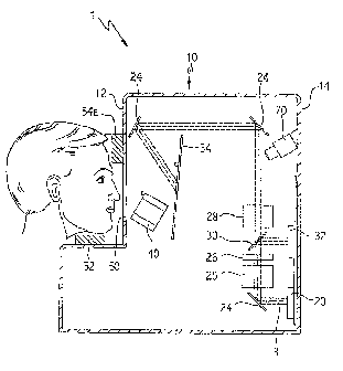
General Description of TestApearatus
21
CA 02863230 2014-09-11
The met form and nature of the apparatus for conducting the method described
herein may
laty, as would be known to one of ordinary skill in the art. An exemplary
anategement of an
tpparatas capable of applying the method described herein is provided below.
The apparatus
nay be modified and altered as would be obvious to one of ordinary skill in
the art without
leviating from the teachings disclosed herein.
In its most basic form, the apparatus comprises a means for genemtirag a
target stimulus,
news for displaying a target stimulus (which is used to meanie the recovery of
visual
rensheivity) and a means for input to allow the subject to convey to the
healthcare provider
nformation regarding the target stimulus (such as that the target stimulus is
visible or the target
Itimulus is not visible). Other functions may be incorporated into the
apparatus, such as a means
for bleaching the test eye, a moans for aligning the teat eye, a MOM for
confirming alignment
ind similar items. In one embodiment, the means for displaying may be an
optical system. In
Rich embodiment, a light source produces a light that is acted on by one or
more optical elements
-.0 prod= the target stimulus and project the target stimulus onto a screen or
other display or
hrough a diffuser for visualization by the subject In an alternate embodiment,
the means for
lisplaying may be an electronic system. In such embodirtunt, the target
stiraulus is produced by
In electronic means and is displayed on a CRT display, a liquid crystal
display, a plasma display
3r an LED display for visualization by the subject Eneh of these embodiments
is described
oelow.
In the embodiment where the means for generating is an optical system, the
optical system
nomprises the elements to generate and act on the target stimulus such that
the target stimulus has
the desired characteristics. The means for generating comprises at least one
of a light source, one
or more optical elements and a screen or other display. The light source will
be used to generate
a light beam which will become the target stimulus, referred to as the target
spot. There may be
multiple or single light sources to generate the light beam. In one
embodiment, the light source is
a bank of light emitting-diodes (LEDs). The light source may also be a
tungsten lamp or any
other appropriate light source. The light source may emit white light and the
light beam cm this
ease white light) produced may be acted upon by various optical elements to
produce a tight
beam of a desired spectrum, or there may be multiple light sources to generate
light of various
wavelengths directly such that the light beam has a particular spectrum of
wavelengths
determined by the light emitted from the selected light source. Such light
sources could be
placed on a means for rotation so that the appropriate light source could be
selected as desired.
The light beam generated by the light source may be acted upon by a series of
optical
elements to produce the target spot A variety of optical elements may be used
in various
combinations to determine the properties of the light beam. These include
directing means to
22
CA 02863230 2014-09-11
limot the light beam, refining means to collimate and shape the light beam,
selecting means to
elect the desired spectrum of the light beam, and modulating means to control
the intensity of
he light beam. In one embodiment, the directing means are mirrors, the
refining means is
Jiving optics, the selecting moans is an optical filter, and the =Mating means
is a neutral
tensity filter or an electronic modulator. Additional optical elements may
also be incorporated,
uch as an optical splitter to direct a portion of the light beam to a
calibration detector to record
he characteristics of the light beam and to ensure the characteristics of; the
light beam are as
lesimd. The target spot is then directed to a means for display, which may be
a screen or other
Tisual display.
In the alternative, the means for generating may be electronic in nature. The
target stimulus
nay be generated by electronic rather than optical means as described above.
In this embodiment
he target stimulus is generated electronically. The electronics produce the
sayropriate
vavelength of light for the target stimulus. Alternatively, a filter may be
inserted over the CRT
lisplay, the liquid crystal display, the plasma display or an LED display, or
other appropriate
lisplay to impart to the target stimulus the appropriate wavelength. The
target stimulus is then
Rapier:4 on a means for display, which may be a CRT or LED screen, or other
appropriate
lisplay.
The apparatus may be portable or fixed in a permanent location_ In one
embodiment, the
cubject may be confined in a testing booth and the apparatus may be a part of
the testing booth or
Amer! in the testing booth. The healthcare provider may be located outside the
testing booth to
amervise the operation of the apparatus. An advantage of this embodiment is
that the healthcare
vovicier will be in normal light during the implementation of the method and
can better monitor
he method
3xemplary Test Apparatus
One embodiment of the apparatus is shown in FIGS. SA and 5B. This embodiment
illustrates
he means for generating as an optical system. The apparatus 1 comprises a
housing 10 having a
front side 12 and a rear side 14 joined by aide walls and bottom and top
walls. The housing 10
38S a viewing opening 50 to receive the head of the subject and allow the
subject to view the
lisplay means, such as screen 34. The viewing opening 50 may be adapted to
eliminate or reduce
ambient light from entering the viewing opening and apparatus. The housing 10
is adapted with
means for alignment to align the subject eye of the subject as desired. In one
embodiment, the
means for alignment comprises a chime& 52 to receive the chin of the subject
The chiruest 52 is
adjustable to aid in the alignment of the subject's eyes with the target spot
16 (as discussed
below). The housing 10 also contains a headrest 54A and 54B to support the
subject's forehead
23
CA 02863230 2014-09-11
while using the machine. Headrests 54A and 54B are selected for use depending
on which eye of
the subject is being tested
The housing 10 contains the basic components of the apparatus. A bleaching
light ammo 40
is provided within the housing 10 to generate the bleaching light 42. The
function of the
bleaching light source 40 is as discussed above. The bleaching light source 40
may be adjusted to
provide a high intensity or a low intensity bleach. Alternatively, the
apparatus 1 may omit the
bleaching light source 40 and the bleaching step carried out independently of
the apparatus.
. In the embodiment
illustrated in FIG. 5A, the light source is a bank of LEDs 20 which emit a
white light beam 3 and an optical element acts on the emitted white tight beam
3 se that the target
spot 16 is of the desired spectrum. The use of LEDs 20 as the light source may
provide several
advantages. First, LEDs are exceeding robust, generate almost no heat load,
require little or no
safety hazard protection, and am very low-cost. In addition, LEDs provide an
opporizmity fix
fine-scale intensity control via electronics, eliminating the complexity and
expense of fine-scale
control via neutral density wedges and other methods.
The light beam 3 is acted upon by One or more optical elements. These optical
dements
include, but are not limited to, directing means to direct the light beam,
refining means to
collimate and shape the light beam, selecting means to select the desired
spectrum of the light
beam, and modulating means to control the intensity of the light beam. In one
embodiment, the
directing means am mirrors, the refining means are shaping optics, the
selecting means is an
optical filter, and the modulating means is a neutral density filter or an
electronic modulator. The
light beam 3 is acted upon by a first mirror 24 to direct the light beam 3 to
the shaping optics 25.
The shaping optics 25 collimates and shapes the light beam 3 so that the
target spot 16 produced
is of the desired size and shape. The operation of such shaping optics 25 is
well known in the art
and is not discussed further herein. As the light beam 3 emerges from the
shaping optics 2511
passes through an optical filter 26 so that the appropriate specimen of light
is selected for
production of the target spot 16. The optical filter 26 may be a color filter.
The operation of such
optical filters 26 is well known in the art and is not discussed further
herein. As the light beam 3
emerges from the optical filter 26, it passes through an optical splitter 30.
The optical splitter 30
directs a portion of the light to a calibration detector 32. The calibration
detector 32 records the
characteristics of the light beam 3 (Such its, but not limited to, the
spectrum and intensity) and
passes a portion of the light beam 3 further along the light path of the
instrtunent The calibration
detector 32 may be a photodiode calibration detector or other calibration
detector as is known in
the art. As the light beam 3 emerges from the optical splitter 30, it is acted
on by neutral density
filter 28. The neutral density filter 28 modulates the beam of light 3 to
produce the desired
intensity. The use of the neutral density filter 28 will allow control of the
intensity of the light
24
CA 02863230 2014-09-11
beam 3 over six logs of dynamic range, with a maximum projected intensity of
¨S cd m.4. As
the light emerges from the neutral density fitter 28, it is farther directed
by one or more mirrors
24 and is ultimately projected OS the target spot 16 onto a screen or other
display. The location of
the target spot can be located at the desired area of the subject's eye as
discussed above. The
directing means may be adjusted to achieve such localization_ In the
embodiment illustrated, The
display is a screen 34. The display can be visualized by the subject
A means for control is in communication with the various components of the
apparatus 1,
such as, but not limited to, the bleaching light source, the light sotnee, the
directing means, the
refining means, the selecting means, and the modulating meant:. In addition,
the means for
control may be in communication vritla the calibration deteoter and the
subject impel 111011.1111 (as
described below). For example, the control means may control the light
emission from the light
source so that the pulses of light emitted by the light source correspond to
the configuration
required by the test method and emissions from the bleaching light source to
ensure that the
percent bleaching desired is obtained. In addition, the control means could
adjust the refining
means, the selecting means and the modulating means to produce a light beam
with the desired
characteristics. Furthermore, the control means may adjust the directing means
to provide
desired localization of the target spot. Therefore, the means for control is
capable of adjusting the
parameters of the components of the apparatus as dictated by the method
described. Furthermore,
the control means also records the status and output of each of the components
of the apparatus.
For example, the control means may record the intensity of the target
stimulus. The control
means also records the input from the subject input means, which is used to
allow the subject to
input his/her responses to the target stimulus, for use in generating the
threshold values. The
control means may further measure and record the time elapsed during the
implementation of the
method (said timing to start in one embodiment immediately after the bleaching
step is
accomplished) and the time at which subject inputs are received from the
subject input means and
the time at which the various parameters of the method are changed (such as
the changing
intensity of the target stimulus). By comparing the timing of the subject
response to the target
stimulus as received from the subject input means and correlating said subject
responses to the
status of the parameters of the apparatus, the control means may then
determine and record the
threshold measurements and execute calculations required for noise reduction
in the threshold
measurements.
A means for comparison may be in communication with the means for controL The
means for
comparison may be separate from or integral with, the means for control. The
means for
comparison may use the threshold measurements and the information from the
components of the
system for subsequent analysis. The means for comparison may be capable of
executing
CA 02863230 2014-09-11
calculation& to fit the threshold measurements to a desired model of dark
adaptation (such as, but
not limited to, the one-linear, one-exponeatial model described above) and
generating a full or
partial dark adaptation model fit and/or the desired index factors from said
threshold
measurements and then recording and storing said information. As discussed
above, the means
for comparison may execute such calculations as the threshold measurements are
collected, or =
may execute such calculations after all desired threshold measurements are
obtained. The means
for comparison may be an external device in communication with the control
means via the
interact
The desired index factors may then be compared to corresponding index factors
in a
comparative database and the result recorded and stored. The comparative
database index factors
and comparison results may be contained within the means for comparison
allowing the process
to be automated or may be separate from and. in communication with the means
for compsrison.
The means for comparison may output the information to a visual display as
desired. The output
may be in the form of a full or partial threshold curve and dark adaptation
model fit or other
graphical format. In addition, the individual index factors may be displayed
as well. The output
may be further conveyed to a storage device man output device, such as a
printer.
The configuration of the embodiment described in FIG. 5 is for illustrative
purposes only.
Other configurations containing additional elements or similar substitutions
for the elements
described may be envisioned. In addition, the order of the elements described
may be re-
arranged as desired.
The apparatus I may also contain a means for confirming alignment of the
subjects test eye.
In one embodiment, such a means is an infrared cane= which can be used to
verify that the
subject's test eye is properly aligned to view the target spot 16 and the
photobleaching light 42.
To aid the subject in achieving such alignment, a. target fixation light 17
and a bleaching fixation =
light 43 may be provided (see FIG. 5B). As the subject fixes the test eye on
the bleaching
fixation light 43, the subject can be assured the test eye is in the proper
position to receive the
desired photobleaching effect Likewise, as the subject fixes the test eye on
the target fixation
light 17, the subject can be assured the test eye is in the proper position to
view the target spot 16.
The target fixation light 17 and the bleaching fixation light 43 may be
produced by additional
light sources, such as LEDs incorporated at the desired locations inside the
apparatus or may be
projected onto the screen 34.
This design of the apparatus 1 will allow investigation of a broad range of
target stimulus
parameters by simple adjustment or change-out of the minors, shaping optics
end mica filters.
The target spot size, location and spectrum therefore can be varied as desired
by the healthcare
provider. Furthermore, the intensity of the bleaching light source can also be
conholled
26
CA 02863230 2014-09-11
Overview of Method Implementation
The use of the apparatus 1 to employ the method of the present disclose= will
now be
discrisasd. The operation of the apparatus and execution of the method can be
viewed as having 5
steps: 1) aligning the subject 2), photobleachin' g of the lest eye; 3)
monitoring recovery of visual
sensitivity (i.e. =topic recovery); 4) optionally fitting the data obtained to
an appropriate model
to generate the dark adaptation parameters; and 5) comparing the threshold
measurements or
optionally the the index factors, such as the dark adaptation parameters, from
the subject to a
comparative database. The steps should not be construed as limiting
descriptions, but are simply
convenient areas for further detailed discussion. Bach of these steps will be
discussed in greater
detail below. Furthermore, the hardware required to carry out each of these
steps need not be
incorporated into the test apparatus, but may be if dashed.
In the alignment process, the subject is aligned by adjustment of the chin
rest 52 vertically,
horizontally or both vertically and horizontally. Correct positioning of the
subject is achieved by
viewing the subject's test eye with an infrared camera 70 mounted inside the
housing 10 while
the subject focuses on the target fixation light 17. The optical system is
arranged such that this
single step aligns the subject correctly with respect to both the bleaching
light 42 and the target
spot 16. The infrared camera 70 can be used as needed to confirm continued
alignment of the
subject FIG. 5B shows a view of one embodiment of the interior of the housing
10 as viewed
through opening 50 and represents the view a subject would encounter on using
the device.
Once alignment of the subject is achieved, the subject's test eye is subject
to a bleaching
protocol by exposure to the bleaching light 42. In this embodiment, the
bleaching light 42 is a
brief high intensity camera flash or electronic strobe (typically 5 to Slog
scot Td/sec for 0.25 ms)
that is generated while the subject is focused on the bleaching fixation light
43 to ensure the
proper pcntion of the rhodopsin molecules of the retina is bleached. The
amount of bleaching
ptoduced can be determined by the healthcare provider by varying the desired
intensity of the
bleaching light 42, which is controlled by the means for control as discussed
above. In one
embodiment, 50% to 98% of the rhodopsin molecules are bleached.
The dark adaptation measurements begin immediately after the bleaching
protocol is
administered by obtaining a series of threshold measurements. With the subject
once again
focusing on the target fixation light 17, the threshold measurements are
obtained. in one
embodiment, the threshold measurements are obtained using a 3-down/1-up
modified staircase
procedure. Starting at a first intensity (such as 4.85 cd in-2), target spots
16 are presented on the
screen 34 to the subject every 2 to 3 sec for a defined duration (such as a
200 ms pulse). If the
subject does not respond to the target spot 16 (such as by activating the
input means), the light
intensity of the target spot 16 remains unchanged until the subject responds.
If the subject
27
CA 02863230 2014-09-11
indicates the target spot 16 is visible (such as by activating the input
means), the light intensity of
the target spot 16 is decreased for each successive pulse in 0.3 log units ("3-
1owel until the
subject stops responding That the target spot 16 is present After the subject
indicates that the
target spot 16 is invisible, the light intensity of the target spot 16 is
increased for each aucc,essive
pulse in steps of 0.1 log units ("1-up") until the subject responds that the
target spot 16 is once
again visible. This light intensity of the target spot 16 at the completion of
this sequence is
defined as the threshold measurement Successive threshold measurements stint
with a target
Spot 16 light intensity 03 log units brighter than the previous threshold
measurement Threshold
m.easurernents are made once or twice every minute for the duration of the
measurement protocol
During this process, the threshold measurements am subjected to a noise
redaction protocol as
discussed above. Other implementations of the staircase protocol may also be
used as described
above and methods other than a staircase procedure may also be employed as
would be known to
one of skill in the art
To focus an rod-mediated function, a target stimulus 16 with a wavelength near
the peak rod
sensitivity (¨ 500 um) is used. Corrective lanes can be introduced between the
test eye and the
trivet spot 16 as appropriate by means of a lens holder inside the machine
(not shown). The
duration of die measurement protocol can be varied and may be terminated in
accordanee with
the decision rules as discussed above.
In one embodiment. the threshold measurements are then fit to a desired model
of dark
adaptation. The desired model may be used to generate one or more index
factors. As discussed
above, the index factors may be a plurality of threshold measurements, a full
or partial threshold
curve or a dark adaptation parameter. Any of the dark adaptation models
described herein or
known to those of skill in the art may be used, such as the two-component, one-
linear one-
exponential model. As previously described, the initial cone-mediated
(photopio) portion of the
threshold curve is modeled with a linear component, and the subsequent rod-
mediated (scotopie)
portion of the curve is modeled with an exponential component. The ocanpatison
means may be
programmed to record the appropriate parameters, to fit the data to the
desired model and to
automatically extract such index factors from the mod& For some subjects with
tate ARtsip,
this two-component model may not provide a satisfactory fit. Insufficient
sensitivity recovery
after the rod-cone break will cause the exponential portion of the model to
fit poorly. For cases
where the two-component model proves inadequate (R2 <0.9), a bilinear fit can
be applied to the
data to accurately estimate the desired dark adaptation parameters, such as
the rod-cone break,
and the slope of the rod recovery will be recorded. The flexibility of
employing multiple models
allows tracking of disease progression more accurately than strict adherence
to a single model.
Alternatively, the threshold measurements may be output to the bealthcare
provider (in the form
28
CA 02863230 2014-09-11
of a partial or full threshold curve, a dark adaptation model fit or table
describing the index
factors) and the healthcare provider may extract the dark adaptation
parameters manually.
After the dashed Wear factors are determined. one or mom of the subject's
index factors
are compared to the corresponding index factors flora individuals in a
comparadve database. In
one embodiment, the subject's dark adaptation parameters are compared to a
referessie range of
the corresponding parameters in the compatativo database. The reference range
may be a
statistical parameter above and below the index factor in the comparative
database, such as the
mean of the selected index factor in the comparative daetbase * two standard
deviations of the
mean. If the subject's dark adaptation psonnater falls outside the reference
range, dark
adaptation is considered impaired and the subject is considered to be at risk
for ARMD or other
disease stales as described herein. If several index factors are estimated and
the subject is
considered Anomie/ on any one of the estimated index factors, dark adaptation
is considered
impaired and the subject is considered to be at risk for ARMD or other disease
states as described
herein. The comparative database is as described above. Alternatively, the
threshold
measureanenbi may be direedy compared to corresponding threshold measurements
in the
comparative database to determine dark adaptation status or dirrase state
without going through
the intermediate model fit and index factor determination.
CONCLUSION
In surnmary, determination of dark adaptation performed by the methods
described above
was shown to be a sensitive indicator of the earliest stages of ARMD.
Therefore, dark adaptation
can be used to identify those individuals are at-risk for ARMD and the other
disease states
described herein or any other disease states that impact nod photoreceptor
function. Furthermore,
dark adaptation can be used to indicate dise.aaf. state severity and/or
progression.
Given the disclosure herein, one of ordinary skill in the art may become aware
of various
other modifications, &Adams, or improvements. Such other modifications,
features and
improvements should be considered part of this disclosure.
EXAMPLES
Example!
Using the =foresee method of the present disclosure, it was shown that
impaired rod¨
mediated dark adaptation accuratdy predicts ARMD and is an eady functional
marker of AMD.
Twenty patients (65 to 79 years old) were examined who at baseline had norms/
retinal health.
29
CA 02863230 2014-09-11
Normal retinal health was based on a grading of photographed fundus appearance
using the
standardized International Classification System. Dating the initial baseline
visit, rod-mediated
dark adaptation was measured using the method described herein The patients
were classified as
having normal or impaired dark adaptation at the baseline visit Impaired dark
adaptation was
defined using the rod-cone bleak parameter as the dark adaptation parameter,
with impaired dark
adaptation being diagnosed when the rod-cone break parameter fell outside the
reference maga
(12 standard deviation of the men) of normal healthy subjects in our
comparative datahaaa Eye
health status was measured in tho subsequent 4 years alter the baseline visit.
Medical records
were examined for changes in the patient's retinal heal& At the end of 4
years, 86% (12/14) of
patients with impaired dark adaptation at baseline received a diagnosis of
ARMD, whereas less
than 17% (1/6) of patients with aonnal dark adaptation at baseline received a
diagnosis of
ARMD. These findings indicate that impaired rod-mediated dark adaptation is a
risk factor for
ARMD and that a method that accurately identifies impaired rod-mediated dark
adaptation can be
used to identify those individuals who are at risk for incident early ARMD.
Example 2
Furthermore, rod-mediated dark adaptation kinetics are markedly slowed in
early ARMD
patients as compared to normal age-matched subjects. Dark adaptation
parameters were obtained
from 20 early ARMD patients (ages 66-88) and 16 healthy subjects (ages 62-79)
as described in
the present disclosure. ARMD status was assigned using a standardized fundus
photography
grading system. On average, the time constant of rod-mediated sensitivity
recovery of dark
adaptation was markedly slowed in ARMD patients. In this study, the time to
complete the test
was on average 16 minutes longer for ARMD patients as compared to healthy
individuals.
Further analysis of the data revealed that 85% of the ARM]) patients exhibited
impaired rod-
mediated dark adaptation as defined by at least one dark adaptation parameter
falling outside + 2
standard deviations of the mean normal value (see Table 3). In contrast, only
20% of the healthy
subjects were classified as exhibiting impaired rod-mediated dark adaptation
Significantly,
cone-mediated visual sensitivity, visual acuity and contrast sensitivity were
classified as impaired
in only 25% of the ARMD patients, indicating impaired rod-mediated dark
adaptation is a more
sensitive indicator of early ARMED than visual sensitivity, visual acuity and
contrast sensitivity.
Example 3
In addition to identifying those individuals at risk for ARMD, the method
described
herein may also be used to detect and determine differences in ARMD disease
severity. Pandas
photographs for a subset of ARMD patients and normal patients were sent to the
Wisconsin
Reading Center for grading in accordance with the Wisconsin Aging-Related
Maculopathy
Grading System (WARMDGS). Based on the result of the fundus photography
grading, three
CA 02863230 2014-09-11
ARMD patients (open square, triangle and diamond) and one normal patient
(closed chyle) were
selected for examination using the method described herein. The three ARMD
patients displayed
different stages of ARMD progress. Patient no. I (open square) and no. 2 (open
triangle)
exhibited soft indistinct drawn with a numimnra size of about 250 ;am and a
coverage area of
about 1500 gm. However, patient no. 2 had 2-times the number of soft drasen
with distribution
further away from the fovea, indicating a progression of ARMD disease. Patient
no. 3 (open
diamond) had a number of bard drusen and a pigment epithelial detachment,
indicating an
additional Progression of ARMD disease over patients no. 1 and no. 2. The
curves and selected
dark adaptation parameters generated from the threshold measurements for
patients 1-3 and the
normal patient were then compared. As can be seen farm FIG. 6, the times for
rod-mediated
sensitivity recovery of dark adaptation for patient nos. 1-3 was significantly
greater than the
normal controL Furthermore, the time required for rod-mediated sensitivity
recovery of dark
adaptation was greater as the ARMD disease severity increased (with time
increasing from
patient no. 1 to patient no. 3). This study indicates that impaired rod-
mediated dark adaptation
can be used not only to determine individuals at-risk for ARMD, but to gauge
ARMD disease
severity and/or progression.
31
CA 02863230 2014-09-11
High Bleach Intensity Low Bleach Intensity
ç-98% bleach) 50% bleach),
Parameter old nounal earl ARM impairment- old normal
early ARM _ impairment
rod-cone break 15.41 min 23.42 min 52% 7.15 min 13.56 ruin 90%
rod lime
5.32 min 13.86 min 1.61% 5.74 min 7.72 min 34%
ocmatant
Table 1- Dark. adaptation parameter differences for high bleach intensity vs
low bleep' h intensity
12" inferior field 5' inferior field
old early early
Parameter liana/ ARm impairment old
normal Aim impairment
rod-cone break 15.61 min 20.48 min 31% 14.82 min 24.03 min 62%
rod time
10.11 min 12.49 min 24% 9.96 min 16.80 min 69%
constant
Table 2- Dark adaptation parameter differences for peripheral vs_ Genial
target spot location
Variables Percentage
Kinetic Variable:
Rod-cone break 75%
2o3 component recovery -4 56%
3' component recovery 0%
Time to baseline 55%
Rod-mediated time constant 65%
Any.dark adaptation kinetics 85%
Steady-State Variables:
Baseline (pre-bleach) scotopic 25%
Photopic sensitivity_over 18 radius 25%
Scottie sensitivity over 18 radius 20%
Contrast sensitivi, 35%
Table 3: Percentage of ARM patients exhibiting impaired rod-mediated dark
adaptation (any
parameter falling outside + 2 standard deviations of the normal mean value)
32